Studies on the interaction between Virus Movement Protein ... · CCB colloidal coomassie blue cDNA...
160
Studies on the interaction between Tobacco Mosaic Virus Movement Protein and Microtubules Inauguraldissertation zur Erlangung der Würde eines Doktors der Philosophie vorgelegt der Philosophisch-Naturwissenschaftlichen Fakultät der Universität Basel von Jamie A. Ashby aus Großbritannien Universität Basel Friedrich Miescher-Institute for Biomedical Research Basel, September 2003
Transcript of Studies on the interaction between Virus Movement Protein ... · CCB colloidal coomassie blue cDNA...

Studies on the interaction between Tobacco Mosaic
Virus Movement Protein and Microtubules
Inauguraldissertation
zur
Erlangung der Würde eines Doktors der Philosophie
vorgelegt der
Philosophisch-Naturwissenschaftlichen Fakultät
der Universität Basel
von
Jamie A. Ashby
aus Großbritannien
Universität Basel
Friedrich Miescher-Institute for Biomedical Research
Basel, September 2003
ii
Genehmigt von der Philosophisch-Naturwissenschaftlichen Fakultät auf Antrag von Prof. Thomas Boller Prof. Manfred Heinlein Prof. Frederick Meins Basel, 25.09.2003
Prof. Marcel Tanner Dekan
iii
Table of contents Page
Acknowledgements.........................................................................vii
Abbreviations .................................................................................viii
Abstract..............................................................................................x
1 Introduction .......................................................................................1
1.1 Overview of RNA trafficking and localization .......................................... 1
1.2 Examples of RNA trafficking and localization ......................................... 2
1.2.1 Ash-1 mRNA in Saccharomyces cerevisiae .......................................................... 2
1.2.2 MBP mRNA in oligodendrocytes ............................................................................ 3
1.2.3 Pair-rule mRNAs in the Drosophila blastoderm embryo...................................... 4
1.2.4 Vg1 mRNA in Xenopus oocytes.............................................................................. 4
1.2.5 Bicoid and Oskar RNAs in Drosophila oogenesis................................................ 6
1.2.6 Protamine-1 mRNA in mouse spermatids ............................................................. 7
1.2.7 Intercellular RNA trafficking in plants.................................................................... 8
1.2.7.1 Long distance RNA trafficking and developmental regulation ................................... 9
1.2.7.2 Systemic RNA trafficking and gene silencing........................................................... 10
1.2.8 Intracellular RNA trafficking in plants.................................................................. 11
1.3 Tobacco Mosaic Virus (TMV) as a model to study intracellular RNA
trafficking in plants ................................................................................................. 13
1.3.1 Structure and organization of the TMV genome ................................................. 14
1.3.2 TMV infection cycle................................................................................................ 15
1.3.3 The role of MP during vRNA trafficking ............................................................... 16
1.3.4 Transport as a ribonucleoprotein complex (RNP) .............................................. 17
1.3.5 Subcellular distribution of MP during infection.................................................. 18
1.3.5.1 Endomembrane association..................................................................................... 19
1.3.5.2 Microtubule association............................................................................................ 20

iv
1.3.5.3 Summary .................................................................................................................. 21
1.3.6 Thesis objectives ................................................................................................... 22
2 Materials and Methods....................................................................24
2.1 Purification of overexpressed MP from E.coli ....................................... 24
2.1.1 Purification strategy............................................................................................... 24
2.1.1.1 Subcellular localization of MP expressed in E.coli ................................................... 24
2.1.1.2 Principle of Immobilized Metal Affinity Chromatography (IMAC) ............................. 24
2.1.1.3 General note about MP purification.......................................................................... 25
2.1.2 Construction of plasmids for MP overexpression.............................................. 26
2.1.2.1 Overview of the QIAexpress vector pQE60........................................................... 26
2.1.2.2 Cloning procedures .................................................................................................. 26
2.1.3 Overexpression of MP ........................................................................................... 27
2.1.4 Isolation of MP-containing inclusion bodies....................................................... 27
2.1.5 Ni 2+-NTA Affinity Chromatography...................................................................... 28
2.1.6 Renaturation of purified MP.................................................................................. 29
2.1.7 Determination of final MP purity by Mass Spectrometry ................................... 29
2.2 In vitro RNA binding assays ................................................................... 30
2.2.1 RNA band-shift assay using a short 32P-labeled probe...................................... 30
2.2.2 Modified conditions suitable for TMV-derived RNA ........................................... 31
2.3 Infection of BY-2 protoplasts and Nicotiana benthamiana with mutant
TMV-derivatives encoding –(H)6 affinity tags........................................................ 32
2.3.1 Construction of TMV derivatives .......................................................................... 32
2.3.1.1 TMV-MP:(H)6:GFP.................................................................................................... 32
2.3.1.2 TMV-MP:GFP:(H)6.................................................................................................... 33
2.3.2 Preparation of BY-2 protoplasts and transfection with viral RNA derived from
TMV-derivatives encoding –(H)6 affinity tags ....................................................................... 33
2.3.3 Infection of Nicotiana benthamiana with viral RNA derived from TMV-
derivatives encoding –(H)6 affinity tags................................................................................ 34
2.3.4 Analysis of infection sites..................................................................................... 35
2.3.5 Subcellular localization of MP............................................................................... 35

v
2.4 In vitro Microtubule-associated protein (MAP) assay ........................... 36
2.4.1 Principle of the MAP assay ................................................................................... 36
2.4.2 Determination of appropriate reaction conditions.............................................. 37
2.4.2.1 MP solubility assay................................................................................................... 37
2.4.2.2 Microtubule stability assay ....................................................................................... 38
2.4.3 MAP assay............................................................................................................... 39
2.4.4 Control reactions to determine the effect of –(H)6 on MT binding in vitro ....... 40
2.4.5 Analysis of Microtubule binding by MP in vitro .................................................. 40
2.5 Visualization of MP:MT complexes formed in vitro............................... 42
2.5.1 Immobilization of MTs in kinesin coated perfusion chambers.......................... 42
2.5.2 Formation and visualization of MP:MT complexes............................................. 43
2.6 Co-precipitation experiments in vitro using immobilized MP as an
affinity bait............................................................................................................... 45
2.6.1 Co-precipitation of MTs and tubulin heterodimers in vitro................................ 45
2.6.2 Co-precipitation of RNA and tubulin heterodimers in vitro ............................... 47
2.7 In vitro kinesin motility assays ............................................................... 50
3 Results .............................................................................................52
3.1 Purification of MP from E.coli ................................................................. 52
3.1.1 Determination of MP purity ................................................................................... 55
3.2 In vitro RNA binding by MP..................................................................... 57
3.2.1 RNA binding behaviour of MP in vitro ................................................................. 57
3.3 Infection of Nicotiana benthamiana and BY-2 protoplasts with TMV
encoding bMP fused to –(H)6 affinity tags............................................................. 60
3.3.1 Infection of BY-2 protoplasts ................................................................................ 60
3.3.2 Infection of N. benthamiana .................................................................................. 63
3.4 Biochemical analysis of MP:MT binding in vitro ................................... 68
3.4.1 Optimization of reaction conditions..................................................................... 68
3.4.2 MAP assay............................................................................................................... 73
3.4.3 Determining the effect of –(H)6 on MT binding.................................................... 75

vi
3.4.4 Analysis of the MT binding properties of MP in vitro ......................................... 77
3.5 Visualization of MP:MT complexes formed in vitro by fluorescence
microscopy.............................................................................................................. 84
3.5.1 Establishment of the perfusion chamber system............................................... 84
3.5.2 Fluorescence microscopy..................................................................................... 85
3.6 Co-precipitation experiments in vitro using immobilized MP as an
affinity bait............................................................................................................... 90
3.6.1 Co-precipitation of MTs and tubulin heterodimers in vitro................................ 90
3.6.2 Co-precipitation of RNA and tubulin heterodimers in vitro ............................... 93
3.7 Apparent modulation of kinesin motor activity by MP.......................... 97
4 Discussion .....................................................................................103
4.1 The MAP-like behaviour of MP.............................................................. 103
4.2 MT-localization of MP in vivo................................................................ 107
4.3 Role of the MP:MT interaction .............................................................. 112
5 References.....................................................................................115
6 Appendix ........................................................................................135
Media and solutions .............................................................................. 135
Primers ................................................................................................... 139
Mathematical and statistical models .................................................... 140
Publication ............................................................................................. 150

vii
Acknowledgements
Firstly, I would like to thank Prof. Manfred Heinlein for providing me with the
opportunity to work in the field of macromolecular trafficking, the freedom to explore
my ideas and the guidance to consolidate my findings. I would also like to express my
gratitude to Prof. Thomas Boller and Prof. Frederick Meins for their critical appraisal
of my work and for their constructive suggestions.
I would like to acknowledge Prof. Andreas Hoenger, Dr. Thomas Wendt and
Dr. Vincent Brondani for their invaluable help with experimental matters, and in
addition, Prof. Barbara Hohn and Dr. Ortrun Mittelsten-Scheid for their most helpful
advice while writing this thesis.
Many thanks to Dean Flanders, Thomas Nyffenegger and Alan Naylor for their
excellent I.T. support, and thanks also to Dr. Daniel Hess and Ms. Anne Ulvestad for
protein analytical services.
I am grateful to past and present friends from the FMI for making my time an
enjoyable one, particularly Dr. Mark Lambermon, Dr. Pawel Pelczar, Ms. Monika
Fasler, Alan Naylor and Dr. James Moore.
My warmest thanks must go to my family. Without their sacrifices and constant
support, this thesis would not have been written.

viii
Abbreviations
ATP adenosine 5’-triphosphate
BES N,N-Bis(2-hydroxyethyl)-2-aminoethanesulfonic acid
BSA bovine serum albumin
BY-2 bright yellow-2
CC companion cell
CCB colloidal coomassie blue
cDNA complementary DNA
CIAP calf intestinal alkaline phophatase
CP coat protein
dpi days post infection/inoculation
dsDNA double-stranded DNA
DTT 1,4-Dithio-DL-threitol
EDTA ethylenediaminetetraacetic acid
EGTA ethylene glycol-bis(2-aminoethyl)-N,N,N’,N’-tetraacetic acid
FITC fluorescein-isothiocyanate
GFP5 green fluorescence protein (variant 5)
GST glutathione-S-transferase
GTP guanosine 5’-triphosphate
GTP-γ-S guanosine 5’-[γ-thio]triphosphate
GuHCL guanidine hydrochloride
hpi hours post infection/inoculation
HPLC high performance liquid chromatography
IPTG isopropyl-β-D-thiogalactoside

ix
MAP Microtubule-associated protein
MES 2-(N-Morpholino)ethanesulfonic acid
MP movement protein (TMV P30)
MT microtubule
MW molecular weight
NaBH4 sodium boroanhydride
NP-40 nonidet-P40 (Nonylphenylpolyethylene glycol)
nt nucleotide
OD optical density
ORF open reading frame
PBS phosphate buffered saline
Pd plasmodesmata
Pipes 1,4-Piperazinediethanesulfonic acid
PTGS post-transcriptional gene silencing
RT-PCR reverse transcription polymerase chain reaction
S.E.M. standard error (of the mean)
SDS-PAGE dodium dodecyl sulfate-polyacrylamide gel electrophoresis
SE sieve element
SEL size exclusion limit
Tris tris-hydroxymethylaminoethane
TRITC tetramethylrhodamine-isothiocyanate
vRNA viral RNA
vRNP viral ribonucleoprotein complex
WT wild-type

x
Abstract
During the invasion of a susceptible host, Tobacco mosaic virus (TMV) transports its
RNA genome from sites of viral synthesis into neighbouring cells, thus potentiating the
spread of infection. In many systems, RNA trafficking is known to be a highly
coordinated process, often involving the complex interplay of numerous factors. In
plants, however, the mechanisms that govern RNA transport are not well understood.
Since viruses have a propensity to exploit pre-existing host machinery for their own
purposes, TMV has become a popular model system for the study of RNA trafficking
in plants. TMV viral RNA (vRNA) is thought to be transported as a ribonucleoprotein
(RNP) with the virally encoded movement protein (MP). Localization studies have
demonstrated a temporal redistribution of MP from endoplasmic reticulum (ER)-
derived replication bodies onto microtubules (MTs) at mid to late stage of infection. A
functional link between the association of MP with microtubules (MT) and RNA
trafficking has been established, and furthermore, this association does not require
other viral components or plant-specific factors, thereby indicating a direct interaction
between MP and MTs. In vitro, purified MP interacts directly with preformed,
dynamically suppressed MTs and has no requirement for MT polymerization or
polymer-specific structural forms. Immunohistochemical studies indicate that MP may
interact with the extreme C-termini of α- and β-tubulin, possibly forming a stabilizing
sheath. Consistent with the proposal that MP functions as a structural microtubule-
associated protein (MAP), MP:MT complexes assembled in vitro are highly stable. A
possible functional overlap between the putative RNA and MT binding domains of MP
has been identified, and although MP:MT complexes are unlikely to support RNA
transport directly, such complexes have been implicated to modulate MT-dependent
molecular motor activity. Collectively, in vitro data are in support of the observation
that MP associates with MTs to high amounts, leading to the hypothesis that MP-
mediated RNA transport and the MP:MT interaction are related, yet functionally
distinguishable processes. Furthermore, based on the similarity of MP to endogenous
MAPs, a new model is proposed in which MP-mediated trafficking may occur in the
form of a translationally competent ER-vesicle, concurrent with the deployment of MP
to the MT surface.

1
1 Introduction
1.1 Overview of RNA trafficking and localization
In order for multicellular organisms to regulate their biological activities, appropriate
information must be communicated in a coordinated manner between tissues, cells and
subcellular compartments. The nature of such information varies widely, however,
eukaryotes have developed sophisticated strategies which allow them to regulate
cellular and developmental events by the selective trafficking and localization of RNA
(Chartrand et al., 2001; Jansen, 2001; Palacios and St Johnston, 2001; Kloc et al.,
2002; Okita and Choi, 2002). RNA localization is known to occur in a variety of cell
types and is thought to be essential for processes such as cell-fate determination, polar
cell growth and spatially restricted protein expression. Intracellular RNA localization
can occur via several mechanisms, which include random diffusion/cytoplasmic
streaming and directional transport on cytoskeleton elements. In both plants and
animals, examples of intercellular RNA trafficking have been described, and in
addition, certain endogenous plant RNAs have been demonstrated to move
systemically, suggesting that RNA trafficking may be involved in non-cell-autonomous
signaling. Accumulating evidence suggests that endogenous RNA trafficking is
mediated by the interaction of specific trans-acting factors with signal elements present
within the RNA (Palacios and St Johnston, 2001). A number of localized RNAs are
found in large ribonucleoprotein (RNP) complexes (Ferrandon et al., 1994; Knowles et
al., 1996; Mouland et al., 2001), and in addition to directing RNAs to their respective
destinations, such RNPs may also be involved in translational regulation (Cooperstock
and Lipshitz, 2001; Johnstone and Lasko, 2001). RNA trafficking and localization is
not only restricted to endogenous RNAs. A number of RNA viruses found in both

2
animal and plant kingdoms are also known to utilize components of the host transport
machinery, presumably in an effort to facilitate the replication and intercellular spread
of their genomes.
1.2 Examples of RNA trafficking and localization
1.2.1 Ash-1 mRNA in Saccharomyces cerevisiae
During cell division in budding yeast, mating-type switching (Strathern and
Herskowitz, 1979) is repressed by the asymmetric accumulation of transcriptional
regulator Ash1p in daughter cell nuclei. (Bobola et al., 1996; Sil and Herskowitz,
1996). Spatial segregation of Ash1p involves the localization of Ash-1 transcripts at the
distal bud tip during late anaphase (Long et al., 1997; Takizawa et al., 1997) and has
been demonstrated to require the gene encoding Myo4p, a class V myosin (Haarer et
al., 1994; Jansen et al., 1996). By using a yeast mutant in which the myosin binding
site on actin is disrupted, it was found that Ash-1 transcripts no longer localized
asymmetrically to the daughter cell (Long et al., 1997), and furthermore, Ash-1 mRNA
appeared to be randomly distributed throughout both mother and daughter cells
following treatment with the actin-depolymerizing drug Latrunculin-A (Takizawa et
al., 1997), again suggesting the involvement of an actin-dependent mechanism.
Compelling evidence to support the role of Myo4p motor transport in Ash-1 mRNA
trafficking was provided by the observation that Myo4p colocalizes with Ash-1
mRNA-containing particles (Bertrand et al., 1998; Takizawa and Vale, 2000), and in
living cells, directly mediates Ash-1 mRNA movement into the bud tip (Bertrand et al.,
1998; Beach et al., 1999). It has emerged that a number of accessory proteins are also
required for Ash-1 translocation, and these are thought to be involved in anchoring Ash-
1 transcripts to Myo4p, stabilizing the actin cytoskeleton and possibly maintaining

3
translational silence until Ash-1 mRNA reaches the bud tip (Munchow et al., 1999;
Bohl et al., 2000; Long et al., 2000; Takizawa and Vale, 2000).
1.2.2 MBP mRNA in oligodendrocytes
Myelin basic protein (MBP) mRNA has been found to localize to the membranes and
myelin of oligodendrocyte peripheral processes, up to 50 µm from the site of RNA
synthesis (Kristensson et al., 1986; Verity and Campagnoni, 1988). By injecting a
fluorescently labeled MBP mRNA into cultured mouse oligodendrocytes, it was
observed that transcripts formed granular structures within the perykaryon cytoplasm
which traveled towards the cellular periphery along narrow processes at an average
speed of ~ 0.2 µm/sec (Ainger et al., 1993). A similar distribution of endogenous MBP
mRNA was also found using in situ hybridization techniques, and furthermore, it was
observed that upon reaching the cell periphery, the movement of MBP mRNA-
containing granules was no longer directional, possibly suggesting the existence of a
multi-step mechanism. Subcellular fractionation experiments indicated that MBP
mRNA was associated with cytoskeleton elements within the cellular processes, and
interestingly, it was found that treatment with the microtubule destabilizing drug
Nocodazole inhibited the translocation of MBP mRNA, however, disruption of the
actin cytoskeleton had no discernible effect (Carson et al., 1997). By inhibiting
kinesin-1 expression using an anti-sense oligonucleotide, it was also found that MBP
mRNA localization was inhibited. Collectively, these data strongly indicate that
anterograde MBP mRNA trafficking in oligodendrocytes requires intact microtubules
and most likely involves the positive-end directed motor protein kinesin-1.

4
1.2.3 Pair-rule mRNAs in the Drosophila blastoderm embryo
During interphase 14 of D. melanogaster embryogenesis, the monolayer of nuclei
found at the cortex of the blastoderm embryo subdivides into apical, nuclear and basal
compartments (Fullilove and Jacobson, 1971; Rickoll, 1976; Turner and Mahowald,
1976). At this stage, RNA transcripts of the pair-rule genes localize to the apical
periplasm in seven transverse stripes along the anteroposterior axis, thereby
establishing segmental patterning (Hafen et al., 1984; Edgar et al., 1986). In two
independent studies, disruption of the microtubule cytoskeleton with colcemid caused
both endogenous pair-rule RNA fushi tarazu (ftz), and injected ftz mRNA to disperse
into all three of the blastoderm layers (Edgar et al., 1987; Lall et al., 1999).
Furthermore, it was found that when preincubated with human or Drosophila
embryonic nuclear extract, injected ftz mRNAs rapidly localized to the apical periplasm
in a microtubule-dependent manner (Lall et al., 1999). Since MTs radiate from apically
positioned centrosomes in blastoderms, it was speculated that pair-rule mRNA
transport requires the minus-end directed motor protein dynein. It was found that upon
coinjection of ftz mRNA with anti-dynein heavy chain antibodies, almost complete
inhibition of ftz localization occurred (Wilkie and Davis, 2001). Moreover, through the
use of hypomorphic dynein mutants, or by disrupting the dynein-dynactin complex
with an excess of dynamitin (Echeverri et al., 1996), trafficking of ftz RNA could be
severely impeded, thereby strongly indicating that active motor transport potentiates
pair-rule mRNA translocation.
1.2.4 Vg1 mRNA in Xenopus oocytes
In X. laevis oocytes, specific maternal RNAs become localized to either the animal or
vegetal cortices, thereby leading to cell polarization and formation of the primary

5
developmental axis in fertilized embryos (Kloc et al., 2001). Vg1 mRNA is distributed
throughout the oocyte early during oogenesis (stages 1 and 2) and localizes to the
vegetal cortex from stage 3 onwards (Melton, 1987; Forristall et al., 1995). Using in
situ hybridization experiments, it was observed that following treatment with the
microtubule disrupting drugs colchicine or nocodazole, Vg1 mRNA no longer localized
to the vegetal cortex during embryogenesis stage 3 (Yisraeli et al., 1990). In contrast,
Vg1 mRNA remained localized to the vegetal hemisphere during stages 5 and 6 of
oogenesis, regardless of microtubule disruption, although transcripts became
mislocalized upon disruption of actin filaments with cytochalasin B. These findings
correlated with fractionation experiments demonstrating Vg1 mRNA release from
detergent-insoluble fractions following nocodazole treatment at stage 3, and
cytochalasin treatment at stages 5/6, respectively. Furthermore, these data support the
notion of a two-step localization mechanism in which Vg1 mRNA is trafficked to the
vegetal hemisphere in a microtubule-dependent manner and anchored proximal to the
cortex using an actin-based mechanism. In addition, it was found that Vg1 association
with microtubules requires Vg1RBP/Vera, a protein which specifically recognizes and
co-localizes with Vg1 mRNA at the vegetal cortex (Schwartz et al., 1992; Elisha et al.,
1995; Deshler et al., 1997). It has been speculated that Vg1 mRNA trafficking may
require minus-end directed motor trafficking along MTs which have their plus-ends
oriented towards the cell interior (Pfeiffer and Gard, 1999). Although such a motor
protein has yet to be identified, is has been demonstrated that Vg1RBP/Vera co-
fractionates with rough endoplasmic reticulum (RER) and Vg1 mRNA localizes to a
subdomain of the vegetal RER during oogenesis states 2 and 3 (Deshler et al., 1997). It
is therefore conceivable that, although localization to the cortex requires the association
of both Vg1RBP/Vera and Vg1 mRNA with intact microtubules, actual translocation in
6
fact results from a microtubule-based membrane trafficking pathway (Dabora and
Scheetz, 1988; Lee and Chen, 1988; Allan and Vale, 1994; Waterman-Storer et al.,
1995).
1.2.5 Bicoid and Oskar RNAs in Drosophila oogenesis
Drosophila oocytes develop in an egg chamber consisting of 15 nurse cells and an
oocyte, surrounded by a monolayer of follicle cells. The nurse cells are connected to
each other, and also to the anterior of the oocyte by large cytoplasmic bridges called
ring canals. During stages 1-7 of oogenesis, bicoid and oskar mRNAs are synthesized
in the nurse cells and transported into the oocyte. Later during mid-stages (8/9),
anteroposterior patterning is specified in part by the anterior localization of bicoid
mRNA and posterior localization of oskar mRNA, respectively (St Johnston et al.,
1989; Wilsch-Brauninger et al., 1997; Riechmann and Ephrussi, 2001; van Eeden et
al., 2001). It has been demonstrated that intact microtubules are required for trafficking
and localization of these transcripts (Pokrywka and Stephenson, 1991; Clark et al.,
1994; Pokrywka and Stephenson, 1995), and in addition, several indirect lines of
evidence support the role of molecular motors in bicoid and oskar mRNA localization
(Brendza et al., 2000; Schnorrer et al., 2000; Palacios and St Johnston, 2002). During
mid-oogenesis, microtubules are normally oriented with their minus-ends proximal to
the anterior pole (Theurkauf et al., 1992). Using a Drosophila mutant with a
misorganized microtubule cytoskeleton, the presence of microtubule minus-ends could
be correlated with bicoid mRNA localized to both poles, thereby suggesting a minus-
end directed motor transport mechanism (Clark et al., 1997; Schnorrer et al., 2000).
Furthermore, disruption of dynein activity by the overexpression of dynamitin led to
bicoid mRNA mislocalization, apparently without compromising the microtubule
7
organization (Duncan and Warrior, 2002; Januschke et al., 2002). Similarly, oskar
mRNA was mislocalized in kinesin deficient mutant Drosophila oocytes (Brendza et
al., 2000), however, direct observations of motor-dependent RNA trafficking have yet
to be described. Interestingly, it has been proposed that kinesin-dependent ooplasmic
streaming (Gutzeit, 1986; Bohrmann and Biber, 1994; Palacios and St Johnston, 2002)
may contribute to the positioning of oskar transcripts during late stages of oogenesis
(Glotzer et al., 1997). When injected into oocytes, fluorescent oskar RNAs appeared to
enter the directional streaming flow and accumulate at the posterior pole, an
observation that correlates with the loss of anteroposteriorly organized microtubules
during late oogenesis (Theurkauf et al., 1992). Although rather speculative, this theory
does highlight the necessity for both trafficking and anchoring machinery during RNA
localization, which in the case of bicoid and oskar mRNA transport, is reflected by the
fact that over 20 accessory factors are likely to be involved (Martin et al., 2003).
1.2.6 Protamine-1 mRNA in mouse spermatids
Independent lines of evidence support the idea that, in order to attain phenotypic
equivalency during spermatogenesis, haploid mice spermatids are able to transmit
certain mRNAs directly through intercellular crossbridges. Indirect evidence for
intercellular RNA transport came from studies using transgenic mice, hemizygous for a
chimaeric gene containing the transcriptional regulatory sequences of protamine-1
(Prm-1) and the coding region for human growth hormone (hGH). It was found that in
mice where only half of the germ cells carried the transgene, the hGH gene product
could be detected in over 90 % of the spermatids, The authors could not, however,
differentiate between translocation of the RNA transcript or the protein itself (Braun et
al., 1989). By exploiting a karyotype of mice that undergo anomalous meiotic
segregation, Caldwell and Handel were able to demonstrate that intercellular Prm-1

8
trafficking was indeed likely to occur. In populations of spermatids that contained
individuals nulli- uni- and disomic for the Prm-1 gene, the distribution of Prm-1
mRNA in whole testis sections was found to be indistinguishable from that of control
mice (Caldwell and Handel, 1991). Interestingly, protamine-1 RNA contains so-called
Y and H sequences (Han et al., 1995), which are known to occur in a number of RNAs
associated with mouse testis/brain RNA-binding protein (TB-RBP). Using
immunocytochemical and in situ labeling techniques, TB-RBP and AKAP4 (an X-
linked RNA containing Y and H sequences) could be co-localized in the spermatid
cytoplasm, and furthermore, within intercellular crossbridges (Morales et al., 2002).
Although inconclusive, this might suggest that a ribonucleoprotein (RNP) transport
complex is involved in AKAP4 trafficking between spermatozoa.
1.2.7 Intercellular RNA trafficking in plants
Plant cells are separated from each other by an extensive extracellular matrix or cell
wall. Although the cell wall is not totally impervious (Baron-Epel et al., 1988), it is
likely to hinder the direct cell-to-cell communication of relatively large molecules such
as mRNAs. Furthermore, because plant cells do not migrate, their spatial environment
becomes a major determinant of cell fate (Steeves and Sussex, 1989; Van den Berg et
al., 1995), and therefore, plants must be able to communicate efficiently between cells
and tissues in order to orchestrate their development. To facilitate this, neighbouring
plant cells are connected via plasmodesmata (Pd), membrane-contiguous cytoplasmic
continuities (Robards and Lucas, 1990). Moreover, due to this symplasmic continuum
between groups of cells, it has been proposed that plants behave as supracellular
organisms (Lucas and van der Schoot, 1993; Lucas et al., 1995; Jackson and Hake,

9
1997; Zambryski and Crawford, 2000), with the capacity to regulate gene expression
above the single cell level (Lucas et al., 2001; Haywood et al., 2002; Heinlein, 2002a).
1.2.7.1 Long distance RNA trafficking and developmental regulation
The potential role of intercellular RNA trafficking in developmental and physiological
processes was highlighted by the finding that mRNA transcripts of phloem-specific
leaf sucrose transporter (SUT-1) had the apparent capacity to move cell-to-cell (Kühn
et al., 1997). Using in situ hybridization techniques, it was possible to localize Sut-1
mRNA to the companion cells (CC), enucleate sieve elements (SE) and plasmodesmata
of potato leaf phloem. Furthermore, since overall SUT-1 expression is repressed by an
antisense construct under the control of a CC-specific promoter (Kuhn et al., 1996), it
was reasonably speculated that Sut-1 mRNA is transcribed in the CC before moving
through the Pd into the SEs. Intercellular RNA trafficking has also been proposed for
Knotted-1 (KN-1), a homeobox transcriptional regulator known to be involved in cell-
fate determination in maize vegetative and floral meristems (Sinha, 1999). KN-1
protein has been directly demonstrated to move between cells of the Arabidopsis shoot
apex (Kim et al., 2002), and furthermore, upon co-injection of tobacco mesophyll cells
with E.coli produced KN-1 protein, in vitro transcribed Kn-1 sense RNA transcripts
were observed to move cell-to-cell (Lucas et al., 1995). Although a direct functional
relationship between Kn-1 RNA movement and cell differentiation was not determined,
the authors were able to demonstrate that intercellular RNA movement was sequence-
specific in this case. Using a similar microinjection approach, Xoconostle-Cazares et
al. could show that when CmPP16 RNA from pumpkin and the corresponding
CmPP16 protein were co-injected into mesophyll cells, rapid intercellular RNA
movement occurred (Xoconostle-Cazares et al., 1999). Furthermore, using RT-PCR, it

10
was possible to detect CmPP16 transcripts in the phloem sap of cucumber scions
heterografted onto pumpkin rootstocks, thereby demonstrating long-distance RNA
trafficking via the plant vasculature. The role of RNA as a cell-non-autonomous
signaling macromolecule is supported by the observation that following long distance
transport, RNAs known to be involved in developmental processes can both
accumulate in meristematic tissues (Ruiz-Medrano et al., 1999), and apparently
influence leaf morphology (Kim et al., 2001). Kim et al. exploited a dominant leaf
mutant of tomato called Mouse ears (Me) in which anomalous fusion of the KNOX
gene LeT6 to the 5’ coding and promoter region of Pyrophosphate-dependent
phosphofructokinase (PFP) results in overexpression of PFP-LeT6 transcripts in leaves
and altered leaf morphology (Chen et al., 1997; Kim et al., 2001). Interestingly, when
wild-type scions were heterografted onto Me mutant rootstocks, new leaves in the scion
developed the Mouse ears morphology, suggesting that the Me phenotype was graft
transmissible. Furthermore, the distribution of PFP-LeT6 transcript in the shoot apical
meristem of heterografted scions was reminiscent of that found in non-grafted Me
plants, suggesting that spatial patterning of RNA may influence developmental
consequences at the shoot apex. Although these data provided strong correlative
evidence that PFP-LeT6 RNA was the signaling molecule responsible for the Me
phenotype in heterografted scions, it remains plausible that overexpression of this
homeotic gene may have led to the misregulation of other developmental signals, such
as phytohormones (Ori et al., 1999; Tsiantis, 2001).
1.2.7.2 Systemic RNA trafficking and gene silencing
In addition to plant developmental coordination, intercellular RNA trafficking has also
been implicated in the potentiation of systemic post-transcriptional gene silencing

11
(PTGS), a sequence-specific defense mechanism by which plants target and degrade
RNAs that are perceived to be foreign or aberrant (Lucas et al., 2001; Waterhouse et
al., 2001; Mlotshwa et al., 2002). Direct evidence for a systemic silencing signal was
provided by grafting experiments where spontaneous gene silencing in transgenic
tobacco rootstocks was transmitted into isogenic scions that did not display silencing
prior to the grafting procedure (Palauqui et al., 1997). In addition, it was found that
systemic silencing could be initiated in transgenic tobacco by the introduction of DNA
which contained sequences homologous to the transgene (Voinnet and Baulcombe,
1997). The apparent absence of exogenous DNA in systemically silenced tissue
supported the concept of a mobile silencing signal, and moreover, since the silencing
events appeared to be sequence-specific in both cases, the systemic signal was
proposed to be a nucleic acid. Consistent with this hypothesis, short (21-26 nt) double-
stranded (ds) RNAs with sequence homology to silenced transcripts have been found to
accumulate in silenced tissues (Hamilton and Baulcombe, 1999; Hamilton et al., 2002),
and although thought to be the cleavage products of longer dsRNA precursors (Tang et
al., 2003), these so-called small interfering RNAs (siRNAs) are sufficient to elicit
sequence-specific gene silencing in both mammalian and plant systems (Elbashir et al.,
2001; Klahre et al., 2002; Vanitharani et al., 2003). The exact nature of the mobile
silencing signal has yet to be elucidated, however, since evidence for systemic
trafficking of DNA or long dsRNAs is mostly lacking, it remains a plausible hypothesis
that long distance gene silencing is mediated by siRNAs or related RNA species.
1.2.8 Intracellular RNA trafficking in plants
In plants, the mechanisms which govern intracellular trafficking and localization of
endogenous RNA remain poorly understood. One of the best studied examples,
however, is the localization of storage protein mRNAs to subcompartments of the

12
rough endoplasmic reticulum (RER) of rice endosperms. During seed development,
endosperms accumulate two classes of storage protein, glutelins and prolamines.
Following translation on polysomes (Kim et al., 1993; Okita et al., 1994; Okita and
Rogers, 1996), glutelin protein is sequestered to a vacuolar compartment via the golgi
complex, and prolamine forms so-called protein bodies within the lumen of
intracisternal RER (Krishnan et al., 1986; Yamagata et al., 1986). Biochemical studies
demonstrated that while glutelin mRNA species predominated in cisternal-ER (C-ER)
derived vesicles, ER fractions enriched for protein bodies (PB-ER), only contained
prolamine mRNAs (Yamagata et al., 1986; Yamagata and Tanaka, 1986). The non-
random distribution of these transcripts was directly confirmed by in situ hydridization
experiments (Li et al., 1993), and moreover, by substitution of the prolamine 3’-
untranslated region (3’-UTR) for that of glutelin, it was shown that the prolamine
chimera was mislocalized to sites reminiscent of endogenous glutelin transcripts (Choi
et al., 2000). This suggested that sequence-specific signaling elements are involved in
mRNA localization during endosperm development, and moreover, disruption of the
protamine AUG start codon resulted in severe RNA mislocalization, indicating that
protamine mRNA may translocate as a ribonucleoprotein (RNP) complex, and possibly
requires translation initiation factors for appropriate ER-targeting. Although prolamine
mRNAs have been shown to associate with detergent-resistant cell fractions containing
PBs and cytoskeleton elements (Muench et al., 1998), direct evidence linking
prolamine mRNA trafficking and active transport is lacking. A putative trans-acting
factor has been identified, however, and this 120 kDa protein binds the prolamine 3’-
UTR and co-sediments with the actin cytoskeleton (Sami-Subbu et al., 2001).
Nevertheless, due to the fact that both polysomes (Davies et al., 1991) and ER-
membranes (Dabora and Scheetz, 1988; Lee and Chen, 1988; Allan and Vale, 1994;

13
Waterman-Storer et al., 1995) have been described to associate with cytoskeletal
filaments, it remains unclear what role the cytoskeleton plays in prolamine localization,
and indeed, whether these subcellular compartments are mutually exclusive during
endosperm development.
1.3 Tobacco Mosaic Virus (TMV) as a model to study
intracellular RNA trafficking in plants
In plants, the molecular mechanisms responsible for intracellular RNA transport
remain largely unknown. Evidence from other systems indicates that RNA trafficking
and localization is a highly coordinated process, capable of directing transcripts to their
respective destinations in an efficient and sequence-specific manner. Although not
ubiquitous, cytoskeleton-dependent mechanisms appear to be a common feature of
intracellular RNA transport in a variety of cell types. Given the propensity of viruses to
exploit pre-existing host machinery, it is not surprising to find that among others, HIV
(Kimura et al., 1996; Liu et al., 1999; Mouland et al., 2000; Mouland et al., 2001),
Vaccinia (Hollinshead et al., 2001; Mallardo et al., 2001; Rietdorf et al., 2001) and
Herpes simplex viruses (Döhner et al., 2002; Mabit et al., 2002; Martin et al., 2002)
also utilize cytoskeleton-based transport in order to facilitate the translocation of their
viral components through the cytoplasm (Sodeik, 2000; Enquist, 2002). Based on this
precept, TMV may also exploit an endogenous transport pathway, and therefore,
provide a useful tool for elucidating the host components involved in plant RNA
trafficking. TMV is a single stranded positive-sense RNA virus which replicates on
ER-derived membranes (Más and Beachy, 1999) before exporting its genome into
neighbouring cells via plasmodesmata (Tomenius et al., 1987; Atkins et al., 1991).
Since these processes have no requirement for coat protein (CP), it appears that TMV

14
viral RNA (vRNA) can move both intra- and intercellularly in a non-encapsidated form
(Dawson et al., 1988; Heinlein et al., 1998). In addition to modulating the size
exclusion limit of Pd (Wolf et al., 1989; Oparka et al., 1997), the 30 kDa movement
protein (MP) of TMV is proposed to mediate vRNA transport in the form of a viral
ribonucleoprotein complex (Dorokhov et al., 1983; Dorokhov et al., 1984; Citovsky et
al., 1990), and therefore, MP may represent an indirect marker for the presence of
vRNA during TMV infection. In support of this hypothesis, preliminary studies show
that during TMV infection, the distribution of vRNA in living plant cells is very similar
to that of MP (Boyko et. al, unpublished observation), an observation previously found
in TMV-infected BY-2 protoplasts (Más and Beachy, 1999).
1.3.1 Structure and organization of the TMV genome
TMV virions are composed of a 6.4 kb single-stranded, positive-sense RNA which is
encapsidated with 2160 helically arranged coat protein (CP) subunits. The genomic
RNA is capped with 7-methyl guanosine at the 5’ terminus and forms a histidine-
accepting transfer RNA (tRNA)-like structure within the 3’-untranslated region (3’-
UTR). Downstream of the 69 nucleotide leader sequence are four open reading frames
(ORFs) encoding the 17.5 kDa CP and three non-structural proteins of 126, 183 and 30
kDa, respectively. The 126 kDa and 183 kDa proteins are translated directly from
genomic RNA and constitute the replicase function of TMV (Lewandowski and
Dawson, 2000). The 126 kDa protein is translated from ORF1 and the 183 kDa protein
results from read-through of the amber stop codon which terminates translation of the
126 kDa protein. The CP and 30 kDa movement protein (MP) are translated from
subgenomic RNAs that are produced during virus replication.
15
1.3.2 TMV infection cycle
Following entry of TMV into a host cell (Shaw, 1999), virus infection is thought to
initiate through a combination of co-translational and co-replicational disassembly
mechanisms. It was shown that in vitro, treatment of virions with mild alkali conditions
resulted in the rapid exposure of ~ 200 nucleotides from the 5’-end of genomic RNA
(Mundry et al., 1991). Furthermore, using a cell-free translation system, it was found
that translation of replicase protein was greatly enhanced by pH and ionic conditions
similar to those found in the plant cytoplasm, thereby supporting the idea of 5’ → 3’
co-translational disassembly in vivo (Wilson, 1984). In addition, it was demonstrated
that 3’ → 5’ disassembly of TMV coincided with synthesis of progeny negative-strand
RNA and required the presence of the replicase proteins (Wilson, 1985). Therefore, it
is likely that the processes of viral disassembly, replicase translation and RNA
synthesis are coupled in vivo. Replication of TMV RNA proceeds through replicase-
dependent synthesis of a complementary negative-strand using the positive-strand as a
template. Subsequently, progeny positive strands are synthesized using the negative
strand as a template, as are the subgenomic RNAs encoding MP and CP (Buck, 1999).
In tobacco protoplasts, negative strand synthesis halts 6-8 hours following inoculation,
however, positive strands continue to be synthesized for a further 8-10 hours (Ishikawa
et al., 1991). In addition to genomic- and subgenomic-length positive-strand RNAs,
replication intermediates isolated from TMV-infected plants appear to include
genomic-length double-stranded RNAs and a number of partly double-stranded and
partly single-stranded RNAs (Nilsson-Tillgren, 1970; Jackson et al., 1971). It remains
unclear, however, whether such double-stranded RNAs (dsRNAs) were formed
spontaneously during the RNA extraction procedure, or actually represent RNA species
present in vivo. Considering that dsRNA can act as a potent elicitor of gene-silencing
16
(Bass, 2000), one might expect TMV to limit dsRNA formation throughout infection.
During early to mid-infection, MP is expressed transiently and accumulates to
relatively low levels (Lehto et al., 1990). In contrast, CP accumulates to high amounts
later during replication (Siegel et al., 1978; Ooshika et al., 1984; Watanabe et al.,
1984; Lehto et al., 1990), and when synthesis of negative-strand RNA has ceased,
encapsidates the genomic positive-strand RNA to form virions (Aoki and Takebe,
1975; Palukaitis et al., 1983).
1.3.3 The role of MP during vRNA trafficking
Following infection of tobacco BY-2 protoplasts with a TMV-derivative lacking the
genes for MP and CP, it was found that TMV-specific RNAs continued to accumulate,
indicating that neither coding sequence contained cis-acting elements required for
replication (Meshi et al., 1987). Furthermore, analysis of local lesion development in
TMV-infected N. tabacum demonstrated that cell-to-cell vRNA spread was abolished
by mutations in the MP gene, but not the CP gene (Meshi et al., 1987; Dawson et al.,
1988). More recent studies have unequivocally proven that the entire CP gene is
dispensable for cell-to-cell movement of TMV (Heinlein et al., 1998). The role of MP
in vRNA trafficking is further supported by functional complementation studies in
which transgenically or virally expressed MPs have the ability to facilitate intercellular
spread of TMV-derivatives encoding transport-defective MPs (Deom et al., 1987; Holt
and Beachy, 1991). Using a variety of experimental approaches, it was also found that
MP has the intrinsic ability to target Pd (Tomenius et al., 1987; Atkins et al., 1991;
Ding et al., 1992; Moore et al., 1992) and rapidly increase their size exclusion limit
(Wolf et al., 1989; Waigmann et al., 1994). This may indicate that MP directly
interacts with putative Pd-targeting factors, however, it was found that MP mutants
unable to mediate the cell-to-cell spread of TMV can still target and accumulate at Pd
17
(Boyko et al., 2000a; Boyko et al., 2000c). Although inconclusive, this would appear
to suggest that such loss of movement function corresponds to events preceding Pd
targeting of the TMV genome, thereby reaffirming the role of MP during intracellular
vRNA transport.
1.3.4 Transport as a ribonucleoprotein complex (RNP)
MP purified from E.coli has been demonstrated to bind both ssRNA and ssDNA in
vitro (Citovsky et al., 1990; Citovsky et al., 1992; Li and Palukaitis, 1996). Such
complexes assume an unfolded, elongated structure which is consistent in diameter (~
2 nm) with dilated Pd (Citovsky et al., 1992; Drygin et al., 1998; Kiselyova et al.,
2001). Similar long and elongated RNP complexes have been isolated from tobacco
plants infected with wild-type TMV (Dorokhov et al., 1983), but could not be extracted
from plants inoculated with virus encoding a temperature-sensitive mutant of MP, and
grown at non-permissive temperature (Dorokhov et al., 1984). The fact that MP binds
ssRNA in a sequence non-specific manner may also account for the observation that
MPs from certain viruses are functionally interchangeable (Deom et al., 1987) and that
MP can facilitate the cell-to-cell spread of a number of co-injected nucleic acids
(Fujiwara et al., 1993; Noueiry et al., 1994; Waigmann et al., 1994; Ding et al., 1995).
MP:vRNA complexes formed in vitro are non-translatable when electroporated into
tobacco protoplasts or when added to in vitro translation reactions (Karpova et al.,
1997). Translation repression is abolished when MP is phosphorylated by protein
kinase-C or by a plant cell-wall-associated kinase (Karpova et al., 1999), and since in
vitro assembled vRNPs can also be translated in planta, it has been speculated that
these complexes are modified during their passage through the Pd (Karpova et al.,
1997). Although direct observations of MP-mediated vRNA trafficking have yet to be

18
made, the collective evidence does support the hypothesis that MP and RNA are co-
transported as a vRNP complex.
1.3.5 Subcellular distribution of MP during infection
When fused with the green fluorescent protein (GFP) of Aequorea victorea, the spatial
distribution of MP in TMV-infected cells can be directly monitored (Heinlein et al.,
1995; Epel et al., 1996). Although the visualization of vRNA has been limited to
indirect techniques such as in situ hybridization (Más and Beachy, 1999), inferences
can be made as to the subcellular structures involved in vRNA trafficking by
determining the distribution of MP:GFP and vRNA in cells at equivalent stages of
TMV-infection. Infection of N. benthamiana with a TMV-derivative lacking the CP
gene and encoding MP:GFP (TMV-MP:GFP) has demonstrated that the subcellular
distribution of MP changes temporally within infected cells (Heinlein et al., 1995;
Oparka et al., 1997; Heinlein et al., 1998). Upon initial infection, MP:GFP localizes to
Pd, a finding consistent with its role in mediating cell-to-cell vRNA transport (Wolf et
al., 1989; Waigmann et al., 1994). As infection progresses, MP:GFP can be observed
to form irregular shaped ER-derived cytoplasmic puncta, which increase in size over
time (Heinlein et al., 1995). The disappearance of these larger cytoplasmic aggregates
(termed inclusion bodies) coincides with the localization of MP:GFP to microtubules
(McLean et al., 1995; Boyko et al., 2000b; Gillespie et al., 2002), and late during
infection, MP:GFP fluorescence is again localized exclusively to Pd. With the obvious
exception of Pd targeting, a very similar pattern of MP:GFP localization has been
observed in TMV-MP:GFP infected BY-2 protoplasts (Heinlein et al., 1998; Más and
Beachy, 1998). Moreover, vRNA was also shown to localize to the intersections of
cortical and cytoplasmic ER strands, larger ER-derived bodies and microtubules in BY-

19
2 protoplasts, indicating that MP and RNA localization may occur concomitantly (Más
and Beachy, 1999).
1.3.5.1 Endomembrane association
In addition to MP and vRNA, the ER-containing inclusion bodies formed during
infection with TMV-MP:GFP also contain replicase proteins (Más and Beachy, 1999).
It is thought that these bodies are formed by the transient recruitment of membranes
from the cortical ER network, coinciding with the accumulation MP (Heinlein et al.,
1998; Reichel and Beachy, 1998). Furthermore, the reversion of aggregated ER back to
tubular ER occurs concurrently with the apparent redistribution of MP:GFP onto
microtubules (Heinlein et al., 1998; Gillespie et al., 2002). Similar changes in the
morphology of the ER also occur upon ectopic expression of MP:GFP, indicating that
inclusion body formation is replicase-independent (Reichel and Beachy, 1998).
Interestingly, infection of BY-2 protoplasts with a TMV-derivative lacking the MP
gene did not abolish the association of TMV-specific RNA with perinuclear ER, but
did prevent the formation of large replicase-containing inclusion bodies (Más and
Beachy, 1999). Furthermore, a deletion mutant of MP lacking 55 amino acids from the
C-terminus does not accumulate in aggregated ER-inclusions, but can still facilitate the
cell-to-cell movement of vRNA (Boyko et al., 2000c). This is in agreement with
previous studies in which an MP-attenuated TMV-derivative was still able to replicate
in planta (Meshi et al., 1987), and suggests that inclusion body formation is not
essential to TMV function. The fact that replicase-containing complexes have been
purified from the membrane fractions of infected cells argues that early replication
events in fact do take place proximal to membranes (Ralph et al., 1971; Nilsson-
Tillgren et al., 1974; Watanabe and Okada, 1986; Young and Zaitlin, 1986; Young et
al., 1987; Osman and Buck, 1996). This is supported by electron microscopy studies

20
illustrating that viral-specific inclusions contain replicase, ER and ribosomes (Martelli
and Russo, 1977; Hills et al., 1987; Saito et al., 1987), and may indicate that at time-
points when large MP:GFP-containing bodies are observed, viral replication and cell-
to-cell spread has already taken place.
1.3.5.2 Microtubule association
In TMV-infected plants and protoplasts, localization of MP to microtubules (MTs)
coincides with the reduction in size, and subsequent disappearance of MP-containing
ER inclusion bodies (Heinlein et al., 1998). Interestingly, when infected plants are
grown at elevated temperatures, the rate of TMV intercellular spread is enhanced
(Lebeurier and Hirth, 1966; Dawson et al., 1975). Although the level of MP expression
does not increase in plants grown at 32 oC (compared to 22 oC), the localization of MP
to MTs occurs earlier during infection, indicating that efficiency of intracellular vRNA
trafficking is linked to the redistribution of MP from sites of synthesis onto MTs
(Boyko et al., 2000b). In BY-2 protoplasts, TMV RNA and MP have been localized to
microtubules at equivalent stages during infection (Heinlein et al., 1998; Más and
Beachy, 1999). In contrast, vRNA is mislocalized in protoplasts infected with TMV
encoding a mutant of MP which does not localize to MTs, but retains RNA binding
activity (Kahn et al., 1998). A number of MP deletion mutants have also been
characterized in planta, and a positive correlation between the localization of MP to
MTs and intercellular vRNA movement has been established (Kahn et al., 1998; Boyko
et al., 2000c). Compelling evidence to support the role of MTs in vRNA trafficking
was provided by the identification of a region within MP that shares sequence-
similarity to α-, β- and γ-tubulins (Boyko et al., 2000a). Single amino acid
substitutions within this so-called ‘tubulin mimicry domain’ are sufficient to confer
temperature-sensitive loss of MT-association and vRNA movement. In α- and β-

21
tubulins, this region of homology forms an exposed loop structure which is thought to
mediate the lateral contacts between microtubule protofilaments (Downing and
Nogales, 1998). Whether MP is capable of making equivalent binding contacts with
tubulin remains unclear, however, under conditions which typically result in the
depolymerization of MTs, such as cold, high molarity salt or Ca2+ treatments, MT
filaments isolated from infected cells are unusually stable (Boyko et al., 2000a).
Biochemical evidence supports the idea that MP can interact directly with MTs
(McLean et al., 1995), and as demonstrated by transient expression assays, the ability
of MP to associate with MTs is independent of viral replication (Heinlein et al., 1998;
Reichel and Beachy, 1998; Kotlizky et al., 2001). Furthermore, MP associates with
MTs when ectopically expressed in mammalian cells, thus indicating that plant-specific
factors are not required for this interaction (Boyko et al., 2000a).
1.3.5.3 Summary
The collective data provide strong correlative evidence that intracellular trafficking of
TMV RNA requires the association of MP with MTs. It is thought that MP mediates
vRNA transport in the form of a vRNP complex, however, the direct visualization of
actively moving vRNA has yet to be made. Nevertheless, MP associates with MTs
when transiently expressed in plant and mammalian cells, and may mediate the
stabilization of MT networks by a specialized direct interaction. Whether MP:MT
complexes directly engage in vRNA transport is unclear, but given the functional
requirement for MP to associate with MTs, further studies of this interaction may
provide a broader understanding of the strategies employed by TMV during successful
infection of susceptible host plants.

22
1.3.6 Thesis objectives
The principle aim of this thesis was to elucidate the underlying mechanisms by which
MP interacts with MTs. In order to fulfill this objective, it was imperative that we
determine whether MP can interact directly with MTs, or whether accessory factors
common to both plant and mammalian systems actually mediate the observed MP:MT
association. Although direct MP:MT binding was previously demonstrated using
purified proteins (McLean et al., 1995), the validity of these results were challenged
following the discovery that MP preparations had a propensity to form disordered, non-
specific aggregates (Dr. L. Brill, personal communication). Furthermore, due to
speculation regarding the involvement of a putative tubulin-mimicry domain present
within MP (Boyko et al., 2000a), we hoped to gain an understanding of the dynamic
behaviour of such an interaction; i.e. determine whether MP interacts with preformed
or actively polymerizing MTs. It was hoped that by studying this interaction using
purified proteins in vitro, the initial interpretation of data would be somewhat
simplified, thereby providing a suitable foundation on which to base further studies
involving multi-component complexes. If MP was indeed found to bind MTs directly,
we wanted to characterize the interaction in more detail by determining the relative
binding affinity and stoichiometry of the reaction. It was believed that under controlled
conditions, comparisons could then be drawn between MP and other known
microtubule-associated proteins (MAPs), particularly with regard to MT stability.
Because the association of MP with MTs correlates with vRNA trafficking, it was
crucial that we understood whether both MP and RNA could participate in the
interaction with MTs, and whether in principle, a minimal MP:RNA:MT complex
would be sufficient to facilitate the translocation of RNA. Several putative models have
emerged to explain the mechanism of vRNA transport during infection (Heinlein,

23
2002b). Of these, it has been proposed that MP acts as a novel type of MAP and is
involved in stabilizing the MT network for the purposes of providing a pathway for
motor–based trafficking, or perhaps as a counter-defense strategy to evade host defense
responses. In an attempt to differentiate between these potential roles, we also thought
it important to test the ability of MT-dependent motor proteins to translocate on
filaments heavily decorated with MP.

24
2 Materials and Methods
2.1 Purification of overexpressed MP from E.coli
2.1.1 Purification strategy
2.1.1.1 Subcellular localization of MP expressed in E.coli
Upon overexpression in E.coli, MP is known to form ‘inclusion bodies’ (Citovsky et
al., 1990; Brill et al., 2000). These dense protein aggregates are highly insoluble under
native conditions, rendering them unsuitable for most common types of preparative
biochemistry. Fortunately however, due to their high stability in the presence of salts
and non-ionic detergents, inclusion bodies can be readily fractionated from the majority
of cellular components by differential centrifugation. Furthermore, inclusion bodies
can be fully solubilized under denaturing conditions, and while this results in only
semi-pure preparations (80-90 % MP), MP prepared in this manner has been
demonstrated to retain biological activity upon renaturation (Citovsky et al., 1990;
Waigmann et al., 1994). It was therefore decided that inclusion body fractionation
would be used as a starting point in the purification of MP from E.coli.
2.1.1.2 Principle of Immobilized Metal Affinity Chromatography (IMAC)
IMAC is a group-specific method for affinity purification of proteins and is based on
the reversible interaction of amino acid side groups with divalent metal cations (Porath
et al., 1975). Typically, a metal chelator, such as Nitrilotriacetic acid (NTA), is
conjugated to a solid matrix where it also occupies 4 of the 6 available binding
coordinates of Ni2+ or Co2+. The remaining coordinates interact strongly with histidine
side groups, particularly when found in tandem repeats of 4–6 residues. Proteins

25
containing polyhistidine runs (“His-tagged proteins”) are able to interact with metal
ions even in the presence of micelle concentrations of non-ionic detergents, high salt
concentrations (> 4 M) and chemical denaturants such as 8 M Urea or 6M GuHCl. It
was due to these advantageous properties of IMAC resins that we constructed a MP
directly fused to 6 consecutive histidine residues, thereby allowing us to further purify
MP isolated from inclusion bodies under both denaturing and high stringency
conditions.
2.1.1.3 General note about MP purification
Although many His-tagged proteins have been successfully purified from crude cellular
extracts (Schmies et al., 2000; Ferguson and Goodrich, 2001; Nunez et al., 2001), a
common problem found is that C-terminally tagged proteins often co-purify with
truncated, immunologically related products. This is thought to be due to imperfect
initiation of translation occurring at methionine codons within the open reading frame,
and in the case of MP, the degree to which these truncated products occur appears to
vary between preparations. To counter this phenomenon, a number of variations in
bacterial growth conditions and purification procedures were applied which included
ion exchange-, immuno affinity- and size exclusion-chromatography, respectively. It
must be noted, however, that MP purification solely by means of inclusion body
isolation and Ni2+-NTA IMAC yielded the most favourable results, and unless
otherwise stated in the text, all preparations of MP:(H)6 used for in vitro assays were
prepared by this method and are thus simply referred to as “MP”.

26
2.1.2 Construction of plasmids for MP overexpression
2.1.2.1 Overview of the QIAexpress vector pQE60
The commercial vector pQE60 (Qiagen) is designed for high level expression of
proteins in E.coli which are fused to a C-terminal GS(H)6 affinity tag. Genes of interest
may be cloned into an NcoI restriction site, allowing the restoration of an ‘in-frame’ 5’
ATG start codon. Expression is under the control of a strong T5 promoter located
upstream of two Lac operator regions and a Shine-Dalgarno sequence. During normal
growth, expression is repressed by high levels of the LacIq gene product which is
constitutively supplied in trans from the pREP4 repressor plasmid present in
commercial E.coli strain M15 (Qiagen; derived from E.coli strain K12). A stop codon
and two transcription terminator sequences directly downstream of the multiple cloning
site facilitate precise termination of protein translation and prevent read-through of
transcription. Plasmids pQE60 and pREP4 confer ampicillin and kanamycin resistance,
respectively, and efficient induction of expression is achieved by applying IPTG to the
growth media.
2.1.2.2 Cloning procedures
Standard molecular biology techniques were employed during the production of MP-
expression constructs and are essentially as described (Sambrook and Russell, 2001).
Plasmid pTf5-NX2 (Boyko et al., 2002) was utilized as a template for PCR-
amplification of the MP gene. Primers containing specific sequence mismatches (see
Appendix II) were used to introduce unique 5’ Eco47III and 3’ BamHI restriction sites
into the MP sequence during PCR. Resulting products were digested with Eco47III
followed by BamHI and full-length products were purified from 1 % agarose gels
(QIAquick gel extraction kit, Qiagen) and quantified by measuring the absorbance at
27
260 nm. Expression vector pQE60 was digested with NcoI and treated with Klenow
fragment, thereby filling in the 5’-overhang and creating a blunt end compatible with
the Eco47III digested PCR product. The vector was further digested with BamHI, gel
purified and quantified. Insert was ligated to vector (at a molar ratio of 3:1) using T4
DNA ligase, according to the manufacturers recommendations and competent E.coli
strain M15 (Qiagen) was directly transformed with ligation mixture and grown
overnight at 37 oC on LB-Agar containing 50 µg/mL of carbenicillin and 25 µg/mL of
kanamycin. The integrity of the resulting expression vector (designated pQTf5-WT)
was determined by plasmid DNA extraction and DNA sequence analysis. Positive
expression clones contained a restored 5’ ATG start codon and coding sequence of MP,
with a 3’-proximal sequence coding for -GS(H)6 directly adjacent to a downstream
TAA stop codon.
2.1.3 Overexpression of MP
A single bacterial colony was used to inoculate 20 mL of 2X YT media containing 50
µg/mL of carbenicillin and 25 µg/mL of kanamycin. Overnight cultures were
subsequently grown at 37 oC with agitation (230 rpm). Large-scale cultures were
prepared by inoculating 400 mL of the above media with 10 mL of overnight culture
and were grown under the described conditions until OD600 reached ~ 0.8 (typically 3
hours). MP overexpression was initiated by the addition of 2 mM of IPTG and cultures
were further grown for 3 hours as described, after which, cells were harvested by
centrifugation at 10,000 x g in a Sorvall GSA rotor for 10 mins at 4 oC.
2.1.4 Isolation of MP-containing inclusion bodies
Cell pellets were resuspended in 15 mL of ice cold lysis buffer (20 mM Tris-CL [pH
8.0], 30 U DNase I, 10 µg/mL RNase A and protease inhibitor cocktail [Promega]),

28
frozen in liquid nitrogen and thawed on water-slush. Lysates were repeatedly sonicated
(50 W) on ice (Labsonic 2000; B. Braun, Melsungen, Germany) and insoluble
inclusion bodies were collected by centrifugation at 13,000 x g in a Sorvall SS34 rotor
for 15 minutes at 4 oC. Inclusion bodies containing MP were washed by sonication in
10 mL of ice cold inclusion body wash buffer (20 mM Tris-CL [pH 8.0], 2 M Urea, 500
mM NaCl, 10 µg/mL RNase A, 2 % (v/v) Triton X-100 and protease inhibitor cocktail)
followed by resedimentation at 25,000 x g in a Sorvall SS34 rotor for 10 minutes at 4
oC. Inclusion bodies were repeatedly washed in this manner until a sediment of
consistent colour was obtained (typically 3 times). Inclusion bodies were then
solubilized by sonication in 5 mL of ice cold solubilization buffer (100 mM NaH2PO4
[pH 7.5], 1 M NaCl, 10 mM Tris base, 8 M Urea and 10 % [v/v] Glycerol) containing 5
mM Imidazole (SB5), followed by incubation on a rotary mixer overnight at 4 oC.
Protein suspensions were cleared of insoluble material by centrifugation at 15,000 x g
in a Sorvall SS34 rotor at 4 oC prior to further purification steps.
2.1.5 Ni 2+-NTA Affinity Chromatography
The cleared protein suspensions were combined with 2.5 mL of Ni2+-NTA sepharose
(Qiagen) by platform shaking for 30 minutes at 4 oC, after which time, the resin and
lysates were packed into disposable 1 cm diameter plastic columns (Bio-Rad
Laboratories) and the resin allowed to settle. Columns were drained by gravity flow
and the Flow-through fractions were collected for later analysis. Non-specifically
bound proteins were subsequently removed by washing the columns twice with 5
column volumes of SB5 (Wash Fractions I and II) followed by twice with 8 column
volumes of SB20 (Wash fractions III and IV). Elution was achieved by gently layering
10 volumes of SB500 onto the top of the resin bed and allowing the buffer to drain
through by gravity flow, while collecting 500 µL fractions (Elution fractions I–VIII).

29
As determined by SDS-PAGE and Colloidal Coomassie Blue (CCB) staining, residual
non-specifically bound proteins co-eluted with MP in the early elution fractions (EI-
IV), while highly purified MP eluted in subsequent fractions, albeit in lower quantities.
2.1.6 Renaturation of purified MP
Elution fractions containing the highest apparent MP purity were renatured by direct
dialysis against 50 mL of deionized water (dH2O) for 30 minutes at room temperature
and then twice against 4 L of dH20 for several hours at 4 oC. Proteins were cleared of
aggregated material by centrifugation at 20,000 x g for 30 min (4°C) in a desktop
microcentrifuge, followed by centrifugation at 100,000 x g for 1 hour in a Beckman
TLA-100.3 rotor at 4°C. A protein concentration assay was performed (Bradford,
1976) and solutions were diluted to 0.1 – 0.5 mg/mL (∼ 3–15 µM) with dH2O before
being rapidly frozen in liquid nitrogen and stored at –80 oC. When specific buffering
was required, MP was rapidly thawed at 30 oC and immediately placed on ice before
the appropriate buffer components were added to the desired concentration. To control
for variations in buffer-induced aggregation, MP was then incubated for at least 30
minutes on ice and centrifuged for 15 minutes at 100,000 x g in a Beckman TLA-100
ultracentrifuge at 4°C before being assayed for protein concentration as above. This
procedure was performed after every aliquot thaw/buffer addition.
2.1.7 Determination of final MP purity by Mass Spectrometry
MP was purified and renatured as above and 10 µg of protein was visually analyzed by
12 % SDS-PAGE and CCB staining. Under sterile conditions, the 30 kDa MP band
was excised from the gel, reduced with 2 % (w/v) DTT, alkylated with 5 % (w/v)
iodoacetamide before the protein was digested overnight at 37 oC with protease Lys-C
in 50 mM ammonium bicarbonate buffer, pH 8.0 (Shevchenko et al., 1996). The
30
extracted peptides were analyzed by capillary liquid chromatography tandem mass
spectrometry (LC-MSMS) using a Magic C18 100 µm x 10 cm HPLC column
(Spectronex, Switzerland) connected online to a Finnigan DecaXP iontrap
(ThermoFinnigan, CA). A linear gradient from 5% to 50% (v/v) of buffer B (0.1%
Formic Acid, 80% Acetonitril in H2O; diluted with 0.1% Formic Acid, 2% Acetonitril
in H2O) was delivered with a Rheos 2000 HPLC system (Flux, Switzerland) at 100
µl/min over 60 minutes. A precolumn flow splitter reduced the flow to approximately
300 nl/min and the peptides were manually loaded with a 10 µl Hamilton syringe on a
peptide captrap (Michrom BioResources, Inc, CA) mounted in the injection loop of the
Mass Spectrometer (MS). The eluting peptides were ionized by electrospray ionization,
detected, and the individual ionized peptides were automatically selected and
fragmented further in the iontrap. Subsequent MSMS spectra, containing sequence
information for the corresponding single peptides, were analyzed in silico using the
program TurboSequest versus a customized database containing the known sequence of
MP (Goelet et al., 1982). Peptide sequences that were not automatically attributable to
MP were manually analyzed, taking into account variations in amino acid oxidation
and/or acetylation. Peptide sequences not assigned by this method were considered to
be non-lysine cleavage products, the result of alternative chemical modifications or of
foreign origin.
2.2 In vitro RNA binding assays
2.2.1 RNA band-shift assay using a short 32P-labeled probe
This method was established to verify the RNA binding activity of our MP
preparations using a method comparable to those previously described for MP in the
literature. A 123 nucleotide [γ-32P]UTP-labeled RNA was produced by in vitro
31
transcription (MAXIscript T7 kit; Ambion Inc.) using a PCR-derived DNA template
corresponding to the 5’-terminal region of the GFP gene containing a synthetic 5’-
proximal T7 promoter sequence. Transcripts were purified by 6% denaturing PAGE
(Stern and Gruissem, 1987) and resuspended in dH2O (specific activity, 1.1 x 10⁷
cpm/µg of RNA). The in vitro RNA-binding assay was performed by incubation of 10
nM (~ 4.4 ng) of RNA probe with increasing amounts of MP (pre-equilibrated with 10
mM Tris-Cl [pH 7.5]) for 30 min at 4 °C in a 10 µL reaction mixture containing 10
mM Tris-Cl, pH 7.5, and 0.1% (v/v) Triton X-100. Samples were then loaded onto 1%
non-denaturing agarose gels (in 45 mM Tris-borate [pH 8.0] and 0.1% [vol/vol] Triton
X-100) and electrophoresed for 2 hours at 6.5 V/cm. Gels were dried at 80 °C and
exposed to X-ray film for 3 hours at -75°C.
2.2.2 Modified conditions suitable for TMV-derived RNA
A 6.7 kb TMV-derived RNA was produced by in vitro transcription (MEGAscript T7
kit, Ambion Inc.) using Acc65I-linearized plasmid pTf5-WT as a template. Following
transcription, template DNA was digested with 2U of DNase I for 15 minutes at 37 oC
and RNA was purified by extraction with a mixture of acid-buffered Phenol (pH 5.5),
Chloroform and Isoamylalchohol (25:24:1 v/v) followed by gel filtration (Microspin
G-25 column, Amersham Biosciences) into RNase-free H2O. RNA was quantified by
measuring the absorbance at 260 nm and binding assays were performed by incubating
10 nM (625 ng) of RNA with increasing amounts of MP for 15 minutes at 4 oC in a
reaction mixture (20 µL) containing 10 mM Tris-Cl (pH 7.5) and 1 mM DTT. In
control reactions, BSA (in 10 mM Tris-Cl, pH 7.5) was substituted for MP. Samples
were loaded onto a 1% non-denaturing agarose gel (in 40 mM Tris-acetate and 1 mM
EDTA) containing 0.1 µg/mL ethidium bromide and were electrophoresed at 12.5

32
V/cm for 45 minutes. Gels were recorded using an EagleEye II UV transluminator
(Stratagene, Ca).
2.3 Infection of BY-2 protoplasts and Nicotiana benthamiana
with mutant TMV-derivatives encoding –(H)6 affinity tags
It was reasonably suggested that the presence of a –(H)6 affinity tag might be
detrimental to MP function during viral infection. To address this question, TMV
derivatives were produced that lacked the coat protein (CP) ORF and encoded either
MP:GFP, MP:(H)6:GFP or MP:GFP:(H)6. Tobacco BY-2 protoplasts and Nicotiana
benthamiana leaf epidermal cells were subsequently infected with in vitro transcribed
infectious RNA derived from these viral constructs.
2.3.1 Construction of TMV derivatives
2.3.1.1 TMV-MP:(H)6:GFP
Plasmid pTf5-NX2 (Boyko et al., 2002) was digested with BlnI for 3 hours at 37 oC,
and linearized DNA was purified (QIAquick PCR purification kit, Qiagen) and
subsequently digested with SalI for 3 hours at 37 oC. To prevent self-ligation of the
linearized plasmid DNA, the 5’-phosphate groups were removed with 10 units of calf
intestinal alkaline phosphatase (CIAP) directly added to the restriction reaction for 15
minutes at 37 oC. The resulting 9.5 kb plasmid DNA was excised from a 1 % agarose
gel, purified (QIAquick gel extraction kit, Qiagen) and quantified by measuring
absorbance at 260 nm. To produce the DNA insert coding for -GS(H)6, two
complementary oligonucleotides (see Appendix II), were synthesized (Microsynth,
Switzerland), desalted (Microspin G-25 column, Amersham Biosciences) and the
oligonucleotides (6 µM) were individually phosphorylated with 10 units of T4

33
Polynucleotide Kinase (PNK) in the supplied reaction buffer for 30 minutes at 37 oC.
The combined oligonucleotides were annealed by heating to 75 oC and slowly cooling
to room temperature. The resulting dsDNA insert containing 5’ SalI and 3’ BlnI
compatible cohesive ends, was ligated to the prepared plasmid DNA at a molar ratio of
10:1, respectively, using T4 DNA ligase. Cells of the E.coli strain DH5α were
transformed with 10 µL of ligation mixture and constructs containing the desired insert
were verified by DNA sequence analysis. Positive constructs were modified (red)
within the hinge region between MP and GFP (blue) to encode the following protein
sequence: MP-PVVDGS(H)6LGPGIS(G)5ILDLK-GFP.
2.3.1.2 TMV-MP:GFP:(H)6
Using a similar approach, a dsDNA (see Appendix) coding for -R(H)6 and containing
cohesive ends compatible with NotI/XhoI digested Tf5-NX2 was prepared and ligated
to vector as described. Positive constructs were identified by DNA sequence analysis
and encoded: MP:GFP-(H)6, with a single amino acid deletion from the C-terminus of
GFP.
2.3.2 Preparation of BY-2 protoplasts and transfection with viral
RNA derived from “His-tagged” TMV constructs
RNA transcripts corresponding to TMV (+) sense strands were produced by in vitro
transcription (MEGAscript T7 kit, Ambion Inc.), using Acc65I digested plasmids pTf5-
MP:GFP (NSPAX), pTf5-MP:(H)6:GFP or pTf5-MP:GFP:(H)6 as templates, in
reactions containing CAP analogue (m7G(5')ppp(5')G, Ambion Inc.). No further RNA
purification was required prior to protoplast transfections. To produce BY-2
protoplasts, 50 mL of tobacco BY-2 suspension cell culture was centrifuged in a Sigma
3K10 swing-bucket centrifuge for 1 minute at 600 rpm (∼ 65 x g) at 20 oC and cells
34
were resuspended in 40 mL 0.4 M D-mannitol and allowed to plasmolyse for 10
minutes at room temperature. Cells were sedimented for an additional minute as above
and were resuspended in 15 mL of enzyme solution (1 % w/v cellulase Onozuka RS
and 0.1 % w/v pectolyase Y23, in 0.4 M D-mannitol) and incubated in a petri dish for 3
hours at 28 oC with occasional agitation. Following digestion, cells were washed twice
with 40 mL 0.4 M D-mannitol and aliquots containing approximately 2 x 10⁶
protoplasts (1-2 ml) were transferred into sterile 10 mL tubes and pelleted as above.
Pellets were resuspended in 600 µL electroporation buffer (0.3 M D-mannitol, 70 mM
KCl, 5 mM MES pH 5.8) and were mixed with viral RNA transcripts (~ 40 µg in 200
µL electroporation buffer) in a 0.4 cm gap electroporation cuvette (Bio-Rad
Laboratories). Protoplasts were transfected by electroporation using the Bio-Rad gene
pulser II, applying infinite resistance (4) at a capacity of 125 µF, and a pulse of 0.3 kV
for ideally 4.5-5.2 ms. The protoplasts were immediately transferred to 10 mL tubes
and incubated on ice for 30 minutes, and subsequently, room temperature for 5
minutes. Then, 10 mL of protoplast culture medium (2.2 mg/mL MS salts, 1 µg/mL
Thiamine-CL, 0.1 µg/mL myoinositol, 10 µg/mL sucrose, 0.2 µg/mL 2,4-
dichlorophenol acetic acid [pH 5.8] and 400 mM D-mannitol) was added. Aliquots of
protoplasts (1 mL) were cultured at 28 oC in the presence of 10 µg/mL actinomycin-D
for 18 hours.
2.3.3 Infection of Nicotiana benthamiana with viral RNA transcripts
derived from “His-tagged” TMV constructs
N. benthamiana plants were grown from seed under conditions of 70 % humidity and
temperature of 22 oC during the 16 hour photoperiod and 20 oC during the dark period.
Expanded leaves of 5 week old plants were mechanically inoculated (in the presence of

35
carborundum) with RNA transcripts prepared as described (Materials and Methods
section 2.3.3) and plants were grown for a further 3 days according to the above growth
regime.
2.3.4 Analysis of infection sites
Infected leaves were illuminated with a BLAK RAY B-100AP UV lamp (UVP Inc.,
Upland, Ca) and images were captured using a Canon EOS-D30 digital camera
equipped with Canon EF 100 mm 1:2.8 macro lens. Images were imported into Adobe
Photoshop 6.0.1 software (Adobe Systems Incorp.) before the total number of pixels
and mean pixel intensity within each respective infection site were determined using
the built-in histogram function. Values obtained from at least two infected leaves were
averaged and pixel intensities corresponding to non-infected leaf tissue (30 readings
per leaf) were used for background corrections. Equivalent metric units for area were
calculated according to the known pixel:area ratio obtained from a calibration image.
2.3.5 Subcellular localization of MP
Following culturing at 28 oC for 18 hours, transfected protoplasts (0.5-1 mL) were
chemically fixed with an equal volume of protoplast fixative (6 % [w/v]
paraformaldehyde and 10 mM EGTA in PBS [pH 7.4]) for 30 minutes at room
temperature. Cells were pelleted by centrifugation at 800 rpm in a Sigma 3K10 swing-
bucket centrifuge for 4 minutes at room temperature and resuspended in 150 µL of the
resulting supernatant. Samples were spun onto Poly-L-Lysine coated glass slides in a
Shandon Cytospin 3 for 2 minutes at 2000 x g and dried overnight at 4 oC in the dark.
For immunohistochemical labeling of tubulin, samples were washed with PBS (pH 7.4)
containing 0.5 % (v/v) Tween-20 and 10 mM EGTA (PBST-E) for 10 minutes at room
temperature, and the cells were then permeabilized with cold methanol (-20 oC) for 10

36
minutes followed by two washing steps with PBST, each for 10 minutes. Monoclonal
anti-α-tubulin antibody (DM1A; EMD Biosciences Inc., Germany) was applied to
samples (1:500 dilution) under humid conditions for 1.5 hours at room temperature.
Slides were washed six times at room temperature with a large volume of PBS before
tetra-rhodamine isothiocyanate TRITC-conjugated secondary anti-mouse IgG antibody
(1:100; Pierce Biotechnology) was applied and the samples were incubated for 1 hour
at room temperature in a darkened humid chamber. After samples were washed as
above and briefly rinsed with dH2O, glass coverslips were mounted onto a drop of
Mowiol (Calbiochem) containing 2.5 % 1,4-diazobicyclo-[2.2.2.]-octane (DABCO) as
an antifade agent. Detached N. benthamiana leaves containing fluorescent infection
sites were directly placed on glass slides and viewed under oil using a 60 X objective
lens. Fluorescence microscopy was carried out on a Nikon Eclipse E800 microscope
equipped with Plan Apochromat objective lenses (Nikon). For specific visualization of
GFP and TRITC fluorescence, DM505 (Nikon) and G-2A (Nikon) filter sets were used,
respectively. Images were acquired and processed using an ORCA-100 progressive
scan interline charge-coupled-device camera (Hamamatsu Photonics, Hamamatsu City,
Japan) and Openlab 3.1 software (Improvision, Coventry, England).
2.4 In vitro Microtubule-associated protein (MAP) assay
2.4.1 Principle of the MAP assay
A relatively simple in vitro method to test whether a protein can interact with
microtubules is cosedimentation (Ackmann et al., 2000). This approach relies on the
fact that purified tubulin dimers can be polymerized in vitro to form true microtubules
with typically 12-15 protofilaments (Bohm et al., 1984; Chretien et al., 1992) and
lengths varying between 2-40 µm (Farrell et al., 1987). Due to the large size of these

37
structures (at least 400 MDa/µm), MTs and associated proteins can be sedimented by
centrifugation, leaving soluble non-binding proteins and unpolymerized tubulin in the
supernatant. The protein content of each fraction can then be quantified by SDS-PAGE
and scanning gel densitometry. If one allows a steady-state in the binding reaction to be
achieved and also assumes independent binding sites on the MT surface, this
interaction can be viewed as classical receptor-ligand equilibrium binding (Klotz,
1946; Scatchard, 1949; Hulme and Birdsall, 1991). Therefore, when the concentration
of polymerized tubulin (receptor) is kept constant and the binding protein (ligand) is
titrated over 3 orders of magnitude, [ligand]bound can be plotted versus. [ligand]free,
yielding a saturation hyberbole according to the Langmuir equation (see Appendix III
for derivation of this, and related equations). The binding stoichiometry (n) and
dissociation constant (Kd) of the ligand can then be solved by non-linear regression
analysis.
2.4.2 Determination of appropriate reaction conditions
Initial attempts to perform the MAP assay demonstrated that MP non-specifically
aggregated in the standard published reaction buffer BRB80 (80 mM Pipes-OH [pH
6.8], 1 mM MgCl2 and 1 mM EGTA) containing 1 mM GTP. To overcome this
problem, the solubility of MP was systematically tested in the presence of each of the
buffer constituents and combinations thereof. Since a fixed MT concentration is also a
requisite for quantitative analysis of binding, conditions that support MT stability were
tested in a similar manner.
2.4.2.1 MP solubility assay
Frozen aliquots of MP in dH2O were rapidly thawed at 30 oC and immediately
centrifuged at 20,800 x g for 20 minutes (4 oC) in an Eppendorf 5417C desktop

38
microcentrifuge and placed on ice. Respective buffer components (summarized in
Table 2) were mixed in 1.5 mL eppendorf tubes at room temperature. Approximately 7
µg of MP was then added to the buffer mixtures (final volume 30 µL), briefly vortexed
and incubated at room temperature for 1 hour. Mixtures were centrifuged for 30
minutes at 20,800 x g in a Sorvall 5404 microcentrifuge (room temperature) and the
supernatants were transferred to fresh eppendorf tubes. Pellets were resuspended in 30
µL of 10 mM Tris-Cl (pH 6.8) and both fractions were analyzed by SDS-PAGE and
CCB staining. When 95 % or more of the MP remained in the supernatant, the protein
was considered soluble. In all cases where MP was insoluble, no MP could be detected
in the supernatant fraction.
2.4.2.2 Microtubule stability assay
Tubulin heterodimers (5 mg/mL) purified from bovine brain (Cytoskeleton Inc.) were
thawed on ice and polymerized in buffer BRB80 containing 1 mM GTP for 45 minutes
at 37 oC in a 10 µL reaction. To suppress the microtubule dynamics, taxol (Paclitaxel,
Sigma) dissolved in anhydrous DMSO was added to a final concentration of 50 µM in
three steps with 10 minute incubations at 37 oC in-between additions. Microtubules
were further stabilized by incubation at room temperature for at least 12 hours. To
remove unpolymerized tubulin, 90 µL of BRB80 containing 25 µM Taxol was added
and the microtubules were sedimented at 20,800 x g in an Eppendorf 5417C
microcentrifuge at room temperature. Aspirated supernatants were discarded and
microtubules were resuspended in 60 µL of the test buffer without GTP, by gentle
pipetting, and were incubated for 30 minutes at room temperature. Polymerized and
depolymerized tubulin was separated by centrifugation at 20,800 x g for 30 minutes at
room temperature and supernatants were transferred to fresh tubes. Pellets were

39
resuspended in 60 µL of 10 mM Tris-Cl (pH 6.8) and both fractions were analyzed by
SDS-PAGE and CCB staining.
2.4.3 MAP assay
Microtubules were polymerized as above with the following modifications. To ensure
the removal of free GTP from the buffer, stabilized MTs were washed twice by
sedimentation and resuspension in BRB80 containing 25 µM Taxol as described. Final
pellets were resuspended in 25 µL of MAP binding buffer (12 mM Pipes-OH [pH
6.85], 0.5 mM MgCl2, 10 % [v/v] Glycerol, 0.01 % [v/v] Tween-20 and 10 µM Taxol)
and a 5 µL aliquot was removed, heat-denatured at 80 oC for 5 minutes and measured
for protein concentration by the method of Bradford (Bradford, 1976). MP in dH2O
was rapidly thawed at 30 oC, incubated on ice for 30 minutes and cleared of non-
specific aggregates by centrifugation at 100,000 x g for 15 minutes in a Beckman TLA-
100 ultracentrifuge at 4°C. An aliquot of the MP was removed and measured for
protein concentration. Binding reactions (25 µL) were performed by incubating 1-3 µM
polymerized tubulin with up to 10 µM MP in MAP binding buffer for 10 minutes at
room temperature. MTs and associated MP were pelleted by centrifugation at 20,800 x
g for 10 minutes in an Eppendorf 5417C centrifuge at room temperature and
supernatants were transferred to fresh tubes. Pellets were resuspended in 25 µL of 10
mM Tris-Cl (pH 6.8) and both fractions were analyzed by SDS-PAGE and CCB
staining. To control for non-specific aggregation effects, aliquots of the MP stock (0.1-
10 µM) were treated in the same manner without MTs, and analyzed by SDS-PAGE
and CCB staining. Each binding reaction was performed at least three times, and for
the lowest concentrations of MP (0.1-3 µM), reactions volumes were scaled-up to

40
better facilitate protein detection on CCB stained gels. For downstream quantification,
0.1-20 µM MP was analyzed directly by SDS-PAGE and CCB staining.
2.4.4 Control reactions to determine the effect of –(H)6 on MT
binding in vitro
MP and MTs were prepared as described for the MAP assay. Binding reactions (50 µL)
were assembled in MAP binding buffer containing 1.5 µM of either; GST-(H)6 (kindly
donated by A. Furmanek, FMI, Basel), recombinant (H)6-Tau 441 (Panvera), BSA
(Pierce Biotechnology) or MP, respectively. Samples were incubated in the presence or
absence of 6 µM polymerized tubulin at room temperature for 30 minutes and MTs
together with associated proteins were sedimented by centrifugation at 20,800 x g in an
Eppendorf 5417C centrifuge for 10 minutes at room temperature. Supernatants were
discarded and the pellets were resuspended in 30 µL of 10 mM Tris-Cl (pH 6.8) and
analyzed by SDS-PAGE and CCB staining. To visually compare the protein amounts,
1.5 µM of each respective protein was analyzed on the same SDS-PAGE gel without
prior treatment.
2.4.5 Analysis of Microtubule binding by MP in vitro
CCB stained SDS-PAGE gels were digitized using a Typhoon 9400 variable mode
imager (Amersham Biosciences) and images were imported into Scion Image beta 4.01
densitometry software (Scion corporation). A standard curve of known MP amounts
was produced by measuring the average pixel intensity (arbitrary units) within a fixed
area containing protein bands of MP (0.1-20 µM). The mean pixel intensity of at least
10 background readings was subtracted from these values which were subsequently
plotted against the corresponding original MP concentration, yielding a straight line (r2
41
= 0.995). MP amounts on sample gels were estimated by measuring the mean pixel
intensity (minus gel background) and reading the µM equivalent from the standard
curve. It should be noted that differences in gel staining resulted in variable values for
[MP], therefore, to validate these data, the apparent [MP]bound and [MP]free
concentrations were summed and plotted against the theoretical starting concentration.
Only data sets producing a straight line relationship (termed ‘internal standard’) were
considered for further analysis. From control gels, the amount of non-specifically
pelleted MP was quantified using the above approach and final values for [MP]bound
and [MP]free were adjusted accordingly (typically < 5% difference). Binding data where
were produced by plotting values for [MP]bound versus [MP]free and fitting a curve to
these data according to either a one-site (Appendix III eq. 11) or two-site (Appendix III
eq. 12) equilibrium binding model, as detailed below:
One-site model: [L]
[L]n v
+=
dK
Two-site model: [L]
[L] n
[L] [L] n
v2
2
1
1
++
+=
dd KK
Where v is the fraction of all MP binding sites on tubulin, n is the total number
equivalent MP binding sites, [L] is the relative concentration of unbound MP and Kd is
the binding dissociation constant of MP
Values for binding stoichiometry (n), dissociation constant (Kd) and relative strength of
curve fitting (r2) were calculated using the built-in non-linear regression analysis
function. To discriminate between the most appropriate binding model, the fits of each
respective curve were compared by one-way analysis of variance (ANOVA), assuming
a null hypothesis that a one-site model is correct (Motulsky and Christopoulos, 2003).

42
For outputs of the regression analyses and ANOVA calculations, please refer to
Appendix III. Scatchard-Rosenthal plots (Scatchard, 1949; Rosenthal, 1967) were
produced by plotting [MP]bound/[MP]free versus [MP]bound and fitting a 3 parameter
rational term to the data (y = a+b/1+cx) using a best-fit trial and error approach.
2.5 Visualization of MP:MT complexes formed in vitro
Initial attempts to visualize MP:MT complexes formed in vitro by antibody labeling
and fluorescence microscopy demonstrated that without fixation, such complexes were
unstable over the course of the labeling procedure. Additionally, when the complexes
were chemically fixed in free solution prior to labeling, extensive MT-bundling and
artefactual MP-aggregation became readily apparent. Therefore, a perfusion chamber
system was established which allowed MTs to be immobilized on glass slides by virtue
of the molecular motor protein kinesin. Complexes formed in the perfusion chamber
could then be manipulated in such a way as to preclude formation of the
aforementioned artefacts while still facilitating immunohistochemical labeling and
fluorescence microscopy.
2.5.1 Immobilization of MTs in kinesin coated perfusion chambers
Perfusion chambers were produced by placing two 50 mm strips of scotch tape
lengthways along a plain glass slide with a gap of 6 mm between each strip. An 18 mm
square glass coverslip was then attached to the tape strips using transparent nail
varnish. The resulting chambers had a volume capacity of 8 ± 2 µL, and buffers could
be perfused through the chamber by pipetting buffer into one side and simultaneously
removing buffer from the other using Whatman paper as an absorbent wick. Rates of
buffer exchange were typically in the range of 5-10 µL per second, however, pilot
experiments using an aqueous dye (bromophenol blue) showed that buffer flow was not

43
laminar along the edges, and therefore, subsequent microscopic analyses were made at
the centre of the chamber in order to attain experimental consistency. Human
(recombinant) GST-conjugated kinesin-1 heavy chain (amino acids 1-379;
Cytoskeleton Inc.) was applied to the chamber (1.7 µg/mL [12 nM]) in kinesin binding
buffer (BRB80 containing 1 mM DTT, 10 µM Taxol and 0.25 mg/mL casein) and
incubated at room temperature for 5 minutes. Unbound proteins were washed out with
approximately 5 chamber volumes (50 µL) of kinesin wash buffer (BRB80 containing
1 mM DTT, 10 µM Taxol and 0.1% BSA) at room temperature. Prepared chambers
could then be stored under humid, darkened conditions for at least a week (4 oC)
without significant loss of MT binding activity. Tubulin was polymerized as described
in Materials and Methods section 2.4.2.2 and diluted with kinesin wash buffer (~ 90
nM tubulin dimer) before 5 volumes were perfused through the chamber and incubated
for 5 minutes at room temperature. Unabsorbed MTs were washed out with 7.5
volumes of MAP binding buffer (12 mM Pipes-OH [pH 6.85], 0.5 mM MgCl2, 10 %
[v/v] Glycerol, 0.01 % [v/v] Tween-20 and 10 µM Taxol). At this stage the chambers
could be stored under darkened, humid conditions at room temperature for up to an
hour.
2.5.2 Formation and visualization of MP:MT complexes
MP in dH2O was rapidly thawed and diluted to 0.1-5 µM with MAP binding buffer (12
mM Pipes-OH [pH 6.85], 0.5 mM MgCl2, 10 % [v/v] Glycerol, 0.01 % [v/v] Tween-20
and 10 µM Taxol) before being cleared of aggregated material for 15 minutes at
100,000 x g in a Beckman TLA 100.3 rotor at 4 oC. Five chamber volumes of MP
solution (10 nM – 5 µM) were perfused into the chamber and incubated at room
temperature for between 1 and 30 minutes. Unbound MP was removed with 10

44
volumes of MAP binding buffer before proteins were fixed for 10 minutes at room
temperature with 1% (v/v) gluteraldehyde in MAP binding buffer. Intact chambers were
immediately immersed in PBS containing 0.5 % (v/v) Tween-20 (PBS-T). The
chambers were disassembled and the slides containing bound proteins were dried for 1
hour at room temperature. All subsequent steps were performed at room temperature.
Free aldehydes were quenched twice for 10 minutes with a large volume of 0.1 % (w/v)
NaBH4 in PBS-T and samples were further washed for 10 minutes with PBS-T. For
immunohistochemical labeling, either rat monoclonal anti-α-tubulin antibody (YOL-
1/34; Camon Labor Service, Wiesbaden, Germany) was used in combination with a
rabbit polyclonal antibody reactive against the N-terminus of MP (anti-MP-N; residues
6-22), or mouse monoclonal anti-α-tubulin antibody (DM1A; EMD Biosciences Inc.,
Darmstadt, Germany) was used in combination with anti-MP-C (MP residues 209-
222), respectively. Primary antibodies were applied to samples (1:200 dilution in PBS-
T) and incubated for 1.5 hours before the slides were washed three times with a large
volume of PBS-T. FITC-conjugated secondary anti-rabbit IgG antibody (1:100; Pierce
Biotechnology) and either TRITC-conjugated secondary anti-rat IgG antibody (1:100;
Jackson Immuno Research laboratories) or TRITC-conjugated secondary anti-mouse
IgG antibody (1:100; Pierce Biotechnology) were applied to the samples and incubated
for 1 hour under darkened conditions. Samples were washed five times with a large
volume of PBS-T before glass coverslips were mounted onto a drop of Mowiol
(Calbiochem), containing 2.5 % 1,4-diazobicyclo-[2.2.2.]-octane (DABCO) as an
antifade agent. Fluorescence microscopy was carried out as previously described
(Materials and Methods section 2.3.4) and final images were prepared using Adobe
Photoshop 6.0.1 software (Adobe Systems Incorp.)

45
2.6 Co-precipitation experiments in vitro using immobilized
MP as an affinity bait
In an attempt to determine whether MP interacts specifically with binding contacts
present on the surface of polymerized tubulin or can interact with unpolymerized
tubulin dimers, a system was established whereby MP was immobilized on Ni2+-NTA-
conjugated sepharose beads and used as an affinity bait to co-precipitate free tubulin
α/β heterodimers or polymerized tubulin in vitro. The effect of RNA on MP:tubulin
binding was also investigated using a similar approach.
2.6.1 Co-precipitation of MTs and tubulin heterodimers in vitro
In order to reduce the likelihood of buffer induced MP-aggregation, 10 µL of bovine
brain tubulin dimers (10 mg/mL; Cytoskeleton Inc.) and bovine serum albumin (BSA;
2 mg/mL; Pierce Biotechnology) were dialysed overnight at 4 oC against 400 mL of
pulldown buffer (40 mM Pipes-OH [pH 6.85], 0.5 mM MgCl2, 10 % [v/v] Glycerol,
0.01 % [v/v] Tween-20 and 5 mM Imidazole). Microtubules were polymerized in vitro
and exchanged into pulldown buffer containing 10 µM Taxol as described (Materials
and Methods section 2.4.2.2) To ensure that MP bound the Ni2+-NTA affinity matrix
by virtue of the –(H)6 tag, 100 µL of MP (0.5 mg/mL in dH2O) was denatured with 1.4
mL of solubilization buffer (SB) without imidazole (100 mM NaH2PO4, [pH 7.5], 1 M
NaCl, 10 mM Tris base, 8 M Urea and 10 % (v/v) Glycerol) and was incubated
overnight at 4 oC. Ni2+-NTA beads (30 µL of 50 % slurry; Qiagen) were equilibrated
with 500 µL of SB, sedimented briefly at 20,800 x g (room temperature) using an
Eppendorf 5417C desktop microcentrifuge and resuspended in 200 µL (~ 5 µg) of
denatured MP solution. MP was allowed to bind the beads for 30 minutes at room
temperature with gentle shaking before unbound protein was removed by sedimenting
46
the beads and aspirating the resulting supernatants. MP was refolded on the beads by
resuspending the pellets in 500 µL of pulldown buffer and incubating for at least 1 hour
at 4 oC, with gentle shaking. Beads were again pelleted and the supernatants discarded
before binding reactions were set-up in the following way. To block non-specific
binding sites, the beads were resuspended in 40 µL of BSA solution (8 µg in pulldown
buffer) and incubated for 10 minutes at 4 oC. Tubulin (5 µg tubulin dimers or MTs in
pulldown buffer containing 50 µM Taxol) was added to a final volume of 50 µL and
incubated at 4 oC for 15 minutes before the beads were pelleted by centrifugation at
20,800 x g for 1 minute in an Eppendorf 5417C desktop microcentrifuge at room
temperature. Supernatants containing unbound material were carefully transferred to
fresh 1.5 mL eppendorf tubes and the pellets containing bound proteins were washed
five times by repeatedly pelleting/resuspending the beads in fresh pulldown buffer
containing 10 µM Taxol (750 µL), with 10 minute incubations at 4 oC between each
centrifugation. Bound proteins were eluted by incubating the beads for 30 minutes at 4
oC in 50 µL of SB containing 500 mM Imidazole before fractions containing bound
and unbound proteins (10 µL) were separated by 12 % SDS-PAGE and transferred to
polyvinylidene diflouride membrane (Bio-Rad Laboratories). Membranes were probed
with either anti-MP-C (reactive against MP residues 209-222) or anti-α-tubulin
(DM1A) followed by peroxidase-conjugated secondary antibody (Pierce
Biotechnology), and the presence of either protein was indirectly detected with
Supersignal West Dura substrate (Pierce Biotechnology) by exposure to X-ray film.
To control for non-specific binding of tubulin to the sepharose beads, the above
procedure was also performed using beads that were not conjugated to MP.

47
2.6.2 Co-precipitation of RNA and tubulin heterodimers in vitro
A 6.7 kb TMV-derived RNA was produced by in vitro transcription (MEGAscript T7
kit, Ambion Inc.) using Acc65I-linearized plasmid pTf5-MP:GFP:(H)6 as a template.
Following transcription, template DNA was digested with 2U of DNase I for 15
minutes at 37 oC and RNA was purified by extraction with a mixture of acid-buffered
Phenol (pH 5.5), Chloroform and Isoamylalchohol (25:24:1 v/v) followed by gel
filtration (Microspin G-25 column, Amersham Biosciences) into RNase-free H2O.
RNA was quantified by measuring the absorbance at 260 nm and verified by 1 % non-
denaturing agarose gel electrophoresis. All other reaction components were prepared as
described in the preceding section. Following the conjugation and refolding of MP on
Ni2+-NTA beads (5 µg MP or 3.25 µM final concentration), non-specific binding sites
were blocked by incubating the beads for 10 minutes at 4 oC in 30 µL of pulldown
buffer containing 10 µg of BSA. As summarized in Table 1, binding reactions (50 µL
final volume) were then set-up using a two-step approach whereby equal volumes of
either tubulin heterodimers (1.63 µM final concentration) or RNA (5 µg or 0.05 µM
final concentration) were first added to the beads and incubated for 15 minutes at 4 oC.
Following this, equal volumes of either tubulin, RNA or pulldown buffer were then
added and incubated for a further 15 minutes at 4 oC. MP was omitted in reactions 5
and 6 in order to control for non-specific binding of either tubulin or RNA to the
sepharose beads. The beads were subsequently pelleted by centrifugation at 20,800 x g
for 1 minute in an Eppendorf 5417C desktop microcentrifuge at 4 oC and the
supernatants were collected. Pelleted beads were washed five times by repeatedly
pelleting/resuspending the beads in ice-cold pulldown buffer containing 10 µM Taxol
(400 µL), with 10 minute incubations on ice between each centrifugation. Bound
components were eluted for 30 minutes at 4 oC with 70 µL of solubilization buffer

48
containing 500 mM Imidazole and 10 µL aliquots of each pellet fraction were analyzed
by SDS-PAGE and Western blotting using antibodies specific against MP and α-
tubulin, respectively. RNA within the remaining eluents was repurified by gel filtration
(Microspin G-25 column, Amersham Biosciences) into RNase-free H2O and
analyzed by 1 % non-denaturing agarose gel electrophoresis and UV-translumination.

49
Reaction 1 2 3 4 5* 6*
First incubation
Tubulin 10 µL 10 µL X X 10 µL 10 µL
RNA X X 10 µL 10 µL X X
Second incubation
Tubulin X X 10 µL X X X
RNA X 10 µL X X X 10 µL
pulldown buffer 10 µL X X 10 µL 10 µL X
(*) MP was omitted from these reactions.
Table 1 Co-precipitation of RNA and tubulin heterodimers in vitro using
immobilized MP as an affinity bait: summary of binding reactions performed
50
2.7 In vitro kinesin motility assays
Perfusion chambers were fabricated as described in Materials and Methods section
2.5.1 and were coated for 3 minutes at room temperature with 10 µL of casein (0.25
mg/mL) and human GST-conjugated kinesin-1 heavy chain (50 µg/mL; Cytoskeleton
Inc.) in motility buffer (50 mM Pipes-OH [pH 6.85], 1 mM EGTA, 2 mM MgCl2,
2 mM ATP, 10 µM Taxol, 10 % [v/v] Glycerol and 150 mM KCl). In order to reduce
oxidative damage to proteins, unbound material was removed with 50 µL of motility
buffer containing an oxygen scavenger system (motility buffer+OS) consisting of 18
µg/mL catalase, 0.1 mg/mL glucose oxidase, 2.25 mg/mL D-glucose and 10 mM DTT.
An equal mixture of TRITC-labeled and unlabeled bovine brain tubulin (Cytoskeleton
Inc.) was polymerized as described in Materials and Methods section 2.4.2.2 and 50 µL
of polymer solution (12.5 µg/mL or 110 nM polymerized dimers in motility buffer+OS)
was perfused through the chamber and incubated for 2 minutes at room temperature.
Unbound MTs were removed with 50 µL of motility buffer+OS followed by 100 µL of
minimal buffer (50 mM Pipes-OH [pH 6.85], 2 mM MgCl2 and 10 µM Taxol) before
various concentrations of MP (pre-equilibrated in minimal buffer) were perfused
through the chamber (50 µL) and incubated for 30 seconds at room temperature.
Perfusion chambers were washed with a further 50 µL of minimal buffer, placed on the
stage of a Nikon Eclipse E800 microscope and fluorescent microtubules on the upper
surface of the chamber were visualized using a G-2A (Nikon) filter set. To initiate
kinesin motor activity, 100 µL of motility buffer+OS (without KCl) was perfused
through the chamber and images of microtubules were recorded every 1.25 seconds
using an ORCA-100 progressive scan interline charge-coupled-device (CCD) camera
(Hamamatsu Photonics, Japan) and Openlab 3.1 software (Improvision, England). For

51
each experimental condition, the mean velocity of at least 30 MTs were manually
calculated by comparing the relative position of each respective MT end at known time
intervals for 2-3 minutes. The statistical difference between mean microtubule
velocities following the various treatments were determined using a two-tailed
unpaired t-test, and the percentage of MTs undergoing any discernible translocation
was also calculated for each respective MP concentration.
52
3 Results
3.1 Purification of MP from E.coli
The MP gene was PCR amplified from a plasmid vector harbouring a cDNA of the
TMV genome and was subcloned into E.coli expression vector pQE60. Upon
expression in E.coli, the resulting movement proteins contained a C-terminal -GS(H)6
affinity tag and was purified by isolation of MP-containing inclusion bodies and Ni2+-
NTA Immobilized Metal Affinity Chromatography (IMAC). Following IMAC,
aliquots of the Flow-through, Wash and Elution fractions were analyzed by SDS-
PAGE and CCB-staining (Fig. 1). As apparent from the Flow-through fraction, MP
isolated from inclusion bodies was only partially purified (< 80 % by visual estimation)
and contained a significant amount of protein that did not interact with the column
matrix (Fig. 1, lane 1). Notably however, not all of the 30 kDa MP was able to bind the
matrix, indicating that either the affinity tag was absent from these particular moieties,
or that the binding capacity of the Ni2+-NTA resin had been exceeded. Wash fraction I
(Fig. 1, lane 2) contained a relatively low amount of MP (taking a 2 X dilution factor
into account) and also detectable levels of contaminating protein. The contaminating
material appeared to diminish in Wash fractions II-IV (Fig. 1, lanes 3-5), although low
amounts of apparently non-specifically bound MP continued to be present. Initially it
was presumed that effective MP elution could be achieved by using a step-wise
Imidazole concentration gradient. This however, proved to be incorrect when a four-
step elution (100–500 mM Imidazole) was performed, since the concentration range
within which residual contaminants were removed broadly overlapped with the elution
profile of MP (not shown). The result was unacceptable losses of MP in early
contaminated eluents and was concomitant with a high dilution (< 0.1 mg/mL) of

53
apparently pure MP in subsequent elution fractions. Further compounding this fact, a
range of methods intended to concentrate MP (including; Centricon protein
concentration [Millipore], microspin-gel filtration [Amersham Biosciences] and
hypertonic-dialysis), proved largely ineffective. A number of washing regimes were
performed in an attempt to overcome this problem, and a compromise was reached
using a single, high concentration of Imidazole (500 mM) and collecting lower fraction
volumes (Materials and Methods section 2.1.5). High amounts of MP (∼ 1 mg/mL)
were indeed observable in the most immediate Elution fractions (EI-IV; Fig. 1, lanes 6-
9), but also contained contaminating proteins. However, in Elution fractions V-VIII
(Fig. 1, lanes 10-13), highly purified MP could be obtained in quantities still sufficient
for the intended downstream applications. It was speculated that as the Imidazole
concentration initially increased within the matrix, proteins making nominal binding
contacts with Ni2+ were preferentially eluted early (i.e. E.coli proteins with accessible
histidines or MP interacting via < 6 histidines), whereas MP interacting by virtue of the
entire His-tag eluted when the Imidazole concentration reached the necessary
threshold.

54
Figure 1 Purification of MP from E.coli by Ni2+-NTA affinity chromatography.
MP was expressed in E.coli and inclusion body proteins were solubilized with
solubilization buffer containing 5 mM Imidazole (SB5). Following IMAC (Materials
and Methods section 2.1.5) 10 µL aliquots of each resulting fraction were analyzed by
SDS-PAGE and CCB-staining. Both contaminating E.coli proteins and MP can be seen
in the Flow-through fraction (lane 1) and after further washing with SB5 (lanes 2 and 3)
and SB20 (lanes 4 and 5), only non-specifically bound MP is detectable. Elution with
SB500 led to fractions containing both MP and E.coli proteins (lanes 6-9), however,
purified MP began to elute in subsequent fractions (lanes 10-13). MP (30 kDa) is
indicated by the star (*).
*
1 2 3 4 5 6 7 8 9 10 11 12 13

55
3.1.1 Determination of MP purity
Following renaturation (Materials and Methods section 2.1.6), 10 µg of MP was
analyzed by SDS-PAGE and CCB-staining. As was typical for most preparations, the
purity of 30 kDa MP was estimated to be above 95 % (Fig. 2b). Western blots probed
with a polyclonal antibody reactive against the C-terminus of MP confirmed that the 30
kDa protein was indeed MP (not shown). According to the SwissProt protein database,
the E.coli. genome encodes nearly 200 proteins with molecular weights ranging
between 29 and 31 kDa. To determine whether any contaminating proteins within this
range were being masked by the presence of 30 kDa MP on SDS-PAGE gels, the
protein content of this band was analyzed by in-gel Lys-C digestion and LC-MSMS
(Materials and Methods section 2.1.7.). Using this highly sensitive method (fmol
detection) it was possible to assign 19 different peptides to MP, confirming 78.7% of
the total amino acid sequence (Fig. 2a and c). Oxidized and acetylated amino acids
within the peptides were taken into account and although the entire sequence of MP
was not covered, the number, and relative abundance of non-assignable peptides was
sufficiently low that the 30 kDa band could be considered pure MP.

56
Fig 2 Determination of MP purity. (a) Base peak LCMS chromatogram of Lys-C
digested MP (10 µg). The peaks represent the most intense ionized peptides eluting
from the HPLC matrix prior to MSMS sequence determination. Oxidized or acetylated
amino acids are indicated as Knox and Kn
Ac, respectively. (b) CCB-stained SDS-PAGE
gel containing 10 µg of MP following renaturation. The box indicates the area of gel
excised for Lys-C in-gel digestion. (c) Peptide sequences attributable to MP, as
determined by LC-MSMS analysis.
(c) Peptide Sequence K1 ALVVK K1Ac AcALVVK K3 VNINEFIDLTK K5ox ILPSMOxFTPVK K5 ILPSMFTPVK K6+7Ac SVMCSKAcVDK K8 IMVHENESLSEVNLLK K10 LIDSGYVCLAGLVVTGEWNLPDNCRGGVSVCLVDK K11 RMERADEATLGSYYTAAAK K14Ox VVPNYAITTQDAMOxK K14 VVPNYAITTQDAMK K15 NVWQVLVNIRNVK K14 +15Ac VVPNYAITTQDAMKAcNVWQVLVNIRNVK K16 MSAGFCPLSLEFVSVCIVYRNNIK K18 (3x) Ox ITNVRDGGPMOxELTEEVVDEFMOxEDVPMOxSIRLAK K18 ITNVRDGGPMELTEEVVDEFMEDVPMSIRLAK K23 NSSNDRSVPNK K25 DFGGMSFK
(b)
Time (min)
K3
K15
K18
(3x)
ox
K10
K
16
K5
K5
ox
K11
K18
(2x)
ox
K8
ox
K14
ox
K1
Ac
K23
K14
K6+
7 A
c
K14
+ 1
5 A
c
K1 K18
K18
ox K
25 ox
10 20 30 40 50 60 70
Rel
ativ
e in
tens
ity
(a)

57
3.2 In vitro RNA binding by MP
Although it is has been documented that tobamovirus MPs bind RNA sequence non-
specifically in vitro (Citovsky et al., 1990), it was imperative that we determined
whether our renatured MP had retained such an activity. One of our principle aims was
to investigate the potential influence that RNA may exert on interactions between MP
and MTs in vitro, and furthermore, by comparing the RNA binding activities of MP
mutants in vitro to the infection phenotypes of corresponding mutant viruses,
inferences might be made as to the mechanism of TMV dysfunction in planta (Boyko
et al., 2002).
3.2.1 RNA binding behaviour of MP in vitro
As demonstrated by the standard RNA band-shift assay (Fig. 3a), MP purified from
E.coli still had the capacity to interact with RNA in vitro. At stoichiometries between
1-2 MP per RNA, MP formed a migratory complex with RNA, which remained
apparent until high concentrations of MP were applied (> 5 MP per RNA). Above this
protein:RNA ratio, there appeared to be a transition from the migratory complex to a
non-migratory form which was retained in the well. Although inconclusive, it is
unlikely that this retarded complex was solely the result of buffer-induced MP
aggregation, since MP treated under almost identical conditions (minus RNA) and
subjected to ultracentrifugation at 100,000 x g, remained soluble in the supernatant (not
shown). As shown in Fig, 3b, reaction conditions were modified to accommodate TMV
genomic RNA and the band-shift assay was repeated. Even at apparently high
MP:RNA stoichiometries (# 50 MP per RNA), no discernible shift in RNA migration
was observed, however, when a greater amount of MP was used, a total retention of
RNA in the well again became apparent. Interestingly, when the stoichiometries were

58
expressed in terms of MP per nucleotide (nt), it was found that this retardation had
occurred at between 1 MP per 68–136 nt which was comparable with the stoichiometry
sufficient to cause a migratory band-shift using the smaller probe (1 MP per 75-150
nucleotides). The fact that the genomic RNA was retained in the well may simply
reflect the sheer theoretical size of the protein-RNA complex (> 5 MDa). It remains
unclear whether the MP-RNA complexes formed by each respective method are
equivalent, although to a certain extent, these results support the commonly held notion
that MP binds RNA in a cooperative manner (Citovsky et al., 1992; Boyko et al.,
2002).
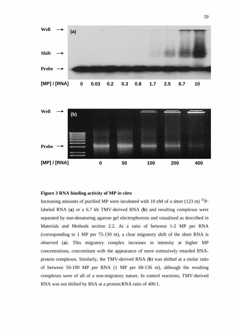
59
Figure 3 RNA binding activity of MP in vitro
Increasing amounts of purified MP were incubated with 10 nM of a short (123 nt) 32P-
labeled RNA (a) or a 6.7 kb TMV-derived RNA (b) and resulting complexes were
separated by non-denaturing agarose gel electrophoresis and visualized as described in
Materials and Methods section 2.2. At a ratio of between 1-2 MP per RNA
(corresponding to 1 MP per 75-150 nt), a clear migratory shift of the short RNA is
observed (a). This migratory complex increases in intensity at higher MP
concentrations, concomitant with the appearance of more extensively retarded RNA-
protein complexes. Similarly, the TMV-derived RNA (b) was shifted at a molar ratio
of between 50-100 MP per RNA (1 MP per 68-136 nt), although the resulting
complexes were of all of a non-migratory nature. In control reactions, TMV-derived
RNA was not shifted by BSA at a protein:RNA ratio of 400:1.
Well
Shift
Probe
[MP] / [RNA]
Well
Probe
[MP] / [RNA]
0 0.03 0.2 0.3 0.8 1.7 2.5 6.7 10
0 50 100 200 400
(b)
(a)

60
3.3 Infection of Nicotiana benthamiana and BY-2 protoplasts
with TMV encoding MP fused to –(H)6 affinity tags
In order to investigate whether TMV harbouring an MP-(H)6 fusion protein remained
infectious or indeed, whether the presence of polyhistidine per se interfered with the
known subcellular localization of MP:GFP, we transfected BY-2 protoplasts with
TMV-derived RNA encoding MP:GFP, MP:(H)6:GFP or MP:GFP:(H)6. Although the
correct localization of MP in BY-2 protoplasts is indicative of MP-mediated cell-to-cell
movement function in planta (Heinlein et al., 1998), this activity was directly
determined by inoculating intact leaves of N. benthamiana plants with the above RNA
transcripts and visualizing infection sites by fluorescence microscopy.
3.3.1 Infection of BY-2 protoplasts
At 18 hours post transfection (hpt), TMV-MP:GFP had clearly replicated and GFP
fluorescence could be localized to MTs (Fig. 4a-c), an observation consistent with the
cell-to-cell movement function of MP in planta. Similarly, both TMV-MP:(H)6:GFP
and TMV-MP:GFP:(H)6 were able to produce an infection phenotype comparable to
TMV-MP:GFP at 18 hpt and localization of –(H)6 tagged MP:GFP with microtubules
could be observed in both cases (Fig. 4f and i). Although not directly demonstrative of
MP cell-to-cell movement function, these data do illustrate that six consecutive
histidine residues do not interfere with MP localization. Expression of free GFP from a
MP and CP attenuated TMV construct results in the diffuse, cytoplasmic localization of
GFP fluorescence (Heinlein et al., 1995). For reasons not fully understood, the degree
of MP:GFP fluorescence found in association with MTs appeared to vary, with some
MTs apparently more heavily decorated with MP:GFP than others. Although the
relative amount of MP:GFP in association with MTs was comparable for the viruses
61
tested (not shown), on microtubules displaying heavy MP:GFP association, there was a
corresponding weak immunohistochemical labeling of tubulin. Although inconclusive,
this observation indicated that along stretches of MTs where tubulin was poorly
labeled, MP was in the proximity of the MT surface in large amounts, or that at least
some of the MP was occupying the antigenic site via a direct interaction, resulting in
steric hindrance of the antibody-tubulin interaction.

62
Figure 4 Infection of BY-2 protoplasts with TMV-derivatives encoding –(H)6
affinity tags. Tobacco BY-2 protoplasts were transfected with RNA derived from
TMV-MP:GFP (a, b and c), TMV-MP:(H)6:GFP (d, e and f) and TMV-MP:GFP:(H)6
(g, h and i), respectively. At 18 hpt, cells were chemically fixed and immuno-labeled
with anti-α-tubulin antibody. As determined by the onset of GFP fluorescence, all three
viruses were considered infectious in protoplasts, and the respective MP:GFP
chimaeras could be clearly associated with filamentous structures (a, d and g).
Antibody labelling of α-tubulin (b, e and h) and analysis of the merged composite
images (c, f and i) confirmed that MP:GFP was localized to microtubules in all three
cases, an observation consistent with the cell-to-cell movement function of MP.
(a) (b) (c)
(d) (e) (f)
(g) (h) (i)
10 µm 3 µm
GFP α-tubulin merge

63
3.3.2 Infection of N. benthamiana
As a test for viral infectivity and MP-mediated intercellular trafficking of vRNA,
infectious RNA in vitro transcribed from pTf5-MP:GFP (NSPAX), pTf5-MP:(H)6:GFP
and pTf5-MP:GFP:(H)6 (Materials and Methods section 2.3.1) were used to inoculate
leaves of N.benthamiana plants. At 3 days post inoculation (dpi), infected leaves were
detached, illuminated with UV light and photographed using a digital camera (Fig. 5a,
c and e). In all three cases, fluorescent infection sites were clearly visible, indicating
that each respective TMV-derived virus retained both the capacity to replicate, and also
to move cell-to-cell. In the case of TMV-MP:(H)6:GFP, the resulting infection sites
were brighter than those caused by either TMV-MP:GFP or TMV-MP:GFP:(H)6 (Fig.
5b), however, this increase in fluorescence did not correlate with an increase in the
average infection site area (Fig. 5a) In contrast, both the average size and relative
brightness of infection sites caused by TMV-MP:GFP:(H)6 were lower than those of
TMV-MP:GFP or TMV-MP:(H)6:GFP (Fig. 5a and b). In order to determine whether –
(H)6 interferes with the known subcellular localization of MP:GFP in planta, infection
sites produced by each respective TMV-derivative were compared by fluorescence
microscopy (Fig. 6b, d and f). The subcellular distribution of TMV-MP:GFP in cells
throughout the infection site were found to be consistent with previous reports of a coat
protein attenuated TMV-derivative virus expressing an MP:GFP fusion protein
(Heinlein et al., 1998). Briefly, in cells adjacent to the leading (outer) edge of infection,
MP:GFP was localized to cell wall sites. As one observed cells progressively closer to
the centre of infection, MP:GFP could be first seen to associate with cytoplasmic
inclusion bodies, then both smaller inclusion bodies and filamentous structures,
filamentous structures only, and in cells closest to the centre, again cell wall proximal
sites. This characteristic range of MP:GFP subcellular distributions throughout the

64
infection site was also observed for MP:GFP expressed from TMV-MP:(H)6:GFP and
TMV-MP:GFP:(H)6, respectively. It should be noted that in almost all cells infected
with TMV-MP:(H)6:GFP, significantly higher amounts of MP:GFP could be detected
in sites adjacent to the cell wall. Although this may account for the overall brightness
of the resulting infection sites, the presence of –(H)6 appeared to have little apparent
effect on the ability of MP:(H)6:GFP or MP:GFP:(H)6 to associate with MTs (Fig. 6b, d
and f). This result also highlights that the reduced rate of TMV-MP:GFP:(H)6 spread
was probably not due to aberrant localization of MP:GFP:(H)6. Although it is apparent
that MP:GFP:(H)6 accumulated to lower levels than the other MP:GFP fusion proteins,
the precise mechanisms governing the level of MP and rate of vRNA movement
remain unclear.

65
Figure 5 Analysis of infection sites on Nicotiana benthamiana leaves caused by
TMV-derivatives encoding –(H)6 affinity tags. Leaves infected with TMV-MP:GFP,
TMV-MP:(H)6:GFP or TMV-MP:GFP:(H)6 were detached 3 days post infection and
recorded under UV illumination by digital photography. The area and average pixel
intensity within resulting infection sites were determined densitometrically, and as
shown in (a), infection sites caused by both TMV-MP:GFP and TMV-MP:(H)6:GFP
were comparable in size (2.48 ± 0.13 and 2.49 ± 0.15 mm2, respectively). In contrast,
infection with TMV-MP:GFP:(H)6 led to an average infection site area of 1.76 ± 0.13
mm2, indicating a reduced rate of vRNA cell-to-cell movement. Relative to MP:GFP
fluorescence (18.1 ± 1.0 arbitrary units), MP:(H)6:GFP appeared to overaccumulate
(35.4 ± 2.1 arb. units), although a corresponding increase in infection site area was not
apparent (b). The depreciated level of MP:GFP:(H)6 fluorescence (9.6 ± 0.6 arb. units)
did correlate with a reduced infection site area, however, the possibility that –(H)6
caused MP to mislocalize could not be discounted without further microscopic
analyses. Error bars represent the standard error of the mean.
(a)
(b)
MP:GFP MP:(H)6:GFP MP:GFP:(H)6

66
Figure 6 Infection of Nicotiana benthamiana leaf epidermal cells with TMV-
derivatives encoding –(H)6 affinity tags. Expanded leaves of N. benthamiana plants
were mechanically inoculated with RNA derived from TMV-MP:GFP (a and b), TMV-
MP:(H)6:GFP (c and d) and TMV-MP:GFP:(H)6 (e and f), respectively. At 3 days post
infection, detached leaves were visualized by UV-digital photography (a, c and e) and
fluorescence microscopy (b, d and f). As determined by the appearance of expanding
(e)
(d) (c)
(f)
30 µm 1 mm
(a) (b)

67
fluorescent infection sites, all three viruses were considered infectious with regard to
viral replication and intercellular trafficking of vRNA. Although of a similar size,
infection sites caused by TMV-MP:(H)6:GFP (c) appeared to be brighter than those due
to TMV-MP:GFP (a), possibly reflecting enhanced MP:(H)6:GFP expression. In
contrast, the infection sites caused by TMV-MP:GFP:(H)6 (e) appeared to be both
smaller, and less fluorescent than TMV-MP:GFP induced sites, however, microscopic
analysis revealed that for each respective virus, the subcellular distribution of MP:GFP
fluorescence was largely consistent with previous reports (Heinlein et al., 1998).
Although inconclusive, MP:(H)6:GFP accumulation in cell wall proximal sites (d)
appeared to account for the relatively high fluorescence of infection sites caused by
TMV-MP:(H)6:GFP. Nevertheless, in cells behind the leading edge (LE) of infection
(arrow), MP:GFP, MP:(H)6:GFP and MP:GFP:(H)6 could be localized to filamentous
structures characteristic of microtubules (b, d and f; Boyko et al., 2000a), thereby
indicating that all three fusion proteins were indistinguishable with regard to MT
association. Although the presence of –(H)6 had subtle effects on the level of MP
accumulation and rate of vRNA trafficking, these data do collectively demonstrate that
viral replication, cell-to-cell trafficking and MP:MT association are not inhibited by –
(H)6 to the point of rendering the TMV-derivative viruses non-viable.

68
3.4 Biochemical analysis of MP:MT binding in vitro
3.4.1 Optimization of reaction conditions
The reaction buffer BRB80 (80 mM Pipes-OH [pH 6.8], 1 mM MgCl2, 1 mM EGTA)
containing GTP is commonly used for MT-based biochemical assays (Ackmann et al.,
2000). Attempts to directly perform the MAP assay (Materials and Methods section
2.4.3) with MP using this buffer system resulted in an apparent non-saturable and
linear binding relationship (not shown). Direct visualization of these ‘complexes’ by
immunohistochemical labeling of α-tubulin and MP followed by fluorescence
microscopy demonstrated that MP had formed large, irregular-shaped aggregates on
and around extensively bundled MTs (not shown). Regardless of the evident MP-MT
interaction, non-specific aggregation could not discounted as the sole determining
factor. Due to this experimental constraint, interpretations of the binding reaction
remained unreliable. To improve the stability of MP, a systematic approach was
established whereby MP solubility was tested in a range of buffers empirically known
to support MT polymerization (Lee and Timasheff, 1977; Waxman et al., 1981;
Pedrotti et al., 1993). Briefly, MP was incubated in premixed buffer, centrifuged and
the pellet and supernatant fractions were analyzed by SDS-PAGE and CCB staining.
Table 2 summarizes a number of MT-compatible conditions within which MP
solubility was tested. As mentioned, buffer BRB80 (+ GTP) induced the otherwise
soluble MP to aggregate. Solubility in this buffer could not be improved by the
presence of 10 % (v/v) glycerol or by non-ionic detergents in micelle concentrations
(Table 2, 2-8). Testing the BRB80 components individually showed that GTP was the
causal agent of MP insolubility when present at 1 mM (Table 2), a concentration
typically used to facilitate net MT stability in vitro. As a possible substitute for GTP,

69
the non-hydrolyzable analogue, Guanosine 5’-[γ-thio]-triphosphate (GTP-γ-S) was also
tested and found to produce a similar level of MP aggregation. Although not shown, it
was also found that a number of salts (e.g. H2PO4-, Na+ and K+) at concentrations
below 50 mM effectively “salted-out” MP from solution. This observation was not
entirely unsurprising given that MP has been shown by subcellular fractionation
experiments to behave as an integral-membrane protein (Reichel et al., 1999), and that
many such proteins have a general propensity to aggregate in low concentrations of salt
(Prof. M. Spiess, personal communication). Additionally, it was found that buffers
containing 80 mM Pipes-OH (pH 6.8) induced a less pronounced, yet detectable
decrease in MP solubility (Table 2, 4-5). Although considered to be minor (< 5 % loss
of MP in pellets), it was speculated that the effects of ~ 150 mM ionic strength (Seitz et
al., 2002), may still lead to extraneous non-specific binding and thus, overestimation of
the MP:MT binding stoichiometry (Timasheff, 2002). MP was therefore tested in more
dilute buffer (12 mM Pipes-OH [pH 6.8]) and was indeed found to remain soluble even
when stored for a period of days at 4 oC. It should be noted that a hydrophobic
component to the MP-MT interaction during infection cannot be ruled out, however, it
was hoped that by reducing the ionic strength of the buffer to a minimum, the
biochemical analysis in vitro could be somewhat simplified. Since GTP was now
known to induce MP aggregation, both GTP and unpolymerized tubulin dimers were
removed from the MT polymer fraction by repeatedly pelleting and resuspending MTs
into GTP-free buffer. In vitro, MTs spontaneously convert between phases of
elongation and shortening, and since MTs require GTP for the elongation phase of their
dynamics, the absence of exogenous GTP results in a net breakdown of MTs over time
(Caplow and Fee, 2003). To counteract this fact, pelleted MTs were resuspended in
GTP-free buffer containing the drug taxol, a strong suppressant of MT dynamics

70
(Wilson et al., 1985; Derry et al., 1995). Glycerol was also included in the MAP
binding buffer to additionally enhance MT stability (Prof. A. Hoenger, personal
communication). Providing the polymerized MTs were allowed to equilibrate in the
presence of taxol and GTP (≥ 12 hours) before being resuspended in MAP binding
buffer (12 mM Pipes-OH [pH 6.85], 0.5 mM MgCl2, 10 % [v/v] Glycerol, 0.01 % [v/v]
Tween-20 and 10 µM Taxol), MTs remained almost totally stabilized in the absence of
GTP for at least 30 minutes following buffer exchange (Fig. 7b).

71
Table 2 Solubility of MP in microtubule-compatible buffers. In order to
determine which component of the reaction buffer BRB80 (80 mM Pipes-OH [pH 6.8],
1 mM MgCl2, 1 mM EGTA [+ 1mM GTP]) was responsible for MP aggregation, MP
was incubated in the above buffer compositions, incubated for 1 hour and subjected to
high-speed centrifugation as described in Materials and Methods section 2.4.2.1. The
resulting pellet and supernatant fractions were analyzed by SDS-PAGE and CCB
staining and if 95% or more of MP remained in the supernatant, the protein was
considered to be soluble in that particular buffer (as indicated). Using this approach it
was possible to identify GTP as the causal agent of MP aggregation (highlighted in
red). All other standard components of BRB80 had no significant effect on the
solubility of MP. As a potential substitute for GTP, the structural analogue GTP-γ-S
was tested and also found to induce comprehensive MP aggregation. Neither micelle
concentrations of non-ionic detergents nor glycerol appeared to have any positive
effect on the solubility of MP.
80 mM Pipes-OH, pH 6.8
12 mM Pipes-OH, pH 6.8
1 mM Mg Cl2
1 mM EGTA
1 mM GTP
1 mM GTP-γ-S
25 µM Taxol
10 % (v/v) Glycerol
1 % (v/v) Nonidet-P40
0.1 % (v/v) Nonidet-P40
1 % (v/v) Triton X-100
0.1 % (v/v) Triton X-100
0.01 % (v/v) Tween-20
Soluble (yes/no)
+
+
+
+
+
N
+
+
+
+
+
+
N
+
+
+
+
+
+
N
+
+
+
+
+
+
N
+
+
+
+
+
+
N
+
+
+
+
+
N
+
+
+
+
+
N
+
+
+
+
+
N
+
+
+
+
+
N
+
+
Y
+
+
+
Y
+
+
Y
+
+
+
Y
+
+
+
+
+
Y
+
+
+
+
+
+
N
+
+
+
+
+
+
N
Component Buffer compositions

72
Figure 7 Examples of the microtubule stability assay. MTs were polymerized as
described in Materials and Methods section 2.4.2.2 and were resuspended in 60 µL of
either 10 mM Tris-Cl (pH 7.5), 1 mM MgCl2 and 25 µM Taxol (a), or MAP binding
buffer (12 mM Pipes-OH [pH 6.85], 0.5 mM MgCl2, 10 % [v/v] Glycerol, 0.01 % [v/v]
Tween-20 and 10 µM Taxol), as shown in panel (b). Samples were incubated for 30
minutes and polymerized tubulin was sedimented by centrifugation. Pellet (P) and
supernatant (S) fractions were analyzed on separate SDS-PAGE gels by CCB staining.
MTs were highly unstable in Tris-based buffers (pH 6.8 or pH 7.5), regardless of the
presence of Taxol. It was found that even upon dilution, and in the absence of GTP,
stable MTs could be maintained when Pipes (pH 6.8-7.0) was employed as the
buffering agent in the presence of Taxol.
P S P S
(a) (b)

73
3.4.2 MAP assay
The microtubule binding activity of MP was tested using the optimized MAP binding
buffer (Materials and Methods section 2.4.3). In order to fulfill the criteria for
saturation binding analysis, increasing amounts of MP was titrated over 3 orders of
magnitude (sub-supramolar) against a fixed concentration (1.1 µM) of polymerized
tubulin dimers. Following incubation, MTs and associated MP were sedimented and
both the pellet and supernatant fractions were analyzed by SDS-PAGE and CCB
staining. As can be seen in Fig. 8, an absence of tubulin in the supernatant fractions
indicated that the MTs remained dynamically stable throughout the course of the
reaction. Saturable binding was apparent as determined by the appearance of MP in
supernatant fractions at a 3:1 molar ratio of MP to tubulin and above. Replicate
experiments using freshly prepared MTs and MP produced directly comparable results.
However, due to the detection threshold of CCB, it was speculated that saturation of
MT binding sites by MP might not be seen at MP concentrations below 3 µM. Reaction
volumes were therefore increased 3-fold for reactions containing between 0.1 and 3
µM MP (not shown). Although more MP could be seen in the supernatant fraction in
this case, the ratio at which MP became visible remained the same at approximately
3:1. To control for buffer-induced aggregation of MP, reactions were repeated in the
absence of tubulin. In some cases very low amounts of MP could be detected in the
pellet fractions and where applicable, these amounts were accounted for when
calculating the amount of MP per lane.

74
Figure 8 Microtubule binding by MP in vitro. Increasing amounts of MP were
incubated with 1.1 µM of polymerized tubulin dimers in MAP binding buffer and MTs
together with associated MP were sedimented by centrifugation. Pellet (top panel) and
supernatant (bottom panel) fractions were analyzed by SDS-PAGE and CCB staining.
Detectable levels of tubulin are exclusively found in the pellet fractions, indicating that
the MTs remained stable under the reaction conditions used. In addition, the presence
of MP in the pellet fractions demonstrates that MP had the capacity to interact directly
with pure MTs. This interaction was saturable as determined by the increasing presence
of MP in the supernatants at an MP:tubulin ratio of 3:1 and above. Although not
shown, when reaction volumes were increased 3-fold, the ratio at which MP was
detectable in the supernatant remained about 3:1, suggesting that the detection
threshold of CCB (∼ 50 ng MP) was not a limiting factor.
Tubulin
MP
MP
0.1 0.25 0.5 1 2 3 4 5 6 7 8 10 [MP], µM

75
3.4.3 Determining the effect of –(H)6 on MT binding
The association of MAPs with MTs is thought to arise predominantly via ionic
interactions (Cassimeris and Spittle, 2001). At a pH of 7.0 (reaction pH = 6.8) the
predicted charge of –(H)6 is + 1.42 (ProtParam, IK@N Bioinformatics), therefore,
using a similar approach to the MAP assay, we tested whether –(H)6 is sufficient to
directly mediate MT binding contacts. Test proteins GST-(H)6, (H)6-Tau 441 or MP-
(H)6 (all at 1.5 µM) were incubated with (+) or without (-) a molar excess of
polymerized tubulin (6 µM) and pelleted material was analyzed by SDS-PAGE and
CCB staining. As expected, approximately all of the MP cosedimented with MTs (Fig.
9, lane 11), as compared to the starting amount of material (Fig. 9, lane 10). In the
absence of tubulin, soluble MP remained exclusively in the supernatant (Fig. 9, lane
12). A similar result was obtained using recombinant (H)6-Tau, a well characterized
neuronal MAP (Fig. 9 lanes, 4-6). In contrast, neither soluble GST-(H)6 (Fig. 9, lanes
1-3) or BSA (Fig. 9, lanes 7-9) appeared to cosediment with tubulin to any significant
degree, taking into account the molar excess of MTs. Although these results
demonstrate that a -(H)6 affinity tag is insufficient to produce stoichiometric MT
binding in vitro, low amounts of GST-(H)6 and BSA did cosediment with tubulin. The
amount of non-specific binding was comparable in both cases, however, the degree to
which –(H)6 contributed to this interaction, and whether a similar non-specific
component is involved in the MP-MT interaction remains unclear.

76
Figure 9 MAP assay control reactions to determine the effect of –(H)6 on
microtubule binding in vitro. Test proteins (1.5 µM) were incubated either with (+) or
without (-) 6 µM polymerized tubulin in MAP binding buffer and subjected to
centrifugation. Pellet fractions were analyzed by SDS-PAGE and CCB staining. The
position of tubulin is denoted by (∗) and for visual comparison, 1.5 µM of each test
protein was directly loaded on the gel without prior centrifugation (input). In the
absence of tubulin (lanes 3,6,9 and 12), the apparent lack of protein in the pellets
indicated that all four test proteins remained soluble under the conditions used. A small
amount of GST-(H)6 was observed to cosediment with MTs (lane 2), however,
compared to the starting amount of material (lane 1) this degree of binding appeared to
be neglible, possibly reflecting a weak non-specific interaction. Similarly, BSA also
did not bind MTs to any significant amount (lane 8), as compared to the starting
amount of protein (lane 7). Conversely, with a molar excess of tubulin, almost
complete binding of (H)6-Tau (lane 5) and MP (lane 11) could be observed. Although a
non-specific component to this binding could not be ruled out, the collective data
suggested that a –(H)6 moiety alone was insufficient to mediate strong interactions with
MTs in vitro.
Input (+) (-) Input (+) (-) Input (+) (-) Input (+) (-)
GST-(H)6 (H)6-Tau BSA MP-(H)6
*
1 2 3 4 5 6 7 8 9 10 11 12

77
3.4.4 Analysis of the MT binding properties of MP in vitro
Following the MAP assay, the amount of MP in the pellet ([MP]bound) and supernatant
([MP]free) fractions was quantified by densitometry of the digitized gels. The amount of
MP in each respective fraction was corrected for non-specifically pelleted MP and,
subsequently, values for [MP]bound were plotted versus [MP]free, yielding a saturation
hyperbole. In an attempt to characterize the equilibrium binding reaction, a curve was
fit to these data according to a simple model describing a receptor:ligand binding
reaction where only one type of ligand binding site exists, and all binding sites of this
type are independent and do not display an allosteric influence on subsequent ligand
binding (Appendix III eq. 11). It must also be assumed when using this model that
there is a single type of monovalent ligand, when values for the binding stoichiometry
and Kd are required. As can be seen in Fig. 10, a curve fit using the one-site model did
not pass smoothly through all data points, a fact reflected in the relatively low value of
r2 = 0.967 (an indicator that actual data deviate from the model-dependent predicted
curve). Nonetheless, non-linear regression analysis of this curve yielded apparent
values of Kd = 71.6 ± 14.5 nM with a stoichiometry of 2.8 ± 0.12 MP/tubulinmonomer.
The Kd value determined by this method was within the range commonly found for
other MAPs (Ackmann et al., 2000), although it should be noted that since a low ionic
strength buffer was employed, and MAP binding is thought to result largely via ionic
interactions, the apparent affinity of MP for MTs may be slightly enhanced. The value
for stoichiometry was considered to be high, therefore, in order to determine whether
the one-site model was indeed appropriate, Scatchard-Rosenthal plots were produced
by plotting [MP]bound/[MP]free versus [MP]bound according to the Scatchard equation
(Appendix III eq. 13; Fig. 11, inset). In theory, adherence to the above criteria would
result in a straight line with a slope of –1/Kd and stoichiometry found at the x-intercept.

78
Manually fitting a curve to these data illustrated a clear deviation from the predicted
straight line relationship (Fig. 11) and in fact resulted in a line of upward curvature.
Scatchard curves of this type can be interpreted in a number of ways (Norby et al.,
1980; Zierler, 1989). For instance, the existence of more than one type of ligand
binding site on a receptor can account for apparently biphasic curves, and are usually
attributable to sites with differing affinities for the same ligand. In this case,
independence is assumed both within and between types of binding site. Alternatively,
one may infer that ligand binding undergoes homotropic allosterism, that is, initial
binding of ligand to a single type of binding site has a negative influence on subsequent
ligand binding to the same type of site. In an attempt to differentiate between the
underlying binding mechanisms, a curve was fit to plots of [MP]bound versus [MP]free
according to a two-site model (Appendix III eq. 12), where binding site independence
was again assumed. In this case, non-linear regression yielded two values for Kd and
stoichiometry (Kd1 = 31.3 ± 16.6 nM; n1 = 1.81 ± 0.55; Kd2 = 1.56 ± 1.34 µM; n2 = 1.56
± 0.436), indicating both a high and low affinity MP-binding site on tubulin. The
overall fit of the line was improved (Fig. 12; r2 = 0.986) as compared to the one-site
model, and comparison of the two curves by one-way analysis of variance (Fig. 13; F =
4.72, P = 0.026) indicated that statistically, a two-site model was indeed the more
appropriate. It should be noted that absolute values obtained from Scatchard plots are
rendered unreliable due a deviation from the Gaussian distribution during data
transformation. Taken together with the statistically strong fit of a two-site model, it is
tempting to conclude that MP binds alternative sites on tubulin, however, whether the
second site actually resides on the surface of tubulin, or occurs via a tubulin:MP:MP
ternary complex remains a matter of conjecture. Additional complexities arise due to
insufficient knowledge regarding the homogeneity of MP. Since it is possible that

79
multivalent or alternative folded states of renatured MP exist, and indeed, the low
affinity site may represent a non-specific binding site, further experimentation is
clearly required in order to make a categorical conclusion about the mechanistic nature
of the MP:MT interaction in vitro.

80
Figure 10 Equilibrium binding of MP to MTs in vitro: one-site model. Equilibrium
binding experiments were performed by incubating purified MP (0.1-10 µM) with in
vitro polymerized tubulin (2.2 µM monomer) before bound and unbound MP was
separated by centrifugation and analyzed by SDS-PAGE and CCB staining. Values for
[MP]bound and [MP]free were determined by densitometry as described in Materials and
Methods section 2.4.5. Plotting [MP]bound versus [MP]free gave a saturation hyperbole
and a curve was fit to these data according to the binding model describing a single
type of non-interacting binding site on tubulin (Materials and Methods section 2.4.5).
The fit curve was analyzed by non-linear regression and yielded an apparent Kd of 71.6
± 14.5 nM, a value comparable to other characterized MAPs. The curve saturated at an
apparent stoichiometry of 2.8 ± 0.12 equivalent binding sites per tubulin monomer.
Because the curve did not pass smoothly through all of the data points (r2 = 0.967;
where 1 indicates a perfect fit), it remains feasible that a one-site model does not
adequately describe these data.

81
Figure 11 “Linearization” of equilibrium binding data: Scatchard-Rosenthal plot.
Equilibrium binding experiments were performed as described in Materials and
Methods section 2.4.3 and values of [MP]bound/[MP]free were plotted versus [MP]bound.
According to the Scatchard equation (inset), equilibrium binding data plotted in this
manner should result in a straight line when the criteria for a single type of
independent, non-interacting ligand binding site are met. These data clearly deviate
from the predicted straight line, and therefore, a curve was fit (y = a+b/1+cx) using a
best-fit trial and error approach (r2 = 0.98). The upwards curvature and apparent
biphasic nature of this line can indicate the following; negative cooperativity
(homotropic steric hindrance) in a one-binding site system, the existence of two or
more independent MP-binding sites per tubulin or a non-specific binding component to
the reaction (Norby et al., 1980). Care should be taken when interpreting Scatchard
plots, since following transformation, data no longer adhere to the assumed Gaussian
distribution.

82
Figure 12 Equilibrium binding of MP to MTs in vitro: two-site model. Equilibrium
binding experiments were performed as described in Materials and Methods section
2.4.3 and in this case, a curve was fit to the plot of [MP]bound versus [MP]free according
to a model describing two types of independent, non-interacting binding sites on
tubulin (Materials and Methods section 2.4.5). Non-linear regression analysis indicated
the existence of a high affinity (Kd1 = 31.3 ± 16.6 nM; n1 = 1.81 ± 0.55) and a low
affinity (Kd2 = 1.56 ± 1.34 µM; n2 = 1.56 ± 0.436, respectively) MP-binding site on
tubulin. The curve appeared to fit the data relatively well (r2 = 0.986), as compared to
using a one-site model (Fig. 10). The possibility that the improved fit resulted from an
increased number of parameters, and therefore inflexion points, could not be
discounted using this analysis alone.

83
altalt
altnullaltnull
/dfSS)df - )/(dfSS -(SS
F =
7/79.0
7)-0.79)/(9 - (1.87 F =
)026.0 , (72.4F == P
Figure 13 Analysis of MP equilibrium binding to MTs in vitro: comparison of
binding data fit to a one-site or two-site model. Equilibrium binding experiments
were performed as described in Materials and Methods section 2.4.3 and curves were
fit to a plot of [MP]bound versus [MP]free according to models describing a single type of
independent binding site (a1) or two different independent binding sites (a2). The fit of
these curves were compared by one-way ANOVA of the non-linear regression data
obtained for each respective curve fit. The F ratio was calculated (b) assuming a null
hypothesis (null) that the one-site model fits the data better, and an alternative
hypothesis (alt) that the two-site model is more appropriate. SS is the sum of the
squares and df is degrees of freedom. Since the F ratio was found to be greater than
one, the null hypothesis could be rejected, which was confirmed by the calculated P-
value of 0.026. It could therefore be statistically inferred that in vitro, MP binding to
MTs is a biphasic reaction involving more than one MP-binding site on tubulin or
possibly resulting due to multivalent/isotypic forms of MP.
(a) (b)

84
3.5 Visualization of MP:MT complexes formed in vitro by
fluorescence microscopy
3.5.1 Establishment of the perfusion chamber system
MTs are highly sensitive to their immediate buffer environment, making the direct
immunolabeling of MAP:MT complexes technically unfeasible without prior fixation.
Initial attempts to fix MP:MT complexes in free solution resulted in extensive and
irregular bundling of MTs (not shown). Although MP appeared to colocalize with MTs
in this case, the presence of large MP aggregates suggested that the interaction may
have occurred largely through non-specific crosslinking, particularly when MP was in
molar excess. A system was therefore established whereby immobilized MTs could be
incubated with MP and unbound protein removed before fixation. Briefly, a simple
perfusion chamber was coated with a mixture of casein and the molecular motor
kinesin. In vitro polymerized MTs were anchored to the kinesin in the absence of ATP,
in order to inhibit motor activity before MP was introduced and allowed to bind MTs.
Unbound protein was washed out and the resulting complexes were rapidly fixed with
glutaraldehyde before α-tubulin and MP were immunohistochemically labeled. One
major drawback to using the perfusion chamber method is that one has no accurate
knowledge of the actual amount of MTs within the chamber, since this is dependent on
both the concentration of kinesin and tubulin, and also the time that each respective
component is absorbed to the chamber. Assuming that every MT was immobilized, the
maximum theoretical tubulin concentration within the chamber would have increased
to 0.5 µM, due to a concentration of the original 90 nM solution. As determined by
visual inspection of rhodamine-labeled MTs before and after absorption, the realistic
concentration was significantly lower than this figure. Therefore, MP was applied at a

85
wide range of concentrations in an attempt to emulate the MP:Tubulin stoichiometries
used during the MAP assay. To test for crossreactivity leading to false MP signals, a
variety of anti-tubulin, anti-MP and secondary antibody combinations were used to
label MTs undecorated with MP. The following combinations were considered
suitable; anti-α-tubulin YOL1/34 (α-tubulin residues 414-422) in combination with
anti-MP-N (MP residues 6-22), or anti-α-tubulin DM1A (α-tubulin residues 426-450)
in combination with anti-MP-C (MP residues 209-222), respectively. Due to poor
labeling efficiency, anti-β-tubulin antibodies were not used.
3.5.2 Fluorescence microscopy
Using either of the above antibody combinations, it was not possible to observe any
significant MP colocalization with MTs at MP concentrations below 200 nM (not
shown). Since estimates of the tubulin concentration were wholly unreliable, it could
only be speculated that the apparent lack of MP co labeling was either due to
insufficient amounts of MP, and consequently a fluorescence signal below the
microscope detection limit, or alternatively, a low signal to noise ratio resulting from
non-specific binding of MP to the chamber surface. In either case, the tubulin staining
produced a smooth, consistent fluorescence signal along the length of the MTs. In
contrast to this, incubations with higher concentrations of MP (0.75-2.5 µM) resulted in
a noticeably punctate tubulin labeling when either YOL1/34 or DM1A anti-tubulin
antibody was employed alone, and in extreme cases, an almost total absence of tubulin
labeling (for examples see Fig. 14, row a). This observation suggested that MP may
have been present at or near the antigenic sites for either antibody (collectively
mapping to α-tubulin residues 414-450), and indeed, immunolabeling of MP:MT
complexes (MP > 0.75 µM) with the antibody combinations described above resulted

86
in apparent MP colocalization along the entire length of the MTs. The distribution of
MP along the MTs was not uniform, however, and along certain stretches of the MTs,
the MP labeling appeared to be particularly strong (Fig. 14, row b). Although the
overall tubulin-specific signal was higher than that of MP, these areas where the MP
signal was stronger clearly correlated with a largely reduced tubulin-specific signal
(Fig. 14, row c and d). Although inconclusive, this result indicated that at least in part,
MP had the capacity to obscure the antigenic site recognised by either anti-tubulin
antibody, and that residues found at either termini of MP were accessible to anti-MP
antibodies, possibly suggesting that these regions of MP were not directly involved in
the MP:MT interaction. Further experimentation, such as the use of truncated or
mutagenized MP would be required in order to test this hypothesis. It should be noted
that upon TMV infection of BY-2 protoplasts, MTs heavily decorated with MP:GFP
are also stained poorly by anti-α-tubulin antibody DM1A, although care should be
taken when drawing conclusions from such dissimilar experimental systems.
Biochemical analysis of the MP:MT interaction strongly suggests that equilibrium
binding deviates from a single ligand, single receptor mechanism. In principle, the
observed non-uniform distribution of MP along MTs in vitro supports this hypothesis,
although it must be conceded that due to extensive washing, the above conditions do
not fully reflect equilibrium binding. Nonetheless, MP complexed with MTs is
recognized by anti-MP antibodies reactive against both N- and C-termini. Considering
the possible interpretations of a ‘two-site’ binding model (Results section 3.4.4), the
above observations favour a binding mechanism whereby MP can interact directly with
tubulin, although, a less ordered binding reaction may occur, possibly resulting in a
tubulin:MP:MP ternary complex. Due to the similar predicted binding stoichiometries
for either mechanism (n1 = 1.81 ± 0.55 and n2 = 1.56 ± 0.436 MP:tubulin dimer), it is

87
not possible using this method to make firm conclusions regarding the relative
abundance of MP on the MT and the corresponding binding mechanism. Similarly, it
remains unclear whether non-uniform MP distributions reflect structural differences
within the MT itself, or indeed, whether MP can interact with β-tubulin.
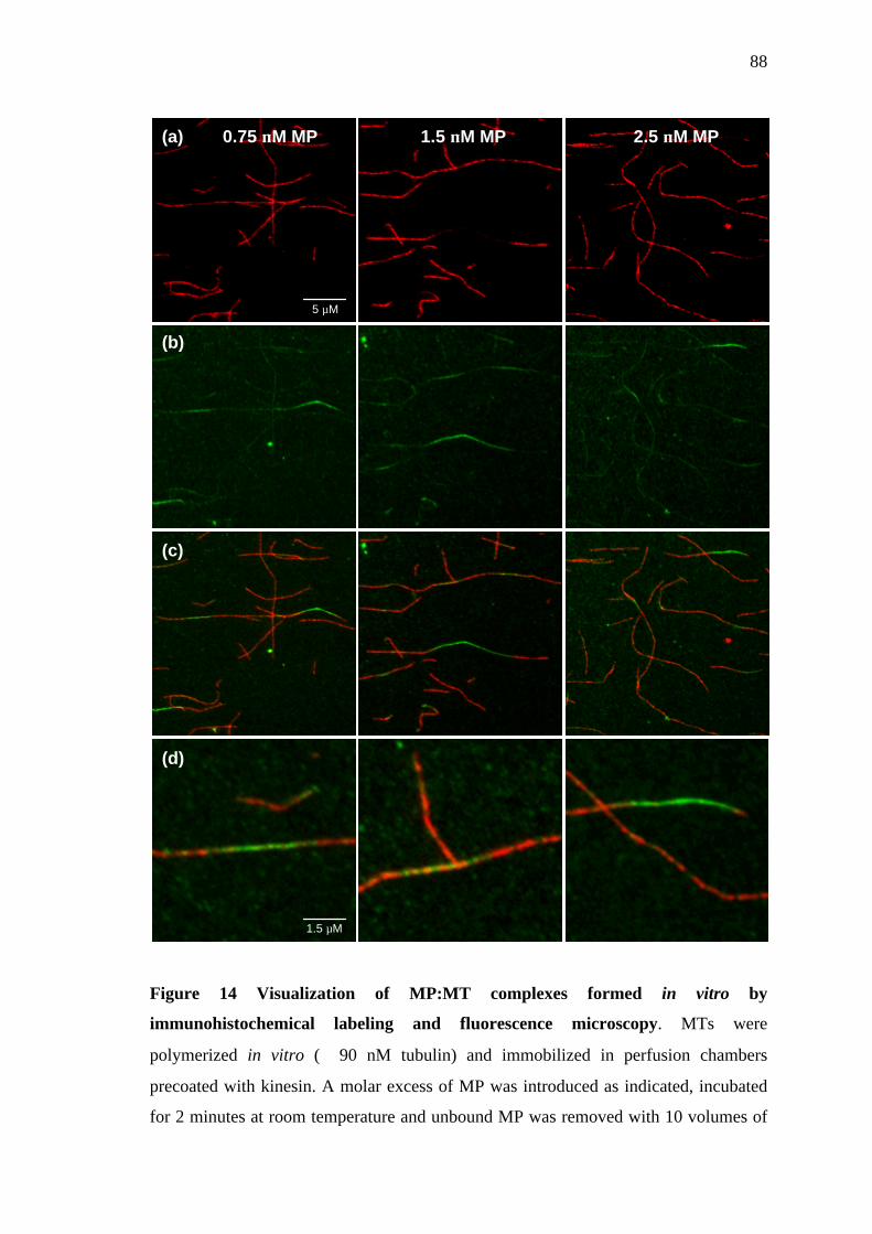
88
Figure 14 Visualization of MP:MT complexes formed in vitro by
immunohistochemical labeling and fluorescence microscopy. MTs were
polymerized in vitro (∼ 90 nM tubulin) and immobilized in perfusion chambers
precoated with kinesin. A molar excess of MP was introduced as indicated, incubated
for 2 minutes at room temperature and unbound MP was removed with 10 volumes of
(a) 0.75 µM MP
(d)
(c)
(b)
2.5 µM MP 1.5 µM MP
5 µM
1.5 µM

89
MAP binding buffer. Resulting MP:MT complexes were fixed and labeled with
antibodies specific for α-tubulin (YOL1/34; row a) and MP (anti-N-term; row b),
respectively. Under the conditions used, MP could be clearly observed to colocalize
with the MTs at a level above that of background fluorescence (row a and b). Although
the overall MP-specific fluorescence was less than the corresponding tubulin signal
(row c and d), the presence of MP could be correlated with a mere punctate labeling of
tubulin,. in contrast to MTs or stretches of MTs undecorated by MP that showed strong
and smooth tubulin labeling (not shown). Moreover, in areas where MP fluorescence
was particularly strong, an almost complete absence of tubulin staining was observed.
Although these data do not strictly reflect equilibrium binding (due to excessive
washing), the non-uniformity of the MP:MT colocalization is in support of biochemical
evidence that suggests in vitro, MP binds MTs via a bi- or possibly multiphasic
mechanism. The precise manner by which MP impedes the anti-tubulin:antigen
interaction remains unclear, although in principle, these data are consistent with
observations of TMV-infected BY-2 protoplasts in which heavy MP:MT colocalization
corresponds to a diminished tubulin immunolabeling.

90
3.6 Co-precipitation experiments in vitro using immobilized
MP as an affinity bait
3.6.1 Co-precipitation of MTs and tubulin heterodimers in vitro
It has been described in the literature that certain ‘tubulin’ binding proteins
preferentially interact with specific structural forms of tubulin such as tubulin dimers
(Spittle and Cassimeris, 1996; Fukata et al., 2002) or microtubules ends (Carvalho et
al., 2003; Howard and Hyman, 2003). Therefore, in order to determine whether MP
can interact with binding contacts found on tubulin dimers or specifically interacts only
with polymerized tubulin, purified MP was immobilized on Ni2+-NTA sepharose beads
and used as an affinity bait for the co-precipitation of either in vitro polymerized MTs
or tubulin dimers. Pellet and supernatant fractions were separated by centrifugation and
were analysed by SDS-PAGE and Western blotting, and to control for non-specific
binding of tubulin to sepharose, the same procedure was followed in the absence of
MP. Western blots probed with anti-MP-C antibody showed that throughout the course
of the binding reaction, MP remained coupled to the affinity matrix in amounts that
were comparable between reactions (Fig. 15a). Probing equivalent Western blots with
anti-α-tubulin antibody showed that even after extensive washing, detectable levels of
tubulin could be found in pellet fractions containing MP (Fig. 15b). In pellet fractions
without MP, no tubulin was detected, thereby indicating that precipitation of tubulin
had occurred via an interaction with MP, and not by virtue of non-specific contacts
with the sepharose beads. Tubulin did not co-precipitate with Ni2+-NTA sepharose
beads in the absence of BSA (not shown), again suggesting that an MP:tubulin
complex had formed, as opposed to an MP:BSA:tubulin ternary complex. Repeating
the experiment confirmed that under the conditions used, MP interacted more

91
favourably with tubulin dimers than MTs, although the reason for this is not fully
understood. Also, it cannot be ruled out that what appeared to be an MP:MT interaction
was actually the result of MT depolymerization and the interaction of MP with tubulin
dimers or short tubulin oligomers. Nevertheless, the fact that soluble tubulin dimers
were able to bind MP in vitro suggests that polymer-specific structural forms of tubulin
are not a prerequisite to MP binding. Furthermore, considering that the –(H)6 tag was in
close proximity to the affinity matrix, it is likely that residues found at the extreme C-
terminus of MP were inaccessible for tubulin binding, suggesting that the MP:tubulin
interaction was not mediated solely by this region of MP, an observation in agreement
with previous reports (Boyko et al., 2000a; Boyko et al., 2000c). Repeating the
experiment confirmed that under the conditions used, MP interacted more favourably
with tubulin dimers than MTs, although the reason for this is not fully understood. It
could be envisaged, however, that while the physical distribution of MP on Ni2+-NTA
beads may have had a nominal effect on the correct orientation and binding of free
tubulin dimers to MP, a net reduction in the number of available MP binding sites per
equivalent (polymerized) tubulin dimer may have occurred, thus resulting in weaker
overall binding. Also, it cannot be ruled out that what appeared to be an MP:MT
interaction was actually the result of MT depolymerization and the interaction of MP
with tubulin dimers or short tubulin oligomers. Nevertheless, the fact that soluble
tubulin dimers were able to bind MP in vitro suggests that polymer-specific structural
forms of tubulin are not a prerequisite to MP binding, and furthermore, considering that
the –(H)6 tag was in close proximity to the affinity matrix, it is doubtful that the
MP:tubulin interaction occurred solely via the extreme C-terminus of MP.

92
Figure 15 Co-precipitation of MTs and tubulin heterodimers in vitro using
immobilized MP as an affinity bait. Ni2+-NTA beads conjugated to MP (+ MP) or
without MP (- MP) were incubated with in vitro polymerized MTs (polymer) or
tubulin α/β dimers (dimers). Pellet (P) and supernatant (S) fractions were separated by
centrifugation and analysed by SDS-PAGE and Western blotting using antibodies
specific against MP or α-tubulin, respectively. As shown in (a), MP remained coupled
to the Ni2+-NTA beads throughout the course of the binding reaction. In pellet fractions
containing MP, detectable amounts of both tubulin polymer and tubulin dimers were
observed (b), although under the conditions used (Materials and Methods section
2.6.1), the MP:dimer interaction appeared to be favoured. In the absence of MP, no
tubulin was detected in the pellets, indicating that tubulin co-sedimention with Ni2+-
NTA beads occurred due to an interaction with MP.
P S P S polymer dimers
anti-MP
(a)
+ MP MP + MP MP polymer dimers
anti-α-tubulin
(b)
(c)
Pellet
Supernatant

93
3.6.2 Co-precipitation of RNA and tubulin heterodimers in vitro
The putative role of RNA in the MP:tubulin interaction in vitro was investigated using
an approach whereby MP (theoretical max. 3.25 µM) was immobilized on Ni2+-NTA
beads and used to co-precipitate combinations of tubulin dimers and RNA. Using a
two-step approach, MP-conjugated beads were first incubated in the presence of
tubulin heterodimers (1.63 µM) or the 6.7 kb in vitro transcribed TMV genomic RNA
(50 nM or ~300 µM polymerized nucleotide). Subsequently, tubulin dimers, RNA or
pulldown buffer were added to the reaction (as summarized in Fig. 16a) and incubated
once again. To control for non-specific binding, RNA or tubulin were incubated with
beads in the absence of MP. Pellet and supernatant fractions were separated by
centrifugation before RNA was analyzed by non-denaturing agarose gel electrophoresis
(Fig. 16c) and proteins were analyzed by SDS-PAGE and Western blotting (Fig. 16b).
When incubated alone with MP-conjugated beads, both tubulin dimers (Fig. 16b1) and
RNA (Fig. 16c4) appeared to co-precipitate with MP-conjugated beads. Although
comparable amounts of MP were found exclusively in the pellet fractions, high levels
of both tubulin and RNA could be detected in corresponding supernatant fractions (not
shown), indicating that saturable binding had been achieved. At the MP:tubulinmonomer
ratio of 1:1, one would expect the majority of tubulin to be found in the pellet fraction
(Results section 3.4.4). Since this was not the case, it was speculated that either MP
was incompletely conjugated to the affinity matrix (i.e. << 3.25 µM) or that upon
refolding, only a subset of MP had retained tubulin binding activity. Nevertheless, in
control reactions where MP was absent, neither tubulin nor RNA (Fig. 16b5 and c6,
respectively) was detected in pellet fractions, which confirmed that co-precipitation of
either component had only occurred due to an interaction with MP. When MP-

94
conjugated beads were pre-incubated with tubulin before the addition of RNA, the
amount of tubulin found in the pellet fraction was comparable to when tubulin was
added alone (Fig. 16b2). However, the amount of detectable RNA in the pellet fraction
was greatly reduced, which indicated that the MP:RNA interaction had been inhibited
by the presence of tubulin (Fig. 16c2). Since RNA was able to bind MP in absence of
tubulin, and indeed was present in a large molar excess over tubulin (when viewed in
terms of nucleotides), it is likely that MP had a greater affinity for tubulin than RNA.
In contrast, when RNA was incubated with MP-conjugated beads preceding the
addition of tubulin, both components appeared to retain the capacity to interact with
MP (Fig. 16c3 and b3, respectively), although to a lower level than when added alone.
One possible explanation for this might be that tubulin out-competed RNA for an
equivalent binding interface on the surface of MP. Although competitive effects cannot
be ruled out, one might expect that at equilibrium, the level of pelleted material would
be similar to when tubulin was added before RNA. Since both components were found
in relatively high levels (and binding saturation had apparently occurred, not shown), it
appeared that in fact a triple complex composed of MP, RNA and tubulin had formed.
This being the case, it would suggest that although binding contacts essential for
tubulin interactions were not entirely sequestered by the presence of RNA, there was a
functional overlap between RNA and tubulin binding domains present within MP.
Whether this putative molecular order of events would govern triple complex
formation on the surface of polymerized tubulin, or indeed, whether such a complex
would be viable in planta remains to be further analyzed.

95
Figure 16 Co-precipitation of RNA and tubulin heterodimers in vitro using
immobilized MP as an affinity bait. Using a two-step approach, Ni2+-NTA beads
conjugated to purified MP (theoretical max. 3.25 µM) were incubated in the presence
of either a 6.7kb in vitro transcribed RNA (50 nM or ~300 µM polymerized nucleotide)
or tubulin heterodimers (1.63 µM). Following this, tubulin, RNA or pulldown buffer
were added to the beads, which were subsequently incubated and sedimented by
centrifugation. Pelleted RNA was analyzed by 1 % non-denaturing agarose gel
electrophoresis (c) and pelleted proteins were analyzed by SDS-PAGE and Western
1 + MP Tubulin Buffer
2 + MP Tubulin RNA
3 + MP RNA Tubulin
4 + MP RNA Buffer
5 - MP Tubulin Buffer
6 - MP Tubulin RNA
Reaction ± MP 1st component 2nd component
1 2 3 4 5 6 1 2 3 4 5 6
anti-MP anti-α-tubulin
2 3 4 6
TMV-MP:GFP:(H)6 RNA (6.7 kb)
(a)
(b)
(c)

96
blotting, using antibodies specific for MP and α-tubulin, respectively (b). The reaction
combinations performed are summarized in Table (a) and correspond with the
numbered lanes in (b) and (c). When added individually, both tubulin dimers (b1) and
RNA (c4) appeared to co-sediment with MP, and in the corresponding supernatant
fractions (not shown), detectable levels of both tubulin and RNA were observed which
suggested that saturable binding had been achieved. In control reactions where MP was
absent (reactions 5 and 6), neither tubulin nor RNA appeared to interact with the
sepharose beads, confirming a direct interaction with MP had occurred in both cases.
When MP-conjugated beads were pre-incubated with tubulin preceding the addition of
RNA, almost saturable binding of tubulin appeared to occur (b2), although under the
conditions used, the capacity of RNA to interact with MP was severely impeded (c2).
In contrast, when RNA was introduced before tubulin, both components appeared to
bind MP (b3 and c3), albeit to a lower level than when incubated alone with MP.
Although inconclusive, these data suggest that at least in principle, an MP:RNA:MT
triple complex is viable, and furthermore, residues within MP required for RNA
binding may functionally overlap with those required to bind tubulin.

97
3.7 Apparent modulation of kinesin motor activity by MP
Since it has been described that microtubule association of MAP2 and tau proteins are
inhibitory to molecular motor activity both in vivo (Ebneth et al., 1998; Trinczek et al.,
1999; Stamer et al., 2002) and in vitro (von Massow et al., 1989; Lopez and Sheetz,
1993; Hagiwara et al., 1994; Seitz et al., 2002), we speculated that the interaction of
MP with MTs may confer a similar effect. In order to test this hypothesis, we utilized
the so-called ‘MT gliding assay’ whereby conventional human kinesin-1 was absorbed
onto a glass surface before fluorescently labeled MTs were introduced in the presence
or absence of purified MP (Materials and Methods section 2.5). Kinesin motor activity
was then initiated with the addition of ATP and the rate of MT translocation was
measured using digital fluorescence microscopy (Fig. 17a) by determining the relative
position of at least 30 MT ends at known time intervals (150 velocity
readings/experimental condition). In addition, the percentage of MTs undergoing any
translocation was calculated for each respective experimental condition. In the absence
of MP, the average velocity of kinesin-dependent MT translocation was found to be
0.20 ± 0.052 µm/sec (Fig. 17b), a value in agreement with previous reports (Seitz et al.,
2002). When 200 nM MP was added to the chamber prior to the initiation of kinesin
motor activity, the average velocity of motile MTs was reduced to 0.16 ± 0.053 µm/sec
(Fig. 17c), an interesting observation given that no discernible MP labeling on MTs
was observed when equivalent MP:MT complexes were immunohistochemically
labeled with an antibody specific for MP. Nevertheless, analysis of the mean values
using a two-tailed unpaired t-test showed them to be statistically distinct (P<0.001, n =
150), which indicated that MP affected a weak inhibition on kinesin-dependent MT
translocation. A more pronounced effect on the rate of MT translocation was observed
98
when 600 nM MP was used (0.14 ± 0.058 µm/sec), and although statistically different
than the mean velocity resulting from 0 and 200 nM MP treatments, respectively
(P<0.01, n = 150), it was apparent that the range of translocation speeds had deviated
slightly from a normal distribution (Fig. 17d). Visual analysis of animated MT
translocations recorded under the described conditions (supplementary data:
www.fmi.ch/members/jamie.ashby) showed that, although the majority of MTs
appeared to smoothly glide across the chamber surface, a number of motile MTs
underwent intermittent stalling when ≥ 600 nM MP was used. Although stalling may
account for the deviation from normally distributed data, it was apparent that due to
this statistically irregular data spread at MP concentrations above 600 nM, reliable
comparisons of average velocities could not be made. It should also be noted that,
although not taken into account during velocity measurements, the percentage of motile
MTs did appear to decrease concomitantly with increasing MP concentration (Fig. 17b-
d). Moreover, when MP was used at either 850 nM or 1.0 µm, the percentage of motile
MTs had decreased so dramatically (30.4 and 4.0 %, respectively) that the measurable
sample population for determining velocity was below a reasonable statistical
minimum. Interestingly, visual analysis of the few MTs actually translocating indicated
that kinesin motor activity was largely unaffected in those isolated cases
(www.fmi.ch/members/jamie.ashby). It is possible that these particular MTs
represented a subset of the population which were only partially decorated with MP.
Although the presence of MP did appear to modulate the overall rate of kinesin-
dependent MT translocation, interpretation of these data with regard to the mechanism
of inhibition remains somewhat tenuous. Under the experimental conditions applied,
kinesin is predicted to be in a dimeric, processive form at a density of ~ 4000
molecules/µm2 (Howard et al., 1993; Moyer et al., 1996). However, we have no

99
accurate knowledge of the polymerized tubulin concentration within the chamber.
Visual comparisons before and after washing the chamber of unbound MTs showed
that only a small percentage of introduced material actually adhered to the surface (not
shown). Since each MT is therefore likely to be in favourable contact with several
hundred kinesin molecules at any given time, one cannot determine whether inhibition
arose due to a reduction in binding frequency between kinesin and tubulin, run length
once bound or indeed modulation of kinesin motor activity itself. Current models for
kinesin inhibition by microtubule-associated proteins favour a mechanism by which
both proteins compete for equivalent binding contacts on β-tubulin, although once
bound to the MT, kinesin is believed to translocate at normal rates (Lopez and Sheetz,
1993; Seitz et al., 2002). It is unclear whether MP inhibits kinesin by a directly
comparable mechanism, since if this were the case, one would not expect to observe a
general decrease in MT translocation velocity at such a high density of kinesin.
Although MP may indeed compete with kinesin for binding sites on tubulin, it remains
plausible that the decreased rate of kinesin motor activity occurs by a more complex
mechanism. In order for dimeric kinesins to translocate processively, one of the motor
head domains must transiently dissociate from tubulin and move to the next available
site in an ATP-dependent manner (Movie 6, www.fmi.ch/members/jamie.ashby). It
could be speculated that the presence of MP on or in proximity to this second site
imposes a steric constraint on such a kinesin motion, or moreover, directly impedes the
so-called “power stroke” (Vale and Milligan, 2000). Intermittent stalling of MTs might
then occur if one or more kinesin molecules are inhibited in this manner at a region of
the MT which is decorated with particularly high levels of MP. Recovery from stalling
might be orchestrated by the collective force of active kinesin motors which physically
disrupt the non-motile kinesin:tubulin interaction or by displacement of the obstructive

100
MP. It was speculated that MP may directly bind to kinesin and inhibit motor activity
by modulation of ATPase activity or steric hindrance. Although direct binding
experiments are still required, preblocking of the perfusion chamber with 500 nM MP
preceding the addition of MTs appeared to have a neglible effect on kinesin-dependent
MT translocation, as determined visually (not shown). In addition, it was proposed that
MP may bind non-specifically to the surface of the chamber, and at high
concentrations, interact with MTs at stoichiometries sufficient to counteract kinesin
force production i.e. anchor MTs to the surface. Non-specific effects of this nature
cannot be discounted as the factor responsible for complete inhibition of motility at
very high MP concentrations (> 600 nM), however, it is known that although MTs
strongly adhere to plain glass surfaces, MT translocation can still be achieved if glass is
preabsorbed with kinesin (von Massow et al., 1989). Nevertheless, further experiments
utilizing very low kinesin amounts or even single molecules (Howard et al., 1989;
Block et al., 1990) at known MP:tubulin ratios are required in order to determine more
precisely the mechanism by which MP apparently inhibits kinesin-1 motor activity in
vitro.

101
Figure 17 Apparent modulation of kinesin motor activity by MP in vitro. TRITC-
labeled MTs (110 nM polymerized dimers) were absorbed to perfusion chambers
precoated with GST-conjugated kinesin-1 and either buffer alone (a and b), 200 nM
MP (c) or 600 nM MP (d) were perfused through the chamber and briefly incubated.
Following the addition of motility buffer+OS, images of “gliding” MTs were recorded by
digital fluorescence microscopy, as illustrated in panel (a) and the velocity of MT
translocation was determined by measuring the distance traveled by at least 30
individual MTs every 1.25 seconds for 2 minutes. Mean values (" S.D.) were
determined from the pooled data (150 measurements per sample) and the percentage of
MTs that underwent any translocation were also calculated for each respective
0 sec
40 sec
80 sec
7 µm
(a) (b)
(c)
(d)

102
experimental condition. As can be seen in panels b and c, upon the introduction of 200
nM MP, the average velocity of kinesin-dependent MT translocation was reduced from
0.20 " 0.052 µm/sec to 0.16 " 0.053 µm/sec, respectively. Comparison of these means
using an unpaired t-test showed that they were statistically unequal (P<0.001, n = 150),
thereby suggesting that MP had a weak inhibitory effect on kinesin motor activity at
this concentration. A further decrease in motility rate was found when 600 nM MP was
used (0.14 " 0.058 µm/sec), and although this figure was found to be statistically lower
(P<0.01, n =150) than mean velocities resulting from 0 nM or 200 nM MP treatments,
the data appeared to deviate slightly from a normal distribution. Irregularities in the
data distribution became more pronounced when MP was employed at > 600 nM (not
shown), and resulting mean values were not analyzed further. The percentage of MTs
undergoing any motility also appeared to decrease with increasing concentrations of
MP and at 1 µm MP, almost complete inhibition of MT motility was observed. Due to
the high density of kinesin molecules (~ 4000 molecules/µm2) and unknown tubulin
concentration, it remains unclear as to the precise mechanism by which MP appeared to
inhibit kinesin motor activity, however, these data may indicate that at low and high
concentrations of MP, inhibition occurs via distinct modes of action.

103
4 Discussion
The experiments described in this thesis were performed in an attempt to further
characterize the association of TMV movement protein (MP) with microtubules (MTs).
At the onset of these studies, the available experimental evidence indicated that the
MP:MT interaction was functionally required for vRNA trafficking. Briefly, MP was
observed to redistribute from ER-derived replication sites onto MTs (Heinlein et al.,
1998; Gillespie et al., 2002) and colocalize with vRNA at equivalent stages of infection
(Más and Beachy, 1999). The appearance of MT-localized MP early during infection
correlated with an increased rate of vRNA trafficking, and in cases where MP did not
associate with MTs, intercellular vRNA spread was abolished (Kahn et al., 1998;
Boyko et al., 2000a; Boyko et al., 2000b). Although these observations provide a
plausible argument for MT-dependent vRNA transport, it remains unclear whether
MP:MT complexes directly engage in vRNA trafficking, or indeed, why the
association of MP with MTs is usually observed relatively late during infection.
Nevertheless, MP associates with MTs in a number of cell types, and based on the
findings of this thesis, is proposed to function as a structural microtubule-associated
protein (MAP). Possible mechanisms leading to the MP:MT association, and the
potential consequences of such an interaction during TMV infection are discussed.
4.1 The MAP-like behaviour of MP
Upon ectopic expression, MP associates with MTs in tobacco leaf epidermal cells, BY-
2 protoplasts, and COS-7 cells (Heinlein et al., 1998; Boyko et al., 2000a). The fact
that viral replication and plant-encoded factors are dispensable for this association
suggests that MP interacts with MTs in a specific manner, either by directly binding to
tubulin, or via an accessory factor common to plants and animals. Furthermore, the

104
distribution of MP along MTs appears to be relatively uniform in most cases, which is
consistent with the MT-localization of other MAPs known to directly interact with
MTs, such as Tau in neurons or MAP65 in higher plants (Drewes et al., 1998; Lloyd
and Hussey, 2001). Following the identification of a region within MP sharing apparent
homology to α- β- and γ-tubulins, it was proposed that MP may associate with MTs
through a novel interaction involving ‘dynamic coassembly’ with polymerizing MTs
(Boyko et al., 2000a). Although tubulin mimicry cannot be discounted as a possible
mechanism for mediating MP:MT interactions, the binding of MP to MTs in vitro is
found to be independent of MT polymerization. Taken together with the localization
data, it would seem that MP exhibits the basic characteristics of a structural MAP, and
conceivably, might even modulate MT-dependent processes. A fundamental property
of structural MAPs is their ability to stabilize MT dynamics (Wiche et al., 1991). MTs
initially form by the polymerization of tubulin dimers liganded to GTP. Through the
hydrolytic activity of β-tubulin, GDP accumulates in the polymer structure and the
resulting tubulin favors a curved configuration (Janosi et al., 2002). MTs are thus
rendered unstable and will rapidly depolymerize unless capped with GTP-tubulin or
otherwise stabilized (Panda et al., 2002; Caplow and Fee, 2003). Structural MAPs are
thought to stabilize MTs by longitudinally crosslinking tubulin subunits and
neutralizing the repulsive negative charges between dimers (Cassimeris and Spittle,
2001). The molecular mechanism by which MP stabilizes MTs is not understood,
however, both in vitro and in vivo, the localization of MP to MTs appears to correlate
with the inability of monoclonal antibodies to recognize C-terminal epitopes on α- or
β-tubulin. Although MAPs have also been shown to bind the C-terminus of tubulin
(Al-Bassam et al., 2002), this apparent similarity to MP may be circumstantial given
that MP shares no discernible sequence homology with any known MAP. Endogenous

105
MAPs interact with the acidic helices of tubulin by virtue of motifs rich in basic amino
acids, and which are commonly arranged in tandem repeats (Olmsted, 1986; Downing,
2000). In MP, a single 42 residue basic domain has also been identified by
computational analysis (Brill et al., 2000). It has been demonstrated that deletion of
this region leads to abolished MT association in TMV-infected plants (Boyko et al.,
2000c), and furthermore, this loss of MT binding corresponds with a change in the
predicted net charge of MP:GFP from + 3.2 to – 2.8 at pH 7.5 (ProtParam,
http://biobench.ph.chbs/aa/ProtParam/index.html). Although it is tempting to speculate
that MP also binds MTs by ionic interactions, care should be taken when interpreting
these findings, particularly as short N-terminal deletions and single point mutations
outside of this basic region also confer a similar mislocalization of MP in vivo (Kahn et
al., 1998; Boyko et al., 2000a; Boyko et al., 2000c). During infection, the events
leading up to the association of MP with MTs are not fully characterized. Therefore, it
could be argued that not all MP mutants are equivalent with regard to dysfunction, as
the observed loss of MT association in vivo could have arisen through the disruption of
other upstream processes. By testing the ability of such mutant proteins to bind MTs in
vitro, it may be possible to discriminate between domains and/or residues within MP
that are essential for MT binding, and those which are involved other mechanisms,
such as ER- or Pd-targeting. As mentioned above, MAPs are thought to stabilize MTs
by traversing tubulin subunits and physically holding them together. Considering that
MP only contains a single putative MT-binding domain, and is significantly smaller
than most MAPs, what would explain the unusually high level of MT stability
potentiated by MP? Perhaps the most attractive model involves the putative tubulin-
mimicry domain of MP (Boyko et al., 2000a). This region shares sequence similarity to
the so-called tubulin ‘M-loop’, which is proposed to mediate lateral contacts between

106
tubulin protofilaments (Nogales et al., 1999). Although rather speculative, MP may
have the ability to emulate M-loop binding contacts, and by an unknown mechanism,
strengthen the inter-protofilament interaction or perhaps counteract the curling forces
imposed by GDP-β-tubulin. A similar stabilization mechanism is thought to occur in
response to the anti-mitotic drug taxol, which binds in proximity to the nucleotide-
binding domain of β-tubulin and makes interactions with the M-loop (Downing, 2000;
Snyder et al., 2001). However, because MP is larger than taxol by over an order of
magnitude, one might expect that direct MP:tubulin binding contacts made at the
protofilament interface would in fact be detrimental to the structural integrity of MTs.
Certainly, one would expect to see aberrations within the microtubule lattice, but
electron micrographs of MP:MT complexes assembled in vitro show filament
structures to be relatively intact at low MP loads (not shown). Nevertheless, residues
found within this mimicry domain of MP seem to be crucial for MP function, as
disruption of this domain by point mutagenesis renders TMV temperature-sensitive
with regard to cell-to-cell movement (Boyko et al., 2000a). An alternative hypothesis
to explain MP-induced MT stability might be the ability of MP to form stable
oligomeric homocomplexes. It is known that in vitro, purified MP has a propensity to
form aggregates unless conditions of low ionic strength are employed or micelle
concentrations of detergent are present in the buffer (Waigmann and Zambryski, 1995;
Brill et al., 2000). Such oligomers were generally considered to have no functional
relevance, however, it is conceivable that MP aggregation in vitro is simply a reflection
of the chemical nature of this protein, and may represent a mode by which MP
potentially ‘overloads’ MTs. Observations of MP:MT complexes in situ illustrate just
how abundant MP is at the MT surface, and furthermore, immuno-gold labeling of
MP:MT complexes formed in vitro has demonstrated that at high stoichiometries, MP
107
appears to form a sheath along the MT surface (not shown). Although further studies
will be required to fully validate these findings, preliminary analyses indicate that these
MP-derived structures are relatively uniform in size, and may be the result of a specific
MP oligomer or at least an ordered, non-specific aggregate. Of course, the high levels
of MP observed in association with MTs in vitro and in vivo may be the result of
binding to sites other than the C-terminal helices. Supercomputer modeling of MTs
using the Poisson-Boltzmann equation has revealed that much of the outer MT surface
has a negative electrostatic potential and may be capable of interacting with the
positively-charged MP (Baker et al., 2001). Given that MP:MT complexes isolated
from TMV-infected protoplasts or assembled in vitro remain stable under conditions of
high ionic strength, a purely electrostatic interaction seems less likely. Unfortunately,
results from the biochemical analysis of MP:MT interactions in vitro do not resolve the
question of MP-induced MT stability further than suggesting that MP forms oligomers
and/or binds multiple sites on the surface of MTs. In order to better characterize the
mechanisms that govern this interaction, it may be necessary to image the complexes
formed during infection at very high resolutions, a technically challenging endeavor.
4.2 MT-localization of MP in vivo
Although a functional relationship between the association of MP with MTs and
intercellular spread of TMV infection is established (Boyko et al., 2000a; Boyko et al.,
2000b), it remains unclear whether such MP:MT complexes can directly mediate
vRNA transport. Furthermore, because MP exhibits the characteristics of a structural
MAP, it has been suggested that late during infection, MP is involved in processes
other than RNA trafficking. Nevertheless, vRNA transport appears to be somehow
linked with the localization of MP to MTs, and therefore, further insight into the
mechanism of MP-mediated RNA trafficking might be gained if one understands the

108
events which lead to formation of MP:MT complexes in vivo. During early stages of
TMV infection, MP localizes to intersections of reticulated ER strands which
subsequently aggregate to form large inclusion bodies. As infection proceeds into the
mid stages, these large inclusions reduce in size and ER returns to a tubular form,
concurrent with the appearance of MP in association with MTs (Heinlein et al., 1998;
Reichel and Beachy, 1998). Because the smaller aggregates appear to align with MTs,
it has been proposed that MP is directly redistributed from ER onto neighbouring MTs
(Heinlein, 2002b). Interestingly, biochemical fractionation experiments indicate that
MP behaves as an integral ER-membrane protein, and consistent with this finding, two
potential transmembrane helices have been identified in MP by computational analysis
(Reichel et al., 1999; Brill et al., 2000). Furthermore, according to the model proposed
by Brill et al. (2000), the putative MT binding region of MP is predicted to be
accessible to the cytoplasm, which might suggest that ER-associated MP contacts the
MT surface directly. Protein mediated interfaces between ER and MTs have been
described in other systems (Terasaki et al., 1986; Brandt et al., 1995; Isenberg and
Niggli, 1998; Maas et al., 2000), and since MT-polymerization has been demonstrated
to drive the modulation of tubulovesicular networks in Xenopus egg extracts
(Waterman-Storer et al., 1995), a similar mechanism has been proposed for the
translocation of ER-associated TMV vRNPs. The results of this thesis indicate that
polymerization is not required for the association of MP with MTs, and therefore,
might suggest that a motor-dependent ER-gliding mechanism is more appropriate
(Waterman-Storer and Salmon, 1998), if it indeed transpires that vRNP trafficking is a
membrane-based process. Regardless, it still remains unclear how MP can apparently
redistribute from small ER puncta onto the entire length of MTs without being
observed in ER strands. Since evidence exists that TMV replication can occur on ER

109
without the formation of visible inclusion bodies (Boyko et al., 2000c), it is
conceivable that low levels of MP are actually transported through ER or translated
proximal to MTs before accumulating on MTs to detectable amounts. Although ER-
trafficking of MP has not been demonstrated, evidence does exist to support the
concept of divergent MP transport pathways. For instance, when constitutively
expressed in transgenic plants, MP fails to localize to MTs although Pd targeting
appears to be largely unaffected (Reichel et al., 1999). Although an interesting
observation, Pd targeting alone is probably insufficient to support intercellular vRNA
trafficking, since movement-deficient MP mutants unable to associate with MTs can
still target and accumulate at Pd (Kahn et al., 1998; Boyko et al., 2000c). A possible
link between the trafficking of vRNA and association of MP with MTs might be found
when one looks at the mechanism by which other structural MAPs are deployed to
MTs. In neurons, the axonal localization of Tau is dependent on 3’ UTR cis-acting
signals that direct Tau mRNA-containing granules to axonal MTs by a mechanism
thought to involve the MT-dependent molecular motor protein KIF3a (Litman et al.,
1994; Behar et al., 1995; Aranda-Abreu et al., 1999; Aronov et al., 2001; Aronov et
al., 2002). Other RNA-containing transport granules have been shown to contain
several proteins, and in addition, translationally repressed ribosome:RNA complexes
(Bassell et al., 1998; Krichevsky and Kosik, 2001). In the case of Tau mRNA, it has
been proposed that RNA movement and protein translation are interdependent
mechanisms, although it is not clear whether Tau protein is synthesized by an axonal
ribosome complex, or by co-transported factors (Aronov et al., 2002). Nevertheless, it
would seem that mRNA transport is a prerequisite to MT binding of Tau protein and it
could be speculated that TMV MP is deployed to MTs using a similar mechanism. A
common feature of RNA localization appears to be that transcripts are translationally

110
repressed during transit and derepressed when the final destination is reached (Jansen,
2001; Kloc et al., 2002). Although it is conceivable that TMV would have little
requirement for such a regulatory mechanism, MP:RNA complexes formed in vitro do
not appear to support translation unless MP is phosphorylated (Karpova et al., 1997;
Karpova et al., 1999). These sites of phosphorylation have not been identified,
however, it is interesting to note that upon isolation of detergent-resistant MT fractions
from TMV-infected protoplasts, MT-associated MP is focused in at least 4 spots of
equivalent molecular weight on 2D-PAGE gels, which is consistent with protein
phosphorylation (not shown). Furthermore, addition of unmodified MP to MT-enriched
plant extracts results in rapid MP phosphorylation (not shown), thus supporting the
concept that MP and TMV-specific might be co-transported along MTs in a
translationally competent manner. How then might this complex arise? Since
ectopically expressed MP also localizes to MTs, other viral components and specific
cis-acting elements within genomic vRNA appear to be dispensable. It is plausible that
sequences present within the MP ORF directly connect the RNP complex to a pre-
existing transport complex, but because MP is translated from a subgenomic RNA, this
would perhaps not fully explain the translocation of genomic vRNA. Alternatively, it
may be possible that by virtue of cytoplasmically accessible domains, MP can directly
interact with a motor protein and bud off from ER in the form of a ribosome-containing
ER vesicle. A similar type of complex has been described to transport the dendrite
localized developmental factor Purα (Ohashi et al., 2002), and may represent an
analogous transport system to that proposed for TMV. Furthermore, preliminary
observations of MP:GFP at very early stages of TMV-infection indicate that MP-
containing granules move within the cytoplasm at rates consistent with motor-
dependent trafficking. Although granule movement along microtubule filaments has

111
yet to be demonstrated, translocation does appear to occur in a saltatory, but directional
fashion (Boyko et al., unpublished observations). If motor-based vesicular trafficking
proves to be the mechanism responsible for vRNA movement, it would possibly clarify
the apparently contradictory observations that MT is required for RNA transport, but
the MP:MT complex is highly stable, and therefore, unlikely to support movement. It
would be logical to propose that as vRNA is transported along MT networks in a
granule, MP is constitutively translated, and due to its intrinsic ability to interact with
MTs, accumulates on the MT surface. Preliminary evidence suggests that in vitro, MP
does not directly compete with either Tau or MAP2 for equivalent binding sites, but
can still apparently interact with MTs. Although it cannot be ruled out that MP
actually bound these MAPs non-specifically, this result indicates that a direct
interaction between MP and MTs in vivo may indeed be possible. Furthermore, the
MAP-like distribution of MP in vivo may actually be a consequence of vRNA
transport, but plays a specialized secondary role during mid to late stages of infection.
Certainly, it will be imperative to distinguish between early and late MP:MT
interactions in order to understand intracellular vRNA movement. A possible way to
achieve this may be to determine the relative localization of TMV RNA and MP in
relation to MT and ER compartments following silencing of the genes encoding kinesin
and kinesin-like proteins. Although a loss of TMV movement and MP/RNA
localization to MTs could result from indirect cytopathic effects, this approach may
provide circumstantial evidence to support motor-dependent transport. Additionally, it
may be possible to identify factors involved in such a transport complex by
biochemical fractionation of MP-containing granules or by immunoprecipitation
experiments.

112
4.3 Role of the MP:MT interaction
In most cases, MP can only be observed to associate with MTs in cells behind the
leading edge of infection. Since the position of cells within the infection site indicates
the temporal stage of viral infection, the MP:MT association is likely to take place
following successful replication and cell-to-cell spread of the viral genome.
Considering that MP may function as a MAP and that MP:MT interactions possibly
represent a secondary feature of MT-based RNA transport, what possible role could the
MP:MT complexes have during infection? Although rather open to interpretation, one
suggestion is that MP provides a stable network of MTs along which viral trafficking
can occur. Because MTs are highly dynamic, developmentally regulated organelles
(Wasteneys, 2002; Shaw et al., 2003), such stabilization may conceivably prolong the
ability of TMV to traffic its genome. Sustained vRNA transport would not seem to be
an essential process, since the viral genome would have probably entered neighbouring
cells prior to this event, however, subtle effects on the efficiency of TMV spread
cannot be discounted. An alternative proposal has been that MT-associated MP is
actually part of a host-directed degradation pathway. It has been demonstrated that MP
is polyubiquitinated in TMV-infected protoplasts, and furthermore, ubiquitination is
enhanced following the addition of 26S proteasome inhibitors (Reichel and Beachy,
2000). Based on MP:GFP localization studies, inhibition of the 26S proteasome also
leads to the apparent redistribution of MP from MTs to the ER-membranes of
protoplasts and leaf epidermal cells during mid stages of infection (Reichel and
Beachy, 2000; Gillespie et al., 2002). While it is highly likely that a host plant would
attempt to defend itself against viral invasion using such a degradation system, certain
aspects regarding the involvement of MTs are unclear. The fact that proteins destined
for proteasomal degradation localize to the perinucleus (Hellmann and Estelle, 2002;

113
Vierstra, 2003) may be incompatible with the organization of plant MTs. Following
cytokinesis, MTs are certainly known to extend from the nucleus toward the cell
cortex, however, throughout most of interphase, plant MTs are arranged cortically
(Wasteneys, 2002). Furthermore, this cortical arrangement is commonly seen when MP
is in association with MTs (Fig.14). It is claimed that because proteasome inhibition
leads to MP distribution patterns consistent with a MP mutant enhanced in movement,
ER-localized MP is indicative of protein that has evaded a MT-based degradation
pathway (Gillespie et al., 2002). However, it is also likely that proteasome inhibition
would result in an overaccumulation of ubiquitinated proteins that were unable to leave
the targeting pathway because the destination complex was inactive. This would then
suggest that 26S proteasome targeting of MP is mediated by ER during infection, and
not MTs. When overexpressed in CHO cells and neurons, the structural MAP Tau is
known to inhibit the kinesin-dependent trafficking of vesicles, ER and mitochondria
(Ebneth et al., 1998; Stamer et al., 2002). Experimental evidence described in this
thesis supports the concept that when abundant on the surface of MTs, MP also has the
ability to inhibit kinesin motor activity. What impact this putative action would have on
viral infection is far from understood, however, it has been speculated that by
inhibiting endogenous MT-dependent trafficking, MP is somehow affecting a counter-
defense strategy. Sequence-specific RNA silencing has been proposed to function as a
plant defense response against invading pathogens (Lindbo et al., 1993; Covey et al.,
1997; Ratcliff et al., 1997; Ratcliff et al., 1999). Once activated by the recognition of
foreign nucleic acids, sequence-specific signals can spread systemically and direct the
targeting and degradation of these RNAs. TMV is an RNA virus which appears to
avoid the effects of gene silencing during infection. It is not clear how TMV evades
this potent defense mechanism, and it may in fact relate to modulation of Pd or simply

114
the speed of viral movement. However, one proposal is that MP decorated MTs cannot
support the trafficking of silencing signals, and thus reduces the effectiveness of
silencing against viral replication. Since the exact nature of silencing signals, and the
mechanism by which they traffic is not fully understood, it must be conceded that this
theory is largely speculative. Nevertheless, it would be interesting to determine
whether silencing responses against infection could be elicited by the introduction of
short dsRNAs with sequence homology to TMV RNA, and furthermore, whether these
responses related in any way to the level of MP in association with MTs.

115
5 References
Ackmann, M., Wiech, H. and Mandelkow, E. (2000) Nonsaturable binding indicates clustering of tau on the microtubule surface in a paired helical filament-like conformation. J Biol Chem 275: 30335-43. Ainger, K., Avossa, D., Morgan, F., Hill, S. J., Barry, C., Barbarese, E. and Carson, J. H. (1993) Transport and localization of exogeneous myelin basic protein mRNA microinjected into oligodendrocytes. J Cell Biol 123: 431-441. Al-Bassam, J., Ozer, R. S., Safer, D., Halpain, S. and Milligan, R. A. (2002) MAP2 and tau bind longitudinally along the outer ridges of microtubule protofilaments. J Cell Biol 157: 1187-96. Allan, V. and Vale, R. (1994) Movement of membrane tubules along microtubules in vitro: evidence for specialised sites of motor attachment. J Cell Sci 107 ( Pt 7): 1885-1897. Aoki, S. and Takebe, I. (1975) Replication of tobacco mosaic virus RNA in tobacco mesophyll protoplasts in vitro. Virology 65: 343-354. Aranda-Abreu, G. E., Behar, L., Chung, S., Furneaux, H. and Ginzburg, I. (1999) Embryonic lethal abnormal vision-like RNA-binding proteins regulate neurite outgrowth and tau expression in PC12 cells. J Neurosci 19: 6907-17. Aronov, S., Aranda, G., Behar, L. and Ginzburg, I. (2001) Axonal tau mRNA localization coincides with tau protein in living neuronal cells and depends on axonal targeting signal. J Neurosci 21: 6577-87. Aronov, S., Aranda, G., Behar, L. and Ginzburg, I. (2002) Visualization of translated tau protein in the axons of neuronal P19 cells and characterization of tau RNP granules. J Cell Sci 115: 3817-27. Atkins, D., Hull, R., Wells, B., Roberts, K., Moore, P. and Beachy, R. N. (1991) The tobacco mosaic virus 30K movement protein in transgenic tobacco plants is localized to plasmodesmata. J Gen Virol. 72: 209-211. Baker, N. A., Sept, D., Joseph, S., Holst, M. J. and McCammon, J. A. (2001) Electrostatics of nanosystems: application to microtubules and the ribosome. Proc Natl Acad Sci U S A 98: 10037-41. Baron-Epel, O., Hernandez, D., Jiang, L. W., Meiners, S. and Schindler, M. (1988) Dynamic continuity of cytoplasmic and membrane compartments between plant cells. J Cell Biol 106: 715-721. Bass, B. L. (2000) Double-stranded RNA as a template for gene silencing. Cell 101: 235-238.
116
Bassell, G. J., Zhang, H., Byrd, A. L., Femino, A. M., Singer, R. H., Taneja, K. L., Lifshitz, L. M., Herman, I. M. and Kosik, K. S. (1998) Sorting of beta-actin mRNA and protein to neurites and growth cones in culture. J Neurosci 18: 251-265. Beach, D. L., Salmon, E. D. and Bloom, K. (1999) Localization and anchoring of mRNA in budding yeast. Curr Biol 9: 569-78. Behar, L., Marx, R., Sadot, E., Barg, J. and Ginzburg, I. (1995) cis-acting signals and trans-acting proteins are involved in tau mRNA targeting into neurites of differentiating neuronal cells. Int J Dev Neurosci 13: 113-27. Bertrand, E., Chartrand, P., Schaefer, M., Shenoy, S. M., Singer, R. H. and Long, R. M. (1998) Localization of ASH1 mRNA particles in living yeast. Mol Cell 2: 437-45. Block, S. M., Goldstein, L. S. B. and Schnapp, B. J. (1990) Bead movement by single kinesin molecules studied with optical tweezers. Nature 348: 348-352. Bobola, N., Jansen, R. P., Shin, T. H. and Nasmyth, K. (1996) Asymmetric accumulation of Ash1p in postanaphase nuclei depends on a myosin and restricts yeast mating-type switching to mother cells. Cell 84: 699-709. Bohl, F., Kruse, C., Frank, A., Ferring, D. and Jansen, R. P. (2000) She2p, a novel RNA-binding protein tethers ASH1 mRNA to the Myo4p myosin motor via She3p. EMBO J 19: 5514-24. Bohm, K. J., Vater, W., Fenske, H. and Unger, E. (1984) Effect of microtubule-associated proteins on the protofilament number of microtubules assembled in vitro. Biochim Biophys Acta 800: 119-26. Bohrmann, J. and Biber, K. (1994) Cytoskeleton-dependent transport of cytoplasmic particles in previtellogenic to mid-vitellogenic ovarian follicles of Drosophila: time-lapse analysis using video-enhanced contrast microscopy. J Cell Sci 107 ( Pt 4): 849-58. Boyko, V., Ashby, J. A., Suslova, E., Ferralli, J., Sterthaus, O., Deom, C. M. and Heinlein, M. (2002) Intramolecular complementing mutations in Tobacco mosaic virus movement protein confirm a role for microtubule association in viral RNA transport. J Virol 76: 3974-3980. Boyko, V., Ferralli, J., Ashby, J., Schellenbaum, P. and Heinlein, M. (2000a) Function of microtubules in intercellular transport of plant virus RNA. Nat Cell Biol 2: 826-832. Boyko, V., Ferralli, J. and Heinlein, M. (2000b) Cell-to-cell movement of TMV RNA is temperature-dependent and corresponds to the association of movement protein with microtubules. Plant J 22: 315-325.
117
Boyko, V., van der Laak, J., Ferralli, J., Suslova, E., Kwon, M.-O. and Heinlein, M. (2000c) Cellular targets of functional and dysfunctional mutants of tobacco mosaic virus movement protein fused to GFP. J Virol 74: 11339-11346. Bradford, M. M. (1976) A rapid and sensitive method for the quantification of microgram quantities of protein utilizing the principle of protein-dye binding. Anal Biochem 72: 248-254. Brandt, R., Leger, J. and Lee, G. (1995) Interaction of tau with the neural plasma membrane mediated by tau's amino-terminal projection domain. J Cell Biol 131: 1327-40. Braun, R. E., Behringer, R. R., Peschon, J. J., Brinster, R. L. and Palmiter, R. D. (1989) Genetically haploid spermatids are phenotypically diploid. Nature 337: 373-6. Brendza, R. P., Serbus, L. R., Duffy, J. B. and Saxton, W. M. (2000) A function for kinesin I in the posterior transport of oskar mRNA and Staufen protein. Science 289: 2120-2122. Brill, L. M., Nunn, R. S., Kahn, T. W., Yeager, M. and Beachy, R. N. (2000) Recombinant tobacco mosaic virus movement protein is an RNA-binding, alpha-helical membrane protein. Proc Natl Acad Sci USA 97: 7112-7117. Buck, K. W. (1999) Replication of tobacco mosaic virus RNA. Philos Trans R Soc Lond B Biol Sci 354: 613-27. Caldwell, K. A. and Handel, M. A. (1991) Protamine transcript sharing among postmeiotic spermatids. Proc Natl Acad Sci U S A 88: 2407-11. Caplow, M. and Fee, L. (2003) Concerning the chemical nature of tubulin subunits that cap and stabilize microtubules. Biochemistry 42: 2122-6. Carson, J. H., Worboys, K., Ainger, K. and Barbarese, E. (1997) Translocation of myelin basic protein mRNA in oligodendrocytes requires microtubules and kinesin. Cell Motil Cytoskeleton 38: 318-28. Carvalho, P., Tirnauer, J. S. and Pellman, D. (2003) Surfing on microtubule ends. Trends Cell Biol 13: 229-37. Cassimeris, L. and Spittle, C. (2001) Regulation of microtubule-associated proteins. Int Rev Cytol 210: 163-226. Chartrand, P., Singer, R. H. and Long, R. M. (2001) RNP localization and transport in yeast. Annu Rev Cell Dev Biol 17: 297-310. Chen, J. J., Janssen, B. J., Williams, A. and Sinha, N. (1997) A gene fusion at a homeobox locus: alterations in leaf shape and implications for morphological evolution. Plant Cell 9: 1289-304.
118
Choi, S. B., Wang, C., Muench, D. G., Ozawa, K., Franceschi, V. R., Wu, Y. and Okita, T. W. (2000) Messenger RNA targeting of rice seed storage proteins to specific ER subdomains. Nature 407: 765-7. Chretien, D., Metoz, F., Verde, F., Karsenti, E. and Wade, R. H. (1992) Lattice defects in microtubules: protofilament numbers vary within individual microtubules. J Cell Biol 117: 1031-40. Citovsky, V., Knorr, D., Schuster, G. and Zambryski, P. (1990) The P30 movement protein of tobacco mosaic virus is a single-stranded nucleic acid binding protein. Cell 60: 637-647. Citovsky, V., Wong, M. L., Shaw, A. L., Venkataram Prasad, B. V. and Zambryski, P. (1992) Visualization and characterization of tobacco mosaic virus movement protein binding to single-starnded nucleic acids. Plant Cell 4: 397-411. Clark, I., Giniger, E., Ruohola-Baker, H., Jan, L. Y. and Jan, Y. N. (1994) Transient posterior localization of a kinesin fusion protein reflects anteroposterior polarity of the Drosophila oocyte. Curr Biol 4: 289-300. Clark, I. E., Jan, L. Y. and Jan, Y. N. (1997) Reciprocal localization of Nod and kinesin fusion proteins indicates microtubule polarity in the Drosophila oocyte, epithelium, neuron and muscle. Development 124: 461-70. Cooperstock, R. L. and Lipshitz, H. D. (2001) RNA localization and translational regulation during axis specification in the Drosophila oocyte. Int Rev Cytol 203: 541-66. Covey, S. N., Al-Kaff, N. S., Langara, A. and Turner, D. S. (1997) Plants combat infection by gene silencing. Nature 385: 781-782. Dabora, S. L. and Scheetz, M. P. (1988) The microtubule dependent formation of a tubulovascular network with characteristics of the endoplasmic reticulum from cultured cell extracts. Cell 54: 27-35. Davies, E., Fillingham, B. D., Oto, Y. and Abe, S. (1991) Evidence for the existence of cytoskeleton-bound polysomes in plants. Cell Biol Int Rep 15: 973-81. Dawson, W. O., Bubrick, P. and Grantham, G. L. (1988) Modifications of the tobacco mosaic virus coat protein gene affecting replication, movement, and symptomatology. Phytopathology 78: 783-789. Dawson, W. O., Schlegel, D. E. and Lung, M. C. Y. (1975) Synthesis of tobacco mosaic virus in intact tobacco leaves systemically inoculated by differential temperature treatment. Virology 65: 565-573. Deom, C. M., Oliver, M. J. and Beachy, R. N. (1987) The 30-kilodalton gene product of tobacco mosaic virus potentiates virus movement. Science 237: 384-389.
119
Derry, W. B., Wilson, L. and Jordan, M. A. (1995) Substoichiometric binding of taxol suppresses microtubule dynamics. Biochemistry 34: 2203-11. Deshler, J. O., Highett, M. I. and Schnapp, B. J. (1997) Localization of Xenopus Vg1 mRNA by Vera protein and the endoplasmic reticulum. Science 276: 1128-31. Ding, B., Haudenshield, J. S., Hull, R. J., Wolf, S., Beachy, R. N. and Lucas, W. J. (1992) Secondary plasmodesmata are specific sites of localization of the tobacco mosaic virus movement protein in transgenic tobacco plants. Plant Cell 4: 915-928. Ding, B., Li, Q., Nguyen, L., Palukaitis, P. and Lucas, W. J. (1995) Cucumber mosaic virus 3a protein potentiates cell-to-cell trafficking of CMV RNA in tobacco plants. Virology 207: 345-353. Döhner, K., Wolfstein, A., Prank, U., Echeverri, C., Dujardin, D., Vallee, R. and Soseik, B. (2002) Function of dynein and dynactin in herpes simplex virus capsid transport. Mol Biol Cell 13: 2795-2809. Dorokhov, Y. L., Alexandrov, N. M., Miroshnichenko, N. A. and Atabekov, J. G. (1983) Isolation and analysis of virus-specific ribonucleoprotein of tobacco mosaic virus-infected tobacco. Virology 127: 237-252. Dorokhov, Y. L., Alexandrova, N. M., Miroshnichenko, N. A. and Atabekov, J. G. (1984) The informosome-like virus-specific ribonucleoprotein (vRNP) may be involved in the transport of tobacco mosaic virus infection. Virology 137: 127-134. Downing, K. H. (2000) Structural basis for the interaction of tubulin with proteins and drugs that affect microtubule dynamics. Annu Rev Cell Dev Biol 16: 89-111. Downing, K. H. and Nogales, E. (1998) Tubulin stucture: insights into microtubule properties and functions. Curr Opin Struct Biol 8: 789-791. Drewes, G., Ebneth, A. and Mandelkow, E. M. (1998) MAPs, MARKs and microtubule dynamics. Trends Biochem Sci 23: 307-11. Drygin, Y. F., Bordunova, O. A., Gallyamov, M. O. and Yaminsky, I. V. (1998) Atomic force microscopy examination of tobacco mosaic virus and virion RNA. FEBS Lett 425: 217-21. Duncan, J. E. and Warrior, R. (2002) The cytoplasmic dynein and kinesin motors have interdependent roles in patterning the Drosophila oocyte. Curr Biol 12: 1982-91. Ebneth, A., Godemann, R., Stamer, K., Illenberger, S., Trinczek, B., Mandelkow, E.-M. and Mandelkow, E. (1998) Overexpression of Tau protein inhibits kinesin-dependent trafficking of vesicles, mitochondria and endoplasmic reticulum: implications for Alzheimer's disease. J Cell Biol 143: 777-794. Echeverri, C. J., Paschal, B. M., Vaughan, K. T. and Vallee, R. B. (1996) Molecular characterization of the 50-kD subunit of dynactin reveals function for the
120
complex in chromosome alignment and spindle organization during mitosis. J Cell Biol 132: 617-33. Edgar, B. A., Kiehle, C. P. and Schubiger, G. (1986) Cell cycle control by the nucleo-cytoplasmic ratio in early Drosophila development. Cell 44: 365-72. Edgar, B. A., Odell, G. M. and Schubiger, G. (1987) Cytoarchitecture and the patterning of fushi tarazu expression in the Drosophila blastoderm. Genes Dev 1: 1226-37. Elbashir, S. M., Harborth, J., Lendeckel, W., Yalcin, A., Weber, K. and Tuschl, T. (2001) Duplexes of 21-nucleotide RNAs mediate RNA interference in cultured mammalian cells. Nature 411: 494-8. Elisha, Z., Havin, L., Ringel, I. and Yisraeli, J. K. (1995) Vg1 RNA binding protein mediates the association of Vg1 RNA with microtubules in Xenopus oocytes. EMBO J 14: 5109-14. Enquist, L. W. (2002) Break ins and break outs: Viral interactions with the cytoskeleton of mammalian cells. Annu Rev Cell Dev Biol 18: 135-161. Epel, B. L., Padgett, H. S., Heinlein, M. and Beachy, R. N. (1996) Plant virus movement protein dynamics probed with a GFP-protein fusion. Gene 173: 75-79. Farrell, K. W., Jordan, M. A., Miller, H. P. and Wilson, L. (1987) Phase dynamics at microtubule ends: the coexistence of microtubule length changes and treadmilling. J Cell Biol 104: 1035-46. Ferguson, H. A. and Goodrich, J. A. (2001) Expression and purification of recombinant human c-Fos/c-Jun that is highly active in DNA binding and transcriptional activation in vitro. Nucleic Acids Res 29: E98. Ferrandon, D., Elphick, L., Nüsslein-Volhard, C. and St Johnston, D. (1994) Staufen protein associates with the 3'UTR of bicoid mRNA to form particles that move in a microtubule-dependent manner. Cell 79: 1221-1232. Forristall, C., Pondel, M., Chen, L. and King, M. L. (1995) Patterns of localization and cytoskeletal association of two vegetally localized RNAs, Vg1 and Xcat-2. Development 121: 201-8. Fujiwara, T., Giesman-Cookmeyer, D., Ding, B., Lommel, S. A. and Lucas, W. J. (1993) Cell-to-cell trafficking of macromolecules through plasmodesmata potentiated by the red clover necrotic mosaic virus movement protein. Plant Cell 5: 1783-1794. Fukata, Y., Itoh, T. J., Kimura, T., Menager, C., Nishimura, T., Shiromizu, T., Watanabe, H., Inagaki, N., Iwamatsu, A., Hotani, H. et al. (2002) CRMP-2 binds to tubulin heterodimers to promote microtubule assembly. Nat Cell Biol 4: 583-91. Fullilove, S. L. and Jacobson, A. G. (1971) Nuclear elongation and cytokinesis in Drosophila montana. Dev Biol 26: 560-77.

121
Gillespie, T., Boevink, P., Haupt, S., Roberts, A. G., Toth, R., Vantine, T., Chapman, S. and Oparka, K. J. (2002) Functional analysis of a DNA shuffled movement protein reveals that microtubules are dispensable for the cell-to-cell movement of tobacco mosaic virus. Plant Cell 14: 1207-1222. Glotzer, J. B., Saffrich, R., Glotzer, M. and Ephrussi, A. (1997) Cytoplasmic flows localize injected oskar RNA in Drosophila oocytes. Curr Biol 7: 326-37. Goelet, P., Lomonossoff, G. P., Butler, P. J., Akam, M. E., Gait, M. J. and Karn, J. (1982) Nucleotide sequence of tobacco mosaic virus RNA. Proc Natl Acad Sci U S A 79: 5818-22. Gutzeit, H. O. (1986) The role of microfilaments in cytoplasmic streaming in Drosophila follicles. J Cell Sci 80: 159-69. Haarer, B. K., Petzold, A., Lillie, S. H. and Brown, S. S. (1994) Identification of MYO4, a second class V myosin gene in yeast. J Cell Sci 107 ( Pt 4): 1055-64. Hafen, E., Kuroiwa, A. and Gehring, W. J. (1984) Spatial distribution of transcripts from the segmentation gene fushi tarazu during Drosophila embryonic development. Cell 37: 833-41. Hagiwara, H., Yorifuji, H., Sato-Yoshitake, R. and Hirokawa, N. (1994) Competition between motor molecules (kinesin and cytoplasmic dynein) and fibrous microtubule-associated proteins in binding to microtubules. J Biol Chem 269: 3581-3589. Hamilton, A., Voinnet, O., Chappel, L. and Baulcombe, D. (2002) Two classes of short interfering RNA in RNA silencing. EMBO J 21: 4671-4679. Hamilton, A. J. and Baulcombe, D. C. (1999) A species of small antisense RNA in posttranscriptional gene silencing in plants. Science 286: 950-952. Han, J. R., Yiu, G. K. and Hecht, N. B. (1995) Testis/brain RNA-binding protein attaches translationally repressed and transported mRNAs to microtubules. Proc Natl Acad Sci U S A 92: 9550-4. Haywood, V., Kragler, F. and Lucas, W. J. (2002) Plasmodesmata: pathways for protein and ribonucleoprotein signaling. Plant Cell Supplement: S303-S325. Heinlein, M. (2002a) Plasmodesmata:dynamic regulation and role in macromolecular cell-to-cell signalling. Curr Opin Plant Biol 5: 543-552. Heinlein, M. (2002b) The spread of Tobacco mosaic virus infection: insights into the cellular mechanism of RNA transport. Cell Mol Life Sci 59: 58-82. Heinlein, M., Epel, B. L., Padgett, H. S. and Beachy, R. N. (1995) Interaction of tobamovirus movement proteins with the plant cytoskeleton. Science 270: 1983-1985.

122
Heinlein, M., Padgett, H. S., Gens, J. S., Pickard, B. G., Casper, S. J., Epel, B. L. and Beachy, R. N. (1998) Changing patterns of localization of the tobacco mosaic virus movement protein and replicase to the endoplasmic reticulum and microtubules during infection. Plant Cell 10: 1107-1120. Hellmann, H. and Estelle, M. (2002) Plant development: regulation by protein degradation. Science 297: 793-797. Hills, G. J., Plaskitt, K. A., Young, N. D., Dunigan, D. D., Watts, J. W., Wilson, T. M. A. and Zaitlin, M. (1987) Immunogold localization of the intracellular sites of structural and nonstructural tobacco mosaic virus proteins. Virology 161: 488-496. Hollinshead, M., Rodger, G., Van Eijl, H., Law, M., Hollinshead, R., Vaux, D. J. T. and Smith, G. L. (2001) Vaccinia virus utilizes microtubules for movement to the cell surface. J Cell Biol 154: 389-402. Holt, C. A. and Beachy, R. N. (1991) In vivo complementation of infectious transcripts from mutant tobacco mosaic virus cDNAs in transgenic plants. Virology 181: 109-117. Howard, J., Hudspeth, A. J. and Vale, R. D. (1989) Movement of microtubules by single kinesin molecules. Nature 342: 154-158. Howard, J., Hunt, A. J. and Baek, S. (1993) Assay of microtubule movement driven by single kinesin molecules. In 'Methods in Cell Biology' 39: 137-147. Howard, J. and Hyman, A. A. (2003) Dynamics and mechanics of the microtubule plus end. Nature 422: 753-8. Hulme, E. C. and Birdsall, N. J. M. (1991). Strategy and tactics in receptor-binding studies. In Receptor-ligand interactions: a practical approach, pp. 63-174. Oxford: Oxford University Press. Isenberg, G. and Niggli, V. (1998) Interaction of cytoskeletal proteins with membrane lipids. Int Rev Cytol 178: 73-125. Ishikawa, M., Meshi, T., Ohno, T. and Okada, Y. (1991) Specific cessation of minus-strand RNA accumulation at an early stage of tobacco mosaic virus infection. J Virol 65: 861-868. Jackson, A. O., Mitchell, D. M. and Siegel, A. (1971) Replication of tobacco mosaic virus. I. Isolation and characterization of double-stranded forms of ribonucleic acid. Virology 45: 182-91. Jackson, D. and Hake, S. (1997) Morphogenesis on the move: cell-to-cell trafficking of plant regulatory proteins. Curr Opin Genet Devel 7: 495-500. Janosi, I. M., Chretien, D. and Flyvbjerg, H. (2002) Structural microtubule cap: stability, catastrophe, rescue, and third state. Biophys J 83: 1317-30.

123
Jansen, R.-P. (2001) mRNA localization: message on the move. Nat Rev Mol Cell Biol 2: 1-10. Jansen, R. P., Dowzer, C., Michaelis, C., Galova, M. and Nasmyth, K. (1996) Mother cell-specific HO expression in budding yeast depends on the unconventional myosin myo4p and other cytoplasmic proteins. Cell 84: 687-97. Januschke, J., Gervais, L., Dass, S., Kaltschmidt, J. A., Lopez-Schier, H., St Johnston, D., Brand, A. H., Roth, S. and Guichet, A. (2002) Polar transport in the Drosophila oocyte requires Dynein and Kinesin I cooperation. Curr Biol 12: 1971-81. Johnstone, O. and Lasko, P. (2001) Translational regulation and RNA localization in Drosophila oocytes and embryos. Annu Rev Genet 35: 365-406. Kahn, T. W., Lapidot, M., Heinlein, M., Reichel, C., Cooper, B., Gafny, R. and Beachy, R. N. (1998) Domains of the TMV movement protein involved in subcellular localization. Plant J 15: 15-25. Karpova, O. V., Ivanov, K. I., Rodionova, P., Dorokhov, Y. L. and Atabekov, J. G. (1997) Nontranslatability and dissimilar behavior in plants and protoplasts of viral RNA and movement protein complexes formed in vitro. Virology 230: 11-21. Karpova, O. V., Rodionova, N. P., Ivanov, K. I., Kozlovsky, S. V., Dorokhov, Y. L. and Atabekov, J. G. (1999) Phosphorylation of tobacco mosaic virus movement protein abolishes its translation repressing ability. Virology 261: 20-24. Kim, J. Y., Yan, Z., Cilia, M., Khalfan-Jagani, Z. and Jackson, D. (2002) Intercellular trafficking of a KNOTTED1 green fluorescent protein fusion in the leaf and shoot meristem of Arabidopsis. Proc Natl Acad Sci USA 99: 4103-4108. Kim, M., Canio, W., Kessler, S. and Sinha, N. (2001) Developmental changes due to long-distance movement of a homeobox fusion transcript in tomato. Science 293: 287-289. Kim, W., Li, X. and Okita, T. (1993) Expression of storage protein multigene families in developing rice endosperm. Plant Cell Physiol 34: 595-603. Kimura, T., Hashimoto, I., Nishikawa, M. and Fujisawa, J. I. (1996) A role for Rev in the association of HIV-1 gag mRNA with cytoskeletal beta-actin and viral protein expression. Biochimie 78: 1075-80. Kiselyova, O. I., Yaminsky, I. V., Karger, E. M., Frolova, O. Y., Dorokhov, Y. I. and Atabekov, J. G. (2001) Visualization by atomic force microscopy if tobacco mosaic virus movement protein-RNA complexes formed in vitro. J Gen Virol 82: 1503-1508. Klahre, U., Crete, P., Leuenberger, S. A., Iglesias, V. A. and Meins, F. J. (2002) High molecular weight RNAs and small interfering RNAs induce systemic posttranscriptional gene silencing in plants. Proc Natl Acad Sci USA 99: 11981-11986.

124
Kloc, M., Bilinski, S., Chan, A. P., Allen, L. H., Zearfoss, N. R. and Etkin, L. D. (2001) RNA localization and germ cell determination in Xenopus. Int Rev Cytol 203: 63-91. Kloc, M., Zearfoss, N. R. and Etkin, L. D. (2002) Mechanisms of subcellular mRNA localization. Cell 108: 533-44. Klotz, I. M. (1946) The application of the law of mass action to binding by proteins: interactions with calcium. Arch Biochem 9: 109-117. Knowles, R. B., Sabry, J. H., Martone, M. E., Deerinck, T. J., Ellisman, M. H., Bassell, G. J. and Kosik, K. S. (1996) Translocation of RNA granules in living neurons. J Neurosci 16: 7812-20. Kotlizky, G., Katz, A., van der Laak, J., Boyko, V., Lapidot, M., Beachy, R. N., Heinlein, M. and Epel, B. L. (2001) A dysfunctional movement protein of Tobacco mosaic virus interferes with targeting of wild type movement protein to microtubules. Mol Plant Microbe Interact 7: 895-904. Krichevsky, A. M. and Kosik, K. S. (2001) Neuronal RNA granules: a link between RNA localization and stimulation-dependent translation. Neuron 32: 683-96. Krishnan, H. B., Franceschi, V. R. and Okita, T. W. (1986) Immunochemical studies on the role of the golgo complex in protein-body formation in rice seeds. Planta 169: 471-480. Kristensson, K., Holmes, K. V., Duchala, C. S., Zeller, N. K., Lazzarini, R. A. and Dubois-Dalcq, M. (1986) Increased levels of myelin basic protein transcripts in virus-induced demyelination. Nature 322: 544-7. Kühn, C., Franceschi, V. R., Schulz, A., Lemoine, R. and Frommer, W. B. (1997) Macromolecular trafficking indicated by localization and turnover of sucrose transporters in enucleate sieve elements. Science 275: 1298-1300. Kuhn, C., Quick, W. P., Schultz, A., Sonnewald, U. and Frommer, W. B. (1996) Companion cell-specific inhibition of the potato sucrose transporter SUT1. Plant Cell & Envir 19: 1115-1123. Lall, S., Francis-Lang, H., Flament, A., Norvell, A., Schupbach, T. and Ish-Horowicz, D. (1999) Squid hnRNP protein promotes apical cytoplasmic transport and localization of Drosophila pair-rule transcripts. Cell 98: 171-80. Lebeurier, G. and Hirth, L. (1966) Effect of elevated temperatures on the development of two strains of tobacco mosaic virus. Virology 29: 385-395. Lee, C. and Chen, L. B. (1988) Dynamic behavior of endoplasmic reticulum in living cells. Cell 54: 37-46. Lee, J. C. and Timasheff, S. N. (1977) In vitro reconstitution of calf brain microtubules: effects of solution variables. Biochemistry 16: 1754-64.

125
Lehto, K., Bubrick, P. and Dawson, W. O. (1990) Time course of TMV 30k protein accumulation in intact leaves. Virology 174: 290-293. Lewandowski, D. J. and Dawson, W. O. (2000) Functions of the 126- and 183-kDa proteins of tobacco mosaic virus. Virology 271: 90-8. Li, Q. and Palukaitis, P. (1996) Comparison of the nucleic acid- and NTP-binding properties of the movement protein of cucumber mosaic cucumovirus and tobacco mosaic tobamovirus. Virology 216: 71-79. Li, X., Franceschi, V. R. and Okita, T. W. (1993) Segregation of storage protein mRNAs on the rough endoplasmic reticulum membranes of rice endosperm cells. Cell 72: 869-79. Lindbo, J. A., Silva-Rosales, L., Proebsting, W. M. and Dougherty, W. G. (1993) Induction of a highly specific antiviral state in transgenic plants: implications for regulation of gene expression and virus resistance. Plant Cell 5: 1749-1759. Litman, P., Barg, J. and Ginzburg, I. (1994) Microtubules are involved in the localization of tau mRNA in primary neuronal cell cultures. Neuron 13: 1463-74. Liu, B., Dai, R., Tian, C. J., Dawson, L., Gorelick, R. and Yu, X. F. (1999) Interaction of the human immunodeficiency virus type 1 nucleocapsid with actin. J Virol 73: 2901-8. Lloyd, C. and Hussey, P. (2001) Microtubule-associated proteins in plants--why we need a MAP. Nat Rev Mol Cell Biol 2: 40-7. Long, R. M., Gu, W., Lorimer, E., Singer, R. H. and Chartrand, P. (2000) She2p is a novel RNA-binding protein that recruits the Myo4p-She3p complex to ASH1 mRNA. EMBO J 19: 6592-601. Long, R. M., Singer, R. H., Meng, X., Gonzalez, I., Nasmyth, K. and Jansen, R. P. (1997) Mating type switching in yeast controlled by asymmetric localization of ASH1 mRNA. Science 277: 383-7. Lopez, L. A. and Sheetz, M. P. (1993) Steric inhibition of cytoplasmic dynein and kinesin motility by MAP2. Cell Motil Cytoskeleton 24: 1-16. Lucas, W. J., Bouche-Pillon, S., Jackson, D. P., Nguyen, L., Baker, L., Ding, B. and Hake, S. (1995) Selective trafficking of KNOTTED1 homeodomain protein and its RNA through plasmodesmata. Science 270: 1980-1983. Lucas, W. J. and van der Schoot, C. (1993) Plasmodesmata and the supracellular nature of plants. New Phytol 125: 435-476. Lucas, W. J., Yoo, B.-C. and Kragler, F. (2001) RNA as a long-distance information macromolecule in plants. Nat Rev Mol Cell Biol 2: 849-857.

126
Maas, T., Eidenmuller, J. and Brandt, R. (2000) Interaction of tau with the neural membrane cortex is regulated by phosphorylation at sites that are modified in paired helical filaments. J Biol Chem 275: 15733-40. Mabit, H., Nakano, M. Y., Prank, U., Saam, B., Döhner, K., Sodeik, B. and Greber, U. F. (2002) Intact microtubules support adenovirus and herpes simplex virus infections. J Virol 76: 9962-9971. Mallardo, M., Schleich, S. and Krijnse Locker, J. (2001) Microtubule-dependent organization of vaccinia virus core-derived early mRNAs into distinct cytoplasmic structures. Mol Biol Cell 12: 3875-91. Martelli, G. P. and Russo, M. (1977) Plant virus inclusion bodies. Adv Virus Res 21: 175-266. Martin, A., O'Hare, P., McLauchlan, J. and Elliott, G. (2002) Herpes simplex virus tegument protein VP22 contains overlapping domains for cytoplasmic localization, microtubule interaction, and chromatin binding. J Virol 76: 4961-70. Martin, S. G., Leclerc, V., Smith-Litiere, K. and Johnston, D. S. (2003) The identification of novel genes required for Drosophila anteroposterior axis formation in a germline clone screen using GFP-Staufen. Development 130: 4201-15. Más, P. and Beachy, R. N. (1998) Distribution of TMV movement protein in single living protoplasts immobilized in agarose. Plant J 15: 835-842. Más, P. and Beachy, R. N. (1999) Replication of tobacco mosaic virus on endoplasmic reticulum and role of the cytoskeleton and virus movement in intracellular distribution of viral RNA. J Cell Biol 147: 945-958. McLean, B. G., Zupan, J. and Zambryski, P. C. (1995) Tobacco mosaic virus movement protein associates with the cytoskeleton in tobacco plants. Plant Cell 7: 2101-2114. Melton, D. A. (1987) Translocation of a localized maternal mRNA to the vegetal pole of Xenopus oocytes. Nature 328: 80-2. Meshi, T., Watanabe, Y., Saito, T., Sugimoto, A., Maeda, T. and Okada, Y. (1987) Function of the 30 kd protein of tobacco mosaic virus: involvement in cell-to-cell movement and dispensability for replication. EMBO J 6: 2557-2563. Mlotshwa, S., Voinnet, O., Mette, M. F., Matzke, M., Vaucheret, H., Ding, S. W., Pruss, G. and Vance, V. B. (2002) RNA silencing and the mobile silencing signal. Plant Cell Supplement: S289-S301. Moore, P., Frenczik, C. A., Deom, C. M. and Beachy, R. N. (1992) Developmental changes in plasmodesmata in transgenic tobacco expressing the movement protein of tobacco mosaic virus. Protoplasma 170: 115-127.
127
Morales, C. R., Lefrancois, S., Chennathukuzhi, V., El-Alfy, M., Wu, X., Yang, J., Gerton, G. L. and Hecht, N. B. (2002) A TB-RBP and Ter ATPase complex accompanies specific mRNAs from nuclei through the nuclear pores and into intercellular bridges in mouse male germ cells. Dev Biol 246: 480-94. Motulsky, H. and Christopoulos, A. (2003). Fitting models to data using linear and non-linear regression: a practical guide to curve fitting: Oxford University Press. Mouland, A. J., Mercier, J., Luo, M., Bernier, L., DesGroseillers, L. and Cohen, E. A. (2000) The double-stranded RNA-binding protein Staufen is incorporated in human immunodeficiency virus type 1: evidence for a role in genomic RNA encapsidation. J Virol 74: 5441-51. Mouland, A. J., Xu, H., Cui, H., Krueger, W., Munro, T. P., Prasol, M., Mercier, J., Rekosh, D., Smith, R., Barbarese, E. et al. (2001) RNA trafficking signals in human immunodeficiency virus type 1. Mol Cell Biol 21: 2133-43. Moyer, M. L., Gilbert, S. P. and Johnson, K. A. (1996) Purification and characterization of two monomeric kinesin constructs. Biochemistry 35: 6321-6329. Muench, D. G., Wu, Y., Coughlan, S. J. and Okita, T. W. (1998) Evidence for a Cytoskeleton-Associated Binding Site Involved in Prolamine mRNA Localization to the Protein Bodies in Rice Endosperm Tissue. Plant Physiol 116: 559-69. Munchow, S., Sauter, C. and Jansen, R. P. (1999) Association of the class V myosin Myo4p with a localised messenger RNA in budding yeast depends on She proteins. J Cell Sci 112 ( Pt 10): 1511-8. Mundry, K. W., Watkins, P. A. C., Ashfield, T., Plaskitt, K. A., Eisele-Walter, S. and Wilson, T. M. A. (1991) Complete uncoating of the 5' leader sequence of tobacco mosaic virus RNA occurs rapidly and is required to initiate cotranslational virus disassembly in vitro. J Gen Virol 72: 769-777. Nilsson-Tillgren, T. (1970) Studies on the biosynthesis of TMV. 3. Isolation and characterization of the replicative form and the replicative intermediate RNA. Mol Gen Genet 109: 246-56. Nilsson-Tillgren, T., Kielland-Brandt, M. C. and Bekke, B. (1974) Studies on the biosynthesis of tobacco mosaic virus. VI. On the subcellular localization of double-stranded viral RNA. Mol Gen Genet 128: 157-169. Nogales, E., Whittaker, M., Milligan, R. A. and Downing, K. H. (1999) High-resolution model of the microtubule. Cell 96: 70-88. Norby, J. G., Ottolenghi, P. and Jensen, J. (1980) Scatchard plot: common misinterpretation of binding experiments. Anal Biochem 102: 318-320. Noueiry, A. O., Lucas, W. J. and Gilbertson, R. L. (1994) Two proteins of a plant virus coordinate nuclear and plasmodesmal transport. Cell 76: 925-932.
128
Nunez, E., Wei, X., Delgado, C., Rodriguez-Crespo, I., Yelamos, B., Gomez-Gutierrez, J., Peterson, D. L. and Gavilanes, F. (2001) Cloning, expression, and purification of histidine-tagged preS domains of hepatitis B virus. Protein Expr Purif 21: 183-91. Ohashi, S., Koike, K., Omori, A., Ichinose, S., Ohara, S., Kobayashi, S., Sato, T. A. and Anzai, K. (2002) Identification of mRNA/protein (mRNP) complexes containing Purα, mStaufen, fragile X protein, and myosin Va and their association with rough endoplasmic reticulum equipped with a kinesin motor. J Biol Chem 277: 37804-10. Okita, T. W. and Choi, S. B. (2002) mRNA localization in plants: targeting to the cell's cortical region and beyond. Curr Opin Plant Biol 5: 553-9. Okita, T. W., Li, X. and Roberts, M. W. (1994) Targeting of mRNAs to domains of the endoplasmic reticulum. Trends Cell Biol 4: 91-96. Okita, T. W. and Rogers, J. C. (1996) Compartmentation of proteins in the endomembrane system of plant cells. Annu Rev Plant Physiol Plant Mol Biol 47: 327-350. Olmsted, J. (1986) Microtubule-associated proteins. Annu Rev Cell Biol 2: 421-457. Ooshika, I., Watanabe, Y., Meshi, T., Okada, Y., Igano, K., Inouye, K. and Yoshida, N. (1984) Identification of the 30 k protein of TMV by immunoprecipitation with antibodies directed against a synthetic peptide. Virology 132: 71-78. Oparka, K. J., Prior, D. A. M., Santa Cruz, S., Padgett, H. S. and Beachy, R. N. (1997) Gating of epidermal plasmodesmata is restricted to the leading edge of expanding infection sites of tobacco mosaic virus. Plant J 12: 781-789. Ori, N., Juarez, M. T., Jackson, D., Yamaguchi, J., Banowetz, G. M. and Hake, S. (1999) Leaf senescence is delayed in tobacco plants expressing the maize homeobox gene knotted1 under the control of a senescence-activated promoter. Plant Cell 11: 1073-80. Osman, T. A. and Buck, K. W. (1996) Complete replication in vitro of tobacco mosaic virus RNA by a template-dependent, membrane-bound RNA polymerase. J Virol 70: 6227-6234. Palacios, I. M. and St Johnston, D. (2001) Getting the message across: the intracellular localization of mRNAs in higher eukaryotes. Annu Rev Cell Dev Biol 17: 569-614. Palacios, I. M. and St Johnston, D. (2002) Kinesin light chain-independent function of the Kinesin heavy chain in cytoplasmic streaming and posterior localisation in the Drosophila oocyte. Development 129: 5473-85. Palauqui, J.-C., Elmayan, T., Pollien, J.-M. and Vaucheret, H. (1997) Systemic aquired silencing: transgene-specific post-transcriptional silencing is transmitted by grafting from silenced stocks to non-silenced scions. EMBO J 16: 4738-4745.

129
Palukaitis, P., Garcia-Arenal, F., Sulzinski, M. A. and Zaitlin, M. (1983) Replication of tobacco mosaic virus. VII. Further characterization of single- and double-stranded virus-related RNAs from TMV-infected plants. Virology 131: 533-545. Panda, D., Miller, H. P. and Wilson, L. (2002) Determination of the size and chemical nature of the stabilizing "cap" at microtubule ends using modulators of polymerization dynamics. Biochemistry 41: 1609-17. Pedrotti, B., Soffientini, A. and Islam, K. (1993) Sulphonate buffers affect the recovery of microtubule-associated proteins MAP1 and MAP2: evidence that MAP1A promotes microtubule assembly. Cell Motil Cytoskeleton 25: 234-42. Pfeiffer, D. C. and Gard, D. L. (1999) Microtubules in Xenopus oocytes are oriented with their minus-ends towards the cortex. Cell Motil Cytoskeleton 44: 34-43. Pokrywka, N. J. and Stephenson, E. C. (1991) Microtubules mediate the localization of bicoid RNA during Drosophila oogenesis. Development 113: 55-66. Pokrywka, N. J. and Stephenson, E. C. (1995) Microtubules are a general component of mRNA localization systems in Drosophila oocytes. Dev Biol 167: 363-370. Porath, J., Carlsson, J., Olsson, I. and Belfrage, G. (1975) Metal chelate affinity chromatography, a new approach to protein fractionation. Nature 258: 598-9. Ralph, R. K., Bullivant, S. and Wojcik, S. J. (1971) Cytoplasmic membranes as a possible site of tobacco mosaic virus RNA replication. Virology 43: 713-716. Ratcliff, F., Harrison, B. D. and Baulcombe, D. C. (1997) A similarity between viral defense and gene silencing in plants. Science 276: 1558-1560. Ratcliff, F. G., MacFarlane, S. A. and Baulcombe, D. C. (1999) Gene silencing without DNA: RNA-mediated cross-protection between viruses. Plant Cell 11: 1207-1215. Reichel, C. and Beachy, R. N. (1998) Tobacco mosaic virus infection induces severe morphological changes of the endoplasmatic reticulum. Proc Natl Acad Sci USA 95: 11169-11174. Reichel, C. and Beachy, R. N. (2000) Degradation of the tobacco mosaic virus movement protein by the 26S proteasome. J Virol 74: 3330-3337. Reichel, C., Más, P. and Beachy, R. N. (1999) The role of the ER and cytoskeleton in plant viral trafficking. Trends Plant Sci 4: 458-463. Rickoll, W. L. (1976) Cytoplasmic continuity between embryonic cells and the primitive yolk sac during early gastrulation in Drosophila melanogaster. Dev Biol 49: 304-10.
130
Riechmann, V. and Ephrussi, A. (2001) Axis formation during Drosophila oogenesis. Curr Opin Genet Dev 11: 374-83. Rietdorf, J., Ploubidou, A., Reckmann, I., Holmström, A., Frischknecht, F., Zettl, M., Zimmermann, T. and Way, M. (2001) Kinesin-dependent movement on microtubules precedes actin-based motility of vaccinia virus. Nature Cell Biol 3: 992-1000. Robards, A. W. and Lucas, W. J. (1990) Plasmodesmata. Annu. Rev. Plant Physiol. Plant Mol Biol 41: 369-419. Rosenthal, H. E. (1967) A graphic method for the determination and presentation of binding parameters in a complex system. Anal Biochem 20: 525-532. Ruiz-Medrano, R., Xoconostle0Cazares, B. and Lucas, W. J. (1999) Phloem long-distance transport of CmNACP mRNA: implications for supracellular regulation in plants. Development 126: 4405-4409. Saito, T., Meshi, T., Takamatsu, N. and Okada, Y. (1987) Coat protein gene sequence of tobacco mosaic virus encodes a host response determinant. Proc Natl Acad Sci USA 84: 6074-6077. Sambrook, J. and Russell, D. W. (2001). Molecular cloning: a laboratory manual: Cold Spring Harbour Laboratory Press. Sami-Subbu, R., Choi, S. B., Wu, Y., Wang, C. and Okita, T. W. (2001) Identification of a cytoskeleton-associated 120 kDa RNA-binding protein in developing rice seeds. Plant Mol Biol 46: 79-88. Scatchard, G. (1949) The attraction of proteins for small molecules and ions. Ann N.Y. Acad Sci 51: 660-662. Schmies, G., Chizhov, I. and Engelhard, M. (2000) Functional expression of His-tagged sensory rhodopsin I in Escherichia coli. FEBS Lett 466: 67-9. Schnorrer, F., Bohmann, K. and Nüsslein-Volhard, C. (2000) The molecular motor dynein is involved in targeting swallow and bicoid RNA to the anterior pole of Drosophila oocytes. Nat Cell Biol 2: 185-190. Schwartz, S. P., Aisenthal, L., Elisha, Z., Oberman, F. and Yisraeli, J. K. (1992) A 69-kDa RNA-binding protein from Xenopus oocytes recognizes a common motif in two vegetally localized maternal mRNAs. Proc Natl Acad Sci U S A 89: 11895-9. Seitz, A., Kojima, H., Oiwa, K., Mandelkow, E.-M., Song, Y.-H. and Mandelkow, E. (2002) Single-molecule investigation of the interference between kinesin, tau and MAP2c. EMBO J 21: 4896-4905. Shaw, J. G. (1999) Tobacco mosaic virus and the study of early events in virus infections. Phil Trans R Soc Lond B 354: 603-611.
131
Shaw, S. L., Kamyar, R. and Ehrhardt, D. W. (2003) Sustained microtubule treadmilling in Arabidopsis cortical arrays. Science 300: 1715-8. Shevchenko, A., Wilm, M., Vorm, O. and Mann, M. (1996) Mass spectrometric sequencing of proteins silver-stained polyacrylamide gels. Anal Chem 68: 850-8. Siegel, A., Hari, V. and Kolacz, K. (1978) The effect of tobacco mosaic virus infection on host and virus-specific protein synthesis in protoplasts. Virology 85: 494-503. Sil, A. and Herskowitz, I. (1996) Identification of asymmetrically localized determinant, Ash1p, required for lineage-specific transcription of the yeast HO gene. Cell 84: 711-22. Sinha, N. (1999) Leaf development in angiosperms. Annu Rev Plant Physiol Plant Mol Biol 50: 419-446. Snyder, J. P., Nettles, J. H., Cornett, B., Downing, K. H. and Nogales, E. (2001) The binding conformation of Taxol in beta-tubulin: a model based on electron crystallographic density. Proc Natl Acad Sci U S A 98: 5312-6. Sodeik, B. (2000) Mechanisms of viral transport in the cytoplasm. Trends Microbiol 8: 465-472. Spittle, C. S. and Cassimeris, L. (1996) Mechanisms blocking microtubule minus end assembly: evidence for a tubulin dimer-binding protein. Cell Motil Cytoskeleton 34: 324-35. St Johnston, D., Driever, W., Berleth, T., Richstein, S. and Nusslein-Volhard, C. (1989) Multiple steps in the localization of bicoid RNA to the anterior pole of the Drosophila oocyte. Development 107 Suppl: 13-9. Stamer, K., Vogel, R., Thies, E., Mandelkow, E. and Mandelkow, E.-M. (2002) Tau blocks traffic of organelles, neurofilaments and APP vesicles in neurons and enhances oxidative stress. J Cell Biol 156: 1051-1063. Steeves, T. A. and Sussex, I. M. (1989). Patterns in plant development. Cambridge: Cambridge University Press. Stern, D. B. and Gruissem, W. (1987) Control of plastid gene expression: 3' inverted repeats act as mRNA processing and stabilizing elements, but do not terminate transcription. Cell 51: 1145-1157. Strathern, J. N. and Herskowitz, I. (1979) Asymmetry and directionality in production of new cell types during clonal growth: the switching pattern of homothallic yeast. Cell 17: 371-81. Takizawa, P. A., Sil, A., Swedlow, J. R., Herskowitz, I. and Vale, R. D. (1997) Actin-dependent localization of an RNA encoding a cell-fate determinant in yeast. Nature 389: 90-3.

132
Takizawa, P. A. and Vale, R. D. (2000) The myosin motor, Myo4p, binds Ash1 mRNA via the adapter protein, She3p. Proc Natl Acad Sci U S A 97: 5273-8. Tang, G., Reinhart, B. J., Bartel, D. P. and Zamore, P. D. (2003) A biochemical framework for RNA silencing in plants. Genes Dev 17: 49-63. Terasaki, M., Chen, L. B. and Fujiwara, K. (1986) Microtubules and the endoplasmic reticulum are highly interdependent structures. J Cell Biol 103: 1557-1568. Theurkauf, W. E., Smiley, S., Wong, M. L. and Alberts, B. M. (1992) Reorganization of the cytoskeleton during Drosophila oogenesis: implications for axis specification and intercellular transport. Development 115: 923-36. Timasheff, S. N. (2002) Protein-solvent preferential interactions, protein hydration, and the modulation of biochemical reactions by solvent components. Proc Natl Acad Sci U S A 99: 9721-6. Tomenius, K., Clapham, D. and Meshi, T. (1987) Localization by immunogold cytochemistry of the virus-coded 30K protein in plasmodesmata of leaves infected with tobacco mosaic virus. Virology 160: 363-371. Trinczek, B., Ebneth, A., Mandelkow, E.-M. and Mandelkow, E. (1999) Tau regulates the attachment/detachment but not the speed of motors in microtubule-dependent transport of single vesicles and organelles. J. Cell Sci. 112: 2355-2367. Tsiantis, M. (2001) Control of shoot cell fate: beyond homeoboxes. Plant Cell 13: 733-8. Turner, F. R. and Mahowald, A. P. (1976) Scanning electron microscopy of Drosophila embryogenesis. 1. The structure of the egg envelopes and the formation of the cellular blastoderm. Dev Biol 50: 95-108. Vale, R. D. and Milligan, R. A. (2000) The way things move: looking under the hood of molecular motor proteins. Science 288: 88-95. Van den Berg, C., Willemsen, V., Hage, W. and Scheres, B. (1995) Cell fate in the Arabidopsis root meristem determined by directional signaling. Nature 378: 62-65. van Eeden, F. J., Palacios, I. M., Petronczki, M., Weston, M. J. and St Johnston, D. (2001) Barentsz is essential for the posterior localization of oskar mRNA and colocalizes with it to the posterior pole. J Cell Biol 154: 511-23. Vanitharani, R., Chellappan, P. and Fauquet, C. M. (2003) Short interfering RNA-mediated interference of gene expression and viral DNA accumulation in cultured plant cells. Proc Natl Acad Sci U S A 100: 9632-6. Verity, A. N. and Campagnoni, A. T. (1988) Regional expression of myelin protein genes in the developing mouse brain: in situ hybridization studies. J Neurosci Res 21: 238-48.

133
Vierstra, R. D. (2003) The ubiquitin/26S proteasome pathway, the complex last chapter in the life of many plant proteins. Trends Plant Sci 8: 135-42. Voinnet, O. and Baulcombe, D. C. (1997) Systemic signaling in gene silencing. Nature 389: 553. von Massow, A., Mandelkow, E.-M. and Mandelkow, E. (1989) Interaction between kinesin, microtubules and microtubule-associated protein 2. Cell Motil Cytoskeleton 14: 562-571. Waigmann, E., Lucas, W., Citovsky, V. and Zambryski, P. (1994) Direct functional assay for tobacco mosaic virus cell-to-cell movement protein and identification of a domain involved in increasing plasmodesmal permeability. Proc Natl Acad Sci USA 91: 1433-1437. Waigmann, E. and Zambryski, P. (1995) Tobacco mosaic virus movement protein-mediated protein transport between trichome cells. Plant Cell 7: 2069-2079. Wasteneys, G. O. (2002) Microtubule organization in the green kingdom: chaos or self-order? J Cell Sci 115: 1345-54. Watanabe, Y., Emori, Y., ooshika, I., Meshi, T., Ohno, T. and Okada, Y. (1984) Synthesis of TMV-specific RNAs and proteins at the early stage of infection in tobacoo protoplasts: transient expression of 30k protein and its mRNA. Virology 133: 18-24. Watanabe, Y. and Okada, Y. (1986) In vitro viral RNA synthesis by a subcellular fraction of TMV-inoculated tobacco protoplasts. Virology 149: 73-74. Waterhouse, P. M., Wang, M.-B. and Finnegan, E. J. (2001) Role of short RNAs in gene silencing. Trends Plant Sci 6: 297-301. Waterman-Storer, C. and Salmon, E. D. (1998) Endoplasmic reticulum membrane tubules are distributed by microtubules in living cells using three distinct mechanisms. Curr Biol 8: 798-806. Waterman-Storer, C. M., Gregory, J., Parsons, S. F. and Salmon, E. D. (1995) Membrane/microtubule tip attachment complexes (TACs) allow the assembly dynamics of plus ends to push and pull membranes into tubulovesicular networks in interphase Xenopus egg extracts. J Cell Biol 130: 1161-9. Waxman, P. G., del Campo, A. A., Lowe, M. C. and Hamel, E. (1981) Induction of polymerization of purified tubulin by sulfonate buffers. Marked differences between 4-morpholineethanesulfonate (Mes) and 1,4-piperazineethanesulfonate (Pipes). Eur J Biochem 120: 129-36. Wiche, G., Oberkanins, C. and Himmler, A. (1991) Molecular structure and function of microtubule-associated proteins. Int Rev Cytol 124: 217-273. Wilkie, G. S. and Davis, I. (2001) Drosophila wingless and pair-rule transcripts localize apically by dynein-mediated transport of RNA particles. Cell 105: 209-19.

134
Wilsch-Brauninger, M., Schwarz, H. and Nusslein-Volhard, C. (1997) A sponge-like structure involved in the association and transport of maternal products during Drosophila oogenesis. J Cell Biol 139: 817-29. Wilson, L., Miller, H. P., Farrell, K. W., Snyder, K. B., Thompson, W. C. and Purich, D. L. (1985) Taxol stabilization of microtubules in vitro: dynamics of tubulin addition and loss at opposite microtubule ends. Biochemistry 24: 5254-62. Wilson, T. M. A. (1984) Cotranslational disassembly of tobacco mosaic virus in vitro. Virology 137: 255-265. Wilson, T. M. A. (1985) Nucleocapsid disassembly and early gene expression by positive-strand RNA viruses. J Gen Virol 66: 1201-1207. Wolf, S., Deom, C. M., Beachy, R. N. and Lucas, W. J. (1989) Movement protein of tobacco mosaic virus modifies plasmodesmatal size exclusion limit. Science 246: 377-379. Xoconostle-Cazares, B., Xiang, Y., Ruiz-Medrano, R., Wang, H. L., Monzer, J., Yoo, B. C., McFarland, K. C., Franceschi, V. R. and Lucas, W. J. (1999) Plant paralog to viral movement protein that potentiates transport of mRNA into the phloem. Science 283: 94-98. Yamagata, H., Tamura, K., Tanaka, K. and Kasai, Z. (1986) Cell-free synthesis of rice prolamin. Plant Cell Physiol 27: 1419-1422. Yamagata, H. and Tanaka, K. (1986) The site of synthesis and accumulation of rice storage proteins. Plant Cell Physiol 27: 135-145. Yisraeli, J. K., Sokol, S. and Melton, D. A. (1990) A two-step model for the localization of maternal mRNA in Xenopus oocytes: involvement of microtubules and microfilaments in the translocation and anchoring of Vg1 mRNA. Development 108: 289-98. Young, N. D., Forney, J. and Zaitlin, M. (1987) Tobacco mosaic virus replicase and replicative structures synthesized in vitro. J Cell Sci 7 (Suppl.): 277-285. Young, N. D. and Zaitlin, M. (1986) An analysis of tobacco mosaic virus replicative structures synthesized in vitro. Plant Mol Biol 6: 455-465. Zambryski, P. and Crawford, K. (2000) Plasmodesmata: gatekeepers for cell-to-cell transport of developmental signals in plants. Annu Rev Cell Dev Biol 16: 393-421. Zierler, K. (1989) Misuse of nonlinear Scatchard plots. Trends Biochem Sci 14: 314-317.

135
Appendix I Media and solutions
Bacterial Media
2 X YT bacterial growth media
16 mg/mL tryptone
10 mg/mL Bacto Yeast Extract
5 mg/mL NaCl
Luria Bertani (LB) Agar for bacterial plates
10 mg/mL tryptone
5 mg/mL Bacto Yeast Extract
10 mg/mL NaCl
15 mg/mL Agar
Buffers used for Inclusion Body Isolation and Ni2+-NTA affinity
chromatography
Lysis Buffer
20 mM Tris-CL, pH 8.0
30 U DNase I (Ambion Inc.)
10 µg/mL RNase A (Promega)
2 X working strength Protease Inhibitor Cocktail, minus EDTA (Boehringer
Mannheim)
136
Inclusion Body Wash Buffer
20 mM Tris-CL, pH 8.0
2 M Urea
500 mM NaCl
10 µg/mL RNase A
2 % (v/v) Triton X-100
1 X working strength Protease Inhibitor Cocktail, minus EDTA
Solubilization buffer (SB5, 20 and 500)
100 mM NaH2PO4, pH 7.5
1 M NaCl
10 mM Tris base (Used to reduce methionine carbomylation due to Urea)
8 M Urea
10 % (v/v) Glycerol
Note: numbers denote the concentration of Imidazole (mM).
Buffers used for binding and visualization of MP:MT complexes
BRB80 (Brinkley Reaction Buffer)
80 mM Pipes-OH, pH 6.8
1 mM MgCl2
1 mM EGTA

137
Kinesin binding buffer
BRB80 containing:
1 mM DTT
10 µM Taxol
0.25 mg/mL casein
Kinesin wash buffer
BRB80 containing:
1 mM DTT
10 µM Taxol
0.1 % (w/v) BSA
MAP binding buffer
12 mM Pipes-OH, pH 6.85
0.5 mM MgCl2
10 % (v/v) Glycerol
0.01 % (v/v) Tween-20
10 µM Taxol
Pulldown buffer
40 mM Pipes-OH, pH 6.85
0.5 mM MgCl2
10 % (v/v) Glycerol

138
0.01 % (v/v) Tween-20
5 mM Imidazole
Buffers used for kinesin motility assays
Motility Buffer
50 mM Pipes-OH, pH 6.85
1 mM EGTA
1 mM MgCl2,
2 mM ATP
10 µM Taxol
150 mM KCl
Minimal buffer
50 mM Pipes-OH, pH 6.85
1 mM MgCl2
10 µM Taxol
Oxygen Scavenger System
Motility buffer containing:
18 µg/mL catalase
0.1 mg/mL glucose oxidase
2.25 mg/mL D-glucose
10 mM DTT
139
Appendix II Primers
Primers for PCR amplication of the MP gene from plasmid pTf5-NX2
Forward primer: 5’ gtttcgcgagcgctctagttgtt 3’
Reverse primer: 5’ cgaggtcaaggatccagcggcc 3’
Primers used for insertion of –(H)6 tags into plasmid pTf5-NX2
TMV-MP:(H)6:GFP Sense strand: 5’ tcgacggttcccatcaccatcaccatcac 3’
Antisense strand: 3’ gccaagggtagtggtagtggtagtggatc 5’
TMV-MP:GFP:(H)6
Sense strand: 5’ ggccgctcatcaccatcaccatcactaa 3’
Antisense strand: 3’ cgagtagtggtagtggtagtgattagct 5’

140
Appendix III Mathematical and statistical models
Saturation binding curve equations
Assuming no chemical catalysis and the law of mass action, a simple bimolecular
binding reaction can be described in the following way
(1)
where [R] is the concentration of receptor (tubulin), [L] is the concentration of ligand
(MP) and [RL] is the concentration of receptor:ligand complex. The probability of
[RL] formation is proportional to k1[R] [L], where k1 is the association rate constant,
and the probability of [RL] breakdown is proportional to k2[RL], where k2 is the
dissociation rate constant, respectively.
Thus, the rate of change of [RL] formation is proportional to the difference in rate
constants.
( ) ( )[RL] [L] [R] t
]RL[21 kk −=
∆∆
(2)
This relates to the free energy of binding in the usual way.
−=∆
substrateproduct
RT G o ln (3a)
−=∆
[L] [R][RL]
RT G o ln (3b)
However, at chemical equilibrium, there is no change in the rate of [RL] formation.
( ) ( )[RL] [L] [R] 0 t
]RL[21 kk −==
∆∆
( ) ( )[RL] [L] [R] 21 kk = (4)
[R] [L] [RL] k1
k2
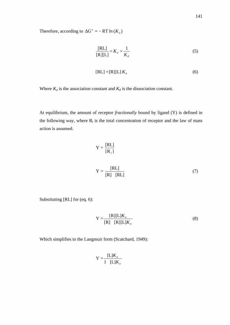
141
Therefore, according to )( RT G oaKln−=∆
d
a KK
1
]L][R[]RL[
== (5)
aK [L] [R] ]RL[ = (6)
Where Ka is the association constant and Kd is the dissociation constant.
At equilibrium, the amount of receptor fractionally bound by ligand (Y) is defined in
the following way, where Rt is the total concentration of receptor and the law of mass
action is assumed.
]R[[RL]
Yt
=
[RL] [R][RL]
Y+
= (7)
Substituting [RL] for (eq. 6):
a
a
KK[L] [R] [R]
[L] [R] Y
+= (8)
Which simplifies to the Langmuir form (Scatchard, 1949):
a
a
KK
[L] 1[L]
Y+
=
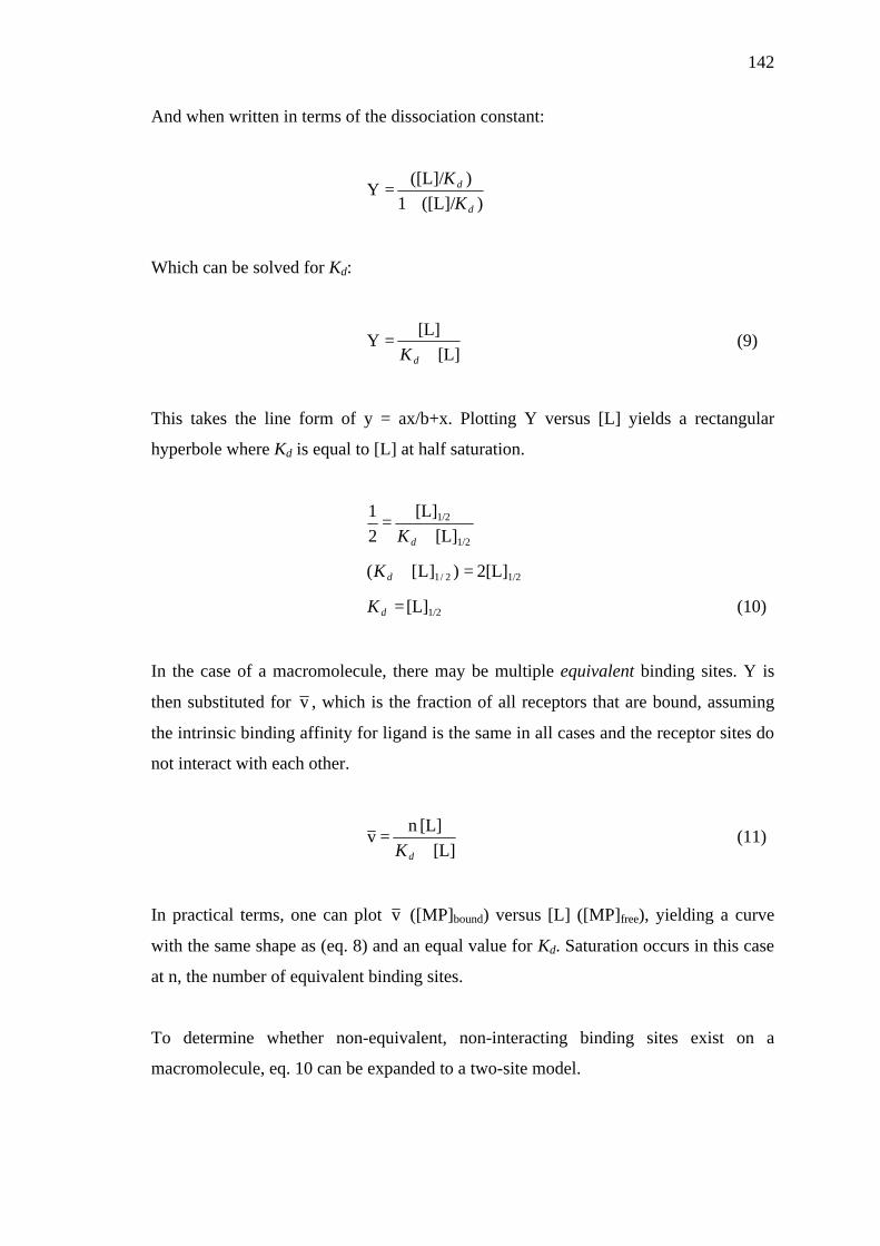
142
And when written in terms of the dissociation constant:
)([L]/ 1
)([L]/ Y
d
d
KK
+=
Which can be solved for Kd:
[L]
[L] Y
+=
dK (9)
This takes the line form of y = ax/b+x. Plotting Y versus [L] yields a rectangular
hyperbole where Kd is equal to [L] at half saturation.
1/2
1/2
[L] [L]
21
+=
dK
1/22/1 2[L] )]L[( =+dK
1/2[L] =dK (10)
In the case of a macromolecule, there may be multiple equivalent binding sites. Y is
then substituted for v , which is the fraction of all receptors that are bound, assuming
the intrinsic binding affinity for ligand is the same in all cases and the receptor sites do
not interact with each other.
[L]
[L]n v
+=
dK (11)
In practical terms, one can plot v ([MP]bound) versus [L] ([MP]free), yielding a curve
with the same shape as (eq. 8) and an equal value for Kd. Saturation occurs in this case
at n, the number of equivalent binding sites.
To determine whether non-equivalent, non-interacting binding sites exist on a
macromolecule, eq. 10 can be expanded to a two-site model.

143
[L] [L] n
[L]
[L] n v
2
2
1
1
++
+=
dd KK (12)
Plots of [L]bound versus [L]free are hyperbolic, but yield Kd and n values for each
respective type of site. Binding data fit to a multiple-site model must be statistically
compared to lower order models by analysis of variance (ANOVA) to ensure the
strength of fit is not simply due to an increase in inflexion points.
Linearization of equilibrium binding data
The Scatchard-Rosenthal plot (Scatchard, 1949; Rosenthal, 1967) can be used as a
visual aid in the interpretation of equilibrium binding data. Deviations from the
predicted straight line give intuitive clues as to the mechanism of binding, e.g. whether
multiple non-equivalent binding sites exist or whether equivalent binding sites display
allosteric effects on subsequent ligand binding (cooperativity).
The one-site equilibrium binding equation (eq. 11) can be manipulated in the following
way to obtain the Scatchard form.
[L] [L]
nv
+=
dK
[L]n [L]) (v =+dK
[L] n) v(- [L]n [L] v - v +=+=dK
ddd KKKn
v-
n) v(-
]L[
v+=
+=
dd KKn
v 1-
]L[
v+= (13)

144
Plots of [L]bound/[L]free versus [L]bound give a straight line (y = mx + b) when the criteria
for a single equivalent, non-interacting binding site are met. The slope equals -1/Kd and
x-intercept equals n. Following transformation, data points (x-axis) do not conform to
the Gaussian distribution, therefore, values for n and Kd are rendered unreliable. Bi-
and multiphasic binding curves are indicative of multiple types of binding site and/or
cooperativity of binding, although multivalent ligands or non-specific binding cannot
be firmly discounted without further experimentation (Norby et al., 1980; Zierler,
1989).
Comparison of binding models by analysis of variance (ANOVA)
One-way ANOVA is a statistical method which allows two or more groups of data to
be compared according to the difference in the mean value between the groups and the
degree of variability within the groups themselves. Since both equilibrium binding
models (eq. 11 and 12) are related, it is possible to directly compare the strength of
curve fit by calculating the F ratio in the following way, where SS is the residual sum-
of-squares (the difference between the observed and predicted curves) and df is the
residual degrees of freedom or difference between the total number of samples and
total number of adjustable parameters within the model (Motulsky and Christopoulos,
2003):
Null hypothesis (null): The simpler model is most appropriate
Alternative hypothesis (alt): The more complex model is more appropriate
altalt
altnullaltnull
/dfSS)df - )/(dfSS -(SS
F =ratio

145
In basic terms, this equation answers the question whether a perceived improved fit
using a complex model is ‘worth’ the additional variables within the modeled term
(decrease in degrees of freedom). F ratio values above 1 indicate that the complex
model is statistically distinct from the simple model and fits the data more strongly. To
determine the probability that random scatter within the data led to the complex model
to fit better, one can calculate the P-value using the an automated algorithm (e.g.
function =FDIST in Microsoft Excel 2000). P-values less than 0.05 (5%) confirm that
the null hypothesis can be reasonably rejected.

146
Non-linear regression outputs (SigmaPlot 2001, SPSS inc.)
One-site model
Nonlinear Regression
[Variables]x = col(1)y = col(2)[Parameters]a = max(y) b = x50(x,y)[Equation]f= a*x/(b+x)fit f to ytolerance=0.000001stepsize=100iterations=100
R = 0.98341850 Rsqr = 0.967 Adj Rsqr = 0.963
Standard Error of Estimate = 0.4553
Analysis of Variance: DF SS MS FRegression 1 54.8617 54.8617 264.6556Residual 9 1.8657 0.2073Total 10 56.7274

147
Two-site model
Nonlinear Regression
[Variables]x = col(1)y = col(2)'Automatic Initial Parameter Estimate Functions[Parameters]a = max(y)/2 b = .5*x50(x,y) c = max(y)/2 d = 1.5*x50(x,y) [Equation]f= a*x/(b+x)+c*x/(d+x)fit f to ytolerance=0.000001stepsize=100iterations=100
R = 0.99297596 Rsqr = 0.986 Adj Rsqr = 0.980
Standard Error of Estimate = 0.3368
Analysis of Variance: DF SS MS FRegression 3 55.9333 18.6444 164.3483Residual 7 0.7941 0.1134Total 10 56.7274
Comparison of kinesin motility rates using the two-tailed unpaired
students t-test
When different groups of data conform to a Gaussian (normal) distribution and the
variance of each group is the same (homoscedastic) , the t-test can be employed in
order to determine whether each respective group has a significantly different mean (µ)
value. In principle, this is achieved by determining the probability that observed
differences between the means are simply due to a high standard deviation (σ), i.e. the
result of variability within one or both groups, or actually arose due to a significant
measurable difference. In the case of apparent kinesin motility rates, velocity data were

148
obtained from reactions performed at different times and with different batches of
reagent. Therefore, the data groups can be considered to be independent and a two-
tailed unpaired t-test can be used with the null hypothesis (null) that the mean MT
velocity of two groups is the same, and an alternative hypothesis (alt) that the means
are different. The t-statistic was manually calculated as described below and when it
was greater than the corresponding value obtained from a t-distribution with the same
degrees of freedom, the null hypothesis was reasonably rejected.
If both groups have the same sample number (n), one calculates the pooled standard
deviation (S), where σ is the standard deviation.
2)(
S2alt
2null σ+σ
= (eq. 14)
From this, the standard error (S.E.) of the difference between the means )( altnull µ−µ is
calculated.
altnull n1
n1
S.D.E.S += (eq.15)
And finally the t-statistic is calculated thus.
( ).D.E.Saltnull µ−µ
=t (eq. 16)
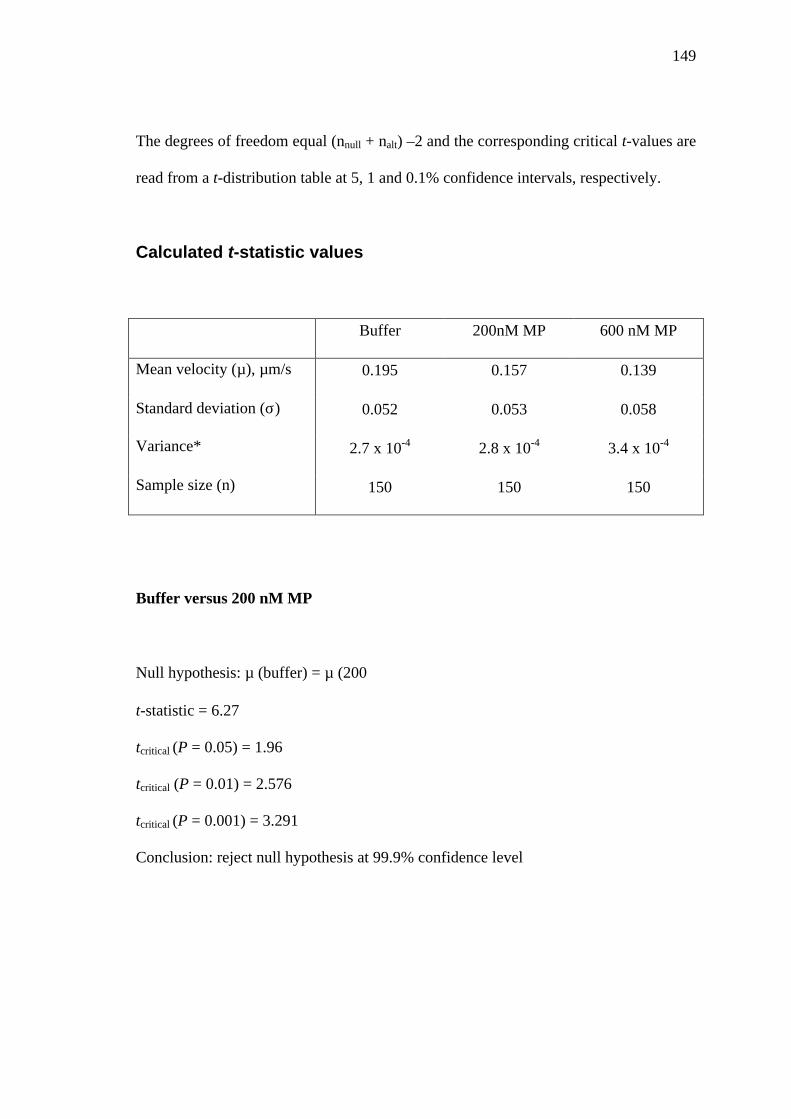
149
The degrees of freedom equal (nnull + nalt) –2 and the corresponding critical t-values are
read from a t-distribution table at 5, 1 and 0.1% confidence intervals, respectively.
Calculated t-statistic values
Buffer 200nM MP 600 nM MP
Mean velocity (µ), µm/s 0.195 0.157 0.139
Standard deviation (σ) 0.052 0.053 0.058
Variance* 2.7 x 10⁻⁴ 2.8 x 10⁻⁴ 3.4 x 10⁻⁴
Sample size (n) 150 150 150
Buffer versus 200 nM MP
Null hypothesis: µ (buffer) = µ (200)
t-statistic = 6.27
tcritical (P = 0.05) = 1.96
tcritical (P = 0.01) = 2.576
tcritical (P = 0.001) = 3.291
Conclusion: reject null hypothesis at 99.9% confidence level

150
200 nM MP versus 600 nM MP
Null hypothesis: µ (200) = µ (600)
t-statistic = 2.81
tcritical (P = 0.05) = 1.96
tcritical (P = 0.01) = 2.576
tcritical (P = 0.001) = 3.291
Conclusion: reject null hypothesis at 99 % confidence level
* To verify that variance values were sufficiently similar, an heteroscedastic (unequal variance) t-test
was employed using Microsoft Excel 2000 software. The results of this analysis were fully consistent
with the above t-statistic values, indicating that a pooled variance t-test was indeed the most appropriate
for these data.
Appendix IV Publication