STUDIES ON HUMAN ANTIBODIES Bx MANUEL E. KAPLAN,~ M.D ...
21
STUDIES ON HUMAN ANTIBODIES IV. PURIFICATION AND PROP~.RrmS O~" A_WTI-A AND A_w~-B OBTAINED BY ABSORPTION AND F.LUTION/~ROMINSOLIYBLE BLOOD GROUP SD-BSTANCES* Bx MANUEL E. KAPLAN,~ M.D., AND ELVIN A. KABAT, PH.D. (From the Departments of Microbiology and Neurology, College of Physicians and Surgeons, Columbia University, the Neurological Institute, Presbyterian Hospital, the Department of Hematology, Mount Sinai Hospital, New York, and the Department of Medicine, Washington University School of Medicine, St. Louis) (Received for publication 4 March 1966) Antibodies formed in response to injection of single antigens may be extra- ordinarily heterogeneous, often consisting of molecules belonging to each of the three major classes of serum immunoglobulins, "yG, "yM, and TA x (1-3). Me- Duffle, et al. (7), studying the properties of human blood group isoantibodies, showed that rabbits immunized with specific precipitates of A substance and human anti-A developed antibodies to three serum proteins; two of these were identified as antibody to "yG- and "yM-immunoglobulins. The third protein re- mained unidentified until it was characterized by Kunkel and Rockey (8) as "yA. Serum from individual human donors may contain anti-A or anti-B of all three immunoglobulin classes (8-10). It would be of great interest to determine the characteristics of the combining sites of the various anti-A immunoglobulins formed in a single individual. The present study reports a new method for purifying anti-A or anti-B using specific insoluble adsorbants, separation of the purified anti-A antibodies into "yG- and ,yM-fractions, and initial observations on the relative capacities of various oligosaccharides involved in the A antigenic determinant to inhibit the precipi- tation of TG- and ~M-anti-A by blood group A substance. Materials and Metkods Blood Group Substances.--Soluble blood group substances were purified from bog gastric mucin, (Wilson Laboratories, Chicago), and human ovarian cysts by previously described * Aided by grants from the National Science Foundation (G-18727 and GB-3675), the Officeof Naval Research, The National Heart Institute (HE 04456 and 5488), and the General Research Support grant of the United States Public Health Service. Present address: The Department of Medicine, Jewish Hospital of St. Louis, St. Louis. 1The nomenclature used is that recommended by the World Health Organization (4). "yD (IgD), recently described by Rowe and Fahey (5), has not yet been shown to exhibit antibody activity (6). 1061 brought to you by CORE View metadata, citation and similar papers at core.ac.uk provided by PubMed Central
Transcript of STUDIES ON HUMAN ANTIBODIES Bx MANUEL E. KAPLAN,~ M.D ...

STUDIES ON HUMAN ANTIBODIES
IV. PURIFICATION AND PROP~.RrmS O~" A_WTI-A AND A_w~-B OBTAINED BY ABSORPTION AND F.LUTION /~ROM INSOLIYBLE BLOOD GROUP SD-BSTANCES*
Bx MANUEL E. KAPLAN,~ M.D., AND ELVIN A. KABAT, PH.D.
(From the Departments of Microbiology and Neurology, College of Physicians and Surgeons, Columbia University, the Neurological Institute, Presbyterian Hospital,
the Department of Hematology, Mount Sinai Hospital, New York, and the Department of Medicine, Washington University School of Medicine,
St. Louis)
(Received for publication 4 March 1966)
Antibodies formed in response to injection of single antigens may be extra- ordinarily heterogeneous, often consisting of molecules belonging to each of the three major classes of serum immunoglobulins, "yG, "yM, and TA x (1-3). Me- Duffle, et al. (7), studying the properties of human blood group isoantibodies, showed that rabbits immunized with specific precipitates of A substance and human anti-A developed antibodies to three serum proteins; two of these were identified as antibody to "yG- and "yM-immunoglobulins. The third protein re- mained unidentified until it was characterized by Kunkel and Rockey (8) as "yA. Serum from individual human donors may contain anti-A or anti-B of all three immunoglobulin classes (8-10).
I t would be of great interest to determine the characteristics of the combining sites of the various anti-A immunoglobulins formed in a single individual. The present study reports a new method for purifying anti-A or anti-B using specific insoluble adsorbants, separation of the purified anti-A antibodies into "yG- and ,yM-fractions, and initial observations on the relative capacities of various oligosaccharides involved in the A antigenic determinant to inhibit the precipi- tation of T G - and ~M-ant i -A by blood group A substance.
Materials and Metkods
Blood Group Substances.--Soluble blood group substances were purified from bog gastric mucin, (Wilson Laboratories, Chicago), and human ovarian cysts by previously described
* Aided by grants from the National Science Foundation (G-18727 and GB-3675), the Office of Naval Research, The National Heart Institute (HE 04456 and 5488), and the General Research Support grant of the United States Public Health Service.
Present address: The Department of Medicine, Jewish Hospital of St. Louis, St. Louis. 1 The nomenclature used is that recommended by the World Health Organization (4).
"yD (IgD), recently described by Rowe and Fahey (5), has not yet been shown to exhibit antibody activity (6).
1061
brought to you by COREView metadata, citation and similar papers at core.ac.uk
provided by PubMed Central

1062 HUMAN ANT~OD~S.
methods (11). Insoluble derivatives of these purified blood group substances were prepared by copolymerization with the N-carboxyanhydride of L-lencine as described by Tsuyuki, et al. (12). 2 The details of this procedure were as follows: Purified blood group substance was dissolved to a concentration of 4 mg/ml in 7A00 M NaHCO3 (pH 8.3). The solution in an Erlenmeyer flask was chilled to approximately 5°C and mixed continuously by magnetic stirring. To it was slowly added solid crystalline N-earboxy-L-leucine anhydride (Pilot Chemi- cals, Inc. Watertown, Massachusetts) in an amount equal to the weight of dissolved blood group substance. Stirring in the cold was continued for an additional 48 hr. The contents of the flask were then centrifuged for 1 hr at 17,000 RP~ in a refrigerated Servall RC-2 centrifuge. The dear supernatant solution was decanted, the precipitate thoroughly dispersed in 25 volumes of ~ 0 o x( NaHC08 and again centrifuged. This step was repeated until the precipitate had been washed 4 times in NaFICOa and 4 times in distilled water. After the last wash the precipitate was lyophiliz~d and stored at room temperature. The insoluble blood group ma- terials so prepared are, hereafter, referred to as polyleucyl blood group substances (PI-BGS) and as polylencyl A (P1-A) and polyleucyl B (P1-B).
Inhibitors.--Haptenic derivatives of purified human ovarian cyst A substance were pre- pared by mild acid hydrolysis (13) and by alkaline cleavage in the presence of sodium bore- hydride (14). Two oligosaccharides, A~I (13), a trisaccharide having the structure:
c~-,-GalNAc-(1 ~ 3)-~-D-Gal-(1 ~ 3)-D-GNAc
and ARL 0.52 (previously called Aa) a pentasaccharide with the structure:
cz-L-Fuc 1 1 2
Ot-D-GalNAc-(1 ~ 3)-f~-D-Gal-(1 ---) 4)-f~-D-GNAc-R
representing the largest and most active fragment of the antigenic determinant of blood group A substance thus far isolated (15-17) were utilized along with N-acetyl-v-galactosamine (GalNAc) to inhibit the precipitation of the purified anti-A by A substance as described earlier (18, 19, 13)?
Antiscra.--Serum R.G., generously provided by Dr. Richard E. Rosenfield of Mount Sinai Hospital, New York, was obtained from a multiparous blood group O female hyper- immunized by repeated blood group A pregnancies. Two separate bleedings from this donor were obtained, the first (R.G.I) dating from June, 1959, and the second (R.G.II) from February, 1962. Serum Ortho 63-2622 was the gift of Dr. Philip Levine and Mr. Glen Hill of Ortho Research Laboratories, Raritan, New Jersey. The donor of this serum was a blood group B individual immunized with hog A substance. These sera were preserved with phenol (0.25%) and merthiolate (1:10,000) and were stored at 4°C. Goat and rabbit antisera to whole human serum and to human 7G-, 7A-, and 7M-immunoglobulins were purchased from Hyland Laboratories, Los Angeles, and from Lloyd Bros., Inc., Cincinnati. Rabbit antisera specific for human kappa (group I) and lambda (group II) polypeptide chains were the gift of Dr. Henry Kunkel of The Rockefeller University. Dr. John Fahey of the National Institutes of Health generously furnished rabbit antiserum to human 7D and serum W.T., a serum con- mining known quantifies of'yD. Antisera specific for human complement components C'q and ~IC/~IA were kindly supplied by Dr. Charles Christian and Dr. Konrad Hsu of the College of Physicians and Surgeons.
2 We are indebted to Dr. Lawrence Levine of Brandeis University who suggested the possible applicability of this method.
* GNAc, N-acetyl-v-glucosamine; Fuc, fucose; and Gal, galactose.

MANUEL E. KAI~LAN AND ELVIN A. KABAT 1063
Carbohydrates.--v-Galactose and sucrose were purchased from Pfanstiehl Chemicals, Waukegan, Illinois, and N-acetyl-v-galactosamine from Nutritional Bioehemicals Corp., Cleveland. Biogel P 10 and P 300 used for gel filtration were obtained from Bio-Rad Labora- tories, Richmond, California.
Analytical Me/hods.--Standard analytical techniques were utilized and the following sub- stances measured by the methods given: methylpentose, Dische and Shettles; hexosamine, E]son and Morgan; and reducing sugar, Park and Johnson (d. 18). The procedure for nitrogen was a modification (14) of the method of Rosevcar and Smith (20).
Separation Procedures.---Gel filtration employing Biogel P 10 and P 300 columns was carried out at room temperature according to the directions supplied by Bin-Rad Laboratories.
Density gradient ultracentrifugation was performed as described by Kunkel (21) utillzi~g a 4 mi linear (10 to 40%) sucrose gradient in a Spinco model L centrifuge with SW 39 rotor for 16 hr at 35,000 m,x~.4 Ten fractions of equal volume (circa 0.45 ml) were collected drop- wise through a pin hole made in the bottom of the tube.
Immunochemical Methods.--Hemagglutination studies were performed with a Takatsy microtitrator (Cooke Engineering Company, Alexandria, Virginia) using 0.025 nil loops and 2% erythrocyte suspensions at room temperature.
Quantitative precipitin analyses (18) were performed by a micropreeipltin technique using the ninhydrin procedure for nitrogen (14).
Agar diffusion studies were carried out at 37°C according to the method of Ouchterlony (22) in 1% gels (Ionagar No. 2, Consolidated Labs., Inc., Chicago) containing 0.5 M glycine (23) and 1:10,000 merthiolate.
Immunoelectrophoresis was performed as described by Grabar and Williams (24) (d. 25). Puri fwation of Isokemagglutinins.--The principle of the method involves the specific ab-
sorption of anti-A or anti-B antibodies by P1-A or PI-B respectively, removal of nonspecific protein by washing, and elution of absorbed antibody by extraction with acid buffer or with sugar haptens at neutral pH. The specific procedure was as follows:
Absorp¢ion: To a given volume of serum containing anti-A was added I mg/mi of P1-A. The serum was thoroughly mixed to suspend the insoluble material evenly, and incubated at 37°C for 1 Jar with frequent mixing. The serum was then slowly rot&ted (10 to 16 RiM) for 1 wk in the refrigerator. The insoluble P1-A was harvested by eentrifugstion and the superuatant serum tested for residual anti-A by hemagglutination and by quantitative precipitin tests. I t was invariably found that more than 90% of the homologous antibody had been removed by the PI-A. With R.G., a type O serum which contained both anti-A and anti-B, absorptions were performed in 2 different ways: in the first instance the serum was absorbed only with P1-A; in the second experiment sequential absorptions with P1-B followed by P1-A were carried out.
After absorption, the P1-BGS-antibody complex was washed repeatedly with cold saline until the washings were essentially devoid of material absorbing at 2800 A.
Elution: Elution of antibody was performed by suspending the packed, washed P1-BGS- antibody precipitate in 2 ml, ~/10 acetate buffer, pH 3.62 at 0°C (26), for I hr with frequent mixing. After eentrifugation at 4°C, the supernatant was immediately transferred to a dialysis casing unknotted at the top and dialyzed against 500 nil cold PO4-buffered saline (~/20, pH 7.3). Fresh, cold acetate buffer (pH 3.62) was rcapplied to the packed sediment and the ex- traction repeated. The second eluate was added to the first in the dialysis bag, and the dialysis fluid changed. A third ehition was performed and the P1-BGS washed several times with small quantities of saline. The combined eluates and saline washes were dialyzed for 6 hr against 1
4 We gratefully acknowledge the expert assistance of Dr. K. Aho in the performance of these studies.

1064 HUMAN ANTIBODIES. IV
liter of ,~/20 PO4-buffered saline (pH 7.3) and overnight against xr/1000, PO4-buffered saline (pH 7.3). Since the antibody yield by acid elution did not exceed 10%, the procedure was changed as follows: washed, packed H-A that had been used for anti-A absorption was sus- pended in 2 to 3 ml of PO4-buffered saline (M/1000, pH 7.2-7.3) containing 1 g of GalNAc, incubated for 1 hr at 37°C, and slowly rotated overnight in the refrigerator. Mter centrifuga- tion, the supematant eluate was removed and the P1-A resuspended in additional GalNAc in buffered saline. This procedure was repeated twice and the resultant eluates pooled. The H-A was washed several times in cold, buffered saline and the washings added to the pooled eluates. The elution procedure for H-B was identical to that for PI-A except that ,-galactose was substituted for GalNAc. The pooled eluates and washings were concentrated by ultrafiltration in the cold under negative pressure through an 8 ml collodion membrane, porosity less than 5 n~u (Membranfiltergesellschaft G6ttingen, Germany, obtained from Schleicher and Schuell, Keene, New Hampshire). Initially, the concentrated, sugar-containing eluates were freed of
TABLE I
AnalyHcal Properties of Purified Blood Group Substances and of Their Insoluble Polyleucyl Derivatives
Material Methylpentose Hexosamine
Hog A H-hog A (I) m-hog A (II)
McDon A H-McDon A
Beach B H-Beach B
% % BGS 10.6 100 5.8 55 6.0 57
21.1 100 7.6 36
19.3 100 8.7 45
%
29.0 16.3 15.3
30.9 11.1
21.3 9.4
% BGS 100 56 53
100 36
I00
44
sugar by repeated cycles of dilution with buffered saline and ultrafiltration. More recently it was found by Dr. Marianne M. Dorner in this laboratory, that a single passage of such a con- centrated eluate through a Biogel P-10 column (I.1 X 60 cm) equilibrated with buffered saline (~r/1000, pH 7.2) served to separate completely hapten (i.e. sugar) from protein. Those chromatographic fractions absorbing at 2800 A were pooled and reconcentrated by ultra- filtration.
R.ESUI,TS
Preparation of and A bsorption with Insoluble Blood Group Substances.--Table I summarizes the analytical properties of the original purified blood group sub- stances and of their insoluble polyleucyl derivatives. Two separate batches of Pl-hog A (I and I I ) were prepared from the same starting materials. When the methylpentose and hexosamine contents of the various polyleucyl compounds are expressed as percentages of those found in the parent blood group sub- stances, it is apparent that the insoluble derivatives contained from 36 % (PI- Beach B) to about 55 % (Pl-hog A) of blood group material.

M A N U E L E. K A P L A N AND E L V I N A. K A B A T 1065
The capacities of the polyleucyl blood group substances to absorb specifically anti-A and anti-B from human sera is illustrated in Table II. Absorption of R.G.I, which contained both anti-A and anti-B, with Pl-hog A was found to re- move 95 % of the anti-A (22 of 23 #g Ab (antibody) N/ml) and about 75 % of the anti-B (3.6 of 4.7 #g Ab N/ml). The absorbed serum agglutinated type B cells to a titer of 1:16 but, even undiluted, would no longer agglutinate type A1 erythrocytes. P1-McDon A absorbed from serum R.G.I more than 95 % of the antibody N which McDon A was capable of precipitating (13.5 of 14.0 #g Ab N/ml). This represented only about 3/~ of the total anti-A precipitable by hog mucin A. P1-A absorption removed most of the anti-B from R.G.I as well.
TABLE H Absorption of Anti-A and Anti-B by Insoluble Polyleucyl Blood Group Substances
Antiserum
R.G.I Unabsorbed Absorbed with Pl-hog A Absorbed with P1-McDon A
R.G.II Unabsorbed Absorbed with H-Beach B Absorbed with PI-Beach B + H-hog A
Ortho 63-2622 Unabsorbed Absorbed with Pl-hog A
Hemngglutinafion titer
A cells B cells Hog A
~g/ml
128 32 23.0 0 16 1 .0 2 16 5.0
128 32 18.4 64 0 17.1
0 0.5
256 0 13.3 0 0.9
Antibody N precipitated by
McDon A Beach B
14.0 4.7 0.7 I.I 0.5 1.8
4.7 0.2
Serum R.G.II, containing 20% less anti-A than R.G.I, was absorbed first with PI-Beach B and then with Pl-hog A. The first absorption removed more than 95 % of the antibody N precipitable by human B substance, (4.5 of 4.7 #g Ab N/re_l) but less than 10% of the Ab N precipitable by hog A (1.3 #g of a total of 18.4 #g Ab N/ml). The rest of the anti-A was removed by a subse- quent absorption with PI-A. These findings indicate that in serum R.G., AB cross-reacting antibody makes up a large proportion of the anti-B and only a small proportion of the anti-A.
Purification and Characterization of Blood Group Antibodies.--Table I I I sum- marizes the absorption and elution steps used in isolating anti-A and anti-B, the yields obtained, and the agglutinating and precipitating properties of the purified antibodies. In the initial study suboptimal quantities (0.5 mg/ml

1066 HUMAN ANTIBODIES. IV
serum) of P1-A were used for absorption, and elution was performed with pH 3.62 acetate buffer. An overall anti-A yield of only 7.3 % was achieved. Subse- quently, antibody recoveries were improved by absorbing with larger quantities of P1-A and eluting with GalNAc at neutral pH.
Both the acid and GalNAc eluates prepared from P1-A-R.G.I antibody com- plex agglutinated type B cells to a modest titer, 1:16; however, neither eluate was precipitated by human B substance.
In an attempt to isolate AB cross-reacting antibody from serum R.G.II, the latter was absorbed with P1-B and elution carried out with galactose, the terminal nonreducing sugar of the B antigenic determinant (19, 27). The re-
TABLE III Preparation and Properti
Serum
R.G.I. R.G.I.
R.G.II
Ortho 63-2622
Volume used
ml
12~ 4. ¢
(a) 16( (b) 15C
223
Total antibody N
Anti-~
2875 1035
2950 2565
2990
Anti-]
~g
563 212
768 38
Absorbant
Pl-hog A* P1-hog A
P1-Beach B Pl-hog A
P1-hog A
Absorption
Residual antibody N
Anti-A Anfi-B
~g go J*g 688 24 N.D.~
45 4 45.2
2735 93 40 75 3 N.D.
2 1 4 7 I
%
21
5
* In this instance only 0.5 mg Pl-hog A/ml serum was used for absorption which a counts for the incomplete removal of anti-A.
:~ Not determined.
sultant eluate, although relatively poor in nitrogen, agglutinated type A and B erythrocytes to fiters of 1: 256 and was incompletely precipitated by both A and B substances. Since no effort was made to differentiate the anti-B from anti-A B, it is not possible to relate, quantitatively, the N content of this eluate to the anti-A or anti-B present in the original serum. Efforts to obtain additional antibody from the P1-B substance by elution with galactinol were unsuccessful.
Fig. 1 A and 1 B demonstrate that P1-A-GalNAc eluates from R.G.II and Ortho 63-2622 contained q,G-, 3'M-, and q,A-immunoglobulins. (All three im- munoglobulins were also detected in the P1-B-Gal eluate from serum R.G.II.) When the immunoelectrophoretic patterns were developed with polyvalent goat anti-human serum, some of the eluates were found to contain trace quanti- ties of albumin; furthermore, the Ortho eluate gave an additional very faint band in the slow/~-region close to, but separate from, the ~/M-line. The sub-
M A N U E L E . K A P L A N A N D E L V I N A. K A B A T 1067
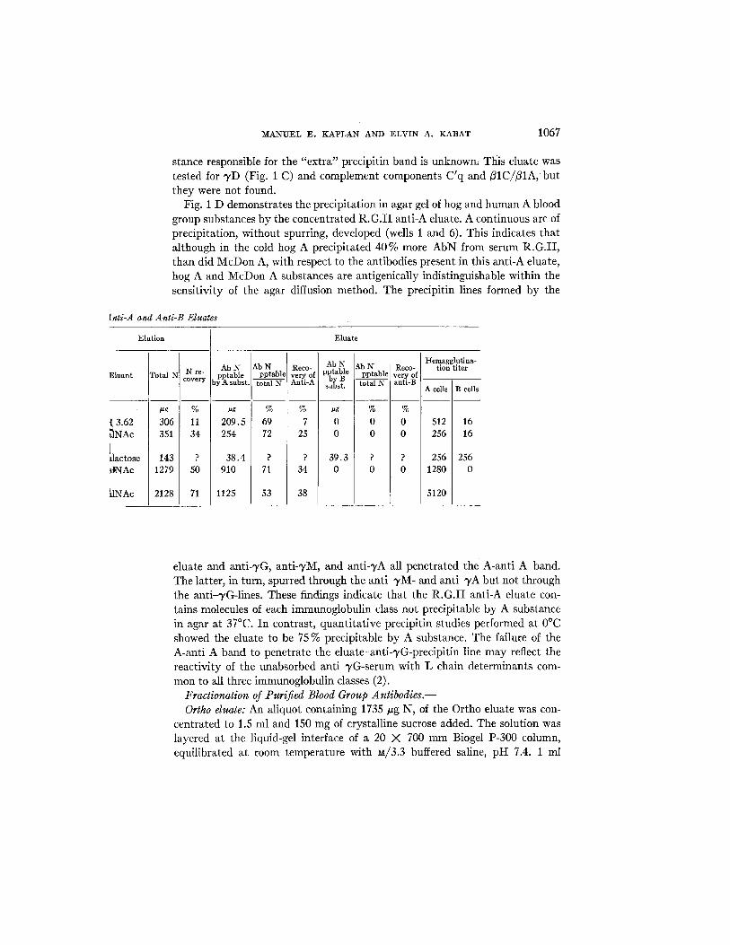
stance responsible for the "extra" precipitin band is unknown, This eluate was tested for ~,D (Fig. 1 C) and complement components C'q and/~IC//~IA, but they were not found.
Fig. 1 D demonstrates the precipitation in agar gel of hog and human A blood group substances by the concentrated R.G.I I anti-A eluate. A continuous arc of precipitation, without spurring, developed (wells 1 and 6). This indicates that although in the cold hog A precipitated 40% more AbN from serum R.G.II, than did McDon A, with respect to the antibodies present in this anti-A eluate, hog A and McDon A substances are antigenically indistinguishable within t he sensitivity of the agar diffusion method. The precipitin lines formed by the
[nti-A and Anti-B Eluates
Elution ] Eluate
Eluant
t 3.62 ~nAc I
~lactose ~tNAc
~.tNAc
Total
#g
306 351
143 1279
2128
N re- covery
% 11 34
? 50
71
Ab N pptable y A subst.
~g
209.5 254
38.4 910
1125
Ab N pptabl¢
% 69 72
? 71
53
Reco- v e r y of Anti-A
% 7
25
? 34
38
I Ab N lAb N
pptable [ pptable b B - -
su~st, fctal N
ug I % 0 0 0 0
39.3 ? 0 0
Reco- very of anti-B
% 0 0
Hemagglutina- tion titer
A ceils B cells
512 16 256 16
256 256 1280 0
5120
eluate and anti-3,G, anti-'yM, and anti-~,A all penetrated the A-anti A band. The latter, in turn, spurred through the anti-3,M- and anti~,A but not through the anti-TG-lines. These findings indicate that the R.G. I I anti-A eluate con- tains molecules of each immunoglobulin class not precipitable by A substance in agar at 37°C. In contrast, quantitative precipitin studies performed at 0°C showed the eluate to be 75 % precipitable by A substance. The failure of the A-anti A band to penetrate the eluate-anti-3,G-precipitin line may reflect the reactivity of the unabsorbed anti-~/G-serum with L chain determinants com- mon to all three immunoglobulin classes (2).
Fractionation of Purified Blood Group Antibodies.-- Ortho eluate: An aliquot containing 1735 #g N, of the Ortho eluate was con-
centrated to 1.5 ml and 150 mg of crystalline sucrose added. The solution was layered at the liquid-gel interface of a 20 X 700 mm Biogel P-300 column, equilibrated at room temperature with M/3.3 buffered saline, pH 7.4. 1 ml

Fro. 1. A. Immunoelectrophoretic patterns of (1) Ortho 63-2622 serum, (2) Ortho-GalNAc eluate, (3) R.G.II serum, and (4) R.G.II-GalNAc eluate; goat anti-human serum was used in the troughs to develop reactions.
B. Immunoelectrophoretic patterns of (1, 4) Ortho 63-2622 serum; (2, 3) Ortho-GalNAc eluate; (5, 8) R.G.II serum; and (6, 7) R.G.II-GalNAc eluate; troughs (a) goat anti-TG- globulin, (b) goat anti--TA-globulin, and (c) goat anti-"/M-globulin.
C. Center well: rabbit anti-TD-serum. Peripheral wells, (1) empty, (2) Ortho 63-2622 serum, (3) Ortho-GalNAc eluate, and (4) W.T. serum ('yD-standard).
D. Center well: R.G.II-GalNAc eluate. Peripheral wells, (1) McDon A substance, (2) goat anti-TM-globulin, (3) goat anti-/A-globulin, (4) McDon A substance, (5) goat anti-'yG- globulin, and (6) hog A substance.
E. Immunodiffusion study of ten fractions resulting from density gradient centrifugation of R.G.II-GalNAe eluate. Trough (a) goat anti-'yM-globulin, (b) goat anti-TA-globulin , and (c) goat anti-TG-globulin.
F. Center well (a): rabbit anti-type K (group I) antiserum. Center well (b): rabbit anti- type L (group II) antiserum. Peripheral wells, (1) type K Bence-Jones protein, (2) R.G.II "),G-fraction, (3) R.G.II "/M-fraction, (4) type L Bence-Jones protein, (5) Ortho "yM-fraction, and (6) Ortho ~/G-fraction.
1068

M A N U E L E. K A P L A N AND E L V I N A. K A B A T 1069
fractions were collected at the rate of 18 per hour and their absorbance at 2800 A measured. The absorption curve showed two broad, incompletely separated peaks. The immunoglobulins present in selected fractions were characterized by means of agar diffusion employing 3'M-, "yG-, and ^/A-specific antisera. Second peak fractions 68 to 137 containing "yG and very small amounts of yA, were pooled, dialyzed against isotonic buffered saline, and concentrated by ultra- filtration to give the "yG-fraction of the Ortho eluate. Fractions 33 to 55, which represented the ascending limb of the first protein peak, were found to be pre- ponderantly ~/M; however, after these fractions had been pooled and concen- trated, trace quantities of ~,G and "yA were detected. Consequently, P-300 gel filtration was repeated under the conditions previously described. Only a single
TABLE IV Properges of the Unfractiona~t Anti-A eluates and f Tlwir Separated 'yG-and "yM- Fracffons
I
Ab N lab s Anti-A
OD OD 2800 A pptable [ pptable hemag- #gN/m] Eluate Nitrogen 2800 A #g N/ml ;by A I gluti- subst. I total N nation tlter
titer
)rtho unfractionated* ~,G-fraction ~/M-fraction
R.G.II unfractionated 3,G-fraction TM-fraction
Intermediate fraction
Pg
180 625 94
985 498 112 15
pg/~
90.2 68.7 62.8
365 148.8 34.1 12.7
0.890 0.655 0.520
I~ .D.
1.492 0.440 0.141
0.00987 0.00953 0.00829
N.~D.
0.0103 0.129 0.111
~tg
91.4 326 40.7
702 343 99.5 10.7
% 51 52 43
71 69 89 71
320 320 160
5120 5120
80 80
0.28 0.21 0.39
0.07 0.03 0.43 0.16
* This represents a dilution of a small portion of the Ortho eluate not subjected to gel filtration.
protein peak containing essentially pure ~M resulted. Fractions 56 to 67 of the first F-300 co|utah were found to consist of mixtures of all three immunoglobulin classes and were not further studied.
R.G. I I duale: An aliquot of this eluate, contninlng 985 pg N, was concen- trated to 0.5 ml, layered on a 4 ml sucrose density gradient, and centrifuged for 16 hr at 35,000 m ~ as previously outlined. Ten 0.45 ml fractions were col- lected and the immunoglobulin content of each fraction determined by agar diffusion (Fig. 1 E). Fractions 1 and 2 consisting almost exclusively of ~/M, and fractions 4 to 6 containing ~/G- and some 5,A-immunoglobulins were pooled. These 2 fractions, as well as fraction 3, which represented a mixture of -IM, ~/G, and ~,A, were dialyzed thoroughly against buffered saline to remove sucrose. The immunochemical properties of these fractions, designated, as R.G. -yM (tubes 1 and 2), "yG (tubes 4 to 6), and intermediate (tube 3) were studied without further fractionation.

1070 H U M A N ANTIBODIES. IV
Table IV summarizes the analytical data on the purified fracfionated anti- bodies and Fig. 2 depicts their precipitation curves with hog A substances. Total N recovery from the R.G.II eluate was 64% with 51% in the ~¢G-frac- tions, 11.4% in the ~,M-fraction, and 1.5 % in the intermediate fraction. From its absorbance at 2800 A, the intermediate Ortho fraction was estimated to contain 148 #g N; thus, an overall N yield of only 50% was achieved from the Ortho eluate. This comparatively poor recovery may be ascribed to protein denaturation due to prolonged exposure of the Ortho eluate to summer room temperatures (28--32°C) during gel filtration.
ORTHO ELUATE R .G .Z [ -GALNAc ELUATE
5.C I--
a. o ba o¢ o. 2.C z
C-- i.c z
o
0 Unfractioneted, 50.4 ,ul X b'O-Froction, 74.6/JI
0 ~'M-Froctlon, 87.5/ul 4 . 0
A 3.0
X
2 .0
1.0
, , , , h , , , , , t 2 3 4 5 6 7 8 9 1 0
MICROGRAMS HOG A ADDED
0 Unfractionated, 10.2jul
0 "SM- Froction, 102.2 jul X YG-Froction, 30.21ul [] ~nterrnediole F r o c t l o n , 2 4 9 . 9 jul
y z / S 0
I 2 3 4 5 6 7 8 9 I0 II 12 1 3 1 4 15 [ 6 1 7 1 8 1 9 2 0 2 1 2 2 23 MICROGRAMS HOG MUGIN A ADDED
FIG. 2. Quantitative precipitin curves of whole anti-A eluates and their 3'M- and "yG- fractions.
When the N content and absorbance at 2800 A of the individual antibody solutions are compared (Table IV, column 4), it is apparent that the value for OD/~g N obtained for the two ~'M-fractions deviate appreciably, and in opposite directions, from the ~G-values.
Precipitability of the Ortho eluate and its fractions by hog A substance was rather low (43 to 52 %) while that of the R.G.II eluate and its fractions was higher, ranging from 69% (~G) to 89% (~M). The A-anfi-A precipitin curves for each eluate and its constituent immunogtobulin fractions (Fig. 2) were very similar and did not deviate in shape from the classical quantitative precipitin c u r v e •
When hemagglutinating activity was expressed as the minimum amount of antibody in tzg N/ml required to give detectable agglutination (Table IV, last

M A N U E L E . K A P L A N A N D E L V I N A. K A B A T 1071
column), the R.G.II eluate was four times as active as the Ortho eluate, the R.G. ~/G-fraction seven times as active as the Ortho ~,G-fraction, while the ac- tivities of the two "rM-fractions were essentially equal. The agglutinating ac- tivities of the Ortho antibody fractions were very similar. Much greater differ-
I00
80
60
40
z o 2c I -
~D "" 0 z
I-- 2= bJ o Ix
fOe
8c
60
40
10
20
ORTHO SERUM, 197.2 )JI + H 0 6 A S U B S T A N C E , 6 .95 jug
GALNA¢ E L U A T E , 5 0 . 4 j u l + HOG A SUBSTANCE, 6.953ug
A
OGalNoc OAsTr c><- GolNAc-(I.->3 ) -~ -GoI-(I.->.3) -GNAc
ELUATE 3 G - F R A C T I O N , 74.6JUl + HOG A SUBSTANCE, 6 . 9 5 J u g
C
I0 Io #o I0
B
D
i i i i
O¢-Fuc ¢; OARLO.52 o<-GoINA¢ - ( I-~.3 )-(3 - GoI-(I-~4)-GNAc-R
E L U A T E ;~M-FRAGTION, 87.5Jul
4 HOG A S U B S T A N G E , 6 . 9 5 jug
,, 01 I0 fo Io to
MILLIMICROMOLES INHIBITOR ADDED
FIG. 3. Inhibition by oligosaccharides of the precipitation by A substance of Ortho anti-A and its purified antibody fractions.
ences were found with the various R.G. fractions (e.g. 0.43 pg N/ml of the R.G. ~M-fraction as compared with 0.03 pg N/ml of the -),G-fraction were re- quired for detectable agglutination.)
Fig. 1 F demonstrates that the Ortho 3'M- and 3'G-, as well as the R.G. 3~M-, fractions contained both type K and L light chains. Although no effort was made to quantitate these determinants, the appearance of the various precipitin bands suggested that the two Ortho fractions contained a preponderance of type L molecules while type K molecules appeared in somewhat greater con-

1072 H U M A N A N T I B O D I E S . I V
< [ - r 0 4 .
IX)
i
o Z ~ i a , = - 0 , o o ~ , i i ~ l ( t ,
7 z = o ~
-I-
o ~ ÷ ~ . J
q ~
~ l l l l l l t l f
°
W O
N •
+
i 1 ~ i i i i i i i _oO o ~ o g o o o o o o
t,,3 ,¢~
U-CO
z = ~ = , ~ - o
z
o n
o i I i ~ 1 i t i i o oO o o R g o o ~o ~ o o -
NOI£1BIHNI 1N'::IO ~ 3 d
I= o
~n_o
o
I= ¢1
"0
~o ~
o ~ I -
o <
¢_o ~ . o
u) o
o
o

MANUEL E. KAPLAN AND ELVIN A. KABAT 1073
centrafion in the R.G. ~M-fracfion. As seen in Fig. 1 F, the Ortho K-anti K lines and the L-anti L line developed by the R.G. ~M fraction appeared rela- tively close to the antigen (i.e. immunoglobuHn) wells and were quite broad and diffuse; the diffuseness was particularly striking on those sides of the precipitin bands facing the antigen wells. These findings are typical of immuno- diffusion precipitin systems containing gross antibody excess (28, 29). The R.G. ^/G-fraction was found, in contrast, to be comprised almost exclusively of group K molecules. Only after prolonged incubation of the gel-diffusion plate did an extremely faint precipitin line develop between the R.G. "yG-fraction and the anfi-L serum. This line is not visible in Fig. 1 F.
TABLE V Millimicromoles of Inhibitor Required for 50°/o Inhibition
Anti-A
Ortho Serum Eluate "yG ~/M
R.G.II Serum Eluate ~/G ~,M
Iahibitor
GalNAc
2,600 5,000
13,000 2,800
25,000 12,000 4,700 4,100
ASH (Trlsaccharlde)
230 420 350
2800
1200 ?
4700 4100
IARL 0,52 (Pentasaccharide,
150 120 76
?
33 60 44
>300
Precipitln-Inhibition Stud/es.--The behavior in quantitative hapten in- hibition studies of the Ortho and R.G.II sera, the anfi-A eluates and their "),C~ and ~M-fracfions is seen in Figs. 3 and 4 and is summarized in Table V. With both sera the pentasaccharide ART. 0.52 was, on a molar basis, a far better inhibitor than the monosaccharide GalNAc; the trisaccharide (A5II) displayed intermediate activity. Similar results have previously been reported from this laboratory with other anfi-A sera (17). Compared with serum R.G., the Ortho serum was considerably more inhibitable by GalNAc and ASII and slightly less inhibitable by ARL 0.52. The unfracfionated Ortho eluate was somewhat less inhlbitable by GalNAc and more inhlbitable by ARL 0.52 than the original serum; the converse findings were noted in the case of serum R.G. and its eluate, i.e. the eluate was slightly more inhlbitable by GalNAc and less inhlbitable by ART. 0.52 than the original serum. In relatively low concentrations, the penta- saccharide effectively inhibited the precipitation of both Ortho and R.G. "yG- fractions (50 % inhibition being achieved with 76 m ~ and 44 m ~ respectively).

1074 ~ u M A N ANTIBODIES. IV
In contrast, even at ART. 0.52 concentrations of 270 m ~ , less than 15 % in- hibition of precipitation of the "},M-fractions could be achieved. Although in- hibition points at higher oligosaccharide concentrations were not obtained be- cause of limited material, the available data suggest that, on a molar basis (a) with both 3,M-anti-A fractions'the penta- and trisaccharide were only as efficient as GalNAc in inhibiting precipitation, (b) with R.G. 3'G the trisac- charide and GalNAc were only ~0oo as active as the pentasaccharide in in- hibiting precipitation (approximately 4700 m#• of the first two and 44 m#M of the last being required for 50% inhibition), and (c) with the Ortho ~/G- anti-A, the pentasaccharide was approximately 4 times as active as the tri- saccharide which in turn was 40 times more active than GalNAc in inhibiting precipitation (50% inhibition points being obtained at concentrations of 76 m/zM, 350 m ~ , and 13,000 muir respectively).
DISCUSSION
Antigens rendered insoluble by various means have been successfully em- ployed as specific immune absorbants to purify homologous antibodies from serum (30, 31, cf. 18). Earlier efforts to isolate human blood group antibodies employing this technique were reported by Isliker (32) and by Goodman (33) who utilized red cell stroma coupled respectively to ion exchange resins and to polyurethane. Although they presented no data regarding the physicochemical properties of the recovered antibodies or the degree of purification achieved, this approach appeared to offer sufficient promise to merit further investigation especially in view of the successful use of Sephadex, an insoluble dextran, in purifying antidextran (25).
The studies herein reported clearly show that specific insoluble BGS are simply prepared by utilizing soluble blood group substances as multifunctional initiators for the polymerization of N-carboxy-L-leucine anhydride. The initial reaction is schematically depicted as follows:
H,C\ CH,
~H,+ (H,N),--BGS o
HsC\ CHa
Blood group substances, represented as BGS (NI-I~) are mucopolysaccharides; the carbohydrate moieties of which contribute approximately 75 to 80% of the weight and antigenic specificity, are bound to peptides that are, apparently, immunologically inert (34). N-Carboxy-L-leucine anhydride reacts with free amino groups, present as the epsilon amino groups of lysine in the peptide portion of the BGS molecule, all of the amino sugars being N-acetylated. The

MANUEL E. KAPLAN AND ELVIN A. KABAT 1075
hypothetical intermediate product (*) resulting from the interaction of 1 molecule of carboxyanhydride with 1 molecule of BGS contains (n - 1) lysyl amino groups and a newly formed NH2 group on the a-carbon of the attached leucine. Additional carboxya~hydride may then react at these sites to propagate the reaction. The final insoluble product is ~lmost certainly heterogeneous, probably consisting of polyleucyl chains of various lengths attached to lysyl residues. If polymerizatiou occurs as outlined, the primary structure of anti- genically active sites should be undisturbed. The ability of these insoluble BGS to combine specifically with blood group antibodies (Table II) supports this concept.
The immunochemical properties of the various soluble blood group substances and of their polyleucyl derivatives were found to be very similar. Thus, McDon A and P1-McDon A were capable of combining with less R.G. anti-A than were hog A or Pl-hog A. Such differences have been found with anti-A sera obtained from individuals hyperlmmunized with hog A substances (11) but have not been seen in instances in which the antigenic stimulation was by heterospecific pregnancies as was the case with R.G.
The findings that P1-A substance absorbed most of the anti-B while P1-B re- moved very little of the anti-A from serum R.G. strongly suggested that much of the anti-B activity in the serum was due to cross-reacting antibody. Ad- ditional support for this conclusion was the observation that the R.G. anti- bodies obtained from P1-B by galactose elution manifested as much anti-A as anti-B activity (Table III). The occurrence of cross-reacting antibodies in the serum of some type O individuals has been documented repeatedly (35, 36). Evidence suggests that the specificity of these antibodies is directed against structural features common to both blood group A and B oligosaccharide determinants (37).
The antibody yield resulting from acid elution of P1-BGS (Table III) was poor; this is in agreement with the observations of Kochwa and Rosenfield (38) who recovered very little antibody when anti-A- or anti-B-sensitized red cell stroma were treated with pH 3.0 glycine buffer. In contrast, GalNAc anti-A eluates were found to contain as much as 70 % of the total antibody N absorbed onto P1-A. Antibody yields may be improved even further if larger haptens such as A5II or ART. 0.52 were used for elution.
The presence of ~/M-, ~/G-, and ^/A-immunoglobulins in the Ortho and R.G. eluates confirms the observations of Kunkel and Rockey (8), Rawson and Abelson (9), and Ishizaka, et al. (10), that, in individual human sera, iso- hemagglutlnins may occur in all 3 immunoglobulin classes. As previously described, the concentrated Ortho eluate contained an additional unidentified protein that migrated in the slow E-region on immunoelectrophoresis. Whether this represents doubling of the ~,M-precipitin line due to K and L determinants or an additional component could not be determined.

1076 HUMAN ANTIBODIES. IV
Except for the 19S 7M-cold agglutinins, of which all reported examples have been type K (39, 40), a variety of purified human antibodies (TG as well as "yM) have been found to contain both K and L light chain determinants in varying proportions. Thus, Mannik and Kunkel (41) observed that 6 purified anti-A and 2 anti-B antibodies exhibited both K and L determinants; of these antibodies 6 were primarily 7G, one "i,M, and one 3'A. Similar findings have been reported for other human antibodies including anti-Rh (40, 41), anti- thyroglobulin (41), antiteichoic acid (41), and rheumatoid factors (39). The present studies differ from the previous ones in that antibodies of a given specificity were separated into 7M- and "rG-fractions and the L chain determi- nants of the latter compared. Both type K and L molecules (with a preponder- ance of L) were found in the two Ortho fractions. The R.G. 7M-fraction also contained both light chain determinants and in this antibody fraction type K molecules appeared to predominate. In striking contrast, the R.G. 7G-fraction was essentially devoid of type L determinants, being made up of type K mole- cules almost exdusively. Although this finding is of unusual interest, its signifi- cance cannot be fully assessed. It is generally believed that a single antibody- producing cell may synthesize either, but not both, type K or type L light chains (42). If, as proposed by Nossal et al. (43), individual antibody-forming cells synthesize first 7M- then 7G-antibodies of a given specificity, the anti- bodies produced, regardless of their configuration, would contain a relatively fixed proportion of K and L light chains. Since, the 7M- and 7G-immunoglobu- lins isolated from serum R.G. differed significantly in this respect, the following possibilities must be considered: (a) a selected population of 7M-producing cells (principally L chain formers) failed to produce 7G; (b) 7G- and 7M-anti- bodies of the same specificity are synthesized in different cells; or (c) the purifi- cation procedures employed resulted in the isolation of selected, phenotypically distinguishable populations of TM- and "yG-immunoglobulins. The last alterna- tive is discussed subsequently in another context.
The purified anti-A antibodies obtained from the Ortho and R.G. sera were incompletely precipitable by hog A substance (Tables III and IV). Similar findings have been reported previously with purified human antidextrans (25). Why the homologous antigens do not totally precipitate these purified anti- bodies is not understood. The problem in part involves difficulties in the analysis of very small amounts of antibody by the ninhydrin method (cf. 25, footnote to Table I). It is possible that such antigen-antibody complexes are unusually soluble or that the antibodies undergo structural changes during purification which render them less precipitable. The importance of solubility factors is emphasized by the results of the gel diffusion study illustrated in Fig. 1 D. The concentrated unfractionated, R.G. anti-A eluate placed in the center well was found, by quantitative precipitin analysis to be 71% precipitable by hog A substance (Table III) but the precipitin bands formed by the eluate and blood

M A N U E L E. K A P L A N AND E L V I N A. K A B A T 1077
group A substances were extensively penetrated by the "yG-anti-~,G, "yM-anti- ~M, and -¢A-anti-~A precipitin lines. This indicates that A-anti A precipitation in agar at 37°C is less complete than that occurring under conditions of the quantitative precipitin assay. The A-anti A complexes in agar at 37°C would appear to be significantly more soluble or dissociable than the immunoglobulin- anti-immunoglobulin precipitates.
The observations indicating that the purified ~,G-anti-A antibodies are, on a molar basis as active, if not more active, than "yM-antibodies in agglutinating human type A1 red cells (Table IV) are at variance with previously published studies (10, 44). Ishizaka, et al. (10), measuring specific antibody content by indirect methods, estimate that the n~inln~um levels of human anti-A in im- munoglobulin concentrates required to induce hemagglutination are, for "¢M, 0.0004 to 0.000g ug AbN/ml, and for wG, 0.01 ttg AbN/ml, AbN concentrations being determined by uptake of radioactive iodine. The present studies suggest minimal hemagglutinating concentrations of 0.39 and 0.43 #g N[ml for ~'M and 0.03 and 0.21/~g N/m1 for ~,G--anti-A antibodies respectively. While the data for wG-antibodies are in substantial agreement, those for the "yM-antibodies differ by a factor of 1000. The reasons for this are not immediately apparent. It is possible that GalNAc elutes only weakly binding, poorly agglutinating ,yM-anti-A from P1-A, while the more strongly binding, actively agglutinating ~,M-molecules resist elution and are not recovered or studied.
The capacity of haptens of various sizes to inhibit precipitation of antibody by homologous antigen (quantitative hapten inhibition technique) is believed to reflect the degree to which the haptens conform to the antibody-combining site (cf. 18). Thus, after it was demonstrated with several antidextran sera having 1, 6 specificity that isomaltohexaose or isomaltoheptaose were, on a molar basis, the most efficient inhibitors of dextran-antidextran precipitation, it was inferred that the maximum size of the antibody combining site was in the range of a hexa- or heptasaccharide (45).
By performing such studies with ~/M- and ~G-anti-A antibodies isolated from individual sera it was hoped to elucidate and compare the combining site characteristics of these two antibody classes. It was found that the precipitation of ~G-anti-A fractions was most effectively inhibited by the pentasaccharide hapten ART. 0.52 while the ~,M-antibodies from the same sera were inhibited to the same degree by equimolar concentrations of monosaccharide (GalNAc), tri- saccharide, and pentasaccharide haptens. These findings suggest that the anti- body-comblning sites of these isolated immunoglobulin fractions differ, the ,yG-sites being sufficiently large to accommodate, at least, a pentasaccharide while the capacity of the smaller wM-sites is limited to GalNAc, the terminal nonreducing sugar of the A-antigenic determinant.
The question arises as to whether the small combining site size of the WM- anti-A is characteristic of all ~,M-anti-A molecules or whether the method of

1078 R U A N ANTIBODIES. IV
isolation involves some selection of molecules with smaller size combining sites. Some evidence of selection is apparent in comparing the whole serum with the eluates as well as with the separated ~,M- and ~,G-fractions (Figs. 2 and 3). Since the available data indicates that "},M-antibodies have a valence of five or six (46, 47) while ~/G-antibodies are bivalent, it is quite possible that the GalNAc extraction of the polyleucyl BGS-anti-A precipitate eluted only "rM- antibodies with small combining sites, e.g. complementary to GalNAc, while simultaneously extracting a more representative sample of the ~'G-anti-A. This, as noted above, could account for the observed differences in hemag- glutinating potency for the "~,M-anti-A in this and other laboratories. Further information on this point will become available when extracts are made with larger haptens such as the pentasaccharide.
Evidence that combining site heterogeneity does, in fact, exist among the TG-anti-A immunoglobulin molecules present in an individual serum is seen in Figs. 3 and 4. A5II, the trisaccharide hapten, inhibited precipitation of the Ortho TG-fraction to a degree intermediate between GalNAc and the penta- saccharide hapten AR~ 0.52. This suggests that more ~,G-molecules are able to react with the trisaccharide than with GalNAc alone and that when fucose is linked to the subterminal galactose of the trisaccharide (forming a branched ollgosaccharide), antibody binding is again substantially increased. With re- spect to the R.G. ~/G-fract~on, in contrast, the trisaccharide hapten was no more inhibitory than was GalNAc. These differences are quite consistent with earlier interpretations (cf. 17, 48) of the role of the fucose side chain on the sub- terminal galactose of AR~ 0.52 in increasing the inhibiting power of the tri- saccharide hapten by stabilizing a preferred conformation of the A determinant. Evidence for preferred conformations of oligosaccharides containing GNAc and GalNAc in solution has been obtained by optical rotatory dispersion studies (48).
SUMMARY
Insoluble blood group substances prepared by copolymerization of soluble blood group substances with N-carboxy-L-leucine anhydride were used to absorb blood group antibodies from two human, high-titered anti-A sera. After the absorbants were washed free of nonspecific serum proteins, blood group antibodies were eluted either with pH 3.62 acetate buffer, or at neutral pH with monosaccharide haptens of the A or B antigenic determinants (N-acetyl-D- galactosamine or D-galactose respectively). The purified anti-A antibodies were characterized, immunoelectrophoretically, as ~/M-, ~/A-, and ~G-immuno- globulins. These were further separated into ~,M- and 3,G-fractions by gel filtration or density gradient centrifugation. Both ~,M- and one of the two ~/G- antibody fractions contained K and L light chain determinants; the remaining ~,G-fraction was comprised, almost totally, of type K molecules.

MANUEL E. KAPLAN AND ELVIN A. KABAT 1079
Precipitability of the purified anti-A immunoglobulins by blood group A substance varied from 43 to 89 %. The agglutinating activity per unit N of the isolated ~'G-anti-A was found to equal, in one case, and to exceed, in the second, that of the "yM-antibodies from the same individuals.
The marked differences between "},M- and "yG-antibody fractions in quanti- tative hapten inhibition studies were interpreted to mean that the antibody- combining site of the isolated eluted h,G-anti-A was significantly larger than that of the eluted TM-anti-A. Whether these data connote differences in combining site size between entire immunoglobulin classes in an individual serum or simply reflect the properties of highly selected antibody populations cannot be stated at present.
BIBLIOGRAPHY
1. Heremans, J., Les Globulines S~riques du Syst~me Gamma, Leur Nature et leur Pathologie, Paris, Masson, 1950.
2. Cohen, S., and Porter, R. R., Structure and biological activity of immuno- globulins, Advances Immunol., 1964, 4, 287.
3. Kabat, E. A., Structure and heterogeneity of antibodies, Acta Hematol., 1966, 36, in press.
4. World Health Organization, Nomenclature for human immunoglobulins, Bull. World Health Organ., 1964, 30, 447.
5. Rowe, D. S., and Fahey, J. L., A new class of human immunoglobulins. I. A unique myeloma protein, ]. Exp. Med., 1965, 121,171.
6. Fahey, J. L., personal communication. 7. McDuffie, F. M., Allen, P. Z., Kabat, E. A., and Williams, C. A., Jr., An im-
munochemical study of the relationship of human blood group isoantibodies to h'l and ~f2 globulins, J. Immunol., 1958, 81, 48.
8. Kunkel, H. G., and Rockey, J. H . , / ~ and other immunoglobulln* in isolated anfi-A antibodies, Proc. Soc. Exp. Biol. and Med., 1963, 113, 278.
9. Rawson, A. J., and Abelson, N. M., Studies of blood group antibodies. VI. The blood group isoantibody activity of "~'IA globulin, J. Immunol., 1964, 93, 192.
10. Ishizaka, K., Ishizaka, T., Lee, E. H., and Fudenberg, H., Immunochemical properties of human -},A isohemagglutinins. I. Comparisons with q,G- and -~M- globulin antibodies, ]. Immunol., 1965, 95, 197.
11. Kabat, E. A., Blood Group Substances, New York, Academic Press, 1956. 12. Tsuyuki, H., Van Kley, H., and Stahmann, M. A., The preparation and physical
properties of polypeptidyl proteins, ]. Am. Chem. So6., 1956, 78, 764. 13. Schiffman, G., Kabat, E. A., and Leskowitz, S., Immunochemical studies on blood
groups. XXVI. The isolation of oligosaccharides from human ovarian blood group A substance, including two disaccharides and a trisaccharide involved in the specificity of the blood group A antigenic determinant, ]. Am. Chem. Soc., 1962, 84, 73.
14. Schiffman, G., Kabat, E. A., and Thompson, W., Immunochemical studies on blood groups. XXX. Cleavage of A, B, and H blood group substances by alkali, Biochemistry, 1964, 3, 113.
15. Lloyd, K. O., and Kabat, E. A., Some products of the degradation of blood group
1080 II'UMAN ANTIBODIES. IV
substances by alkaline borohydride, Biochem. and Biophysic. Research Com- mun., 1964, 5, 385.
16. Lloyd, K. 0., Kabat, E. A., Layug, E. J., and Gruezo, F., Immunochemical studies on blood groups. XXXIV. Structures of some oligosaccharides produced by alkaline degradation of blood group A, B, and H substances, Biochemistry, 1966, 5, 1489.
17. Lloyd, K. O., Kabat, E. A., and Rosenfield, R. E., lmraunochemical studies on blood groups. XXXV. The activity of fucose containing oligosaccharides iso- lated from blood group A, B and H substances by alkaline degradation, Bio- chemistry, 1966, 5, 1502.
18. Kabat, E. A., Kabat and Mayer's Experimental Immunochemistry, Springfield, Illinois, Charles C. Thomas, Publisher, 2nd edition, 1961.
19. Kabat, E. A., and Leskowitz, S., Immtmochemical studies on blood groups. XVII. Structural units involved in blood group specificity, J. Am. Chem. Soc., 1955, 77, 5159.
20. Rosevear, J. W., and Smith, E. L., Glycopeptides. I. Isolation and properties of glycopeptides from a fraction of human v-globulin, Y. Biol. Chem., 1961, 236, 425.
21. Kunkel, H. G., Macroglobulins and high molecular weight antibodies, in The Plasma Proteins, (Putnam, F. W., editor) New York, Academic Press, 1959, 225.
22. Ouchterlony, O., Antigen-antibody reactions in gels, Acta Path. et Microbiol. Scand., 1948, 25, 186.
23. Halbert, S. P., Swick, L., and Sonn, C., The use of precipitin analyses in agar for the study of human streptococcal infections. II. Ouchterlony and Oaldey techniques, J. Exp. Med., 1955, 101,557.
24. Grabar, P., and Williams, C. A., Jr., M6thode permettant, l'etude conjuge des propri~t~s ~lectrophor6tiques et immunochemiques d'un m61ange de prot6ines. Application au s6rum sanguln, Biochlm. et Biophysica Acta, 1953, 10, 193.
25. Schlossman, S. F., and Kabat, E. A., Specific fractionation of a population of antidextran molecules with combining sites of various sizes, J. Exp. Med., 1962, 116, 535.
26. Kunkel, H. G., Mannik, M., and Williams, R. C., Individual specificity of iso- lated antibodies, Science, 1963, 140, 1218.
27. Watkins, W. M., and Morgan, W. T. J., Inhibition by simple sugars of enzymes which decompose the blood group substances, Nature, 1955, 175, 676.
28. Oudin, J., Specific precipitation in gels and its application to immunochemical analysis, Methods Med. Research, 1952, 5, 335.
29. Crowle, A. J., Immunodiffusion, New York, Academic Press, 1961. 30. Campbell, D. H., Luescher, E., and Lerman, L. S., The use of an immunologicaUy
specific adsorbant for purification of antibody, Proc. Nat. Acad. Sc., 1951, 37, 575.
31. Isliker, H. C., The chemical nature of antibodies, Advances Protein Chem., 1957, 12, 387.
32. Isliker, H. C., Purification of antibodies by means of antigens linked to ion ex- change resins, Ann. New York Aead. So., 1953, 57, 225.

MANUEL E. KAPLAN AND ELVIN A. KABAT 1081
33. Goodman, H. S., Chromatographic fractionation and characterization of anti- body based on the thermal dissociation of the specific antigen-antibody com- plex, Nature, 1962, 193, 1.
34. Carsten, M. E., and Kabat, E. A., Immunochemical studies on blood groups. XIX. The amino acids of blood group substances, J. Am. Chem. Sot., 1956, 78, 3083.
35. Landsteiner, K., and Witt, D. H., Observations on the human blood groups. Irregular reactions. Isoagglutinins in sera of group 4. The factor A1, J. Immunol., 1926, 11,221.
36. Race, R. R., and Sanger, R., Blood Groups in Man, Oxford, BlackweU Scientific Publication, 1962.
37. Schiffman, G., and Howe, C., The specificity of blood group A-B cross-reacting antibody, J. Immunol., 1965, 94, 197.
38. Kochwa, S., and Rosenfield, R. E., Immunochemical studies of the Rh system. I. Isolation and characterization of antibodies, J. Immunol., 1964, 92, 682.
39. Franklin, E. C., and Fudenberg, H. H., Antigenic heterogeneity of human Rh antibodies, rheumatoid factors, and cold agglutinins, Arch Biochem. and Bio- physics, 1964, 3, 433.
40. Leddy, J. P., and Bakemeier, R. F., Structural aspects of human erythrocyte autoantibodies, J. Exp. Med., 1965, 121, 1.
41. Mannik, M., and Kunkel, H. G., Localization of antibodies in group I and group I I "y-globulins, J. Exp. Med., 1963, 118, 817.
42. Bernier, G. M., and Cebra, J. J., Polypepfide chains of human gamma globulin. Cellular localization by fluorescent antibody, Science, 1964, 144, 1590.
43. Nossal, G. J. V., Szenberg, A., Ada, G. L., and Austin, C., Single cell studies on 19S antibody production, J. Exp. Med., 1964, 119,485.
44. Greenbury, C. L., Moore, D. H., and Nunn, L. A. C., Reaction of 7S and 19S components of immune rabbit antisera with human group A and AB red cells, Immunology, 1963, 6,421.
45. Kabat, E. A., The upper limit for the size of the human antidextran combining site, J. Immunol., 1960, ~ , 82.
46. Onoue, K., Yagi, Y., Grossberg, A. L., and Pressman, D., Number of binding sites of rabbit macroglobulin antibody and its subunits, Immunochemistry~ 1965, 9., 401.
47. Humphrey, J. H., personal communication. 48. Beychok, S., and Kabat, E. A., Optical Activity and conformation of carbo-
hydrates. I. Optical rotatory dispersion studies on immunochemically reactive amino sugars, and their glycosides, milk oligosaccharides, oligosaccharides of glucose, and blood group substances, Biochemistry, 1965, 4, 2565.