Structure of the Proteasome - Unisciel
39
Structure of the Proteasome Tobias Jung and Tilman Grune Institute of Nutrition, Friedrich Schiller University, Jena, Germany I. Introduction .................................................................................. 1 II. The 20S Proteasome ........................................................................ 2 A. The Proteasomal a-Subunits ......................................................... 7 B. The Proteasomal b-Subunits ......................................................... 10 C. Intracellular Assembly of the 20S Proteasome ................................... 12 D. Modeling of the 20S Proteasomal Proteolysis .................................... 15 III. Regulation of the 20S Proteasome ....................................................... 15 A. The 19S Regulator ...................................................................... 16 B. The Immunoproteasome .............................................................. 16 C. The Thymus-Specific Proteasome (Thymoproteasome) ........................ 19 D. The 11S Regulator ...................................................................... 20 E. The Hybrid Proteasome (PA28–20S–PA700) ..................................... 21 F. The PA200 Regulator Protein ........................................................ 23 IV. Conclusion .................................................................................... 25 References .................................................................................... 26 The ubiquitin-proteasomal system is an essential element of the protein quality control machinery in cells. The central part of this system is the 20S proteasome. The proteasome is a barrel-shaped multienzyme complex, con- taining several active centers hidden at the inner surface of the hollow cylinder. So, the regulation of the substrate entry toward the inner proteasomal surface is a key control mechanism of the activity of this protease. This chapter outlines the knowledge on the structure of the subunits of the 20S proteasome, the binding and structure of some proteasomal regulators and inducible proteasomal subunits. Therefore, this chapter imparts the knowl- edge on proteasomal structure which is required for the understanding of the following chapters. I. Introduction In order to maintain the functionality and the viability of a cell, most of the cellular proteins are subjected to a highly regulated turnover. To realize this, proteins that are misfolded, (oxidatively) damaged, or no longer required, have to be recognized and removed. 1–4 Removal of proteins is usually realized via proteolytic degradation. The most important proteolytic intracellular system of the cytosol is the proteasomal system, an evolutionarily very old and distributed machinery that was found to be present even in many of the oldest bacteria, as Progress in Molecular Biology Copyright 2012, Elsevier Inc. and Translational Science, Vol. 109 1 All rights reserved. DOI: 10.1016/B978-0-12-397863-9.00001-8 1877-1173/12 $35.00
Transcript of Structure of the Proteasome - Unisciel
Structure of the Proteasome
Progress in Molecular Biologyand Translational Science, Vol. 109 1DOI: 10.1016/B978-0-12-397863-9.00001-8
Tobias Jung and Tilman Grune
Institute of Nutrition, Friedrich SchillerUniversity, Jena, Germany
I. I
ntroduction ...... ... .. .. ... .. ... .. ... .. ... .. .. ... .. ... .. ... .. .. ... .. ... .. ... .. ... .. .. ... .. ...Copyright 20All
1877
12, Elrights-1173
1
sevre
/12
II. T
he 20S Proteasome ...... .. ... .. ... .. ... .. .. ... .. ... .. ... .. .. ... .. ... .. ... .. ... .. .. ... .. ... 2 A . T he Proteasomal a-Subunits ...... .. .. ... .. ... .. ... .. .. ... .. ... .. ... .. ... .. .. ... .. ... 7 B . T he Proteasomal b-Subunits ...... .. .. ... .. ... .. ... .. .. ... .. ... .. ... .. ... .. .. ... .. ... 1 0 C . I ntracellular Assembly of the 20S Proteasome ..... ... .. ... .. ... .. ... .. .. ... .. ... 1 2 D . M odeling of the 20S Proteasomal Proteolysis .... .. ... .. ... .. ... .. ... .. .. ... .. ... 1 5I
II. R egulation of the 20S Proteasome...... .. ... .. ... .. ... .. .. ... .. ... .. ... .. ... .. .. ... .. ... 1 5 A . T he 19S Regulator .... .. ... .. ... .. ... .. .. ... .. ... .. ... .. .. ... .. ... .. ... .. ... .. .. ... .. ... 1 6 B . T he Immunoproteasome ...... .. ... .. .. ... .. ... .. ... .. .. ... .. ... .. ... .. ... .. .. ... .. ... 1 6 C . T he Thymus-Specific Proteasome (Thymoproteasome) .... ... .. ... .. .. ... .. ... 1 9 D . T he 11S Regulator .... .. ... .. ... .. ... .. .. ... .. ... .. ... .. .. ... .. ... .. ... .. ... .. .. ... .. ... 2 0 E . T he Hybrid Proteasome (PA28–20S–PA700) ..... .. ... .. ... .. ... .. ... .. .. ... .. ... 2 1 F . T he PA200 Regulator Protein ..... .. .. ... .. ... .. ... .. .. ... .. ... .. ... .. ... .. .. ... .. ... 2 3I
V. C onclusion ...... .. ... .. .. ... .. ... .. ... .. ... .. .. ... .. ... .. ... .. .. ... .. ... .. ... .. ... .. .. ... .. ... 2 5 R eferences ...... .. ... .. .. ... .. ... .. ... .. ... .. .. ... .. ... .. ... .. .. ... .. ... .. ... .. ... .. .. ... .. ... 2 6The ubiquitin-proteasomal system is an essential element of the proteinquality control machinery in cells. The central part of this system is the 20Sproteasome. The proteasome is a barrel-shaped multienzyme complex, con-taining several active centers hidden at the inner surface of the hollow cylinder.So, the regulation of the substrate entry toward the inner proteasomal surfaceis a key control mechanism of the activity of this protease.
This chapter outlines the knowledge on the structure of the subunits ofthe 20S proteasome, the binding and structure of some proteasomal regulatorsand inducible proteasomal subunits. Therefore, this chapter imparts the knowl-edge on proteasomal structure which is required for the understanding of thefollowing chapters.
I. Introduction
In order to maintain the functionality and the viability of a cell, most of thecellular proteins are subjected to a highly regulated turnover. To realize this,proteins that are misfolded, (oxidatively) damaged, or no longer required, haveto be recognized and removed.1–4 Removal of proteins is usually realized viaproteolytic degradation. The most important proteolytic intracellular system ofthe cytosol is the proteasomal system, an evolutionarily very old and distributedmachinery that was found to be present even in many of the oldest bacteria, as
ier Inc.served.$35.00

2 JUNG AND GRUNE
well as in plants and animals. The central part of the proteasomal system is the20S ‘‘core’’ proteasome, a large multisubunit and multicatalytic protease, aswell as several different regulators that can change the activity of the specificityof the ‘‘core’’ particle.
In the following sections, we describe the structure and function of the 20Sproteasome and its regulators. For a better differentiation between the varia-tions of the ‘‘proteasome,’’ the 20S ‘‘core’’ particle is always referred to as‘‘proteasome,’’ and the other forms, according to the regulators that are attachedto that ‘‘core.’’
II. The 20S Proteasome
The 20S proteasome represents the catalytic part of the proteasomalsystem, a highly regulated group of proteins that perform degradation ofdamaged or misfolded proteins, regulation of their life spans,1–6 and ‘‘qualitycontrol’’ of newly synthesized proteins7–12 that are involved in regulation of thecell cycle,5 gene expression,13–17 immune responses,18–23 responses to (oxida-tive) stress,24–28 and carcinogenesis.29–31 Furthermore, the nuclear protein isinvolved in the maintenance of chromatin and influences DNA repair.32–34
So an evermore increasing spectrum of cellular functions are related to theproteasomal system.
The term ‘‘20S’’ results from the sedimentation constant of the proteasome‘‘core’’ particle.35 The mammalian form of this particle is of a cylindricalstructure of about 100�160 A that contains four homologous rings (twoalpha (a-) and two beta (b-)rings, arranged in the sequence a-b-b-a), whichare built of seven different subunits each. The three-dimensional structureof the large protease of several organisms has been investigated extensively viaX-ray crystallography.36–41
Two basic forms of the proteasome are known: the ancestral one that isfound in Archaea bacteria like Thermoplasma acidophilum and the evolution-arily higher form of yeast, plants, and animals.
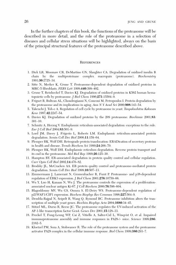
As the evolutionary higher form, the ancestral proteasome contains fourheptameric rings, arranged in the common a-b-b-a sequence, but in thisancestral proteasome each ring contains seven equal subunits, so only onea- and b-subunit are present (see Fig. 1). It is obvious that from these simpleforms of the proteasome a more complex one evolved via the divergence ofthe single subunits into several homologous ones. So the evolutionary higherform of the 20S proteasome contains 14 different subunits overall (a1–a7 andb1–b7), showing molecular masses between 20 and 30 kDa (see Table I),summarizing to a molecular weight of some 700 kDa. While the catalyticcenters are located in the inner b-rings, the outer a-rings of the proteasome

a-ring
a-ring
b-ring
b-ringb
b
aa
Top viewSide view Perspective view
5 nm
FIG. 1. The structure of the archaea 20S proteasome from Thermoplasma acidophilum. Thisfigure shows a basic model of the archaea proteasomal structure. As shown on the left, theproteasome contains four homologous rings in the sequence a-b-b-a. Each ring contains sevenidentical subunits: the a-ring only a-, the b-ring only b-subunits, as shown in the central image. Theright panel shows the arrangement of the a-subunits in a vertical view onto an a-ring.
STRUCTURE OF THE PROTEASOME 3
are responsible for the regulation of substrate entrance to the inner proteolyticchamber, as well as for recognition and binding of the substrates themselves. Sothe a-subunits are able to change both the activity and specificity of theproteasome. The proteolytic centers found in the inner rings are encoded bythree different b-subunits (b1, b2, and b5). Thus, due to the symmetric arrange-ment of the different rings, the inner chamber contains 6 different proteolyticcenters, protected inside the proteasome in the evolutionary higher form of theproteasome, but 14 in the ancestral one. The inside of the proteasome issubdivided into two ‘‘ante chambers’’ (between the a- and b-rings) and onesingle ‘‘main chamber,’’ found between the two b-rings (see Fig. 2). The ‘‘mainchamber’’ is also the location of the catalytic centers.
As the proteasome is today referred to as a proteolytic system, severalregulators are binding to the core proteasome, modulating the proteasomalactivity. Today, a set of several different proteasomal regulators are known, allbinding to the a-subunits of the outer proteasomal rings. The 11S regulatorparticle, in most organisms termed ‘‘PA28’’ or ‘‘REG,’’ is formed of threedifferent subunits (PA28a, PA28b, and PA28g arranged in several diversecombinations). In Trypanosoma brucei, this ATP-independent regulator iscalled ‘‘PA26.’’ Another important regulator is the ATP-dependent ‘‘19S,’’ alsoknown as ‘‘PA700’’ regulator; its analogue in archaea is termed ‘‘PAN.’’ Severalother regulators are known, including the nuclear regulator ‘‘PA200,’’ which isknown in three different isoforms (PA200i, PA200ii, and PA200iii) and

TABLE I
HERE, THE MOLECULAR MASSES (AFTER POSTTRANSCRIPTIONAL PROCESSING, AS FOUND IN THE
ASSEMBLED WHOLE 20S ‘‘CORE’’ PROTEASOME) OF THE PROTEASOMAL SUBUNITS FROM BOTH HUMAN
AND YEAST PROTEASOME ARE LISTED, AS WELL AS THE SUBUNITS OF TWO PROTEASOMAL REGULATOR CAPS
(11S AND 19S)
20S ‘‘core’’ proteasome
Systematic S. cerevisiae Homo sapiens Mass [kDa] Literature
a1 C7/Prs2 HsPROS27/HsIota 27.5 42
a2 Y7 HsC3 25.9 43,44
a3 Y13 HsC9 29.5 45,46
a4 Pre6 HsC6/XAPC7 27.9 47,48
a5 Pup2 HsZeta 26.4 49–51
a6 Pre5 HsC2/HsPROS30 30.2 52
a7 C1/Prs1 HsC8 28.4 53–56
b1 Pre3 HsDelta/Y 25.3 (21.9) 57–59
b1i – Lmp2 23.2 (20.9) 60–63
b2 Pup1 Z 20.0 (24.5) 64–66
b2i – Mecl1 28.9 (23.8) 67,68
b3 Pup3 HsC10-II 22.9 69,70
b4 C11 / Pre1 HsC7-I 22.8 69,71
b5 Pre2 X/MB1 N/A (22.4) 71
b5i – Lmp7 30.4 (21.2) 67,72–74
b6 C5/Prs3 HsC5 26.5 (23.3) 75,76
b7 Pre4 HsN3/HsBPROS26 29.2 (24.4) 69,77,78
11S (PA28) activator cap
Systematic Other names Mass [kDa] Literature
11S subunit a REGa or PA28a 28.723 79,80
11S subunit b REGb or PA28b 27.348 20
11S subunit g REGg or PA28g 30.886 81–84
19S (PA700) regulator cap
Systematic Other names Mass [kDa] Literature
ATPase-subunitsRpt1 S7 or p48, Mss1, Yta3, Cim5 48.633 85
Rpt2 S4 or p56Yhs4, Yta5, Mts2 49.184 86–88
Rpt3 S6b or p48, Tbp7, Yta2, Ynt1, MS73 47.336 89,90
Rpt4 S10b or p42, Sug2, Pcs1, Crl13, CADp44 44.173 91,92
Rpt5 S6a or p50, Tbp1, Yta1 49.118 93,94
Rpt6 S8 or p45, Trip1, Sug1, Cim3, Crl3, Tby1, Tbp10 45.653 95–97
Non-ATPase-subunitsRpn1 S2 or p97, Trap2, Nas1, Hrd2, Rpd1, Mts4 100.199 98
Rpn2 S1 or p112, Sen3 105.866 99,100
Rpn3 S3 or p58, Sun2 61.005 101
(Continues)
4 JUNG AND GRUNE

TABLE I (Continued)
19S (PA700) regulator cap
Systematic Other names Mass [kDa] Literature
Rpn4 Son1 or Ufd5 60.152 102–104
Rpn5 P55 or Nas5 52.904 105–107
Rpn6 S9 or p44.5 47.447 108–110
Rpn7 S10a or p44, HUMORF07 45.531 111–113
Rpn8 S12 or p40, Mov-34, Nas3 37.060 114
Rpn9 S11 or p40.5, Les1, Nas7 42.945 115–117
Rpn10 S5a or p54, ASF1, Sun1, Mcb1, Mbp1 40.736 118–120
Rpn11 S13 or Poh1, Mpr1, Par1 34.577 121–123
Rpn12 S14 or p31, Nin1, Mts3 30.004 98,124,125
Rpn15 DSS1 or SHFM1 (in human), SEM1 (in yeast) 8.146 126,127
The molecular masses in brackets represent the peptide mass before posttranscriptional processing.According to their position (a- or b-ring) in the mature proteasome, the systematic names of the subunits weredefined. The ‘‘i’’ in the systematic names indicates an g-interferon-‘‘inducible’’ proteasomal subunit. Themolecular masses of the proteasome are according to Coux et al.,128 and the information about the regulatorcaps is according to Finley et al.129 and Baumeister et al..130 The molecular weights of the single proteins weretaken from the corresponding literature and checked using the site ‘‘www.wolframalpha.com.’’ Molecular weightsfrom ‘‘www.wolframalpha.com’’ were very similar to the weights taken from the literature, except for the subunitsRpt1 (57.199 kDa according to www.wolfram.com), Rpn4 (24.551 kDa), and Rpn12 (39.481 kDa).
STRUCTURE OF THE PROTEASOME 5
contributes to spermatogenesis131 and DNA repair.132 However, only PA200iseems to bind to the proteasome, while the other two isoforms can be found innuclear foci133 without any proteasomal interaction.
Furthermore, PR39 and PI31, which work as cellular proteasomal inhibi-tors, are known. PR39 is a short peptide of only 39 amino acids, first extractedfrom porcine bone marrow, and functions as a noncompetitive inhibitor, both inyeast and mammals. Its mechanism of inhibition is unique: via binding to thea7-subunit of the proteasome, an allosteric change of the whole proteasomalstructure is induced that decreases its proteolytic activity and affects thebinding to the 19S-regulator.134 PI31, a mammalian protein first discoveredby DeMartino,135 competes with the a- and b-form of PA28 for binding of theproteasome.136
Several other proteins interact with the proteasomal system and are able toregulate the proteasomal proteolytic activities. The heat shock protein 90(Hsp90) is another known cellular proteasomal regulator.137–139 Furthermore,the proteasomal activity seems to be regulated by phosphorylation of its sub-strates or of components of the proteasomal system itself53,95,140,141 and thenuclear proteasome by poly-ADP-ribose.142–144
As mentioned, the proteolytic activity is localized inside the proteasome inthe main chamber. While only the subunits b1, b2, and b5 show proteolyticactivity, the others do not; furthermore, in some mammalian cells, the active

a
b
a2a3
a4a5
a6
a1 a7
b4
b56
b7
b4b3
b2b1
a3 a2 a1a4
Forechamber
Forechamber
Main chamber
a-ring
b-ring
b-ring
b-ring
Arc
haea
(T
. aci
doph
ilum
)Y
east
(S
. cer
evis
ia)
Forechamber
Forechamber
Main chamber
a-ring
b-ring
b-ring
b-ring
Simplified Model
b
X-Ray (open)X-Ray
FIG. 2. Structures of the archaea and eukaryotic 20S proteasomes. Here, the structures of thearchaea and the eukaryotic proteasomes are compared. The upper line of images shows a simpledescriptive model (left), a highly detailed model calculated from X-ray structure analysis (middle),and the inside of the archaea 20S proteasome (from Thermoplasma acidophilum) after removal ofseveral a- and b-subunits. The simple ball model shows the a-rings in pink and the b-rings inturquoise, while the more detailed one alternates those colors in order to accentuate the singlerings. The right image shows the inner structure of the prokaryotic proteasome, subdivided into twofore chambers between the a- and b-rings, and the main chamber formed by the two b-rings. Thebottom row of images shows the same for the eukaryotic 20S proteasome from Saccharomycescerevisiae. This type of proteasome contains seven different a- and b-subunits, each arranged in twoa- and b-rings. The single subunits are color coded in the same way in the simple ball model on theleft and the more complex models from X-ray structure analysis (middle and right). The right imagereveals the inner structure of the yeast 20S proteasome, showing the same subdivision into two foreand one main chamber, as found in the archaea proteasome too.
6 JUNG AND GRUNE
subunits can be replaced by their g-interferon (IFN-g)-inducible isoforms b1i,b2i, and b5i. However, in fact, that is not a replacement but a de novo synthesisof new proteasomes. The presence of the IFN-g-inducible isoforms of theproteasome results in a change of the fragment length of the product ofoligopeptides. It is surmised that the inducible proteasomal forms play asignificant role in the antigen presentation of the adaptive immune response.Interestingly, in the thymus, a specific third variation of the b5-subunit wasrecently found by Hirano, the replacement of the b5-subunit by the so-calledb5t one.145 The b-subunits of the proteasome provide their own class of

STRUCTURE OF THE PROTEASOME 7
proteases that show no evolutionary relation to other known proteases but avery high relationship to each other, suggesting a common ancestor.36 Accord-ing to the arrangement of the proteasomal subunits as found in the archaeicform from T. acidophilum, the different subunits have been divided into twoclasses, the a- and b-subunits.146 Normally, Eubacteria contain no 20S protea-some. Nevertheless, in a subgroup of Eubacteria, the so-called actinobacteria,proteasomal genes and even proteasomes were found. In those actinobacteria,HslVU (also known as ClpQY), an ATP-dependent hexameric protease147,148
that shows structural similarities to the b-subunits of the 20S proteasome, wasidentified. It is a dimer containing both the proteasome-related protease HsllVand HslU, an ATPase.149
The proteasome is not the only proteolytic system of the cell, but one of themost important ones. Among the others are the lysosomal system,150,151 contain-ing many different cathepsins,152–154 and the calpains associated with thecytoskeleton. While the main task of the lysosomal system includes the degra-dation of intracellular organelles, the proteasomal system is the most importantone regarding the recognition and degradation of (damaged) proteins. It isassumed that between 70% and 90%155,156 of the misfolded, (oxidatively)damaged, or no longer needed proteins are degraded via the proteasomalpathway. While the pure degradation of dysfunctional or misfolded proteinsoccurs in an ATP-independent way, the regulatory degradation of functionalproteins is ATP-dependent and,moreover, the proteins targeted for degradationhave to be labeled with a chain of ubiquitin molecules (polyubiquitination).
In mammalian cells, the amount of proteasome can be up to 1% of thewhole protein pool (in liver and kidney cells).157 The proteasome can be foundboth in the cytosolic and in the nuclear compartment of a cell, and can also bebound to the endoplasmic reticulum (ER) as well as being in association withthe cytoskeleton.158 The mammalian form of the 20S proteasome was firstdiscovered and isolated from human red blood cells by Harris in 1968 andtermed ‘‘cylindrin’’159,160 following the shape of the large protein complex.Other scientists have termed it ‘‘macroxyproteinase,’’161–163 ‘‘hollow cylinder’’protein,164,165 ‘‘multicatalytic proteinase complex,’’166–170 or ‘‘prosome.’’158,171
Today, the term ‘‘proteasome’’ is used the most often.
A. The Proteasomal a-Subunits
Both recognition and access of the substrate into the inner proteolyticchamber of the proteasome is regulated by the a-rings. After contact with asubstrate, an (oxidatively) damaged/misfolded, and (partly) defolded protein, aconformational change of the a-rings is induced that virtually ‘‘unlocks’’ thegate they form to control and regulate substrate entrance to the inner chamber.That gate is formed by the N-terminal ends of the three subunits a2, a3, anda4.
172 The N-terminal ends of those three subunits are pointing in the direction

8 JUNG AND GRUNE
of the proteasomal symmetric axis and block mechanically the entrance to theproteolytic centers formed by the two b-rings. Incubation of isolated protea-somes with low concentrations of sodium dodecylsulfate (SDS), an agent thatinduces a slight defolding of the proteasomal subunits, resulted in a significantincrease of proteolytic activity, caused by an opening of the proteasomal gatethat facilitates substrate entrance128 (Fig. 3).
Similar effects could be induced by repeated freezing–thawing cycles aswell as under conditions of low ionic strength. Those experimental resultssuggested an involvement of structural changes of the a-subunits that regulateproteasomal substrate entrance. A further possibility of substrate entrancemodulation is the binding of a regulatory subunit (like 11S, 19S, or PA200) tothe proteasomal a-rings. Such a binding can increase proteasomal activity up to10-fold173,174 and induce a change in substrate specificity, which is induced bythe maximal opening of the substrate channel of the proteasome via a confor-mational rearrangement of the blocking N-terminal ends of some a-subunits.
a5
a6
a7
a1a2
a3
a7
a1a2
a3
a4
a5
a4
5 nm
a6
FIG. 3. Gating of the eukaryotic 20S proteasome. This image shows a structural rearrangementof the a-rings of the yeast 20S proteasome without and after activation. Activation can be inducedby binding of a substrate protein, short oligopeptides, or regulating proteins (in this case, Blm10;see the text). After activation, the substrate accessibility of the proteasome increases, mainly due toan opening of the gate that is formed by the N-terminal structures of the a-subunits. Please note themassive reorganization of the N-terminus of a3 (yellow). After activation, the a-rings open a channelof about 13 A to the inner proteolytic centers. The small icon in the middle shows the point of view(arrow) from which the reader is looking at the depicted a-rings.

STRUCTURE OF THE PROTEASOME 9
The ‘‘activation’’ of the proteasome seems to be mediated via the binding of thea-rings to hydrophobic amino acids (normally buried inside the correctlyfolded/native form of a globular and water-soluble proteins) that are exposedby (oxidatively) damaged or unfolded/misfolded proteins; that binding inducesthe conformational change that results in an opening of the proteasomal gate.175
The proteasomal gate shows a maximal diameter of about 13 A (archaeaproteasome) in its maximal opened state: this is sufficient to enable the en-trance of a defolded substrate protein, usually represented by a single chain ofamino acids. The individual N-terminal ends of the single gating subunitsreveal unique three-dimensional structures that are essential for the gating ofthe proteasome. Some of the involved structures are highly conserved ineukaryotic cells. The so-called YDR-motif (Tyr8-Asp9-Arg10) can be found inevery single a-subunit, as well as in archaea and eukaryotic cells, and may beworking as a joint, bending the gating structures in order to modulate protea-somal activities.172 The most important part seems to be played by thea3 subunit: a3DN mutant of yeast, which miss the last nine amino acids(GSRRYDSRT) that are found in the wild type, and show a permanentlyincreased proteasomal activity that cannot be modulated/increased any furtherby SDS-exposure of the proteasome.172 However, the characteristics ofsubstrate binding or degradation are not or significantly less affected. Incontrast, the a7DNmutant did not reveal any significant increase in proteolyticactivity, while the a3a7DN mutant induced significantly more activity in caseindegradation than either of the single deletions.176 So, especially the YDRmotive of the a3-subunits seems to be essential for stabilization of the gate,involving allosteric effects that affect also the subunits a2 and a4.
Interestingly, in the archaea proteasome (from T. acidophilum), where thea-rings are built of seven identical subunits, some oligopeptides revealed theability to induce a conformational change in the N-termini of the a-subunitsresulting in an opening of the gate. In this conformational change, the last 13amino acids are involved as well as a slight turn (about 4�) of every singlesubunit.
The same gate-opening reconfiguration can be induced by the attachmentof PAN in its ATP-bound confirmation, as cryo-electron microscopic experi-ments have revealed.177 The oligopeptides mentioned above (seven or moreamino acid residues) are the different C-terminal sequences of PAN sub-units178 that are able to bind the gaps between the single a-subunits of theproteasome. Those residues are termed the HbXYmotifs. After binding of PANor an oligopeptide, the gate of the archaea proteasome ‘‘opens’’ by a structuralrearrangement. The diameter of the ‘‘closed’’ gate is usually about 9 A and thusnotably smaller than the channel directing to the inner proteolytic chamber ofthe proteasome with a diameter of about 23 A. As mentioned earlier, after‘‘activation’’ the diameter of the open gate increases to about 13–20 A. A similar

10 JUNG AND GRUNE
mechanism was revealed in studies of the PA26-binding: the gate structureswere opened by the C-termini of PA26 and a so-called activation loop thatinduces both a movement of the N-terminal structures of the a-rings and a faintturning of every a-subunit.177
Usually, the 20S proteasome is found in the cell in its ‘‘inactive’’ state butcan be ‘‘activated’’ by regulators, unfolded proteins, or proteasomal substrates.Furthermore, it has to be discriminated between the proteasomal peptidaseand protease activities: while ‘‘peptidase activities’’ represent the degradationof small oligopeptides and are almost independent of the gate status, ‘‘proteaseactivities’’ stand for the degradation of a whole unfolded protein and areconsiderably dependent on the gate status. This suggests that the gatinga-subunits have only little interaction with small peptide fragments but a keyfunction in the degradation of whole protein substrates.
B. The Proteasomal b-Subunits
In contrast to the gating/regulating function of the proteasomal a-units, themain task of the b-units is the proteolytic process itself.The ancient archaea proteasome contains seven identical b-subunits in one
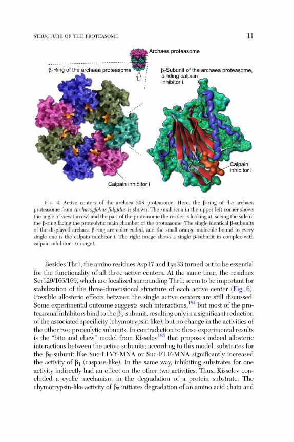
ring and thus seven proteolytic centers, too. In contrast, further developedyeast and the mammalian proteasomes contain only three different centers perb-subunit ring, localized on the subunits b1, b2, and b5. In 2002, Unno et al.proposed a novel N-terminal nucleophile hydrolase activity,40,41 formed by theThr8 residue of subunit b7, after the analysis of bovine proteasomes’ X-raycrystal structure. The proteolytic centers of the active subunits are found in theinner chamber of the 20S proteasome complex. Figure 4 shows the catalyticcenters of the archaea proteasome. Each of the three known proteolyticsubunits shows a different preference for substrate binding:
� b1 Shows a peptidyl–glutamyl–peptide hydrolysing activity (caspase-likeactivity, cleaving after acidic amino acids, thus also termed as ‘‘post-glutamyl-peptide hydrolytic’’ activity).179
� b2 Shows a trypsin-like activity, cleaving after basic amino acids.� b5 Shows a chymotrypsin-like activity and cleaves after neutral aminoacids.180
In all three active subunits, the active center is formed by the N-terminus(Thr1) of the corresponding proteasomal b-subunits (Fig. 5). Noteworthy, theproteases that show similar specificities like trypsin181 or chymotrypsin182
are serine-proteases. Typical products of proteasomal degradation are oligo-peptides with lengths between 2 and 35 amino acids.183 That distributionshows three different maxima: 2–3, 8–10, and 20–30 amino acids, while theaverage length is about 8–12 residues.86
Archaea proteasome
β-Subunit of the archaea proteasome,
inhibitor i.
β-Ring of the archaea proteasomebinding calpain
Calpain inhibitor i
Calpaininhibitor i
FIG. 4. Active centers of the archaea 20S proteasome. Here, the b-ring of the archaeaproteasome from Archaeoglobus fulgidus is shown. The small icon in the upper left corner showsthe angle of view (arrow) and the part of the proteasome the reader is looking at, seeing the side ofthe b-ring facing the proteolytic main chamber of the proteasome. The single identical b-subunitsof the displayed archaea b-ring are color coded, and the small orange molecule bound to everysingle one is the calpain inhibitor i. The right image shows a single b-subunit in complex withcalpain inhibitor i (orange).
STRUCTURE OF THE PROTEASOME 11
Besides Thr1, the amino residues Asp17 and Lys33 turned out to be essentialfor the functionality of all three active centers. At the same time, the residuesSer129/166/169, which are localized surrounding Thr1, seem to be important forstabilization of the three-dimensional structure of each active center (Fig. 6).Possible allosteric effects between the single active centers are still discussed:Some experimental outcome suggests such interactions,184 but most of the pro-teasomal inhibitors bind to the b5-subunit, resulting only in a significant reductionof the associated specificity (chymotrypsin like), but no change in the activities ofthe other two proteolytic subunits. In contradiction to these experimental resultsis the ‘‘bite and chew’’ model from Kisselev185 that proposes indeed allostericinteractions between the active subunits; according to this model, substrates forthe b5-subunit like Suc-LLVY-MNA or Suc-FLF-MNA significantly increasedthe activity of b1 (caspase-like). In the same way, inhibiting substrates for oneactivity indirectly had an effect on the other two activities. Thus, Kisselev con-cluded a cyclic mechanism in the degradation of a protein substrate. Thechymotrypsin-like activity of b5 initiates degradation of an amino acid chain and

Eukaryotic (yeast) 20S proteasome
2
15
Bortezomib
¢5
Bortezomib(on 1)
Bortezomib(on ¢2)
Upp
er-r
ing
()
Low
er-r
ing
()
-ringa
a¢
bb
bb
b
bb
b¢b
bb
b¢
-ring¢-ring-ring
FIG. 5. Active centers of the eukaryotic 20S proteasome.
12 JUNG AND GRUNE
triggers further cleavage by the b1-subunit (showing peptidyl–glutamyl–peptidehydrolysing activity); during b1-mediated substrate cleavage (‘‘chewing’’), b5activity is inhibited. If no further cleavage by b1 is possible, then b5 is ‘‘reacti-vated,’’ starting the cycle over and over again. According to Kisselev’s model,allosteric interactions between the active subunits are essential for substratedegradation.185 However, knockout mutants of yeast revealed that active protea-somal subunits are important for cellular survival and functionality (cell division)and showed that the proteolytic capacities of the single subunits vary in theirimportance: b5>>b2>b1. This results in the fact that double knockout mutantsb1/b5 and b1/b2 are still viable, while b2/b5 are not.
186,187
C. Intracellular Assembly of the 20S Proteasome
The first step in proteasome assembly is the association of the a-ring. Inorder to prevent any unspecific oligomerization of the a-subunits, this processis guided by different chaperones. These chaperones are called proteasomeassembly chaperones (PAC). Until now four different forms of these chaper-ones are known: PAC 1–4 (in humans)188–190 and their equivalents from yeast,

β5-subunit of the yeast20S proteasome
Active center of the β5-subunit of theyeast 20S proteasome
Bortezomib
Thr1
Lys33
Ser129
Asp166
Asp17
Ser169
FIG. 6. Amino acids in the active center of the eukaryotic 20S proteasome. The small icon inthe upper left corner shows the part of the whole 20S proteasome (from Saccharomyces cerevisiae)that is shown in the enlarged image: the two b-rings. Some of the subunits are removed in order toexpose the main chamber with its six proteolytic centers overall. The upper b-ring still contains b1(red), b2 (yellow), and b5 (blue), and the lower ring only b50 (blue). Every single of those activesubunits binds a bortezomib molecule (purple) to its active center. The two bortezomib moleculesthat are bound to the removed active subunits of the lower b-ring (b10 and b20) are shown in orange.Here, the b5-subunit of a 20S proteasome (from S. cerevisiae) is shown. The left part of the imageshows the whole structure, and the right part the active center and the most important amino acidsthat are involved. The center binds a single molecule of bortezomib.
STRUCTURE OF THE PROTEASOME 13
termed proteasome biogenesis-associated protein (Pba) 1–4.191,192 However,such a terminology is not uniquely used and so sometimes in yeast, Pba1 iscalled POC1, Pba2 POC2 or PAC2, Pba3 PAC3 or Dmp2188 or POC3, andfinally Pba4 Dmp1 or POC4. These chaperones form two different heterodi-mers to become active: the first one is PAC1–PAC2 in humans and Pba1–Pba2(POC1–POC2) in yeast; the second one is PAC3–PAC4 in humans and Pba3–Pba4 (Dmp1–Dmp2) in yeast.189 PAC1–PAC2 are involved in the assembly ofthe a-ring. The PAC1–PAC2 heterodimer first binds to the subunits a5 and a7,followed by stepwise incorporation of a6 and a1 (both bind on the a7 side),then followed by a2 (binds to a1), a3 (binds a2), and a4 (binds both to a3 anda5), driven by mutual interactions.193 This assembly is supplemented by thePAC3–PAC4 heterodimer, attaching to the a2-subunit.
After this, the PAC3–PAC4 heterodimer (in yeast Pba3–Pba4/Dmp1–Dmp2) provides incorporation of the single b-subunits one after the other. Ina first step, PAC3–PAC4 binds and incorporates first b2 and then b3. In further

14 JUNG AND GRUNE
steps, the subunits b4, b5, b6, b1, and b7145 are incorporated. At this stage, the
active b-subunits (b1, b2, and b5) are still in their inactive form (prob1, prob2,and prob5), due to a later removed prosequence. b6 and b7 are not proteolyticactive, but nevertheless contain a prosequence. The PAC3–PAC4 heterodimerdetaches from the complex after b3 is incorporated. This ends up with theformation of a so-called half proteasome built with one a- and one b-ring. Thecorresponding intermediates of a complete a-ring binding different b-subunitsincluding the so-called half-mer (a complete a-ring ring with all b-subunits incor-porated except b7) have already been identified, suggesting the sequential charac-teristic of that assembly.194 Another identified intermediate is the so-called 13S(built with a complete a-ring and the subunits b2, b3, and b4).195 Until now, theexact order of b-subunit incorporation in yeast is still unclear. In yeast, Ump1 bindsafter b2, b3, and b4 are already recruited in the complete a-ring, while in humansit binds the a-ring together with b2.
145
So, two of those ‘‘half proteasomes’’ finally assemble at the holoproteasome,the functional 20S ‘‘core’’ particle. In yeast, assembly of the holoproteasome ismediated by the proteasome maturation factor Ump1196,197 (also called ‘‘pro-teassemblin’’193,198 or ‘‘POMP’’,199–201 for proteasome maturation protein). Thehuman form of Ump1 is termed ‘‘hUmp1.’’ One Ump1 binds to every single‘‘half proteasome.’’ After the last b-subunit (b7) is incorporated into the ‘‘half-mer,’’ the dimerization of two ‘‘half mers’’ is induced. That whole complex (alsotermed ‘‘16S intermediate’’) now contains a complete a- and b-ring, as well asone Ump1 and the PAC1–PAC2 heterodimer. In this process, the extendedC-terminus of b7 from one ‘‘half proteasome’’ interacts between the b1- andb2-subunit of the opposite b-ring from the other ‘‘half proteasome.’’ Incorpo-ration of b7 seems to be a rate-limiting step in this process, since overexpressionof b7 massively increases 16S dimerization.202 Assembly of the holoproteasomeis followed by an autocatalytic ‘‘activation’’ of the proteolytic b-subunits: TheirN-terminal ends are degraded (setting free the N-terminal Thr1 on b1, b2, andb3), followed by degradation of both Ump1 molecules inside the proteolyticchamber as the first substrate of the functional 20S holoproteasome. After this,the two attached PAC1–PAC2 heterodimers are degraded, too.
Another protein involved in 16S dimerization is Hsc73: one is bound to each16S, and detaches after assembly of both 16S. Since intracellular immunoloca-lization revealed a stable binding of Ump1 to the outer membrane of the ER,199
it is supposed that the complete a-ring is bound to Ump1 and is not releasedbefore all of the b-subunits are recruited. Asmuch as 75–88% of the proteasomematuration intermediates were colocalized with the ER membrane, whileformation of the holoproteasome seems to take place in the cytosol of the cell.203
Interestingly, in contrast, assembly of the archaea proteasome (fromT. acidophilum), which only contains a single type of both a- and b-subunits,is independent of any chaperone proteins: a coexpression of both subunits in

STRUCTURE OF THE PROTEASOME 15
Escherichia coli resulted in the formation of functional archaea 20S protea-somes.36,146 This auto-assembly is enabled by characteristic loops (L-loops) ofthe a-subunits that can assemble with other a-subunits. The b-subunits have nosuch structure and, thus, their assembly depends of a complete a-ring thatfunctions as an assembly ‘‘platform.’’204
D. Modeling of the 20S Proteasomal Proteolysis
In order to describe 20S proteasomal (with and without regulators) degra-dation, several mathematical models have been developed. For the predictionof antigens presented by the immune system, a model describing cleavage sitesand fragment length of a given oligopeptide may be useful. For this purpose,two main strategies exist: one concentrates on the predictions of the fragmentsgenerated during proteolytic degradation of a substrate, though the results maybecome inaccurate if the produced fragments overlap205; the other strategy isfocused on the prediction of potential cleavage sites, returning the probabilityof the occurrence of a specific fragment.206 In this model, the producedfragments are normally determined by a potential cleavage site between twoamino acids with respect to one or two residues neighboring that locus on eachside. In contrast, other models try to simulate the process of degradation from amechanistic point of view, considering kinetic rates in order to calculate thevelocity of proteolytic degradation for a given substrate protein. The computedresults have to be proven in an empiric way, while the models are based on‘‘learning sets’’ that result from the fragments occurring after degradation of aknown protein.
III. Regulation of the 20S Proteasome
In order to prevent uncontrolled and unregulated proteolytic degradationin a cell, the proteasomal degradation has to be carefully regulated. Therefore,during evolution, a set of regulators has developed that are able to control bothrecognition and degradation of proteasomal substrates. The regulated entranceinto the proteolytic inner chamber of the 20S core proteasome is realized viathe gating a-rings that may be ‘‘activated’’ by binding to the exposed hydro-phobic stretches of oxidatively damaged proteins. Usually, these hydrophobicstructures are buried inside the soluble proteins, but after (oxidative) modifi-cation, these structures may be exposed, whereas native and correctly foldedproteins have to be targeted (via polyubiquitination) for proteasomal degrada-tion. The most important regulator for the recognition of ubiquitin-labeledproteins is the so-called 19S regulator complex that cooperates with anothercellular machinery, the ubiquitination system.

16 JUNG AND GRUNE
A. The 19S Regulator
The 19S regulator, also known as ‘‘PA700’’ or ‘‘proteasome activator700 kDa’’207–210 is built up of two main structures: a ring-shaped base thatbinds to the a-rings of the 20S ‘‘core’’ proteasome and a lid that recognizes andbinds polyubiquitinated proteins, thus regulating substrate entrance to the 20Sproteasome.
The base ring contains at least 10 different subunits (Rpt1–Rpt6, Rpn1,Rpn2, Rpn10, and Rpn13). The lid contains nine subunits (Rpn3, Rpn5–Rpn9,Rpn11, Rpn12, and Rpn15) that are also termed ‘‘DSS1’’ or ‘‘SHFM1’’ inhumans, and ‘‘SEM1’’ in yeast. The Rpt-subunits show an ATPase activity,while the Rpn-subunits do not. Furthermore, Rpn11 in the lid contains aZn2þ-dependent proteolytic center that is able to catalyze the proteolyticdegradation of polyubiquitin chains that label native substrate proteins fordegradation; after this, the ubiquitin molecules are released for reuse of thepolyubiquitination machinery.
The Rpt2 (in humans also called S4 or p56, and in yeast YTA5 or mts2),Rpt3 (human form termed as S6, Tbp7, or P48, and the yeast form as YTA2),and Rpt5 (S60 or Tbp1 in humans, and YTA1 in yeast) subunits of the base ringplay a role in gate opening of the attached a-subunits of the 20S proteasome,211
while Rpn10 (S5a or Mbp1 in humans, and SUN1,MCB1, or pus1 in yeast) andRpn13 (ADRM1 in humans, and DAQ1 in yeast) function as polyubiquitinreceptors.211 The main role of the small protein ubiquitin is the labeling ofnative proteins for proteasomal degradation, in order to regulate their intra-cellular amount of life span.
One 19S regulator might attach to each of the a-rings of the 20S ‘‘core’’proteasome, forming a large particle of about 2 MDa,212 termed the ‘‘26Sproteasome.’’ In a mechanism that is known from other regulators of theproteasome, the 19S particle makes substrate access to the ‘‘core’’ particleeasier by ‘‘opening’’ the gating a-rings. It has been shown in yeast that theRpt2-ATPase of the base ring is involved in this process.86 Until now, no datafrom X-ray crystallography of the 19S regulator are available211 and only someof the interactions of the single subunits are known213; thus, in Fig. 7, only ahypothetical structure of the 19S regulator cap can be displayed.
B. The Immunoproteasome
A special inducible form of the 20S proteasome is called the immunopro-teasome (i20S). The i20S proteasome can be induced by IFN-g, and on the siteof the subunits b1, b2, and b5 their inducible equivalents (b1i, b2i, and b5i) arelocated.140,214–219 To achieve this, proteasomes have to be synthetized denovo.220,221 Further inducers are both tumor necrosis factor alpha (TNF-a)222,223 and lipopolysaccharides.224 In general, the three inducible b-subunits

19S regulator cap
Base ring of the19S regulator cap
α-ring
β-ring
β-ring
α-ring
Eukaryotic (yeast) 20S “core” proteasome
Lid of the19S regulator cap
FIG. 7. A model of the structure of the eukaryotic 26S proteasome. This image shows a modelof a eukaryotic 20S ‘‘core’’ proteasome (from Saccharomyces cerevisiae) bound to two 19S regulatorcaps. Since there are no data from X-ray structure analysis for the 19S regulator, this is just a modelshowing the very basic shape of 19S that is divided into a ‘‘base’’-ring containing six subunits bindingthe a-ring of the 20S ‘‘core’’ proteasome and a ‘‘lid’’ containing nine subunits, responsible forrecognition, binding, and unfolding (in an ATP-dependent way) of polyubiquitinated substrates,feeding them into the 20S proteasome for terminal degradation. Overall, the 19S regulator capcontains some 18 different subunits (see Table I), 10 for the whole ‘‘base’’ structure and nine for the‘‘lid.’’
STRUCTURE OF THE PROTEASOME 17
are homologs of the constitutive ones and are indicated by an additional ‘‘i’’(i20S); other names found in the literature for those inducible proteasomalsubunits are low-molecular-weight protein 2 (LMP2, for b1i), multicatalyticendopeptidase-like-complex-1 (MELC1, for b2i), and LMP7 (for for b5i).
225–227
At the same time, the so-called 11S regulator (also known as PA28, PA26, orREG) is induced by the same cellular signal cascades. The major function ofthe immunoproteasome is the production of a specific short oligopeptideproduct pattern that can be presented by the major histocompatibility complexclass I (MHC-I) on the cell’s surface in immune response. Typical products of

18 JUNG AND GRUNE
the immunoproteasome are short protein fragments, made up of about 8–10amino acids that are optimized for MHC-I-presentation. Since i20S inductionmainly depends on the amount of cytokines that are released in the tissue, it issuggested that immunoproteasomes mainly release new self-determinants thatprevent autoimmune response in the surrounding uninfected cells.228 On theother hand, Yewdell proposed in 2005228 that the main functions of i20S are notthe generation of MHC-I-presented antigens and that further research in thisfield is needed.
INF-g induces both the proteasome maturation factor Ump1 and theinducible forms of the proteolytic proteasomal subunits; paradoxically, themRNA of Ump1 is increased, while the amount of free Ump1 decreases (asfound in HeLa cells) and the half-life of that protein is reduced from 82 to only21 min.229 It turns out that this is caused by a massively increased proteasomeformation that enhanced degradation of Ump1, which is the first proteasomalsubstrate (see above) after assembly of the holoproteasome. Consideringthis, the turnover of Ump1 can be used as an indicator for the formation rateof functional proteasomes. The formation of the immunoproteasome is turnedinto a highly dynamic process due to two main factors. The first one is thefact that the processed b5i has a higher affinity to Ump1 than the propeptideprob5i, which generates a higher rate of i20S formation compared to the one ofc20S (this fact suggests two different binding sites).229 The second factor is themuch shorter half-life of i20S (about 27 h) compared to c20S (about 8–12days).230–232 The interplay of these two factors allows both a very quick expres-sion and a fast removal of i20S. Seven days of continuous stimulation cancompletely replace c20S by i20S,21 while after a shorter stimulus the ratio ofc20S/i20S decreases.
However, after expression of the inducible subunits, the de novo assembledproteasomes do not always contain all six inducible catalytic subunits; manyforms are found that only show between 1 and 5 of the inducible bi-subunits,while the others are the constitutive ones. Whether there is a special need orfunction for such intermediate proteasomes is still unclear. Nevertheless, bothb5i and Ump1 seem to be essential for the formation of i20S: cells that do notexpress b5i or Ump1 are not able to form i20S, even after IFN-g treatment.
In Ump1-knockdown cells, proteasomal-mediated proteolysis decreasesrapidly to 60% after 24 h and to 40% after 48 h,229 while the overall amountof cellular proteasomes is reduced significantly.233 In the same way, an over-expression of Ump1 increases the proteasomal-mediated cellular proteoly-sis.200 The number of bi-subunits increases constantly over time in muscletissue of aged rats compared to young ones (a three- to sixfold increase wasfound). The same results were detected for neurons, astrocytes, and endothe-lial cells in the hippocampus of elderly humans (about 70 years of age) com-pared to a younger control group (about 42 years of age).234 Considering this,

STRUCTURE OF THE PROTEASOME 19
the inducible b-subunits seem to accumulate over time in cells and tissues thatnormally only contain the ‘‘housekeeping’’ form of the proteasome (c20S) andespecially in cells prone to postmitotic aging, as neurons and muscle cells.
C. The Thymus-Specific Proteasome(Thymoproteasome)
Another specific proteasomal subunit is the so-called b5t, which was firstdiscovered in mice, exclusively in cortical thymic epithelial cells (cTECs).235 b5tplays an important role in the positive selection of thymocytes.236 Accordingly, theterm ‘‘thymoproteasome’’ was suggested for the ‘‘b1i, b2i, and b5t-configuration’’of the 20S proteasome. It turns out that the thymoproteasome is responsible forthe generation of short antigenic oligopeptides presented on the cell surface,resulting in the positive selection of CD8þ T-cells. The self-peptide productionis dependent on the thymoproteasome and is essential for the development of animmune-competent repertoire of CD8þ T-cells.237
Genomic analyses of the gene coding b5t (PSMB11) was performed bySutoh et al..238 It turned out that teleost fish have two functional copies ofPSMB11 (PSMB11a and PSMB11b), while chickens, turkeys, and zebra fishlost PSMB11, expressing neither thymo- nor immunoproteasomes. In mam-mals, reptiles, amphibians, and teleost fishes, PSMB11 is located close toPSMB5, which codes b5 of the constitutive 20S proteasome. These resultssuggest that PSMB11 may originate from the older PSMB5 by tandem dupli-cation. b5t shows a close relation to both b5 and b5i and was found to beincorporated in about 20% of the thymic proteasomes.
In proteasomes containing b5t, the inducible subunits b1i and b2i arepreferentially incorporated compared to the constitutive ones b1 and b2. Anti-gens that are presented by the MHC-I239–241 complexes show hydrophobicC-termini that function as an anchor in MHC-I binding242 and that result fromthe characteristics of b5t-mediated cleavage. In contrast to b5 and b5i, theproteolytic center of b5t contains hydrophobic amino residues that reduce thechymotrypsin-like proteasomal activity by 60–70%, without any effect onthe other two activities.235 The maximal velocity of proteolysis, as well as theMichaelis constant, is lower in b5t compared to both b5 and b5i. The result is asignificantly decreased amount of oligopeptides released with a hydrophobicC-terminus that are preferably incorporated in the binding grove of MHC-I.Thus, b5t seems to reduce the amount of MHC-I-presentable antigens.
The result is both a lowered production and presentation of MHC-I-boundoligopeptides and thus a decreased interaction of cTECs with the ab-T-cellantigen receptor, which causes a higher probability of positive selection ofthose cells.235,236

20 JUNG AND GRUNE
b5i-deficient mouse models revealed an imperfect development of CD8þ
T-cells and a resulting decrease of those cells by about 80%,243 suggesting thatb5i may enhance the selection of CD8þ T-cells. However, the amount ofantigen-loaded MHC-I molecules presented on the surface of b5t-deficientcells did not change. The lysosomal cathepsin S is a necessary factor in antigenpresentation in most cells, but in contrast, this task is performed by cathepsin Lin thymus cortical epithelial cells. After deletion of cathepsin L in those cells,the selection of CD4þ was reduced without any influence on the amount ofMHC-II. The presentation of different antigenic oligopeptides by MHC-I (inCD8þ cells mediated by the proteasome) and MHC-II (in CD4þ cells medi-ated by the lysosomal system/cathepsin L) decides the positive or negativeselection of mature T-cells. T-cells showing a high affinity to self-antigens aresorted out, otherwise causing autoimmune reactions. In contrast, T-cells with alow affinity to MHC molecules will maturate, while mediocre affinity usuallytriggers positive selection.
D. The 11S Regulator
The 11S regulator of the proteasome, also called ‘‘PA28,’’ ‘‘REG,’’ or ‘‘PA26’’(in T. brucei), has different structures. Three different subunits of the 11Sactivator are known: PA28a, PA28b, and PA28g. There are hexameric orheptameric structures described and, in addition to that, under defined condi-tions, various homo- or heteropolymerization products of the individual sub-units are formed. Results indicated first that the 11S regulator has an a3b3structure, where both subunits were arranged alternatively.244,245 However,later, an a3b4 complex was detected. This particle interestingly contains a b–bdimer, but no a-subunit dimer.246 There seem to exist several 11S forms incells, as PA28a3b3, PA28a4b3, PA28a3b4 (in each case with alternating arrange-ment of the a- and b-subunits), and PA28g7.
247 If PA28a and PA28b subunitsare mixed in vitro in a ratio of a to b of 1.2, both PAa3b4 and PAa4b3 can bedetected.246 The PA28a-heptamer is instable, but can be formed in vitro,whereas the PA28b-heptamer cannot. However, PA28g forms a stableheptamer.248,249
The base diameter of the 11S regulator is some 90 A and the complex isabout 60 A in height; with a central cavity 20–30 A wide. In general, the various11S regulators are able to bind to the outer a-rings of the proteasome andchange substrate degradation properties. However, like the degradation by the20S proteasome, the degradation of substrates by the 11S–20S complex is ATP-independent (Fig. 8), suggesting that only unfolded proteins are substrates.247
11S binding increases the b2-catalyzed cleavage about 10-fold and the b1- andb5-catalyzed cleavages by about 50-fold.250,251 The PA28g-isoform activatesonly b2.
248 Binding of PA28 changes the conformation of the proteasome,thereby making it more efficient in proteolytic activity.252

a-ring
a-ring
a-ring
b-ring
b-ring
b-ring
11Sregulatorparticle
11Sregulatorparticle
20S
(ye
ast)
pro
teas
ome
11Sregulator particle
FIG. 8. The immunoproteasome and the 11S regulator. Here, the structure of the so-calledimmunoproteasome, a eukaryotic 20S ‘‘core’’ proteasome capped with two 11S regulators, is shownon the left side of the image. The right part shows a cross-section of the 11S regulator cap and an a-and b-ring of the 20S proteasome (a half proteasome). Please note that the gate of the ‘‘core’’proteasome is ‘‘opened’’ and a channel through the regulator cap (arrow above the structure)enables substrate access to the main chamber of 20S and the proteolytic centers within.
STRUCTURE OF THE PROTEASOME 21
Interestingly, PA28a and PA28b are located only in the cytosol, but all threePA28 isoforms can be found in the nucleus.253,254 The PA28g isoform activatesonly b2.
248 The PA28a,b regulators seem to be involved in the generation ofoligopeptides in the immune response (see also immunoproteasome). So,PA28b knockout animals have a reduced immune function.255 Interestingly,PA28g-knockout animals show malfunctions in cell cycle regulation and apo-ptosis,255,256 due to the role of PA28g7 in the degradation of nuclear lysine-freeproteins.257,258
E. The Hybrid Proteasome (PA28–20S–PA700)
The hybrid proteasome, thus termed by Tanahashi et al.,259 contains both aPA700 (19S) and a PA28 (11S) regulator cap. Each of the regulators is bound toeither end of the 20S proteasome. Tanahashi et al. determined the relativeamounts of the different possible hybrid proteasomes (see Fig. 9). The 11Sregulator is present in its hexameric form (ab)3 and the two different hepta-meric versions (ab)3a and (ab)3b in hybrid proteasomes, whereas the

41 ± 5%20S
proteasome
20 ± 9%Immuno
Proteasome(11S–20S–11S)
24 ± 9%Hybrid
proteasome(19S–20S–11S)
15 ± 3%26S
proteasome(19S–20S–19S)
FIG. 9. The relative amount of proteasome types as found in the cytosol of HeLa cells. Here,the relative amount of proteasomal types are shown, starting with the uncapped 20S (on theleft, ‘‘20S proteasome’’) and the amounts of the different fractions like 26S proteasome (19S–20S–19S, second from left), hybrid proteasome (19S–20S–11S, second from right), and the immu-noproteasome (11S–20S–11S, on the right).
22 JUNG AND GRUNE
heptameric PA28g (g7) is not. Via immunoprecipitation it is possible to isolatethe hybrid proteasomal forms that can be induced in cells in an IFN-g-mediated way.260 ATP is needed for attachment of PA28 to the 20S coreproteasome, as well as in the ATP-dependent protein degradation of thePA28–20S–PA700 complex.259 Considering the ATP-dependence of the regu-lator ‘‘core’’ particle attachment, the formation of PA28–20S–PA700 is verysimilar to that of PA700–20S–PA700 (the 26S proteasome).
Though the exact cellular function of those hybrid proteasomes is stillunknown, it might be possible that the proteolytic specificities of the coreproteasome bound to 19S change by binding of an additional PA28 regulator,260
causing a different set of oligopeptide products produced during proteolyticdegradation of a substrate. The proteolytic activity of 26S was found to behigher than that shown by the hybrid forms. However, a cooperation ofimmunoproteasome and hybrid forms of the proteasome in antigen processingmight be possible, since both proteasomal forms can be induced via IFN-g. So,it was suggested that most of the proteolysis required for MHC-I antigenpresentation is performed via the 26S proteasome or the so-called hybrid

STRUCTURE OF THE PROTEASOME 23
proteasomes (PA28–20S–19S). Since the PA28 activator cap is not able tomediate the degradation of natively folded proteins, it appears that the sub-strate protein has to be unfolded first in an ATP-dependent way that ismediated by the 19S-regulator cap.
Whether the short oligopeptide products that are released after 19S–20S-mediated proteolytic degradation are further processed by another protea-some, perhaps containing a PA28 regulator protein, is still unclear. Accordingto the ‘‘molecular coupling hypothesis’’, a hybrid proteasome is attached to aTAP1–TAP2 complex (TAP, transporter associated with antigen processing)protein channel in the ER membrane via the PA28 regulator cap. So, a poly-ubiquitinated and natively folded antigenic protein is recognized and unfoldedby the 19S regulator cap and then guided into the core proteasome (in thisspecial case, an immunoproteasome), where it is degraded. Fragment lengthsas well as the fragment characteristics are influenced by the PA28 proteasomalregulator cap that delivers antigenic oligopeptide fragments directly into theTAP1–TAP2 protein complex. The advantage of such a direct transport is theprotection of the fragments from cytosolic proteases. Further processing of theantigenic fragments (like N-terminal trimming) is done via ER-resident pro-teases as endoplasmic reticulum aminopeptidase associated with antigen pro-cessing and endoplasmic reticulum aminopeptidase 1 and 2. This proposedmechanism was inspired by the identification of the highly conserved ‘‘KEKE’’-motifs at the distal side of PA28 that are not involved in binding or activation ofthe 20S ‘‘core’’ proteasome. KEKE motifs may be involved in protein–proteininteractions and have been found in four subunits of the 20S proteasome andfive subunits of the 19S proteasomal regulator cap. Furthermore, they havebeen found in both HSP90 and calnexin, two other proteins that play a role inepitope loading of MHC-I proteins. Another hypothesis gaining from thoseresults suggests that heat shock proteins may be involved in immune re-sponse.261,262 However, this idea has been disproven in 2006 using the SIIN-FEKL epitope of ovalbumin: experiments revealed that there is no promotionof its MHC-I presentation.263
F. The PA200 Regulator Protein
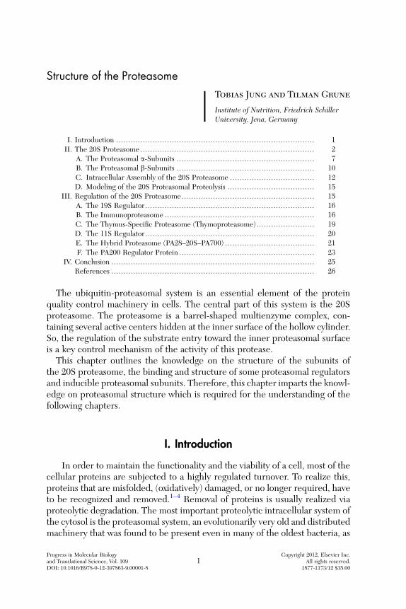
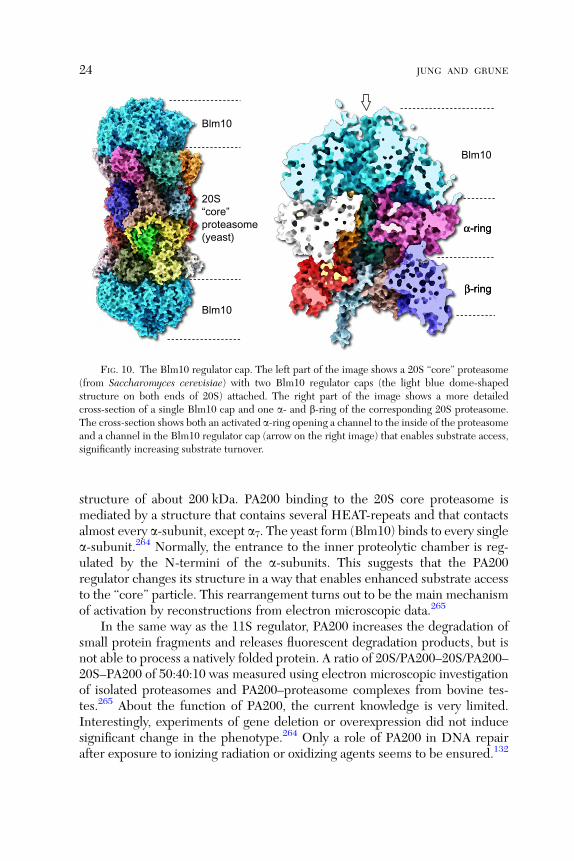
The PA200 proteasomal activator cap is exclusively found in the nucleus ofmammalian cells. The yeast homolog (from Saccharomyces cerevisiae), knownas Blm10, shows a sequence homology of about 20%264 to the mammalianform. First investigations of the PA200 structure and its binding to the 20S coreproteasome were done using electron microscopy. Three-dimensional recon-structions of the gained data showed a slightly asymmetric dome structure(100 A in diameter, as the 20S proteasomal a-ring, and about 60 A in high)with an inner cavity that sits on one or both the a-rings like a cap265 (Fig. 10).Differently from the other proteasomal regulators, PA200 is a monomeric
Blm10
α-ring
β-ring
Blm10
20S“core”proteasome(yeast)
Blm10
α-ring
β-ring
FIG. 10. The Blm10 regulator cap. The left part of the image shows a 20S ‘‘core’’ proteasome(from Saccharomyces cerevisiae) with two Blm10 regulator caps (the light blue dome-shapedstructure on both ends of 20S) attached. The right part of the image shows a more detailedcross-section of a single Blm10 cap and one a- and b-ring of the corresponding 20S proteasome.The cross-section shows both an activated a-ring opening a channel to the inside of the proteasomeand a channel in the Blm10 regulator cap (arrow on the right image) that enables substrate access,significantly increasing substrate turnover.
24 JUNG AND GRUNE
structure of about 200 kDa. PA200 binding to the 20S core proteasome ismediated by a structure that contains several HEAT-repeats and that contactsalmost every a-subunit, except a7. The yeast form (Blm10) binds to every singlea-subunit.264 Normally, the entrance to the inner proteolytic chamber is reg-ulated by the N-termini of the a-subunits. This suggests that the PA200regulator changes its structure in a way that enables enhanced substrate accessto the ‘‘core’’ particle. This rearrangement turns out to be the main mechanismof activation by reconstructions from electron microscopic data.265
In the same way as the 11S regulator, PA200 increases the degradation ofsmall protein fragments and releases fluorescent degradation products, but isnot able to process a natively folded protein. A ratio of 20S/PA200–20S/PA200–20S–PA200 of 50:40:10 was measured using electron microscopic investigationof isolated proteasomes and PA200–proteasome complexes from bovine tes-tes.265 About the function of PA200, the current knowledge is very limited.Interestingly, experiments of gene deletion or overexpression did not inducesignificant change in the phenotype.264 Only a role of PA200 in DNA repairafter exposure to ionizing radiation or oxidizing agents seems to be ensured.132

STRUCTURE OF THE PROTEASOME 25
So, in response to ionizing radiation, PA200 is expressed and accumulates in itshybrid form on chromatin.133 After PA200 knockdown, cells showed genomicinstabilities and reduced survival. The genome-stabilizing functions of PA200seem to be a result of its ability to enhance the proteasomal b1-mediatedpeptidyl—glutamyl-like proteolysis.266 However, deletion of Blm10 in theyeast A364a-strain did not result in any effect on growth or viability aftertreatment of the cells with DNA-damaging agents like bleomycin or phleomy-cin.264 Furthermore, no increased susceptibility of A364a to UV- or g-irradia-tion, methyl methane sulfonate, camptothecin, or hydroxyurea could bedetected. However, overexpression of Blm10 resulted in a reduced growth,but this can be an effect of increased binding to the 20S proteasome, thusdetracting activity from other cellular functions of that protease. The presenceof PA200 in the nucleus enables the formation of another proteasome complex:in yeast, Blm10 is able to form PA200–20S–19S.132,267 After HeLa treatmentwith ionizing irradiation, immunoprecipitation revealed a coprecipitation ofPA200 in complex with 20S–19S, even if the amounts of 20S and 19S were notincreased. Thus, irradiation seems to induce the formation of the PA200–20S–19S complex in a DNA-damage response-mediated way.266 Twenty-four hoursafter irradiation, the PA200–20S–19S complex showed an accumulation onchromatin. The trypsin-like (mediated by the b2-subunit of the proteasome)activity associated with the chromatin showed a sixfold increase, and thepeptidyl–glutamyl-like one (mediated by the b1-subunit) up to a 19-fold in-crease, accompanied by a five- to eightfold increased amount of 20S on thechromatin.268 That accumulation seems to be independent of ATM (a PI3-likekinase),269,270 which starts the signal cascade after irradiation-mediated stressvia triggering of the tumor suppressor p53.271,272 One important function ofPA200 might be an enhancement of the b1 activity of the proteasome that isessential for the cellular survival after exposure to ionizing irradiation.266
IV. Conclusion
As shown in this chapter, the proteasomal system is complex and far frombeing well understood. We referred in this chapter only to the principalstructures of the proteasome, not describing the interaction with the ubiquiti-nation machinery, or other proteasomal regulators; it becomes clear that onecan hardly imagine any aspect of cellular life not related to the function of theubiquitin–proteasome system. The importance of this system is further under-lined by the fact that it appeared early in evolution, as in Archae bacteria, andevolved to a more complex structure with new regulators and specializedsubunits. The evolution toward function and organ-specific isoforms under-lines this chapter.
26 JUNG AND GRUNE
In the further chapters of this book, the functions of the proteasome will bedescribed in more detail, and the role of the proteasome in a selection ofdiseases and cellular stress situations will be highlighted, always on the basisof the principal structural features of the proteasome described above.
References
1. Dick LR, Moomaw CR, DeMartino GN, Slaughter CA. Degradation of oxidized insulin Bchain by the multiproteinase complex macropain (proteasome). Biochemistry1991;30:2725–34.
2. Sitte N, Merker K, Grune T. Proteasome-dependent degradation of oxidized proteins inMRC-5 fibroblasts. FEBS Lett 1998;440:399–402.
3. Grune T, Reinheckel T, Davies KJ. Degradation of oxidized proteins in K562 human hema-topoietic cells by proteasome. J Biol Chem 1996;271:15504–9.
4. Friguet B, Bulteau AL, Chondrogianni N, Conconi M, Petropoulos I. Protein degradation bythe proteasome and its implications in aging. Ann N Y Acad Sci 2000;908:143–54.
5. Takeuchi J, Toh-e A. Regulation of cell cycle by proteasome in yeast. Tanpakushitsu KakusanKoso 1997;42:2247–54.
6. Davies KJ. Degradation of oxidized proteins by the 20S proteasome. Biochimie 2001;83:301–10.
7. Schmitz A, Herzog V. Endoplasmic reticulum-associated degradation: exceptions to the rule.Eur J Cell Biol 2004;83:501–9.
8. Lord JM, Davey J, Frigerio L, Roberts LM. Endoplasmic reticulum-associated proteindegradation. Semin Cell Dev Biol 2000;11:159–64.
9. Plemper RK, Wolf DH. Retrograde protein translocation: ERADication of secretory proteinsin health and disease. Trends Biochem Sci 1999;24:266–70.
10. Plemper RK, Wolf DH. Endoplasmic reticulum degradation. Reverse protein transport andits end in the proteasome. Mol Biol Rep 1999;26:125–30.
11. Hampton RY. ER-associated degradation in protein quality control and cellular regulation.Curr Opin Cell Biol 2002;14:476–82.
12. Brodsky JL, McCracken AA. ER protein quality control and proteasome-mediated proteindegradation. Semin Cell Dev Biol 1999;10:507–13.
13. Zimmermann J, Lamerant N, Grossenbacher R, Furst P. Proteasome- and p38-dependentregulation of ERK3 expression. J Biol Chem 2001;276:10759–66.
14. Wu Y, Luo H, Kanaan N, Wu J. The proteasome controls the expression of a proliferation-associated nuclear antigen Ki-67. J Cell Biochem 2000;76:596–604.
15. Blagosklonny MV, Wu GS, Omura S, El-Deiry WS. Proteasome-dependent regulation ofp21WAF1/CIP1 expression. Biochem Biophys Res Commun 1996;227:564–9.
16. Dembla-Rajpal N, Seipelt R, Wang Q, Rymond BC. Proteasome inhibition alters the tran-scription of multiple yeast genes. Biochim Biophys Acta 2004;1680:34–45.
17. Stitzel ML, Durso R, Reese JC. The proteasome regulates the UV-induced activation of theAP-1-like transcription factor Gcn4. Genes Dev 2001;15:128–33.
18. Preckel T, Fung-Leung WP, Cai Z, Vitiello A, Salter-Cid L, Winqvist O, et al. Impairedimmunoproteasome assembly and immune responses in PA28-/- mice. Science 1999;286:2162–5.
19. Kloetzel PM, Soza A, Stohwasser R. The role of the proteasome system and the proteasomeactivator PA28 complex in the cellular immune response. Biol Chem 1999;380:293–7.

STRUCTURE OF THE PROTEASOME 27
20. Schwarz K, Eggers M, Soza A, Koszinowski UH, Kloetzel PM, Groettrup M. The proteasomeregulator PA28alpha/beta can enhance antigen presentation without affecting 20S proteasomesubunit composition. Eur J Immunol 2000;30:3672–9.
21. Khan S, van den Broek M, Schwarz K, de Giuli R, Diener PA, Groettrup M. Immunoprotea-somes largely replace constitutive proteasomes during an antiviral and antibacterial immuneresponse in the liver. J Immunol 2001;167:6859–68.
22. Osterloh P, Linkemann K, Tenzer S, Rammensee HG, Radsak MP, Busch DH, et al. Protea-somes shape the repertoire of Tcells participating in antigen-specific immune responses. ProcNatl Acad Sci USA 2006;103:5042–7.
23. Borissenko L, Groll M. Diversity of proteasomal missions: fine tuning of the immuneresponse. Biol Chem 2007;388:947–55.
24. Mathew A, Mathur SK, Morimoto RI. Heat shock response and protein degradation: regula-tion of HSF2 by the ubiquitin-proteasome pathway. Mol Cell Biol 1998;18:5091–8.
25. Sulahian R, Sikder D, Johnston SA, Kodadek T. The proteasomal ATPase complex is requiredfor stress-induced transcription in yeast. Nucleic Acids Res 2006;34:1351–7.
26. Kahn NW, Rea SL, Moyle S, Kell A, Johnson TE. Proteasomal dysfunction activates thetranscription factor SKN-1 and produces a selective oxidative-stress response in Caenorhab-ditis elegans. Biochem J 2008;409:205–13.
27. Hahn JS, Neef DW, Thiele DJ. A stress regulatory network for co-ordinated activation ofproteasome expression mediated by yeast heat shock transcription factor. Mol Microbiol2006;60:240–51.
28. Bregegere F, Milner Y, Friguet B. The ubiquitin-proteasome system at the crossroads ofstress-response and ageing pathways: a handle for skin care? Ageing Res Rev 2006;5:60–90.
29. Clawson GA. Protease inhibitors and carcinogenesis: a review. Cancer Invest 1996;14:597–608.
30. Scheffner M,Whitaker NJ. Human papillomavirus-induced carcinogenesis and the ubiquitin-proteasome system. Semin Cancer Biol 2003;13:59–67.
31. Moriishi K, Mochizuki R, Moriya K, Miyamoto H, Mori Y, Abe T, et al. Critical role ofPA28gamma in hepatitis C virus-associated steatogenesis and hepatocarcinogenesis. Proc NatlAcad Sci USA 2007;104:1661–6.
32. Walters KJ, Lech PJ, Goh AM, Wang Q, Howley PM. DNA-repair protein hHR23a alters itsprotein structure upon binding proteasomal subunit S5a. Proc Natl Acad Sci USA2003;100:12694–9.
33. Bergink S, Severijnen LA, Wijgers N, Sugasawa K, Yousaf H, Kros JM, et al. The DNA repair-ubiquitin-associated HR23 proteins are constituents of neuronal inclusions in specific neuro-degenerative disorders without hampering DNA repair. Neurobiol Dis 2006;23:708–16.
34. Krogan NJ, Lam MH, Fillingham J, Keogh MC, Gebbia M, Li J, et al. Proteasome involve-ment in the repair of DNA double-strand breaks. Mol Cell 2004;16:1027–34.
35. Hough R, Pratt G, Rechsteiner M. Purification of two high molecular weight proteases fromrabbit reticulocyte lysate. J Biol Chem 1987;262:8303–13.
36. Lowe J, Stock D, Jap B, Zwickl P, Baumeister W, Huber R. Crystal structure of the 20Sproteasome from the archaeon T. acidophilum at 3.4 A resolution. Science 1995;268:533–9.
37. Stock D, Ditzel L, Baumeister W, Huber R, Lowe J. Catalytic mechanism of the 20Sproteasome of Thermoplasma acidophilum revealed by X-ray crystallography. Cold SpringHarb Symp Quant Biol 1995;60:525–32.
38. Groll M, Ditzel L, Lowe J, Stock D, Bochtler M, Bartunik HD, et al. Structure of 20Sproteasome from yeast at 24 A resolution. Nature 1997;386:463–71.
39. Tomisugi Y, Unno M, Mizushima T, Morimoto Y, Tanahashi N, Tanaka K, et al. New crystalforms and low resolution structure analysis of 20S proteasomes from bovine liver. J Biochem2000;127:941–3.

28 JUNG AND GRUNE
40. Unno M, Mizushima T, Morimoto Y, Tomisugi Y, Tanaka K, Yasuoka N, et al. The structure ofthe mammalian 20S proteasome at 2.75 A resolution. Structure 2002;10:609–18.
41. Unno M, Mizushima T, Morimoto Y, Tomisugi Y, Tanaka K, Yasuoka N, et al. Structuredetermination of the constitutive 20S proteasome from bovine liver at 2.75 A resolution.J Biochem 2002;131:171–3.
42. Petit F, Jarrousse AS, Dahlmann B, Sobek A, Hendil KB, Buri J, et al. Involvement ofproteasomal subunits zeta and iota in RNA degradation. Biochem J 1997;326(Pt 1):93–8.
43. Tokunaga F, Aruga R, Iwanaga S, Tanaka K, Ichihara A, Takao T, et al. The NH2-terminalresidues of rat liver proteasome (multicatalytic proteinase complex) subunits, C2, C3 and C8,are N alpha-acetylated. FEBS Lett 1990;263:373–5.
44. Du J, Mitch WE, Wang X, Price SR. Glucocorticoids induce proteasome C3 subunit expres-sion in L6 muscle cells by opposing the suppression of its transcription by NF-kappa B. J BiolChem 2000;275:19661–6.
45. Feist E, Dorner T, Kuckelkorn U, Schmidtke G, Micheel B, Hiepe F, et al. Proteasome alpha-type subunit C9 is a primary target of autoantibodies in sera of patients with myositis andsystemic lupus erythematosus. J Exp Med 1996;184:1313–8.
46. Castano JG, Mahillo E, Arizti P, Arribas J. Phosphorylation of C8 and C9 subunits of themulticatalytic proteinase by casein kinase II and identification of the C8 phosphorylation sitesby direct mutagenesis. Biochemistry 1996;35:3782–9.
47. Dong J, Chen W, Welford A, Wandinger-Ness A. The proteasome alpha-subunit XAPC7interacts specifically with Rab7 and late endosomes. J Biol Chem 2004;279:21334–42.
48. Mukherjee S, Dong J, Heincelman C, Lenhart M, Welford A, Wandinger-Ness A. Functionalanalyses and interaction of the XAPC7 proteasome subunit with Rab7. Methods Enzymol2005;403:650–63.
49. Mayau V, Baron B, Buttin G, Debatisse M. Twelve genes, including the unassigned proteasomezeta subunit gene, ordered within the human 1p13 region. Mamm Genome 1998;9:331–3.
50. Van GG, De JC, Pype S, Van CW, Julliams A, Vanderhoeven I, et al. Alzheimer’s diseaseassociated presenilin 1 interacts with HC5 and ZETA, subunits of the catalytic 20S protea-some. Neurobiol Dis 1999;6:376–91.
51. Jorgensen L, Hendil KB. Proteasome subunit zeta, a putative ribonuclease, is also found as afree monomer. Mol Biol Rep 1999;26:119–23.
52. KaniaMA,DeMartino GN, BaumeisterW, Goldberg AL. The proteasome subunit, C2, containsan important site for binding of the PA28 (11S) activator. Eur J Biochem 1996;236:510–6.
53. Bose S, Stratford FL, Broadfoot KI, Mason GG, Rivett AJ. Phosphorylation of 20S protea-some alpha subunit C8 (alpha7) stabilizes the 26S proteasome and plays a role in theregulation of proteasome complexes by gamma-interferon. Biochem J 2004;378:177–84.
54. Gerards WL, Enzlin J, Haner M, Hendriks IL, Aebi U, Bloemendal H, et al. The humanalpha-type proteasomal subunit HsC8 forms a double ringlike structure, but does not assem-ble into proteasome-like particles with the beta-type subunits HsDelta or HsBPROS26. J BiolChem 1997;272:10080–6.
55. GerardsWL, de JongWW, Bloemendal H, BoelensW. The human proteasomal subunit HsC8induces ring formation of other alpha-type subunits. J Mol Biol 1998;275:113–21.
56. Shu F, Guo S, Dang Y, Qi M, Zhou G, Guo Z, et al. Human aurora-B binds to a proteasomealpha-subunit HC8 and undergoes degradation in a proteasome-dependent manner.Mol CellBiochem 2003;254:157–62.
57. Chung PA, Johnson J, Khramtsov NV, Upton SJ. Cloning and molecular characterization of agene encoding a Cryptosporidium parvum putative 20S proteasome beta1-type subunit.DNASeq 2000;11:309–14.

STRUCTURE OF THE PROTEASOME 29
58. Lequeu J, Simon-Plas F, Fromentin J, Etienne P, Petitot AS, Blein JP, et al. Proteasomecomprising a beta1 inducible subunit acts as a negative regulator of NADPH oxidase duringelicitation of plant defense reactions. FEBS Lett 2005;579:4879–86.
59. Madding LS, Michel JK, Shockley KR, Conners SB, Epting KL, Johnson MR, et al. Role ofthe beta1 subunit in the function and stability of the 20S proteasome in the hyperthermophilicarchaeon Pyrococcus furiosus. J Bacteriol 2007;189:583–90.
60. Dissemond J, Goette P, Moers J, Lindeke A, Goos M, Ferrone S, et al. Immunoproteasomesubunits LMP2 and LMP7 downregulation in primary malignant melanoma lesions: associa-tion with lack of spontaneous regression. Melanoma Res 2003;13:371–7.
61. Wang HX, Wang HM, Lin HY, Yang Q, Zhang H, Tsang BK, et al. Proteasome subunit LMP2is required for matrix metalloproteinase-2 and -9 expression and activities in human invasiveextravillous trophoblast cell line. J Cell Physiol 2006;206:616–23.
62. Mishto M, Bellavista E, Santoro A, Stolzing A, Ligorio C, Nacmias B, et al. Immunoprotea-some and LMP2 polymorphism in aged and Alzheimer’s disease brains. Neurobiol Aging2006;27:54–66.
63. Krause S, Kuckelkorn U, Dorner T, Burmester GR, Feist E, Kloetzel PM. Immunoprotea-some subunit LMP2 expression is deregulated in Sjogren’s syndrome but not in otherautoimmune disorders. Ann Rheum Dis 2006;65:1021–7.
64. Schweisguth F. Dominant-negative mutation in the beta2 and beta6 proteasome subunitgenes affect alternative cell fate decisions in the Drosophila sense organ lineage. Proc NatlAcad Sci USA 1999;96:11382–6.
65. Ramos PC, Marques AJ, London MK, Dohmen RJ. Role of C-terminal extensions of subunitsbeta2 and beta7 in assembly and activity of eukaryotic proteasomes. J Biol Chem 2004;279:14323–30.
66. Baldisserotto A, Marastoni M, Trapella C, Gavioli R, Ferretti V, Pretto L, et al. Glutaminevinyl ester proteasome inhibitors selective for trypsin-like (beta2) subunit. Eur J Med Chem2007;42:586–92.
67. Scheffler S, Kuckelkorn U, Egerer K, Dorner T, Reiter K, Soza A, et al. Autoimmunereactivity against the 20S-proteasome includes immunosubunits LMP2 (beta1i), MECL1(beta2i) and LMP7 (beta5i). Rheumatology (Oxford) 2008;47:622–6.
68. Eleuteri AM, Kohanski RA, Cardozo C, Orlowski M. Bovine spleen multicatalytic proteinasecomplex (proteasome). Replacement of X, Y, and Z subunits by LMP7, LMP2, and MECL1and changes in properties and specificity. J Biol Chem 1997;272:11824–31.
69. Nothwang HG, Tamura T, Tanaka K, Ichihara A. Sequence analyses and inter-speciescomparisons of three novel human proteasomal subunits, HsN3, HsC7-I and HsC10-II,confine potential proteolytic active-site residues. Biochim Biophys Acta 1994;1219:361–8.
70. Nishimura C, Tamura T, Akioka H, Tokunaga F, Tanaka K, Ichihara A. cDNA cloning of ratproteasome subunit RC10-II, assumed to be responsible for trypsin-like catalytic activity.FEBS Lett 1993;336:462–6.
71. Reidlinger J, Pike AM, Savory PJ, Murray RZ, Rivett AJ. Catalytic properties of 26 S and 20 Sproteasomes and radiolabeling of MB1, LMP7, and C7 subunits associated with trypsin-likeand chymotrypsin-like activities. J Biol Chem 1997;272:24899–905.
72. Egerer T, Martinez-Gamboa L, Dankof A, Stuhlmuller B, Dorner T, Krenn V, et al. Tissue-specific up-regulation of the proteasome subunit beta5i (LMP7) in Sjogren’s syndrome.Arthritis Rheum 2006;54:1501–8.
73. Caudill CM, Jayarapu K, Elenich L, Monaco JJ, Colbert RA, Griffin TA. T cells lackingimmunoproteasome subunits MECL-1 and LMP7 hyperproliferate in response to polyclonalmitogens. J Immunol 2006;176:4075–82.

30 JUNG AND GRUNE
74. Heink S, Fricke B, Ludwig D, Kloetzel PM, Kruger E. Tumor cell lines expressing theproteasome subunit isoform LMP7E1 exhibit immunoproteasome deficiency. Cancer Res2006;66:649–52.
75. Trachtulec Z, Hamvas RM, Forejt J, Lehrach HR, Vincek V, Klein J. Linkage of TATA-bindingprotein and proteasome subunit C5 genes in mice and humans reveals synteny conservedbetween mammals and invertebrates. Genomics 1997;44:1–7.
76. Rodriguez-Vilarino S, Arribas J, Arizti P, Castano JG. Proteolytic processing and assembly ofthe C5 subunit into the proteasome complex. J Biol Chem 2000;275:6592–9.
77. Kopp F, Kristensen P, Hendil KB, Johnsen A, Sobek A, Dahlmann B. The human proteasomesubunit HsN3 is located in the inner rings of the complex dimer. J Mol Biol 1995;248:264–72.
78. Thomson S, Rivett AJ. Processing of N3, a mammalian proteasome beta-type subunit.Biochem J 1996;315(Pt 3):733–8.
79. Knowlton JR, Johnston SC,Whitby FG, Realini C, Zhang Z, RechsteinerM, et al. Structure ofthe proteasome activator REGalpha (PA28alpha). Nature 1997;390:639–43.
80. Zhang Z, Clawson A, Realini C, Jensen CC, Knowlton JR, Hill CP, et al. Identification of anactivation region in the proteasome activator REGalpha. Proc Natl Acad Sci USA1998;95:2807–11.
81. Zhang Z, Zhang R. Proteasome activator PA28 gamma regulates p53 by enhancing its MDM2-mediated degradation. EMBO J 2008;27:852–64.
82. Kanai K, Aramata S, Katakami S, Yasuda K, Kataoka K. Proteasome activator PA28gammastimulates degradation of GSK3-phosphorylated insulin transcription activator MAFA. J MolEndocrinol 2011;47:119–27.
83. Pratt G, Rechsteiner M. Proteasomes cleave at multiple sites within polyglutamine tracts:activation by PA28gamma(K188E). J Biol Chem 2008;283:12919–25.
84. Zannini L, Buscemi G, Fontanella E, Lisanti S, Delia D. REGgamma/PA28gamma protea-some activator interacts with PML and Chk2 and affects PML nuclear bodies number. CellCycle 2009;8:2399–407.
85. Szabo A, Pal M, Deak P, Kiss P, Ujfaludi Z, Pankotai T, et al. Molecular characterization of theRpt1/p48B ATPase subunit of the Drosophila melanogaster 26S proteasome. Mol GenetGenomics 2007;278:17–29.
86. Kohler A, Cascio P, Leggett DS, Woo KM, Goldberg AL, Finley D. The axial channel of theproteasome core particle is gated by the Rpt2 ATPase and controls both substrate entry andproduct release. Mol Cell 2001;7:1143–52.
87. Ueda M, Matsui K, Ishiguro S, Kato T, Tabata S, Kobayashi M, et al. Arabidopsis RPT2aencoding the 26S proteasome subunit is required for various aspects of root meristemmaintenance, and regulates gametogenesis redundantly with its homolog, RPT2b. Plant CellPhysiol 2011;52:1628–40.
88. Lee KH, Minami A, Marshall RS, Book AJ, Farmer LM, Walker JM, et al. The RPT2 Subunitof the 26S Proteasome Directs Complex Assembly, Histone Dynamics, and Gametophyte andSporophyte Development in Arabidopsis. Plant Cell 2011;23:4298–317.
89. Kumar B, Kim YC, DeMartino GN. The C terminus of Rpt3, an ATPase subunit of PA700 (19 S)regulatory complex, is essential for 26 S proteasome assembly but not for activation. J Biol Chem2010;285:39523–35.
90. Amoroso MR, Matassa DS, Laudiero G, Egorova AV, Polishchuk RS, Maddalena F, et al.TRAP1 and the proteasome regulatory particle TBP7/Rpt3 interact in the endoplasmicreticulum and control cellular ubiquitination of specific mitochondrial proteins. Cell DeathDiffer 2011;19:592–604.
91. Lim HS, Cai D, Archer CT, Kodadek T. Periodate-triggered cross-linking reveals Sug2/Rpt4as the molecular target of a peptoid inhibitor of the 19S proteasome regulatory particle. J AmChem Soc 2007;129:12936–7.

STRUCTURE OF THE PROTEASOME 31
92. Han Y, Cao H, Jiang J, Xu Y, Du J, Wang X, et al. Rice ROOT ARCHITECTURE ASSOCI-ATED1 binds the proteasome subunit RPT4 and is degraded in a D-box and proteasome-dependent manner. Plant Physiol 2008;148:843–55.
93. Gallois JL, Guyon-Debast A, Lecureuil A, Vezon D, Carpentier V, Bonhomme S, et al. TheArabidopsis proteasome RPT5 subunits are essential for gametophyte development and showaccession-dependent redundancy. Plant Cell 2009;21:442–59.
94. Lee SY, Mota-Peynado A, Roelofs J. Loss of Rpt5 protein interactions with the core particleand Nas2 protein causes the formation of faulty proteasomes that are inhibited by Ecm29protein. J Biol Chem 2011;286:36641–51.
95. Zhang F, Hu Y, Huang P, Toleman CA, Paterson AJ, Kudlow JE. Proteasome function isregulated by cyclic AMP-dependent protein kinase through phosphorylation of Rpt6. J BiolChem 2007;282:22460–71.
96. Satoh K, Sasajima H, Nyoumura KI, Yokosawa H, Sawada H. Assembly of the 26S proteasomeis regulated by phosphorylation of the p45/Rpt6 ATPase subunit. Biochemistry2001;40:314–9.
97. Chouduri AU, Tokumoto T, Dohra H, Ushimaru T, Yamada S. Functional and biochemicalcharacterization of the 20S proteasome in a yeast temperature-sensitive mutant, rpt6-1. BMCBiochem 2008;9:20.
98. Tomko Jr. RJ, Hochstrasser M. Incorporation of the rpn12 subunit couples completion ofproteasome regulatory particle lid assembly to lid-base joining. Mol Cell 2011;44:907–17.
99. Zmijewski JW, Banerjee S, Abraham E. S-glutathionylation of the Rpn2 regulatory subunitinhibits 26 S proteasomal function. J Biol Chem 2009;284:22213–21.
100. Arlt A, Minkenberg J, Kruse ML, Grohmann F, Folsch UR, Schafer H. Immediate early gene-X1 interferes with 26 S proteasome activity by attenuating expression of the 19 S proteasomalcomponents S5a/Rpn10 and S1/Rpn2. Biochem J 2007;402:367–75.
101. Joshi KK, Chen L, Torres N, Tournier V, Madura K. A proteasome assembly defect in rpn3mutants is associated with Rpn11 instability and increased sensitivity to stress. J Mol Biol2011;410:383–99.
102. Wang X, Xu H, Ha SW, Ju D, Xie Y. Proteasomal degradation of Rpn4 in Saccharomycescerevisiae is critical for cell viability under stressed conditions. Genetics 2010;184:335–42.
103. Dohmen RJ, Willers I, Marques AJ. Biting the hand that feeds: Rpn4-dependent feedbackregulation of proteasome function. Biochim Biophys Acta 2007;1773:1599–604.
104. Wang L, Mao X, Ju D, Xie Y. Rpn4 is a physiological substrate of the Ubr2 ubiquitin ligase.J Biol Chem 2004;279:55218–23.
105. Yu Z, KleifeldO, Lande-Atir A, BsoulM, KleimanM, KrutauzD, et al. Dual function of Rpn5 intwo PCI complexes, the 26S proteasome and COP9 signalosome.Mol Biol Cell 2011;22:911–20.
106. Book AJ, Smalle J, Lee KH, Yang P, Walker JM, Casper S, et al. The RPN5 subunit of the 26sproteasome is essential for gametogenesis, sporophyte development, and complex assembly inArabidopsis. Plant Cell 2009;21:460–78.
107. Yen HC, Espiritu C, Chang EC. Rpn5 is a conserved proteasome subunit and required forproper proteasome localization and assembly. J Biol Chem 2003;278:30669–76.
108. Isono E, Saito N, Kamata N, Saeki Y, Toh E. Functional analysis of Rpn6p, a lid component ofthe 26 S proteasome, using temperature-sensitive rpn6 mutants of the yeast Saccharomycescerevisiae. J Biol Chem 2005;280:6537–47.
109. Santamaria PG, Finley D, Ballesta JP, Remacha M. Rpn6p, a proteasome subunit fromSaccharomyces cerevisiae, is essential for the assembly and activity of the 26 S proteasome.J Biol Chem 2003;278:6687–95.
110. Lier S, Paululat A. The proteasome regulatory particle subunit Rpn6 is required for Dro-sophila development and interacts physically with signalosome subunit Alien/CSN2. Gene2002;298:109–19.

32 JUNG AND GRUNE
111. Sha Z, Yen HC, Scheel H, Suo J, Hofmann K, Chang EC. Isolation of the Schizosacchar-omyces pombe proteasome subunit Rpn7 and a structure-function study of the proteasome-COP9-initiation factor domain. J Biol Chem 2007;282:32414–23.
112. Lee BJ, Kwon SJ, Kim SK, Kim KJ, Park CJ, Kim YJ, et al. Functional study of hot pepper 26Sproteasome subunit RPN7 induced by Tobacco mosaic virus from nuclear proteome analysis.Biochem Biophys Res Commun 2006;351:405–11.
113. Isono E, Saeki Y, Yokosawa H, Toh-e A. Rpn7 Is required for the structural integrity of the 26 Sproteasome of Saccharomyces cerevisiae. J Biol Chem 2004;279:27168–76.
114. Thompson HG, Harris JW, Brody JP. Post-translationally modified S12, absent in transformedbreast epithelial cells, is not associated with the 26S proteasome and is induced by proteasomeinhibitor. Int J Cancer 2004;111:338–47.
115. Jin H, Li S, Villegas Jr. A. Down-regulation of the 26S proteasome subunit RPN9 inhibits viralsystemic transport and alters plant vascular development. Plant Physiol 2006;142:651–61.
116. Takeuchi J, Toh-e A. Genetic dissection of the yeast 26S proteasome: cell cycle defects causedby the Deltarpn9 mutation. Biochimie 2001;83:333–40.
117. Takeuchi J, Fujimuro M, Yokosawa H, Tanaka K, Toh-e A. Rpn9 is required for efficientassembly of the yeast 26S proteasome. Mol Cell Biol 1999;19:6575–84.
118. Riedinger C, Boehringer J, Trempe JF, Lowe ED, Brown NR, Gehring K, et al. Structureof Rpn10 and its interactions with polyubiquitin chains and the proteasome subunit Rpn12.J Biol Chem 2010;285:33992–4003.
119. Zhang D, Chen T, Ziv I, Rosenzweig R, Matiuhin Y, Bronner V, et al. Together, Rpn10 andDsk2 can serve as a polyubiquitin chain-length sensor. Mol Cell 2009;36:1018–33.
120. Walters KJ, Zhang N. Rpn10 protects the proteasome from Dsk2. Mol Cell 2008;32:459–60.121. Guterman A, Glickman MH. Complementary roles for Rpn11 and Ubp6 in deubiquitination
and proteolysis by the proteasome. J Biol Chem 2004;279:1729–38.122. Spataro V, Simmen K, Realini CA. The essential 26S proteasome subunit Rpn11 confers
multidrug resistance to mammalian cells. Anticancer Res 2002;22:3905–9.123. Verma R, Aravind L, Oania R, McDonald WH, Yates III JR, Koonin EV, et al. Role of Rpn11
metalloprotease in deubiquitination and degradation by the 26S proteasome. Science2002;298:611–5.
124. Smalle J, Kurepa J, Yang P, Babiychuk E, Kushnir S, Durski A, et al. Cytokinin growthresponses in Arabidopsis involve the 26S proteasome subunit RPN12. Plant Cell 2002;14:17–32.
125. Takeuchi J, Toh-e A. Genetic evidence for interaction between components of the yeast 26Sproteasome: combination of a mutation in RPN12 (a lid component gene) with mutations inRPT1 (an ATPase gene) causes synthetic lethality. Mol Gen Genet 1999;262:145–53.
126. Josse L, Harley ME, Pires IM, Hughes DA. Fission yeast Dss1 associates with the proteasomeand is required for efficient ubiquitin-dependent proteolysis. Biochem J 2006;393:303–9.
127. Wei SJ, Williams JG, Dang H, Darden TA, Betz BL, Humble MM, et al. Identification of aspecific motif of the DSS1 protein required for proteasome interaction and p53 proteindegradation. J Mol Biol 2008;383:693–712.
128. Coux O, Tanaka K, Goldberg AL. Structure and functions of the 20S and 26S proteasomes.Annu Rev Biochem 1996;65:801–47.
129. Finley D, Tanaka K, Mann C, Feldmann H, Hochstrasser M, Vierstra R, et al. Unifiednomenclature for subunits of the Saccharomyces cerevisiae proteasome regulatory particle.Trends Biochem Sci 1998;23:244–5.
130. Baumeister W, Walz J, Zuhl F, Seemuller E. The proteasome: paradigm of a self-compartmentalizing protease. Cell 1998;92:367–80.
131. Khor B, Bredemeyer AL, Huang CY, Turnbull IR, Evans R, Maggi Jr. LB, et al. Proteasomeactivator PA200 is required for normal spermatogenesis. Mol Cell Biol 2006;26:2999–3007.
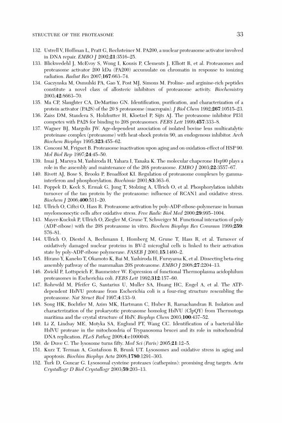
STRUCTURE OF THE PROTEASOME 33
132. Ustrell V, Hoffman L, Pratt G, RechsteinerM. PA200, a nuclear proteasome activator involvedin DNA repair. EMBO J 2002;21:3516–25.
133. Blickwedehl J, McEvoy S, Wong I, Kousis P, Clements J, Elliott R, et al. Proteasomes andproteasome activator 200 kDa (PA200) accumulate on chromatin in response to ionizingradiation. Radiat Res 2007;167:663–74.
134. Gaczynska M, Osmulski PA, Gao Y, Post MJ, Simons M. Proline- and arginine-rich peptidesconstitute a novel class of allosteric inhibitors of proteasome activity. Biochemistry2003;42:8663–70.
135. Ma CP, Slaughter CA, DeMartino GN. Identification, purification, and characterization of aprotein activator (PA28) of the 20 S proteasome (macropain). J Biol Chem 1992;267:10515–23.
136. Zaiss DM, Standera S, Holzhutter H, Kloetzel P, Sijts AJ. The proteasome inhibitor PI31competes with PA28 for binding to 20S proteasomes. FEBS Lett 1999;457:333–8.
137. Wagner BJ, Margolis JW. Age-dependent association of isolated bovine lens multicatalyticproteinase complex (proteasome) with heat-shock protein 90, an endogenous inhibitor. ArchBiochem Biophys 1995;323:455–62.
138. ConconiM, Friguet B. Proteasome inactivation upon aging and on oxidation-effect of HSP 90.Mol Biol Rep 1997;24:45–50.
139. Imai J, Maruya M, Yashiroda H, Yahara I, Tanaka K. The molecular chaperone Hsp90 plays arole in the assembly and maintenance of the 26S proteasome. EMBO J 2003;22:3557–67.
140. Rivett AJ, Bose S, Brooks P, Broadfoot KI. Regulation of proteasome complexes by gamma-interferon and phosphorylation. Biochimie 2001;83:363–6.
141. Poppek D, Keck S, Ermak G, Jung T, Stolzing A, Ullrich O, et al. Phosphorylation inhibitsturnover of the tau protein by the proteasome: influence of RCAN1 and oxidative stress.Biochem J 2006;400:511–20.
142. Ullrich O, Ciftci O, Hass R. Proteasome activation by poly-ADP-ribose-polymerase in humanmyelomonocytic cells after oxidative stress. Free Radic Biol Med 2000;29:995–1004.
143. Mayer-Kuckuk P, Ullrich O, Ziegler M, Grune T, Schweiger M. Functional interaction of poly(ADP-ribose) with the 20S proteasome in vitro. Biochem Biophys Res Commun 1999;259:576–81.
144. Ullrich O, Diestel A, Bechmann I, Homberg M, Grune T, Hass R, et al. Turnover ofoxidatively damaged nuclear proteins in BV-2 microglial cells is linked to their activationstate by poly-ADP-ribose polymerase. FASEB J 2001;15:1460–2.
145. Hirano Y, Kaneko T, Okamoto K, Bai M, Yashiroda H, Furuyama K, et al. Dissecting beta-ringassembly pathway of the mammalian 20S proteasome. EMBO J 2008;27:2204–13.
146. Zwickl P, Lottspeich F, Baumeister W. Expression of functional Thermoplasma acidophilumproteasomes in Escherichia coli. FEBS Lett 1992;312:157–60.
147. Rohrwild M, Pfeifer G, Santarius U, Muller SA, Huang HC, Engel A, et al. The ATP-dependent HslVU protease from Escherichia coli is a four-ring structure resembling theproteasome. Nat Struct Biol 1997;4:133–9.
148. Song HK, Bochtler M, Azim MK, Hartmann C, Huber R, Ramachandran R. Isolation andcharacterization of the prokaryotic proteasome homolog HslVU (ClpQY) from Thermotogamaritima and the crystal structure of HslV. Biophys Chem 2003;100:437–52.
149. Li Z, Lindsay ME, Motyka SA, Englund PT, Wang CC. Identification of a bacterial-likeHslVU protease in the mitochondria of Trypanosoma brucei and its role in mitochondrialDNA replication. PLoS Pathog 2008;4:e1000048.
150. de Duve C. The lysosome turns fifty. Med Sci (Paris) 2005;21:12–5.151. Kurz T, Terman A, Gustafsson B, Brunk UT. Lysosomes and oxidative stress in aging and
apoptosis. Biochim Biophys Acta 2008;1780:1291–303.152. Turk D, Guncar G. Lysosomal cysteine proteases (cathepsins): promising drug targets. Acta
Crystallogr D Biol Crystallogr 2003;59:203–13.

34 JUNG AND GRUNE
153. Turk V, Turk B, Turk D. Lysosomal cysteine proteases: facts and opportunities. EMBO J2001;20:4629–33.
154. Roberts R. Lysosomal cysteine proteases: structure, function and inhibition of cathepsins.Drug News Perspect 2005;18:605–14.
155. Rock KL, GrammC, Rothstein L, Clark K, Stein R, Dick L, et al. Inhibitors of the proteasomeblock the degradation of most cell proteins and the generation of peptides presented onMHCclass I molecules. Cell 1994;78:761–71.
156. Lee DH, Goldberg AL. Proteasome inhibitors: valuable new tools for cell biologists. TrendsCell Biol 1998;8:397–403.
157. Peters JM. Proteasomes: protein degradation machines of the cell. Trends Biochem Sci1994;19:377–82.
158. Scherrer K, Bey F. The prosomes (multicatalytic proteinases; proteasomes) and their rela-tionship to the untranslated messenger ribonucleoproteins, the cytoskeleton, and cell differ-entiation. Prog Nucleic Acid Res Mol Biol 1994;49:1–64.
159. Harris JR. Release of a macromolecular protein component from human erythrocyte ghosts.Biochim Biophys Acta 1968;150:534–7.
160. Harris JR. The isolation and purification of a macromolecular protein component from thehuman erythrocyte ghost. Biochim Biophys Acta 1969;188:31–42.
161. Pacifici RE, Salo DC, Davies KJ. Macroxyproteinase (M.O.P.): a 670 kDa proteinase complexthat degrades oxidatively denatured proteins in red blood cells. Free Radic Biol Med1989;7:521–36.
162. Pacifici RE, Davies KJ. Protein degradation as an index of oxidative stress.Methods Enzymol1990;186:485–502.
163. Salo DC, Pacifici RE, Lin SW, Giulivi C, Davies KJ. Superoxide dismutase undergoesproteolysis and fragmentation following oxidative modification and inactivation. J Biol Chem1990;265:11919–27.
164. Malech HL, Marchesi VT. Hollow cylinder protein in the cytoplasm of human erythrocytes.Biochim Biophys Acta 1981;670:385–92.
165. White MD, Ralston GB. The ‘hollow cylinder’ protein of erythrocyte membranes. BiochimBiophys Acta 1979;554:469–78.
166. Rivett AJ. The multicatalytic proteinase. Multiple proteolytic activities. J Biol Chem1989;264:12215–9.
167. Rivett AJ. The multicatalytic proteinase complex. Revis Biol Celular 1989;20:113–23.168. Rivett AJ. Themulticatalytic proteinase ofmammalian cells.ArchBiochemBiophys 1989;268:1–8.169. Orlowski M. The multicatalytic proteinase complex, a major extralysosomal proteolytic sys-
tem. Biochemistry 1990;29:10289–97.170. Rivett AJ, Sweeney ST. Properties of subunits of the multicatalytic proteinase complex
revealed by the use of subunit-specific antibodies. Biochem J 1991;278(Pt 1):171–7.171. Nothwang HG, Coux O, Bey F, Scherrer K. Prosomes and their multicatalytic proteinase
activity. Eur J Biochem 1992;207:621–30.172. Groll M, Bajorek M, Kohler A, Moroder L, Rubin DM, Huber R, et al. A gated channel into
the proteasome core particle. Nat Struct Biol 2000;7:1062–7.173. Adams GM, Crotchett B, Slaughter CA, DeMartino GN, Gogol EP. Formation of
proteasome-PA700 complexes directly correlates with activation of peptidase activity.Biochemistry 1998;37:12927–32.
174. GlickmanMH, Rubin DM, Fried VA, Finley D. The regulatory particle of the Saccharomycescerevisiae proteasome. Mol Cell Biol 1998;18:3149–62.
175. Liu CW, CorboyMJ, DeMartino GN, Thomas PJ. Endoproteolytic activity of the proteasome.Science 2003;299:408–11.
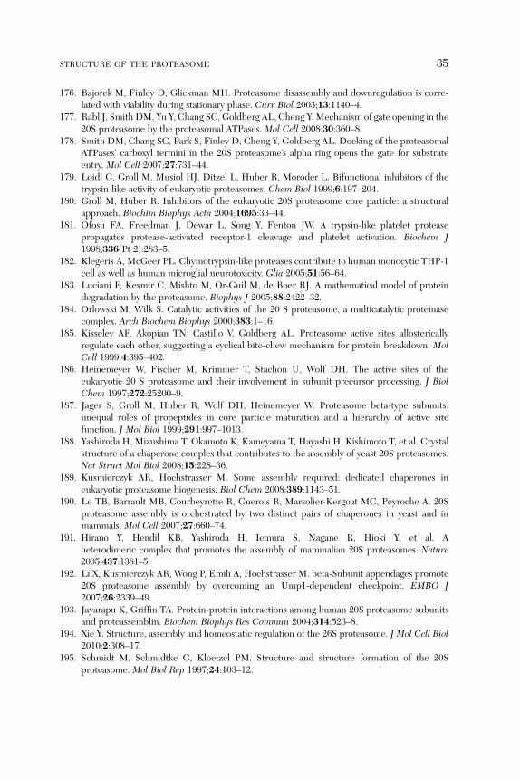
STRUCTURE OF THE PROTEASOME 35
176. Bajorek M, Finley D, Glickman MH. Proteasome disassembly and downregulation is corre-lated with viability during stationary phase. Curr Biol 2003;13:1140–4.
177. Rabl J, Smith DM, Yu Y, Chang SC, Goldberg AL, Cheng Y.Mechanism of gate opening in the20S proteasome by the proteasomal ATPases. Mol Cell 2008;30:360–8.
178. Smith DM, Chang SC, Park S, Finley D, Cheng Y, Goldberg AL. Docking of the proteasomalATPases’ carboxyl termini in the 20S proteasome’s alpha ring opens the gate for substrateentry. Mol Cell 2007;27:731–44.
179. Loidl G, Groll M, Musiol HJ, Ditzel L, Huber R, Moroder L. Bifunctional inhibitors of thetrypsin-like activity of eukaryotic proteasomes. Chem Biol 1999;6:197–204.
180. Groll M, Huber R. Inhibitors of the eukaryotic 20S proteasome core particle: a structuralapproach. Biochim Biophys Acta 2004;1695:33–44.
181. Ofosu FA, Freedman J, Dewar L, Song Y, Fenton JW. A trypsin-like platelet proteasepropagates protease-activated receptor-1 cleavage and platelet activation. Biochem J1998;336(Pt 2):283–5.
182. Klegeris A, McGeer PL. Chymotrypsin-like proteases contribute to human monocytic THP-1cell as well as human microglial neurotoxicity. Glia 2005;51:56–64.
183. Luciani F, Kesmir C, Mishto M, Or-Guil M, de Boer RJ. A mathematical model of proteindegradation by the proteasome. Biophys J 2005;88:2422–32.
184. Orlowski M, Wilk S. Catalytic activities of the 20 S proteasome, a multicatalytic proteinasecomplex. Arch Biochem Biophys 2000;383:1–16.
185. Kisselev AF, Akopian TN, Castillo V, Goldberg AL. Proteasome active sites allostericallyregulate each other, suggesting a cyclical bite-chew mechanism for protein breakdown. MolCell 1999;4:395–402.
186. Heinemeyer W, Fischer M, Krimmer T, Stachon U, Wolf DH. The active sites of theeukaryotic 20 S proteasome and their involvement in subunit precursor processing. J BiolChem 1997;272:25200–9.
187. Jager S, Groll M, Huber R, Wolf DH, Heinemeyer W. Proteasome beta-type subunits:unequal roles of propeptides in core particle maturation and a hierarchy of active sitefunction. J Mol Biol 1999;291:997–1013.
188. Yashiroda H, Mizushima T, Okamoto K, Kameyama T, Hayashi H, Kishimoto T, et al. Crystalstructure of a chaperone complex that contributes to the assembly of yeast 20S proteasomes.Nat Struct Mol Biol 2008;15:228–36.
189. Kusmierczyk AR, Hochstrasser M. Some assembly required: dedicated chaperones ineukaryotic proteasome biogenesis. Biol Chem 2008;389:1143–51.
190. Le TB, Barrault MB, Courbeyrette R, Guerois R, Marsolier-Kergoat MC, Peyroche A. 20Sproteasome assembly is orchestrated by two distinct pairs of chaperones in yeast and inmammals. Mol Cell 2007;27:660–74.
191. Hirano Y, Hendil KB, Yashiroda H, Iemura S, Nagane R, Hioki Y, et al. Aheterodimeric complex that promotes the assembly of mammalian 20S proteasomes. Nature2005;437:1381–5.
192. Li X, Kusmierczyk AR, Wong P, Emili A, Hochstrasser M. beta-Subunit appendages promote20S proteasome assembly by overcoming an Ump1-dependent checkpoint. EMBO J2007;26:2339–49.
193. Jayarapu K, Griffin TA. Protein-protein interactions among human 20S proteasome subunitsand proteassemblin. Biochem Biophys Res Commun 2004;314:523–8.
194. Xie Y. Structure, assembly and homeostatic regulation of the 26S proteasome. J Mol Cell Biol2010;2:308–17.
195. Schmidt M, Schmidtke G, Kloetzel PM. Structure and structure formation of the 20Sproteasome. Mol Biol Rep 1997;24:103–12.

36 JUNG AND GRUNE
196. McIntyre J, Baranowska H, Skoneczna A, Halas A, Sledziewska-Gojska E. The spectrum ofspontaneousmutations caused by deficiency in proteasomematurase Ump1 in Saccharomycescerevisiae. Curr Genet 2007;52:221–8.
197. Chen Q, Thorpe J, Dohmen JR, Li F, Keller JN. Ump1 extends yeast lifespan and enhancesviability during oxidative stress: central role for the proteasome? Free Radic Biol Med2006;40:120–6.
198. Griffin TA, Slack JP, McCluskey TS, Monaco JJ, Colbert RA. Identification of proteassemblin,a mammalian homologue of the yeast protein, Ump1p, that is required for normal proteasomeassembly. Mol Cell Biol Res Commun 2000;3:212–7.
199. Fricke B, Heink S, Steffen J, Kloetzel PM, Kruger E. The proteasome maturation proteinPOMP facilitates major steps of 20S proteasome formation at the endoplasmic reticulum.EMBO Rep 2007;8:1170–5.
200. Chondrogianni N, Gonos ES. Overexpression of hUMP1/POMP proteasome accessory pro-tein enhances proteasome-mediated antioxidant defence. Exp Gerontol 2007;42:899–903.
201. Hoefer MM, Boneberg EM, Grotegut S, Kusch J, Illges H. Possible tetramerisation of theproteasome maturation factor POMP/proteassemblin/hUmp1 and its subcellular localisation.Int J Biol Macromol 2006;38:259–67.
202. Marques AJ, Glanemann C, Ramos PC, Dohmen RJ. The C-terminal extension of the beta7subunit and activator complexes stabilize nascent 20 S proteasomes and promote theirmaturation. J Biol Chem 2007;282:34869–76.
203. Schmidtke G, Schmidt M, Kloetzel PM. Maturation of mammalian 20 S proteasome: purifi-cation and characterization of 13 S and 16 S proteasome precursor complexes. J Mol Biol1997;268:95–106.
204. Gallastegui N, Groll M. The 26S proteasome: assembly and function of a destructive machine.Trends Biochem Sci 2010;35:634–42.
205. Holzhutter HG, Frommel C, Kloetzel PM. A theoretical approach towards the identificationof cleavage-determining amino acid motifs of the 20 S proteasome. J Mol Biol 1999;286:1251–65.
206. Ginodi I, Vider-Shalit T, Tsaban L, Louzoun Y. Precise score for the prediction of peptidescleaved by the proteasome. Bioinformatics 2008;24:477–83.
207. DeMartino GN, Moomaw CR, Zagnitko OP, Proske RJ, Chu-Ping M, Afendis SJ, et al.PA700, an ATP-dependent activator of the 20 S proteasome, is an ATPase containing multiplemembers of a nucleotide-binding protein family. J Biol Chem 1994;269:20878–84.
208. Lam YA, DeMartino GN, Pickart CM, Cohen RE. Specificity of the ubiquitin isopeptidase inthe PA700 regulatory complex of 26 S proteasomes. J Biol Chem 1997;272:28438–46.
209. Strickland E, Hakala K, Thomas PJ, DeMartino GN. Recognition of misfolding proteinsby PA700, the regulatory subcomplex of the 26 S proteasome. J Biol Chem2000;275:5565–72.
210. Liu CW, Millen L, Roman TB, Xiong H, Gilbert HF, Noiva R, et al. Conformationalremodeling of proteasomal substrates by PA700, the 19 S regulatory complex of the 26 Sproteasome. J Biol Chem 2002;277:26815–20.
211. Tanaka K. The proteasome: overview of structure and functions. Proc Jpn Acad Ser B PhysBiol Sci 2009;85:12–36.
212. Glickman MH, Ciechanover A. The ubiquitin-proteasome proteolytic pathway: destructionfor the sake of construction. Physiol Rev 2002;82:373–428.
213. Chen C, Huang C, Chen S, Liang J, Lin W, Ke G, et al. Subunit-subunit interactions in thehuman 26S proteasome. Proteomics 2008;8:508–20.
214. Ortiz-Navarrete V, Seelig A, GernoldM, Frentzel S, Kloetzel PM, Hammerling GJ. Subunit ofthe ‘20S’ proteasome (multicatalytic proteinase) encoded by the major histocompatibilitycomplex. Nature 1991;353:662–4.

STRUCTURE OF THE PROTEASOME 37
215. Aki M, Shimbara N, Takashina M, Akiyama K, Kagawa S, Tamura T, et al. Interferon-gammainduces different subunit organizations and functional diversity of proteasomes. J Biochem1994;115:257–69.
216. Groettrup M, Khan S, Schwarz K, Schmidtke G. Interferon-gamma inducible exchanges of20S proteasome active site subunits: why? Biochimie 2001;83:367–72.
217. Piccinini M, Mostert M, Croce S, Baldovino S, Papotti M, Rinaudo MT. Interferon-gamma-inducible subunits are incorporated in human brain 20S proteasome. J Neuroimmunol2003;135:135–40.
218. Stratford FL, Chondrogianni N, Trougakos IP, Gonos ES, Rivett AJ. Proteasome response tointerferon-gamma is altered in senescent human fibroblasts. FEBS Lett 2006;580:3989–94.
219. Stohwasser R, Giesebrecht J, Kraft R, Muller EC, Hausler KG, Kettenmann H, et al.Biochemical analysis of proteasomes frommouse microglia: induction of immunoproteasomesby interferon-gamma and lipopolysaccharide. Glia 2000;29:355–65.
220. Kloetzel PM. Antigen processing by the proteasome. Nat Rev Mol Cell Biol 2001;2:179–87.221. Kruger E, Kuckelkorn U, Sijts A, Kloetzel PM. The components of the proteasome system and
their role inMHC class I antigen processing.Rev Physiol Biochem Pharmacol 2003;148:81–104.222. Llovera M, Garcia-Martinez C, Agell N, Lopez-Soriano FJ, Argiles JM. TNF can directly
induce the expression of ubiquitin-dependent proteolytic system in rat soleus muscles.Biochem Biophys Res Commun 1997;230:238–41.
223. Lin SY, ChenWY, Lee FY, Huang CJ, Sheu WH. Activation of ubiquitin-proteasome pathwayis involved in skeletal muscle wasting in a rat model with biliary cirrhosis: potential role ofTNF-alpha. Am J Physiol Endocrinol Metab 2005;288:E493–501.
224. Nelson JE, Loukissa A, Altschuller-Felberg C, Monaco JJ, Fallon JT, Cardozo C. Up-regulationof the proteasome subunit LMP7 in tissues of endotoxemic rats. J Lab Clin Med 2000;135:324–31.
225. Driscoll J, Brown MG, Finley D, Monaco JJ. MHC-linked LMP gene products specificallyalter peptidase activities of the proteasome. Nature 1993;365:262–4.
226. Glynne R, Powis SH, Beck S, Kelly A, Kerr LA, Trowsdale J. A proteasome-related genebetween the two ABC transporter loci in the class II region of the human MHC. Nature1991;353:357–60.
227. Cerundolo V, Kelly A, Elliott T, Trowsdale J, Townsend A. Genes encoded in the majorhistocompatibility complex affecting the generation of peptides for TAP transport. Eur JImmunol 1995;25:554–62.
228. Yewdell JW. Immunoproteasomes: regulating the regulator. Proc Natl Acad Sci USA2005;102:9089–90.
229. Heink S, Ludwig D, Kloetzel PM, Kruger E. IFN-gamma-induced immune adaptation of theproteasome system is an accelerated and transient response. Proc Natl Acad Sci USA2005;102:9241–6.
230. Tanaka K, Ichihara A. Half-life of proteasomes (multiprotease complexes) in rat liver. BiochemBiophys Res Commun 1989;159:1309–15.
231. Tanaka K, Ichihara A. Autodegradation of rat liver proteasomes (large multicatalytic protein-ase complexes). Biochem Biophys Res Commun 1989;158:548–54.
232. Ichihara A, Tanaka K. Roles of proteasomes in cell growth. Mol Biol Rep 1995;21:49–52.233. Witt E, Zantopf D, Schmidt M, Kraft R, Kloetzel PM, Kruger E. Characterisation of the newly
identified human Ump1 homologue POMP and analysis of LMP7(beta 5i) incorporation into20 S proteasomes. J Mol Biol 2000;301:1–9.
234. Mishto M, Santoro A, Bellavista E, Sessions R, Textoris-Taube K, Dal PF, et al. A structuralmodel of 20S immunoproteasomes: effect of LMP2 codon 60 polymorphism on expression,activity, intracellular localisation and insight into the regulatory mechanisms. Biol Chem2006;387:417–29.

38 JUNG AND GRUNE
235. Murata S, Sasaki K, Kishimoto T, Niwa S, Hayashi H, Takahama Y, et al. Regulation of CD8þ
T cell development by thymus-specific proteasomes. Science 2007;316:1349–53.236. Murata S, Takahama Y, Tanaka K. Thymoproteasome: probable role in generating positively
selecting peptides. Curr Opin Immunol 2008;20:192–6.237. Nitta T, Murata S, Sasaki K, Fujii H, Ripen AM, Ishimaru N, et al. Thymoproteasome shapes
immunocompetent repertoire of CD8þ T cells. Immunity 2010;32:29–40.238. Sutoh Y, KondoM, Ohta Y, Ota T, Tomaru U, Flajnik MF, et al. Comparative genomic analysis
of the proteasome beta5t subunit gene: implications for the origin and evolution of thymo-proteasomes. Immunogenetics 2012;64:49–58.
239. Fruh K, Yang Y. Antigen presentation by MHC class I and its regulation by interferon gamma.Curr Opin Immunol 1999;11:76–81.
240. York IA, Rock KL. Antigen processing and presentation by the class I major histocompatibilitycomplex. Annu Rev Immunol 1996;14:369–96.
241. Pamer E, Cresswell P. Mechanisms of MHC class I—restricted antigen processing. Annu RevImmunol 1998;16:323–58.
242. Young AC, Nathenson SG, Sacchettini JC. Structural studies of class I major histocompati-bility complex proteins: insights into antigen presentation. FASEB J 1995;9:26–36.
243. Bevan MJ. Immunology. The cutting edge of T cell selection. Science 2007;316:1291–2.244. Song X, Mott JD, von KJ, Pramanik B, Slaughter CA, et al. A model for the quaternary
structure of the proteasome activator PA28. J Biol Chem 1996;271:26410–7.245. Ahn JY, Tanahashi N, Akiyama K, HisamatsuH, Noda C, Tanaka K, et al. Primary structures of
two homologous subunits of PA28, a gamma-interferon-inducible protein activator of the 20Sproteasome. FEBS Lett 1995;366:37–42.
246. Zhang Z, Krutchinsky A, Endicott S, Realini C, Rechsteiner M, Standing KG. Proteasomeactivator 11S REG or PA28: recombinant REG alpha/REG beta hetero-oligomers are hepta-mers. Biochemistry 1999;38:5651–8.
247. Rechsteiner M, Realini C, Ustrell V. The proteasome activator 11 S REG (PA28) and class Iantigen presentation. Biochem J 2000;345(Pt 1):1–15.
248. Realini C, Jensen CC, Zhang Z, Johnston SC, Knowlton JR, Hill CP, et al. Characterization ofrecombinant REGalpha, REGbeta, and REGgamma proteasome activators. J Biol Chem1997;272:25483–92.
249. Li J, Rechsteiner M. Molecular dissection of the 11S REG (PA28) proteasome activators.Biochimie 2001;83:373–83.
250. Di CD. Human erythrocyte contains a factor that stimulates the peptidase activities ofmulticatalytic proteinase complex. Ital J Biochem 1992;41:213–24.
251. Kuehn L, Dahlmann B. Proteasome activator PA28 and its interaction with 20 S proteasomes.Arch Biochem Biophys 1996;329:87–96.
252. Dick TP, Ruppert T, Groettrup M, Kloetzel PM, Kuehn L, Koszinowski UH, et al. Coordi-nated dual cleavages induced by the proteasome regulator PA28 lead to dominant MHCligands. Cell 1996;86:253–62.
253. Soza A, Knuehl C, Groettrup M, Henklein P, Tanaka K, Kloetzel PM. Expression andsubcellular localization of mouse 20S proteasome activator complex PA28. FEBS Lett1997;413:27–34.
254. Wojcik C, Tanaka K, Paweletz N, Naab U, Wilk S. Proteasome activator (PA28) subunits,alpha, beta and gamma (Ki antigen) in NT2 neuronal precursor cells and HeLa S3 cells. Eur JCell Biol 1998;77:151–60.
255. Murata S, Kawahara H, Tohma S, Yamamoto K, Kasahara M, Nabeshima Y, et al. Growthretardation in mice lacking the proteasome activator PA28gamma. J Biol Chem 1999;274:38211–5.
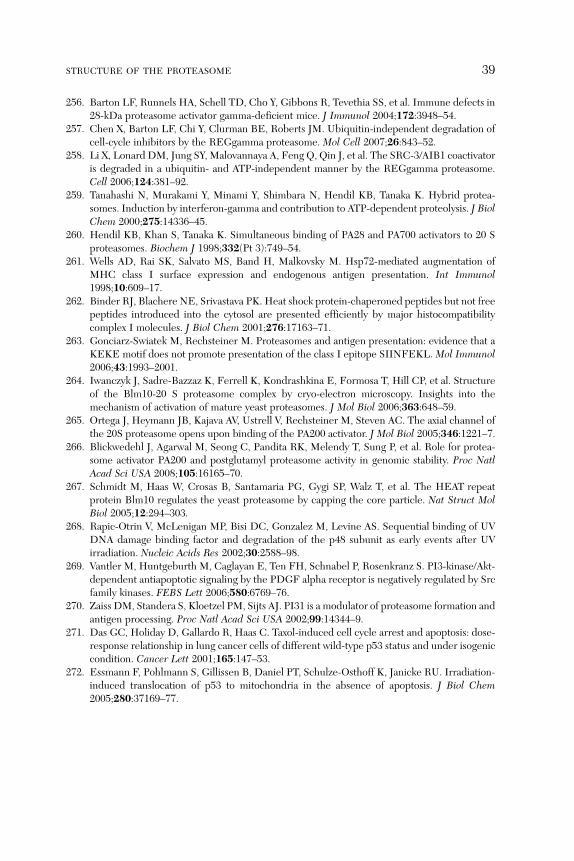
STRUCTURE OF THE PROTEASOME 39
256. Barton LF, Runnels HA, Schell TD, Cho Y, Gibbons R, Tevethia SS, et al. Immune defects in28-kDa proteasome activator gamma-deficient mice. J Immunol 2004;172:3948–54.
257. Chen X, Barton LF, Chi Y, Clurman BE, Roberts JM. Ubiquitin-independent degradation ofcell-cycle inhibitors by the REGgamma proteasome. Mol Cell 2007;26:843–52.
258. Li X, Lonard DM, Jung SY, Malovannaya A, Feng Q, Qin J, et al. The SRC-3/AIB1 coactivatoris degraded in a ubiquitin- and ATP-independent manner by the REGgamma proteasome.Cell 2006;124:381–92.
259. Tanahashi N, Murakami Y, Minami Y, Shimbara N, Hendil KB, Tanaka K. Hybrid protea-somes. Induction by interferon-gamma and contribution to ATP-dependent proteolysis. J BiolChem 2000;275:14336–45.
260. Hendil KB, Khan S, Tanaka K. Simultaneous binding of PA28 and PA700 activators to 20 Sproteasomes. Biochem J 1998;332(Pt 3):749–54.
261. Wells AD, Rai SK, Salvato MS, Band H, Malkovsky M. Hsp72-mediated augmentation ofMHC class I surface expression and endogenous antigen presentation. Int Immunol1998;10:609–17.
262. Binder RJ, Blachere NE, Srivastava PK. Heat shock protein-chaperoned peptides but not freepeptides introduced into the cytosol are presented efficiently by major histocompatibilitycomplex I molecules. J Biol Chem 2001;276:17163–71.
263. Gonciarz-Swiatek M, Rechsteiner M. Proteasomes and antigen presentation: evidence that aKEKE motif does not promote presentation of the class I epitope SIINFEKL. Mol Immunol2006;43:1993–2001.
264. Iwanczyk J, Sadre-Bazzaz K, Ferrell K, Kondrashkina E, Formosa T, Hill CP, et al. Structureof the Blm10-20 S proteasome complex by cryo-electron microscopy. Insights into themechanism of activation of mature yeast proteasomes. J Mol Biol 2006;363:648–59.
265. Ortega J, Heymann JB, Kajava AV, Ustrell V, Rechsteiner M, Steven AC. The axial channel ofthe 20S proteasome opens upon binding of the PA200 activator. J Mol Biol 2005;346:1221–7.
266. Blickwedehl J, Agarwal M, Seong C, Pandita RK, Melendy T, Sung P, et al. Role for protea-some activator PA200 and postglutamyl proteasome activity in genomic stability. Proc NatlAcad Sci USA 2008;105:16165–70.
267. Schmidt M, Haas W, Crosas B, Santamaria PG, Gygi SP, Walz T, et al. The HEAT repeatprotein Blm10 regulates the yeast proteasome by capping the core particle. Nat Struct MolBiol 2005;12:294–303.
268. Rapic-Otrin V, McLenigan MP, Bisi DC, Gonzalez M, Levine AS. Sequential binding of UVDNA damage binding factor and degradation of the p48 subunit as early events after UVirradiation. Nucleic Acids Res 2002;30:2588–98.
269. Vantler M, Huntgeburth M, Caglayan E, Ten FH, Schnabel P, Rosenkranz S. PI3-kinase/Akt-dependent antiapoptotic signaling by the PDGF alpha receptor is negatively regulated by Srcfamily kinases. FEBS Lett 2006;580:6769–76.
270. Zaiss DM, Standera S, Kloetzel PM, Sijts AJ. PI31 is a modulator of proteasome formation andantigen processing. Proc Natl Acad Sci USA 2002;99:14344–9.
271. Das GC, Holiday D, Gallardo R, Haas C. Taxol-induced cell cycle arrest and apoptosis: dose-response relationship in lung cancer cells of different wild-type p53 status and under isogeniccondition. Cancer Lett 2001;165:147–53.
272. Essmann F, Pohlmann S, Gillissen B, Daniel PT, Schulze-Osthoff K, Janicke RU. Irradiation-induced translocation of p53 to mitochondria in the absence of apoptosis. J Biol Chem2005;280:37169–77.