Structural study of ExsA, the regulator of Type III … · iii structure of ExsD. We surprisingly...
53
Structural study of ExsA, the regulator of Type III Secretion System of Pseudomonas aeruginosa Yi Xiao Master of Science in Biological Sciences Florian D. Schubot Ann M. Stevens David R. Bevan Bingyu Zhao April 25, 2013 Blacksburg, Virginia, USA Keywords: Pseudomonas aeruginosa, Type III secretion system, ExsA, structure, crystallization, ExsD, ExsACDE pathway Copyright 2013, Yi Xiao
Transcript of Structural study of ExsA, the regulator of Type III … · iii structure of ExsD. We surprisingly...
Structural study of ExsA, the regulator of Type III Secretion
System of Pseudomonas aeruginosa
Yi Xiao
Master of Science
in
Biological Sciences
Florian D. Schubot
Ann M. Stevens
David R. Bevan
Bingyu Zhao
April 25, 2013
Blacksburg, Virginia, USA
Keywords: Pseudomonas aeruginosa, Type III secretion system, ExsA, structure,
crystallization, ExsD, ExsACDE pathway
Copyright 2013, Yi Xiao

Structural study of ExsA, the regulator of Type III Secretion
System of Pseudomonas aeruginosa
Yi Xiao
Abstract
The Type III secretion system (T3SS) of Pseudomonas aeruginosa uses a needle-like
protein apparatus to detect eukaryotic host cells and translocate effectors directly into the
host cell. The effectors are also known as cytotoxins, which cause disruption of a series
of signaling events in the host cell, facilitating the infection by P. aeruginosa. As the
T3SS is antigenic and the expression of T3SS is energy-consuming, it is highly regulated
where several regulatory proteins interact with each other and control the expression of
T3SS genes. Among these proteins, ExsA, the master regulator of T3SS in P. aeruginosa,
is of great importance as it is a transcriptional activator that activates the expression of all
T3SS genes. Also, as ExsA belongs to the AraC protein family which only exists in
bacteria and fungi, it makes an excellent potential target for drugs against P. aeruginosa
related infections. With a combination of molecular biology tools and structural biology
methods, we solved the N-terminal domain structure of the ExsA protein in P.
aeruginosa. The model of the ExsA N-terminal domain has enriched our knowledge
about ExsA dimerization and can serve as the base for mapping the interaction interfaces
on ExsA and ExsD. Further, we have found two homologues of ExsA by structural
alignment, which share a lot of similarities and have conserved amino acid residues that
are important for ligand binding. The fact that both of these two proteins are regulated by
small ligands rather than proteins also raises the possibility that ExsA may have a second
regulatory mechanism under which ExsA is regulated by a small ligand, which so far has
not been observed or reported by researchers. In order to map the binding site of ExsA on
its anti-activator ExsD, we removed the coiled-coil region (amino acid residue 138-202,
the potential binding site) of ExsD, based on the

iii
structure of ExsD. We surprisingly found that the ExsD variant without the
coiled-coil region readily inhibits ExsA-dependent in vitro transcription. This result rules
out other possibilities and makes us focus on the N-terminus and adjacent regions of
ExsD for the interface with ExsA. Moreover, in order to gain a comprehensive
understanding of the dynamics of the regulation of T3SS in P. aeruginosa, we have
begun to build a mathematical model of the T3SS regulatory pathways. We are
measuring the cellular concentrations of T3SS regulatory proteins with quantitative
molecular biology methods such as quantitative western blot, quantitative PCR and
quantitative mass spectrometry. We have determined the cellular level of ExsA and ExsD
proteins under different physiological conditions, and found that some factors such as
temperature have a significant impact on the levels of ExsA and ExsD. This study has
thus unveiled some unknown features of the T3SS of P. aeruginosa and its related
infections.

iv
Acknowledgements
I am particularly grateful to Dr. Florian Schubot, my advisor, for his encouragements
and guidance. Dr. Schubot expressed a genuine dedication to his students; and that was
the key to my achievements at Virginia Tech.
I would like to thank my committee members, Dr. Ann Stevens, Dr. David Bevan
and Dr. Bingyu Zhao, for their support, encouragements and advices.
I would like to thank Dr. Howard Robinson, for his help with X-ray diffraction
assays and data collection.
I would like to recognize the assistance provided by Xing Jing and Cory Bernhards
on this project, and the other members of the lab for the friendly and professional
atmosphere.
I have a special thought for my wife and my parents. They had waited to see this day.
I am sincerely grateful to my family for their support through every choice I have made
in my life.
v
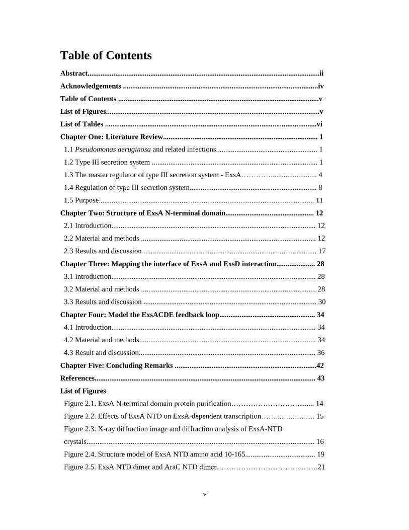
Table of Contents
Abstract..............................................................................................................................ii
Acknowledgements ..........................................................................................................iv
Table of Contents .............................................................................................................v
List of Figures....................................................................................................................v
List of Tables ...................................................................................................................vi
Chapter One: Literature Review.................................................................................... 1
1.1 Pseudomonas aeruginosa and related infections....................................................... 1
1.2 Type III secretion system .......................................................................................... 1
1.3 The master regulator of type III secretion system - ExsA…………......................... 4
1.4 Regulation of type III secretion system..................................................................... 8
1.5 Purpose..................................................................................................................... 11
Chapter Two: Structure of ExsA N-terminal domain................................................ 12
2.1 Introduction............................................................................................................... 12
2.2 Material and methods ............................................................................................... 12
2.3 Results and discussion .............................................................................................. 17
Chapter Three: Mapping the interface of ExsA and ExsD interaction..................... 28
3.1 Introduction............................................................................................................... 28
3.2 Material and methods ............................................................................................... 28
3.3 Results and discussion .............................................................................................. 30
Chapter Four: Model the ExsACDE feedback loop.................................................... 34
4.1 Introduction............................................................................................................... 34
4.2 Material and methods................................................................................................ 34
4.3 Result and discussion................................................................................................ 36
Chapter Five: Concluding Remarks .............................................................................42
References........................................................................................................................ 43
List of Figures
Figure 2.1. ExsA N-terminal domain protein purification………………………......... 14
Figure 2.2. Effects of ExsA NTD on ExsA-dependent transcription……..................... 15
Figure 2.3. X-ray diffraction image and diffraction analysis of ExsA-NTD
crystals............................................................................................................................ 16
Figure 2.4. Structure model of ExsA NTD amino acid 10-165...................................... 19
Figure 2.5. ExsA NTD dimer and AraC NTD dimer……………………………..…….21

vi
Figure 2.6. Superposition of ExsA NTD dimers in the same asymmetric unit……….....22
Figure 2.7. Amino acid residues involved in ExsA dimerization......................................23
Figure 2.8. Superposition of ExsA NTD-ToxT NTD and ExsA NTD-AraC NTD…...... 25
Figure 2.9. Structural-based sequence alignment of ExsA NTD with ToxT and AraC... 27
Figure 3.1. Purification of ExsD 138-202aa and ExsD Δ138-202aa ……….………...…30
Figure 3.2. Titration of ExsA NTD in ExsA dependent transcription.............................. 31
Figure 3.3. Titration of ExsD variants in ExsA-dependent in vitro transcription.............32
Figure 3.4. ExsD trimer and ExsD coiled coil deleted region trimer .............................. 33
Figure 4.1. The exsA exsC and pscB RNA levels in both wild type strain and exsD
knockout strain grown at both 37°C and 30°C, in the presence of EGTA or not…….…37
Figure 4.2. Standard curve created by adding 0µg-30µg purified ExsA protein..............38
Figure 4.3. ExsA concentration in cell. Both wild type (WT) and ΔExsD mutant (-D)
grown at both 37°C and 30°C ………………………………………………………...…39
Figure 4.4. ExsD concentration in cell. Wild type strain grown at both 37°C and 30°C...40
List of Tables
Table 2.1. Data collection and refinement statistics for ExsA N-terminal domain…..….17
1
Chapter One: Literature Review
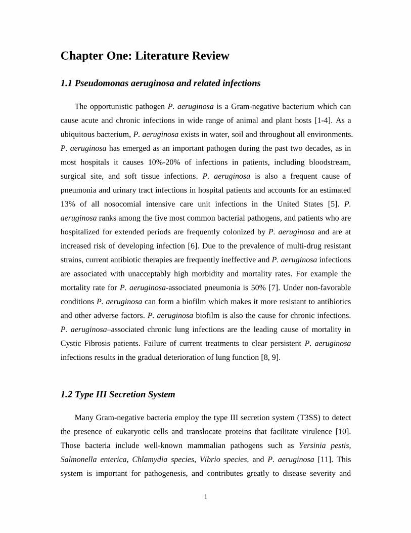
1.1 Pseudomonas aeruginosa and related infections
The opportunistic pathogen P. aeruginosa is a Gram-negative bacterium which can
cause acute and chronic infections in wide range of animal and plant hosts [1-4]. As a
ubiquitous bacterium, P. aeruginosa exists in water, soil and throughout all environments.
P. aeruginosa has emerged as an important pathogen during the past two decades, as in
most hospitals it causes 10%-20% of infections in patients, including bloodstream,
surgical site, and soft tissue infections. P. aeruginosa is also a frequent cause of
pneumonia and urinary tract infections in hospital patients and accounts for an estimated
13% of all nosocomial intensive care unit infections in the United States [5]. P.
aeruginosa ranks among the five most common bacterial pathogens, and patients who are
hospitalized for extended periods are frequently colonized by P. aeruginosa and are at
increased risk of developing infection [6]. Due to the prevalence of multi-drug resistant
strains, current antibiotic therapies are frequently ineffective and P. aeruginosa infections
are associated with unacceptably high morbidity and mortality rates. For example the
mortality rate for P. aeruginosa-associated pneumonia is 50% [7]. Under non-favorable
conditions P. aeruginosa can form a biofilm which makes it more resistant to antibiotics
and other adverse factors. P. aeruginosa biofilm is also the cause for chronic infections.
P. aeruginosa–associated chronic lung infections are the leading cause of mortality in
Cystic Fibrosis patients. Failure of current treatments to clear persistent P. aeruginosa
infections results in the gradual deterioration of lung function [8, 9].
1.2 Type III Secretion System
Many Gram-negative bacteria employ the type III secretion system (T3SS) to detect
the presence of eukaryotic cells and translocate proteins that facilitate virulence [10].
Those bacteria include well-known mammalian pathogens such as Yersinia pestis,
Salmonella enterica, Chlamydia species, Vibrio species, and P. aeruginosa [11]. This
system is important for pathogenesis, and contributes greatly to disease severity and

2
persistence of acute infections, as relative risk of mortality is associated with expression
of T3SS secretory proteins. Disruption of the T3SS leads to a dramatic attenuation as a P.
aeruginosa T3SS knockout strain shows reduced virulence [12-14].
The T3SS of P. aeruginosa consists of 43 coordinately regulated genes that encode
components of the needle-like secretion apparatus, a translocon, and factors that regulate
secretion [15, 16]. T3SS secretion apparatus is sometimes known as the “molecular
syringe”, whose secretion apparatus exports toxins across the bacterial cell envelope. The
translocon of the T3SS is responsible for injecting these toxins into the host cell [10]. The
T3SS can be divided into several parts: I. Proteins that structurally build the T3SS
molecular syringe. II. Proteins (cytotoxins) that are translocated into host cells. III.
Chaperones that help the secretion of their partners. IV. Proteins that regulate the T3SS
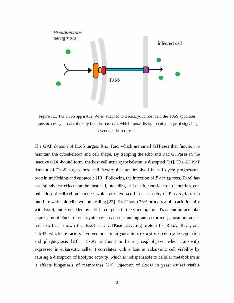
expression and the secretion process [17]. The T3SS apparatus is structurally complex.
More than twenty distinct proteins assemble into a type of molecular syringe: two rings
(also known as the bases) embedded into inner membrane and outer membrane,
respectively; a connector component to link the two rings and a needle component
outside of the membrane to inject potent cytotoxins (effectors) directly into host cells.
(Figure 1.1) Thirty-six genes that are involved in the synthesis and regulation of the T3SS
are encoded from five operons clustered together in P. aeruginosa chromosome. The
structural components of the T3SS apparatus are broadly conserved among the different
pathogens [11].
The nature and function of translocated cytotoxins vary so as to fit the need of
various organisms. The T3SS of P. aeruginosa translocates four known cytotoxins (ExoS,
ExoT, ExoU, and ExoY) into host cells [18]. ExoS has been found to be a bifunctional
protein have two functional domains: the amino-terminal domain, when expressed in host
cells, results in the disruption of actin filaments in a dose-dependent manner, while
expression of the carboxyl-terminal domain of ExoS in eukaryotic cells leads to the
ADP-ribosylation of Ras and uncoupling the Ras-mediated signal transduction [19, 20].
3
Figure 1.1. The T3SS apparatus. When attached to a eukaryotic host cell, the T3SS apparatus
translocates cytotoxins directly into the host cell, which cause disruption of a range of signaling
events in the host cell.
The GAP domain of ExoS targets Rho, Rac, which are small GTPases that function to
maintain the cytoskeleton and cell shape. By trapping the Rho and Rac GTPases in the
inactive GDP-bound form, the host cell actin cytoskeleton is disrupted [21]. The ADPRT
domain of ExoS targets host cell factors that are involved in cell cycle progression,
protein trafficking and apoptosis [19]. Following the infection of P.aeruginosa, ExoS has
several adverse effects on the host cell, including cell death, cytoskeleton disruption, and
reduction of cell-cell adherence, which are involved in the capacity of P. aeruginosa to
interfere with epithelial wound healing [22]. ExoT has a 76% primary amino acid identity
with ExoS, but is encoded by a different gene in the same operon. Transient intracellular
expression of ExoT in eukaryotic cells causes rounding and actin reorganization, and it
has also been shown that ExoT is a GTPase-activating protein for RhoA, Rac1, and
Cdc42, which are factors involved in actin organization, exocytosis, cell cycle regulation
and phagocytosis [23]. ExoU is found to be a phospholipase, when transiently
expressed in eukaryotic cells, it correlates with a loss in eukaryotic cell viability by
causing a disruption of lipolytic activity, which is indispensable to cellular metabolism as
it affects biogenesis of membranes [24]. Injection of ExoU in yeast causes visible
4
membrane damage on different organelles and fragmentation of vacuoles, and injection
of ExoU in mammalian cells results in irreversible cellular membrane damage and rapid
cell death [25]. ExoU is likely to be involved in acute infection of P. aeruginosa, as it is
found that an ExoU knockout strain no longer causes acute cytotoxicity [26]. ExsY has
been found to be an adenylate cyclase which requires activation by calmodulin in the
eukaryotic cells. Infection of CHO cells with ExoY-producing strains of P. aeruginosa
resulted in the intracellular accumulation of cAMP [27]. In sum, the cytotoxins delivered
into host cells by P. aeruginosa through T3SS can trigger a range of events including a
rearrangement of the actin cytoskeleton, disruption of cell signaling and apoptosis,
causing epithelial damage and bacterial dissemination, which are thought to facilitate the
infection.
1.3 The master regulator of Type III Secretion System - ExsA
The AraC-type transcription factor ExsA is the master regulator that controls the
expression of all T3SS-related genes [28, 29]. AraC-family transcriptional regulators
control a range of cellular processes, including the regulation of T3SS expression in a
number of other pathogens [30, 31]. These proteins are characterized by their conserved
AraC domains that interact with specific DNA sequences near the -35 promoter region
[32] and facilitate the recruitment of RNA polymerase to the transcription initiation site
[33-35]. The regulation of AraC-type transcription factors varies greatly. Most of proteins
in the AraC family are regulated by small molecule ligands that bind to the amino
terminal domain of the protein. For instance, AraC is a transcription regulator that
controls the expression of genes required to uptake and catabolize arabinose. The
structure of AraC N-terminal domain has been solved so it is possible to explain the
behavior of AraC protein [36]. The AraC protein consists of an N-terminal regulatory
domain and a C-terminal DNA binding domain. The two domains are functionally
independent as they retain their functions when fused to other proteins. The N-terminal
domain of AraC contains an anti-parallel eight-stranded β barrel which is linked to two
anti-parallel α helices, each about 20 amino acid residues in length. Arabinose is the

5
ligand for AraC that controls its DNA binding behavior [37]. The arabinose binding site
is located in the pocket formed by the β barrel. AraC functions as a dimer. The
dimerization site of AraC protein lies in the α helices [38]. AraC has alternative ways to
bind DNA, depending on whether it is arabinose-bound or not. With arabinose bound to
the N-terminal regulatory domain of AraC, two AraC molecules form a dimer and bind to
the adjacent I1 and I2 sites on the promoter, allowing for the transcription from the pBAD
and pC operons [38]. However, in the absence of arabinose, the AraC dimer adopts a
different conformation and binds to I1 and O2 sites on the DNA, which are widely
separated. As a result, the DNA forms a loop and the transcription from pBAD and pC
operons is inhibited. (Figure 1.2)
Figure 1.2. A. in the absence of arabinose, AraC dimer represses transcription by forming a DNA
loop. B. in the presence of arabinose, the AraC dimer activates transcription.
Interestingly, ExsA and several other regulators of T3SSs constitute a distinct
sub-family that is regulated through protein-protein interactions. Other examples include
the functional ortholog of ExsA in V. parahaemolyticus, InvF in S. enterica, and MixeE
in S. flexineri [39, 40]. However, this does not exclude the possibility that proteins in this
sub-family can also associate with carbohydrates like AraC.
ExsA constitutes a particularly attractive target for the development of novel
therapeutic options because it belongs to the AraC family of proteins, which is only
represented in bacteria and fungi but not in higher eurkaryotes. Drugs designed to target

6
ExsA are supposed to repress the virulence of P. aeruginosa, and may have no side
effects on normal functions of other factors in human cell. Thus, a detailed understanding
of ExsA’s structure, function and regulation will form the foundation for future drug
development efforts.
ExsA protein consists of an ~100 amino acid carboxy-terminal domain (CTD) and a
~200 amino acid amino-terminal domain (NTD). The two domains are connected by a
flexible linker. The CTD of ExsA contains two helix-turn-helix DNA binding motifs and
has been shown to bind a range of T3SS gene promoters and recruit the RNA polymerase
component σ70
to initiate transcription [41]. The NTD of ExsA is known to have the
functions of oligomerization and ligand binding, which is similar to the functions of AraC
N-terminal domain. ExsA controls T3SS genes by directly binding to the promoters and
activating the transcription of all T3SS genes. All ExsA-dependent promoters contain
hexamers that match the consensus -35 and -10 region of σ70
-dependent promoters.
Compared to the normal 17 bp spacing between the -35 and -10 regions, it is about 21~22
bp for ExsA-dependent promoters, and the spacing is critical for ExsA-dependent
transcription. Two ExsA binding sites are upstream of the T3SS genes. An alignment of
ExsA-dependent promoters revealed that the ExsA consensus binding site is located
around the conserved guanine and cytosine at -47 and -45 positions [42]. Further
determinants for ExsA binding include a conserved adenine-rich region centered at the
-51 position and several highly conserved nucleotides around the -35 region. The data
suggest that there are two distinct ExsA binding sites, one overlapping the -35 region and
the other binding site at the adenine-rich position. This finding is confirmed by the
nucleotide substitution assay which shows a significant decrease of ExsA-dependent
transcription when the nucleotides in the -35 region and -55 region are replaced [40].

7
Figure 1.3. The ExsA-DNA binding sites on ExsA-dependent promoters. The binding site 1
overlaps with the -35 consensus and the binding site 2 overlaps with the adenine-rich region
upstream of binding site 1.
The ExsA CTD alone can bind ExsA-dependent promoters, shown in the
electrophoretic mobility shift assay (EMSA), but the affinity is about five-fold lower than
ExsA total protein. In another nucleotide substitution assay, ExsA was shown to bind
preferentially to the -35 region, as the substitution assay shows that nucleotide
substitutions in the -55 region do not affect the ExsA-DNA binding while substitutions in
-35 region almost completely eliminate the binding [42]. By further quantification of the
shifts in EMSA, it has been found that ExsA preferentially binds to the first binding site
(-35 region), then it recruits another ExsA to the the second binding site (-55 region).
Although ExsA CTD can bind to the the first binding site, it fails to recruit another ExsA,
as it does not have the NTD for ExsA-ExsA interaction. In the T3SS gene expression
assay, both ExsA and ExsA CTD can activate the transcription of the reporter gene, while
ExsA NTD cannot as it lacks the DNA binding domain [43]. Thus, the current model of
ExsA-dependent transcription activation is: when T3SS is induced, one ExsA binds to the
first binding site (-35 region) of the promoter, dimerizes with another ExsA and recruits it
to the second binding site (-55 adenine rich region) of the promoter. The ExsA dimer then
recruits RNA polymerase to the promoter to initialize transcription. ExsA dimerization
leads to a higher affinity for σ70
but is not required, since an ExsA monomer alone can
also activate transcription of ExsA-dependent genes albeit at a lower efficiency [43].

8
1.4 Regulation of Type III Secretion System
The expression of the T3SS is under organized regulation as it is highly
energy-consuming. The transcriptional activator ExsA is regulated through
protein-protein interactions. The most studied signal cascade is the
ExsA-ExsC-ExsD-ExsE (ExsACDE) feedback loop that associates the activation of ExsA
directly with the activation of the T3SS apparatus. The genes that encode the four
regulatory proteins ExsA, ExsD, ExsC and ExsE are distributed over two operons. exsA,
exsC, and exsE are all found in one operon, while exsD is co-expressed with eleven
structural components of the T3SS [44].
ExsD is a 31.4 kD protein that acts as an anti-activator in T3SS regulation. ExsD can
bind and inhibit ExsA [45]. Instead of blocking the DNA binding sites on
ExsA-dependent promoters, the binding of ExsD results in the reduced DNA binding
activity of ExsA, as EMSA results show a significantly reduced DNA binding activity of
ExsA when it is bound to ExsD. Also, in an in vitro transcription assay, adding ExsD
results in direct inhibition of ExsA-dependent transcription. ExsA and ExsD, when
co-purifed from E.coli, are co-eluted from a gel filtration column [46]. Analytical
ultracentrifugation data suggest ExsD forms a 1:1 complex with ExsA to inhibit the
transcriptional activity of ExsA [46]. ExsD can also oligomerize in the cells. The ExsD
trimer was found to be less efficient in inhibitory functions than a monomer [46]. Thus,
there exists equilibrium between ExsD monomer and trimer, which is thought to be
another regulatory strategy. The structure of ExsD was recently determined by our lab.
Surprisingly, ExsD is found to resemble the structure of KorB, a DNA binding protein
that acts as a transcription repressor. This suggests that ExsD may have functions other
than binding to ExsA and inhibiting its transcription activity. However, it remains
unknown what the function of ExsD is as a DNA binding protein [45, 47].
ExsC is a 16 kD protein that acts as an anti-anti-activator in the T3SS regulation.
ExsC can bind ExsD and form a 2:2 complex [48]. Also, ExsC functions as a T3SS
chaperone by forming a 2:1 complex with ExsE, a secreted protein that prevents ExsC

9
from binding to ExsD, when the T3SS is “off” [48]. No other functions have been found
for ExsE.
Figure 1.4. A, B. ExsA, ExsD, ExsC, ExsE’s interactions when host cell is/is not attached. C.
Depiction of the gene arrangement of exsA, exsC, exsD, and exsE in the P. aeruginosa genome.
exsC, exsE, and exsA are co-transcribed, while exsD is the first gene in an operon that also
encodes eleven structural components of the T3SS.
T3SS is normally suppressed under non-permissive conditions, such as high Ca2+
or
not being attached to eukaryotic cells [49]. At this stage, ExsC preferentially binds ExsE,
and ExsD binds ExsA and inhibits ExsA-dependent transcription, thus the expression of
T3SS is “off”. Under inducing conditions (such as low Ca2+
or contact of P. aeruginosa
with eukaryotic cells), the activation of T3SS apparatus results in the translocation of

10
ExsE from P. aeruginosa to host cells, and ExsC is released by ExsE. ExsC then binds
and sequesters ExsD to liberate ExsA, which in turn activates the expression of T3SS
genes [44]. As ExsA also activates the expression of exsC, exsE and exsD, the products
will change the cellular concentration of ExsC, ExsE, and ExsD, which regulate the
ExsA-dependent transcription. In sum, ExsA regulates the T3SS via a feedback loop
together with three other regulatory proteins.
Although the ExsACDE model is widely accepted, there are many details that are not
included into the model. For example, the ExsD trimer is thought not to be able to bind
ExsA so there may be distinct stable steady states between ExsD monomer and ExsD
trimer when P. aeruginosa is induced or not. (Figure 1.5)
Figure 1.5. ExsD trimer (left) and monomer (right).
Another problem with the current ExsACDE model is the lack of quantitative data.
The concentrations of ExsA, ExsD, ExsC, and ExsE in the cell have not been measured
thus there is no mathematical model available for the ExsACDE feedback loop to
describe the existing equilibrium states under inducing or non-inducing conditions. An
experiment has shown that ExsD has a higher affinity for ExsA when both of them are
co-expressed in the cell. When they are purified separately and mixed, the affinity
between ExsA and ExsD is much lower [44]. This result suggests that ExsD may bind to
ExsA during translation. Furthermore, we would also like to know the concentration of
ExsA and ExsD in a cell. As we do not know the turnover rate of ExsA or ExsD for now,

11
we cannot answer the question whether ExsD can facilitate the degradation of ExsA in
cell, in which case ExsD should exist in low concentrations in the cell.
The current ExsACDE model does not consider the effects of other important
parameters which may affect the regulation T3SS. Most in vitro experiments on T3SS
regulation were performed under 30oC. However, based on the result from the in vitro
ExsA-dependent transcription assays, the transcription activity of ExsA is different at
30oC and 37
oC. As P. aeruginosa targets warm-blooded hosts, it is of great importance to
study the regulation of T3SS under 37oC, which is the body temperature for humans. It is
possible that the turnover rate of ExsA may increase when the environmental temperature
goes from 30oC to 37
oC, which may affect the overall landscape of T3SS expression.
1.5 Purpose
The specific aims of this project were to gain a deeper understanding of T3SS
regulation, within which the T3SS master regulator ExsA was the focus of the study.
ExsA N-terminal domain protein was purified and crystallized and its structure was
solved in order to study the function and protein-protein interactions of ExsA. With the
structure of the ExsA N-terminal domain as the basis, we found the dimerization interface
of ExsA. Based on the structure alignments, we found homologous proteins, which
provide information about how ExsA could be regulated (Chapter 2). Also, we
constructed several ExsD variants based on the structure of ExsD and tested them in the
ExsA-dependent in vitro transcription assay, in order to find the ExsA-ExsD interaction
interface (Chapter 3). Finally, the cellular concentrations of ExsA and ExsD were
measured in order to obtain parameters that would help build the mathematical model of
ExsACDE regulatory pathway (Chapter 4).

12
Chapter Two: Structure of the ExsA N-terminal
domain
2.1 Introduction
The experimental (structural and molecular analysis) part of this project was
driven by three specific aims. First, ExsA N-terminal domain (ExsA NTD) protein
was purified and crystallized for X-ray diffraction analysis, upon which the structural
model of ExsA NTD was solved. As the locations of the ExsA-ExsA and ExsA-ExsD
interfaces are not known, understanding the molecular basis for these interactions will
be invaluable for the development of small molecules that mimic the behavior of
ExsD to suppress the virulence of P.aeruginosa. The AraC-type transcription factors
regulate virtually all aspects of bacterial gene expression, but these proteins are
notoriously difficult to crystallize, as a result of their low solubility and temperature
sensitivity. While the overall structure of the DNA-binding domain of ExsA can be
easily predicted, no structural information exists for the regulatory domain. Therefore,
critical questions such as the location of the dimer interface and the identity of
residues involved in regulator binding have been difficult to answer. Further, it is also
unclear how ExsA recruits the RNA polymerase to the promoters. The structure
model of ExsA NTD will help reveal the answers for some of these questions.
2.2 Material and Methods
2.2.1 ExsA N-terminal domain preparation
The gene encoding ExsA with a TEV protease cleavage site upstream of the 5’ end was
amplified from P. aeruginosa genomic DNA and introduced into the pDEST-His-MBP
expression plasmid via Gateway cloning technology (Invitrogen). The recombinant
plasmid expressed residues 2–278 of the wild-type ExsA protein sequence without
modifications. Escherichia coli BL21 (DE3) cells were transformed with the plasmid and
induced with 1mM IPTG at an OD600 of 0.6. The cells were incubated for 12 h at 18oC,
13
harvested by centrifugation and the pellet was stored at -80 oC. Thawed cells were lysed
by sonication in binding buffer (500 mM NaCl, 25 mM imidazole pH7.4, 50 mM
Tris-HCl pH7.4, 2 mM DTT), containing 1 mM of PMSF. The cell lysate was clarified by
centrifugation at 15000g for 1 hour at 4oC and applied onto a 30 ml HisTrap Ni-NTA
column (GE Healthcare) (wash buffer: 150 mM NaCl, 25 mM imidazole pH7.4, 50 mM
Tris-HCl pH 7.4, 2 mM DTT. elution buffer: 150 mM NaCl, 250 mM imidazole pH 7.4,
50 mM Tris-HCl pH 7.4, 2 mM DTT). The eluate was then applied to an anion exchange
column (wash buffer: 50 mM NaCl, 25 mM Tris-HCl pH 7.4, 2 mM DTT elution buffer:
1 M NaCl, 25 mM Tris-HCl pH 7.4). TEV protease was added to cleave the His6 Maltose
binding protein tag. The product ExsA is further purified using a HiTrap Ni-NTA column
(wash buffer: 500 mM NaCl, 25 mM imidazole pH 7.4, 50 mM Tris-HCl pH 7.4, 2 mM
DTT. elution buffer: 500 mM NaCl, 250 mM imidazole pH 7.4, 50 mM Tris-HCl pH 7.4,
2 mM DTT). ExsA full-length protein was digested by Thermolysin (20 µg ml-1
) at
30oC for 1 hour, leaving only the N-terminal domain (verified by mass spectrometry).
The reaction was then quenched by adding 2mM CaCl2. The product was applied onto a
Superdex260 gel-filtration column (GE Healthcare) (buffer: 500 mM NaCl, 25 mM
Tris-HCl pH 7.4, 2mM TCEP). The purified ExsA N-terminal domain protein was
concentrated to 1.5 mg ml-1
. The selenomethionine-substituted ExsA (SeMet ExsA) was
produced using the saturation of the methionine biosynthetic pathway protocol [50] and
subsequently treated in the same manner as the original ExsA to yield ExsA NTD. To
determine whether the purified ExsA NTD was properly folded, it was applied to the
ExsA-dependent in vitro transcription assay as an inhibitor. The ExsA-dependent in vitro
transcription assay is described in Cory Bernhard’s work [46].

14
Figure 2.1. A. ExsA-MBP-His6 recombinant protein and TEV cleavage site. B.
ExsA-MBP-His6 recombinant protein and ExsA & MBP-His6 after TEV cleavage (left
gel). ExsA and MBP-His6 were separated by passing through a Ni2+
column (right gel).
The flow through contained ExsA and the eluate contained His6-MBP. C. After limited
proteolysis, ExsA N-terminal domain (ExsA NTD) was generated and further purified by
passing through a gel filtration column.

15
As only properly folded protein can yield crystals, we wanted to show the purified
ExsA NTD protein is properly folded and functional. A titration of ExsA NTD protein in
the ExsA-dependent in vitro transcription assay is done to show that the ExsA NTD
protein can inhibit ExsA dependent transcription in a dose-dependent manner, which
indicates that the purified ExsA NTD protein competes for the dimerization site of
full-length ExsA, resulting in a reduced transcription activity as the DNA binding motif is
located in the C-terminal domain of ExsA. The result indicates the ExsA NTD protein is
properly folded and functional. (Figure 2.2)
Figure 2.2. A. Titration of ExsA N-ternimal domain in ExsA-dependent in vitro transcription. The
reporter values were based on the density of radio-labeled mRNA transcript bands. B. The
radio-labeled mRNA transcripts.

16
2.2.2 Crystallization and data collection
Crystallization of the ExsA NTD was performed using the hanging-drop
vapor-diffusion method at 25oC. (Figure 2.3) Crystals of ExsA NTD were obtained using
a reservoir solution containing 1.6 M MgSO4, 0.1 M MES pH 6.5 and 0.1 M EGTA. The
crystallization droplet consisted of 3ul protein solution (1.5 mg ml-1
ExsA NTD protein,
500 mM NaCl, 25 mM Tris-HCl pH 7.4, 2 mM TCEP) and 1 µl reservoir solution.
Rod-shaped crystals of ExsA NTD appeared after several days. SeMet ExsA NTD was
crystallized under identical conditions. The crystals were flash-cooled in liquid nitrogen
after soaking in a cryoprotection solution containing 90% reservoir solution and 10% (v/v)
glycerol. X-ray data were collected at beamline X29A (National Synchrotron Light
Source, Brookhaven National Laboratory) using an ADSC Q315 CCD detector.
Figure 2.3. X-ray diffraction image and diffraction
analysis of ExsA-NTD crystals.
2.2.3 Structure determination and refinement
The X-ray diffraction data were processed using the XDS program package. Initial
phases for SeMet ExsA N-terminal domain were obtained by Phaser using the multiple

17
anomalous dispersion (MAD) [51]. Model building was performed using COOT [52],
and Phenix program suite was used for structure solution and refinement [53].
Data-collection statistics
Space group P43212
No. of moledules in asymmetric unit 2
Uni-cell parameters (Å)
a=69.820 b=69.820 c=191.580 α=90.00
β=90.00 γ=90.00
Mosaicity 0.4
Resolution range (Å) 65.82-2.9
Observed reflections 209145
Unique reflections 11232
Average multiplicity 18.6
Completeness(%) 100.00%
Rmerge 0.105
< I/σ(I)> 19.5
Refinement statistics
Resolution range (Å) 65.599-2.9
No. of reflections 11125
R-work 0.2367
R-free 0.2785
R.m.s. deviations
Bond lengths(Å) 0.004
Bond angles( o ) 0.822
Ramachandran plot(%)
Favored 90.27
Allowed 9.4
Outliers 0.34
Table 2.1. Data collection and refinement statistics for ExsA N-terminal domain.
2.3 Results and Discussion
2.3.1 Characterizing of the structure of ExsA N-terminal domain
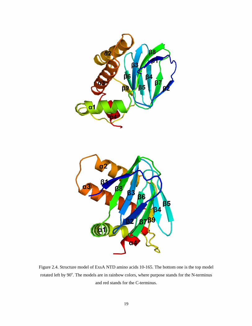
We have solved the 2.9 Å resolution crystal structure of ExsA N-terminal domain.
The crystal contains two ExsA dimers per asymmetric unit. The ExsA N-terminal domain
18
contains an eight-stranded antiparallel β barrel (β1- β8). The length of the β strands varies
but together they form the barrel with an open end. The β barrel is followed by a linker
that has a long α helix (α1) followed by another β strand (β9). β9 is then followed by 3 α
helices (α2 – α4), α1 and α2 are approximately 15 amino acid residues in length, while α4
is shorter and the amino acid residues following α4 are disordered. The three α helices are
packed against the surface of the β barrel. (Figure 2.4)
19
Figure 2.4. Structure model of ExsA NTD amino acids 10-165. The bottom one is the top model
rotated left by 90o. The models are in rainbow colors, where purpose stands for the N-terminus
and red stands for the C-terminus.

20
2.3.2 Characterizing the ExsA dimer
In the crystal there are two ExsA N-terminal domain molecules. Using the 2-fold
symmetry of the crystal the two independent molecules may be used to generate two
slightly distinct dimers that each might represent the biological dimer of ExsA. In both
dimers two α helices (α2 and α3) of one molecule interact with same helices from the
other molecule in reversed orientation, forming a four-helical bundle at the interface.
While the sequence similarity between the regulatory domains of ExsA and AraC is very
low the overall folds are quite similar as discussed below. Interestingly, the arrangement
of the two ExsA NTD dimers closely resembles that observed for a dimer of the
regulatory domain of AraC (PDB code: 2ARC), with the two pairs of α helices acting as
the dimerization interface. (Figure 2.5)

21
Figure 2.5. A. The two ExsA N-terminal domain dimer found in the crystal. B. AraC N-terminal
domain dimer.
22
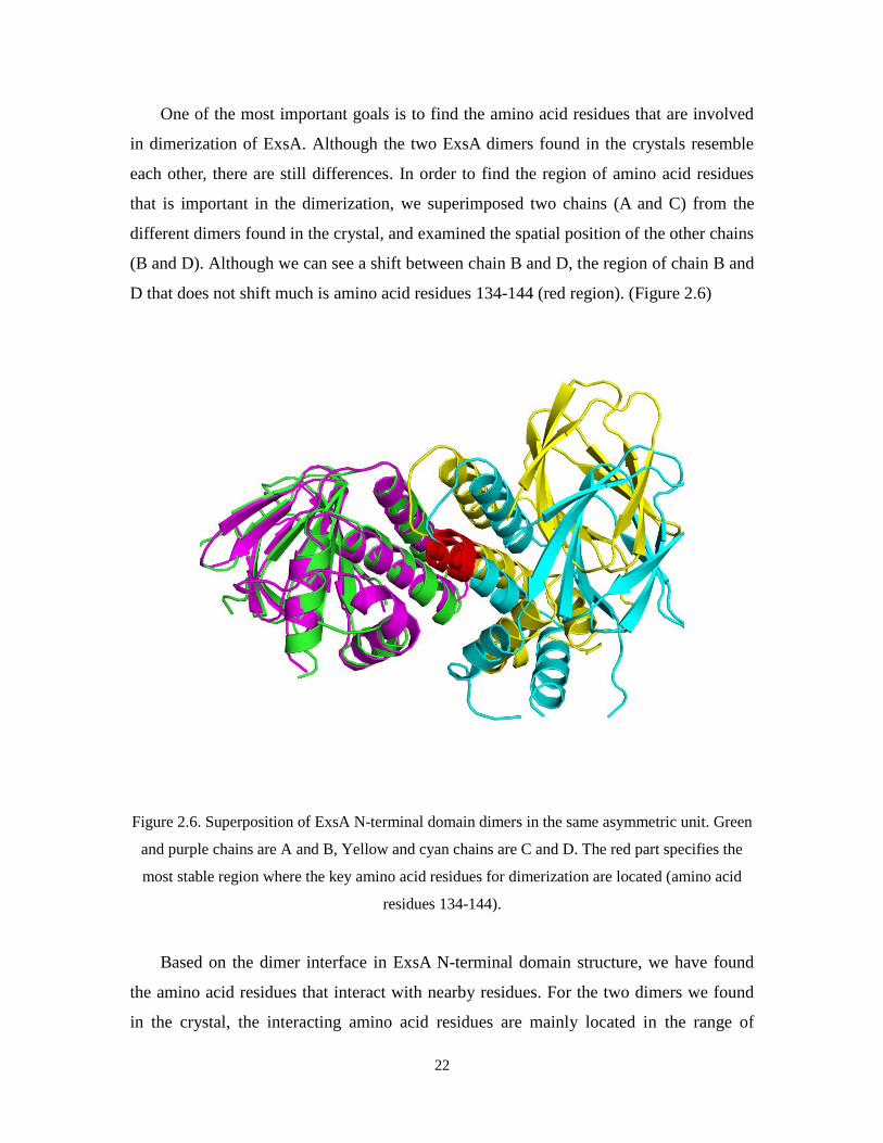
One of the most important goals is to find the amino acid residues that are involved
in dimerization of ExsA. Although the two ExsA dimers found in the crystals resemble
each other, there are still differences. In order to find the region of amino acid residues
that is important in the dimerization, we superimposed two chains (A and C) from the
different dimers found in the crystal, and examined the spatial position of the other chains
(B and D). Although we can see a shift between chain B and D, the region of chain B and
D that does not shift much is amino acid residues 134-144 (red region). (Figure 2.6)
Figure 2.6. Superposition of ExsA N-terminal domain dimers in the same asymmetric unit. Green
and purple chains are A and B, Yellow and cyan chains are C and D. The red part specifies the
most stable region where the key amino acid residues for dimerization are located (amino acid
residues 134-144).
Based on the dimer interface in ExsA N-terminal domain structure, we have found
the amino acid residues that interact with nearby residues. For the two dimers we found
in the crystal, the interacting amino acid residues are mainly located in the range of

23
residues 135-151. The results obtained from two dimers overlap but one dimer has more
amino acid residues involved in dimerization than the other, suggesting the dimerization
of ExsA is dynamic. One interesting finding we have is the R91 on the α1 helix is
involved in the dimerization of ExsA. I.t interacts with the R91 and E143 on the other
molecule of ExsA dimer (Figure 2.7). Our collaborator Timothy Yahr (University of Iowa)
has corroborated this interface demonstrating that several residues at this interface are
indeed critical for ExsA dimerization (personal communication).
Figure 2.7. Amino acid residues involved in ExsA dimerization. A and C are two molecules of
one of the dimers found in the crystal. B and D are two molecule of the other dimer.

24
2.3.3 Characterizing ExsA N-terminal domain homologues
Based on the structure of ExsA NTD, we would also like to know the interface of the
ExsA and ExsD interaction. We expected that the homologues of ExsA might share some
similarities not only in structure by also in function. In order to find structural
homologues of ExsA NTD, we applied structure alignment to the ExsA NTD structure
against structures in the online protein databank [54]. The structure alignment attempts to
find similarities between two structures, based not on the sequence alignment but on the
tertiary structure of the molecules, thus it can establish homology of proteins with low
sequence similarity, where evolutionary relationships between proteins can be hardly
detected by standard sequence alignment techniques. Structural alignment can therefore
be used to imply evolutionary relationships between proteins that share very little
common sequence [55]. The method we used for evaluating the similarity between
proteins is Distance Aligned matrix method (DALI). DALI is a common and popular
structure alignment method that breaks the input structures into hexapeptide fragments
and calculates a distance matrix by evaluating the contact patterns between successive
fragments [56]. Sequence alignment on ExsA does not yield any proteins with high
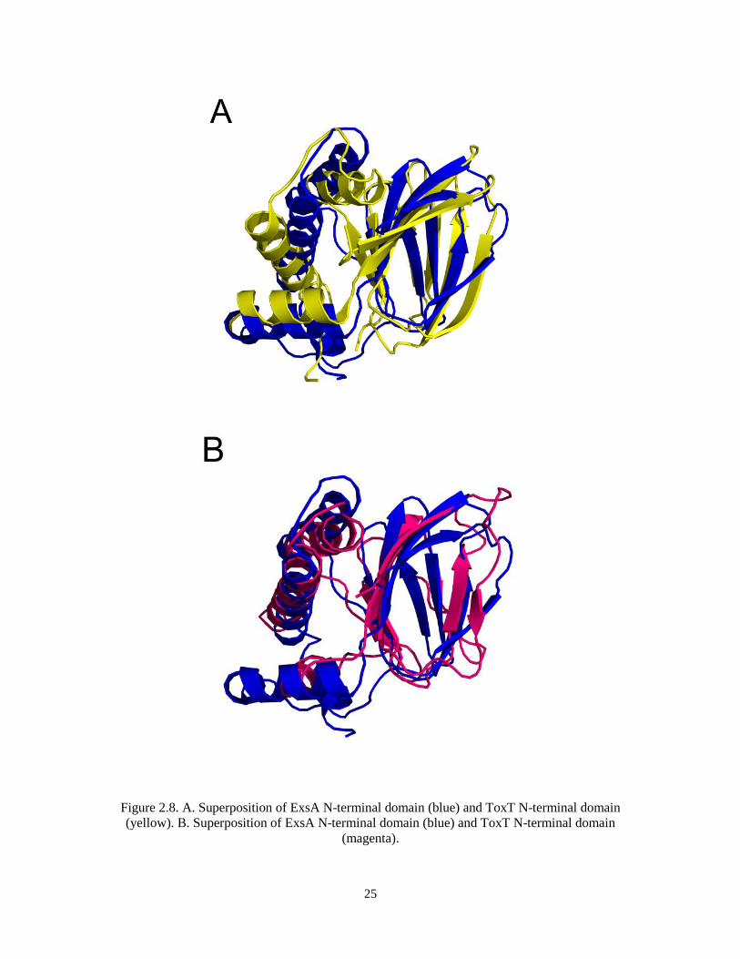
similarity, but structure alignment on ExsA NTD structure on DALI server finds two
similar structures, ToxT (PDB code: 3GBG, r.m.s.d 3.0) and AraC NTD (PDB code:
2ARC, r.m.s.d 3.3). Both ToxT and AraC belong to the AraC family. ToxT is a protein
found in V. cholera, which is a Gram-negative bacterium that can cause an acute
intestinal infection known as cholera. In addition, ToxT regulates two primary virulence
factors of V. cholera [57]. AraC is the regulator of arabinose catabolism in E. coli [58].
(Figure 2.8)
25
Figure 2.8. A. Superposition of ExsA N-terminal domain (blue) and ToxT N-terminal domain
(yellow). B. Superposition of ExsA N-terminal domain (blue) and ToxT N-terminal domain
(magenta).

26
From the structures we find that the N-terminal domain of ExsA resembles that of
ToxT, and they are both similar to the structure of N-terminal domain of AraC. All three
of the proteins contain the anti-parallel β barrel, and the shape and volume of the barrel
are similar to one another. In AraC, the β barrel contains the binding site for arabinose,
and there is an N-terminal arm (amino acid residues 1-10) to “cover” the “opening” of the
barrel which is supposed to stabilize the binding of arabinose via hydrogen bonds. The
N-terminal arm is not found in either ExsA or ToxT. The ToxT structure is found to be
bound to a sixteen-carbon fatty acid cis-palmitoleate, which is found to be a repressor for
ToxT. Y20, F22 and F33 on the N-terminal domain, as well as V261, M259, M269 and
Y266 of the C-terminal domain of ToxT help stabilize the ligand in the pocket [59]. So
far there is no evidence to show that ExsA can bind any small ligands, however, a close
comparison between the binding pocket between AraC and ExsA reveals some conserved
features. The R38, H80, Y82, H93 and W95 in AraC are the residues that form hydrogen
bonds with arabinose directly and indirectly [38], while in ExsA, R25, N27, Y33, W17
and W77 are corresponding residues that are at the similar position in the space, which
may also form hydrogen bonds with a ligand when it is bound (Figure 2.9). As for ToxT,
K28 in ExsA is close to K31 in ToxT, which forms salt bridges with the negatively
charged carboxylate head group of the fatty acid as well as K230 from the C-terminal
domain of ToxT. However, the binding pocket of ExsA is occupied by several bulky side
chains. Thus, it is less likely for ExsA to accommodate a large ligand such as a fatty acid.
Another possibility is that the β barrel pocket is where the interaction between ExsA and
its regulator ExsD occurs. ExsD may bind to the pocket and change the conformation of
ExsA which in turn prevents it from activating transcription. Other than the β barrel, all
three proteins contain two helices that are aligned anti-parallel. These two α helices are
the interface for dimerization of ExsA and AraC with arabinose bound to it, while ToxT
was found to be monomer in the crystal.

27
Figure 2.9. Structural-based sequence alignment of ExsA N-terminal domain with ToxT and AraC.
Amino acid residues marked yellow are the ones involved in ligand binding (potential ligand
binding residues for ExsA).

28
Chapter Three: Mapping the interface of the ExsA and
ExsD interaction
3.1 Introduction
After we solved the structure of the ExsA NTD, we next asked where the interface
the ExsA and ExsD interaction is. There are two potential binding sites on ExsD, one is
the amino terminus of ExsD because we believe it to be overlapping or in the vicinity of
the ExsC binding site. The other is the coiled-coil region of ExsD (amino acid residues
138-202), because such regions are frequently involved in protein-protein interactions.
Having developed an ExsA-specific in vitro transcription assay, we tested a series of
truncated ExsD variants for their ability to interfere with ExsA function. Using the ExsD
structure as a guide, we generated two recombinant ExsD variant proteins, one of which
is the two α helices (amino acid residues 138-202), and the other part is the rest of the
ExsD with the gap of amino acid residues 138-202 bridged by four glycines.
3.2 Material and Methods
3.2.1 Preparation of ExsD coiled-coil region deleted variant
The ExsD coiled-coil deleted region (ExsD Δ138-202aa) gene is constructed via two
rounds of PCRs. In the first PCR reaction, ExsD 1-137aa was amplified with primers
forward: 5’-GTGGAGAACCTGTACTTCCAGGGTATGGAGCAGGAAGAC-3’,
backward: 5’-C TTCGCCAGTGCCGATCCTCCTCCTCCCTGGTCGAGCAGGCT-3’,
ExsD 203-276aa was amplified with primers forward: 5’-CGGGTCAACCTCGGAGGA
GGAGGATCGGCACTGGCG-3’, backward: 5’- GGGGACAACTTTGTACAAGAAAG
TTGCTCATA CTGGCAGAGCTGA-3’ from P. aeruginosa genomic DNA. At the end of
ExsD 1-137aa and the beginning of the ExsD 203-276aa there was a short DNA sequence
encoding 4 continuous glycines. The two products obtained from the first PCR were then
used in the second PCR as templates to amplify the ExsD Δ138-202 with primers forward:
5’-GT GGAGAACCTGTACTTCCAGGGTATGGAGCAGGAAGAC-3’, backward: 5’-
29
GGGGACAACTTTGTACAAGAAAGTTGCTCATACTGGCAGAGCTGA-3’. The
ExsD Δ138-202aa gene was then introduced into pDONR201 plasmid and later
introduced into the pDEST-His-MBP expression plasmid via Gateway cloning technique.
E. coli BL21 (DE3) cells were transformed with the plasmid and induced with 1mM
IPTG at an OD600 of 0.6. The cells were incubated for 12 h at 18oC, harvested by
centrifugation and the pellet was stored at -80 oC. Thawed cells were lysed by sonication
in binding buffer (500 mM NaCl, 25 mM imidazole pH7.4, 50 mM Tris-HCl pH 7.4, 2
mM DTT), containing 50 mM of PMSF. The cell lysate was clarified by centrifugation at
15000g for 1 hour at 4oC and applied onto a HisTrap Ni-NTA column (GE Healthcare)
(wash buffer: 150 mM NaCl, 25 mM imidazole pH 7.4, 50 mM Tris-HCl pH 7.4, 2 mM
DTT. Elution buffer: 150 mM NaCl, 250 mM imidazole pH 7.4, 50 mM Tris-HCl pH 7.4,
2 mM DTT). The coiled coil region of ExsD was found to be unstable and most of it
degraded during purification. However, the coiled-coil deleted region of ExsD was stable.
TEV protease was added into the eluate to cleave the MBP-His6 tag, and the product
ExsD coiled coil deleted protein was further purified using a HiTrap Ni-NTA column
(wash buffer: 150 mM NaCl, 25 mM imidazole pH 7.4, 50 mM Tris-HCl pH 7.4, 2 mM
DTT. elution buffer: 150 mM NaCl, 250 mM imidazole pH 7.4, 50 mM Tris-HCl pH 7.4,
2 mM DTT). (Figure 3.1) The ExsA-dependent in vitro transcription assay is described in
Cory Bernhard’s work [46].

30
Figure 3.1. A. Purification of ExsD 138-202aa. B. Purification of ExsD Δ138-202aa.
3.3 Results and Discussion
3.3.1 Characterizing the inhibitory effect of ExsD Δ138-202aa in
ExsA-dependent in vitro transcription assay
The ExsD Δ138-202aa deleted protein was applied to ExsA-dependent in vitro
transcription assay at 37oC, with full-length ExsD protein as control. The result shows
that ExsD Δ138-202aa inhibits ExsA-dependent transcription in a dose dependent manner.
Surprisingly, ExsD Δ138-202aa has a higher inhibitory effect on ExsA-dependent
transcription than ExsD full-length protein. (Figure 3.2) As indicated by the result, 5
µM of ExsD Δ138-202aa has a similar inhibitory effect of 50 µM ExsD full-length
protein, suggesting that the coiled coil region of ExsD may not have any effect on
ExsD-ExsA interaction.

31
Figure 3.2. Titration of ExsA N-ternimal domain in ExsA dependent transcription
The result is contrary to our expectation, as we thought the coiled-coil region of
ExsD may act as the interface for the ExsD-ExsA association, since coiled coil regions
are normally involved in protein-protein interaction (for example, ExsA dimerization).
However, it indicates the fact that ExsA and ExsC may compete for the binding site on
ExsD, as ExsC is found to bind the first 40 amino acid residues of the N-terminus of
ExsD. In order to test it, ExsD amino acid residues 1-46 (ExsD 1-46aa, purchased peptide)
and ExsD Δ1-20 amino acid residues (ExsD Δ1-20aa, purified ExsD protein digested in
limited proteolysis) were also tested in the ExsA-dependent in vitro transcription assay by.
In Cory Bernhards’ work in our laboratory, ExsD 1-46aa and ExsD Δ1-20aa were shown
to only inhibit ExsA-dependent transcription at a high concentration (>30 µM),
suggesting ExsD 1-46aa and its adjacent region of ExsD are both important for ExsA
inhibition. (Figure 3.3)

32
Figure 3.3. Titration of ExsD variants in ExsA-dependent in vitro transcription. ExsD Δ20: ExsD
1-20aa deleted region. ExsD 1-46: purchased ExsD 1-46aa peptide. ExsD Δ138-202: ExsD
coiled-coil (amino acid residues 138-202) deleted region. ExsD WT: full-length ExsD protein.
Based on the result of the ExsA-dependent in vitro transcription assay, the ExsD
coiled-coil deleted protein has an inhibitory effect on ExsA that is similar to that of ExsD
M59R, which is a mutant that exists as a monomer (determined by analytical gel filtration
assay). We think that the coiled-coil region of ExsD may stabilize the ExsD trimer, as we
know that it is the ExsD monomer that inhibits the ExsA-dependent transcription. Also,
we found that the yield of ExsD Δ138-202aa was lower than that of ExsD full-length
protein, which is probably due to the reduced stability of the variant protein. Now we
have shown that the coiled-coil region of ExsD does not inhibit ExsA. It may have the
function of stabilizing the ExsD trimer, however, it remains unknown whether the
coiled-coil region of ExsD has any other functions. (Figure 3.4)
33
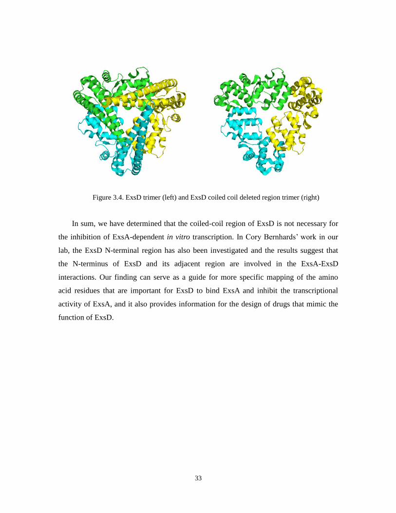
Figure 3.4. ExsD trimer (left) and ExsD coiled coil deleted region trimer (right)
In sum, we have determined that the coiled-coil region of ExsD is not necessary for
the inhibition of ExsA-dependent in vitro transcription. In Cory Bernhards’ work in our
lab, the ExsD N-terminal region has also been investigated and the results suggest that
the N-terminus of ExsD and its adjacent region are involved in the ExsA-ExsD
interactions. Our finding can serve as a guide for more specific mapping of the amino
acid residues that are important for ExsD to bind ExsA and inhibit the transcriptional
activity of ExsA, and it also provides information for the design of drugs that mimic the
function of ExsD.

34
Chapter Four: Model the ExsACDE feedback loop
4.1 Introduction
Our last aim is to build a mathematical model of the ExsACDE regulatory pathway.
It is an energy-consuming process for P. aeruginosa to produce the components of T3SS.
Thus for the most part of the life of P. aeruginosa the T3SS genes are down-regulated
unless it has access to host cells. ExsA activates the transcription of T3SS system
proteins when P. aeruginosa is induced at low Ca2+
condition or attached to a eukaryotic
cell. The knowledge of the levels of transcription and translation of the T3SS regulatory
factors including ExsA, ExsD, ExsC and ExsE under different physiological conditions
will yield important information about how T3SS system regulation work, and a
mathematical model can be built with ordinary differential equations based on the
parameters derived from quantitative molecular biology assays such qPCR and qWestern
blot, which measure the relative/absolute level of RNA and protein in cell. With the
model, we will be able to predict the behaviors of P. aeruginosa virulence under different
circumstances.
4.2 Material and Methods
An initial experiment was done to determine the RNA level of exsA in P. aeruginosa
when cultured at either 37oC or 30
oC, as the in vitro transcription assays done in our lab
indicate there is a difference in the inhibitory effect of ExsD on ExsA at different
temperatures. We also wanted to know the change of exsA RNA level when P. aeruginosa
is cultured under T3SS inducing conditions. As we want to know the regulation of ExsA
on both PexsC and PexsD promoters, we designed primers that amplify exsC, exsA and pscB
genes. P. aeruginosa PAO1 and a PAO1 exsD knockout strain were cultured in LB
overnight. On the second day, 100 µl of overnight culture was added to the TSB
containing 100 mM monosodium glutamate and 1% glycerol. EGTA (2mM) is added to
the culture to create the low Ca2+
condition which stimulates the expression of T3SS
genes. Cells are harvested with the OD ~ 0.8 by centrifugation at 4500g for 1min. Total
35
RNAs are isolated from cells with Trizol reagent followed by a DNase treatment
(Promega RQ1 RNase-free DNase), cDNAs are obtained by reverse transcription (High
Capacity cDNA Reverse Transcription Kits, Applied Biosystems). Relative mRNA levels
of exsA, exsC and pscB are determined by qPCR with rpoD as reference gene with
BIORAD C1000 Thermal Cycler. Primers: exsA forward: 5’-ATGTCGGTCCTGCGGCA
ACTGAGC-3’, backward: 5’-GCGCGGCGAAACCCCATAGACACT-3’. exsC forward:
5’-AGCGGCAGCGTCTGTTGCTGGAG-3’, backward: 5’-GGGTCAGTTGCGCTGCG
AGAATCT-3’. pscB forward: TCGATGCGCAGGTGGTACGAA, backward: TGGATCA
TCTGTTGAGCGGATTGG. rpoD forward: 5’-GGGCGAAGAAGAAATGGTC-3’,
backward: 5’-CAGGTGGCGTAGGTGGAGAA-3’.
The protein level of ExsA and ExsD were determined by quantitative western
blotting. P. aeruginosa PAO1 and PAO1 exsD knockout strain were cultured in LB
overnight. On the second day, 100 µl of overnight culture was added to the Tryptic Soy
Broth containing 100mM monosodium glutamate and 1% glycerol. EGTA (2 mM) is
added to the culture to create the low Ca2+
condition which stimulates the expression of
T3SS genes. Cells are harvested with the OD ~ 0.8 by centrifugation at 4500g for 15min.
For each pellet 1 ml of lysis buffer (150 mM NaCl, 50 mM Tris-HCl pH 8.0, 10 mM
lysozyme) were added and cells were resuspended by pipetting. Cells were incubated on
ice for 10 minutes, followed by sonification with a BRANSON sonifier 450 for 30
seconds (duty cycle: constant, output: 3). Precipitations were cleared by centrifugation at
14000 rpm at 4oC for 30 minutes. Total protein concentration was measured with
Bradford. For quantitative western blot, cell lysate containing 80 mg total protein was
applied to SDS-PAGE. Proteins on the gel were then transferred to an Immuno-Blot
PVDF membrane (BIORAD). The membrane was blocked with 5% non-fat milk in
TBST (150 mM NaCl, 20 mM Tris-HCl pH 8.0, 0.05% Triton X-100) for 30 minutes.
ExsA/ExsD antibody was added to the blocking solution in 1:10000 dilution. After
incubating the membrane overnight, the membrane was washed three times with TBST
(10 minutes each time). Immun-Star Goat Anti-Rabbit (GAR)-HRP secondary antibody
(BIORAD) was added to blocking solution and the membrane was incubated for 2 hours.
After washing three times with TBST, SuperSignal West Pico Chemiluminescent

36
Substrate (Thermo) was added to the membrane and the membrane was developed with
film. Band density was measured with ImageQuant (GE Healthcare).
4.3 Results and Discussion
As expected, exsA, exsC and pscB RNA levels increased significantly when cells
were cultured under T3SS inducing condition at both 30oC and 37
oC for wild type P.
aeruginosa, compared to cells cultured under non-inducing conditions. In the exsD
knockout strain, the RNA levels of exsA, exsC and pscB are also higher than those in the
wild type strain, which agree with the fact that the T3SS is deregulated in an exsD
knockout strain. The RNA levels of exsA, exsC and pscB do not increase much when the
exsD knockout strain is cultured under T3SS inducing condition, indicating that the
ExsACDE regulation is non-functional without ExsD. When cells are grown at 37oC,
exsA, exsC and pscB RNA levels are significantly higher than those in cells grown at
30oC. Interestingly, the RNA levels of exsA, exsC and pscB at 37
oC without induction are
close to the levels of exsA, exsC and pscB RNA at 30oC with induction. This result may
suggest that T3SS expression is activated at 37oC, which is the body temperature of
human beings. (Figure 4.1) For most work on T3SS the experiments are done at 30oC.
But the recent RNA sequencing study on the transcriptome of P. aeruginosa suggests that
the T3SS genes are activated at 37oC, which agrees with our findings [60]. In sum, our
result suggests a different mechanism that regulates the expression of T3SS at 37oC,
when P. aeruginosa enters the host’s body.

37
Figure 4.1. The exsA exsC and pscB RNA levels in both wild type strain (w) and exsD knockout
strain (-D) grown at both 37°C and 30°C, in the presence of EGTA (+E) or not. Amount of
mRNAs is determined by quantitative PCR. The experiment is done in triplicates, with values
normalized to rpoD.
Though mRNA level can represent the transcription rate of genes, it does not
necessarily indicate the protein levels in the cell, as translation and protein turnover rates
are not taken into consideration. Thus we wanted to determine the cellular concentrations
of ExsA and ExsD from cells cultured at either 37°C or 30°C, under either T3SS inducing
condition or not. Quantitative western blotting method was used for protein measurement.
Based on the density of the protein band, the ImageQuant software can determine the
relative amount of protein. Thus, before measuring the amount of ExsA/ExsD proteins, a
standard curve is created with purified ExsA/ExsD proteins using quantitative western
blot, which demonstrate good linearity between band density and amount of protein
(Figure 4.2).

38
Figure 4.2. Standard curve created by adding 0µg-30µg purified ExsA protein.
Next we measured the concentration of ExsA protein in P. aeruginosa grown at 37oC
and 30oC. At 37
oC, the amount of ExsA in the wild type strain is about half of that in the
exsD knockout strain, which agrees with qPCR results. The estimated cellular
concentration of ExsA is 16 nM in the wild type strain and the concentration of ExsA is
31 nM in the exsD knockout strain at 37oC. ExsA is not detected in wild type cells when
grown at 30oC under non-inducing conditions. In the exsD knockout strain of P.
aeruginosa, the concentration of ExsA protein is 27 nM (Figure 4.3). This is expected
result as we think the T3SS expression is shut down when P. aeruginosa is in the
non-inducing environment. However, ExsA expression is detected at 37oC, suggesting
that the T3SS is on at 37oC, even without induction. Our finding is interesting as it
implies that the temperature may have an impact on the regulation of the T3SS.

39
Figure 4.3. The ExsA protein levels in both wild type (WT) and ΔexsD mutant (-D) grown at both
37°C and 30°C within 60 µg total proteins. Amount of ExsA protein is determined by quantitative
western blot. A and B are duplicate western blot results, each is blotted with a standard curve,
respectively. C. Quantified results of A and B, cellular concentration of ExsA.
Further, we would also like to know the concentration of ExsA and ExsD in cell
when the T3SS is induced or not. This may answer the question of how temperature
affects the regulation of T3SS. To induce T3SS, we create a low Ca2+
condition by adding
EGTA to the cultures (2 mM EGTA final). The quantitative western blot result show
something interesting about ExsD: at 30oC, ExsD could not be detected when P.
aeruginosa was grown under the non-inducing environment. However, ExsD was 25 nM
in cells when T3SS was induced at 30oC. At 37
oC, cellular concentration of ExsD was 17
nM when T3SS was not induced, compared to 11 nM when T3SS was induced, which
agrees with the qPCR result. (Figure 4.4) The same experiment was done for ExsA, the
image was in poor quality due to the poor antibody against ExsA thus the bands could not
be quantified. However, the film generally showed almost the same pattern as ExsD
quantitative western blot result.

40
Figure 4.4. Quantitative western blot showing cellular concentration of ExsD in wild type strain
grown at both 37°C and 30°C. The cell lysate samples loaded contained 80 µg total proteins. A
and B are duplicate western blot results, each is blotted with a standard curve, respectively. C.
Quantified results of A and B, cellular concentration of ExsD.
Combining the qPCR and quantitative western blot results, we draw the conclusion
that the transcription level and translational level of T3SS proteins basically agree with
each other. For example, both ExsA and ExsD could not be detected by quantitative
western blot when the cells are grown 30oC without induction, and the RNA level of exsA
and pscB were the lowest. However, there seemed to be a threshold for protein
translation. For example, exsA, exsC and pscB RNA levels increased greatly when T3SS
was induced at 37oC, however, the increase was not reflected by the protein levels of
ExsA and ExsD in the same condition: the protein levels were close to or even a little
lower than those of cells grown at 37oC without induction. From the results we conclude
that temperature may have a significant impact on the T3SS regulation. At 37oC, as the
overall transcription and expression of T3SS genes increases, T3SS is turned on even
under non-inducting condition. When P. aeruginosa finds access to host tissue, T3SS of P.
aeruginosa is also up-regulated so P. aeruginosa can attack host cells. As the temperature
of the environment also increases, T3SS-related genes are further up-regulated so the
virulence of P. aeruginosa also increases. While at room temperature (25oC -30
oC), T3SS

41
of P. aeruginosa is down-regulated: the transcription of T3SS-related genes is low, and
expression of T3SS related proteins is minimal.
In order to model the ExsACDE feedback loop, we need to know the cellular
concentration of ExsA, ExsD, ExsC and ExsE under different conditions (considering
temperature, inducing/non-inducing, etc.). So far we have determined the cellular
concentration of ExsA and ExsD. We would like to apply the quantitative mass
spectrometry to detect ExsC and ExsE, as we have no antibodies for them. Quantitative
mass spectrometry measures the concentration of proteins in the similar way as
quantitative western blot: it creates a standard curve for peptides of the target protein then
calculates the concentration of the protein in cell lysate based on the height of its unique
peptide peak. Mass spectrometry has several advantages over quantitative western
blotting: it is more sensitive than quantitative western blotting and requires no antibodies.
The standard curve can be created by using the short peptides generated from the purified
target protein or synthesized peptides. After we get the concentration of ExsA, ExsD,
ExsC and ExsE in cell under different conditions, this data will be used as parameters to
model the process of ExsACDE signaling pathway. We will consider more details into the
current ExsACDE model, such as protein-protein dissociation constants, and protein
synthesis and degradation rates. With this model, we would be able to predict the
behavior of P. aeruginosa virulence thus more effective and efficient strategies can be
developed to treat P. aeruginosa related infections.

42
Chapter Five: Concluding remarks
This project has studied features of the regulation of type III secretion system of
Pseudomonas aeruginosa in many aspects. We solved the structure of ExsA N-terminal
domain, which is the regulatory domain of the master regulator of type III secretion
system. Based on the structure of the ExsA N-terminal domain, we found the
dimerization interface of ExsA, as well as the crucial amino acid residues that mediate
this dimerization. We also found structural homologues of ExsA using structure
alignment against the protein structures in the protein databank. The results of this work
shed light on the structural basis of ExsA behavior and the mechanisms under which
ExsA may be regulated by other proteins. The structure of the ExsA N-terminal domain
also can serve as a guide for the design of drugs against P. aeruginosa related infections.
Other than ExsA, this project also contributed to mapping of ExsA-ExsD interface.
One variant of ExsD without the coiled-coil region was shown to be a better inhibitor for
ExsA-dependent in vitro transcription than wild type ExsD. The result did not support our
hypothesis about the function of the ExsD coiled-coil region, suggesting this region may
facilitate other functions of ExsD as more and more evidence indicates that ExsD has
other roles besides being the inhibitor of ExsA.
Our project also looked at some features of type III secretion system regulation,
which were ignored by other researchers. The results of our quantitative experiments
testing the level of ExsA and ExsD in cell show that temperature has a significant impact
on the regulation of type III secretion system of P. aeruginosa. The result indicates the
expression of T3SS genes is activated at the body temperature of host even without host
cell contact. This result supplements the current understanding about the regulation of
T3SS of P. aeruginosa.
43
References
1. Church, D., et al., Burn wound infections. Clin Microbiol Rev, 2006. 19(2): p.
403-34.
2. Richards, M.J., et al., Nosocomial infections in combined medical-surgical
intensive care units in the United States. Infect Control Hosp Epidemiol, 2000.
21(8): p. 510-5.
3. Garau, J. and L. Gomez, Pseudomonas aeruginosa pneumonia. Curr Opin Infect
Dis, 2003. 16(2): p. 135-43.
4. Johnson, L.E., et al., Pseudomonas aeruginosa bacteremia over a 10-year period:
multidrug resistance and outcomes in transplant recipients. Transpl Infect Dis,
2009. 11(3): p. 227-34.
5. Gaynes, R., J.R. Edwards, and S. National Nosocomial Infections Surveillance,
Overview of nosocomial infections caused by gram-negative bacilli. Clin Infect
Dis, 2005. 41(6): p. 848-54.
6. Trautmann, M., P.M. Lepper, and M. Haller, Ecology of Pseudomonas aeruginosa
in the intensive care unit and the evolving role of water outlets as a reservoir of
the organism. Am J Infect Control, 2005. 33(5 Suppl 1): p. S41-9.
7. Depuydt, P., et al., Outcome in bacteremia associated with nosocomial
pneumonia and the impact of pathogen prediction by tracheal surveillance
cultures. Intensive Care Med, 2006. 32(11): p. 1773-81.
8. Sadikot, R.T., et al., Pathogen-host interactions in Pseudomonas aeruginosa
pneumonia. Am J Respir Crit Care Med, 2005. 171(11): p. 1209-23.
9. Wine, J.J., The genesis of cystic fibrosis lung disease. J Clin Invest, 1999. 103(3):
p. 309-12.
10. Hauser, A.R., The type III secretion system of Pseudomonas aeruginosa: infection
by injection. Nat Rev Microbiol, 2009. 7(9): p. 654-65.
11. Schechter, L.M., et al., Pseudomonas syringae type III secretion system targeting
signals and novel effectors studied with a Cya translocation reporter. J Bacteriol,
2004. 186(2): p. 543-55.
12. Hauser, A.R., et al., Type III protein secretion is associated with poor clinical
outcomes in patients with ventilator-associated pneumonia caused by
Pseudomonas aeruginosa. Crit Care Med, 2002. 30(3): p. 521-8.
13. Hsu, D.I., et al., Fluoroquinolone-resistant Pseudomonas aeruginosa: risk factors
for acquisition and impact on outcomes. J Antimicrob Chemother, 2005. 55(4): p.
535-41.
14. Roy-Burman, A., et al., Type III protein secretion is associated with death in
lower respiratory and systemic Pseudomonas aeruginosa infections. J Infect Dis,
2001. 183(12): p. 1767-74.
15. Coburn, B., I. Sekirov, and B.B. Finlay, Type III secretion systems and disease.
Clin Microbiol Rev, 2007. 20(4): p. 535-49.
16. Frank, D.W., The exoenzyme S regulon of Pseudomonas aeruginosa. Mol
Microbiol, 1997. 26(4): p. 621-9.
17. Galle, M., I. Carpentier, and R. Beyaert, Structure and Function of the Type III
Secretion System of Pseudomonas aeruginosa. Curr Protein Pept Sci, 2012. 13(8):
44
p. 831-42.
18. Matz, C., et al., Pseudomonas aeruginosa uses type III secretion system to kill
biofilm-associated amoebae. ISME J, 2008. 2(8): p. 843-52.
19. Angus, A.A., et al., The ADP-ribosylation domain of Pseudomonas aeruginosa
ExoS is required for membrane bleb niche formation and bacterial survival within
epithelial cells. Infect Immun, 2010. 78(11): p. 4500-10.
20. Maresso, A.W., et al., Pseudomonas aeruginosa ExoS ADP-ribosyltransferase
inhibits ERM phosphorylation. Cell Microbiol, 2007. 9(1): p. 97-105.
21. Pederson, K.J., et al., The amino-terminal domain of Pseudomonas aeruginosa
ExoS disrupts actin filaments via small-molecular-weight GTP-binding proteins.
Mol Microbiol, 1999. 32(2): p. 393-401.
22. Garrity-Ryan, L., et al., The ADP ribosyltransferase domain of Pseudomonas
aeruginosa ExoT contributes to its biological activities. Infect Immun, 2004.
72(1): p. 546-58.
23. Cowell, B.A., et al., ExoT of cytotoxic Pseudomonas aeruginosa prevents uptake
by corneal epithelial cells. Infect Immun, 2000. 68(1): p. 403-6.
24. Phillips, R.M., et al., In vivo phospholipase activity of the Pseudomonas
aeruginosa cytotoxin ExoU and protection of mammalian cells with
phospholipase A2 inhibitors. J Biol Chem, 2003. 278(42): p. 41326-32.
25. Sato, H. and D.W. Frank, ExoU is a potent intracellular phospholipase. Mol
Microbiol, 2004. 53(5): p. 1279-90.
26. Finck-Barbancon, V., et al., ExoU expression by Pseudomonas aeruginosa
correlates with acute cytotoxicity and epithelial injury. Mol Microbiol, 1997.
25(3): p. 547-57.
27. Yahr, T.L., et al., ExoY, an adenylate cyclase secreted by the Pseudomonas
aeruginosa type III system. Proc Natl Acad Sci U S A, 1998. 95(23): p.
13899-904.
28. Yahr, T.L. and M.C. Wolfgang, Transcriptional regulation of the Pseudomonas
aeruginosa type III secretion system. Mol Microbiol, 2006. 62(3): p. 631-40.
29. Lee, E.J., D.J. Evans, and S.M. Fleiszig, Role of Pseudomonas aeruginosa ExsA
in penetration through corneal epithelium in a novel in vivo model. Invest
Ophthalmol Vis Sci, 2003. 44(12): p. 5220-7.
30. Gallegos, M.T., et al., Arac/XylS family of transcriptional regulators. Microbiol
Mol Biol Rev, 1997. 61(4): p. 393-410.
31. Gallegos, M.T., C. Michan, and J.L. Ramos, The XylS/AraC family of regulators.
Nucleic Acids Res, 1993. 21(4): p. 807-10.
32. Egan, S.M., Growing repertoire of AraC/XylS activators. J Bacteriol, 2002.
184(20): p. 5529-32.
33. Wickstrum, J.R. and S.M. Egan, Amino acid contacts between sigma 70 domain 4
and the transcription activators RhaS and RhaR. J Bacteriol, 2004. 186(18): p.
6277-85.
34. Dhiman, A. and R. Schleif, Recognition of overlapping nucleotides by AraC and
the sigma subunit of RNA polymerase. J Bacteriol, 2000. 182(18): p. 5076-81.
35. Griffith, K.L. and R.E. Wolf, Jr., A comprehensive alanine scanning mutagenesis
of the Escherichia coli transcriptional activator SoxS: identifying amino acids
important for DNA binding and transcription activation. J Mol Biol, 2002. 322(2):

45
p. 237-57.
36. Lobell, R.B. and R.F. Schleif, DNA looping and unlooping by AraC protein.
Science, 1990. 250(4980): p. 528-32.
37. Niland, P., R. Huhne, and B. Muller-Hill, How AraC interacts specifically with its
target DNAs. J Mol Biol, 1996. 264(4): p. 667-74.
38. Soisson, S.M., et al., Structural basis for ligand-regulated oligomerization of
AraC. Science, 1997. 276(5311): p. 421-5.
39. Skurnik, M. and P. Toivanen, LcrF is the temperature-regulated activator of the
yadA gene of Yersinia enterocolitica and Yersinia pseudotuberculosis. J Bacteriol,
1992. 174(6): p. 2047-51.
40. Vakulskas, C.A., K.M. Brady, and T.L. Yahr, Mechanism of transcriptional
activation by Pseudomonas aeruginosa ExsA. J Bacteriol, 2009. 191(21): p.
6654-64.
41. Vakulskas, C.A., E.D. Brutinel, and T.L. Yahr, ExsA recruits RNA polymerase to
an extended -10 promoter by contacting region 4.2 of sigma-70. J Bacteriol, 2010.
192(14): p. 3597-607.
42. Brutinel, E.D., et al., Characterization of ExsA and of ExsA-dependent promoters
required for expression of the Pseudomonas aeruginosa type III secretion system.
Mol Microbiol, 2008. 68(3): p. 657-71.
43. Brutinel, E.D., C.A. Vakulskas, and T.L. Yahr, Functional domains of ExsA, the
transcriptional activator of the Pseudomonas aeruginosa type III secretion system.
J Bacteriol, 2009. 191(12): p. 3811-21.
44. Zhou, X., M.E. Konkel, and D.R. Call, Regulation of type III secretion system 1
gene expression in Vibrio parahaemolyticus is dependent on interactions between
ExsA, ExsC, and ExsD. Virulence, 2010. 1(4): p. 260-72.
45. McCaw, M.L., et al., ExsD is a negative regulator of the Pseudomonas
aeruginosa type III secretion regulon. Mol Microbiol, 2002. 46(4): p. 1123-33.
46. Bernhards, R.C., et al., Self-trimerization of ExsD limits inhibition of the
Pseudomonas aeruginosa transcriptional activator ExsA in vitro. FEBS J, 2013.
280(4): p. 1084-94.
47. Bernhards, R.C., et al., Structural evidence suggests that antiactivator ExsD from
Pseudomonas aeruginosa is a DNA binding protein. Protein Sci, 2009. 18(3): p.
503-13.
48. Dasgupta, N., et al., A novel anti-anti-activator mechanism regulates expression
of the Pseudomonas aeruginosa type III secretion system. Mol Microbiol, 2004.
53(1): p. 297-308.
49. Horsman, S.R., R.A. Moore, and S. Lewenza, Calcium chelation by alginate
activates the type III secretion system in mucoid Pseudomonas aeruginosa
biofilms. PLoS One, 2012. 7(10): p. e46826.
50. Doublie, S., Preparation of selenomethionyl proteins for phase determination.
Methods Enzymol, 1997. 276: p. 523-30.
51. Hendrickson W, O.C., Phase determination from multiwavelength anomalous
diffraction measurements. Methods in Enzymology, 1997(276): p. 494–523.
52. Emsley, P. and K. Cowtan, Coot: model-building tools for molecular graphics.
Acta Crystallogr D Biol Crystallogr, 2004. 60(Pt 12 Pt 1): p. 2126-32.
53. Adams, P.D., et al., PHENIX: building new software for automated

46
crystallographic structure determination. Acta Crystallogr D Biol Crystallogr,
2002. 58(Pt 11): p. 1948-54.
54. Berman, H.M., et al., The Protein Data Bank. Nucleic Acids Res, 2000. 28(1): p.
235-42.
55. Zhang, Y. and J. Skolnick, The protein structure prediction problem could be
solved using the current PDB library. Proc Natl Acad Sci U S A, 2005. 102(4): p.
1029-34.
56. Holm, L. and C. Sander, Mapping the protein universe. Science, 1996. 273(5275):
p. 595-603.
57. DiRita, V.J., et al., Regulatory cascade controls virulence in Vibrio cholerae.
Proc Natl Acad Sci U S A, 1991. 88(12): p. 5403-7.
58. Miyada, C.G., L. Stoltzfus, and G. Wilcox, Regulation of the araC gene of
Escherichia coli: catabolite repression, autoregulation, and effect on araBAD
expression. Proc Natl Acad Sci U S A, 1984. 81(13): p. 4120-4.
59. Lowden, M.J., et al., Structure of Vibrio cholerae ToxT reveals a mechanism for
fatty acid regulation of virulence genes. Proc Natl Acad Sci U S A, 2010. 107(7):
p. 2860-5.
60. Dotsch, A., et al., The Pseudomonas aeruginosa transcriptome in planktonic
cultures and static biofilms using RNA sequencing. PLoS One, 2012. 7(2): p.
e31092.

47