Structural Biology Related to HIV/AIDS - 2016 June 23 – Friday, June 24, 2016! Ruth L. Kirschstein...
230
Thursday, June 23 – Friday, June 24, 2016 Ruth L. Kirschstein Auditorium Structural Biology Related to HIV/AIDS - 2016 Natcher Conference Center Bethesda, Maryland 1
Transcript of Structural Biology Related to HIV/AIDS - 2016 June 23 – Friday, June 24, 2016! Ruth L. Kirschstein...
Thursday, June 23 – Friday, June 24, 2016!Ruth L. Kirschstein Auditorium
Structural Biology Related to HIV/AIDS - 2016
Natcher Conference Center Bethesda, Maryland
1
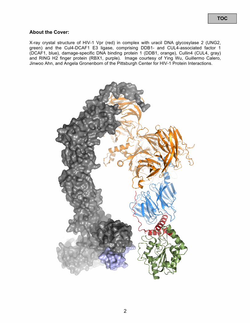
About the Cover:
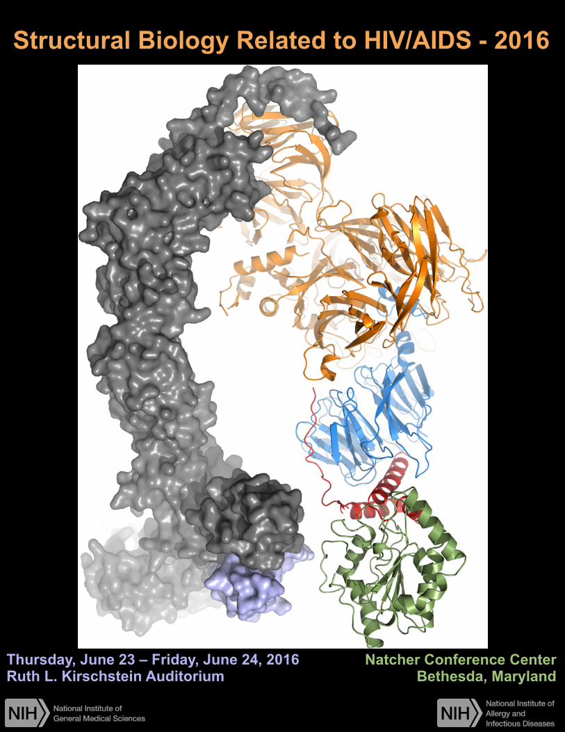
X-ray crystal structure of HIV-1 Vpr (red) in complex with uracil DNA glycosylase 2 (UNG2, green) and the Cul4-DCAF1 E3 ligase, comprising DDB1- and CUL4-associated factor 1 (DCAF1, blue), damage-specific DNA binding protein 1 (DDB1, orange), Cullin4 (CUL4, gray) and RING H2 finger protein (RBX1, purple). Image courtesy of Ying Wu, Guillermo Calero, Jinwoo Ahn, and Angela Gronenborn of the Pittsburgh Center for HIV-1 Protein Interactions.
2
Table of Contents About the Cover ............................................................................................................... 2 Table of Contents ............................................................................................................ 3 Meeting Agenda ............................................................................................................... 4 About the Poster Sessions ............................................................................................. 8 Thursday Posters ............................................................................................................ 9
Computational Methods and Modeling ................................................................... 9 Antibody-Envelope Interactions and Virus Spread ................................................ 9 Envelope Structure .................................................................................................. 10 Targeting Envelope ................................................................................................. 12 Uncoating and TRIM5α ............................................................................................ 13 Reverse Transcription ............................................................................................. 15 Vif and APOBEC ...................................................................................................... 16 Nuclear Entry ........................................................................................................... 18 Integrase ................................................................................................................... 18 Vpr ............................................................................................................................. 19 Provirus and Transcription ..................................................................................... 19
Friday Posters ............................................................................................................... 22
RNA Splicing ............................................................................................................ 22 RRE and Rev ............................................................................................................ 22 RNA Structure .......................................................................................................... 23 RNA Packaging ........................................................................................................ 25 Gag Trafficking and Assembly ............................................................................... 26 Gag-Membrane Interaction ..................................................................................... 27 Protease and Inhibition ........................................................................................... 28 Maturation ................................................................................................................ 31 Capsid ....................................................................................................................... 32 Membrane Accessory Proteins .............................................................................. 34
Speaker Abstracts ......................................................................................................... 35 Poster Abstracts, Thursday ......................................................................................... 69 Poster Abstracts, Friday ............................................................................................. 139 List of Participants ...................................................................................................... 209 Map of NIH Campus ..................................................................................................... 227 Parking and Security Procedures .............................................................................. 228
3
Agenda - Structural Biology Related to HIV/AIDS – 2016
DAY 1 8:00 – 8:15 Carl Dieffenbach (National Institute of Allergy and Infectious Diseases)
Opening Remarks
Session I: Reports from Specialized Centers (P50);; Session Chair: Wes Sundquist
8:15 – 9:15 The Center for HIV RNA Studies (CRNA)
Michael Summers (University of Maryland Baltimore County) Influence of 5’-start Site Heterogeneity and Capping on HIV-1 RNA Structure and Fate
Victoria D'Souza (Harvard University) Understanding Translational Regulation in HIV-1
Owen Pornillos (University of Virginia) Crystal Structure of the CA-SP1 Assembly and Maturation Switch
Sanford Simon (Rockefeller University) Steps in the Assembly of HIV-1
9:15 – 10:15 The Center for HIV Accessory and Regulatory Complexes (HARC)
Nevan Krogan (University of California San Francisco) The HARC Center: Progress and Overview
Alex Marson (University of California San Francisco) Genome Engineering Primary Human T Cells to Test Function of Host Factors in HIV Pathogenesis
Alan Frankel (University of California San Francisco) Functional Segregation of Overlapping Genes in HIV
Charles Craik (University of California San Francisco) Identifying Recombinant Antibodies to Challenging HIV Related Targets
10:15 – 10:45 BREAK
Session II: Reports from Specialized Centers (P50) continued;; Session Chair: Irwin Chaiken
10:45 – 11:45 The HIV Interaction and Viral Evolution Center (HIVE)
Arthur Olson (The Scripps Research Institute) HIVE Center: HIV Interactions and Viral Evolution of Drug Resistance
Mamuka Kvaratskhelia (Ohio State University) HIV-1 Integrase Binds the Viral RNA Genome and Is Essential During Virion Morphogenesis
4
Eddie Arnold (Rutgers University) A New Class of Allosteric HIV-1 Integrase Inhibitors Identified by Crystallographic Fragment Screening of the Catalytic Core Domain
Crystal Structure of Prototype Foamy Virus PR-RT Polyprotein Shows Unique Architecture Among Retroviruses: Implications for Function
Stefan Sarafianos (University of Missouri, Columbia) Visualization of Viral RNA and DNA Dynamics During HIV infection
11:45 – 1:00 LUNCH
1:00 – 3:00 POSTER SESSION
Session III: Reports from Specialized Centers (P50) continued;; Session Chair: Alan Frankel
3:00 – 4:00 The Pittsburgh Center for HIV Protein Interactions (PCHPI)
Tatyana Polenova (University of Delaware) Dynamic Regulation of HIV-1 Capsid Assembly, Maturation, and Interactions with Host Factors by Integrated MAS NMR and MD Simulations
Jacek Skowronski (Case Western Reserve University) HIV-1 and HIV-2 Exhibit Divergent Interactions with DNA Repair Enzymes
Guillermo Calero (University of Pittsburgh) To Repair or Not Repair: The X-Ray Structure of the DDB1-DCAF1-Vpr-UNG2 Complex
4:00 – 5:00 The Center for the Structural Biology of Cellular Host Elements in Egress, Trafficking, and Assembly of HIV (CHEETAH)
Wes Sundquist (University of Utah) Introduction to the Center
Michael Kay (University of Utah) D-Peptide Fusion Inhibitor Protects Against High-Dose SHIV Challenge
Tom Hope (Northwestern University) Visualizing HIV Uncoating in Living Cells
Barbie Ganser-Pornillos (University of Virginia) Structural Studies of TRIM5α
5:00 ADJOURN FOR DAY
5
DAY TWO
Session IV: Invited Speakers, Session Chair: Elizabeth Church
8:00 – 8:30 Debora Marks (Harvard Medical School) Structure and Function of RNA and Proteins from Natural Sequence Variation
8:30 – 9:00 Kushol Gupta (University of Pennsylvannia) Structural Basis for Inhibitor-Induced Aggregation of HIV Integrase
Session V: Selected Posters, Session Chair: Alice Telesnitsky
9:00 – 9:30 Lars-Anders Carlson (University of California Berkeley) Biochemical Reconstitution of Selective HIV-1 Genome Packaging
9:30 – 10:00 Pamela Bjorkman (California Institute of Technology) Structure of a Natively-Glycosylated HIV-1 Env Reveals a New Mode for VH1-2 Antibody Recognition of the CD4 Binding Site Relevant to Vaccine Design
10:00 – 10:30 BREAK
Session V: Targeting Envelope, Session Chair: Arthur Olson
10:30 – 11:15 P01: Structure-Based Antagonism of HIV-1 Envelope Function in Cell Entry
Irwin Chaiken (Drexel University) Overview of the Program and Recent Progress
Ernesto Freire (Johns Hopkins University) Conformational Transformations Triggered by Different Inhibitor Classes
Alon Herschhorn (Dana-Farber Cancer Institute) An Intermediate State of the HIV-1 Envelope Glycoproteins on the Entry Pathway
Session VI: Protease Drug Resistance, Angela Gronenborn:
11:15 – 12:00 P01: The Interdependency of Drug Resistance Evolution and Drug Design: HIV-1 Protease
Celia Schiffer (UMass Medical School) Project Overview
Rieko Ishima (University of Pittsburgh) Establish a Strategy to Elucidate Molecular Mechanisms Leading to Drug Resistance
Daniel Bolon (UMass Medical School) Systematic Exploration of Mutational Pathways to Drug Resistance in HIV Protease
6
12:00 – 1:00 LUNCH (SAB Lunch, Room B;; Please bring your lunch.)
1:00 – 3:00 POSTER SESSION (1:00 – 3:00 NIGMS Centers Scientific Review Board;; Room B)
Session VIII: Selected Posters Cont., Session Chair: Celia Schiffer
3:00 – 3:30 Ursula Schulze-Gahmen (University of California Berkeley) Insights into HIV-1 Proviral Transcription from an Integrative Structure of the Tat:AFF4:P-TEFb:TAR Complex
3:30 – 4:00 Guney Boso (Sanford Burnham Institute) Arrayed Analysis of Immune Evasion: High Content Imaging Screen Reveals Novel Targets of HIV-1 Vpu
4:00 – 4:30 Gregory Melikyan (Emory University) Real-Time Imaging of Single HIV-1 Uncoating in Cells
4:30 – 5:00 Markus Thali (University of Vermont) More is Less: Enhancing Virus-Induced Membrane Fusion to Inhibit Viral Spread
5:00 ADJOURN MEETING
Mark Your Calendars!
Structural Biology Related to HIV/AIDS – 2017 Thursday June 29 – Friday June 30, 2017
Natcher Conference Center, Bethesda, Maryland
7

About the Poster Sessions
Thursday 1:00 – 3:00 T-numbered posters
Friday 1:00 – 3:00 F-numbered posters
Posters may be put up the morning of your session and left all day. Please remember to remove your poster from the board at the end of the assigned day. There are too many for posters to be left up for the whole meeting.
8

Thursday Posters
Computational Methods and Modeling
T1. Identifying and Incorporating Water-Mediated Interactions in Drug Discovery Jiaye Guo,1 and Robert C. Rizzo2,3,4 1Graduate Program in Biochemistry and Structural Biology, Stony Brook University, Stony Brook, New York 11794;; 2Department of Applied Mathematics & Statistics, Stony Brook University, Stony Brook, New York 11794;; 3Institute of Chemical Biology & Drug Discovery, Stony Brook University, Stony Brook, New York 11794;; 4Laufer Center for Physical & Quantitative Biology, Stony Brook University, Stony Brook, New York 11794
T2. A Genetic Algorithm for DOCK to Aid in De Novo Design Courtney D. Singleton1, Lauren E. Prentis2, and Robert C. Rizzo3,4,5 1Department of Pharmacology, 2Department of Biochemistry & Structural Biology, 3Department of Applied Mathematics & Statistics, 4Institute of Chemical Biology & Drug Discovery, 5Laufer Center for Physical & Quantitative Biology, Stony Brook University, Stony Brook, NY 11794
T3. FightAIDS@Home Phase II: Refinement of Massive HIV Virtual Screening Experiments Using Large-Scale Molecular Dynamics Simulations William F. Flynn1,2, Junchao Xia2, Nanjie Deng2, Stefano Forli3, Arthur Olson3, and Ronald M. Levy2 1Department of Physics and Astronomy, Rutgers University, Piscataway, NJ;; 2Center for Biophysics and Computational Biology, Department of Chemistry, Temple University, Philadelphia, PA;; 3Molecular Graphics Laboratory, Department of Molecular Biology, The Scripps Research Institute, La Jolla, CA
T4. Animating the Science of HIV Janet Iwasa Department of Biochemistry, University of Utah
T5. Coarse-grained (CG) Computer Models of Key Stages in the HIV-1 Lifecycle J. M. A. Grime and G. A. Voth 1Department of Chemistry, Institute for Biophysical Dynamics, James Franck Institute, and Computation Institute, The University of Chicago, Chicago, Illinois 60637, USA
Antibody – Envelope Interactions and Virus Spread
T6. Modeling Affinity Maturation of Anti-HIV Antibodies Targeting gp120 Dzmitry Padhorny and Dima Kozakov Laufer Center for Physics and Structural Biology, Stony Brook University
T7. The Broadest bnAbs Target More Conserved HIV-1 Env Epitopes Hongjun Bai, Merlin L. Robb, Nelson L. Michael and Morgane Rolland US Military HIV Research Program, Silver Spring, Maryland 20910, USA
9
T8. Structural Analysis of an HIV-1 Broadly Neutralizing V3-Glycan B-cell Lineage Fera D1, Bonsigniori M2, Kreider E3,Meyerhoff R2, Bradley T2, Wiehe K2, Alam SM2, Hwang KK2, Saunders KO2, Zhang R2, Gladden MA2, Monroe A2, Kumar A2, Xia SM2, Cooper M2, Jette CA1, Pier BW1, Montefiori DC, Trama A, Liao HX2, Kepler TB4, Gao F2, Shaw GM3, Hahn B3, Moody MA2, Gao F2, Mascola JR, Haynes BF2, and Harrison SC1,5 1Laboratory of Molecular Medicine and 5HHMI, Boston Children’s Hospital, Harvard Medical School, Boston, MA;; 2Department of Medicine, Duke University School of Medicine, Duke University Medical Center, and Duke Human Vaccine Institute, Durham, NC;; 3Departments of Medicine and Microbiology, Perelman School of Medicine, University of Pennsylvania, Philadelphia, PA;; 4Departments of Microbiology, and Mathematics and Statistics, Boston University, Boston, MA;; 5Vaccine Research Center, National Institutes of Allergy and Infectious Diseases, National Institutes of Health, Bethesda, MD
T9. A Protective Role for CD169 in Limiting Systemic Spread of a Pathogenic Retrovirus Pradeep D. Uchil1, Ruoxi Pi1, John D. Ventura1, Kelsey A. Haugh1, Xaver Sewald2, and Walther Mothes1* 1Department of Microbial Pathogenesis, Yale University School of Medicine, New Haven, CT 06510;; 2Max von Pettenkofer-Institute, Ludwig-Maximilians-University Munich, Munich, Germany;; *corresponding author
T10. Functional Interplay Between Murine Leukemia Virus Glycogag, Surface Glycoprotein and Serinc5 Modulates Virus Entry Yadvinder S Ahi1, Shu Zhang1, Delphine Muriaux2, Amin Feizpour3, Björn M Reinhard3, Rahm Gummuluru3, and Alan Rein1 1NCI, HIV DRP, Frederick, MD;; 2CNRS, Membrane Domains and Viral Assembly, Montpellier, France;; 3Boston University, Boston, MA
T11. Multi-Dimensional Profiling of Primary Human CD4+ T Memory Cells Lara Manganaro1, Jeffrey R. Johnson2,3,4, Patrick Hong1, Benhur Lee1, Nevan Krogan2,3,4 and Viviana Simon1,5,6 1Department of Microbiology, Icahn School of Medicine at Mount Sinai, New York, NY, USA;; 2Gladstone Institute of Virology and Immunology, 1650 Owens Street, San Francisco, CA 94158, USA;; 3University of California, San Francisco, CA 94158, USA;; 4QB3, California Institute for Quantitative Biosciences, San Francisco, CA 94158, USA;; 5Division of Infectious Diseases, Department of Medicine, Icahn School of Medicine at Mount Sinai, New York, New York, USA;; 6Global Health and Emerging Pathogens Institute, Icahn School of Medicine at Mount Sinai, New York, New York, USA
Envelope Structure
T12. Understanding Glycan Type Specificity in Highly Glycosylated Proteins Artem Krantsevich and David F. Green Stony Brook University
10
T13. Insights into Structures and Dynamics of Variable Regions of Major Subtypes of HIV-1 Gp120 Tuoling Qiu1, and David F. Green1, 2 1Chemistry Department, Stony Brook University, Stony Brook, NY 11794;; 2Department of Applied Mathematics and Statistics, Stony Brook University, Stony Brook, NY 11794
T14. Mapping the Conformational Space of Glycoconjugate-Linked Carbohydrates Xindi Li1 and David F. Green1, 2, 3, 4 1Department of Applied Mathematics & Statistics, 2Department of Chemistry, 3Laufer Center for Physical and Quantitative Biology, 4Graduate Program in Biochemistry and Structural Biology, Stony Brook University, Stony Brook, New York 11794-3600, United States
T15. Deciphering the Mechanisms of HIV-1 Entry Using Novel Env Heterotrimers Mukta D. Khasnis and Michael Root Department of Biochemistry and Molecular Biology, Kimmel Cancer Center, Thomas Jefferson University, Philadelphia, PA
T16. Structural Dynamics of HIV Env Glycoproteins: A Link Between Structural, Functional, and Phenotypic Variation Among Isolates Kelly K. Lee1, Hans Verkerke1, Miklos Guttman1, James A. Williams1, Yu Liang1, Thaddeus M. Davenport1, Cassie Simonich1,2, Shiu-Lok Hu1, and Julie Overbaugh1,21University of Washington, 2Fred Hutchinson Cancer Research Center, Seattle, WA
T17. On the Role of the V3 Loop in the Conformational Thermodynamics of Bridging Sheet Formation in HIV-1 Gp120: On-the-fly Parameterization Free-energy Calculations of the SOSIP BG505 Protomer Alexis Paz1, Matthew Cameron1, and Cameron F. Abrams1,2 1Department of Chemical and Biological Engineering, Drexel University, 3141 Chestnut St., 19104, Philadelphia, PA (US);; 2Department of Biochemistry and Molecular Biology, Drexel College of Medicine, 25 North 15th Street, Philadelphia, PA (US)
T18. Single-Molecule FRET Delineates Asymmetric Trimer Conformations During HIV-1 Entry Xiaochu Ma1, Alon Herschhorn2,3, John D. Ventura1, Daniel S. Terry4, Jason Gorman5, Jonathan R. Grover1, Xinyu Hong1, Zhou Zhou4, Hong Zhao4, Roger B. Altman4, James Arthos6, Peter D. Kwong5, Joseph Sodroski2,3,7, Scott C. Blanchard4, Walther Mothes1*, and James B. Munro8* 1Department of Microbial Pathogenesis, Yale University School of Medicine, New Haven, CT 06536, USA;; 2Department of Cancer Immunology and Virology, Dana-Farber Cancer Institute, Boston, MA 02215, USA;; 3Department of Microbiology and Immunobiology, Harvard Medical School, Boston, MA 02115, USA;; 4Department of Physiology and Biophysics, Weill Cornell Medical College of Cornell University, New York, NY 10021, USA;; 5Vaccine Research Center, National Institute of Allergy and Infectious Diseases, National Institutes of Health, Bethesda, MD 20892, USA;; 6Laboratory of Immunoregulation, National Institute of Allergy and Infectious Diseases, National Institutes of Health, Bethesda, MD 20892, USA;; 7Department of Immunology and Infectious Diseases, Harvard T. H. Chan School of Public Health, Boston, MA 02115, USA;; 8Department of Molecular Biology and Microbiology, Tufts University School of Medicine, Boston, MA 02111, USA;; *Correspondence to: Walther Mothes ([email protected]) and James Munro ([email protected])
11
T19. Investigating Conformational Transitions in HIV-1 Env Using Combinations of CD4 Antagonists, Chemokine Receptor Antagonists and Fusion Inhibitors Koree W. Ahn and Michael J. Root Thomas Jefferson University, United States of America
Targeting Envelope
T20. Cyclic Peptide Triazole Rigid-Receptor Docking and Molecular Dynamics Simulation in Three Different Gp120 States: Comparison of Targets for Future HIV-1 Antagonist Optimization Francesca Moraca1, Adel A. Rashad2, Kriti Acharya2, Irwin Chaiken2 and Cameron F. Abrams1 1Department of Chemical and Biological Engineering, Drexel University, 3141 Chestnut St., 19104, Philadelphia, PA (US);; 2Department of Biochemistry and Molecular Biology, Drexel College of Medicine, 25 North 15th Street, Philadelphia, PA (US)
T21. Generation and Characterization of HIV-1 Escape Mutants to Peptide Triazole Entry Inhibitors Andrew P. Holmes1,2, Adel Ahmed2, Lauren D. Bailey2, Katie Kercher1, William Dampier1, Michael Nonnemacher1, Michael Root3, and Irwin Chaiken2 1Department of Microbiology and Immunology, Drexel University College of Medicine;; 2Department of Biochemistry and Molecular Biology, Drexel University College of Medicine;; 3Department of Biochemistry and Molecular Biology, Thomas Jefferson University
T22. Optimization of Macrocyclic Peptide Triazole HIV-1 Inactivators Adel Ahmed Rashad*, Kriti Acharya*, Rachna Aneja, Ann Haftl and Irwin Chaiken Department of Biochemistry & Molecular Biology, Drexel University College of Medicine, Philadelphia, Pennsylvania 19102;; *Corresponding authors: [email protected], [email protected]
T23. Structure-Based Optimization of Small-Molecule CD4-Mimics: Inhibitors of HIV-1 Entry Sharon M. Kirk, Melissa C. Grenier, Althea E. Gaffney, Bruno Melillo, Amos B. Smith, III Department of Chemistry, University of Pennsylvania
T24. DeNovo Design and Refinement of Inhibitors Targeting HIV Entry Brian C. Fochtman1 William J. Allen2 and Robert C. Rizzo2,3,4 1Department of Biochemistry and Cellular Biology, 2Department of Applied Mathematics & Statistics, 3Institute of Chemical Biology & Drug Discovery, 4Laufer Center for Physical & Quantitative Biology, Stony Brook University, Stony Brook, NY 11794
T25. HIV-1 Lytic Inactivation by Dual Acting Virus Entry Inhibitor Occurs Through Combined Interactions with gp120 and gp41 Subunits of Virus Env Protein Trimer Bibek Parajuli1, Kriti Acharya1, Reina Yu1, Adel A. Rashad1, Cameron F. Abrams2, Irwin M. Chaiken1* 1Department of Biochemistry and Molecular Biology, Drexel University College of Medicine, Philadelphia, Pennsylvania 19102, United States;; 2Department of Chemical and Biological Engineering, Drexel University, Philadelphia, Pennsylvania 19104, United States
12
T26. Design, Synthesis, and Biological Evaluation of Helical Spiroligomers Targeting HIV-1 Gp41 Cheong, J. E. and Schafmeister, C. E. Department of Chemistry, Temple University
T27. Molecular Dynamics Simulations of HIVgp41 Reveal Energetically Favorable Interfaces for Small-Molecule Inhibitors T. Dwight McGee Jr.1, and Robert Rizzo1,2,31Department of Applied Mathematics & Statistics, 2Institute of Chemical Biology & Drug Discovery, 3Laufer Center for Physical & Quantitative Biology, Stony Brook University, Stony Brook, NY 11794
T28. Expression of HERV-K108 Envelope Interferes with HIV-1 Production Sandra Terry1, Alvaro Cuesta-Dominguez1, Lara Manganaro1, Daria Brinzevich1, Viviana Simon1,2, and Lubbertus C.F. Mulder1, 2 1Department of Microbiology, 2Global Health and Emerging Pathogens Institute, Icahn School of Medicine at Mount Sinai, New York, NY
Uncoating and TRIM5α
T29. Minor Sequence Differences in HIV-1NL4-3 and HIV-1LAI Capsid Cause Distinct Capsid Uncoating and Host Cell Infectivity Phenotypes Douglas K. Fischer1,2,4, Simon C. Watkins3,4, Masahiro Yamashita4,5, Tatyana Polenova4,6, and Zandrea Ambrose1,2,41Molecular Virology and Microbiology Program, 2Division of Infectious Diseases, Department of Medicine, 3Department of Cell Biology and Physiology, and 4Pittsburgh Center for HIV Protein Interactions, University of Pittsburgh School of Medicine, Pittsburgh, PA;; 5Aaron Diamond AIDS Research Center, New York, NY;; and 6Department of Chemistry and Biochemistry, University of Delaware, Newark, DE
T30. Real-Time Imaging of Single HIV-1 Uncoating in Cells Ashwanth C. Francis1, Mariana Marin1, Jiong Shi2, Christopher Aiken2 and Gregory B. Melikyan1,3 1Department of Pediatric, Emory University School of Medicine, Atlanta, GA;; 2Department of Pathology, Microbiology and Immunology, Vanderbilt University, Nashville, TN;; 3Children’s Healthcare of Atlanta, Atlanta, GA
T31. Correlation of Infectivity and Imaged Individual HIV Particle Behavior Validates the Early Uncoating Model During HIV Infection João I. Mamede, Gianguido C. Cianci, Meegan Anderson, Thomas J Hope Northwestern University Feinberg School of Medicine, Department of Cell and Molecular Biology, Chicago, IL
13
T32. Development of Cryo-CLEM Methods to Elucidate the Intracellular Structure of TRIM5α Bodies Stephen D. Carter1, Shrawan K. Mageswaran1, Joao I. Mamede3, Tom J. Hope3, Joachim Frank4,5, Zachary Freyberg6,7, and Grant J. Jensen1,2 1Division of Biology and 2Howard Hughes Medical Institute (HHMI), California Institute of Technology, Pasadena, CA 91125, USA;; 3Department of Cell and Molecular Biology, Northwestern University, Chicago, IL 60611-3008;; 4HHMI;; Department of Biochemistry and Molecular Biophysics, Columbia University, New York, New York 10032, USA;; 5Department of Biological Sciences, Columbia University, New York, New York 10027, USA;; 6Department of Psychiatry, College of Physicians & Surgeons, Columbia University, New York, New York 10032, USA;; 7Division of Molecular Therapeutics, New York State Psychiatric Institute, New York, New York 10032, USA
T33. Characterization of TRIM5 Assembly and Activation Using Chimeric “MiniTRIMs” Jonathan M. Wagner1, Marcin D. Roganowicz1, Katarzyna Skorupka1, Steven L. Alam2, Devin Christensen2, Ginna Doss1, Yueping Wan1, Gabriel A. Frank3, Barbie K. Ganser-Pornillos1, Wesley I. Sundquist2, and Owen Pornillos1 1Department of Molecular Physiology and Biological Physics, 2Department of Biochemistry, University of Utah, Salt Lake City, UT 84112, U.S.A.;; 3Faculty of Biology, Technion - Israel Institute of Technology, Haifa 320003, Israel
T34. Analysis of TRIM5α SPRY Domain Packing Against Its Coiled-Coil Domain Marcin D. Roganowicz1, Santanu Mukherjee2, Katarzyna Skorupka1, Damian Dawidowski3, David S. Cafiso3, Edward M. Campbell2, Owen Pornillos1 1Department of Molecular Physiology and Biological Physics, University of Virginia, Charlottesville, VA;; 2Department of Microbiology and Immunology, Stritch School of Medicine, Loyola University Chicago, Maywood, IL;; 3Department of Chemistry, University of Virginia, Charlottesville, VA
T35. Dynamic Allostery in HIV-1 Capsid Interactions with Restriction Factor TRIM5 Revealed by Magic Angle Spinning NMR Caitlin M Quinn1,2, Mingzhang Wang1,2, Jinwoo Ahn2,3, Angela Gronenborn2,3, and Tatyana Polenova1,2 1University of Delaware, Department of Chemistry and Biochemistry, Newark, DE;; 2Pittsburgh Center for HIV Protein Interactions, University of Pittsburgh School of Medicine, 1051 Biomedical Science Tower 3, 3501 5th Ave., Pittsburgh, PA;; 3Department of Structural Biology, University of Pittsburgh School of Medicine, 3501 Fifth Ave., Pittsburgh, PA
T36. Unbiased Genome Wide Screens for Host Cofactors Involved in Viral Restriction Daniel W. Cyburt, Clifton L. Ricana, and Marc C. Johnson Department of Molecular Microbiology and Immunology, University of Missouri-Columbia
14
Reverse Transcription
T37. Investigation of NMR Spectral Changes upon Homodimer Formation of HIV-1 Reverse Transcriptase Ryan L. Slack1, Naima G. Sharaf1, Michael A. Parniak2, Jinwoo Ahn1, Angela M. Gronenborn1, Rieko Ishima1 1Department of Structural Biology and 2Department of Microbiology and Molecular Genetics, University of Pittsburgh, School of Medicine, PA-15260, USA
T38. Biochemical and Cellular Characterization of the SAMHD1 Ortholog, Caenorhabditis Elegans ZCK177.8 Lydia R. Studdard, Tatsuya Maehigashi and Baek Kim Center for Drug Discovery, Emory School of Medicine, Pediatrics, Atlanta, GA
T39. Effect of Nucleic Acid Sequence on DNA Polymerization and NNRTI Inhibitory Mechanisms of HIV-1 Reverse Transcriptase O Ukah1, A Huber2, E Michailidis3, K Das4, MA Parniak5, K Singh1, E Arnold4, and S Sarafianos1,6 1University of Missouri, Molecular Microbiology and Immunology, Columbia, MO, United States;; 2University of Missouri, Veterinary Pathobiology, Columbia, MO, United States;; 3Rockefeller University, Virology and Infectious Disease, New York, NY, United States;; 4Rutgers University, Chemistry and Chemical Biology, Piscataway, NJ, United States;; 5University of Pittsburgh, Microbiology and Molecular Genetics, Pittsburgh, PA, United States;; 6University of Missouri, Biochemistry, Columbia, MO
T40. HIV-1 Capsid Facilitates Reverse Transcription by Retaining Reverse Transcriptase Within the Core Janani Varadarajan and Christopher Aiken Department of Pathology, Microbiology and Immunology, Vanderbilt University School of Medicine, Nashville, TN 37232
T41. 3-Hydroxypyrimidine-2,4-diones as Novel HIV-1 RNase H Inhibitors Karen A. Kirby1,2, Jing Tang3, Sanjeev K. V. Vernekar3, Bulan Wu3, Andrew D. Huber1,4, Mary C. Casey1,2, Juan Ji1,2, Eva Nagy5, Lena Miller5, Qiongying Yang1,2, Michael A. Parniak5, Zhengqiang Wang3, and Stefan G. Sarafianos1,2,6 1C.S. Bond Life Sciences Center, University of Missouri, Columbia, MO;; 2Dept. of Molecular Microbiology & Immunology, University of Missouri School of Medicine, Columbia, MO;; 3Center for Drug Design, University of Minnesota, Minneapolis, MN;; 4Dept. of Veterinary Pathobiology, University of Missouri School of Medicine, Columbia, MO;; 5Dept. of Microbiology & Molecular Genetics, University of Pittsburgh School of Medicine, Pittsburgh, PA;; 6Dept. of Biochemistry, University of Missouri, Columbia, MO
15
T42. Two Distinct Modes of Metal Ion Binding in the Nuclease Active Site of a Viral DNA-Packaging Terminase: Insight into the Two-Metal-Ion Catalytic Mechanism of RNaseH-like Nucleotidyltransferases Haiyan Zhao1, Zihan Lin1, Anna Y. Lynn1, Brittany Varnado1, John A. Beutler2, Ryan P. Murelli3, Stuart F.J. Le Grice4 and Liang Tang1 1Department of Molecular Biosciences, University of Kansas, 1200 Sunnyside Avenue, Lawrence, KS 66045;; 2Molecular Targets Laboratory, National Cancer Institute, Frederick, MD 21702;; 3Department of Chemistry, Brooklyn College, City University of New York, Brooklyn, NY 11210;; 4Basic Research Laboratory, National Cancer Institute, Frederick, MD 21702
T43. Characterization the C-Terminal Nuclease Domain of Herpes Simplex Virus Pul15 as a Target of Nucleotidyltransferase Inhibitors Takashi Masaoka1, Haiyan Zhao2, Danielle R. Hirsch3,4, Michael P. D'Erasmo3,4, Christine Meck3,4, Brittany Varnado2, Marvin J. Meyers5 Joel Baines6, John A. Beutler7, Ryan P. Murelli3,4, Liang Tang2 and Stuart F.J. Le Grice1 1Basic Research Laboratory, National Cancer Institute, Frederick, MD 21702, USA;; 2Department of Molecular Biosciences, University of Kansas, Lawrence, KS 66045, USA;; 3Department of Chemistry, Brooklyn College, City University of New York, Brooklyn, NY 11210, USA;; 4Department of Chemistry, The Graduate Center, City University of New York, New York, NY, 10016, USA;; 5Department of Chemistry, St. Louis University, St. Louis, MO 63103;; 6School of Veterinary Medicine, Louisiana State University, Baton Rouge, LA 70803 USA;; 7Molecular Targets Laboratory, National Cancer Institute, Frederick, MD 21702, USA
Vif and APOBEC
T44. Molecular Characterization of a Unique Restriction Factor, APOBEC3H Jennifer Bohn1, Theodora Hatziioannou2, and Janet Smith11Dept. of Biological Chemistry & Life Sciences Institute, University of Michigan, Ann Arbor, MI;; 2Aaron Diamond AIDS Research Center, The Rockefeller University, New York, NY
T45. NMR Structure of the APOBEC3B Catalytic Domain: Structural Basis for Substrate Binding and DNA Deaminase Activity In-Ja L. Byeon1, Chang-Hyeock Byeon1, Tiyun Wu2, Mithun Mitra2, Dustin Singer2, Judith G. Levin2 and Angela M. Gronenborn1 1Department of Structural Biology and Pittsburgh Center for HIV Protein Interactions, University of Pittsburgh School of Medicine, Pittsburgh, PA 15260, United States;; 2Section on Viral Gene Regulation, Program in Genomics of Differentiation, Eunice Kennedy Shriver National Institute of Child Health and Human Development, National Institutes of Health, Bethesda, MD 20892, United States
T46. The RNA Binding Specificity of Human APOBEC3 Proteins Mimics that of HIV-1 Nucleocapsid Ashley York1,2*, Sebla B Kutluay3*, Manel Errando4, and Paul D Bieniasz1,2,5 1Aaron Diamond AIDS Research Center, The Rockefeller University, New York, NY;; 2The Rockefeller University, Laboratory of Retrovirology, New York, NY;; 3Washington University School of Medicine in St. Louis, Department of Molecular Microbiology, St. Louis, MO;; 4Department of Physics, Washington University in St. Louis, St. Louis, MO, 5Howard Hughes Medical Institute, Aaron Diamond AIDS Research Center, New York, NY;; *equal contributions
16
T47. Identification of Recombinant Antibodies for Functional and Structural Analysis of HIV-Host Complexes Natalia Sevillano1, Hai Ta1, Jennifer Binning1, Florencia La Greca1, Judd Hultquist2, Nathalie Caretta1, Amber Smith3, Xuefeng Ren4, Bei Yang4, Bhargavi Jayaraman1, Shumin Yang1, Yifan Cheng3, Nevan J. Krogan2, John Gross1 and Charles S. Craik1 1Department of Pharmaceutical Chemistry, University of California San Francisco, San Francisco, USA;; 2Department of Cellular and Molecular Pharmacology, University of California, San Francisco, CA, USA;; 3Department of Biochemistry and Biophysics, University of California, San Francisco, CA, USA;; 4Department of Molecular and Cell Biology, University of California, Berkeley, CA, USA
T48. VCBC-Specific Fabs Inhibit Ubiquitination and Degradation of A3 Proteins Jennifer M. Binning1, Amber Smith4,5, Natalia Sevillano1, Judd F. Hultquist2,3, Nathalie Caretta Cartozo1, Hai Ta1, Nevan J. Krogan2,3, Yifan Cheng4,5, Charles S. Craik1, John D. Gross1 1Department of Pharmaceutical Chemistry, University of California, San Francisco, California 94158, USA;; 2Department of Cellular and Molecular Pharmacology, University of California, San Francisco, San Francisco, California 94158, USA;; 3J. David Gladstone Institute, San Francisco, CA 94158;; 4Howard Hughes Medical Institute, UCSF, San Francisco, CA 94158, USA;; 4Howard Hughes Medical Institute, UCSF, San Francisco, CA 94158, USA;; 5Department of Biochemistry and Biophysics, UCSF, San Francisco, CA 94158, USA
T49. Cleavable Cross-linkers, Multistage Mass Spectrometry, and the Structural Characterization of APOBEC3-Vif-CRL5 Complexes Robyn M Kaake1, Seung Joong Kim3, Ignacia Echeverria3, Linda Chelico4, John D Gross3, Andrej Sali3, Lan Huang5, Nevan J Krogan1,2 1The J David Gladstone Institutes, Institute for Virology and Immunology, San Francisco, CA;; 2University of California San Francisco, Dept. of Cellular and Molecular Pharmacology, San Francisco, CA;; 3University of California San Francisco, Dept. of Pharmaceutical Chemistry, San Francisco, CA;; 4University of Saskatchewan, College of Medicine, Saskatoon, Canada;; 5University of California Irvine, Dept. of Physiology & Biophysics, Irvine, CA
T50. Global Landscape of Ubiquitylation and Phosphorylation Changes in Response to HIV-1 Infection Identifies a Novel Substrate of Vif Jeffrey Johnson1,2, David Crosby1, Judd Hultquist1, Erik Verschuren3, Lara Manganaro4, Billy Newton1, Tasha Johnson1, Tricia Lundrigan1, Viviana Simon4, Alan Frankel1, and Nevan Krogan1,2* 1University of California San Francisco;; 2Gladstone Institutes;; 3Genentech, Inc.;; 4Icahn School of Medicine at Mount Sinai;; *Corresponding author
T51. Rerouting Resistance: Escaping Restriction Using Alternative Cellular Pathways Aya Khwaja, Meytal Galilee, Ailie Marx, and Akram Alian Faculty of Biology, Technion – Israel Institute of Technology, Haifa 320003, Israel
17
Nuclear Entry
T52. HIV-1 Interaction with CypA Regulates Use of FG-Nucleoporins for Nuclear Entry Guangai Xue1, Shih Lin Goh2, Hyun Jae Yu1, Anna T. Gres3, KyeongEun Lee1, Stefan G. Sarafianos3, Jeremy Luban2, and Vineet N. KewalRamani1 1Basic Research Laboratory, National Cancer Institute, Frederick, MD;; 2Program in Molecular Medicine, University of Massachusetts Medical School, Worcester, MA;; 3Bond Life Sciences Center, MMI, Biochemistry, University of Missouri, Columbia, MO
T53. HIV-1 Nuclear Trafficking is Altered by Cytoplasmic CPSF6 Expression in a Capsid-Dependent Manner Zhou Zhong1, Callen Wallace2, Christopher Kline3, Simon C. Watkins2, and Zandrea Ambrose1,3 1Molecular Virology and Microbiology Program, 2Department of Cell Biology, and 3Department of Medicine, University of Pittsburgh School of Medicine, Pittsburgh, PA 15261
Integrase
T54. HIV-1 Capsid Protein Modulates the Activity of Preintegration Complexes Muthukumar Balasubramaniam1, Amma Addai1, Jing Zhou2, Jui Pandhare1, Christopher Aiken2, and Chandravanu Dash1 1Center for AIDS Health Disparities Research, Meharry Medical College, Nashville, TN;; 2Department of Pathology, Microbiology and Immunology, Vanderbilt University School of Medicine, Nashville, TN
T55. Modeling Ligand Binding to an Allosteric Site in the Catalytic Core Domain of HIV-1 Integrase Using Absolute and Relative Free Energy Methods Nanjie Deng,1 Stefano Forli,2 Joseph Bauman,3 James Fuchs,4 Mamuka Kvaratskhelia,4 Alan Engelman,5 Eddy Arnold,3 Art Olson,2 and Ronald Levy11Center for Biophysics and Computational Biology, Temple University, Philadelphia, PA;; 2The Scripps Research Institute, La Jolla, CA;; 3Center for Advanced Biotechnology and Medicine, Rutgers University, Piscataway, NJ;; 4College of Pharmacy, Ohio State University, Columbus, OH;; 5Dana-Farber Cancer Institute, Harvard Medical School, Boston, MA
T56. HIV-1 Integrase Strand Transfer Inhibitors that Reduce Susceptibility to Drug Resistant Mutant Integrases Xue Zhi Zhao,1 Hannah Peters,1 Steven J. Smith,2 Daniel P. Maskell,4 Mathieu Metifiot,3 Valerie E. Pye,4 Katherine Fesen,3 Christophe Marchand,3 Yves Pommier,3 Peter Cherepanov,4,5 Stephen H. Hughes,3 and Terrence R. Burke, Jr.1 1Chemical Biology Laboratory and 2HIV Dynamics and Replication Program, Center for Cancer Research, National Cancer Institute, National Institutes of Health, Frederick, MD 21702;; 3Developmental Therapeutics Branch and Laboratory of Molecular Pharmacology, Center for Cancer Research, National Cancer Institute, National Institutes of Health, Bethesda, MD 20892;; 4Clare Hall Laboratories, The Francis Crick Institute, Blanche Lane, South Mimms, EN6 3LD, UK;; 5Imperial College London, St-Mary’s Campus, Norfolk Place, London, W2 1PG, UK
18
T57. Selectivity for Strand-Transfer Over 3'-Processing and Susceptibility to Clinical Resistance of HIV-1 Integrase Inhibitors Are Both Driven by Key Enzyme-DNA Interactions in the Active Site Mathieu Métifiot1, Barry C. Johnson2, Evgeny Kiselev1, Laura Marler1, Xue Zhi Zhao3, Terrence R. Burke Jr3, Christophe Marchand1, Stephen H. Hughes2 and Yves Pommier1 1Developmental Therapeutics Branch, Center for Cancer Research, National Cancer Institute, National Institutes of Health, Bethesda, MD 20892;; 2HIV Drug Resistance Program, Frederick National Laboratory for Cancer Research, Center for Cancer Research, National Cancer Institute-Frederick, National Institutes of Health, Frederick, MD 21702;; 3Chemical Biology Laboratory, Frederick National Laboratory for Cancer Research, Center for Cancer Research, National Cancer Institute-Frederick, National Institutes of Health, Frederick, MD 21702
T58. Using Fragment-Based Analysis of Virtual Screenings to Characterize Binding Sites Richard K. Belew1, Stefano Forli2, David Goodsell2, T. J. O’Donnell3, and Arthur Olson2 1Univ. California – San Diego ([email protected]);; 2The Scripps Research Institute;; 3gNova
Vpr
T59. Structure-Selective Endonuclease MUS81/EME1 Downregulation and G2 Cell Cycle Arrest are Independent Functions of Vpr Xiaohong Zhou, Maria DeLucia, and Jinwoo Ahn Department of Structural Biology and Pittsburgh Center for HIV Protein Interactions, University of Pittsburgh School of Medicine, Pittsburgh, PA 15260, USA
T60. To Repair or Not Repair: The X-ray Structure of the DDB1-DCAF1-Vpr-UNG2 Complex Ying Wu1, Xiaohong Zhou1, Christopher O. Barnes2, Maria DeLucia1, Aina E. Cohen3, Angela M. Gronenborn1, Jinwoo Ahn1, and Guillermo Calero1* 1Department of Structural Biology and Pittsburgh Center for HIV Protein Interactions, University of Pittsburgh School of Medicine, Pittsburgh, PA 15260, USA;; 2Department of Pharmacology and Chemical Biology, University of Pittsburgh School of Medicine, Pittsburgh, PA 15260, USA;; 3Stanford Synchrotron Radiation Lightsource, Menlo Park, CA 94025, USA;; *Correspondence: [email protected] (J.A.), [email protected] (G.C.)
Provirus and Transcription
T61. Human T-Cell Leukemia Virus Type 1 Proviral Load and Genome Structure in Chronically Infected T-Cell Lines Morgan E Meissner1,2, LeAnn Oseth5, Jessica L Martin1,3, Luiza Mendonca1, Wei Zhang1,4, Louis M Mansky1,2,3 1University of Minnesota, Institute for Molecular Virology, Minneapolis, MN, 2University of Minnesota, Molecular, Cellular, Developmental Biology, and Genetics Graduate Program, Minneapolis, MN, 3University of Minnesota, Pharmacology Graduate Program, Minneapolis, MN, 4 University of Minnesota, Characterization Facility, Minneapolis, MN, 5University of Minnesota, Cytogenomics Core, Minneapolis, MN
19
T62. Structural Basis of an Evolved Cre Recombinase that Excises HIV DNA Gretchen Meinke1, Janet Karpinski2, Frank Buchholz2 and Andrew Bohm1 1Department of Developmental, Molecular and Chemical Biology, Tufts University School of Medicine, Boston, MA, 02111, USA;; 2Medical Systems Biology, UCC, Medical Faculty Carl Gustav Carus, TU Dresden, Germany
T63. DNA-PK Inhibition Potently Represses HIV Transcription and Replication Geetaram Sahu1, Kalamo Farley1, Gary Simon1 and Mudit Tyagi1, 2 1Division of Infectious Diseases, Department of Medicine, George Washington University, Washington, DC 20037;; 2Department of Microbiology, Immunology and Tropical Medicine, George Washington University, Washington, DC 20037
T64. Identification of Smac Mimetics as a Novel Class of HIV-1 Latency Reversing Agents Lars Pache1, Miriam S. Dutra1, Adam M. Spivak2, John M. Marlett3, Jeffrey P. Murry3§, Young Hwang4, Ana M. Maestre5, Lara Manganaro5, Mitchell Vamos1, Peter Teriete1, Laura J. Martins2, Renate König1,6, Viviana Simon5, Alberto Bosque2, Ana Fernandez-Sesma5, Nicholas D. P. Cosford1, Frederic D. Bushman4, John A. T. Young3#, Vicente Planelles2 and Sumit K. Chanda1 1Sanford Burnham Prebys Medical Discovery Institute, La Jolla, CA 92037;; 2University of Utah School of Medicine, Salt Lake City, UT 84112;; 3The Salk Institute for Biological Studies, La Jolla, CA 92037;; 4Perelman School of Medicine at the University of Pennsylvania, Philadelphia, PA 19104;; 5Icahn School of Medicine at Mount Sinai, New York, NY 10029;; 6Paul-Ehrlich-Institut, 63225 Langen, Germany;; §Present address: Gilead Sciences, Foster City, CA 94404;; # Present address: F. Hoffmann-La Roche Ltd, 4070 Basel, Switzerland
T65. Visualization of Transcriptional Activation from HIV Integration Sites in Latently Infected Cells Obiaara Ukah1,2, Maritza Puray-Chavez1,2, Juan Ji1, Stefan G Sarafianos1,2,3,4 1University of Missouri, C.S. Bond Life Sciences Center, Columbia, MO, 2University of Missouri, Molecular Microbiology & Immunology, Columbia, MO, 3University of Missouri, Veterinary Pathobiology, Columbia, MO, 4University of Missouri, Biochemistry, Columbia, MO
T66. Heterogenic Transcription Start Sites of HIV-1 and Their Influence on RNA Fates Siarhei Kharytonchyk1, Philip Smaldino1, Sarah Monti2, Michael F. Summers2, and Alice Telesnitsky1 1Department of Microbiology and Immunology, University of Michigan Medical School, Ann Arbor MI 48109-0620;; 2Howard Hughes Medical Institute and Department of Chemistry and Biochemistry, University of Maryland Baltimore County, 1000 Hilltop Circle, Baltimore, MD 21250
20
T67. Insights into HIV-1 Proviral Transcription from an Integrative Structure of the Tat:AFF4:P-Tefb:TAR Complex Ursula Schulze-Gahmen1, Ignacia Echeverria2, Goran Stjepanovic1, Yun Bai1, Huasong Lu1, Dina Schneidman-Duhovny 2, Jennifer A. Doudna1,3,4,5, Qiang Zhou1, Andrej Sali2, and James H. Hurley1,5 1Department of Molecular and Cell Biology and California Institute of Quantitative Biosciences, University of California, Berkeley, Berkeley, United States;; 2Department of Bioengineering and Therapeutic Sciences, Department of Pharmaceutical Chemistry, and California Institute of Quantitative Biosciences, University of California San Francisco, San Francisco, United States;; 3Howard Hughes Medical Institute, University of California, Berkeley, Berkeley, United States;; 4Department of Chemistry, University of California, Berkeley, Berkeley, United States;; 5Molecular Biophysics and Integrated Bioimaging Division, Lawrence Berkeley National Laboratory, Berkeley, United States
T68. Probing Structural Dynamics and Kinetics of HIV-1 RNA Recognition to Guide RNA Drug Design Nicole Orlovsky, Isaac Kimsey, Terrence Oas, and Hashim M. Al-Hashimi Department of Biochemistry, Duke University, Durham, NC
T69. Structure Based Methods to Target HIV-1 Transactivation Response Element RNA Laura R. Ganser1, Janghyun Lee4, Bharathwaj Sathyamoorthy5, Hal P. Bogerd2, Yi Xue1, Dawn K. Merriman3, Aman Kansal1, Paul Bieniasz6, Bryan R. Cullen2, Hashim M. Al-Hashimi1 1Department of Biochemistry, 2Department of Molecular Genetics, and 3Department of Chemistry, Duke University, Durham, NC 27710;; 4Department of Chemistry, Korea Advanced Institute of Science and Technology, Deejeon, 34141 South Korea;; 5Department of Chemistry, Indian Institute of Science Education and Research Bhopal, Bhopal 462-066, India;; 6Aaron Diamond AIDS ResearchCenter, Laboratory of Retrovirology and Howard Hughes Medical Institute, The Rockefeller University, New York, NY 10016
T70. Virtual Screening Aided Design, Synthesis and SAR Study on Amiloride Derivatives as Probes for HIV-1 RNA Umuhire Juru, A.1, Patwardhan, N.N.1, Eubanks, C.S.1, Kapral G.J.1, Lee, J. 2, Ganser, L.2, Sathyamoorthy B.2, Al-Hashimi, H. 2, and Hargrove, A.E.1 1Department of Chemistry, Duke University, Durham, NC;; 2Department of Biochemistry, Duke University School of Medicine, Durham, NC
21
Friday Posters
RNA Splicing
F1. A “U2AF Homology Motif” of Tat-SF1, a Host Cofactor for HIV-1 RNA Splicing, Recognizes the Human SF3b1 Spliceosome Subunit and the HIV-1 Rev Protein Steven Horner, Sarah Loerch, and Clara L. Kielkopf Center for RNA Biology, University of Rochester School of Medicine & Dentistry, Rochester, NY 14642
F2. Combined NMR and SAXS Studies of HIV and SIV Splicing Elements Christopher E. Morgan, Niyati Jain, and Blanton S. Tolbert Department of Chemistry, Case Western Reserve University, Cleveland, OH
F3. Splicing in a Panel of HIV-1 Transmitted/Founder Virus Ann Emery1 and Ronald Swanstrom2
1Genetics and Molecular Biology Curriculum, 2Department of Biochemistry and Biophysics, University of North Carolina Chapel Hill
F4. RNA Specificity of HIV Splicing Factor Revealed by Global Analysis of its Binding Landscape Niyati Jain, Hsuan-Chun Lin, Christopher E. Morgan, Michael E. Harris, and Blanton S. Tolbert Case Western Reserve University
F5. Global Synonymous Mutagenesis Identifies Novel Cis-Acting RNA Sequences that Regulate HIV-1 Splicing and Replication Matthew Takata1,2, Steven Soll1,2,3, Ann Emery4,5, Daniel Blanco-Melo1,2, Ronald Swanstrom4,5 and Paul D. Bieniasz1,2,3 1Aaron Diamond AIDS Research Center, The Rockefeller University, New York, NY;; 2The Rockefeller University, Laboratory of Retrovirology, New York, NY;; 3Howard Hughes Medical Institute, Aaron Diamond AIDS Research Center, New York, NY;; 4Department of Microbiology and Immunology, University of North Carolina at Chapel Hill, Chapel Hill, North Carolina, USA;; 5Department of Biochemistry and Biophysics, University of North Carolina at Chapel Hill, Chapel Hill, North Carolina, USA
RRE and Rev
F6. Structure-Function Studies on the HIV-1 Rev Response Element Ina O'Carroll1,2, Yashna Thappeta1, Lixin Fan3, Edric Ramirez-Valdez1, Sean Smith1, Yun-Xing Wang3, Alan Rein1 1HIV Dynamics and Replication Program, National Cancer Institute, Frederick, MD;; 2 Chemistry Department, United States Naval Academy, Annapolis, MD;; 3National Cancer Institute, Structural Biophysics Laboratory, Frederick, MD
22
F7. Role of DEAD-box Protein DDX1 in Assembly of Rev-RRE Nuclear Export Complexes Rajan Lamichhane, John Hammond, Raymond Pauszek, Ingemar Pedron, Edwin van der Schans, James R. Williamson and David P. Millar Department of Integrative Structural and Computational Biology, The Scripps Research Institute, La Jolla CA 92037
F8. Cryo-Electron Microscopy of HIV-1 RNAs Zhaoming Su1, Kaiming Zhang1, Muyuan Chen1, Sarah Keane2, Jan Marchant2, Steve Ludtke1, Michael Schmid1, Michael F. Summers2, and Wah Chiu11National Center for Macromolecular Imaging, Verna and Marrs McLean Department of Biochemistry and Molecular Biology, Baylor College of Medicine, Houston, TX 77030;; 2Howard Hughes Medical Institute and Department of Chemistry and Biochemistry, University of Maryland Baltimore County, 1000 Hilltop Cricle, Baltimore, Maryland 21250
RNA Structure
F9. A Combined Chemical and Phylogenetic Approach for HIV-1 and SIV RNA Secondary Structure Prediction Within and Among Infected Hosts Brittany D. Rife1,2, Carla N. Mavian1,2, Susanna L. Lamers3, David J. Nolan1,2, Sergei Kosakovsky Pond4, and Marco Salemi1,2 1Department of Pathology, Immunology, and Laboratory Medicine, University of Florida, Gainesville, FL, USA;; 2Emerging Pathogens Institute, University of Florida, Gainesville, FL, USA;; 3Bioinfoexperts, LLC, Thibodaux, LA, USA;; 4Department of Biology, Temple University, Philadelphia, PA, USA.
F10. Impact of the HLA B*57 Allele on Intra-Host HIV-1 Capsid-Coding RNA Secondary Structure Diversity Brittany D. Rife1,2, Carla N. Mavian1,2, Susanna L. Lamers3, David J. Nolan1,2, Frederick M. Hecht5, Annika C. Karlsson4, and Marco Salemi1,2 1Department of Pathology, Immunology, and Laboratory Medicine, University of Florida, Gainesville, FL, USA;; 2Emerging Pathogens Institute, University of Florida, Gainesville, FL, USA;; 3Bioinfoexperts, LLC, Thibodaux, LA, USA;; 4Karolinska Institutet, Stockholm, Sweden;; 5UCSF Positive Health Program, San Francisco General Hospital, University of California, San Francisco, California, USA
F11. Conserved Global Structure and Function of Genomic RNA 5´-UTR Across Prototypic HIV-1 Subtypes Roopa Comandur, Erik D. Olson, William A. Cantara, Joshua E. Hatterschide, Christopher P. Jones, and Karin Musier-Forsyth Department of Chemistry and Biochemistry, Center for Retrovirus Research and Center for RNA Biology, The Ohio State University, Columbus, OH 43210
23
F12. HIV-1 Translation is Positively Regulated by Higher Order Conformation of 5’RNA Ioana Boeras1, Zhenwei Song2, Xiao Heng2, Sarah Monti3, Aaron Rendahl1, and Kathleen Boris-Lawrie1 1University of Minnesota, Saint Paul, MN;; 2University of Missouri, Columbia, MO;; 3University of Maryland, Baltimore County, Baltimore, MD
F13. Identification of the Extended Dimer Interface of the HIV-1 5′ Leader Sarah C. Keane,1 Verna Van,1 Heather M. Frank,1 Carly Sciandra,1 Sayo McCowin,1 Justin Santos,1 Xiao Heng,2 and Michael F. Summers1 1Howard Hughes Medical Institute (HHMI) and Department of Chemistry and Biochemistry, University of Maryland Baltimore County (UMBC). 1000 Hilltop Circle, Baltimore, MD 21250, USA;; 2Department of Biochemistry, University of Missouri, Columbia, MO, 65211, USA
F14. Identifying Conserved RNA Binding Motifs for PSF/SFPQ, a Critical Host Factor for HIV-1 Replication Gatikrushna Singh1, Brittany Rife2, Marco Salemi2, Leslie Parent3 and Kathleen Boris-Lawrie1 1University of Minnesota, Saint Paul, MN;; 2University of Florida, Gainesville, FL;; 3Penn State College of Medicine, Hershey, PA
F15. Renal Risk Variants of APOL1 RNA Contribute to Podocyte Injury by Activating Protein Kinase R Koji Okamoto1, Jason W. Rausch2, Joon-Yong Chung3, Avi Z. Rosenberg4, Stephen M. Hewitt3, Eisei Noiri5, Stuart F.J. Le Grice2, Maarten Hoek6, Cheryl A. Winkler7 and Jeffrey B. Kopp1 1Kidney Disease Section, NIDDK, NIH, 2Reverse Transcriptase Biochemistry Section, Basic Research Program, FNLCR;; 3Experimental Pathology Lab, Laboratory of Pathology, Center for Cancer Research, NCI, NIH;; 4Department of Pathology, Johns Hopkins Medical Institutions;; 5Department of Nephrology, Endocrinology, Hemodialysis & Apheresis, University Hospital, The University of Tokyo;; 6Merck Research Laboratories, Merck and Co.;; 7Basic Research Laboratory, Center for Cancer Research, NCI, Leidos
F16. Structural Characterization of Large RNAs from HIV-1 Using NMR Jan Marchant1, Roald Teuben1, Lindsay Glang1, Geraldine Ezeka1, Michael Lopresti1, Kaiming Zhang2, Zhaoming Su2, Wah Chiu2 and Michael Summers1 1Howard Hughes Medical Institute, University of Maryland Baltimore County;; 2Baylor College of Medicine
F17. Development of Site-Specifically Labeled Nucleotides to Address Problems in NMR Spectroscopy Andrew P. Longhini, Regan M. Leblanc, and Theodore K. Dayie University of Maryland, College Park
24
F18. Variation in KSHV-Encoded microRNA Sequence Affect the Levels of Mature microRNAs in Kaposi Sarcoma Lesions Vickie Marshall1, Nazzarena Labo1, Joanna Sztuba-Solinska2, Elena M Cornejo-Castro1, Karen Aleman3, Kathleen M Wyvill3, Lynne McNamara4, Stuart FJ Le Grice2, Robert Yarchoan3, Thomas S Uldrick3, Mark N Polizzotto3, Patrick MacPhail4 and Denise Whitby1 1AIDS and Cancer Virus Program, Leidos Biomedical, Frederick National Laboratory for Cancer Research, Frederick MD 21702;; 2Basic Research Program, National Cancer Institute, Frederick, MD 21702;; 3HIV and AIDS Malignancy Branch, National Institutes of Health, Bethesda, MD;; 4Clinical HIV Research Unit, Department of Internal Medicine, University of the Witwatersrand, Johannesburg, South Africa
RNA Packaging
F19. 5′ Start Site Heterogeneity of the HIV-1 RNA and its Effect on Structure and Function Joshua Brown, Seungho Choi, Michael Lopresti, Hannah Carter, Aishwarya Iyer, Jana Hiji, Nicolas Bolden, Lindsay Glang, and Michael Summers University of Maryland Baltimore County
F20. DHX9/RHA Binding to the PBS-Segment of the Genomic RNA during HIV-1 Assembly Bolsters Virion Infectivity Ioana Boeras1, Zhenwei Song2, Andrew Moran2, Jarryd Franklin2, William Clay Brown3, Marc Johnson4, Kathleen Boris-Lawrie1 and Xiao Heng2 1Department of Veterinary and Biomedical Sciences, University of Minnesota, Saint Paul, MN 55108, USA;; 2Department of Biochemistry, University of Missouri, Columbia, MO 65211, USA;; 3Center for Structural Biology, Life Sciences Institute, University of Michigan, Ann Arbor, MI 48109, USA;; 4Department of Molecular Microbiology and Immunology, University of Missouri, Columbia, MO 65211, USA
F21. RNA Structure Provides Insights into Mechanism of Selective Genome Packaging by Retroviral Gag Erik D Olson1, Tiffiny Rye-McCurdy1, Brian R Thompson1, Ioulia Rouzina1, Leslie Parent2, and Karin Musier-Forsyth1 1Department of Chemistry and Biochemistry, Center for RNA Biology, and Center for Retroviral Research, The Ohio State University, Columbus, OH;; 2Penn State College of Medicine, Departments of Medicine and Microbiology and Immunology, Hershey, PA
F22. Probing HTLV-1 Matrix-Viral Genomic RNA Interactions William A. Cantara, Weixin Wu, and Karin Musier-Forsyth Department of Chemistry and Biochemistry, Center for RNA Biology, and Center for Retroviral Research, The Ohio State University, Columbus, OH 43210
F23. Cellular MicroRNAs are Packaged into HIV-1 Virions Hal P. Bogerd1, Edward M. Kennedy1, Adam W. Whisnant2 and Bryan R. Cullen1 1Department of Molecular Genetics and Microbiology, Duke University Medical Center, Durham, NC 27710, USA;; 2 Institute of Virology, University of Würzburg, Würzburg, Germany
25
F24. In Vitro Selective Binding between the HIV-1 Packaging Signal and Gag is Driven by a Delicate Balance between Specific and Non-Specific Interactions Mauricio Comas-Garcia1, Siddhartha A.K. Datta1, Rajat Varma2 and Alan Rein1 1HIV DRP, National Cancer Institute, Frederick, MD 21702, USA;; 2Laboratory of Systems Biology, National Institute of Allergic and Infectious Diseases, Bethesda, MD, 20892
F25. HIV-1 Gag Co-Localization with Unspliced vRNA in the Nucleus Occurs During or Shortly After Transcription Kevin M. Tuffy1, Rebecca J. Kaddis Maldonado1, Breanna Rice1, Alan Cochrane3, and Leslie J. Parent1,2 1Departments of Medicine and 2Microbiology & Immunology, Penn State College of Medicine, Hershey, PA;; 3Department of Molecular Genetics, University of Toronto, Toronto, Ontario, Canada
F26. Readily Accessible Multiplane Microscopy: 3D Tracking the HIV-1 Genome in Living Cells Michelle S. Itano, Marina Bleck, Daniel S. Johnson, and Sanford M. Simon Laboratory of Cellular Biophysics, The Rockefeller University, New York, NY
F27. Biochemical Reconstitution of Selective HIV-1 Genome Packaging Lars-Anders Carlson1, Yun Bai1,3, Sarah C. Keane2, Jennifer A. Doudna1, and James H. Hurley1 1Department of Molecular and Cell Biology, University of California, Berkeley;; 2University of Maryland, Baltimore County;; 3Current address: School of Life Science and Technology, ShanghaiTech University, Shanghai, 201210, China
F28. Screening Potential Small Molecule Inhibitors Against the Core Encapsidation Signal of HIV Using Nuclear Magnetic Resonance Julie Nyman, Jessica Zaki, and Michael Summers UMBC, Department of Chemistry and Biochemistry
Gag Trafficking and Assembly
F29. Characterizing the Host Cell Factors Involved in HIV-1 Gag Trafficking to Sites of Virus Assembly Rachel Van Duyne, Philip R. Tedbury, and Eric O. Freed Virus-Cell Interaction Section, HIV Dynamics and Replication Program, NCI-Frederick, MD
F30. Retroviral Gag Puncta Biogenesis and Quantitative Measurements of Gag Stoichiometry Isaac Angert, John Eichorst, Jessica L Martin, Wei Zhang, Louis M Mansky, and Joachim D Mueller Institute for Molecular Virology, University of Minnesota, Minneapolis, MN 55455
F31. HTLV-1 and HIV-1 CA-CA Interactions Involved in Virus Particle Assembly Jessica L Martin, Rachel Marusinec, Louis M Mansky University of Minnesota, Institute for Molecular Virology, Minneapolis, MN
26
F32. Design and Characterization of Enveloped Protein Nanoparticles Jörg Votteler1, Cassie Ogohara2, Sue Yi2, David Belnap1, David Baker2, Neil P. King2, and Wesley I. Sundquist1 1CHEETAH Center and Department of Biochemistry, University of Utah, Salt Lake City, Utah 84113;; 2Institute for Protein Design, University of Washington, Seattle, Washington 98195
F33. Virus-like Particles of Immature HIV-1 Assembled on Bacteriophage-derived Templates Pooja Saxena1, Li He2†, Andrey Malyutin1‡, Siddhartha A. K. Datta3, Alan Rein3, Kevin M. Bond1, Martin F. Jarrold1, Alessandro Spilotros4, Dmitri Svergun4, Trevor Douglas1 and Bogdan Dragnea1 1Department of Chemistry, Indiana University, Bloomington, IN 47405, USA;; 2Department of Molecular and Cellular Biochemistry, Indiana University, Bloomington, IN 47405, USA;; 3National Cancer Institute, P.O. Box B, Building 535, Frederick, MD 21702-1201, USA;; 4European Molecular Biology Laboratory-DESY, Notkestrasse 85, Geb. 25a, 22603 Hamburg, Germany;; Present addresses: †Department of Genetics, Yale School of Medicine, 333 Cedar Street, New Haven, CT 06510;; ‡Columbia University Medical Center, Department of Biochemistry and Molecular Biophysics, 650 W 168th St, Black Building 2-221, New York, NY 10032
Gag-Membrane Interaction
F34. Matrix Mutations Responsible for Retargeting Gag to MVBs also Decrease Matrix’s Affinity for tRNALys3 Christy R. Gaines, Amalia Rivera-Oven, Emre Tkacik, Ally Yang, Alecia Achimovich, Tawa Alabi, and Michael Summers Howard Hughes Medical Institute at UMBC
F35. HIV-1 Matrix-31 Membrane Binding Peptide Interacts Differently with Membranes Containing PS vs. PI(4,5)P2 Lauren O’Neil1, Kathryn Andenoro1, Isabella Pagano1, Laura Carroll1, Leah Langer1, Zachary Dell1, Davina Perera2, Bradley W. Treece1, Frank Heinrich1,3, Mathias Lösche1,3,4, John F. Nagle1 and Stephanie Tristram-Nagle1 1Biological Physics Group, Physics Department, Carnegie Mellon University, Pittsburgh, PA 15213;; 2Biomedical Engineering, Douglass College, Rutgers University, New Brunswick, NJ 08901;; 3National Institute of Standards and Technology Center for Neutron Research, Gaithersburg, MD 20899;; 4Biomedical Engineering, Carnegie Mellon University, Pittsburgh, PA 15213
F36. Cholesterol Enhancement of Retroviral Gag Protein-Membrane Interaction: Mechanistic Insights Milka Doktorova1, Fred Heberle2,3, Richard Kingston4, John Katsaras3, Gerald Feigenson5, Harel Weinstein6, Volker Vogt5, and Robert Dick5 1Weill Cornell Medical College, Tri-Institutional Program in Computational Biology and Medicine;; 2Oak Ridge National Laboratory, Biology and Soft Matter Division;; 3University of Tennessee, Bredesen Center for Interdisciplinary Research and Graduate Education;; 4School of Biological Sciences, University of Auckland;; 5Cornell University, Department of Molecular Biology and Genetics;; 6Weill Cornell Medical College, Department of Physiology and Biophysics
27
F37. Molecular Determinants of Retroviral Gag Membrane Assembly Frank Heinrich,1,2 Marilia Barros,1 Rebecca Eells,1 Ioannis Karageorgos,3,4 Hirsh Nanda,1,2 Robert A. Dick,5 Volker M. Vogt,5 Sid A. K. Datta,6 Alan Rein,6 and Mathias Lösche1,2 1Dept. of Physics and Dept. of Biomedical Engineering, Carnegie Mellon University;; 2The NIST Center for Neutron Research;; 3Biomolecular Measurement Division, NIST;; 4Institute for Bioscience and Biotechnology Research (IBBR);; 5Dept. of Molecular Biology and Genetics, Cornell University;; 6HIV Dynamics and Replication Program, Center for Cancer Research, National Cancer Institute, NIH
F38. Membrane Charge and Order Influence Membrane Binding of the Retroviral Structural Protein Gag Yi Wen, Robert A. Dick, Gerald W. Feigenson and Volker M. Vogt Department of Molecular Biology and Genetics, Cornell University, Ithaca NY 14853
Protease and Inhibition
F39. Co-Crystallization of Nucleotides with HIV Protease Reveals a Potential Mechanism for Rate Enhancement in the Presence of RNA Tiefenbrunn, T1., Happer, M.2, Lin, Y.-C.2, Elder, J. H.2, Stout, C. D.1,* Departments of 1ICSB, 2IMS, TSRI, 10550 N. Torrey Pines Rd., La Jolla, CA 92037;; *Deceased April 2016
F40. Analyzing the Hydration Structure of HIV-1 Protease using Molecular Dynamics Simulations Florian Leidner,1 Janet Paulsen,1 Nese KurtYilmaz,1 Yves Muller,2 and Celia Schiffer1 1Department of Biochemistry and Molecular Pharmacology, University of Massachusetts Medical School, Worcester, MA, USA;; 2Division of Biotechnology, Friedrich-Alexander University Erlangen-Nürnberg, Erlangen, Germany
F41. Design, Stereoselective Synthesis and Evaluation of HIV-1 Protease Inhibitors Incorporating Novel P2ʹ′ Groups Linah N. Rusere,1 Akbar Ali,1 Sook-Kyung Lee,2 Ronald Swanstrom,2 and Celia A. Schiffer1 1Department of Biochemistry and Molecular Pharmacology, University of Massachusetts Medical School, Worcester, Massachusetts 01605, United States;; 2Department of Biochemistry and Biophysics, and the UNC Center for AIDS Research, University of North Carolina at Chapel Hill, Chapel Hill, NC 27599, United States
F42. Long-Range Structural Perturbation upon Active-Site Inhibitor Interaction on HIV-1 Protease Shahid N Khan, John D Persons, Michel T Guerrero, and Rieko Ishima Department of Structural Biology, University of Pittsburgh School of Medicine
28
F43. Exploring Surface Sites on HIV Protease as Targets for Inhibitors: From Computation to Biological Activity Tiefenbrunn, T.1, Forli, S.1, Happer, M.2, Gonzalez, A3., Baksh, M. M.4, Chang, M. W.5, Tsai, Y.-S.3, Lin, Y.-C.2, Perryman, A. L.1, Rhee, J.-K.4, De Vera, I.6, Kojetin, D.6, Torbett, B.E.5, Olson, A. J.1, Soltis, M.3, Elder, J. H.2, Finn, M. G.4, and Stout, C. D.1,* Departments of 1ICSB, 2IMS, 5MEM, TSRI, 10550 N. Torrey Pines Rd., La Jolla, CA 92037;; 3SSRL, SLAC, 2575 Sand Hill Road MS 99, Menlo Park, CA 95124;; 4School of Chemistry & Biochemistry, Georgia Tech, 901 Atlantic Drive, Atlanta, GA 30332;; 6Department of Molecular Therapeutics, TSRI Florida, 130 Scripps Way, Jupiter, FL 33458;; *Deceased April 2016
F44. Interdependence of Inhibitor Recognition in HIV-1 Protease Sub-Sites Janet Paulsen, Florian Leidner, Akbar Ali, and Celia Schiffer Department of Biochemistry and Molecular Pharmacology, University of Massachusetts Medical School, Worcester, MA, USA
F45. Improving Inhibitor Design to Counter Drug Resistance: Lessons from HIV-1 and HCV Protease Inhibitors Nese Kurt Yilmaz1, Ronald Swanstrom2 and Celia A. Schiffer1 1Department of Biochemistry and Molecular Pharmacology, University of Massachusetts Medical School, 364 Plantation Street, Worcester, MA 01605, USA;; 2Department of Biochemistry and Biophysics, and the UNC Center for AIDS Research, University of North Carolina at Chapel Hill, Chapel Hill, North Carolina 27599, USA
F46. Mapping The Fitness Landscapes of Drug-Resistance in HIV Protease Jeffrey I Boucher1, Troy W Whitfield1, Sook-Kyung Lee3,4, Ronald Swanstrom2,3,4, Celia A Schiffer1, and Daniel NA Bolon1 1Department of Biochemistry and Molecular Pharmacology, University of Massachusetts Medical School, Worcester MA 01605, USA;; 2Department of Microbiology and Immunology, 3Department of Biochemistry and Biophysics, and the 4UNC Center for AIDS Research, University of North Carolina at Chapel Hill, Chapel Hill, NC 27599, USA
F47. The HIV-1 Late Domain-2 S40A Polymorphism in Antiretroviral Exposed Individuals Influences Protease Inhibitor Susceptibility Susan M Watanabe1, Viviana Simon2, Brittney R Kemp3, Satoshi Machihara3, Kimdar Sherefa Kemal4, Binshan Shi5, Brian Foley6, Barbara Weiser7,8, Harold Burger7,8, Kathryn Anastos4, Chaoping Chen3, and Carol A Carter1 1Stony Brook University, Dept. of Molecular Genetics & Microbiology, Stony Brook, NY;; 2Icahn School of Medicine at Mount Sinai, Dept. of Microbiology, Global Health and Emerging Pathogens Institute, New York, NY;; 3Colorado State University, Dept. of Biochemistry and Molecular Biology, Denver, CO;; 4Albert Einstein College of Medicine, Dept. of Medicine, New York, NY;; 5Albany College of Pharmacy and Health Sciences, Dept. of Health Sciences, Albany, NY;; 6Los Alamos National Laboratory, Theoretical Biology and Biophysics Dept., Los Alamos, NM;; 7University of California Davis, Dept. of Medicine, Sacramento, CA;; 8Sacramento VA Medical Center, Dept. of Medicine, Sacramento, CA
29
F48. The Contribution of Mutations Outside of the Protease Appeared During DRV Selection in Conferring Resistance to Highly Potent Protease Inhibitors Sook-Kyung Lee1, Ean Spielvogel1, Shuntai Zhou1, Jesse Crayle1, Celia Schiffer2, and Ronald Swanstrom1
1Department of Biochemistry and Biophysics, and the UNC Center for AIDS Research, University of North Carolina, Chapel Hill, NC;; 2Department of Biochemistry and Molecular Pharmacology, University of Massachusetts, Worcester, MA
F49. Effects of Natural Polymorphisms of non-B HIV-1 Protease on Protein Conformations Trang Tran1, Zhanglong Liu1, Ben M. Dunn2, and Gail E. Fanucci1 1Department of Chemistry, University of Florida, Gainesville, FL 32611;; 2Department of Biochemistry and Molecular Biology, University of Florida, Gainesville, FL 32610
F50. Inference of Epistatic Effects and the Development of Drug Resistance in HIV-1 Protease William F. Flynn1,2, Allan Haldane2, and Ronald M. Levy2 1Department of Physics and Astronomy, Rutgers University, Piscataway, NJ;; 2Center for Biophysics and Computational Biology, Department of Chemistry, Temple University, Philadelphia, PA
F51. Altering the Conformational Landscape as a Mechanism for Evolution in HIV-1 Protease Zhanglong Liu, Lingna Hu, Linh Pham, Trang T. Tran, Xi Huang, Ben M. Dunn, and Gail E. Fanucci Department of Chemistry, University of Florida, Gainesville, FL 32611
F52. Selection to Confirm Novel Resistance Pathways to Potent New UMASS HIV-1 Protease Inhibitor Ean Spielvogel1, Sook-Kyung Lee1, Shuntai Zhou1, J Paulsen2, Celia Schiffer2, and Ronald Swanstrom1
1Department of Biochemistry and Biophysics, and the UNC Center for AIDS Research, University of North Carolina, Chapel Hill, NC;; 2Department of Biochemistry and Molecular Pharmacology, University of Massachusetts, Worcester, MA
F53. Mechanistic Characterization of Inhibitor Resistance in HIV-1 Protease via Machine Learning and Atomistic Simulation Debra A. Ragland1, Troy W. Whitfield2,3, Nese Kurt-Yilmaz1, Konstantin Zeldovich1,3 and Celia A. Schiffer1 1Department of Biochemistry and Molecular Pharmacology, University of Massachusetts Medical School, Worcester MA 01655;; 2Department of Medicine, University of Massachusetts Medical School, Worcester MA 01655;; 3Program in Bioinformatics and Integrative Biology, University of Massachusetts Medical School, Worcester MA 01655
30
Maturation
F54. The Race Against Protease Activation Defines the Role of ESCRTs in HIV Budding Mourad Bendjennat1 and Saveez Saffarian1,2,3,# 1Department of Physics and Astronomy, University of Utah, Salt Lake City, Utah, USA;; 2Center for Cell and Genome Science, University of Utah, Salt Lake City, Utah, USA;; 3Department of Biology, University of Utah, Salt Lake City, Utah, USA;; #[email protected]
T55. Resistance Pathways for Potent and Broadly Active HIV-1 Maturation Inhibitors;; Insights into Gag Structure During Assembly and Maturation Emiko Urano1, Sherimay D. Ablan1, Justin Kaplan1, Nishani Kuruppu1, Juan Fontana2, Alasdair C. Steven2, Mingzhang Wang3, Caitlin Quinn3, Tatyana Polenova3, David E. Martin4, T. J. Nitz4, Carl T. Wild4, and Eric O. Freed1 1HIV Dynamics and Replication Program, NCI, Frederick, MD;; 2Department of Chemistry and Biochemistry, University of Delaware, Newark, DE;; 3NIAMS, NIH, Bethesda, MD;; 4DFH Pharma, Inc., Gaithersburg, MD
F56. Dynamic Regulation of HIV-1 Capsid Maturation by Integrated Magic Angle Spinning NMR and Molecular Dynamics Simulations Mingzhang Wang1,2, Caitlin M. Quinn1,2, Juan R. Perilla4, Huilan Zhang1,2, Guangjin Hou1, Rupal Gupta1,2, Sherimay Ablan5, Emiko Urano5, Jinwoo Ahn2,3, In-Ja Byeon2,3, Christopher Aiken2,6, Klaus Schulten4, Angela M. Gronenborn2,3, Eric O. Freed5, and Tatyana Polenova 1,2 1University of Delaware, Department of Chemistry and Biochemistry, Newark, DE;; 2Pittsburgh Center for HIV Protein Interactions, University of Pittsburgh School of Medicine, 1051 Biomedical Science Tower 3, 3501 5th Ave., Pittsburgh, PA;; 3Department of Structural Biology, University of Pittsburgh School of Medicine, 3501 Fifth Ave., Pittsburgh, PA;; 4University of Illinois, Theoretical and Computational Biophysics Group, Urbana, IL;; 5HIV Dynamics and Replication Program, Center for Cancer Research, National Cancer Institute, Frederick, MD;; 6Department of Pathology, Microbiology and Immunology, Vanderbilt University Medical Center, Nashville, TN
F57. Identification of a Novel Element in HIV-1 CA Critical for Assembly and Maturation Mariia Novikova1, Muthukumar Balasubramaniam1, Sagar Kudchodkar1, Ferri Soheilian2, Anna T Gres3, Karen A Kirby3, Juan Fontana4, Alasdair C. Steven4, Stefan G Sarafianos3, and Eric O Freed1 1Virus-Cell Interaction Section, HIV Dynamics and Replication Program, NCI-Frederick, Frederick, MD;; 2Electron Microscope Laboratory, Cancer Research Technology Program, Leidos Biomedical Research, Inc., Frederick National Laboratory for Cancer Research, Frederick, MD;; 3University of Missouri, C.S. Bond Life Sciences Center, Columbia, MO;; 4Laboratory of Structural Biology Research, National Institute of Arthritis and Musculoskeletal and Skin Diseases, NIH, Bethesda, MD
F58. Crystal Structure of an HIV Assembly and Maturation Switch Jonathan Wagner, Kaneil Zadrozny, Jakub Chrustowicz, Michael Purdy, Mark Yeager, Barbie Ganser-Pornillos, and Owen Pornillos Department of Molecular Physiology and Biological Physics, University of Virginia School of Medicine
31
F59. Structural Analysis of CA-SP1-NC Assemblies by Magic Angle Spinning NMR Ryan W. Russell1,2, Christopher L. Suiter1,2, Guangjin Hou1,2, Caitlin M. Quinn1,2, Angela M. Gronenborn2,3, Mingzhang Wang1,2, Manman Lu1,2, Jinwoo Ahn2,3, Sherimay D. Ablan4, Emiko Urano4, Eric O. Freed4, and Tatyana Polenova1,21Department of Chemistry and Biochemistry, University of Delaware, Newark, DE;; 2Pittsburgh Center for HIV Protein Interactions, University of Pittsburgh School of Medicine, 1051 Biomedical Science Tower 3, 3501 5th Ave., Pittsburgh, PA;; 3Department of Structural Biology, University of Pittsburgh School of Medicine, 3501 Fifth Ave., Pittsburgh, PA;; 4HIV Dynamics and Replication Program, Center for Cancer Research, National Cancer Institute, Frederick, MD
F60. Dynamic Characterization of The Spacer Peptide 1 (SP1) in Immature HIV-1 Capsid Protein Assemblies Juan R Perilla1, Boon Chong Goh1, Mingzhang Wang2, Caitlinn Quinn2, Manman Lu2, Tatyana Polenova2, Klaus Schulten1 1University of Illinois at Urbana-Champaign, Physics and Beckman Institute, Urbana, IL;; 2University of Delaware, Chemistry, Newark, DE
Capsid
F61. Segmental Labeling of HIV-1 Capsid Protein for Solid State NMR Spectroscopy Sebanti Gupta and Robert Tycko Laboratory of Chemical Physics, National Institute of Diabetes and Digestive and Kidney diseases, National Institutes of Health, MD 20892, USA
F62. Crystal Structures of, P38A, P38A/T216I, E45A and E45A/R132T HIV-1 Capsid Proteins Highlight the Plasticity of HIV-1 Capsid Anna T. Gres1,2, Dandan Liu1,3, Karen A. Kirby1,3, Qiongying Yang1,3, Juan R. Perilla4, Klaus Schulten4, Jiong Shi5,6, Christopher Aiken5, John J. Tanner2,6, Xiaofeng Fu7, Peijun Zhang7, and Stefan G. Sarafianos1,3,6 1Bond Life Sciences Center, 2Chemistry, 3Molecular Microbiology & Immunology, University of Missouri, Columbia, MO;; 4Physics & Beckman Institute, University of Illinois at Urbana-Champaign, Urbana, IL;; 5Pathology, Microbiology & Immunology, Vanderbilt University, School of Medicine, Nashville, TN;; 6Biochemistry, University of Missouri, Columbia, MO;; 7Structural Biology, University of Pittsburgh, School of Medicine, Pittsburgh, PA
32
F63. Functionally Important Dynamics in HIV-1 Capsid Assemblies: Atomic-Level Understanding by Integrated MAS NMR, MD, and Density Functional Theory Huilan Zhang1,2, Rupal Gupta1,2, Guangjin Hou1,2, Manman Lu1,2, Jinwoo Ahn2,3, In-Ja Byeon2,3, Christopher J. Langmead4, Ivan Hung5, Peter L. Go’kov5, Zhehong Gan5, William Brey5, Marc Caporini6, Melanie Rosay6, Werner Maas6, Jochem Struppe6, David A. Case7, Hartmut Oschkinat8, Guido Pintacuda9, Anne Lesage9, Angela M. Gronenborn2,3, and Tatyana Polenova1,2 1Department of Chemistry and Biochemistry, University of Delaware, Newark, Delaware;; 2Pittsburgh Center for HIV Protein Interactions, University of Pittsburgh School of Medicine, 1051 Biomedical Science Tower 3, 3501 5th Ave., Pittsburgh, PA;; 3Department of Structural Biology, University of Pittsburgh School of Medicine, 3501 Fifth Ave., Pittsburgh, PA;; 4Computer Science Department, Carnegie Mellon University, 5000 Forbes Avenue, Pittsburgh, PA;; 5National High Magnetic Field Laboratory, Florida State University, Tallahassee, FL;; 6Bruker Biospin Corporation, 15 Fortune Drive, Billerica, MA;; 7BioMaPS Institute and Department of Chemistry and Chemical Biology, Rutgers University, 147 Frelinghuysen Road, Piscataway, NJ;; 8Leibniz-Institut für Molekulare Pharmakologie, Robert-Roessle-Str. 10, 13125 Berlin, Germany;; 9Centre de RMN à Très Hauts Champs, UMR 5280 CNRS / Ecole Normale Supérieure de Lyon, 5 rue de la Doua, 69100 Villeurbanne (Lyon), France
F64. Toward Atomic-Resolution Structure of Conical CA A204C Assemblies by Magic Angle Spinning NMR Xingyu Lu1,2, Guangjin Hou1,2, Huilan Zhang1,2, Manman Lu1,2, Mingzhang Wang1,2, Jinwoo Ahn2,3, In-Ja L. Byeon2,3, Angela M. Gronenborn2,3, and Tatyana Polenova1,21Department of Chemistry and Biochemistry, University of Delaware, Newark, DE;; 2Pittsburgh Center for HIV Protein Interactions, University of Pittsburgh School of Medicine, 1051 Biomedical Science Tower 3, 3501 5th Ave., Pittsburgh, PA;; 3Department of Structural Biology, University of Pittsburgh School of Medicine, 3501 Fifth Ave., Pittsburgh, PA
F65. Chemical Nature and Physical Properties of the HIV-1 Capsid from All-Atom Molecular Dynamics Simulations Juan R Perilla and Klaus Schulten University of Illinois at Urbana-Champaign, Physics and Beckman Institute, Urbana, IL
F66. Virtual Screening of HIV-1 Mature Capsid Protein Pierrick Craveur, Stefano Forli, and Arthur Olson Department of Integrative Structural and Computational Biology, The Scripps Research Institute, La Jolla, California, United States of America
33
F67. Dynamic Allostery Governs Cyclophilin A - HIV-1 Capsid Interplay Manman Lu1,2, Guangjin Hou1,2, Huilan Zhang1,2, Christopher L. Suiter1,2, Jinwoo Ahn2,3, In-Ja L. Byeon2,3, Juan R. Perilla4, Christopher J. Langmead5, Ivan Hung6, Peter L. Gor’kov6, Zhehong Gan6, William Brey6, Christopher Aiken2,7, Peijun Zhang2,3, Klaus Schulten4, Angela M. Gronenborn2,3, and Tatyana Polenova1,2 1Department of Chemistry and Biochemistry, University of Delaware, Newark, DE;; 2Pittsburgh Center for HIV Protein Interactions, University of Pittsburgh School of Medicine, 1051 Biomedical Science Tower 3, 3501 5th Ave., Pittsburgh, PA;; 3Department of Structural Biology, University of Pittsburgh School of Medicine, 3501 Fifth Ave., Pittsburgh, PA;; 4Center for Biophysics and Computational Biology and Beckman Institute for Advanced Science and Technology and Departments of Physics, Chemistry, and Biochemistry, University of Illinois at Urbana-Champaign, Urbana, Illinois;; 5School of Computer Science, Computational Biology Department, Carnegie Mellon University, 5000 Forbes Ave., Pittsburgh, PA;; 6National High Magnetic Field Laboratory, Florida State University, Tallahassee, FL;; 7Department of Pathology, Microbiology and Immunology, Vanderbilt University Medical Center, Nashville, TN
Membrane Accessory Proteins
F68. Expression and Purification of Pentameric Vpu Yi-Liang Liu1, Ignacio Asial1,2, and Robert M. Stroud11Department of Biochemistry and Biophysics, University of California, San Francisco, California, USA;; 2School of Biological Sciences, Nanyang Technological University, Singapore, Singapore
F69. Arrayed Analysis of Immune Evasion: High Content Imaging Screen Reveals Novel Targets of HIV-1 Vpu Guney Boso, Prashant Jain, Paul de Jesus, Quy Nguyen and Sumit Chanda Sanford Burnham Prebys Medical Discovery Institute
F70. Studies of Membrane-Bound HIV Nef to Elucidate its Role in T-Cell Activation Mathias Lösche1,2, Michael S. Kent3, and Thomas E. Smithgall4 1Dept. of Physics and Dept. of Biomedical Engineering, Carnegie Mellon University;; 2The NIST Center for Neutron Research;; 3Sandia National Laboratories;; and 4Dept. of Microbiology and Molecular Genetics, University of Pittsburgh School of Medicine
34
Influence of 5’-Start Site Heterogeneity and Capping on HIV-1 RNA Structure and Fate
Michael F. Summers1, Joshua D. Brown1, Siarhei Kharytonchyk2, Sarah Monti1, Thao Tran1, Steven Choi1, Michael Lopresti1, Hannah Carter1, Aisha Iyer1, Jana Hiji1, Nicolas Bolden1, and Alice Telesnitsky2
1Howard Hughes Medical Institute and Department of Chemistry and Biochemistry, University of Maryland Baltimore County, 1000 Hilltop Circle, Baltimore, MD 21250;; 2Department of Microbiology and Immunology, University of Michigan Medical School, Ann Arbor MI 48109-0620
All lentiviral genomes are transcribed from an integrated proviral DNA that contains a stretch of three sequential guanosines at the U3/R junction, any of which could potentially serve as the transcription start site. We have obtained evidence that the 5´-ends of HIV-1 transcripts produced in infected cells are heterogeneous, and that 5´-end heterogeneity influences RNA fate. Cells infected with HIV-1 produce 5´-capped HIV-1 RNAs that begin with one, two or three guanosines (1G, 2G, and 3G, respectively), with the 1G product selectively packaged into virions and the 2G/3G RNAs enriched on polysomes, apparently preferred for translation. In vitro studies indicate that 5´-end heterogeneity influences the structure of the RNA: Native gel electrophoresis studies indicate that the 5´-capped 1G leader readily forms dimers whereas 5´-capped 2G and 3G RNAs adopt stable monomers. Site directed mutagenesis and 2H-edited NMR studies indicate that the two RNAs adopt significantly different structures and suggest a mechanism that may explain how the presence of a single additional 5´-guanosine can have such a significant influence on structure and fate.
35
Understanding Translational Regulation in HIV-1 Pham Vincent, Wu Marie Tang-Pei, and D’Souza Victoria Dept. of Molecular and Cellular Biology, Harvard University
Efficient replication in HIV-1 is dependent upon the maintenance of the ratio between structural (Gag) and enzymatic (Pol) proteins. Pol is expressed exclusively as a Gag-Pol fusion by ribosomal frameshifting. This mechanism occurs infrequently and only affects 5-10% of translating ribosomes, allowing the virus to maintain the critical Gag to Gag-Pol ratio. The frameshifting event requires a cis-acting element located downstream of the frameshifting site on the mRNA. In my talk, I will present NMR, SAXS, UV melting and cell-based data that suggest the presence of an alternate conformation that could potentially play a role in determining the frameshifting frequency.
36
Crystal Structure of the CA-SP1 Assembly and Maturation Switch Owen Pornillos University of Virginia
Virus assembly and maturation proceeds through the programmed operation of molecular switches, which trigger both local and global structural rearrangements to produce infectious particles. HIV-1 contains an assembly and maturation switch – the CA-SP1 switch – that spans the C-terminal domain (CTD) of the capsid (CA) region and the first spacer peptide (SP1) of the precursor structural protein, Gag. In collaboration with Barbie Ganser-Pornillos and Mark Yeager at the CHEETAH Center, our group developed ways to crystallize the CTD-SP1 fragment of HIV-1 Gag into its immature lattice form, which is composed of CTD-SP1 hexamers connected by CTD dimers. In the resulting crystal structure, the CTD-SP1 hexamer appears like a goblet, in which the cup comprises the CTD and an ensuing type II beta-turn, and the stem comprises a 6-helix bundle. The 6-helix bundle regulates proteolysis during maturation, and our results indicate that HIV-1 maturation inhibitors prevent unfolding of the CA-SP1 junction and thereby deny access of the viral protease to its substrate. An exciting finding is that nucleation and assembly of the immature Gag lattice appears to be dependent on cooperative folding of both the beta-turn and 6-helix bundle. This supports a model wherein binding of the downstream NC domain to high-affinity sites in the genomic RNA dimer (or an RNA monomer in a dimerization-competent state) promotes folding of the CA-SP1 junction and directly nucleates a Gag hexamer.
37
Steps in the Assembly of HIV-1 Sanford Simon Rockefeller University
The biogenesis of HIV-1 was examined from transcription of the genome in the nucleus to scission of the virion at the surface of the cell. Genome dimers were observed in the cytosol. After docking of the genome at the membrane, Gag was observed to be recruited, coincident with the recruitment of 7SL RNA. After Gag recruitment, there was a transient recruitment of ESCRT-III proteins, and, roughly contemporaneously, recruitment of Vps4. Only after ESCRTs and Vps4 could not be detected at the membrane, was there scission of the nascent virion from the cell. Occasionally there were repeated waves of recruitment of ESCRT-III which occurred with repeated waves of Vps4. Once scission occurred, there was no further recruitment of ESCRTs or Vps4.
38
The HARC Center: Progress and Overview Yifan Cheng1,2,3, Charles Craik2,4, Jennifer Doudna2,3,5, Alan Frankel1,2, John Gross2,4, James Hurley2,5, Matt Jacobson2,4, Nevan Krogan2,6, Matija Peterlin7, and Robert Stroud1,2 1Department of Biochemistry and Biophysics, UCSF, San Francisco, CA 94158;; 2California Institute for Quantitative Biosciences, QB3;; 3Howard Hughes Medical Institute, UCSF, San Francisco, CA 94158;; 4Department of Pharmaceutical Chemistry, UCSF, San Francisco, CA 94158;; 5Department of Molecular and Cell Biology, UC Berkeley, Berkeley, CA 94720;; 6Department of Cellular and Molecular Pharmacology, UCSF, San Francisco, CA 94158;; 7Department of Medicine, UCSF School of Medicine, San Francisco, CA 94158
The HARC Center, as part of a large collaborative effort, has developed and applied technological advances towards the goal of achieving comprehensive structural views of HIV-host complexes that are key to essential regulatory and accessory functions. The HIV and host proteins we study are a highly challenging set of targets, often possessing unfolded or dynamic regions that co-fold or adapt to their partners. Themes of plasticity, remodeling and the use of diverse interfaces and partners, as well as evolutionary variability are repeated throughout the projects involving Rev, Vif and Tat complexes. The PR and Vpu projects also present unique challenges related to their activities and environments. Technological approaches to identify and characterize these HIV-human protein complexes, including cryo-EM, mass spectrometry, computational techniques, integrative modeling and Fab-based biochemistry, in addition to genetic approaches such as CRISPR, virus selection and study of human cohort and evolutionary data, are enabling HIV-host complex structure determinations that would not have been possible without this integrated approach. Overall, this work is increasing mechanistic understanding of the virus life cycle and identifying new targets and protein interfaces for intervention as well as expanding the technologies that can be brought to bear on such problems. Through this work, we are achieving a deeper understanding of how the virus subverts the host machinery to control transcription, RNA trafficking, protein degradation, and other essential cellular processes. Updates of several of these ongoing works will be discussed.
39
Genome Engineering Primary Human T Cells to Test Function of Host Factors in HIV Pathogenesis Judd F. Hultquist1,2,3*, Kathrin Schumann4*, Jonathan M. Woo4,5, Lara Manganaro6, Jennifer Doudna5,7,8,9,10, Viviana Simon6,11,12, Nevan J. Krogan1,2,3, and Alexander Marson4,5,13,14 * Equal Contribution;; 1Department of Cellular and Molecular Pharmacology, University of California, San Francisco, CA 94158, USA;; 2California Institute for Quantitative Biosciences, QB3, University of California, San Francisco, CA 94158, USA;; 3J. David Gladstone Institutes, San Francisco, CA 94158, USA;; 4Diabetes Center, University of California, San Francisco, CA 94143, USA;; 5Innovative Genomics Initiative, University of California, Berkeley, CA 94720, USA;; 6Department of Microbiology, Icahn School of Medicine at Mount Sinai, New York, NY 10029, USA;; 7Department of Molecular and Cell Biology, University of California, Berkeley, California;; 8Howard Hughes Medical Institute, University of California, Berkeley, Berkeley, California;; 9Department of Chemistry, University of California, Berkeley, California;; 10Lawrence Berkeley National Laboratory, Physical Biosciences Division, Berkeley, California;; 11Global Health and Emerging Pathogens Institute, Icahn School of Medicine at Mount Sinai, New York, NY 10029, USA;; 12Division of Infectious Diseases, Department of Medicine, Icahn School of Medicine at Mount Sinai, New York, NY 10029, USA;; 13Division of Infectious Diseases, Department of Medicine, University of California, San Francisco, CA 94143, USA;; 14UCSF Helen Diller Family Comprehensive Cancer Center, University of California, San Francisco, CA 94158, USA
Gene editing in primary human cells has potential to reveal host dependency factors for HIV and other pathogens. We recently developed a method to edit the genome of primary human T cells by electroporation of CRISPR/Cas9 ribonucleoproteins (RNPs). Here, we have adapted this methodology to a high-throughput platform for the efficient, multiplex editing of candidate host factors. Using this platform coupled with in vitro HIV infection assays, we disrupted the HIV co-receptors CXCR4 and CCR5 to engineer T cells with tropism-dependent blocks to infection. Targeting intra-cellular dependency factors that act after viral entry, including the integration factor LEDGF and TNPO3, involved in viral export, conferred tropism-independent HIV resistance. We also demonstrated that CRISPR/Cas9 RNPs can disrupt multiple factors simultaneously, providing combinatorial resistance to infection. Ultimately, this strategy can generate HIV-resistant T cells that could contribute to next-generation cell-based therapies aimed at curing HIV infection. Arrayed genome editing in primary T cells will allow unbiased functional studies of host genes encoding proteins that physically interact with HIV proteins. Genetic interaction mapping of host and HIV factors will complement proteomic interaction maps. Looking forward, coupling targeted genome editing in human T cells with genomics and proteomics will enable detailed mechanistic studies of the molecular interactions between host factors and HIV.
40
Functional Segregation of Overlapping Genes in HIV Jason D. Fernandes1,2, Tyler B. Faust1,3, Nicolas J. Strauli4,5, Cynthia Smith1, David C. Crosby1, Robert Nakamura1, Ryan D. Hernandez4, Alan D. Frankel1 1Department of Biochemistry and Biophysics 2Program in Pharmaceutical Sciences and Pharmacogenomics 3Tetrad Program, Department of Biochemistry and Biophysics 4Department of Bioengineering and Therapeutic Science 5Biomedical Sciences Graduate Program University of California, San Francisco, CA 94158
Overlapping genes pose an evolutionary dilemma as one region of DNA evolves under the selection pressures of multiple proteins. The degree of constraint that each gene exerts on the other has long remained unclear. To explore this problem, we utilized three complementary datasets to analyze the essential, overlapping HIV-1 genes tat and rev. First, we analyzed conservation of patient viruses;; next, we measured the functional activity of every residue of each protein via alanine scanning;; finally, we measured the fitness of engineered, non-overlapped viruses containing every Tat and Rev point mutation. We find a “selfish” organization in which overlapped sites strongly favor a single gene, and where this organization preferentially eliminates unfit genotypes, doubling the proportion of possible fit genotypes. Thus, HIV minimizes the constraint of overlapping genes and redirects that constraint to its own advantage.
41
Identifying Recombinant Antibodies to Challenging HIV Related Targets Natalia Sevillano1, Jennifer Binning1, Hai Ta1, Florencia La Greca1, Judd Hultquist2,3, Amber Smith4,5, Nathalie Caretta Cartozo1, Nevan Krogan2,3, Yifan Cheng4,5, John D. Gross1 and Charles Craik1 1Department of Pharmaceutical Chemistry, UCSF, San Francisco, CA 94158;; 2Department of Cellular and Molecular Pharmacology, UCSF, San Francisco, CA 94158;; 3J. David Gladstone Institute, San Francisco, CA 94158;; 4Howard Hughes Medical Institute, UCSF, San Francisco, CA 94158;; 5Department of Biochemistry and Biophysics, UCSF, San Francisco, CA 94158
Selective tools for studying HIV-host complexes involved in regulatory and accessory functions is essential for understanding their role in the virus life cycle and for identifying new targets and protein interfaces for intervention. Targeting specific HIV-host complexes for structural studies has been challenging due to the high degree of conformational flexibility that confounds efforts to achieve structural homogeneity. Using the large binding surface on antibodies and their value as diagnostics and therapeutics, we developed phage-displayed, recombinant antibody fragment (Fab) libraries and protocols to identify fully renewable, recombinant antibody-based binders to conformational states of HIV-host proteins and complexes. We have optimized methods to speed the reliable characterization of phage display selected antibody fragments to make the technology more generally applicable. In particular, we have developed a phage display biopanning procedure to identify Fabs for protein complexes so that the Fabs can be rapidly grouped based on relative affinities using enzyme linked immunosorbent assay (ELISA) and unpurified Fabs. This procedure greatly speeds the prioritization of candidate binders to the target and aids in subsequent structure determinations and functional assays. We have recently targeted several HIV-host complexes including the APOBEC3 (A3) family of restriction factors that protect hosts from retroviruses and retroelements. A3 proteins are polynucleotide cytosine deaminases that restrict by mutating viral cDNA. The lentiviral Vif protein promotes spread of virus in the host by targeting A3 family members for degradation by the 26S proteasome. To do so, Vif must hijack an E3 ligase from the Cullin-RING ligase (CRL) superfamily. In primates, the function of the Vif-E3 requires a noncanonical CRL subunit, core-binding factor beta, whereas this requirement is absent or different in non-primate species. Using a reconstituted and characterized HIV-1 Vif substrate receptor complex (Vif-CBFβ-ELOB-ELOC complex (VCBC), we biopanned an Fab phage displayed library to identify high-affinity Fab fragments for the HIV-1 Vif complex to facilitate EM structural studies and functional studies with A3 substrates. In particular, one Fab we identified can inhibit Vif activity in vitro and in cells, blocking neutralization of A3F and A3C but not A3G. Our findings document a useful strategy for resolving and inhibiting Vif complexes, and to experimentally establish the feasibility of generating inhibitors that directly target a specific interface of HIV Vif. Agents that inhibit Vif-A3 interactions would restore the restriction potential of A3 family members and be useful as anti-HIV therapeutics. The approach and methodology developed can hopefully be applied to other HIV-host targets and complexes to develop valuable reagents for subsequent studies.
42
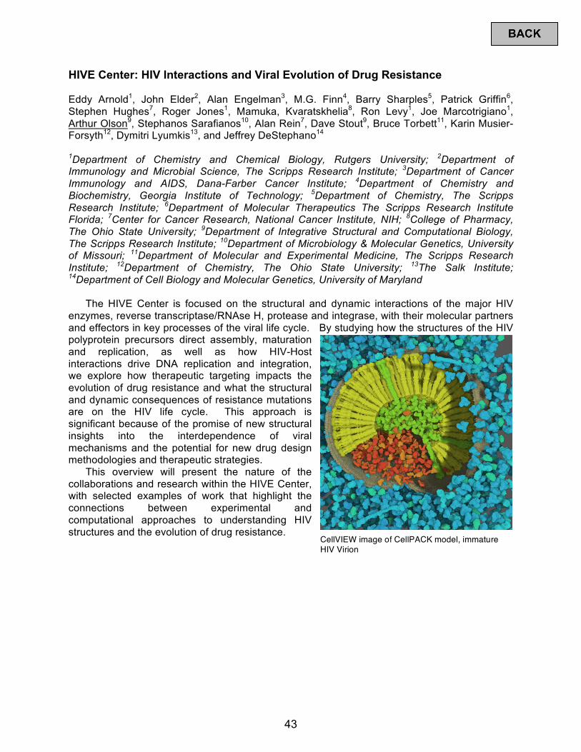
HIVE Center: HIV Interactions and Viral Evolution of Drug Resistance Eddy Arnold1, John Elder2, Alan Engelman3, M.G. Finn4, Barry Sharples5, Patrick Griffin6, Stephen Hughes7, Roger Jones1, Mamuka, Kvaratskhelia8, Ron Levy1, Joe Marcotrigiano1, Arthur Olson9, Stephanos Sarafianos10, Alan Rein7, Dave Stout9, Bruce Torbett11, Karin Musier-Forsyth12, Dymitri Lyumkis13, and Jeffrey DeStephano14 1Department of Chemistry and Chemical Biology, Rutgers University;; 2Department of Immunology and Microbial Science, The Scripps Research Institute;; 3Department of Cancer Immunology and AIDS, Dana-Farber Cancer Institute;; 4Department of Chemistry and Biochemistry, Georgia Institute of Technology;; 5Department of Chemistry, The Scripps Research Institute;; 6Department of Molecular Therapeutics The Scripps Research Institute Florida;; 7Center for Cancer Research, National Cancer Institute, NIH;; 8College of Pharmacy, The Ohio State University;; 9Department of Integrative Structural and Computational Biology, The Scripps Research Institute;; 10Department of Microbiology & Molecular Genetics, University of Missouri;; 11Department of Molecular and Experimental Medicine, The Scripps Research Institute;; 12Department of Chemistry, The Ohio State University;; 13The Salk Institute;; 14Department of Cell Biology and Molecular Genetics, University of Maryland
The HIVE Center is focused on the structural and dynamic interactions of the major HIV enzymes, reverse transcriptase/RNAse H, protease and integrase, with their molecular partners and effectors in key processes of the viral life cycle. By studying how the structures of the HIV polyprotein precursors direct assembly, maturation and replication, as well as how HIV-Host interactions drive DNA replication and integration, we explore how therapeutic targeting impacts the evolution of drug resistance and what the structural and dynamic consequences of resistance mutations are on the HIV life cycle. This approach is significant because of the promise of new structural insights into the interdependence of viral mechanisms and the potential for new drug design methodologies and therapeutic strategies.
This overview will present the nature of the collaborations and research within the HIVE Center, with selected examples of work that highlight the connections between experimental and computational approaches to understanding HIV structures and the evolution of drug resistance.
CellVIEW image of CellPACK model, immature HIV Virion
43
HIV-1 Integrase Binds the Viral RNA Genome and is Essential During Virion Morphogenesis Jacques J. Kessl1,2,3*, Sebla B. Kutluay4,5*, Dana Townsend5, Stephanie Rebensburg2,3, Alison Slaughter1,3, Ross C. Larue2,3, Nikoloz Shkriabai2,3, Nordine Bakouche2,3, James R. Fuchs1,3, Paul D. Bieniasz4,6,7, Mamuka Kvaratskhelia1,2,3 1HIVE Center, 2Center for Retrovirus Research, 3College of Pharmacy, The Ohio State University, Columbus, OH 43210;; 4CRNA Center, 5Department of Molecular Microbiology, Washington University School of Medicine, St. Louis, MO 63110;; 6Laboratory of Retrovirology, 7Howard Hughes Medical Institute, Aaron Diamond AIDS Research Center, The Rockefeller University, New York, NY 10016, USA;; *These authors contributed equally
While an essential role of HIV-1 integrase (IN) for integration of viral cDNA into human chromosome is established, studies with IN mutants and allosteric IN inhibitors (ALLINIs) have suggested that IN can also influence viral particle maturation. However, it has remained enigmatic as to how IN contributes to virion morphogenesis. Using crosslinking-immunoprecipitation sequencing we have uncovered that IN binds the viral RNA genome in virions. These interactions have specificity as IN exhibits distinct preference for select viral RNA structural elements. We show that IN substitutions that selectively impair its binding to viral RNA result in eccentric, non-infectious virions without affecting nucleocapsid-RNA interactions. Likewise, ALLINIs impair IN binding to viral RNA in virions of wild type but not escape mutant virus. These results reveal an unexpected biological role of IN binding to the viral RNA genome during virion morphogenesis and elucidate the mode of action of ALLINIs.
44
A New Class of Allosteric HIV-1 Integrase Inhibitors Identified by Crystallographic Fragment Screening of the Catalytic Core Domain Disha Patel,1 Janet Addae-Antwi,2 Pratibha C. Koneru,3 Erik Serrao,4 Stefano Forli,5 Jacques Kessl,3 Lei Feng,3 Nanjie Deng,6 Ronald M. Levy,6 James R. Fuchs,2 Arthur J. Olson,5 Alan N. Engelman,4 Joseph D. Bauman,1 Mamuka Kvaratskhelia,3 and Eddy Arnold1 1Center for Advanced Biotechnology and Medicine, Rutgers University, Piscataway, NJ;; 2Division of Medicinal Chemistry and Pharmacognosy, The Ohio State University, Columbus, OH;; 3Center for Retrovirus Research and College of Pharmacy, The Ohio State University, Columbus, OH;; 4Department of Cancer Immunology and Virology, Dana-Farber Cancer Institute and Department of Medicine, Harvard Medical School, Boston, MA;; 5Department of Integrative Structural and Computational Biology, The Scripps Research Institute, La Jolla, CA;; 6Center for Biophysics and Computational Biology, Temple University, Philadelphia , PA
HIV-1 integrase (IN) is essential for virus replication and represents an important multifunctional therapeutic target. Recently discovered quinoline-based allosteric IN inhibitors (ALLINIs) potently impair HIV-1 replication and are currently in clinical trials. ALLINIs exhibit a multimodal mechanism of action by inducing aberrant IN multimerization during virion morphogenesis and competing with IN for binding to its cognate cellular cofactor LEDGF/p75 during early steps of HIV-1 infection. However, quinoline-based ALLINIs have a low genetic barrier for the selection of resistant phenotypes, which highlights a need for discovery of second generation inhibitors. Using crystallographic screening of a library of 971 fragments against the HIV-1 IN catalytic core domain (CCD) followed by a fragment expansion approach, we have identified thiophenecarboxylic acid-derivatives that bind at the CCD-CCD dimer interface at the principal LEDGF/p75 binding pocket. The most active fragment inhibited LEDGF/p75-dependent HIV-1 IN activity in vitro with an IC50 of 72 μM and impaired HIV-1 infection of T cells at an EC50 of 36 μM. These findings are significant because the identified lead fragment with a relatively small molecular weight (221 Da) provides an optimal building block for developing a new class of inhibitors. Furthermore, while structurally distinct thiophenecarboxylic acid-derivatives target a similar pocket at the IN dimer interface as the quinoline-based ALLINIs, the lead fragment 5 inhibited IN mutants that confer resistance to quinoline-based compounds. Collectively, our findings provide a plausible path for structure-guided development of second generation ALLINIs.
45
Crystal Structure of Prototype Foamy Virus PR-RT Polyprotein Shows Unique Architecture Among Retroviruses: Implications for Function Jerry Joe Harrison,1 Steve Tuske,1 Kalyan Das,1 Joseph D. Bauman,1 Paul L. Boyer,2 Stephen H. Hughes,2 and Eddy Arnold1 1Center for Advanced Biotechnology and Medicine, and Departments of Chemistry and Chemical Biology, and Medicinal Chemistry, Rutgers University, Piscataway, NJ;; 2HIV Drug Resistance Program, National Cancer Institute, Frederick, MD
Retroviruses conventionally synthesize their proteins from polycistronic mRNAs into polyproteins, and the polyproteins are proteolytically processed by the viral proteases into functional entities. We have engaged in systematic structural study of the Pol polyprotein of prototypic foamy virus (PFV), a surrogate of HIV. Here, we report the crystal structure of the protease-reverse transcriptase (PR-RT) polyprotein of PFV that functions as the viral transcription and processing machinery. While the monomeric PFV PR exhibits similar architecture as the HIV-1 PR, it contains a C-terminal extension containing two helices, associated with the equivalent of HIV-1 RT helices E and F beneath the base of the palm subdomain. The PR is anchored to the RT adjacent to the fingers and palm subdomains by these two helices via a long loop. This arrangement allows the PR to swing open to enable dimerization without disturbing the integrity of the RT;; like in HIV, PFV PR is active as a dimer. The structural components (the fingers, palm, and thumb subdomains) are highly conserved in all retroviruses and retrotransposon RTs that carry out the addition of deoxynucleotides to a DNA strand using a common catalytic mechanism. The organization of the polymerase (fingers, palm, thumb, connection, and RNase H) in PFV PR-RT is significantly different when compared to other retroviral polymerases. Three functional entities PR, RT, and RNase H are connected by flexible linkers. Notably, the thumb C-terminus and RNase H N-terminus are connected to the connection subdomain via long flexible linkers that permit spatial rearrangement of the subdomains. The novel spatial arrangements of the thumb, connection, and RNase H in the current structure suggest that the PFV PR-RT would undergo significant conformational rearrangements upon nucleic acid binding. This structure opens up the frontiers for modeling the polyproteins of other retroviruses such as HIV-1 to enhance our understanding of retroviral polyprotein processing.
46
Visualization of Viral RNA and DNA Dynamics During HIV Infection Maritza Puray-Chavez1,2, Andrew D. Huber1,3, Dandan Liu1,2, Obiaara Ukah1,2, Vincent Yapo1,2, Juan Ji1, Jennifer J. Wolf1,2, Gregory A. Sowd4, Alan N. Engelman4 and Stefan G. Sarafianos1,2,3,5
1Christopher S. Bond Life Sciences Center, 2Molecular Microbiology & Immunology, 3Veterinary Pathobiology, University of Missouri, Columbia, MO;; 4Department of Cancer Immunology and Virology, Dana-Farber Cancer Institute, Boston, MA;; 5Biochemistry, University of Missouri, Columbia, MO
To better understand the HIV life cycle, we developed a fluorescence microscopy approach which utilizes branched DNA in situ hybridization technology on fixed cells to visualize single copies of viral RNA (vRNA) and DNA (vDNA). Viral nucleic acids were observed following infection at relatively low multiplicity of infection (MOI, typically 0.5) in semi-synchronized or synchronized cells. Target cells included HeLa-derived TZM-bl, lymphocytes (H9, Jurkat, MT-2), monocytes (THP-1), HEK293T, and primary cells.
Protocols were developed to simultaneously monitor in individual fixed cells unspliced and spliced+unspliced vRNA, negative sense vDNA, and integrated proviral DNA. Specificity was confirmed a) by the absence of fluorescence signal in uninfected cells, b) susceptibility of the vRNA, but not of the vDNA fluorescence signal to RNase A treatment of the samples prior to hybridization, and c) susceptibility of the vDNA, but not of the vRNA fluorescence signal to DNase I treatment prior to hybridization. Specific detection of HIV-1 and HIV-2 enabled visualization of HIV-1/HIV-2 co-infection events in individual cells.
Reverse transcription in TZM-bl cells was generally completed by ~10 hours post infection (hpi) as judged by the gradual disappearance of vRNA signal, presumably due to RNase H activity, with concomitant increase of nascent vDNA in the cytoplasm. vRNA signal was rescued in the presence of an RNase H inhibitor. We commonly observed co-localization of HIV capsid (CA) with vRNA and nascent vDNA that were presumably encapsidated. Both cytoplasmic and proviral DNA signals disappeared upon EFdA, or AZT treatment, whereas raltegravir specifically suppressed proviral/nuclear, but not cytoplasmic vDNA.
At low MOIs, we typically observed one or two integration sites. Spliced vRNA was observed exported from the nucleus prior to unspliced vRNA. Unspliced RNA transcripts began to appear at ~12 hpi, centered at individual transcription sites and at times appearing as transcriptional “bursts” co-localized with integration sites. Notably, not all integrated DNA sites appeared transcriptionally active. Large amounts of nascent unspliced RNA transcripts accumulated in the nucleus until ~14-16 hpi, presumably when Rev enabled their highly efficient export from the nucleus. CA protein also appeared in the cytoplasm at ~14-16 hpi. At ~24 hpi, new virions appeared to infect nearby cells. We have expanded this approach to monitor distribution of vRNA and vDNA upon knockdown or knockout of host factors, including CPSF6, LEDGF/p75, CypA, or MOV10, or upon treatment with additional antivirals targeting reverse transcriptase, integrase, or CA.
47
Dynamic Regulation of HIV-1 Capsid Assembly, Maturation, and Interactions with Host Factors by Integrated MAS NMR and MD Simulations Manman Lu1,2, Huilan Zhang1,2, Guangjin Hou1,2, Rupal Gupta1,2, Caitlin Quinn1,2, Mingzhang Wang1,2, Jinwoo Ahn2,3, In-Ja Byeon2,3, Sherimay Ablan4, Juan R. Perilla5, Christopher J. Langmead6, David A. Case7, Christopher Aiken2,8, Eric O. Freed4, Peijun Zhang2,3, Klaus Schulten5, Angela M. Gronenborn2,3, and Tatyana Polenova1,2 1Department of Chemistry and Biochemistry, University of Delaware, Newark, Delaware 19716;; 2Pittsburgh Center for HIV Protein Interactions, University of Pittsburgh School of Medicine, 1051 Biomedical Science Tower 3, 3501 Fifth Ave., Pittsburgh, PA 15261;; 3Department of Structural Biology, University of Pittsburgh School of Medicine, 3501 Fifth Ave., Pittsburgh, PA 15261;; 4HIV Dynamics and Replication Program, Center for Cancer Research, National Cancer Institute, Frederick, MD 21702;; 5University of Illinois, Theoretical and Computational Biophysics Group, Urbana, IL 61801;; 6Computer Science Department, Carnegie Mellon University, Gates Hillman Center, 5000 Forbes Avenue, Pittsburgh, PA;; 7BioMaPS Institute and Department of Chemistry and Chemical Biology, Rutgers University, 147 Frelinghuysen Road, Piscataway, NJ 08854-8087;; 8Department of Pathology, Microbiology and Immunology, Vanderbilt University Medical Center, Nashville, TN 37232
HIV-1 capsids, assembled from ~1,500 copies of the capsid (CA) protein, are an integral part of mature virions [1, 2]. Conical in shape, capsids enclose the viral genetic material (two copies of RNA) together with several proteins that are essential for viral replication. In the assembled state, capsids are remarkably dynamic, with the CA residue motions occurring over a range of timescales from nano- to milliseconds [3-6]. These motions are functionally important for capsid’s assembly, viral maturation, and interactions with host factors [3, 6]. In this talk, I will discuss the recent progress in atomic-resolution structural and dynamics studies of CA assemblies by magic angle spinning (MAS) NMR spectroscopy. I will present an integrated MAS NMR and MD simulations approach to probe functionally important motions in assemblies of CA and their complexes with host factor Cyclophilin A (CypA), as well as assemblies of CA-SP1 maturation intermediates. I will discuss the role of dynamic allosteric regulation in capsid’s assembly, maturation, and escape from the CypA dependence. Our studies demonstrate that the integration of experimental NMR methods and theory yields quantitative, atomic-level insights into the dynamic processes that govern the HIV-1 capsid’s function. 1. Briggs J. A. G., Simon M. N., Gross I., Krausslich H. G., Fuller S. D., Vogt V. M., Johnson M. C. Nat
Struct Mol Biol. 11(7);; p. 672-675 (2004). 2. Sundquist W. I., Krausslich H. G. Csh Perspect Med. 2(7) (2012). 3. Byeon I. J. L., Hou G. J., Han Y., Suiter C. L., Ahn J., Jung J., Byeon C. H., Gronenborn A. M., Polenova
T. J Am Chem Soc. 134(14);; p. 6455-6466 (2012). 4. Zhao G. P., Perilla J. R., Yufenyuy E. L., Meng X., Chen B., Ning J. Y., Ahn J., Gronenborn A. M.,
Schulten K., Aiken C., Zhang P. J. Nature. 497(7451);; p. 643-646 (2013). 5. Bayro M. J., Chen B., Yau W. M., Tycko R. J Mol Biol. 426(5);; p. 1109-1127 (2014). 6. Lu M. M., Hou G. J., Zhang H. L., Suiter C. L., Ahn J., Byeon I. J. L., Perilla J. R., Langmead C. J.,
Hung I., Gor'kov P. L., Gan Z. H., Brey W., Aiken C., Zhang P. J., Schulten K., Gronenborn A. M., Polenova T. P Natl Acad Sci USA. 112(47);; p. 14617-14622 (2015).
48

HIV-1 and HIV-2 Exhibit Divergent Interactions with DNA Repair Enzymes Jacek Skowronski Case Western Reserve School of Medicine, Cleveland, OH
In non-dividing host cells HIV is targeted by intrinsic anti-viral defense mechanisms that introduce marks of damage into viral cDNA, thereby tagging it for processing by the cellular DNA repair machinery whose role(s) in HIV replication cycle is not well understood. HIV-1 virion-associated accessory virulence factor Vpr hijacks CRL4DCAF1 E3 ubiquitin ligase and coordinates interactions with the postreplication DNA repair machinery, but the extent of its interactions with DNA repair proteins hasn’t been thoroughly characterized. Here, using proteomics and functional assays we identify HLTF, a postreplication DNA repair helicase, as a new common target of HIV-1/SIVcpz Vpr proteins. We show that HIV-1 Vpr reprograms CRL4DCAF1 E3 to direct HLTF for proteasome-dependent degradation, in a manner that is independent from previously reported Vpr interactions with base excision repair enzyme UNG2 and crossover junction endonuclease MUS81, which Vpr also directs for degradation via CRL4DCAF1 E3. Thus, separate functions of HIV-1 Vpr usurp CRL4DCAF1 E3 to remove key enzymes in three distinct DNA repair pathways. In contrast, we find that HIV-2 Vpr is unable to efficiently program HLTF or UNG2 for degradation. Our findings reveal complex interactions between HIV-1 and the DNA repair machinery, suggesting that DNA repair plays important roles in HIV-1 life cycle. The divergent interactions of HIV-1 and HIV-2 with DNA repair enzymes imply that these viruses use different strategies to guard their DNA genomes and/or facilitate their replication in the host.
49
To Repair or Not Repair: The X-Ray Crystal Structure of the DDB1-DCAF1-Vpr-UNG2 Complex Guillermo Calero, Ying Wu, Xiaohong Zhou, Christopher O. Barnes, Maria DeLucia, Aina E. Cohen, Angela M. Gronenborn, Jinwoo Ahn University of Pittsburgh, Pittsburgh, Pennsylvannia
The HIV-1 accessory protein Vpr is required for efficient viral infection of macrophages and promotion of viral replication in T-cells. The biological activities of Vpr are closely tied to the interaction with DCAF1, a cellular substrate receptor of the Cullin4-RING E3 ubiquitin ligase (CRL4) of the host ubiquitin proteasome-mediated protein degradation pathway. At present, the molecular details of how Vpr usurps the protein degradation pathway have not been delineated. Here, we present the crystal structure of the DDB1-DCAF1-HIV-1-Vpr-Uracil-DNA glycosylase (UNG2) complex. The structure reveals how Vpr engages with DCAF1 and creates the special binding interface for UNG2 recruitment, distinct from how the related Vpx proteins recruit SAMHD1 for degradation. Vpr and Vpx utilize similar N-terminal and helical regions to bind the substrate receptor, while distinctly different regions target the specific cellular substrates. Furthermore, Vpr employs molecular mimicry of DNA by a variable loop for specific recruitment of the UNG2 substrate.
50
Introduction to the CHEETAH Center Wes Sundquist Department of Biochemistry, University of Utah
51
D-Peptide Fusion Inhibitor Protects Against High-Dose SHIV Challenge Michael S. Kay Department of Biochemistry, University of Utah School of Medicine
Peptides excel at disrupting “undruggable” protein-protein interactions, but their in vivo fragility hampers their therapeutic potential. Our lab develops mirror-image D-peptide inhibitors that resist proteases and enjoy extended half-lives. We employ high-throughput mirror-image phage display screening and structure-based design to identify D-peptides that block viral entry. Our anti-HIV D-peptide (cholesterol-conjugated PIE12-trimer) is a pM inhibitor of viral entry and is highly active against in the macaque SHIV infection model for both prevention and treatment. This D-pepitde is now in advanced preclinical development and is ideally suited for sustained-release formulation.
52
Visualizing HIV Uncoating in Living Cells Thomas J. Hope Departments of Cell and Molecular Biology, OB/GYN, and Biomedical Engineering, Northwestern University
Individual viral particle tracking and analysis has become widely utilized as a tool to study the cell biology of the early steps of HIV replication. Although this approach has advanced the field, it has faced the important criticism that it is not possible to filter the observations to focus on the particles that ultimately infect a cell. Utilizing variable time lapse imaging of multiple fields of cells over periods up to 36 hours allows the identification of productively infected cells as visualized by reporter virus expression. Infection with very low MOI allows conditions where there is less than one labeled particle per cell. In this way correlation of individual particle behavior can be directly connected to infection. We have recently developed the use of intravirion fluid phase markers to determine the integrity of the HIV conical capsid core. To visualize dynamic changes in capsid integrity and composition, we utilized the HIV-iGFP construct. During viral maturation of HIV-iGFP, the GFP is liberated from Gag;; a minority population of the free GFP is trapped in the capsid, while the remaining free GFP is located outside of the capsid. With this technique, the loss of the fluid phase GFP occurs in two steps, with fusion and upon the loss of capsid core integrity. Live-cell microscopy of HIV-iGFP virions with a second color labeled viral core protein such as Vpr or Integrase allows for the precise timing of these two steps thereby revealing the kinetics, localization and composition of the early steps of HIV-1 infection. Our studies reveal that the time between fusion and capsid integrity loss, for both HIV and VSV-G mediated fusion, in tissue culture and primary cells (macrophages and T cells), is approximately 25 minutes. Also, capsid integrity loss occurs entirely in the cytoplasm. However, we also detect a second species, which does not experience a change in capsid integrity over 2 hours of imaging. Through viral challenge with less than one particle per cell over a long period of time, we were able to image individual particle capsid integrity loss that produces a viable infection. This analysis revealed that all particles associated with cellular infection showed changes in capsid integrity ~25 minutes. Together, these observations validate the early cytoplasmic uncoating model.
53
Structural Studies of TRIM5α Assembly Barbie K. Ganser-Pornillos Dept. of Physiology and Biological Physics, University of Virginia
TRIM5α is a restriction factor that recognizes the incoming capsids of retroviruses. Although the exact mechanism of restriction is still poorly understood, capsid recognition leads to accelerated capsid dissociation, disrupted reverse transcription, and enhanced interferon signaling.
TRIM5α is a member of a large family of proteins that contain a conserved RBCC motif consisting of a RING domain, one or two B-box domains, and a coiled-coil domain. TRIM5α also has a C-terminal SPRY domain that directly binds the viral capsid. Efficient capsid recognition requires higher-order assembly of TRIM5α, which is mediated by the RBCC motif. We have previously shown that TRIM5-21R, an artificial rhesus TRIM5 chimera that contains the RING domain of human TRIM21, spontaneously assembles into large hexagonal nets. Assembly of the TRIM5-21R hexagonal net can be induced in vitro by mimics of the HIV-1 capsid. We have therefore proposed that complementarity between the TRIM nets and underlying CA hexagonal lattice drives avid capsid recognition.
We now report that hexagonal lattice assembly is a conserved property of TRIM5α homologs from different mammalian species. 2D hexagonal arrays of HIV-1 CA can induce assembly of HIV-restrictive TRIM5α isoforms, but not non-restrictive isoforms. Imaging and analysis by cryoEM and cryoET revealed that the hexagonal TRIM lattice wraps around the curved CA lattices in HIV-1 CA tubes and purified cores. The SPRY domain was localized by cryoET analysis to the 2-fold symmetric hexagon edges in the TRIM lattice, and a combination of crystallographic, biochemical, and virological experiments demonstrated that the 3-fold symmetric vertices consist of B-box 2 domain trimers. Finally, we suggest how the lattice achieves the structural flexibility necessary for TRIM5 to accommodate the variable curvature and pleomorphism of retroviral capsids. Thus, our studies have revealed important principles that explain how TRIM5 proteins recognize retroviral capsid.
54
Structure and Function of RNA and Proteins from Natural Sequence Variation Debora S. Marks Systems Biology & CBMI, Raymond and Beverly Sackler Laboratory for Computational Biology, Harvard Medical School
Modern genome sequencing and synthesis can acquire and generate tremendous molecular diversity in a day, but our ability to navigate and interpret the exponentially large space of possible biological sequences remains limited. We mine the evolutionary sequence record to derive precise information about the function and structure of RNAs and RNA-protein complexes. As in protein structure prediction, we use maximum entropy global probability models of sequence co-variation to infer evolutionarily constrained nucleotide-nucleotide interactions within RNA molecules and nucleotide-amino acid interactions in RNA-protein complexes. The predicted contacts allow all-atom blinded 3D structure prediction at good accuracy for several known RNA structures and RNA-protein complexes. For unknown structures, we predict contacts in 160 non-coding RNA families. Beyond 3D structure prediction, evolutionary couplings help identify important functional interactions—e.g., at switch points in riboswitches and at a complex nucleation site in HIV. Aided by increasing sequence accumulation, evolutionary coupling analysis can accelerate the discovery of functional interactions and 3D structures involving RNA.
55
Structural Basis for Inhibitor-Induced Aggregation of HIV Integrase Kushol Gupta2*, Vesa Turkki1*, Scott Sherrill-Mix1, Young Hwang1, Grant Eilers1, Louis Taylor, Charlene McDanal4, Ping Wang4, David Temelkoff4, Robert T. Nolte3, Emile Velthuisen4, Jerry Jeffrey4, Gregory D. Van Duyne2+ and Frederic D. Bushman1+ 1From the Perelman School of Medicine, University of Pennsylvania. Department of Microbiology 3610 Hamilton Walk Philadelphia, PA 19104-6076 U.S.A.;; 2Department of Biochemistry and Biophysics 242 Anatomy-Chemistry Building Philadelphia, PA, 19104-6059 U.S.A.;; 3Protein Cellular and Structural Science GlaxoSmithKline Collegeville, PA 19426 U.S.A.;; 4HIV DPU Infectious Disease Therapy Area Unit GlaxoSmithKline RTP, NC 27709 U.S.A.;; *these authors contributed equally;; +corresponding authors
The allosteric integrase (IN) inhibitors (ALLINIs) target the viral-encoded IN protein and interfere with HIV replication via disruption of viral particle assembly late during HIV replication. To investigate their inhibitory mechanism, we crystallized full-length HIV-1 IN bound to the ALLINI GSK1264, and determined the structure of the complete inhibitor interface at 4.4 Å resolution. In this structure, GSK1264 is buried between the catalytic core domain of one IN dimer and the CTD of an adjacent IN dimer. The amino-terminal domain is not resolved in the structure but does not participate in GSK1264 binding. The GSK1264 binding interface is rich in residues implicated in IN oligomerization and ALLINI sensitivity, indicating likely functional significance. The IN-IN interaction mediated by GSK1264 leads to formation of an open polymer in the crystal, a polymerization reaction that is readily reproduced in solution with purified components. To probe ALLINI function more broadly, we compared the properties of several ALLINIs in biochemical, virological, and electron microscopic assays. Several ALLINI escape mutations encode IN substitutions at or near the inhibitor binding site, and these also resulted in decreased IN oligomerization in vitro. The results support a mechanism where ALLINIs disrupt viral particle maturation by promoting formation of the IN polymers observed in the IN-GSK1264 crystal structure. Additionally, the results support a structural model for the catalytically inactive IN tetramer discussed in several previous studies. Identification of the interface responsible for polymer formation provides data useful for improving HIV inhibitors and helps explain a wealth of previous studies of HIV IN.
56
Biochemical Reconstitution of Selective HIV-1 Genome Packaging Lars-Anders Carlson1, Yun Bai1,3, Sarah C. Keane2, Jennifer A. Doudna1, and James H. Hurley1 1Department of Molecular and Cell Biology, University of California, Berkeley;; 2University of Maryland, Baltimore County;; 3Current address: School of Life Science and Technology, ShanghaiTech University, Shanghai, 201210, China
HIV-1 Gag selectively packages the genomic viral RNA into assembling virus particles in the context of a large excess of cytosolic human RNAs. As Gag assembles on the plasma membrane, the HIV-1 genome is enriched relative to cellular RNAs by a mechanism which is incompletely understood. We used a minimal system consisting of purified RNAs, recombinant myristylated HIV-1 Gag and giant unilamellar vesicles to recapitulate the selective packaging of the 5' untranslated region of the HIV-1 genome in the presence of excess competitor RNA. In this assay, the Rev-responsive element and a non-viral control RNA were not selected for to the same degree. Mutations in the CA-CTD domain of Gag which subtly affect the self-assembly of Gag on membranes abrogated RNA selectivity. We further found that tRNA suppresses Gag membrane binding less when Gag has bound viral RNA. The ability of HIV-1 Gag to selectively package its RNA genome and its self-assembly on membranes are thus interdependent on one another.
57
Structure of a Natively-Glycosylated HIV-1 Env Reveals a New Mode for VH1-2 Antibody Recognition of the CD4 Binding Site Relevant to Vaccine Design Harry B. Gristick1*, Lotta von Boehmer2*, Anthony P. West Jr.1, Michael Schamber1, Michel C. Nussenzweig2,3, and Pamela J. Bjorkman1 1Division of Biology and Biological Engineering, California Institute of Technology, Pasadena, CA 91125, USA;; 2Laboratory of Molecular Immunology and 3Howard Hughes Medical Institute, The Rockefeller University, New York, NY 10065, USA;; *These authors contributed equally.
The trimeric HIV-1 envelope glycoprotein (Env), the only target of neutralizing antibodies on HIV-1, includes glycans constituting up to 50% of its mass attached to 30±3 potential N-linked glycosylation sites (PNGSs) per gp120-gp41 protomer. Viral glycans are generally non-immunogenic because they are assembled by host cell machinery, thus the carbohydrates decorating the surface of Env constitute a “glycan shield” that reduces access to underlying immunogenic protein epitopes. Structural studies of broadly neutralizing antibodies (bNAbs) bound to Env trimers have revealed mechanisms by which bNAbs targeting various epitopes penetrate the glycan shield to either accommodate or include N-glycans in their epitopes. Although accessibility to the conserved host receptor (CD4) binding site (CD4bs) is restricted by surrounding glycans, VRC01-class bNAbs mimic CD4 binding to share a common mode of gp120 binding and glycan accommodation using a VH1-2*02-derived variable heavy (VH) domain. While attractive candidates for immunogen design, features of VRC01-class bNAbs such as a high degree of somatic hypermutation (SHM) and a short (5-residue) light chain (LC) complementarity determining region 3 (CDRL3) (found in only 1% of human LCs) suggest they might be difficult to elicit through vaccination. Here we present a crystal structure of a fully- and natively-glycosylated Env trimer, which we use to define the complete epitopes of IOMA, a new class of CD4-mimetic bNAb, and the V3-loop-directed bNAb 10-1074. Although derived from VH1-2*02, IOMA has a normal-length CDRL3 and includes fewer SHMs than VRC01-class bNAbs, suggesting it may be easier to elicit than VRC01-class bNAbs. Analysis of the native glycan shield on HIV-1 Env allows the first full description of the interplay between heterogeneous untrimmed high-mannose and complex-type N-glycans within the CD4bs, V3-loop, and other epitopes on Env, revealing antibody-vulnerable glycan holes and roles of complex-type N-glycans on Env that are relevant to vaccine design.
58
Overview and Progress Summary for P01GM056550: Structure-Based Antagonism of HIV-1 Envelope Function in Cell Entry Irwin Chaiken1, Cameron Abrams1, Ernesto Freire2, Wayne Hendrickson3, Walther Mothes4, Joseph Sodroski5 and Amos B. Smith III6 1Drexel University;; 2Johns Hopkins University;; 3Columbia University;; 4Yale University;; 5Harvard University;; 6University of Pennsylvania
Our Program employs a synergistic approach to investigate strategies for inhibiting HIV-1 gp120 in the metastable virus Env protein trimer. We identify and develop gp120 inhibitory compositions, and at the same time use inhibitors to define fundamental concepts, mechanisms and targetable vulnerabilities in Env. It is now well established that Env can traverse an energetically favorable cascade of host cell CD4 and co-receptor interactions that in turn integrate conformational rearrangements leading to virus-cell and cell-cell membrane fusion, entry and infection. A hallmark achievement of the Program has been to identify stages along this “activation trajectory” at which different classes of molecules can block progression to entry. A prime example has been the development of small molecule CD4 mimetics (SMCMs) that, while minimizing enhancement of infection in CD4-negative cells, are able to sensitize Env gp120 for neutralizing antibody binding, with promising consequences for prevention and treatment. At the same time, Env inhibitors have been identified that can divert the conformationally dynamic Env trimer towards an “inactivation trajectory”. A recently developed path of work has led to macrocyclic peptide triazoles (cPTs) that dual antagonize gp120 receptor binding sites, disrupt Env trimer assembly before cell encounter and inactivate virus, including with antagonist variants that appear to trigger Env transformations similar to those found in the activation trajectory. The thermodynamic behavior of SMCMs and cPTs are unique and reflect the different conformational transformation pathways used. The binding sites for inhibitors and inactivators are being investigated by a multi-disciplinary approach including crystallography, computation, mutagenesis and patterns of virus resistance. Intriguingly, SMCMs and cPTs share overlapping binding sites, including in the context of emerging trimer structures, yet with different functional outcomes. The structural mechanisms of binding are being used to advance inhibitor designs. Overall, these efforts are expanding our understanding of both the structural and dynamic mechanisms of the HIV-1 Env protein entry machine, and molecular strategies available to hijack the machinery built into the Env protein for potential use in disease intervention and prevention.
59
Conformational Transformations Triggered by Different Inhibitor Classes Arne Schön and Ernesto Freire Johns Hopkins University
Cell infection by HIV-1 requires the activation of the envelope glycoprotein of HIV-1 into an infection competent state. At the molecular level, this process involves binding of gp120 to the cell surface receptor CD4, and a conformational change triggered by that same binding event. The nature of this conformational change has been examined thermodynamically and characterized as a process involving the folding of intrinsically disordered domains. The complex nature of the mechanism of action of gp120 suggests different strategies to inhibit cell entry, including competitive inhibition and/or trapping gp120 into an unproductive conformation. In this Program Project, competitive and non-competitive inhibitors have been identified and their antiviral potency is being optimized. The coupling between binding and conformational equilibrium requires not only optimization of the binding affinity, but of the conformational effects of binding. In the case of competitive inhibitors (small molecule CD4 mimetics) it is required that the inhibitors do not become agonists and trigger the same changes as CD4. In the case of non-competitive inhibitors (peptide triazoles and macrocyclic peptide triazoles) it is necessary to drive gp120 into an unproductive conformation with as much free energy as possible. Each class of inhibitor is characterized by a distinct thermodynamic signature that serves as a blueprint for optimization. In this presentation, the thermodynamic characteristics of both classes of inhibitors will be discussed.
60
1
An Intermediate State of the HIV-1 Envelope Glycoproteins on the Entry Pathway Alon Herschhorn1,2, Xiaochu Ma3, Christopher Gu1, John D. Ventura3, Bruno Melillo4, Daniel S. Terry5, Amos B. Smith III4, Scott C. Blanchard5, James B. Munro6, Walther Mothes3, Andrés Finzi7 and Joseph Sodroski1,2,8
1Department of Immunology Cancer and Virology, Dana-Farber Cancer Institute, Boston, MA 02215, USA;; 2Department of Microbiology and Immunobiology, Harvard Medical School, Boston, MA 02215, USA;; 3Department of Microbial Pathogenesis, Yale University School of Medicine, New Haven, CT 06536, USA;; 4Department of Chemistry, University of Pennsylvania, Philadelphia, PA 19104, USA;; 5Department of Physiology and Biophysics, Weill Cornell Medical College of Cornell University, New York, NY 10021, USA;; 6Department of Molecular Biology and Microbiology, Tufts University School of Medicine and Sackler School of Graduate Biomedical Sciences, Boston, MA 02110, USA;;7Centre de Recherche du CHUM and Department of Microbiology, Infectiology and Immunology Université de Montréal, Montreal, Quebec, Canada;; Department of Microbiology and Immunology, McGill University, Montreal, Quebec, Canada;; 8Department of Immunology and Infectious Diseases, Harvard T. H. Chan School of Public Health, Boston, MA 02115, USA
Primary human immunodeficiency virus (HIV-1) envelope glycoprotein (Env) trimers (gp120/gp41)3 typically exist in a metastable closed conformation (State 1). Binding the CD4 receptor triggers Env to undergo extensive conformational changes to mediate virus entry. We identified specific gp120 residues that control HIV-1 Env conformation. Changes in these residues regulate transitions to downstream conformations and allow the Env to sample a functional conformation (State 2) intermediate between State 1 and the full CD4-bound state (State 3). State 2 was an obligate intermediate for all transitions between State 1 and State 3 and increased State 2 occupancy was associated with lower energy barriers between the states. State-2-enriched Envs required lower CD4 concentrations to trigger virus entry and exhibited an improved ability to infect primary macrophages. These Envs were resistant to several broadly neutralizing antibodies and small-molecule inhibitors. Thus, State 2 is an Env conformation on the virus entry pathway;; frequent sampling of State 2 increases the adaptability of HIV-1 to different host cell receptor levels and immune environments. Our results provide new insights into the conformational regulation of HIV-1 entry.
61
Project Overview - The Interdependency of Drug Resistance Evolution and Drug Design: HIV-1 Protease, a Case Study R. Swanstrom2, W. Sherman3, A. Ali1, K. Zeldovich1, N. Kurt-Yilmaz1, S Lee2, R. Ishima4, J. Luban1, D. Bolon1, C. Schiffer1 1UMass Medical School, Worcester, MA;; 2University of North Carolina School of Medicine, Chapel Hill, NC;; 3Schrodinger, Inc.;; 4School of Medicine, University of Pittsburgh
In this project we are elucidating the underlying interdependencies under which drug resistance is selected using HIV-1 protease as a model system. Drug resistance occurs when the balance favors viral replication between proteolytic activity within the virus and enzyme inhibition. We combine analysis of sequence diversity, structure, dynamics and energetics to elucidate the underlying pathways and additional macromolecular pressures that contribute to drug resistance.
In this year we have probed the interdependency of resistant variants that arose from viral passaging, through a combination of structural, dynamic and mutational methods. In each case we bridge experimental and computational techniques and are developing a database where multiple data sets can be probed to elucidate their interdependencies.
62
Establish a Strategy to Elucidate Molecular Mechanisms Leading to Drug Resistance R. Swanstrom2, W. Sherman3, A. Ali1, K. Zeldovich1, N. Kurt-Yilmaz1, S Lee2, R. Ishima4, J. Luban1, D. Bolon1, C. Schiffer1 1UMass Medical School, Worcester, MA;; 2University of North Carolina School of Medicine, Chapel Hill, NC;; 3Schrodinger, Inc.;; 4School of Medicine, University of Pittsburgh
We hypothesized that mutations in the flaps and in regions outside the active site of the protease confer its drug resistance by altering the enzyme’s flexibility and structure. However, mechanisms of the conformational changes upon mutation remain unknown presumably because only minor conformational changes are observed. To test the hypothesis, it is critical to establish the methodology to detect small conformational changes but yet may be sensitive to mutations or small differences in inhibitor pharmacophores. In combination of NMR and MD simulations, we approach to detect such small conformational changes that we could not be detected previously.
63
Systematic Exploration of Mutational Pathways to Drug Resistance in HIV Protease R. Swanstrom2, W. Sherman3, A. Ali1, K. Zeldovich1, N. Kurt-Yilmaz1, S Lee2, R. Ishima4, J. Luban1, D. Bolon1, C. Schiffer1 1UMass Medical School, Worcester, MA;; 2University of North Carolina School of Medicine, Chapel Hill, NC;; 3Schrodinger, Inc.;; 4School of Medicine, University of Pittsburgh
We investigated the interdependence of mutations involved in the evolution of drug resistance in HIV protease by designing a library with all possible combinations of an 11-fold strongly resistant mutant and analyzing how each mutant impacts protease function with and without drug selection. Our results reveal a rugged functional landscape where the effects of mutations are strongly dependent on the genetic background in which they occur.
64
Insights into HIV-1 Proviral Transcription from an Integrative Structure of the Tat:AFF4:P-TEFb:TAR Complex Ursula Schulze-Gahmen1, Ignacia Echeverria2, Goran Stjepanovic1, Yun Bai1, Huasong Lu1, Dina Schneidman-Duhovny 2, Jennifer A. Doudna1,3,4,5, Qiang Zhou1, Andrej Sali2, and James H. Hurley1,5 1Department of Molecular and Cell Biology and California Institute of Quantitative Biosciences, University of California, Berkeley, Berkeley, United States;; 2Department of Bioengineering and Therapeutic Sciences, Department of Pharmaceutical Chemistry, and California Institute of Quantitative Biosciences, University of California San Francisco, San Francisco, United States;; 3Howard Hughes Medical Institute, University of California, Berkeley, Berkeley, United States;; 4Department of Chemistry, University of California, Berkeley, Berkeley, United States;; 5Molecular Biophysics and Integrated Bioimaging Division, Lawrence Berkeley National Laboratory, Berkeley, United States
HIV-1 Tat hijacks the human superelongation complex (SEC) to promote proviral transcription. HIV-1 Tat forms a physical complex with P-TEFb (Cyclin T1 and CDK9), AFF1/4, and HIV-1 TAR RNA. Solution structures of peptide:TAR complexes and a crystal structure of Tat:AFF4:P-TEFb are known, but the structure of the active Tat:AFF4:P-TEFb:TAR complex is not. We used hydrogen-deuterium exchange, small angle x-ray scattering, and selective 2’-hydroxyl acylation analyzed by primer extension to determine the integrative structure of the complex. The structure reveals direct contacts between helix α2 of AFF4 and TAR, along with contributions of Cyc T1 TRM and Tat ARM to TAR major groove binding. Point mutations in helix α2 of AFF4 reduced TAR binding to Tat-AFF4-P-TEFb in solution, and the corresponding mutations in AFF1 reduce Tat transactivation in cell-based reporter assays. These findings provide a structural framework for a critical complex in the regulation of HIV-1 latency.
65
Arrayed Analysis of Immune Evasion: High Content Imaging Screen Reveals Novel Targets of HIV-1 Vpu Guney Boso, Prashant Jain, Paul de Jesus, Quy Nguyen and Sumit Chanda Sanford Burnham Prebys Medical Discovery Institute
One of the mechanisms by which HIV-1 evades immune restriction is through targeting of interferon stimulated genes (ISGs) such as APOBEC3G and BST-2 via viral accessory proteins. In an effort to test the hypothesis that there are additional ISGs that are targeted by HIV-1, we developed a high content imaging platform that enables the arrayed analysis of the stability of proteins in the presence or absence of a second protein. We assessed the performance of this platform to identify novel cellular targets of HIV-1 Vpu. We co-transfected 950 V5-tagged ISGs into HEK293 cells together with FLAG-tagged Vpu, or LacZ. Cells were then analyzed for V5 and FLAG staining intensities to reveal 40 proteins that were significantly destabilized after the expression of Vpu, but not LacZ. We confirmed degradation of a subset of these hits in the same system by immunoblotting analysis +/- proteasome inhibitor. We also found that at least five of these proteins are degraded in the HIV-1 infected cells in a Vpu dependent manner. One of these hits, UBE2L6 is an E2 ligase for ISG15 conjugation. Interestingly, we observed a Vpu dependent decrease in the levels of ISG15 conjugates in multiple cell types suggesting that HIV-1 targets this antiviral mechanism as an immune evasion strategy. Moreover we showed that overexpression of two of the hits (CD99 and PLP2) lead to a substantial decrease in HIV-1 infectivity. Our studies suggest that CD99 and PLP2 restrict viral infectivity by causing production of viral particles that are defective in cellular binding/entry.
66
Real-Time Imaging of Single HIV-1 Uncoating in Cells Ashwanth C. Francis1, Mariana Marin1, Jiong Shi2, Christopher Aiken2 and Gregory B. Melikyan1,3 1Department of Pediatric, Emory University School of Medicine, Atlanta, GA;; 2Department of Pathology, Microbiology and Immunology, Vanderbilt University, Nashville, TN;; 3Children’s Healthcare of Atlanta, Atlanta, GA
HIV-1 uncoating, a necessary step in infection, which involves shedding of the capsid protein from the core complex encasing the viral genome, is poorly understood. To elucidate this critical step of HVI-1 entry, we developed a novel strategy to visualize HIV-1 uncoating using a fluorescently tagged oligomeric form of a capsid-binding host protein cyclophilin A (CypA-DsRed), which is specifically packaged into virions through the high-avidity binding to capsid (CA). Single virus imaging reveals that CypA-DsRed remains associated with cores after permeabilization/removal of the viral membrane. Importantly, CypA-DsRed and CA are lost concomitantly from the cores in vitro and in living cells. The rate of CypA-DsRed loss is modulated by the core stability and is accelerated upon the initiation of reverse transcription. The majority of single cores lose CypA-DsRed shortly after viral fusion, while a small fraction remains intact for several hours. Single particle tracking at late times post-infection reveals a gradual loss of CypA-DsRed which is dependent on reverse transcription. Uncoating occurs both in the cytoplasm and at the nuclear membrane. To conclude, the novel CypA-DsRed based imaging assay enables time-resolved visualization of single HIV-1 uncoating in living cells and provides important clues regarding its spatio-temporal regulation.
67
More is Less: Enhancing Virus-Induced Membrane Fusion to Inhibit Viral Spread Menelaos Symeonides1,2, Evan T. Hoffman1, Leslie A. Sepaniac1, Alon Herschhorn3, Joseph G Sodroski3, Thorsten R. Mempel4, and Markus Thali1,2 1Graduate Program in Cell and Molecular Biology, University of Vermont, Burlington, VT 05405, USA;; 2Department of Microbiology and Molecular Genetics, University of Vermont, Burlington, VT 05405, USA;; 3Department of Cancer Immunology and AIDS, Dana-Farber Cancer Institute, Harvard Medical School, Boston, MA 02114, Massachusetts, USA;; 4Center for Immunology and Inflammatory Diseases, Division of Rheumatology, Allergy and Immunology, Massachusetts General Hospital, Harvard Medical School, Boston, MA 02114, USA
Interactions between the HIV-1 envelope glycoprotein (Env) on viral particles and the viral receptor/coreceptor on target cells trigger fusion of viral and host cell membranes, thus mediating virus entry. Transmission of viral particles occurs very efficiently if infected (virus producing) and uninfected (target) T lymphocytes transiently align, thus forming the so-called virological synapse (VS). When producer and target cell contact each other during VS formation they could, in principle, fuse and form a syncytium. Several safeguards put in place in virus producing cells, however, ensure that the majority of encounters between infected and uninfected cells resolve without fusion. Consequently, even if syncytia do form early in the infection process (as recently shown by intravital imaging of HIV-infected BLT mice by the Mempel group;; Nature 2012;; 490;;283-7), they do not grow beyond the four or five nuclei stage. Based on this finding and also based on our more recent in vitro analyses of how fusion-regulating factors affect HIV-1 transmission, we hypothesize that enhancing Env-induced membrane fusion and thus promoting the formation of large T cell syncytia will impair viral spread.
Using fusion enhancers that were identified when the Sodroski lab performed a screen to isolate fusion inhibitors (Nat Chem Biol 2014;; 10:845-852), we have started to test this idea by quantifying syncytia formation first in traditional 2D cell culture systems but then also in a simple 3D cell culture system of which we recently showed that it allows us to recapitulate HIV-1 Env-induced small syncytia in vitro (Viruses 2015;; 7:6590-6603). Preliminary results of these studies, together with recent work by investigators studying varicella zoster virus (Plos Path 2014;; 10:e1004173) or measles virus (Genes Dev 2015;; 27:2356-2366), lend support to the concept that boosting (rather than repressing) membrane fusion, one of the key viral functions, can compromise virus production and thus that fusion enhancement merits to be considered as a potential anti-viral strategy.
Supported by 9R01GM117839-06 to MT
68
T1. Identifying and Incorporating Water-Mediated Interactions in Drug Discovery
Jiaye Guo,1 and Robert C. Rizzo2,3,4
1Graduate Program in Biochemistry and Structural Biology, Stony Brook University, Stony Brook, New York 11794;; 2Department of Applied Mathematics & Statistics, Stony Brook University, Stony Brook, New York 11794;; 3Institute of Chemical Biology & Drug Discovery, Stony Brook University, Stony Brook, New York 11794;; 4Laufer Center for Physical & Quantitative Biology, Stony Brook University, Stony Brook, New York 11794
In many disease-related systems, water-mediated interactions in the binding pocket play a critical role in fortifying hydrogen bonding between protein receptors and ligands. Polar functionalities in rationally designed ligands (e.g. cyclic urea inhibitors of HIV-1 protease) can mimic water-mediated interactions to improve binding affinity. The interaction energy contribution of bridging water molecules can be observed by comparing molecular footprints (defined as per-residue energy decompositions) computed with and without waters present. In this work we present a semi-automated protocol which aims to quantitatively identify important crystallographic water molecules ("bridging waters") that mediate hydrogen bonding between ligands and their protein targets. Knowledge from a set of reported bridging waters has been applied to refine the protocol. The protocol was subsequently applied to a test set of unexplored crystallographic water molecules to help identify potential bridging waters. The goal is to guide large-scale virtual screenings with molecular footprints of solvated ligands to identify new drug leads which mimic water-mediated interactions.
69
T2. A Genetic Algorithm for DOCK to Aid in De Novo Design Courtney D. Singleton1, Lauren E. Prentis2, and Robert C. Rizzo3,4,5 1Department of Pharmacology, 2Department of Biochemistry & Structural Biology, 3Department of Applied Mathematics & Statistics, 4Institute of Chemical Biology & Drug Discovery, 5Laufer Center for Physical & Quantitative Biology, Stony Brook University, Stony Brook, NY 11794
Genetic algorithms (GAs) are evolution-based optimization algorithms that have emerged as a powerful tool for atomic-level de novo design. Specifically, GA-based methods can be employed to construct novel small molecules with compatibility for biologically-relevant protein targets by chemically alter a starting ensemble through mating and mutations. The GA currently in development for the program DOCK6 incorporates 3D ligand geometry and binding interactions, as quantified by different scoring functions, to tailor the evolution of new molecules for improved binding in the context of a defined binding site. As proof-of-principle, the GA has been applied to several systems, including HIV glycoprotein 41 and HIV Protease, to evaluate the behavior of the method with various starting ensembles and binding site shapes. Progress in GA development and behavior on various protein-ligand systems will be presented.
70
T3. FightAIDS@Home Phase II: Refinement of Massive HIV Virtual Screening Experiments Using Large-Scale Molecular Dynamics Simulations William F. Flynn1,2, Junchao Xia2, Nanjie Deng2, Stefano Forli3, Arthur Olson3, and Ronald M. Levy2 1Department of Physics and Astronomy, Rutgers University, Piscataway, NJ;; 2Center for Biophysics and Computational Biology, Department of Chemistry, Temple University, Philadelphia, PA;; 3Molecular Graphics Laboratory, Department of Molecular Biology, The Scripps Research Institute, La Jolla, CA
Using the volunteered computing power of IBM’s World Community Grid (WCG), the TSRI-based FightAIDS@Home research team has conducted the world’s largest virtual screening experiment over the past decade. By screening more than 20 billion compounds against HIV enzymes, the Olson laboratory has identified many potential hit molecules. These potential hits are now being refined using statistical mechanics-based, computationally intensive binding free energy calculations developed by the Levy group under FightAIDS@Home, Phase II. With nearly 100 volunteered CPU-years per day, Phase II of FightAIDS@Home aims to develop novel methodologies to improve enrichment in virtual screening and predict binding modes more accurately. FightAIDS@Home Phase II launched in October 2015, and the first computational experiments are testing different molecular dynamics (MD) sampling schema designed for the WCG’s heterogeneous computing environment. Preliminary results for HIV-1 Integrase complexed with several ligands show binding free energies computed on the WCG are qualitatively consistent with replica exchange molecular dynamics (REMD) simulations on conventional HPC hardware, and the massive scale of the computations allow simulations to overcome free energy barriers in conformational space that may restrict conventional simulations from exploring correct binding modes.
This work would not be possible without the support of IBM. Please visit https://secure.worldcommunitygrid.org/about_us/viewAboutUs.do for more information.
71
T4. Animating the Science of HIV Janet Iwasa Department of Biochemistry, University of Utah
The Science of HIV project seeks to create scientifically accurate and visually stunning animations of the HIV life cycle, highlighting the findings of the P50 Centers for HIV/AIDS-Related Structural Biology. The primary method of outreach to the public will be a multimedia-rich website (http://ScienceofHIV.org). The final animation, which will be approximately 5-10 minutes in length, will tell the compelling story of how a virus is able to hijack its host cell in molecular detail. The visualization will be made available to students, educators and the public in a variety of media contexts, and may act as a powerful means to display the impressive progress that has been made in understanding the molecular mechanisms of HIV infection.
72
T5. Coarse-grained (CG) Computer Models of Key Stages in the HIV-1 Lifecycle J. M. A. Grime and G. A. Voth 1Department of Chemistry, Institute for Biophysical Dynamics, James Franck Institute, and Computation Institute, The University of Chicago, Chicago, Illinois 60637, USA
The HIV-1 viral lifecycle requires the generation and release of a viral particle, or virion, from an infected cell. Prior to viral particle release, several thousand copies of the Gag polypeptide aggregate to form an interconnected lattice at the cytosolic leaflet of the infected cell membrane. This “immature” lattice helps to package viral RNA, and assists in the budding of an immature viral particle. Subsequent to viral particle release, a complicated process of maturation converts the initially non-infectious viral particle into the mature and infectious form. Maturation includes the self-assembly of many copies of the capsid protein to generate a “capsid” structure that surrounds the viral RNA. The lack of a suitable capsid precludes viral infectivity.
In order to study the critical stages of the HIV-1 lifecycle, we use simplified “ultra-coarse-grained” (UCG) computational models of key molecular components to examine various self-assembly behaviors. The simulations recapitulate various supramolecular structural elements, suggesting that the application of such simplified models can provide a valuable complement to experimental studies of viral maturation.
73
T6. Modeling Affinity Maturation of Anti-HIV Antibodies Targeting gp120 Dzmitry Padhorny and Dima Kozakov Laufer Center for Physics and Structural Biology, Stony Brook University
Understanding antibody affinity maturation on structural level is of great importance for immunology, particularly for the design of efficacious humoral vaccines for influenza and HIV-1. In this work we predict the changes in binding affinity due to amino acid mutations during affinity maturation. This is done by calculation of approximate interaction partition function of antigen-antibody interactions using Fast Fourier Transform based sampling with accurate molecular mechanics based potentials. We present our results on the data on neutralization of Zm176.66, Ker2018.11, Q23.17, Yu2 and Q842.d12 HIV strains by a series of broadly neutralizing anti-HIV antibody variants targeting gp120 protein. Our prediction results show good correlation with experiment, and while further development and testing is required, this demonstrates that in silico structural modeling of antibody maturation is feasible.
74
T7. The Broadest bnAbs Target More Conserved HIV-1 Env Epitopes Hongjun Bai, Merlin L. Robb, Nelson L. Michael and Morgane Rolland US Military HIV Research Program, Silver Spring, Maryland 20910, USA
HIV-1 vaccines that could trigger the maturation of broadly neutralizing antibody responses (bnAbs) would likely be potent. To gain insight for vaccine design, we analyzed the relationship between HIV-1 Env diversity and the neutralization breadth of bnAbs.
We characterized HIV diversity in datasets of independent sequences corresponding to HIV-1 subtypes A1 (n = 229), B (n = 1,302), C (n = 972) and CRF01_AE (n= 454) and a group M dataset reflecting the global distribution of HIV-1 sequences (n = 241). We estimated the Ab accessibility of each Env site based on the Env trimer 4TVP and found that sites accessible to Ab (n = 324) were more variable than other Env sites (Shannon Entropy = 0.41 vs 0.05, p <0.001).
Next, we focused on the contact sites on Env that defined the epitopes corresponding to 17 bnAbs and analyzed the relationship between the epitope diversity and the neutralization breadth of the bnAbs (as determined experimentally (Georgiev, Science, 2013). There was no relationship between epitope diversity and neutralization breadth of bnAbs (Spearman ρ = -0.022, p = 0.930). However, when we took into account the number of Ab atoms contacting each site of the epitope, we found that the broadest bnAbs targeted less diverse epitopes (ρ = -0.683, p = 0.003 for gp M dataset;; subtype specific results were similar: 0.497 (HIV-1 C) ≤ ρ ≤ 0.711 (HIV-1 A1), p ≤ 0.044).
These results highlight how bnAbs target conserved epitopes with strong interactions between the bnAbs and the contact residues on Env. While at the individual level, HIV-1 diversification is critical to the development of bnAbs, we found that, at the population level, the broadest bnAbs target the most conserved epitopes, thereby potentially allowing cross-reactivity with heterologous Env proteins. These findings support vaccine strategies focusing on conserved elements of the virus.
75
T8. Structural Analysis of an HIV-1 Broadly Neutralizing V3-Glycan B-cell Lineage Fera D1, Bonsigniori M2, Kreider E3,Meyerhoff R2, Bradley T2, Wiehe K2, Alam SM2, Hwang KK2, Saunders KO2, Zhang R2, Gladden MA2, Monroe A2, Kumar A2, Xia SM2, Cooper M2, Jette CA1, Pier BW1, Montefiori DC, Trama A, Liao HX2, Kepler TB4, Gao F2, Shaw GM3, Hahn B3, Moody MA2, Gao F2, Mascola JR, Haynes BF2, and Harrison SC1,5 1Laboratory of Molecular Medicine and 5HHMI, Boston Children’s Hospital, Harvard Medical School, Boston, MA;; 2Department of Medicine, Duke University School of Medicine, Duke University Medical Center, and Duke Human Vaccine Institute, Durham, NC;; 3Departments of Medicine and Microbiology, Perelman School of Medicine, University of Pennsylvania, Philadelphia, PA;; 4Departments of Microbiology, and Mathematics and Statistics, Boston University, Boston, MA;; 5Vaccine Research Center, National Institutes of Allergy and Infectious Diseases, National Institutes of Health, Bethesda, MD
Proposed strategies to elicit HIV broadly neutralizing antibodies (bnAbs) require a deeper understanding of the evolution of the immune response to vaccination or infection. In HIV infected individuals, viruses and B-cells evolve together, creating a virus-antibody “arms race.” We describe here an analysis of the virus-antibody arms race for a V3-base, glycan-dependent bnAb lineage, called DH270. DH270.1 is a bnAb that depends, like other bnAbs of its class, on the N332 glycan for binding and neutralization. N332 dependent bnAbs also rely on different envelope (Env) segments for their interaction, and so they have different binding footprints. To understand antibody affinity maturation in the DH270 lineage, we determined crystal structures of Fabs for the UCA, several mature members, and a mature member in complex with a man-9 glycan. A low resolution negative stain electron microscopy (EM) 3D reconstruction was also determined for DH270.1 in complex with HIV Env.
Structural analysis of DH270.1 and other lineage members revealed a similar overall architecture, and suggests that development of broad neutralization of this lineage may have arisen due to changes in the biochemical nature resulting from amino acid side chain changes. Binding kinetics of mutants of DH270.1 and Env, together with a crystal structure of man-9 glycan in complex with a more mature form of DH270.1, and a negative stain EM 3D reconstruction of DH270.1 with a SOSIP.664 Env trimer, indicate that three DH270.1 Fabs bind to one trimer with a distinct binding footprint when compared to other V3-glycan bnAbs. Higher resolution structures using cryo-electron microscopy are currently underway to provide greater insights into the evolutionary arms race between the DH270 lineage, cooperating antibody lineages (from the same infected individual) that aided the development of the DH270 lineage, and the infecting virus. These data will further suggest how to design immunogens to induce bnAb development.
Supported by NIH/NIAID-funded Center For HIV/AIDS Vaccine Immunology-Immunogen Discovery grant (CHAVI-ID;; UM1 AI100645) and F32 fellowship (1F32AI116355-01).
76
T9. A Protective Role for CD169 in Limiting Systemic Spread of a Pathogenic Retrovirus Pradeep D. Uchil1, Ruoxi Pi1, John D. Ventura1, Kelsey A. Haugh1, Xaver Sewald2, and Walther Mothes1* 1Department of Microbial Pathogenesis, Yale University School of Medicine, New Haven, CT 06510;; 2Max von Pettenkofer-Institute, Ludwig-Maximilians-University Munich, Munich, Germany;; *corresponding author
CD169 expressed on surfaces of subcapsular sinus and metallophillic macrophages can capture and promote retrovirus trans-infection of B cells. Whether this process is protective or detrimental to the host is not known. Here, we used pathogenic splenomegaly-inducing retrovirus, the Friend Virus Complex (FVC) to study how CD169 shapes the outcome of retrovirus infection. Our data revealed that while CD169 promoted infection at the primary draining lymph node, surprisingly, it limited systemic retroviral spread. Interfering with CD169-function enhanced viral dissemination to secondary draining sites and accelerated mortality in susceptible mouse strains. This was due to predominant infection of erythroblasts where FVC manifests its pathogenesis in absence of CD169-directed delivery of viruses to lymphocytes. In contrast, both efficient infection and spread of non-pathogenic Friend MLV was primarily CD169-dependent as it targets lymphocytes and not erythroblasts for replication. Thus, our data reveal that CD169 plays a protective role in the host because it limits systemic spread and restricts virus infection locally to primary draining lymph nodes.
77
T10. Functional Interplay Between Murine Leukemia Virus Glycogag, Surface Glycoprotein and Serinc5 Modulates Virus Entry Yadvinder S Ahi1, Shu Zhang1, Delphine Muriaux2, Amin Feizpour3, Björn M Reinhard3, Rahm Gummuluru3, and Alan Rein1 1NCI, HIV DRP, Frederick, MD;; 2CNRS, Membrane Domains and Viral Assembly, Montpellier, France;; 3Boston University, Boston, MA
Murine leukemia virus (MLV) is a prototypical gammaretrovirus. In addition to the three polyproteins, Gag, Pol and Env, many gammaretroviruses also code for “Glycogag” (gGag), translated from a CUG codon located upstream and in-frame with the Gag open reading frame. gGag significantly enhances MLV infectivity, but this effect depends on the type of Env glycoprotein on the virus (Pizzato, 2010). Remarkably, gGag complements Nef deficient HIV-1 by protecting the virus from the novel host restriction factors Serinc5 and Serinc3 (Rosa et al, 2015;; Usami et al, 2015).
We showed that gGag traffics through endoplasmic reticulum and Golgi. We also confirmed the finding of Pizzato that gGag significantly enhances the infectivity of virus with xenotropic and amphotropic, but not ecotropic Env. We found that gGag enhances the infectivity of the virus with 10A1, GALV and RD114 Envs. In contrast, however, gGag was found to be detrimental to the infectivity of virus with Ebola glycoprotein. The infectivity of MLV(xeno) lacking gGag could be rescued in trans by a remarkably low amount of gGag expression plasmid, as if gGag acts in a catalytic fashion to enhance infectivity. We also found that the S2 protein of equine infectious anemia virus could replace gGag in these experiments.
We developed an extremely sensitive assay to measure entry of MLVs. Using this assay, we found that gGag affects the virus infectivity by impacting its entry into target cells. We found that Serinc5 blocks infectivity of MLV(Xeno) lacking gGag. On the other hand, Serinc5 improves the infectivity of MLV(Ebola) with gGag. Thus, the counteraction of serinc5 by gGag results in contrasting effects on the infectivity of the virus with xenotropic and Ebola glycoprotein. It is possible that gGag alters the lipid composition of the virus particles, thus impacting virus entry mediated by only certain Envs. Understanding how the lipid profile of MLVs is altered by gGag and Serinc5 may provide insight into the mechanism of gGag function.
78
T11. Multi-Dimensional Profiling of Primary Human CD4+ T Memory Cells Lara Manganaro1, Jeffrey R. Johnson2,3,4, Patrick Hong1, Benhur Lee1, Nevan Krogan2,3,4 and Viviana Simon1,5,6
1Department of Microbiology, Icahn School of Medicine at Mount Sinai, New York, NY, USA;; 2Gladstone Institute of Virology and Immunology, 1650 Owens Street, San Francisco, CA 94158, USA;; 3University of California, San Francisco, CA 94158, USA;; 4QB3, California Institute for Quantitative Biosciences, San Francisco, CA 94158, USA;; 5Division of Infectious Diseases, Department of Medicine, Icahn School of Medicine at Mount Sinai, New York, New York, USA;; 6Global Health and Emerging Pathogens Institute, Icahn School of Medicine at Mount Sinai, New York, New York, USA
Human CD4+ T cells constitute the long-lived HIV reservoir, which prevents virus eradication in HIV infected patients treated with highly active antiretroviral therapy (HAART). Emerging evidence suggests that the HIV reservoir is formed early upon infection within the CD4+ T memory cell population. The CD4+ T memory cell population is heterogeneous with some cells maintaining the ability for self-renewal (central memory or stem cells). We hypothesized that the different CD4+ T memory cells differ with respect to their ability to support productive infection, cell cycle and proliferation status.
We used Cytometry by Time of Flight (CyTOF) to compare the immuno-phenotype, cell cycle profiles and proliferation capacity of primary human CD4+ T cells stimulated with IL2 or IL15 and infected with replication competent HIV. A custom antibody cocktail was used to detect >20 surface and intracellular markers including HIV p24 using sample multiplexing. In addition, we performed quantitative phospho-proteomics of primary human CD4+ T cells treated with IL2 and IL15 in order to define signaling pathways specific for the given immuno-phenotype.
The CyTOF analysis revealed that T memory stem cells (TSCM) display high proliferation capacity, which was further amplified in the presence of IL15. Moreover, TSCM efficiently supported HIV infection. The susceptibility to infection was enhanced by IL15 with TSCM constituting up to 9% of the infected cell population. Cell cycle analysis showed a skewing of the infected cell populations towards the G2 phase.
The phosphoproteomic analysis of CD4+ T cells found that 231 proteins were differentially phosphorylated in IL15 compared to IL2. The main processes activated by IL15 were RNA metabolism, transcription regulation, chromatin remodeling, signal transduction and cytoskeleton remodeling. Of note, we also detected the up-regulation of SAMHD1 phosphorylation upon IL15 stimulation.
Our data indicate that TSCM are cycling and support productive infection. Moreover, the TSCM pool is dynamic and expands in response to IL15. This cytokine activates several pathways favorable to infection (transcription and chromatin remodeling) and counteracts the antiviral activity of SAMHD1. A global characterization of the features of infected T memory cells including CD4+ T stem cells is a key factor for the development of new strategies to eliminating the reservoir.
79
T12. Understanding Glycan Type Specificity in Highly Glycosylated Proteins Artem Krantsevich and David F. Green Stony Brook University
Despite the fact that the vast majority of glycans in most known glycoproteins are of complex type, some glycoproteins have a clear tendency to contain high-mannose and hybrid structures as well. For example, HIV envelope glycoprotein gp120 usually has about 24 glycans, and almost half of them are either high-mannose or hybrid. Statistical analysis of gp120 suggests that: 1) glycosylation sites placed closer to the C-terminus of the protein tend to carry high-mannose/hybrid glycans;; 2) glycosylation sites placed in flexible loops tend to carry complex glycans;; and 3) glycosylation sites placed in regions of secondary structure tend to carry high-mannose/hybrid glycans. In order to develop a mechanistic understanding of the origin of these differences, we developed a stochastic chemical-kinetic model of the N-linked glycosylation pathway in the Golgi Apparatus. This model provides some understanding of how the distribution of glycan types depends on: 1) concentration of glycans in the Golgi Apparatus;; 2) input to the Golgi Apparatus;; and 3) parameters that describe certain glycan modifications.
80
T13. Insights into Structures and Dynamics of Variable Regions of Major Subtypes of HIV-1 Gp120 Tuoling Qiu1, and David F. Green1, 2 1Chemistry Department, Stony Brook University, Stony Brook, NY 11794;; 2Department of Ap-plied Mathematics and Statistics, Stony Brook University, Stony Brook, NY 11794
HIV-1 envelope glycoprotein gp120 subunits, together with transmembrane glycoprotein gp41, form trimeric spikes on the viral surface. These spikes play a crucial role in initiating HIV-1 infection by binding of gp120 to CD4 receptor and subsequently to co-receptors (CCR5 or CXCR4) on target cells. Gp120 consists of 5 functionally important hypervariable regions (V1–V5) that are highly flexible and heavily glycosylated. However, all the previously reported crystal structures of gp120 are missing part of the variable regions, especially for V1 and V4. Here, we constructed gp120 models with complete variable regions for major subtypes of HIV-1. The mod-els were produced with a combination of ab initio modeling and knowledge based methods using MODELLER program. Models were selected and verified with DOPE potential, PROCHECK and other tools. Our models in general show well folded V2-V3 beta barrel structures and relatively disordered V1 and V4 regions. Models were then carefully minimized and subjected to molecular dynamics (MD) simulations in order to investigate the conformational mobility of variable regions, especially the V2-V3 beta barrel structures, with or without the binding of antibodies.
81
T14. Mapping the Conformational Space of Glycoconjugate-Linked Carbohydrates Xindi Li1 and David F. Green1, 2, 3, 4 1Department of Applied Mathematics & Statistics, 2Department of Chemistry, 3Laufer Center for Physical and Quantitative Biology, 4Graduate Program in Biochemistry and Structural Biology, Stony Brook University, Stony Brook, New York 11794-3600, United States
It is well understood that carbohydrates and glycoconjugates play crucial roles in biological processes such as coagulation, fertilization, apoptosis and immunity, yet they still remain understudied. Saccharides’ conformational flexibility, especially considering their nonlinear backbones, anomeric carbons, and different glycosidic linkage points with various monosaccharide units, increase the difficulty of fully understanding their behaviors in glycan-involved interactions. However, their functional specificity, such as recognition between glycans and other glycans, enzymes or lectins, is highly dependent on their conformational preferences. Hence the accurate prediction of glycan conformations is becoming more and more urgent in carbohydrate chemistry.
In particular, HIV glycoprotein gp120 is densely covered with glycans which account for half of its total mass and several families of broadly neutralizing antibodies (bnAbs) has been discovered showing inhibition for HIV infection in cell culture through binding to different glycan moieties. Unfortunately, available experimental data is far from enough and interpretations about them have no uniform criteria and bear intrinsic drawbacks. However, using molecular mechanism methods with libraries of peptides rotamers, we are able to generate enough meaningful data and gain insights into structures and functions of proteins and nucleic acids. Hence, to set up libraries for glycans, our approach is to get detailed maps of the free energy landscape for all the biologically important disaccharides and trisaccharides and then explore them using statistical models such as Gaussian Mixture model (GM) and bivariate Von Mises Mixture (VM) model. Instead of molecular dynamics (MD) simulations which would only visit certain conformations during a typical MD run, we use Monte Carlo (MC) simulation to sample from the whole conformational space. Our preliminary results from oligosaccharide simulations show clear convergence patterns for glycosidic linkages. Both GM model and VM model work well in describing their distributional characteristics while the latter also provides estimates that properly account for the circular nature of angular data.
82
T15. Deciphering the Mechanisms of HIV-1 Entry Using Novel Env Heterotrimers Mukta D. Khasnis and Michael Root Department of Biochemistry and Molecular Biology, Kimmel Cancer Center, Thomas Jefferson University, Philadelphia, PA
The process of HIV-1 entry involves fusion of the viral and cellular membranes, and is mediated by the viral surface glycoprotein Env (gp120/gp41), which is arranged as a trimer-of-heterodimers. The gp120 subunits interact with CD4 and chemokine receptors (CoR) and coordinate structural changes in the gp41 subunits, which in turn catalyze membrane fusion. Each Env trimer contains three CD4 and three CoR binding sites, but entry requires less than three binding events for each of these cellular proteins. Here, we developed a strategy to assess the impact of multiple CD4 and CoR binding events on Env trimer activity. HIV-1 containing Env heterotrimers were generated from virus-producing cells that expressed two Env species: one of which is defective in binding to CD4 or CoR, and the other which contains mutations in the cytoplasmic tail domain (CTD) of gp41 that impair Env incorporation into budding virions. We generated two novel Env heterotrimers which allowed us to study the impact of either 1) a single CD4 binding event in the context of multiple possible CoR binding events, or 2) a single CoR binding event in the context of multiple possible CD4 binding events. As expected, inhibitor titrations showed the amount of CD4 binding per trimer influenced CoR binding. Surprisingly, we also found that the amount of CoR binding per trimer impacts CD4 binding. We were also able to alter the levels of CD4 and CoR on target cells through the use of the binding antagonists D23.2 and AMD3100, respectively. We are currently generating infectivity profiles by applying HIV-1, which contain our novel Env heterotrimers, to these target cells. These infectivity profiles will inform us of subtle changes in receptor-mediated HIV-1 entry when Env can only perform minimal CD4 or CoR binding events.
83
T16. Structural Dynamics of HIV Env Glycoproteins: A Link Between Structural, Functional, and Phenotypic Variation Among Isolates Kelly K. Lee1, Hans Verkerke1, Miklos Guttman1, James A. Williams1, Yu Liang1, Thaddeus M. Davenport1, Cassie Simonich1,2, Shiu-Lok Hu1, and Julie Overbaugh1,2 1University of Washington, 2Fred Hutchinson Cancer Research Center, Seattle, WA
The HIV-1 envelope glycoprotein (Env) mediates viral entry into host cells and is the sole target of neutralizing antibodies. While functional differences such as receptor reactivity and antigenicity are prevalent among Env from diverse HIV-1 isolates, we have only a nascent understanding of the structural basis for those differences. This gap in understanding stems from the limited number of native-like trimeric forms of Env that are available for structural analysis. In addition, the constructs that have been studied were in part selected for their relative stability and compatibility with high-resolution structure determination methods such as X-ray crystallography and cryo-EM. The recent near-atomic resolution structures of the engineered native-like Env trimers (“SOSIP” trimers) have provided unprecedented insight into Env architecture, revealing how the subunits interact, delineating neutralization epitopes, and even resolving significant portions of the glycan shield. The static structures, however, only tell a part of the story. Under native conditions, the Env fusion glycoprotein is animated by structural dynamics at all levels of its organizational hierarchy. Even at equilibrium, the trimeric complex samples a range of conformational states, transiently exposing or disrupting conformational epitopes. It is in these more dynamic aspects that largely define key elements of variation among Envs and hence viral phenotypes.
To understand the structural basis for HIV Env diversity, one needs to examine a wide range of Env trimers from divergent isolates and structural analytical tools that are amenable to studying the glycoprotein complexes under native solution conditions where differences are more likely to be evident. Using a novel purification approach for isolating native-like soluble trimers based upon their biochemical properties (exposure of charged and hydrophobic surfaces as well as size) rather than specific antigenic traits (as current mAb-based purification approaches require), we have been composed a panel of native-like SOSIP-engineered Env trimers from diverse HIV-1 isolates sampled from clades A, B, and C. To study the Env assemblies under native conditions, we use a set of analytical techniques including Hydrogen/Deuterium-exchange mass spectrometry (HDX-MS), X-ray generated radical footprinting, small-angle X-ray scattering, and electron microscopy. In a series of studies using HDX-MS and quantitative antibody-Env binding assays, we have resolved significant isolate-specific differences in local epitope order among Envs, and we demonstrated that local structural dynamics of conformational epitopes inversely correlates with the association rate of antibodies targeting those epitopes. Isolate-specific differences in Env dynamics thus directly impact its antigenic profile.
Through these integrative analytical approaches, we are beginning to identify the links between structure, dynamics, isolate-specific variation, and antigenicity. We see this information as providing a structure-based framework for understanding viral phenotypes. It also will aid in the optimization of Env-based vaccines.
84
T17. On the Role of the V3 Loop in the Conformational Thermodynamics of Bridging Sheet Formation in HIV-1 Gp120: On-the-fly Parameterization Free-energy Calculations of the SOSIP BG505 Protomer Alexis Paz1, Matthew Cameron1, and Cameron F. Abrams1,2 1Department of Chemical and Biological Engineering, Drexel University, 3141 Chestnut St., 19104, Philadelphia, PA (US);; 2Department of Biochemistry and Molecular Biology, Drexel College of Medicine, 25 North 15th Street, Philadelphia, PA (US) An important structural feature of the CD4-bound form of the HIV-1 envelope glycoprotein gp120 subunit is the so-called “bridging sheet”, which is an ordered array of the β2, β1, β21, and β20 strands collectively acting as the requisite co-receptor binding site. In the recent crystal structures of the soluble trimeric SOSIP BG505 constructs [1], the bridging sheet is not formed, with the structure suggesting that flipping of the β1﹣β2 motif by 180 degrees around its long axis would lead to its formation. However, this might require substantial rearrangement of the V1/V2 loop that separates β1 and β2. To better understand the thermodynamic requirements of bridging sheet formation in the SOSIP context, we performed all-atom explicitly solvated molecular dynamics (MD) simulations and related free-energy calculations, using the method of on-the-fly free-energy parameterization (OTFP) [2]. For expediency, we consider a single gp120 protomer excluding gp41 residues. We indeed show that it is energetically prohibitive to form bridging sheet in SOSIP because it is resisted by the lack of motion of V1/V2 to accommodate the flip. However, a major contribution to this structural rigidity of V1/V2 is its interface with the V3 loop, and we reasoned that displacing the main body of the V3 loop away from the surface of the core protein would free the V1/V2 to be flexible enough to allow the β1﹣β2 flip. After displacing the V3 loop using steered MD, we found that the state in which bridging sheet is folded is indeed the thermodynamically most stable conformation. The free energy change upon formation is similar to that of core monomer gp120 with no V1/V2 loop. Our results suggest that CD4 encounter with SOSIP gp120 is alone not sufficient to trigger bridging sheet formation, but that V3 must also first be deployed from its native conformation embedded in a cavity formed by V1/V2. This in turn raises the possibility that the envelope conformational cascade associated with entry begins first with V3 deployment and interaction with co-receptor before CD4 encounter. We speculate that the V1/V2-V3 interface therefore possibly represents an interesting new target for compounds that might allosterically inhibit viral entry. 1. Julien JP, Cupo A, Sok D, Stanfield RL, Lyumkis D, Deller MC, et al. Crystal Structure of a Soluble
Cleaved HIV-1 Envelope Trimer. Science. 2013;;342:1477-83. 2. Paz SA, Abrams CF. Free Energy and Hidden Barriers of the beta-Sheet Structure of Prion Protein. J
Chem Theory Comput. 2015;;11:5024-34.
85
T18. Single-Molecule FRET Delineates Asymmetric Trimer Conformations During HIV-1 Entry Xiaochu Ma1, Alon Herschhorn2,3, John D. Ventura1, Daniel S. Terry4, Jason Gorman5, Jonathan R. Grover1, Xinyu Hong1, Zhou Zhou4, Hong Zhao4, Roger B. Altman4, James Arthos6, Peter D. Kwong5, Joseph Sodroski2,3,7, Scott C. Blanchard4, Walther Mothes1*, and James B. Munro8* 1Department of Microbial Pathogenesis, Yale University School of Medicine, New Haven, CT 06536, USA;; 2Department of Cancer Immunology and Virology, Dana-Farber Cancer Institute, Boston, MA 02215, USA;; 3Department of Microbiology and Immunobiology, Harvard Medical School, Boston, MA 02115, USA;; 4Department of Physiology and Biophysics, Weill Cornell Medical College of Cornell University, New York, NY 10021, USA;; 5Vaccine Research Center, National Institute of Allergy and Infectious Diseases, National Institutes of Health, Bethesda, MD 20892, USA;; 6Laboratory of Immunoregulation, National Institute of Allergy and Infectious Diseases, National Institutes of Health, Bethesda, MD 20892, USA;; 7Department of Immunology and Infectious Diseases, Harvard T. H. Chan School of Public Health, Boston, MA 02115, USA;; 8Department of Molecular Biology and Microbiology, Tufts University School of Medicine, Boston, MA 02111, USA;; *Correspondence to: Walther Mothes ([email protected]) and James Munro ([email protected])
HIV-1 entry into cells requires binding of the viral envelope glycoprotein (Env) to receptor CD4 and coreceptor. Imaging of individual Env molecules on native virions revealed that Env trimers are dynamic, spontaneously transitioning between three distinct conformations. Binding of CD4 and coreceptor surrogate antibody 17b promotes opening of the closed Env (State 1) to stabilize an activated conformation (State 3) by way of at least one structural intermediate (State 2). Here, using single-molecule Fluorescence Resonance Energy Transfer (smFRET), we identify this intermediate as an asymmetric conformation where only a single CD4 molecule engages the Env trimer and individual protomers adopt distinct conformations. We further show that complete activation is enhanced by oligomeric CD4, suggesting that local clustering of the receptor may promote entry.
86
T19. Investigating Conformational Transitions in HIV-1 Env Using Combinations of CD4 Antagonists, Chemokine Receptor Antagonists and Fusion Inhibitors Koree W. Ahn and Michael J. Root Thomas Jefferson University, United States of America
HIV-1 entry proceeds through large structural changes in the trimeric viral glycoprotein Env (gp120/gp41). The interaction of gp120 subunits with cellular CD4 and chemokine receptors coordinates gp41 conformational transitions that culminate in fusion of viral and cellular membranes. Entry can be blocked by antagonists of CD4-gp120 interaction (RAs, e.g. darpin D23.2), antagonists of chemokine receptor-gp120 interaction (coRAs, e.g. AMD3100, TAK779) and inhibitors of gp41 conformational changes (fusion inhibitors, e.g. C37, 5-Helix). Our previous work on the activity of coRAs/fusion inhibitor combinations confirmed that synergy arises from coRA-dependent alterations in fusion kinetics and depends on fusion inhibitor target site, fusion inhibitor affinity and the stoichiometry of chemokine receptor-Env engagement. Here, we investigated the activities of RA/fusion inhibitor and RA/coRA combinations. We hypothesized that both inhibitor combinations would display synergistic activity since CD4 binding exposes chemokine receptor binding sites on gp120, which, in turn, control fusion kinetics. Indeed, we found that D23.2 and C37 displayed a robust synergy against EnvHXB2 that mirrored the properties of AMD3100/C37 synergy, including dependence on the stoichiometry of CXCR4 binding. However, contrary to our hypothesis, D23.2 and AMD3100 displayed antagonistic activity against EnvHXB2. Furthermore, the synergistic and antagoinistic activities of RA/fusion inhibitor and RA/coRA combinations were dependent on viral strain, unlike the synergistic activity previously observed for coRA/fusion inhibitor combinations. Our findings minimized the importance of CD4-induced exposure of chemokine receptor binding sites in determining the activity of RA drug combinations. Instead, combinatorial activities correlated better with CD4-induced gp120 shedding, suggesting that synergy/antagonism arises from kinetic competition between Env inactivation, gp120-chemokine receptor binding, and gp41 structural changes. Implications for the role of CD4 and chemokine receptor binding in coordinating Env structural changes are discussed.
Funded NIH R01 GM66682 to MJR
87
T20. Cyclic Peptide Triazole Rigid-Receptor Docking and Molecular Dynamics Simulation in Three Different Gp120 States: Comparison of Targets for Future HIV-1 Antagonist Optimization Francesca Moraca1, Adel A. Rashad2, Kriti Acharya2, Irwin Chaiken2 and Cameron F. Abrams1 1Department of Chemical and Biological Engineering, Drexel University, 3141 Chestnut St., 19104, Philadelphia, PA (US);; 2Department of Biochemistry and Molecular Biology, Drexel College of Medicine, 25 North 15th Street, Philadelphia, PA (US)
AIDS is caused by the Human Immunodeficiency Virus type 1 (HIV-1) and is a serious health concern worldwide. HIV-1 cell entry is gated by surface glycoprotein envelope spike (Env) trimers of gp120/gp41 heterodimers. The entry mechanism begins with binding of cellular CD4 to gp120, which conformationally decrypts a binding site on gp120 for the co-receptor (CCR5 or CXCR4). Co-receptor binding triggers a conformational cascade that exposes gp41 N-terminal peptide for insertion into the target cell membrane, followed by a refolding event that brings the viral and cell membranes into close proximity to promote virus-cell fusion. Molecules designed to interfere with any one of these steps are collectively referred to as entry inhibitors. Among them, peptide triazoles (PT) are known to bind to gp120, inhibit binding at both CD4 and co-receptor sites on the Env, trigger gp120 shedding and therein lead to irreversible inactivation of the virus before cell encounter. Linear and metabolically stable cyclic peptide triazoles (PT and cPT, respectively) have been extensively studied in our group both theoretically and experimentally, using a combination of mutagenesis experiments, synthetic design, conformational analysis, docking and molecular dynamics (MD) simulations. Here we present a conformational analysis, rigid-receptor docking and MD simulation of the cPT AAR029F in 4 different available targets: (i) monomeric gp120/SMCM complex (PDB ID: 4I53 – target 1);; (ii) trimeric SOSIP (PDB ID: 4NCO – target 2);; (iii) monomeric gp120/F105 complex (PDB ID: 3HI1 – target 3);; (iv) trimeric JR-FL (PDB ID: 5FUU – target 4). AAR029F is characterized by a prototypic PT pharmacophore I-X-W, where X is adamantyl triazolePro. We sought to identify suitable protein targets to guide further PT/cPT optimization given the current lack of a gp120-PT/cPT co-crystal structure. Target 1 was used as a negative control, since it is known that PTs bind before the formation of the bridging sheet (present only when gp120 is in the CD4-bound state, namely the activated state), and inhibit its formation. Docking in targets 2 and 3 gave structures in line with the putative mechanism of action of preventing bridging sheet formation and also consistent with the effects of mutagenesis on binding. The docking on target 4 has been performed in all the 3 protomers of the trimer, since they are not identical. Comparing the first docking pose in protomers A, B and C, only the docking in protomer C showed results in line with the previously performed mutagenesis experiment. The MD simulations of AAR029F/5FUU-protomer C and AAR029F/3HI complex are ongoing. The MD simulation on AAR029F/4NCO shows that the cPT induces a stabilization of the protein in its binding site relative to the apo protein. In conclusion, all docking complexes in targets 2 to 4 show that the I-X-W pharmacophore is responsible for major contacts of the cPT to the protein. To discriminate among targets 2, 3 and 4, future work will involve performing Free Energy Perturbation (FEP) simulations in all complexes, namely AAR029F/4NCO, AAR029F/3HI1 and AAR029F/5FUU-protomer C, upon mutating S375 and W112 and computing ΔΔG’s of binding. Previously mutagenesis studies demonstrated that those two residues are important for PT binding to SOSIP, and we will estimate the impact of their mutation in the most stable AAR029F/4NCO, AAR029F/3HI1 and AAR029F/5FUU-protomer C complexes derived from the MD simulations. 1. R. Aneja, A.A. Rashad et al., J. Med. Chem., 2015, 58(9), 3843-3858 2. K. Acharya, A.A. Rashad, F. Moraca et al., in prep. 3. A.A. Rashad, R.V. Kalyana Sudaram, et al., J. Med.Chem., 2015, 58(18):7603-7608
88
T21. Generation and Characterization of HIV-1 Escape Mutants to Peptide Triazole Entry Inhibitors Andrew P. Holmes1,2, Adel Ahmed2, Lauren D. Bailey2, Katie Kercher1, William Dampier1, Michael Nonnemacher1, Michael Root3, and Irwin Chaiken2 1Department of Microbiology and Immunology, Drexel University College of Medicine;; 2Department of Biochemistry and Molecular Biology, Drexel University College of Medicine;; 3Department of Biochemistry and Molecular Biology, Thomas Jefferson University
We have initiated a study to evolve resistant viruses to multiple classes of peptide triazole entry inhibitors in order to understand the mechanisms of escape and inactivation. The macrocyclic and sulfhydryl-containing peptides used in this study are both capable of inactivating the virus through gp120 shedding, but the latter class has the additional ability to induce cell-free virolysis. Generation of HIV-1 escape mutants was accomplished through virus passaging on an immortalized T-cell line with dose escalation of inhibitor. Over the course of approximately four months, we were able to establish resistant virus cultures that grow under peptide concentrations that are two orders of magnitude greater than the starting concentration. Traditional Sanger sequencing of the escape mutant HIV-1 genome revealed a similar escape profile for each class despite the difference between their inactivating effects. Additionally, deep sequencing was carried out to quantitatively monitor the evolution of various quasispecies in each culture and determine the likelihood that mutations arise together. Mutations within the binding site and around the HIV-1 Env protein were discovered, including at the gp120-gp41 interface and the trimer apex, which supports our hypothesis that peptide triazoles actuate inactivating pressure throughout the protein complex. From this pool of mutations, we generated individual and combinational mutations in a pseudoviral system by site-directed mutagenesis to investigate the mechanism of escape. We have shown that small changes in the side chain at a pivotal location between the peptide binding cavities, V255I/T, have a profound effect on inhibitor function. We believe these mutations restrict conformational perturbation necessary for inactivation, which is likely caused by affecting inhibitor binding as evidenced by molecular docking on the trimer in the presence and absence of the V255 mutations.
89
T22. Optimization of Macrocyclic Peptide Triazole HIV-1 Inactivators Adel Ahmed Rashad*, Kriti Acharya*, Rachna Aneja, Ann Haftl and Irwin Chaiken Department of Biochemistry & Molecular Biology, Drexel University College of Medicine, Philadelphia, Pennsylvania 19102;; *Corresponding authors: [email protected], [email protected] HIV-1 entry inhibition remains an urgent need for AIDS drug discovery and development. We previously reported the discovery of cyclic peptide triazoles (cPTs) that retain the HIV-1 irreversible inactivation functions of the parent linear peptides (PTs). We also showed that the proteolytic susceptibility of cPTs was massively reduced compared to that of the corresponding linear PTs. Here, we have followed up with structural optimization and minimization to produce a next generation of cPTs. We identified potent cPT analogues by replacing the Trp and Ile residues in the pharmacophore of first generation cPTs. To increase the drug-like nature of cPTs, we aimed to replace the ferrocene moiety through examination of different substituents at the triazole ring. This led to identification of smaller aromatic rings with similar potencies compared to the parent ferrocene-containing cPTs. We also identified smaller 5-residue cPTs that were weaker but still retained the dual host cell receptor binding antagonism properties of the 6-residue cPTs. We further evaluated the binding of both linear (UM15) and cPT (AARO29b) to two structurally defined HIV-1 trimeric protein constructs;; i) BG505 SOSIP.664 gp140 (PDB:4NCO) - a highly stable variant that resembles native virus spikes in binding to CD4 receptor as well as known conformationally-dependent gp120 antibodies and ii) JRFL Env(-)ΔCT (EMD-5447) – a heavily glycosylated envelope trimer in its uncleaved and unliganded state. These optimization/minimization steps have led to understanding key structural requirements for PT activities, in general. In turn, this understanding is being used to identify smaller non-peptide scaffolds that could lead to third generation cPT-derived HIV-1 inhibitors. 1. Aneja R, Rashad AA, Li H, Kalyana Sundaram RV, Duffy C, Bailey LD, Chaiken I. J Med Chem. 2015,14;;58(9):3843-3858. 2. Rashad AA, Kalyana Sundaram RV, Aneja R, Duffy C, Chaiken I. J Med Chem. 2015, 24;;58(18):7603-7608. 3. Chaiken I, Rashad AA. Future Med Chem. 2015, 7(17):2305-2310.
90
T23. Structure-Based Optimization of Small-Molecule CD4-Mimics: Inhibitors of HIV-1 Entry Sharon M. Kirk, Melissa C. Grenier, Althea E. Gaffney, Bruno Melillo, Amos B. Smith, III Department of Chemistry, University of Pennsylvania
With approximately 36.9 million people living with HIV worldwide and an additional 2.0 million new infections reported each year, the need to derive novel strategies for the prevention of transmission and suppression of viral reservoirs remains of critical importance. Our program aims to develop potent small-molecule inhibitors of HIV-1 cellular entry based on our growing understanding of the structural and functional mechanism of the HIV-1 envelope glycoprotein (Env) complex. Towards this objective, structure-based optimization has led to the development of potent and broadly active small-molecule CD4 mimics (exemplified by (+)-(R,R)-BNM-IV-147), which upon Env binding can elicit an immune response in HIV-1 infected individuals. Continued synthetic efforts around the indane scaffold and critical oxalamide linker look to further increase the therapeutic potential of these small molecules, aimed at specifically targeting HIV-1 infected cells.
91
T24. DeNovo Design and Refinement of Inhibitors Targeting HIV Entry Brian C. Fochtman1 William J. Allen2 and Robert C. Rizzo2,3,4 1Department of Biochemistry and Cellular Biology, 2Department of Applied Mathematics & Statistics, 3Institute of Chemical Biology & Drug Discovery, 4Laufer Center for Physical & Quantitative Biology, Stony Brook University, Stony Brook, NY 11794
In this work we demonstrate the potential utility of a DeNovo platform we have added to the DOCK6 small ligand modelling program. DeNovo small molecule design uses fragments to computationally generate small molecules specifically tailored to a given target and offers a promising alternative to virtual screening. Additionally, such algorithms can provide novel hypotheses during the lead refinement process by sampling fragments at user-defined “R” group on known active scaffolds. As validation, we explore the ability of DeNovo DOCK to rebuild a testset of 663 small molecule – protein complexes taken from the SB2012 testset under various conditions. When using restricted fragment libraries we are able to rebuild a high percentage of co-crystallized ligands in their cognate receptor in a “native-like” pose. For larger, generic fragment libraries we are able to build ensembles of molecules that score well in footprint, grid, or pharmacophore spaces. Examples of DeNovo DOCK being used to target HIVgp41 through both denovo design and refinement are also presented.
92
T25. HIV-1 Lytic Inactivation by Dual Acting Virus Entry Inhibitor Occurs Through Combined Interactions with gp120 and gp41 Subunits of Virus Env Protein Trimer Bibek Parajuli1, Kriti Acharya1, Reina Yu1, Adel A. Rashad1, Cameron F. Abrams2, Irwin M. Chaiken1* 1Department of Biochemistry and Molecular Biology, Drexel University College of Medicine, Philadelphia, Pennsylvania 19102, United States;; 2Department of Chemical and Biological Engineering, Drexel University, Philadelphia, Pennsylvania 19104, United States
We recently reported discovery of a recombinant chimera, denoted DAVEI (Dual Acting Virus Entry Inhibitor), which is able to selectively cause specific and potent lytic inactivation of both pseudotyped and fully infectious HIV-1 virions. The chimera is composed of the lectin cyanovirin-N (CVN) fused to the 20-residue membrane-proximal external region (MPER) of HIV-1 gp41. Since the Env gp120-binding CVN domain on its own is not lytic, we sought here to determine how the MPER(DAVEI) domain is able to endow the chimera with virolytic activity. We used a protein engineering strategy to identify molecular determinants of MPER(DAVEI) important for function. Recombinant mutagenesis and truncation demonstrated that the MPER(DAVEI) domain could be significantly minimized without loss of function. Dependence of lysis on specific MPER sequences of DAVEI, determination of minimal linker length, and competition by a simplified MPER surrogate peptide suggested that the MPER domain of DAVEI interacts with the Env spike trimer, likely with the gp41 region. This conclusion was supported by observation of binding of biotinylated MPER surrogate peptide to Env protein expressed on cells and by comparison of maximum inter-domain spacing in DAVEI to high-resolution structures of Env. The finding that MPER (DAVEI) in CVN-MPER linker sequences can be minimized without loss of virolytic function provides an improved experimental path to construct size-minimized DAVEI chimeras and molecular tools to determine how simultaneous engagement of gp120 and gp41 by these chimeras can disrupt the metastable virus Env spike.
93
T26. Design, Synthesis, and Biological Evaluation of Helical Spiroligomers Targeting HIV-1 Gp41 Cheong, J. E. and Schafmeister, C. E. Department of Chemistry, Temple University
Spiroligomers, chains of bis-amino acids monomers coupled through pairs of amide bonds, are shape-programmable molecules that can be used to control the length, direction, and shape of the scaffold. These trans-4-hydroxy-L-proline derived molecules can be diversely functionalized and connected for site-specific interactions such as catalysis and molecular recognition applications. We have previously demonstrated that spiroligomers are capable of mimicking the α-helices to be potent inhibitors of protein-protein interaction that binds and stabilizes the target protein (Mdm2) in vivo. In this study, new class of spiroligomers has been developed through C-2 alkylation for access to all possible diastereomers. With these the highly functionalized spiroligomers, potential therapeutics for targeting the envelope transmembrane protein gp41 in HIV-1 was designed and implemented as small molecule for biological evaluation to inhibit the gp41-mediated HIV entry process. Infection inhibition is in the process of being determined.
94
T27. Molecular Dynamics Simulations of HIVgp41 Reveal Energetically Favorable Interfaces for Small-Molecule Inhibitors T. Dwight McGee Jr.1, and Robert Rizzo1,2,3
1Department of Applied Mathematics & Statistics, 2Institute of Chemical Biology & Drug Discovery, 3Laufer Center for Physical & Quantitative Biology, Stony Brook University, Stony Brook, NY 11794
Human Immunodeficiency Virus gp41 (HIVgp41) plays an essential role in viral entry and is an attractive drug target. In this work, Molecular Dynamics simulations of the FDA-approved peptide inhibitor Fuzeon (T20) complexed with the N heptad repeat trimer (NHR) of HIVgp41 embedded in an explicit DOPC membrane were performed to identify regions along the NHR interface that could interact energetically favorably with small molecules. The time-averaged van der Waals and electrostatic interactions between T20 and HIVgp41 were computed revealing a putative site termed LDWAW, which are the residues of T20 that interpolate a pocket proximal to the lipid interface. As a proof-of-principle, a preliminary virtual screen of ca. 100K ligands from the commercially available ZINC database to this site suggest that this region can be employed to prioritize potential small-molecule drug leads for subsequent experimental testing to arrest viral entry.
95
T28. Expression of HERV-K108 Envelope Interferes with HIV-1 Production Sandra Terry1, Alvaro Cuesta-Dominguez1, Lara Manganaro1, Daria Brinzevich1, Viviana Simon1,2, and Lubbertus C.F. Mulder1, 2 1Department of Microbiology, 2Global Health and Emerging Pathogens Institute, Icahn School of Medicine at Mount Sinai, New York, NY
The HML-2 group of HERV-Ks includes retroviruses that have entered the germ-line relatively late during human evolution. Such characteristic makes them more likely to have maintained intact open reading frames with the ability to express functional proteins. Approximately 90 full-length or near full-length HML-2 HERV-Ks have recently been identified and their genomic localization has been established. Expression of HERV-Ks has been linked to different pathological conditions, including HIV infection. HERV-K108 is one of the best-studied HERV-Ks, it retains all open reading frames intact and its expression is often found to be prevalent in transformed tissues such as melanomas. Given that endogenous retroviral proteins were among the first restriction factors discovered, we asked whether HERV-K envelopes could have a measurable influence on any of HIV-1 replication steps. In this study we observe that the expression of the envelope of certain HERV-Ks has a negative effect on HIV production. The interference activity was limited to type-2 HERV-Ks and absent in type-1. The inhibition is not a general phenomenon as GFP and Ebola VP40 expression were not affected. While all the HIV-1 strains tested were sensitive to HERV-K-108 inhibition, SIVs exhibited various degrees of sensitivity. Using site directed mutagenesis, we identify four specific residues within HERV-K108 Env as being responsible for the HIV-1 production inhibition. Indeed, changing the identity of all four residues within the HERV-K envelope rescued HIV-1 production. These findings demonstrate HERV Envs can limit HIV-1 viral production. Further studies will be needed to investigate the cellular circumstances under which HERV Envs are expressed, and how harnessing these protective elements may interfere with HIV-1 life cycle.
96
T29. Minor Sequence Differences in HIV-1NL4-3 and HIV-1LAI Capsid Cause Distinct Capsid Uncoating and Host Cell Infectivity Phenotypes Douglas K. Fischer1,2,4, Simon C. Watkins3,4, Masahiro Yamashita4,5, Tatyana Polenova4,6, and Zandrea Ambrose1,2,4
1Molecular Virology and Microbiology Program, 2Division of Infectious Diseases, Department of Medicine, 3Department of Cell Biology and Physiology, and 4Pittsburgh Center for HIV Protein Interactions, University of Pittsburgh School of Medicine, Pittsburgh, PA;; 5Aaron Diamond AIDS Research Center, New York, NY;; and 6Department of Chemistry and Biochemistry, University of Delaware, Newark, DE
The continued successful treatment of individuals infected with human immunodeficiency virus type 1 (HIV-1) requires the discovery of novel antiviral targets to counter the ongoing development of drug resistance. After host cell entry, HIV-1 reverse transcribes its RNA genome into double-stranded DNA, which is then translocated to the nucleus for integration. A key aspect of this HIV-1 life cycle stage is the dissociation of the viral capsid protein, which is cleaved from the viral Gag polyprotein and shields the viral genome, in a tightly regulated process called uncoating. Our lab developed a fluorescence assay to visualize and examine the kinetics of HIV-1 capsid uncoating within infected cells. This assay stains 5-ethynyl uridine (EU) incorporated into the viral RNA genome with an EU-specific fluorescent dye that requires opening of the capsid. The fluorescent signal shows the initiation of uncoating after HIV-1 cell entry and is colocalized with fluorescently tagged integrase packaged into the virus in trans. This assay revealed that common lab-adapted HIV-1 strains NL4-3 and LAI exhibit different uncoating kinetics in a Gag-dependent manner. An examination of HIV-1 capsid mutants with demonstrated cell cycle and/or Cyclosporine A dependence and impaired host factor interactions further showed that the two HIV-1 strains have different Gag-dependent infectivity phenotypes with these capsid mutants in multiple human cell lines and in primary human CD4+ T cells. Additional data suggests the four amino acids that differ between HIV-1NL4-3 and HIV-1LAI capsid play an integral role in these phenotypes. Work is ongoing to characterize the specific capsid amino acid differences between these HIV-1 strains that are responsible for these changes in uncoating and infectivity, and to determine the mechanism(s) through which these distinct phenotypes are produced. Studies are also underway to examine potential structural alterations in assembled capsid by solid-state NMR. A better understanding of how different HIV-1 strains interact with host cell processes can provide valuable insight into aspects of the virus life cycle that might be targeted by novel therapeutics.
97
T30. Real-Time Imaging of Single HIV-1 Uncoating in Cells Ashwanth C. Francis1, Mariana Marin1, Jiong Shi2, Christopher Aiken2 and Gregory B. Melikyan1,3 1Department of Pediatric, Emory University School of Medicine, Atlanta, GA;; 2Department of Pathology, Microbiology and Immunology, Vanderbilt University, Nashville, TN;; 3Children’s Healthcare of Atlanta, Atlanta, GA
HIV-1 uncoating, a necessary step in infection, which involves shedding of the capsid protein from the core complex encasing the viral genome, is poorly understood. To elucidate this critical step of HVI-1 entry, we developed a novel strategy to visualize HIV-1 uncoating using a fluorescently tagged oligomeric form of a capsid-binding host protein cyclophilin A (CypA-DsRed), which is specifically packaged into virions through the high-avidity binding to capsid (CA). Single virus imaging reveals that CypA-DsRed remains associated with cores after permeabilization/removal of the viral membrane. Importantly, CypA-DsRed and CA are lost concomitantly from the cores in vitro and in living cells. The rate of CypA-DsRed loss is modulated by the core stability and is accelerated upon the initiation of reverse transcription. The majority of single cores lose CypA-DsRed shortly after viral fusion, while a small fraction remains intact for several hours. Single particle tracking at late times post-infection reveals a gradual loss of CypA-DsRed which is dependent on reverse transcription. Uncoating occurs both in the cytoplasm and at the nuclear membrane. To conclude, the novel CypA-DsRed based imaging assay enables time-resolved visualization of single HIV-1 uncoating in living cells and provides important clues regarding its spatio-temporal regulation.
98
T31. Correlation of Infectivity and Imaged Individual HIV Particle Behavior Validates the Early Uncoating Model During HIV Infection João I. Mamede, Gianguido C. Cianci, Meegan Anderson, Thomas J Hope Northwestern University Feinberg School of Medicine, Department of Cell and Molecular Biology, Chicago, IL
Individual viral particle tracking and analysis has become widely utilized as a tool to study the cell biology of HIV replication. Although this approach has advanced the field, it has faced the important criticism that it is not possible to filter the observations to focus on the particles that ultimately infect a cell. Utilizing variable time lapse imaging of multiple fields of cells over periods up to 36 hours allows the identification of productively infected cells as visualized by reporter virus expression. Infection with very low MOI allows conditions where there is less than one labeled particle per cell. In this way correlation of individual particle behavior can be directly connected to infection. We have recently developed the use of intravirion fluid phase markers to determine the integrity of the HIV conical capsid core. To visualize dynamic changes in capsid integrity and composition, we utilized the HIV-iGFP construct. During viral maturation of HIV-iGFP, the GFP is liberated from Gag;; a minority population of the free GFP is trapped in the capsid, while the remaining free GFP is located outside of the capsid. With this technique, the loss of the fluid phase GFP occurs in two steps, with fusion and upon the loss of capsid core integrity. Live-cell microscopy of HIV-iGFP virions with a second color labeled viral core protein such as Vpr or Integrase allows for the timing of these two steps thereby revealing the kinetics, localization and composition of HIV-1 early steps of infection.
The timing of uncoating remains under discussion with some models proposing that uncoating happens early. Other models suggest that the intact capsid docks at the nuclear pore thereby protecting the viral genome from detection by innate sensors in the cytoplasm. The differences of these models combined with the wide assay variability and the lack of data for the kinetics and localization of uncoating, has led to debate over recent years. Our data reveals that the time between fusion and capsid integrity loss, for both HIV and VSV-G mediated fusion, in tissue culture and primary cells (macrophages and T cells), is approximately 25 minutes. Also, capsid integrity loss occurs entirely in the cytoplasm. However, we also detect a second species, which does not experience a change in capsid integrity over 2 hours of imaging. Through viral challenge with less than one particle per cell over a long period of time, we were able to image individual particle capsid integrity loss that produces a viable infection. This analysis revealed that all particles associated with cellular infection showed changes in capsid integrity ~25 minutes. Together, these observations validate the early cytoplasmic uncoating model. The ability to follow uncoating at the single infectious particle level opens up many opportunities to define and characterize the earliest steps of the HIV lifecycle and how the virus interacts with innate host defenses.
99
T32. Development of Cryo-CLEM Methods to Elucidate the Intracellular Structure of TRIM5α Bodies Stephen D. Carter1, Shrawan K. Mageswaran1, Joao I. Mamede3, Tom J. Hope3, Joachim Frank4,5, Zachary Freyberg6,7, and Grant J. Jensen1,2 1Division of Biology and 2Howard Hughes Medical Institute (HHMI), California Institute of Technology, Pasadena, CA 91125, USA;; 3Department of Cell and Molecular Biology, Northwestern University, Chicago, IL 60611-3008;; 4HHMI;; Department of Biochemistry and Molecular Biophysics, Columbia University, New York, New York 10032, USA;; 5Department of Biological Sciences, Columbia University, New York, New York 10027, USA;; 6Department of Psychiatry, College of Physicians & Surgeons, Columbia University, New York, New York 10032, USA;; 7Division of Molecular Therapeutics, New York State Psychiatric Institute, New York, New York 10032, USA
The mammalian TRIM5α protein is a host restriction factor that inhibits retroviral replication in a species-dependent manner. HIV-1 restriction by rhesus (rh) TRIM5α prevents the viral pre-integration complex from entering the nucleus by binding and targeting the HIV-1 capsid at multiple stages in the cytoplasm. The exact mechanism by which rh-TRIM5α restriction occurs is uncertain, but in vivo live fluorescence analysis has suggested the importance of dynamic TRIM5α bodies. To understand the role of TRIM5α bodies in HIV-1 restriction we developed cryogenic correlated light microcopy and electron microscopy (cryo-CLEM) methods to visualize cytoplasmic TRIM5α-YFP bodies in HeLa cells. This procedure allows for the visualization of localized fluorescently-tagged proteins down to the level of individual organelles. However, during the methods development process, we discovered in frozen cells that there are many bright sources of autofluorescence. Hypothesizing that autofuorescence would exhibit a broader spectrum of fluorescence than fluorescence from fluorescent proteins, we developed a quantitative approach to discriminate between autofluorescence and real fluorescent protein signal by looking at both fluorescent intensities across different channels. To validate this new methodology, we targeted a number of fluorophore-tagged organelle protein markers including DSRed2-Mito and chromogranin A-GFP in order to specifically label and image already known structures, such as secretory granules and mitochondria.
100
T33. Characterization of TRIM5 Assembly and Activation Using Chimeric “MiniTRIMs” Jonathan M. Wagner1, Marcin D. Roganowicz1, Katarzyna Skorupka1, Steven L. Alam2, Devin Christensen2, Ginna Doss1, Yueping Wan1, Gabriel A. Frank3, Barbie K. Ganser-Pornillos1, Wesley I. Sundquist2, and Owen Pornillos1 1Department of Molecular Physiology and Biological Physics, 2Department of Biochemistry, University of Utah, Salt Lake City, UT 84112, U.S.A.;; 3Faculty of Biology, Technion - Israel Institute of Technology, Haifa 320003, Israel
TRIM5α is a retroviral restriction factor that is able to intercept incoming HIV capsids and thereby accelerate uncoating and disrupt reverse transcription. In addition, TRIM5α acts as a cytosolic pattern recognition receptor that activates the cell’s anti-viral response via ubiquitin-mediated interferon signaling. The first critical step for TRIM5α function is capsid recognition. TRIM5α dimers must self assemble on the surface of capsids, creating a hexagonal network that positions capsid binding domains over repeating epitopes on the capsid surface leading to highly avid binding. The current model of TRIM5 self assembly presumes that trimerization of the B-box domains is responsible for higher order oligomerization. B-box trimerization also brings their associated RING domains into close proximity, resulting in activation of the RING domain and ubiquitin transfer. Structural confirmation of B-box trimer formation has been very challenging due to the difficulty of purifying and crystallizing full-length TRIM5 proteins. In order to prevent formation of insoluble TRIM5α assemblies during crystallization and biochemical experimentation, we created a chimeric “miniTRIM” construct consisting of the B-box domain and part of the coiled-coil domain dimer. These miniTRIMs were readily purified and crystallized in both dimeric and trimeric oligomeric states. These structures are consistent with the current model of B-box mediated hexagonal TRIM5 lattices, and provide insight into how B-box interactions activate the RING E3 ligase activity for ubiquitin signaling.
101
T34. Analysis of TRIM5α SPRY Domain Packing Against Its Coiled-Coil Domain Marcin D. Roganowicz1, Santanu Mukherjee2, Katarzyna Skorupka1, Damian Dawidowski3, David S. Cafiso3, Edward M. Campbell2, Owen Pornillos1 1Department of Molecular Physiology and Biological Physics, University of Virginia, Charlottesville, VA;; 2Department of Microbiology and Immunology, Stritch School of Medicine, Loyola University Chicago, Maywood, IL;; 3Department of Chemistry, University of Virginia, Charlottesville, VA
The rhesus TRIM5α protein is a cytosolic restriction factor that inhibits various retroviruses, including HIV-1, and is one of the best studied members of RBCC family. It consists of an N-terminal RING domain harboring E3 ligase activity, crucial for inhibition of reverse transcription;; a B-box 2 domain responsible for high-order oligomerization;; a coiled-coil domain that mediates homodimerization by formation of antiparallel hairpin structure;; and a C-terminal SPRY (or B30.2) effector domain that is pivotal for direct binding to HIV-1 capsid and thus viral restriction. The coiled-coil domain is linked to the SPRY domain via the L2 linker. We and others have proposed that the terminal L2 helices pack against the central regions of the coiled-coil dimer in order to position the SPRY domains for optimal capsid recognition and viral restriction. By computationally combining available structures of the B-Box 2/coiled-coil dimer and the isolated SPRY domain, we predicted two classes of residues that are responsible for either coiled-coil dimerization or take part in packing of L2 helices against coiled-coil. Alanine screening of these side chains revealed impaired capsid binding, inhibited restriction activity, and sometimes lowered protein stability. Additionally, the mutants producing defects in coiled-coil dimerization were impaired in cytoplasmic body formation. In future studies, we will use electron paramagnetic resonance experiments to investigate the mobility of the L2 helices and SPRY domains, both for the protein alone and protein bound to capsid-mimicking CA tubes. Initial studies revealed that the L2 helices are highly mobile, when no SPRY domain is present.
102
T35. Dynamic Allostery in HIV-1 Capsid Interactions with Restriction Factor TRIM5 Revealed by Magic Angle Spinning NMR Caitlin M Quinn1,2, Mingzhang Wang1,2, Jinwoo Ahn2,3, Angela Gronenborn2,3, and Tatyana Polenova1,2 1University of Delaware, Department of Chemistry and Biochemistry, Newark, DE;; 2Pittsburgh Center for HIV Protein Interactions, University of Pittsburgh School of Medicine, 1051 Biomedical Science Tower 3, 3501 5th Ave., Pittsburgh, PA;; 3Department of Structural Biology, University of Pittsburgh School of Medicine, 3501 Fifth Ave., Pittsburgh, PA
The restriction factor protein TRIM5α (tripartite motif isoform 5α) is a powerful retroviral inhibitor with species-specific potency against HIV.1 Upon fusion of the viral particle with the host cell plasma membrane, TRIM5α targets the retroviral capsid (CA) and induces premature disassembly of the capsid thus inhibiting retroviral replication.2 While biochemical and structural studies have lent insight into TRIM5α-capsid interactions, atomic-level characterization of TRIM5α interactions with the assembled capsid has not been possible to date. Magic angle spinning (MAS) NMR is uniquely suited to access this information and has provided atomic-resolution structural and dynamics information in several HIV-1 protein assemblies as shown by us and by others.3 We present direct, residue-specific evidence of intermolecular interactions between the CC-SPRY (coiled-coil and PRY/SPRY) domain of TRIM5α and tubular assemblies of cross-linked hexameric CA. We demonstrate that the capsid undergoes numerous structural perturbations upon binding of TRIM5α, including conformational and dynamic changes both at the binding interface and distal to this interface, as evidenced by spectral changes including chemical shift perturbations, changes in linewidths, and the appearance or disappearance of multiple peaks for several residues. The binding of TRIM5α results in global attenuation of capsid dynamics. We further observe perturbations at the intermolecular interfaces identified as being essential for capsid stability and viral infectivity. The fact that structural and dynamic changes occur throughout the entire CA protein as the result of TRIM5α binding, rather than being limited to the protein-protein interface, suggests that TRIM5α employs several mechanisms to promote instability of the capsid lattice, and ultimately induce its premature disassembly. This work was supported by the National Institutes of Health (P50GM082251 and F32GM113452) and is a contribution from the Pittsburgh Center for HIV Protein Interactions. We acknowledge the support of the NSF CHE0959496 grant for acquisition of the 850 MHz NMR spectrometer and of the NIH P30GM110758-01, P30GM103519 grant for the support of core instrumentation infrastructure at the University of Delaware. 1. Stremlau, M.;; Owens, C. M.;; Perron, M. J.;; Kiessling, M.;; Autissier, P.;; Sodroski, J. (2004) Nature
427(6977): 848. 2. Yang, H.;; Ji, X.;; Zhao, G.;; Ning, J.;; Zhao, Q.;; Aiken C.;; Gronenborn, A. M.;; Zhang, P.;; Xiong, Y
(2012) Proc Nat Acad Sci USA 109(45): 18372. 3. Lu, M.;; Hou, G.;; Zhang, H.;; Suiter, C. L.;; Ahn, J.;; Byeon, I. L.;; Perilla, J. R.;; Langmead, C. J.;; Hung,
I.;; Gor’kov, P. L.;; Gan, Z.;; Brey, W.;; Aiken, C.;; Zhang, P.;; Schulten, K.;; Gronenborn, A. M.;; Polenova, T. (2015) Proc Nat Acad Sci USA. 112, 14617;; Han, Y.;; Hou, G.;; Suiter, C. L.;; Ahn, J.;; Byeon, I. L.;; Lipton, A. S.;; Burton, S.;; Hung, I.;; Gor’kov, P. L.;; Gan, Z.;; Brey, W.;; Rice, D.;; Gronenborn, A. M.;; Polenova, T. (2013) J. Am. Chem. Soc. 135, 17793.
103
T36. Unbiased Genome Wide Screens for Host Cofactors Involved in Viral Restriction Daniel W. Cyburt, Clifton L. Ricana, and Marc C. Johnson Department of Molecular Microbiology and Immunology, University of Missouri-Columbia
TRIM5 and Fv1 are retroviral restriction factors that block infection early in the viral lifecycle soon after viral entry. TRIM5α from rhesus monkey (rhTRIM5α) restricts human immunodeficiency virus (HIV-1), human TRIM5 (huTRIM5α) restricts N-tropic murine leukemia virus (MLV) but not B-tropic MLV, and N-tropic Friend virus susceptibility factor-1 (Fv1N) restricts B-tropic MLV but not N-tropic MLV. These blocks are all saturable and occur post-fusion but prior to integration. Interestingly, it has been reported that some of these restriction factors exhibit variable levels of viral restriction depending on the cell line they are introduced into. This suggests that these restriction factors may utilize cellular cofactors during retroviral restriction that are present in some cell lines but absent in others. Previous studies have identified cofactors for TRIM5, but none of these have since been shown to be required for restriction. To identify potential cellular cofactors required for restriction, we are performing a genome-wide screen using the human and murine versions of the CRISPR/Cas9 GeCKO library. These libraries were used to target 19,052 human and 20,661 murine genes in 293FT cells expressing huTRIM5α or rhTRIM5α, or N-tropic murine cells. These CRISPR cell library populations are challenged with restricted virus carrying a selectable marker. Antibiotic resistance is used to select for cells from the resistant population that have become sensitive to infection due to knockout of a critical cofactor. After each round, the selected populations are tested for enhanced viral susceptibility. Once resistant populations have lost their resistance, genomic DNA from these selected cells will be isolated and the CRISPR guide RNA sequence will be identified. This sequence will point to gene knockouts associated with loss of restriction. Each of these candidate cofactor knockouts will be validated individually, outside the context of the genome-wide screen.
Use of this non-biased approach, coupled with an infectivity-based screening mechanism, should allow a wider search than has been previously possible, and the added ability to focus only on required cofactors for retroviral restriction.
104
T37. Investigation of NMR Spectral Changes upon Homodimer Formation of HIV-1 Reverse Transcriptase Ryan L. Slack1, Naima G. Sharaf1, Michael A. Parniak2, Jinwoo Ahn1, Angela M. Gronenborn1, Rieko Ishima1 1Department of Structural Biology and 2Department of Microbiology and Molecular Genetics, University of Pittsburgh, School of Medicine, PA-15260, USA
Experimental NMR data obtained for large proteins in solution often suffer from line-broadening, resonance overlap and signal losses. To address these practical challenges, labeling schemes and optimized pulse sequences are used to enhance sensitivity of the data acquired;; and designed assignment strategies are used to improve experimental tractability. We have used these approaches to characterize changes in HIV-1 Reverse Transcriptase (RT), the multidomain enzyme responsible for the viral DNA polymerase and ribonuclease activities required for replication.
RT is encoded as a 66 kDa polypeptide in the Gag-Pol polyprotein and matured to form a heterodimer composed of 66 kDa (p66) and 51 kDa (p51) subunits. The p51 subunit is generated by proteolytic cleavage of the C-terminal ribonuclease H domain (RNH) from a p66 protein (1-6). Evidence suggests that this proteolytic maturation step proceeds from an immature p66 homodimer (7-10), however little structural information has been reported for this homodimeric intermediate.
We previously established that the overall structures of the Thumb and RNH domains within p66 are very similar to those of the isolated domains in solution (11). Building upon this work, we preformed TROSY-HSQC experiments of [2H,15N] labelled samples to characterize the homodimer interface, as well as the residues and domains affected by p66 homodimerization. Additionally, HMQC experiments of [methyl-13C] Methionine labeled samples were conducted to investigate the effect of buffer ionic strength on subunit dimerization. This coupling of labeling schemes with experimental selection and designed assignment strategy make elucidation of structural information feasible for large proteins in solution. (Supported by: the National Institutes of Health P50GM082251 and R01GM105401) 1. Chattopadhyay D, Evans DB, Deibel MR, Vosters AF, Eckenrode FM, et al. 1992. J Biol Chem,
267:14227-32 2. Katz RA, Skalka AM. 1994. Ann rev biochem 63:133-73 3. Divita G, Rittinger K, Geourjon C, Deleage G, Goody RS. 1995. J Mol Biol, 245:508-21 4. Coffin JM, et al. 1997. In Retroviruses, ed. JM Coffin, SH Hughes, HE Varmus. Cold Spring Harbor
(NY). 5. Herschhorn A, Hizi A. 2010. Cellular and molecular life sciences : CMLS, 67:2717-47 6. Hizi A, Herschhorn A. 2008. Virus research 134:203-20 7. Sluis-Cremer N, Arion D, Abram ME, Parniak MA. 2004. Int J Biochem Cell biol, 36:1836-47 8. Tomasselli AG, Sarcich JL, Barrett LJ, Reardon IM, Howe WJ, et al. 1993. Protein sci, 2:2167-76 9. Wang J, Smerdon SJ, Jager J, Kohlstaedt LA, Rice PA, et al. 1994. Proc Nat Acad Sci U S A,
91:7242-6 10. Wapling J, Moore KL, Sonza S, Mak J, Tachedjian G. 2005. Journal of virology 79:10247-57 11. Sharaf NG, Poliner E, Slack RL, Christen MT, Byeon IJ, et al. 2014. Proteins
105
T38. Biochemical and Cellular Characterization of the SAMHD1 Ortholog, Caenorhabditis Elegans ZCK177.8 Lydia R. Studdard, Tatsuya Maehigashi and Baek Kim Center for Drug Discovery, Emory School of Medicine, Pediatrics, Atlanta, GA
SAM domain and HD domain containing protein 1 (SAMHD1) was identified as a myeloid specific host restriction factor against HIV-1. The dNTP triphosphohydrolase (dNTPase) activity of SAMHD1 limits dNTP levels particularly in macrophages and non-dividing cells by degrading dNTPs into deoxynucleosides and triphosphates, which in turn suppresses viral reverse transcription kinetically. Recently, its involvement in a neuro-developmental genetic disorder, Aicardi Goutieres Syndrome (AGS) has been indicated, as several mutations in human SAMHD1 gene were identified in AGS patients. Caenorhabditis elegans (C. elegans) encodes a SAMHD1 ortholog, ZK177.8, which reportedly also induces developmental defects upon the gene knockdown. Here we demonstrate that ZK177.8 is also an allosterically-regulated dNTPase. ZK177.8 is predominantly found as a dimer in a solution;; however incubation with increasing amount of dGTP induced the formation of a higher oligomeric state (tetramer). Its dNTPase activity was also only observed upon incubation with dGTP or GTP as seen in human SAMHD1. ZK177.8 recognized both 2’ OH and triphosphate of the dNTP substrates for the dNTP hydrolysis as it’s unable to degrade NTPs or various nucleotide chain terminators. Finally, we examined the ability of ZK177.8 to lower the cellular dNTP levels and restrict HIV-1 in a human macrophage cell line lacking SAMHD1. In conclusion, this study suggests that C. elegans ZK177.8 can serve as a model for the SAMHD1-mediated AGS.
106
T39. Effect of Nucleic Acid Sequence on DNA Polymerization and NNRTI Inhibitory Mechanisms of HIV-1 Reverse Transcriptase O Ukah1, A Huber2, E Michailidis3, K Das4, MA Parniak5, K Singh1, E Arnold4, and S Sarafianos1,6 1University of Missouri, Molecular Microbiology and Immunology, Columbia, MO, United States;; 2University of Missouri, Veterinary Pathobiology, Columbia, MO, United States;; 3Rockefeller University, Virology and Infectious Disease, New York, NY, United States;; 4Rutgers University, Chemistry and Chemical Biology, Piscataway, NJ, United States;; 5University of Pittsburgh, Microbiology and Molecular Genetics, Pittsburgh, PA, United States;; 6University of Missouri, Biochemistry, Columbia, MO
HIV-1 reverse transcriptase (RT) is a well-studied enzyme that is the target of nonnucleoside RT inhibitors (NNRTIs), a class of antiviral agents used to treat HIV infection. Numerous biochemical and structural studies have provided valuable insights into the mechanisms by which NNRTIs affect the activity of HIV RT. Structural comparisons of RT in complex with various NNRTIs have revealed differences in the conformation of DNA-interacting structural regions of RT, such as the p66 thumb and the DNA primer grip. Moreover, a recent crystal structure of RT in complex with DNA and nevirapine (Das et al. NSMB, 19 253-9) has shown that NNRTI binding distorts the dNTP-binding site that comprises both protein and DNA components, and also repositions the 3' end of the DNA primer. Hence, we hypothesized that variations in DNA sequence can modulate the efficacy of NNRTIs and their mechanism of inhibition.
To test this hypothesis, we determined the position of RT on DNA of various sequences using a site-specific hydroxyl radical foot printing assay in the presence and absence of different concentrations of various NNRTIs. This approach revealed surprising changes in RT-DNA binding with lateral displacements of template/primers that were dependent on the nucleic acid sequences, as well as on the type on NNRTI bound. To determine whether these site-specific changes correlate with differences in the ability of RT to biochemically recognize the dNTP and DNA substrates, we performed transient-state kinetic analysis and demonstrated significant sequence-specific changes in DNA- and dNTP-binding affinities (Kd.DNA and Kd.dNTP respectively) and catalytic turnover (kpol).
Taken together, our data suggest that (a) NNRTIs have significant effects on RT-DNA binding conformations, which are greatly influenced by the DNA sequence and the type of NNRTI, and that (b) the DNA polymerization properties of RT vary significantly with DNA sequence.
107
T40. HIV-1 Capsid Facilitates Reverse Transcription by Retaining Reverse Transcriptase Within the Core Janani Varadarajan and Christopher Aiken Department of Pathology, Microbiology and Immunology, Vanderbilt University School of Medicine, Nashville, TN 37232
During entry into target cells, the human immunodeficiency virus 1 (HIV-1) releases the viral capsid harboring its RNA genome into the cytoplasm. A productive HIV-1 infection involves two critical early steps: disassembly or “uncoating” of the capsid and conversion of the single-stranded RNA genome into double-stranded DNA by the viral reverse transcriptase (RT). Mutations altering the intrinsic stability of the viral capsid frequently result in impaired infectivity and viral DNA synthesis is impaired in viruses with unstable cores. This indicates that uncoating is a temporally-regulated process ensuring efficient reverse transcription. The HIV-1 RT is a low processivity enzyme that pauses and dissociates frequently from the RNA and DNA templates during viral DNA synthesis. Furthermore, we have observed that RT dissociates from the core at the same rate as the capsid protein during uncoating of purified HIV-1 cores in vitro. Based on these observations, I hypothesized that the viral capsid retains RT within the core to ensure completion of reverse transcription prior to undergoing uncoating. To test this hypothesis, we performed biochemical analysis of HIV-1 cores isolated from wild type (WT) and mutant cores containing unstable, and hyperstable capsids, and performed functional assays of reverse transcription in vitro. Relative to the WT, cores with unstable capsids harbored reduced levels of RT, whereas hyperstable cores contained elevated levels of RT. This suggests that RT is released from the unstable cores prematurely. Further, presence of capsid destabilizing antiviral compound resulted in impaired in vitro reverse transcription in isolated WT cores but not in hyperstable cores. These results indicate that the integrity of the viral capsid is critical for the completion of viral DNA synthesis. The correlation between the capsid stability, RT levels and reverse transcription efficiency supports the hypothesis that the viral capsid serves as a container that retains RT within the core to ensure completion of reverse transcription.
108
T41. 3-Hydroxypyrimidine-2,4-diones as Novel HIV-1 RNase H Inhibitors Karen A. Kirby1,2, Jing Tang3, Sanjeev K. V. Vernekar3, Bulan Wu3, Andrew D. Huber1,4, Mary C. Casey1,2, Juan Ji1,2, Eva Nagy5, Lena Miller5, Qiongying Yang1,2, Michael A. Parniak5, Zhengqiang Wang3, and Stefan G. Sarafianos1,2,6 1C.S. Bond Life Sciences Center, University of Missouri, Columbia, MO;; 2Dept. of Molecular Microbiology & Immunology, University of Missouri School of Medicine, Columbia, MO;; 3Center for Drug Design, University of Minnesota, Minneapolis, MN;; 4Dept. of Veterinary Pathobiology, University of Missouri School of Medicine, Columbia, MO;; 5Dept. of Microbiology & Molecular Genetics, University of Pittsburgh School of Medicine, Pittsburgh, PA;; 6Dept. of Biochemistry, University of Missouri, Columbia, MO
The reverse transcriptase (RT) enzyme of HIV-1 plays a critical role in viral replication, using polymerase and RNase H (RNH) functions to convert the single-stranded RNA viral genome into double-stranded DNA for subsequent integration into the host genome. All currently FDA-approved drugs against RT only target the polymerase function of this enzyme. The RNH activity of RT is the last enzymatic function of HIV-1 yet to be targeted by approved therapies and is therefore an attractive antiviral target.
Our team is using a structure-based design approach to improve existing leads and develop new RNH inhibitors (RNHIs) with increased potency. We have synthesized several compounds based on the 3-hydroxypyrimidine-2,4-dione (HPD) scaffold. Although several analogues demonstrated mid-nanomolar (150-900 nM) potency in RT-associated RNH inhibition assays, none were able to inhibit HIV-1 in cell-based assays. Further design of HPD analogues resulted in compounds with low nanomolar (50 nM) potency in RT-associated RNH assays and one compound with low micromolar efficacy in cell-based assays. This lead compound does not inhibit integrase in in vitro strand transfer assays. Crystal structures of several HPD analogues in complex with RT provide insights into the molecular details of RNH inhibition. Information gained from these structures should help guide design efforts based on these scaffolds that will potentially give rise to new inhibitors with enhanced antiviral potency.
109
T42. Two Distinct Modes of Metal Ion Binding in the Nuclease Active Site of a Viral DNA-Packaging Terminase: Insight into the Two-Metal-Ion Catalytic Mechanism of RNaseH-like Nucleotidyltransferases Haiyan Zhao1, Zihan Lin1, Anna Y. Lynn1, Brittany Varnado1, John A. Beutler2, Ryan P. Murelli3, Stuart F.J. Le Grice4 and Liang Tang1 1Department of Molecular Biosciences, University of Kansas, 1200 Sunnyside Avenue, Lawrence, KS 66045;; 2Molecular Targets Laboratory, National Cancer Institute, Frederick, MD 21702;; 3Department of Chemistry, Brooklyn College, City University of New York, Brooklyn, NY 11210;; 4Basic Research Laboratory, National Cancer Institute, Frederick, MD 21702
Many double-stranded DNA viruses, including those of herpesviruses, encode DNA-packaging molecular machines, designated, terminases, whose nuclease domain resolves concatemeric DNA into genome-length units. The terminase catalytic subunit comprises an N-terminal ATPase domain that powers DNA translocation by translating chemical energy from ATP hydrolysis into physical movement of DNA, and a C-terminal nuclease domain that processes the DNA. Terminase nucleases belong to the superfamily of nucleotidyltransferases, members of which include RNases H, reverse transcriptases, integrases, topoisomerases, DNA and RNA polymerases, transposases, Holliday-junction resolvases, RNAi slicer Argonaute and CRISPR Cas nucleases, and are fundamental to an array of biological processes ranging from DNA replication, recombination and repair, RNA maturation, processing and interference, to host defense, cell death and virus genome packaging.
RNase H-like nucleases share a common 2-metal-ion catalytic mechanism where the two metal ions are jointly coordinated by the scissile phosphate and two active-site carboxylates, bisecting the scissile phosphate. Metal ion A is proposed to deprotonate a water molecule to form OH, which is aligned for a nucleophilic attack on the scissile phosphate, while Metal ion B stabilizes the transition state pentacovalent phosphate. However, a detailed catalytic mechanism establishing the precise roles of these metal ions in pentacovalent phosphate formation remains to be established experimentally. The two metal ions were ~3.4-4 Å apart in previous structures of RNase H-like nucleases, and thought to have to move closer, allowing the nucleophile to access the scissile phosphate for phosphodiester bond cleavage. Such a closer metal-metal distance has, however, not been experimentally observed. Our high resolution crystallographic analysis of the bacteriophage Sf6 terminase domain (gp2C) reveals a metal ion binding mode with a unique coupled-octahedral configuration, exhibiting an ultra-short metal-metal distance of 2.42 Å. Such a distance is unusual given the 1.6 Å atomic radius of Mg2+ and is among the shortest metal-metal distances in biological systems thus far documented. This provides a structural basis for requirement of Mg2+ or Mn2+ in RNase H-like nucleases to support catalysis. Such an ultra-short metal-metal distance may generate a highly positive electrostatic niche that drives formation of the pentacovalent phosphate transition state.
110
T43. Characterization the C-Terminal Nuclease Domain of Herpes Simplex Virus Pul15 as a Target of Nucleotidyltransferase Inhibitors Takashi Masaoka1, Haiyan Zhao2, Danielle R. Hirsch3,4, Michael P. D'Erasmo3,4, Christine Meck3,4, Brittany Varnado2, Marvin J. Meyers5 Joel Baines6, John A. Beutler7, Ryan P. Murelli3,4, Liang Tang2 and Stuart F.J. Le Grice1 1Basic Research Laboratory, National Cancer Institute, Frederick, MD 21702, USA;; 2Department of Molecular Biosciences, University of Kansas, Lawrence, KS 66045, USA;; 3Department of Chemistry, Brooklyn College, City University of New York, Brooklyn, NY 11210, USA;; 4Department of Chemistry, The Graduate Center, City University of New York, New York, NY, 10016, USA;; 5Department of Chemistry, St. Louis University, St. Louis, MO 63103;; 6School of Veterinary Medicine, Louisiana State University, Baton Rouge, LA 70803 USA;; 7Molecular Targets Laboratory, National Cancer Institute, Frederick, MD 21702, USA
Herpes simplex viruses 1 and 2 (HSV-1 and HSV-2, respectively) are closely related enveloped alphaherpesviruses with large, double-stranded DNA genomes encoding ~80 proteins. The natural product α-hydroxytropolones manicol and β-thujaplicinol inhibit replication of HSV-1 and HSV-2 at nontoxic concentrations. Because these were originally developed as divalent metal-sequestering inhibitors of the ribonuclease H activity of HIV-1 reverse transcriptase, α-hydroxytropolones likely target related HSV proteins of the nucleotidyltransferase superfamily, which share an “RNase H-like” fold. One potential candidate is the C-terminal nuclease domain of pUL15 (pUL15C), a component of the viral terminase molecular motor complex. Crystallographic analysis and modeling studies of pUL15C suggested a binding cleft that could accommodate ~14 bp of duplex DNA, within which residues Arg517, Arg695, Lys700, and Lys701 were proposed to interact with the phosphate backbone.
In this work, we extended crystallographic analysis by examining pUL15C-mediated hydrolysis of short DNA duplexes and found that a “minimal” 14 bp duplex containing an A:T-rich segment flanked by G:C-rich segments is required for efficient cleavage. In addition to defining a minimal substrate requirement, this strategy facilitated construction of a high-throughput dual-probe fluorescence assay for rapid kinetic analysis of pUL15C. Kinetic studies with pUL15C variant Lys700Ala showed that this mutation affected neither binding of duplex DNA nor binding of α-hydroxytropolone to the active site but caused a 17-fold reduction in the turnover rate (kcat), possibly by slowing conversion of the enzyme−substrate complex to the enzyme−product complex and/or inhibiting dissociation from the hydrolysis product. Finally, with a view of pUL15-associated nuclease activity as an antiviral target, the dual-probe fluorescence assay, in combination with differential scanning fluorimetry, was used to characterize inhibition by three structural classes of 33 small molecules that target divalent metal at the active site. This approach yielded IC50 values ranging from 0.12 µM to compound 49.1 µM and showed generally a good correlation between thermal stabilizing effects and IC50 values. Further kinetic analysis of pUL15C inhibition revealed that α-hydroxytropolone acts as a non-competitive inhibitor. Our study may provide the foundation for the development and study of a new class of anti-herpes drugs.
111
T44. Molecular Characterization of a Unique Restriction Factor, APOBEC3H Jennifer Bohn1, Theodora Hatziioannou2, and Janet Smith1 1Dept. of Biological Chemistry & Life Sciences Institute, University of Michigan, Ann Arbor, MI;; 2Aaron Diamond AIDS Research Center, The Rockefeller University, New York, NY
Restriction factors play a critical role in protecting the host from viral infections such as HIV-1. Restriction factors in the APOBEC3 family recognize minus strand (-) DNA (ssDNA) of the HIV-1 genome generated during reverse transcription. This ssDNA is a substrate for zinc-dependent cytidine deamination by APOBEC3 enzymes, resulting in hypermutation of the HIV-1 genome, leading to either degradation of the cDNA or incoherent protein translation. In addition to the cytidine deaminase activity, APOBEC3 proteins bind to host and viral RNAs to facilitate encapsidation into new virions. HIV-1 has adapted mechanisms to counteract these restriction factors. The HIV-1 protein Vif neutralizes restriction activity by associating with APOBEC3 enzymes in order to recruit host ubiquitin ligase machinery to tag APOBEC3 enzymes for proteasomal degradation.
APOBEC3H (A3H) is a unique member of the APOBEC3 family in that it potently restricts HIV-1 infectivity using only a single deaminase domain. We have generated protein expression constructs for several natural polymorphic variants of pig-tailed macaque A3H, which exhibit varying levels of antiviral activity in cells. A3H variants will be recombinantly expressed and purified for crystallographic studies, gel-based deamination activity assays, RNA binding and protein-protein interaction studies. The use of natural polymorphic variants identified in a relevant model system for HIV-1 infection, pig-tailed macaques, will allow us to correlate our biochemical and structural studies of pig-tailed macaque A3H to functionality while characterizing this unique restriction factor on an atomic level.
112
T45. NMR Structure of the APOBEC3B Catalytic Domain: Structural Basis for Substrate Binding and DNA Deaminase Activity In-Ja L. Byeon1, Chang-Hyeock Byeon1, Tiyun Wu2, Mithun Mitra2, Dustin Singer2, Judith G. Levin2 and Angela M. Gronenborn1 1Department of Structural Biology and Pittsburgh Center for HIV Protein Interactions, University of Pittsburgh School of Medicine, Pittsburgh, PA 15260, United States;; 2Section on Viral Gene Regulation, Program in Genomics of Differentiation, Eunice Kennedy Shriver National Institute of Child Health and Human Development, National Institutes of Health, Bethesda, MD 20892, United States
Human APOBEC3B (A3B) is a member of the APOBEC3 (A3) family of cytidine deaminases, which function as DNA mutators and restrict viral pathogens and endogenous retrotransposons. Recently, A3B was identified as a major source of genetic heterogeneity in several human cancers. Here, we determined the solution NMR structure of the catalytically active C-terminal domain (CTD) of A3B and performed detailed analyses of its deaminase activity. The core of the structure comprises a central five-stranded β-sheet with six surrounding helices, common to all A3 proteins. The structural fold is most similar to that of A3A and A3G-CTD, with the most prominent difference found in loop 1. The catalytic activity of A3B-CTD is ~15-fold less than that of A3A, although both exhibit similar pH dependence. Interestingly, A3B-CTD with an A3A loop 1 substitution had significantly increased deaminase activity, while a single residue change (H29R) in A3A loop 1 reduced A3A activity to the level seen with A3B-CTD. This establishes that loop 1 plays an important role in A3-catalyzed deamination by precisely positioning the deamination-targeted C into the active site. Overall, our data provide important insights into the determinants for the activities of individual A3 proteins and facilitate understanding of their biological function.
113
T46. The RNA Binding Specificity of Human APOBEC3 Proteins Mimics that of HIV-1 Nucleocapsid Ashley York1,2*, Sebla B Kutluay3*, Manel Errando4, and Paul D Bieniasz1,2,5 1Aaron Diamond AIDS Research Center, The Rockefeller University, New York, NY;; 2The Rockefeller University, Laboratory of Retrovirology, New York, NY;; 3Washington University School of Medicine in St. Louis, Department of Molecular Microbiology, St. Louis, MO;; 4Department of Physics, Washington University in St. Louis, St. Louis, MO, 5Howard Hughes Medical Institute, Aaron Diamond AIDS Research Center, New York, NY;; *equal contributions
The APOBEC3 (A3) cytidine deaminases are antiretroviral proteins, whose targets include human immunodeficiency virus type-1 (HIV-1). Their incorporation into viral particles is critical for antiviral activity and is driven by interactions with the RNA molecules that are packaged into virions. However, it is unclear whether or how A3 proteins preferentially target RNA molecules that are destined to be packaged. Using cross-linking immunoprecipitation sequencing (CLIP-seq), we determined the RNA binding preferences of the A3F, A3G and A3H proteins. We found that A3 proteins bind preferentially to RNA segments with particular properties, both in cells and in virions. Specifically, A3 proteins target RNA sequences that are G and/or A-rich and are not scanned by ribosomes during translation. Comparative analyses of HIV-1 Gag, nucleocapsid (NC) and A3 RNA binding to the HIV-1 genome in cells and virions revealed the striking finding that A3 proteins partially mimic the RNA binding specificity of the HIV-1 NC protein. These findings suggest a model for A3 incorporation into HIV-1 virions in which and NC-like RNA binding specificity is determined by nucleotide composition rather than sequence. This model reconciles the promiscuity of A3 RNA binding that has been observed in previous studies with a presumed advantage that would accompany selective binding to RNAs that are destined to be packaged into virions.
114
T47. Identification of Recombinant Antibodies for Functional and Structural Analysis of HIV-Host Complexes Natalia Sevillano1, Hai Ta1, Jennifer Binning1, Florencia La Greca1, Judd Hultquist2, Nathalie Caretta1, Amber Smith3, Xuefeng Ren4, Bei Yang4, Bhargavi Jayaraman1, Shumin Yang1, Yifan Cheng3, Nevan J. Krogan2, John Gross1 and Charles S. Craik1 1Department of Pharmaceutical Chemistry, University of California San Francisco, San Francisco, USA;; 2Department of Cellular and Molecular Pharmacology, University of California, San Francisco, CA, USA;; 3Department of Biochemistry and Biophysics, University of California, San Francisco, CA, USA;; 4Department of Molecular and Cell Biology, University of California, Berkeley, CA, USA
Monoclonal antibodies (mAbs) are ubiquitous in biomedical research and medicine. Synthetic antibodies (recombinant antibodies, rAbs) can be created in the laboratory, completely eliminating animals from the antibody-production process. They have several advantages over the traditional hybridoma-based antibodies, including control over the state of the antigen that allows identification of antibodies to conformational states of the antigen, rapid identification of antibody binders allowing automation for high throughput production and ability to develop antibodies to highly toxic or non-immunogenic proteins. Besides their therapeutic potential, rAbs are also powerful research tools widely used in structural biology.
A fully human naïve Fab (fragment antigen-binding) phage-display library with a diversity of 4.1 x 10¹⁰ was constructed by the Craik laboratory using methods previously described. We have optimized the protocols for phage display panning for fast verification of binders and initial characterization of the epitope by semi-quantitative and competitive ELISAs without the purification of the Fabs. Using these optimized protocols, we have successfully generated Fabs that are useful for both functional and structural analysis against a wide range of protein targets including proteases, membrane proteins and protein complexes. We have also identified recombinant Fabs for HIV related targets: Vif-CBFβ-ELOB-ELOC complex (VCBC);; the clathrin adaptor complex AP-2 and Nef;; the endosomal sorting complexes required for transport I (ESCRT-I);; and Rev-RNA Complexes.
For VCBC complex, we have identified six tight-binding Fabs, with binding constants in the nanomolar range, we have grouped these Fabs in three classes corresponding to independent epitopes. Two of these Fabs, 3C9 and 1D1 inhibit the ubiquitination of different APOBEC in vitro and in vivo. We are also using combinations of two or three of these Fabs for structural analysis of the complexes using negative stain and single particle electron cryo microscopy (cryoEM).
115
T48. VCBC-Specific Fabs Inhibit Ubiquitination and Degradation of A3 Proteins Jennifer M. Binning1, Amber Smith4,5, Natalia Sevillano1, Judd F. Hultquist2,3, Nathalie Caretta Cartozo1, Hai Ta1, Nevan J. Krogan2,3, Yifan Cheng4,5, Charles S. Craik1, John D. Gross1 1Department of Pharmaceutical Chemistry, University of California, San Francisco, California 94158, USA;; 2Department of Cellular and Molecular Pharmacology, University of California, San Francisco, San Francisco, California 94158, USA;; 3J. David Gladstone Institute, San Francisco, CA 94158;; 4Howard Hughes Medical Institute, UCSF, San Francisco, CA 94158, USA;; 4Howard Hughes Medical Institute, UCSF, San Francisco, CA 94158, USA;; 5Department of Biochemistry and Biophysics, UCSF, San Francisco, CA 94158, USA
The severe pathogenesis associated with HIV is due in large part to its potent inhibition of host immune mechanisms. Among the known lentiviral accessory proteins, the virally encoded Vif protein plays an important role in counteracting the antiviral effects of host APOBEC3 (A3) innate immune proteins. Vif primarily antagonizes A3 by hijacking a cellular Cullin-RING ubiquitin ligase, resulting in the ubiqutination and subsequent targeting of A3 for proteasomal degradation. Vif-mediated inhibition of A3 is critical to lentiviral pathogenesis;; therefore, targeting Vif is a promising strategy for the development of novel HIV inhibitors. To assess the potential of Vif as a therapeutic target, we generated Fabs against the HIV-1 Vif/EloB/EloC/CBF-β (VCBC) complex and tested their ability to inhibit A3 ubiquitination. Using our in vitro ubiquitination assay, we established that two Fabs, termed 3C9 and 1D1, successfully blocked VCBC-mediated A3 ubiquitination. Specifically, 3C9 inhibited ubiquitination of the A3F C-terminal domain (CTD), whereas 1D1 inhibited both A3F-CTD and A3G ubiquitination. An intracellularly expressed single-chain variant of 3C9 was able to inhibit Vif-mediated A3F degradation in celluo and, importantly, can restore A3F packaging into virions. Similarly, a single chain variant of 1D1 can restore levels of all tested A3 proteins in celluo, which is consistent with our in vitro results;; however, the 1D1-restored A3 proteins were unable to be packaged into virions. We have thus identified VCBC-specific Fabs that bind the HIV-1 Vif complex with high affinity and specificity, and have the capacity to function as Vif-E3 ligase inhibitors. Our findings implicate Vif as a promising therapeutic target and experimentally establish the feasibility of generating inhibitors directed against the Vif-E3 complex.
116
T49. Cleavable Cross-linkers, Multistage Mass Spectrometry, and the Structural Characterization of APOBEC3-Vif-CRL5 Complexes Robyn M Kaake1, Seung Joong Kim3, Ignacia Echeverria3, Linda Chelico4, John D Gross3, Andrej Sali3, Lan Huang5, Nevan J Krogan1,2 1The J David Gladstone Institutes, Institute for Virology and Immunology, San Francisco, CA;; 2University of California San Francisco, Dept. of Cellular and Molecular Pharmacology, San Francisco, CA;; 3University of California San Francisco, Dept. of Pharmaceutical Chemistry, San Francisco, CA;; 4University of Saskatchewan, College of Medicine, Saskatoon, Canada;; 5University of California Irvine, Dept. of Physiology & Biophysics, Irvine, CA
APOBEC3 cytidine deaminases (e.g. A3G, A3F, and A3H-II) potently restrict HIV infection via lethal hypermutation of virus genomes. HIV-1 Vif co-opts a cellular transcription factor (CBFβ) and together acts as a non-endogenous substrate receptor for Cullin5-RING E3 ligases (CRL5). These hybrid Vif-CRL5 complexes target A3 restriction factors for ubiquitylation and subsequent proteasomal degradation. Elucidation of A3-Vif-CRL5 structures can highlight druggable protein interfaces and identify new targets for HIV therapies. Structural characterization of A3-Vif-CRL5 complexes by more traditional approaches (e.g. crystallography, NMR, EM) has proven difficult in part due to the transient nature of enzyme-substrate interactions, the stability and solubility of A3 proteins, and the inherent conformational heterogeneity and flexibility of CRL5 complexes. Structural cross-linking mass spectrometry (XL-MS) is an alternative methodology in which MS-identified cross-linked peptides are used to map protein-protein interaction interfaces and provide distance constraints for integrative structural determination. Importantly XL-MS: 1) stabilizes transient interactions;; 2) accommodates conformational heterogeneity;; 3) is compatible with most in solution conditions;; 4) operates at relatively low concentrations and absolute amounts;; and 5) has potential for in cell applications. Here, we use a specialized in vitro XL-MS strategy to interrogate the structure of A3-Vif-CRL5 complexes. Briefly, proximate lysine (K) residues of target complexes are cross-linked with MS cleavable cross-linker DSSO1, proteins are separated by SDS-PAGE and in gel trypsin digested, and the resulting complex mixture of cross-linked and non-cross-linked peptides are analyzed by multistage MS (MSn). Cross-linked peptides are unambiguously identified by traditional bottom-up MS search algorithms (Protein Prospector) and in house software1,2. In all we identify 4244 redundant and 157 unique K-K linkages, including 21 inter-molecular linkages between A3G-Vif and 39 between the other Vif-CRL5 components. Using integrative modeling to incorporate known structures and homology models with XL-MS data, we have generated a working structural model for the A3G-Vif-CRL5 complex. Our model corroborates published structures and genetic interface mapping and additionally captures previously undefined detail, thus demonstrating the feasibility of DSSO-MSn methods for characterization of virus-host complexes. 1. Kao, A., et al., Mol Cell Proteomics. 2011, 10(1): M110.002212. 2. Kao, A., et al., Mol Cell Proteomics. 2012, 11(12): 1566-77.
117
T50. Global Landscape of Ubiquitylation and Phosphorylation Changes in Response to HIV-1 Infection Identifies a Novel Substrate of Vif Jeffrey Johnson1,2, David Crosby1, Judd Hultquist1, Erik Verschuren3, Lara Manganaro4, Billy Newton1, Tasha Johnson1, Tricia Lundrigan1, Viviana Simon4, Alan Frankel1, and Nevan Krogan1,2* 1University of California San Francisco;; 2Gladstone Institutes;; 3Genentech, Inc.;; 4Icahn School of Medicine at Mount Sinai;; *Corresponding author
The human immunodeficiency virus type-1 (HIV-1) extensively remodels its host environment in order to create conditions optimal for viral replication and immune evasion. The virus has evolved to hijack the ubiquitin proteasome system to promote the degradation of antiviral factors such as APOBEC3G and tetherin. HIV-1 also modulates myriad signaling pathways affecting the cell cycle, transcriptional regulation, and innate immune processes. Identifying proteins whose post-translational modification state is altered during the course of infection helps to gain a comprehensive understanding of the pathways and processes modulated by the virus.
We applied quantitative proteomics to globally measure changes in host protein ubiquitylation and phosphorylation in response to HIV-1 infection. Additionally, mutant viruses were profiled to map the activity of post-translational modification events to specific accessory genes. This approach was able to identify known substrates of HIV-mediated ubiquitination and degradation, including APOBEC3C, tetherin, and CD4, as well as known phosphorylation events that are the hallmark of cell cycle arrest induced by HIV-1. Additionally, hundreds of novel ubiquitylation and phosphorylation events were observed to change significantly in response to HIV infection.
Among the novel ubiquitlyation events that were identified, we found that the protein phosphatase 2A (PP2A) B56 regulatory subunit family was targeted for ubiquitylation and degradation during HIV-1 infection. This activity was mapped to the HIV-1 accessory gene Vif. Co-transfection, ubiquitin immunoprecipitation, and Western blotting experiments validated these findings. Experiments testing a panel of Vifs derived from clinical HIV-1 isolates found differences between lentiviral Vif alleles in their ability to degrade B56 that were not correlated with other defined Vif activities. Phosphoproteome analysis of cells infected with wild-type and Vif-deficient virus identified a putative substrate of PP2A B56 that is required for proper innate immune sensing of HIV-1 and is under strong positive selection. The Vif gene has been previously shown to suppress innate immune sensing pathways in monocyte-derived macrophages and dendritic cells. Our systems biology approach led us to a specific hypothesis addressing how this suppression is actually accomplished, and current experiments seek to test this hypothesis.
118
T51. Rerouting Resistance: Escaping Restriction Using Alternative Cellular Pathways Aya Khwaja, Meytal Galilee, Ailie Marx, and Akram Alian Faculty of Biology, Technion – Israel Institute of Technology, Haifa 320003, Israel
The AIDS pandemic continues to devastate tens of millions of people around the world as the HIV-1 virus continues to stubbornly evade intrinsic host and therapeutically administered opposition. Therapeutic strategies are shifting from traditional approaches of targeting viral proteins, which continues to mutate and gain resistance, to an interruption of key host-virus interactions. We hypothesize, however, that redundancies in cellular mechanisms may afford the virus with an important survival advantage by exploiting alternative cellular machineries when the default pathway is blocked by antiviral therapeutics or cellular restriction (1,2). HIV-1 has indeed been shown to exploit alternative cellular routes in escaping drugs targeting vital cellular cofactors such as escaping Maraviroc inhibition of CCR5 by utilizing the alternative CXCR4 coreceptor, and adapting to PF74 targeting of viral capsid by altering the pattern of cellular cofactor utilization during nuclear entry.
We propose that virus flexibility in rerouting within essential cellular pathways will lead to a new type of stubborn resistance, ‘rerouting resistance,’ and that truly effective anti-AIDS drugs can only be designed when a more holistic understanding of host–virus interactome, including potential rerouting landscapes, is adopted. We also propose an approach in which cross-family differences can aid highlighting resistance-mechanisms accessible to challenged HIV-1 by mutational adaptation. In this approach, we suggest exploring how related viruses coevolved in their natural environment to uncover hidden alternative routes and replication patterns potentially accessible to emergent HIV-1 strains.
To illustrate the viability of the ‘rerouting’ concept we will discuss the molecular mechanisms by which mutant viral proteins may regain stability and folding. Fitness viral proteins may employ either (or both) an intrinsic mechanism, through the accumulation of coevolved mutations that induce compensatory conformational changes, or by an extrinsic mode via the interaction with molecular chaperoning proteins promoting the acquisition of the functional folded state. We examined the intrinsic mechanism prospect and, in addition to providing a novel crystal structure of FIV capsid C-terminal domain, we reveal an alternative but correlated pattern of coevolved substitutions, which when otherwise uncoupled and individually substituted into HIV-1 capsid impairs virus infectivity. Flexibility of lentiviral Vif protein in exploiting various cofactors presents a viable example on extrinsic mechanisms of protein evolution. While lentiviral Vif hijacks the E3-ubiquitin complex in evading APOBEC3 cellular-restriction, analysis suggests flexibility in exploiting a diversity of cellular cofactors providing alternative HIV-1 rescue mechanisms.
Pathway targeting is apparently not unique to HIV–host interactions and not even an innovation of parasitic pathogens, but rather a common biologic principle. Therefore, embracing the ‘rerouting’ concept not only has obvious applicability to numerous pathways utilized by HIV-1 replication, and virology in general, but also will open up new horizons for exploring pathway-specific alterations in normal and diseased cellular states, including cancer. 1. Marx, A., and Alian, A. (2015) The Road Less Traveled: HIV's Use of Alternative Routes through
Cellular Pathways. Journal of virology 89, 5204-5212 2. Marx, A., and Alian, A. (2015) Rerouting Resistance: Escaping Restriction Using Alternative Cellular
Pathways. Trends Microbiol 23, 595-597
119
T52. HIV-1 Interaction with CypA Regulates Use of FG-Nucleoporins for Nuclear Entry Guangai Xue1, Shih Lin Goh2, Hyun Jae Yu1, Anna T. Gres3, KyeongEun Lee1, Stefan G. Sarafianos3, Jeremy Luban2, and Vineet N. KewalRamani1 1Basic Research Laboratory, National Cancer Institute, Frederick, MD;; 2Program in Molecular Medicine, University of Massachusetts Medical School, Worcester, MA;; 3Bond Life Sciences Center, MMI, Biochemistry, University of Missouri, Columbia, MO
We performed a small-interfering RNA screen targeting all known human nucleoporins (Nups) to assess effects on HIV-1 infection. We identified Nup35 and POM121 as potential HIV-1 cofactors not previously detected in prior screens. HIV-1 reliance on Nup35 or POM121 was linked to the viral capsid protein (CA). Depletion of either protein impaired infection by wild-type (WT) HIV-1 but not virus bearing N74D or P90A mutant CA. Initial experiments focused on Nup35. CRISPR/Cas9 knockout experiments confirmed a contribution by Nup35 to HIV-1 infection, and quantitative PCR detection of reverse transcription products in Nup35 knockdown cells indicated a block to viral nuclear entry. Nup35 and POM121 are phenylalanine-glycine Nups (FG-Nups). Approximately one-third of the Nups are FG-Nups, including HIV-1 cofactors Nup153 and Nup358. FG-Nups contain multiple FG-dipeptides, maintain the nuclear diffusion barrier, and provide docking sites for nuclear transport receptors (NTRs). It was previously demonstrated that the N74 pocket of CA interacts with FG-motifs from CPSF6 and Nup153. We examined the contribution of Nup35 FG-dipeptides in supporting HIV-1 infection. While ectopically expressed WT Nup35 restored HIV-1 infection in Nup35 knockdown cells, Nup35 with a singular FG disruption was incapable of rescuing infection in the cells depleted of endogenous protein. Notably, HIV-1 interaction with cyclophilin A (CypA) regulated dependence on the FG-Nups. Disruption of the interaction between CA and CypA either by cyclosporine A (CsA) treatment or CypA knockdown restored WT HIV-1 infectivity in Nup35 knockdown cells. Moreover, using a microscopy assay for HIV-1 nuclear entry, we observed evidence of CA accumulation in the nucleus of Nup35, POM121, and Nup153 knockdown cells only after treatment with CsA. We hypothesize that CypA use by HIV-1 alters its CA conformation and exposes the N74 containing pocket to mediate interactions with different FG-Nups present in the nuclear pore complex (NPC). We propose that HIV-1 exploits successive FG interactions between its core, likely containing hundreds of CA molecules, and FG-Nups to achieve transfer through the NPC. In this model, the HIV-1 core directly functions as a NTR capable of multiple low affinity interactions with FG-dipeptides.
120
T53. HIV-1 Nuclear Trafficking is Altered by Cytoplasmic CPSF6 Expression in a Capsid-Dependent Manner Zhou Zhong1, Callen Wallace2, Christopher Kline3, Simon C. Watkins2, and Zandrea Ambrose1,3 1Molecular Virology and Microbiology Program, 2Department of Cell Biology, and 3Department of Medicine, University of Pittsburgh School of Medicine, Pittsburgh, PA 15261
Trafficking of HIV-1 particles to the nucleus is poorly understood. Previous studies showed that depletion of transportin-3 (TNPO3) or expression of a truncated form of a TNPO3 nuclear importin, cleavage polyadenylation specific factor 6 (CPSF6-358), inhibits nuclear entry of HIV-1 DNA. CPSF6 binds to both TNPO3 and HIV-1 capsid and is expressed mainly in the cell nucleus. We hypothesized that nuclear or peri-nuclear localization of CPSF6 is required for proper viral trafficking and relocalization of CPSF6 to the cell periphery alters trafficking of viral DNA. Using fluorescently labeled integrase packaged into HIV-1 particles, we performed live-cell swept field confocal imaging, total internal reflection fluorescence microscopy, and fixed cell confocal imaging of infected HeLa cells expressing CPSF6-GFP or CPSF6-358-GFP. We compared WT HIV-1 with a capsid mutant, N74D, which does not bind to CPSF6. Three-dimensional reconstruction of infected cells was performed and tracking data of HIV-1 particles in cells was analyzed. We observed the formation of higher order complexes of cytoplasmic CPSF6 associated with microtubules after WT HIV-1 infection. Initial co-localization of fluorescent integrase with CPSF6 higher-order complexes was observed, followed by the viral particle movement away on microtubules. However, aggregation of CPSF6 did not occur when cells were infected with the capsid mutant or treated with a small molecule inhibitor, PF74, which prevents capsid from binding to CPSF6. While WT HIV-1 particles had relatively linear tracks in cells expressing nuclear CPSF6, relocalization of CPSF6 to the cell periphery altered the cytoplasmic trafficking of viral particles. Our data suggests that premature access of CPSF6 by HIV-1 capsid leads to the formation of higher order complexes and alteration of cytoplasmic trafficking towards the nucleus.
121
T54. HIV-1 Capsid Protein Modulates the Activity of Preintegration Complexes Muthukumar Balasubramaniam1, Amma Addai1, Jing Zhou2, Jui Pandhare1, Christopher Aiken2, and Chandravanu Dash1 1Center for AIDS Health Disparities Research, Meharry Medical College, Nashville, TN;; 2Department of Pathology, Microbiology and Immunology, Vanderbilt University School of Medicine, Nashville, TN
HIV-1 capsid (CA) is a multifunctional viral protein and an attractive target for novel therapeutics. CA forms the conical viral capsid, which surrounds the viral ribonucleoprotein complex and facilitates early post entry steps in infection. While it is well established that the CA is involved in reverse transcription and nuclear entry, genetic studies provide indirect evidence that CA also controls HIV-1 integration. Recent studies also show that HIV-1 CA co-stains with integrase (IN) in the cytoplasm, at/near the nuclear membrane and even inside the nucleus of infected cells. Moreover, a recent report indicated that CA can enter the nucleus in association with HIV-1 preintegration complexes. However, data supporting a direct and functional link between HIV-1 CA and integration are lacking. To probe a role of CA in integration, we employed a biochemical approach that measures integration activity and composition of HIV-1 pre-integration complexes (PICs) isolated from the cytoplasm (Cy-PICs) and nucleus (Nu-PICs) of acutely infected cells. We used the antiviral compound PF74 that binds to CA and known to alter capsid stability in vitro. In a T cell line, our data show that 2 µM PF74 inhibits infection by 90% activity but has minimal effect on reverse transcription, indicating that the compound alters subsequent steps of HIV-1 infection. Using Alu PCR-based assays of HIV 1 integration in target cells, we observed that infection in the presence of 2 µM PF74 resulted in decreased levels of integrated HIV-1 DNA, likely owing to inhibitory effects on both nuclear entry and integration. We hypothesized that PF74 inhibits the activity of Cy-PICs by promoting capsid retention. Surprisingly, the PICs exhibited markedly increased elevated levels of integration activity despite normal levels of viral DNA. The PF74-induced increase in integration activity was not exhibited by PICs isolated from cells inoculated with a PF74-resistant HIV-1 mutant (5Mut), demonstrating that the effect was a consequence of PF74 acting on the viral capsid. We propose that PF74 enhances PIC activity by partial destabilization of the core, thus uncloaking the PICs for integration. Alternatively, PF74 may stabilize the viral capsid in target cells, thereby protecting the reverse transcribed viral genome and leading to more efficient integration. To distinguish between these hypotheses, we are currently assaying the specific integration activity of PICs of specific CA mutants with hyperstable and unstable capsid. Activity measurements of PICs of the E45A mutant with a hyperstable capsid show significantly reduced integration activity relative to the wild type PICs. Interestingly, PIC activity of the double mutant E45A/R132T with a revertant phenotype exhibited elevated levels of viral DNA integration compared to E45A PICs. These data suggest that increasing capsid stability renders reduced integration activity of PICs. Collectively, these studies strongly suggest a direct and functional role of HIV-1 CA in viral DNA integration.
122
T55. Modeling Ligand Binding to an Allosteric Site in the Catalytic Core Domain of HIV-1 Integrase Using Absolute and Relative Free Energy Methods Nanjie Deng,1 Stefano Forli,2 Joseph Bauman,3 James Fuchs,4 Mamuka Kvaratskhelia,4 Alan Engelman,5 Eddy Arnold,3 Art Olson,2 and Ronald Levy1 1Center for Biophysics and Computational Biology, Temple University, Philadelphia, PA;; 2The Scripps Research Institute, La Jolla, CA;; 3Center for Advanced Biotechnology and Medicine, Rutgers University, Piscataway, NJ;; 4College of Pharmacy, Ohio State University, Columbus, OH;; 5Dana-Farber Cancer Institute, Harvard Medical School, Boston, MA
HIV-1 integrase (IN) is essential for virus replication and represents an important therapeutic target. Recently discovered quinoline-based allosteric IN inhibitors (ALLINIs), which compete with LEDGF/p75 for binding to IN, potently impair HIV-1 replication and have been evaluated in clinical trials. ALLINIs inhibit HIV-1 by inducing aberrant IN multimerization, which impairs the encapsidation of the viral ribonucleoprotein complex within the viral capsid during virus maturation. Because ALLINIs have a relatively low genetic barrier for the evolution of resistance phenotypes, there is a need for discovery of second generation inhibitors. Using crystallographic fragment screening against the HIV-1 IN catalytic core domain (CCD) followed by a fragment expansion approach, we have identified thiophenecarboxylic acid (TPCA)-derivatives that bind at the CCD-CCD dimer interface at the principal LEDGF/p75 binding pocket.
We have performed binding free energy simulations on three TPCA derivatives bound at the LEDGF-binding pocket of IN. The goal of the computational study is to predict the binding affinities and binding modes of these compounds in solution and to understand the energetics and the effects of ALLINI resistant mutations H171T and A128T on the binding of the fragments. The calculations predict that compounds 5 and 8 bind significantly more strongly compared with compound 1, and that the predicted binding dissociation constants (Kd) of 1, 5 and 8 show good correlation with experimentally measured IC50s. The calculations also show that the H171T mutation only slightly modifies the binding affinities of the three compounds, by less than 2 fold. In contrast, H171T is reported to weaken the binding of quinoline based ALLINI BI-D by ~65 fold. The lack of susceptibility of the TPCA compounds to H171T is believed to be related to the absence of a tert-butoxy moiety in these compounds, as the tert-butoxy functional group in BI-D forms strong intermolecular H-bonds with the side chain of H171. Similar to prior reports of quinoline-based compounds, the A128T mutation was estimated to have a very small effect on the binding affinities of the fragments studied here.
We have also carried out large scale relative binding free energy calculations using a novel thermodynamic cycle that we call the Common Intermediate State Method to predict a priori the binding affinities of a series of ligands designed from the recently reported pyridine-based ALLINI KF115. The method utilizes common intermediate states to avoid difficult alchemical transformations like ring expansion and breaking in the calculation of relative free energy. Here we focused our effort on a small library of KF115 derivatives with varying R1 groups, and have identified several of them that show better or comparable binding affinities compared with the parental inhibitor.
123
T56. HIV-1 Integrase Strand Transfer Inhibitors that Reduce Susceptibility to Drug Resistant Mutant Integrases Xue Zhi Zhao,1 Hannah Peters,1 Steven J. Smith,2 Daniel P. Maskell,4 Mathieu Metifiot,3 Valerie E. Pye,4 Katherine Fesen,3 Christophe Marchand,3 Yves Pommier,3 Peter Cherepanov,4,5 Stephen H. Hughes,3 and Terrence R. Burke, Jr.1 1Chemical Biology Laboratory and 2HIV Dynamics and Replication Program, Center for Cancer Research, National Cancer Institute, National Institutes of Health, Frederick, MD 21702;; 3Developmental Therapeutics Branch and Laboratory of Molecular Pharmacology, Center for Cancer Research, National Cancer Institute, National Institutes of Health, Bethesda, MD 20892;; 4Clare Hall Laboratories, The Francis Crick Institute, Blanche Lane, South Mimms, EN6 3LD, UK;; 5Imperial College London, St-Mary’s Campus, Norfolk Place, London, W2 1PG, UK
HIV integrase (IN) strand transfer inhibitors (INSTIs) are among the newest anti-AIDS drugs. There are currently three FDA-approved HIV-1 INSTIs: Merck’s Raltegravir (RAL) (October 2007);; Gileads’ Elvitegravir (EVG) (August 2012) and GlaxoSmithKlines’ Dolutegravir (DTG) (August 2013). However, the emergence of drug-resistant IN mutants emphasizes the need to develop additional agents that have improved efficacies against the existent resistant mutants. We developed non-cytotoxic naphthyridine-containing INSTIs that retain low nanomolar IC50 values against HIV-1 variants harboring all of the major INSTI-resistant mutations. We found by analyzing crystal structures of inhibitors bound to the highly homologous prototype foamy virus (PFV) integrase, that the most successful inhibitors show striking mimicry of the bound viral DNA prior to 3’-processing and the bound host DNA prior to strand transfer. Using this concept of “bi-substrate mimicry” we developed a new broadly effective inhibitor that not only mimics aspects of both the bound target and viral DNA, but also more completely fills the space they would normally occupy. Maximizing shape complementarity and recapitulating structural components encompassing both of the IN DNA substrates could serve as a guiding principle for the development of new INSTIs.
124
T57. Selectivity for Strand-Transfer Over 3'-Processing and Susceptibility to Clinical Resistance of HIV-1 Integrase Inhibitors Are Both Driven by Key Enzyme-DNA Interactions in the Active Site Mathieu Métifiot1, Barry C. Johnson2, Evgeny Kiselev1, Laura Marler1, Xue Zhi Zhao3, Terrence R. Burke Jr3, Christophe Marchand1, Stephen H. Hughes2 and Yves Pommier1 1Developmental Therapeutics Branch, Center for Cancer Research, National Cancer Institute, National Institutes of Health, Bethesda, MD 20892;; 2HIV Drug Resistance Program, Frederick National Laboratory for Cancer Research, Center for Cancer Research, National Cancer Institute-Frederick, National Institutes of Health, Frederick, MD 21702;; 3Chemical Biology Laboratory, Frederick National Laboratory for Cancer Research, Center for Cancer Research, National Cancer Institute-Frederick, National Institutes of Health, Frederick, MD 21702
Integrase strand transfer inhibitors (INSTIs) are highly effective against HIV infections. Co-crystal structures of the prototype foamy virus intasome have shown that all three FDA-approved drugs, raltegravir, elvitegravir and dolutegravir, act as interfacial inhibitors during the strand transfer integration step. However, these structures give only a partial sense for the limited inhibition of the 3´-processing reaction by INSTIs and how INSTIs can be modified to overcome drug resistance, notably against the G140S-Q148H double mutation. Based on biochemical experiments with modified oligonucleotides, we demonstrate that both the viral DNA +1 and -1 bases, which flank the 3´-processing site, play a critical role for 3´-processing efficiency and inhibition by raltegravir and dolutegravir. In addition, the G140S-Q148H (SH) mutant integrase, which has a reduced 3´-processing activity becomes more active and more resistant to inhibition of 3´-processing by raltegravir and dolutegravir in the absence of the -1 and +1 bases. Molecular modeling of HIV-1 integrase, together with biochemical data, indicate that the conserved residue Q146 in the flexible loop of HIV-1 integrase is critical for productive viral DNA binding through specific contacts with the virus DNA ends in the 3´-processing and strand transfer reactions. The potency of integrase inhibitors against 3´-processing and their ability to overcome resistance is discussed.
125
T58. Using Fragment-Based Analysis of Virtual Screenings to Characterize Binding Sites Richard K. Belew1, Stefano Forli2, David Goodsell2, T. J. O’Donnell3, and Arthur Olson2 1Univ. California – San Diego ([email protected]);; 2The Scripps Research Institute;; 3gNova
In order to help analyze the data from our large FightAIDS@home virtual screens we have developed ADChemCast, a representation of a protein structure in terms of receptor-ligand interactions found to be shared across AutoDock docking of many ligands in a virtual library. Details of the specific interactions between residue and ligand atoms underlying the docking are localized to a virtual fragmentation of the ligands using RECAP. While neither the residue-ligand atom interaction features nor the fragmentation influence AutoDock's docking energy calculation, they do support post hoc analysis of patterns of receptor interaction with fine-grained features of ligand components.
We have used ADChemCast as the basis for classification of proteins included in the Database of Useful Decoys -- Enhanced (DUD-E) experimental set, and demonstrated strong performance against competitive results. We use interpretable machine learning methods to provide insight into just how active compounds can be described in terms of residue-ligand atom interaction features and fragments. In addition to exploiting dominant hydrogen bond interactions, identifying consistent patterns in ligand's atoms' van der Waals (vdW) interactions across the receptor surfaces has proven critical. In the case of the HIV-1 reverse transcriptase enzyme (part of DUD-E), we also demonstrate relevance of these results by showing the overlap of these same features with approved inhibitors.
We also describe current efforts to extend these methods to investigate quinoline-based allosteric HIV-1 integrase (IN) inhibitors (ALLINIs) and related pyridine-based multimeric IN inhibitors (MINIs). Known potent inhibitors are used as queries against ADChemCast indices of the virtual libraries to retrieve library compounds that share similar fragments and similar binding features to suggest new compounds for biological assay.
126
T59. Structure-Selective Endonuclease MUS81/EME1 Downregulation and G2 Cell Cycle Arrest are Independent Functions of Vpr Xiaohong Zhou, Maria DeLucia, and Jinwoo Ahn Department of Structural Biology and Pittsburgh Center for HIV Protein Interactions, University of Pittsburgh School of Medicine, Pittsburgh, PA 15260, USA
HIV-1 Vpr regulates multiple activities in virus life cycles, including nuclear import of pre-integration, induction of cell cycles arrest, stimulation of virus transcription and downregulation of host factors. Recently, Vpr was reported to downregulate structure-selective endonuclease MUS81. Evolutionarily conserved MUS81 forms a complex with EME1, and further associates with another endonuclease SLX4/SLX1 to form a four-subunit complex of MUS81/EME1/SLX4/SLX1, coordinating distinctive biochemical activities of both endonucleases in DNA repair. Vpr was previously reported to bind SLX4 to mediate downregulation of MUS81, which appears to link to G2 arrest in cycling cells. However, the detail mechanism underlying the MUS81 downregulation is not clear yet. Here we show that the MUS81/EME1 is downregulated by ubiquitin-proteasome pathway, via Vpr hijacking the host CRL4-DCAF1 E3 ubiquitin ligase. Multiple Vpr variants from HIV-1 and SIV downregulate both MUS81 and its cofactor EME1. We also find a C-terminally truncated Vpr mutant and single residue mutants, which lost G2 arrest activity, are still capable of downregulating MUS81 and EME1, suggesting that G2 arrest and MUS81/EME1 downregulation are independent functions of Vpr. Interestingly, neither the interaction of MUS81/EME1 with DCAF1/Vpr nor their downregulation is dependent on SLX4/SLX1. Therefore, information from this study underlines how Vpr regulates host factors downregulation and may be help in developing anti-HIV-1 strategies.
127
T60. To Repair or Not Repair: The X-ray Structure of the DDB1-DCAF1-Vpr-UNG2 Complex Ying Wu1, Xiaohong Zhou1, Christopher O. Barnes2, Maria DeLucia1, Aina E. Cohen3, Angela M. Gronenborn1, Jinwoo Ahn1, and Guillermo Calero1* 1Department of Structural Biology and Pittsburgh Center for HIV Protein Interactions, University of Pittsburgh School of Medicine, Pittsburgh, PA 15260, USA;; 2Department of Pharmacology and Chemical Biology, University of Pittsburgh School of Medicine, Pittsburgh, PA 15260, USA;; 3Stanford Synchrotron Radiation Lightsource, Menlo Park, CA 94025, USA;; *Correspondence: [email protected] (J.A.), [email protected] (G.C.)
Uracil-DNA glycosylase (UNG2) is crucial in preserving genome fidelity and stability by repairing uracil-DNA lesions during replication. The role of UNG2 during AIDS pathogenesis has remained controversial over several decades. The recruitment of UNG2 into virus particles by HIV-1 accessory protein R (Vpr) is important for efficient viral infection of macrophages. In addition, Vpr has been shown to promote UNG2 degradation through the host ubiquitin proteasome pathway via interacting with DDB1- and CUL4A-associated factor 1 (DCAF1), a cellular substrate receptor of the Cullin4-RING E3 ubiquitin ligase machinary (CRL4). Here, we present the crystal structure of HIV-1 Vpr and human UNG2 in complex with the CRL4 receptor-adaptor proteins DDB1-DCAF1. The structure reveals the elegant mechanism employed by Vpr to “mimic” damaged-DNA, when binding to UNG2, thereby inhibiting its catalysis activity. Moreover, Vpr also steers UNG2 towards destruction by loading it onto CRL4 ubiquitin ligase machinary.
128
T61. Human T-Cell Leukemia Virus Type 1 Proviral Load and Genome Structure in Chronically Infected T-Cell Lines Morgan E Meissner1,2, LeAnn Oseth5, Jessica L Martin1,3, Luiza Mendonca1, Wei Zhang1,4, Louis M Mansky1,2,3 1University of Minnesota, Institute for Molecular Virology, Minneapolis, MN, 2University of Minnesota, Molecular, Cellular, Developmental Biology, and Genetics Graduate Program, Minneapolis, MN, 3University of Minnesota, Pharmacology Graduate Program, Minneapolis, MN, 4 University of Minnesota, Characterization Facility, Minneapolis, MN, 5University of Minnesota, Cytogenomics Core, Minneapolis, MN
Human T-cell leukemia virus type-1 (HTLV-1) infects about 15 million people worldwide and results in an incurable leukemia in about 5% of individuals infected. Progress in the study of the mechanisms of viral replication and virus particle structure have been limited given the difficulty in propagating the virus in cell culture. MT-2 cells have been extensively used as a source of infectious HTLV-1 particles. However, a limitation is that these cells have multiple proviruses, some of which contain genome perturbations that could produce non-infectious particles with altered virus particle morphologies. To address this potential limitation in the use of MT-2 cells as a homogenous source of infectious HTLV-1 particles, a panel of cell lines were analyzed for their proviral content by using fluorescence in-situ hybridization. Analysis of this panel identified the SP cell line as a promising cell line that contained only for 4 proviruses, compared to other cell lines that harbored as many as 21 proviruses. Proviral integration sites in SP cells were determined via Splinkerette PCR analysis. Nucleotide sequencing was used to confirm the proviral structure, particularly for intact Gag and protease coding sequences. Cryo-electron microscopy of virus particles produced from SP cells provides evidence of HTLV-1 particles with variable particle morphologies. Ongoing studies are directed at investigating relative virus infectivity of particles compared to that from MT-2 cells. Taken together, our studies indicate that the SP cell line represents a promising cell line for the production of HTLV-1 particles produced from intact proviral sequences. Such a reagent provides a standardized cell line for use in experiments investigating authentic virus particle morphology.
M.M. is supported by the Institute for Molecular Virology Training Program (NIH T32 AI083196), University of Minnesota-Twin Cities.
129
T62. Structural Basis of an Evolved Cre Recombinase that Excises HIV DNA Gretchen Meinke1, Janet Karpinski2, Frank Buchholz2 and Andrew Bohm1 1Department of Developmental, Molecular and Chemical Biology, Tufts University School of Medicine, Boston, MA, 02111, USA;; 2Medical Systems Biology, UCC, Medical Faculty Carl Gustav Carus, TU Dresden, Germany
We are pursuing a genome editing approach to excise the HIV provirus from infected cells using engineered versions of the Cre recombinase. These engineered recombinases, termed Tre and Brec1, recognize unique 34 bp (base-pair) sequences present in the HIV long terminal repeats (LTRs). Upon LTR binding, these recombinases can then excise the provirus. Like Cre, they do not need additional co-factors, and they act with single-nucleotide precision and do not rely on error-prone repair mechanisms for chromosomal ligation after removal of the provirus DNA. Thus, a Cre-based therapy may prove safer than nuclease-based therapies such as CRISPR-Cas9. This novel approach does not require that the targeted cells be actively producing the virus, and therefore it can, in principle, act on latently-infected cells.
For recombination to occur, a synaptic tetrameric complex comprised of four molecules of Cre interacting with two loxP sites is formed. LoxP consists of two perfect, inverted 13 bp repeats flanking an 8 bp spacer. As proof-of-principle, using directed evolution techniques, the Buchholz lab engineered a recombinase that recognizes a 34 bp sequence found within HIV-1 subtype A (termed loxLTR). This recombinase termed Tre, contains 19 mutations relative to Cre. The loxLTR site differs in 17 of the 34 positions relative to loxP, and is asymmetric. To understand how the engineered Tre interacts with loxLTR, we have solved the X-ray structure of Tre/loxLTR complex using a catalytically inactive point mutant (Y324F). We have analyzed the protein-DNA interactions and compared them to wt Cre/loxP structures. In addition, we can analyze how Tre interacts with non-identical sequences. For example, Arg 94 (Tre) forms a hydrogen bond to the N7 atom of Ade in one half-site, whereas at the other half-site, steric collisions with a Thy would occur, so the Arg 94 side-chain must move and it interacts instead with the phosphate backbone.
The Buchholz lab has recently engineered a second generation recombinase termed Brec1 that recognizes a 34 bp sequence that occurs in > 80 % of all HIV LTRs, and, hence may prove to be a more useful anti-HIV therapeutic than Tre. When delivered via a lentiviral vector, Brec1 was shown to efficiently excise integrated provirus from patient derived cells. No deleterious effects to mice were observed even when Brec1 was constitutively overexpressed. Crystal structures of Tre, Brec1 and related recombinases will allow us to better understand how to alter specificity of these enzymes and enhance their activity and utility.
130
T63. DNA-PK Inhibition Potently Represses HIV Transcription and Replication Geetaram Sahu1, Kalamo Farley1, Gary Simon1 and Mudit Tyagi1, 2 1Division of Infectious Diseases, Department of Medicine, George Washington University, Washington, DC 20037;; 2Department of Microbiology, Immunology and Tropical Medicine, George Washington University, Washington, DC 20037
DNA-dependent protein kinase (DNA-PK), a nuclear protein kinase that specifically requires association with DNA for its kinase activity, plays important roles in the regulation of different DNA transactions, including transcription, replication and DNA repair, as well as in the maintenance of telomeres. We reported DNA-PK facilitated HIV transcription by interacting with and phosphorylating the carboxyl terminal domain (CTD) of the RNA polymerase II (RNAP II) complex recruited to HIV LTR. In our current study, DNA-PK inhibition via highly specific small molecule inhibitors replicated the shRNA-mediated abrogation of both HIV transcription and replication in HIV infected myeloid and lymphoid cell lines, the main cell types targeted by HIV. These inhibitors also impaired HIV replication in primary CD4+ T-cells. Pre-treatment of the HIV-infected cell lines with the DNA-PK inhibitors also resulted in severe impairment of the phosphorylation of the serine 2 and serine 5 of the RNAP II CTD. Chromatin immunoprecipitation (ChIP) analysis showed that DNA-PK inhibition led to the establishment of transcriptionally repressive heterochromatin structures at the HIV LTR. These findings confirm the important role of DNA-PK in HIV transcription and replication. Moreover, we found that DNA-PK inhibitors successfully limit the reactivation of latent HIV proviruses in patients’ PBMCs. This observation presents a strong evidence for the inclusion of transcription inhibitors, such as DNA-PK inhibitors as supplements to HAART regimens, in order to further enhance restriction of HIV replication, besides limiting transcription from proviruses and resultant deleterious effects from viral proteins.
131
T64. Identification of Smac Mimetics as a Novel Class of HIV-1 Latency Reversing Agents Lars Pache1, Miriam S. Dutra1, Adam M. Spivak2, John M. Marlett3, Jeffrey P. Murry3§, Young Hwang4, Ana M. Maestre5, Lara Manganaro5, Mitchell Vamos1, Peter Teriete1, Laura J. Martins2, Renate König1,6, Viviana Simon5, Alberto Bosque2, Ana Fernandez-Sesma5, Nicholas D. P. Cosford1, Frederic D. Bushman4, John A. T. Young3#, Vicente Planelles2 and Sumit K. Chanda1 1Sanford Burnham Prebys Medical Discovery Institute, La Jolla, CA 92037;; 2University of Utah School of Medicine, Salt Lake City, UT 84112;; 3The Salk Institute for Biological Studies, La Jolla, CA 92037;; 4Perelman School of Medicine at the University of Pennsylvania, Philadelphia, PA 19104;; 5Icahn School of Medicine at Mount Sinai, New York, NY 10029;; 6Paul-Ehrlich-Institut, 63225 Langen, Germany;; §Present address: Gilead Sciences, Foster City, CA 94404;; # Present address: F. Hoffmann-La Roche Ltd, 4070 Basel, Switzerland
Replication of HIV-1 is highly dependent on components of the host cell machinery. To identify constitutively expressed factors that limit HIV-1 replication we selected a set of candidate genes that have the potential to impede viral replication based on a meta-analysis of multiple genome-wide screening datasets. Employing a targeted RNAi screen we identified 139 factors that interfere with early stages of HIV-1 replication. A subset of these factors were subjected to additional validation and mapped to specific stages of the viral life cycle.
One of these factors, BIRC2, limits HIV-1 transcription through an NF-κB-dependent mechanism. BIRC2 is a negative regulator of non-canonical NF-κB signaling, suggesting that this pathway activates HIV-1 transcription. Consistently, activation of non-canonical NF-κB signaling through BIRC2 depletion enhances HIV-1 replication. Interestingly, treatment of latently infected Jurkat (JLat) cells with small-molecule BIRC2 antagonists known as Smac mimetics led to a reactivation of the virus in this latency model. Moreover, Smac mimetics acted synergistically with HDAC inhibitors to reverse latency in JLat cell lines. We further confirmed the latency-reversing activity of these compounds in the more physiological context of resting CD4+ T cells isolated from aviremic HIV-infected patients. We have found that Smac mimetic treatment, in combination with the HDAC inhibitor panobinostat, resulted in synergistic activation of the latent reservoir in this ex vivo system.
While previous studies have demonstrated that canonical NF-κB signaling is utilized by HIV-1 to increase viral replication, the role of the non-canonical NF-κB pathway has been more elusive. Taken together, our findings suggest that non-canonical NF-κB signaling increases HIV transcription. Moreover, activating the non-canonical NF-κB pathway by targeting BIRC2 with Smac mimetics appears as a promising new therapeutic strategy to reactivate latent HIV-1 while limiting toxicity risks associated with systemic activation of canonical NF-κB signaling.
132
T65. Visualization of Transcriptional Activation from HIV Integration Sites in Latently Infected Cells Obiaara Ukah1,2, Maritza Puray-Chavez1,2, Juan Ji1, Stefan G Sarafianos1,2,3,4 1University of Missouri, C.S. Bond Life Sciences Center, Columbia, MO, 2University of Missouri, Molecular Microbiology & Immunology, Columbia, MO, 3University of Missouri, Veterinary Pathobiology, Columbia, MO, 4University of Missouri, Biochemistry, Columbia, MO
The HIV latent reservoir (LR) in patients is a major obstacle preventing successful eradication of viral infection. Methods that are currently available to detect the frequency of integrated viral DNA (vDNA) in latently infected cells fail to detect or accurately identify all transcriptionally silent replication competent proviruses. As a result, the size of the HIV LR may be overestimated if performing PCR amplification of vDNA, because this method can also detect replication incompetent viruses. Alternatively, the LR size may be underestimated if viral outgrowth assays are performed, because they may fail to reactivate all replication competent proviruses. Therefore, a sensitive method for direct visualization of integrated vDNA and the viral RNA (vRNA) transcribed from these integration sites would help to accurately estimate the size of the LR in patients.
Our lab has developed an assay to simultaneously detect single copies of vDNA and single copies of vRNA transcribed from integrated vDNA by using a known branched DNA in situ hybridization method on fixed latently infected HIV cells. To validate this assay, we used JLat cells, a model of HIV latency that produces vRNA and GFP upon stimulation with latency reversing agents (LRAs) including phorbol 12-myristate 13-acetate (PMA) and Ionomycin, TNF-ɑ, or romidepsin, which allowed visualization of spliced and unspliced vRNA that could be monitored in a time-dependent manner. The development of this sensitive methodology for visualization of single presumptively integrated vDNA copies and furthermore, visualizing reactivation of transcriptionally silent proviruses via detection of single copies of spliced or unspliced vRNA transcribed from said integration sites, will help define the size of the LR and can ultimately be applied as a screening tool for future LRAs.
133
T66. Heterogenic Transcription Start Sites of HIV-1 and Their Influence on RNA Fates Siarhei Kharytonchyk1, Philip Smaldino1, Sarah Monti2, Michael F. Summers2, and Alice Telesnitsky1
1Department of Microbiology and Immunology, University of Michigan Medical School, Ann Arbor MI 48109-0620;; 2Howard Hughes Medical Institute and Department of Chemistry and Biochemistry, University of Maryland Baltimore County, 1000 Hilltop Circle, Baltimore, MD 21250
Three guanine bases (G454, G455, and G456) reside at the beginning of the R region in HIV-1 DNA, and controversial data exists on which of these three “G”s serves as the transcription start site (TSS) during HIV-1 transcription initiation. Using an RNA protection assay (RPA) we show that the nucleotide where HIV-1 initiates transcription is variable. Our findings show that in both transiently transfected and chronically infected cells two major classes of HIV-1 transcripts beginning with G454 (3G-form) and G456 (1G-form) are present. Minor amounts of the 2G-form (beginning from G455) can also be seen in the cells. However, only 1G-RNA selectively packages into virions. Moreover, when the HIV-1 TSS is modified by mutagenesis so that the 1G-form becomes a minor RNA subspecies in the cells, this form is still preferentially packaged into virus particles. Sucrose gradient fractionation of the chronically infected cell cytoplasm revealed enrichment of the polysomal fraction with HIV-1 3G and 2G RNAs, suggesting that these RNA forms are preferentially translated.
134
T67. Insights into HIV-1 Proviral Transcription from an Integrative Structure of the Tat:AFF4:P-Tefb:TAR Complex Ursula Schulze-Gahmen1, Ignacia Echeverria2, Goran Stjepanovic1, Yun Bai1, Huasong Lu1, Dina Schneidman-Duhovny 2, Jennifer A. Doudna1,3,4,5, Qiang Zhou1, Andrej Sali2, and James H. Hurley1,5 1Department of Molecular and Cell Biology and California Institute of Quantitative Biosciences, University of California, Berkeley, Berkeley, United States;; 2Department of Bioengineering and Therapeutic Sciences, Department of Pharmaceutical Chemistry, and California Institute of Quantitative Biosciences, University of California San Francisco, San Francisco, United States;; 3Howard Hughes Medical Institute, University of California, Berkeley, Berkeley, United States;; 4Department of Chemistry, University of California, Berkeley, Berkeley, United States;; 5Molecular Biophysics and Integrated Bioimaging Division, Lawrence Berkeley National Laboratory, Berkeley, United States
HIV-1 Tat hijacks the human superelongation complex (SEC) to promote proviral transcription. HIV-1 Tat forms a physical complex with P-TEFb (Cyclin T1 and CDK9), AFF1/4, and HIV-1 TAR RNA. Solution structures of peptide:TAR complexes and a crystal structure of Tat:AFF4:P-TEFb are known, but the structure of the active Tat:AFF4:P-TEFb:TAR complex is not. We used hydrogen-deuterium exchange, small angle x-ray scattering, and selective 2’-hydroxyl acylation analyzed by primer extension to determine the integrative structure of the complex. The structure reveals direct contacts between helix α2 of AFF4 and TAR, along with contributions of Cyc T1 TRM and Tat ARM to TAR major groove binding. Point mutations in helix α2 of AFF4 reduced TAR binding to Tat-AFF4-P-TEFb in solution, and the corresponding mutations in AFF1 reduce Tat transactivation in cell-based reporter assays. These findings provide a structural framework for a critical complex in the regulation of HIV-1 latency.
135
T68. Probing Structural Dynamics and Kinetics of HIV-1 RNA Recognition to Guide RNA Drug Design Nicole Orlovsky, Isaac Kimsey, Terrence Oas, and Hashim M. Al-Hashimi Department of Biochemistry, Duke University, Durham, NC
Since the discovery of non-coding RNAs as key players and regulators of many biological processes and disease states, an increasing effort has been geared toward targeting RNA structure with small molecules for therapeutics. In particular, the HIV-1 transactivation response element (TAR) RNA has been at the focus of RNA drug discovery aimed at disrupting the TAR-Tat protein interaction that is essential for full-length transcription elongation and proliferation of the virus. However, few of these efforts have resulted in bioactive drugs with clinical potential, highlighting the need for further understanding of RNA-binding interactions to aid in effective drug design. In parallel, there is recent evidence that optimization of drug-target residence time may be important to drug efficacy, but this remains largely unexplored for RNA targets. Here, we apply a combination of NMR and fluorescence-based techniques to examine transient RNA-small molecule complexes, gaining insight into both the kinetics and thermodynamics underlying RNA recognition. Our approach enables simultaneous measurement of binding affinity, association and dissociation rates, as well as structural characterization of the ligand-bound RNA.
136
T69. Structure Based Methods to Target HIV-1 Transactivation Response Element RNA Laura R. Ganser1, Janghyun Lee4, Bharathwaj Sathyamoorthy5, Hal P. Bogerd2, Yi Xue1, Dawn K. Merriman3, Aman Kansal1, Paul Bieniasz6, Bryan R. Cullen2, Hashim M. Al-Hashimi1 1Department of Biochemistry, 2Department of Molecular Genetics, and 3Department of Chemistry, Duke University, Durham, NC 27710;; 4Department of Chemistry, Korea Advanced Institute of Science and Technology, Deejeon, 34141 South Korea;; 5Department of Chemistry, Indian Institute of Science Education and Research Bhopal, Bhopal 462-066, India;; 6Aaron Diamond AIDS ResearchCenter, Laboratory of Retrovirology and Howard Hughes Medical Institute, The Rockefeller University, New York, NY 10016
Non-coding RNAs are vital to the regulation of many cellular processes, making RNA an attractive target for next-generation therapeutics. HIV-1 transactivation response element (TAR) RNA is one example of a non-coding RNA critical to cellular regulation and disease progression. TAR binds to the viral trans-activator protein, Tat, resulting in transcriptional enhancement necessary for viral replication and pathogenesis. There has been significant effort towards identifying small molecules that can block the TAR-Tat interaction, however, the limited number of successes highlights the importance of developing new methods to target RNA.
First, we describe parallel experimental and computational screening of >100,000 small molecules against TAR RNA followed by NMR screening of select compounds. The experimental high throughput screening (HTS) provided a rich data set with which we evaluated computational docking against a dynamic ensemble of apo-TAR RNA conformations previously determined by combining solution state Nuclear Magnetic Resonance (NMR) data and molecular dynamics simulations (Frank,2009;; Stelzer, 2011). We find that ensemble-based docking predicts experimentally verified TAR-binding small molecules, including non-aminoglycosides, with an area under the curve (AUC) of the receiver operator characteristic (ROC) curve of AUC=0.92. This is compared to AUC=0.82-0.55 when docking against any one conformation. NMR chemical shift mapping experiments qualitatively support the conformational preferences of small molecules predicted by docking. Thus, ensemble-based docking can be applied to a flexible non-coding RNA target with accuracies comparable to those obtained from targeting well-folded proteins. However, preliminary cell-based studies did not reveal significant anti-viral activity for the small molecules.
As a novel approach to targeting RNA, we propose to target short-lived alternate conformations of TAR, termed excited states (ESs). TAR has two distinct excited states that are characterized by non-canonical mispairs that sequester residues necessary for the TAR-Tat interaction and that form unique pockets which may offer improved binding specificity. Preliminary mutational data suggests that stabilizing the ES2 of TAR prevents the TAR-Tat interaction in vitro and drastically reduces Tat-dependent trans-activation in cell-based assays. Further, we have generated a preliminary structural model of TAR ES2 using a combination of structure prediction and NMR chemical shifts of the ES and ES-stabilizing mutants. This model suggests that TAR ES2 has favorable and unique binding pockets. Future directions will include refining the ES2 TAR structural ensemble and carrying out virtual screening against it in search of molecules that stabilize this structure and have anti-viral activity.
137
T70. Virtual Screening Aided Design, Synthesis and SAR Study on Amiloride Derivatives as Probes for HIV-1 RNA Umuhire Juru, A.1, Patwardhan, N.N.1, Eubanks, C.S.1, Kapral G.J.1, Lee, J. 2, Ganser, L.2, Sathyamoorthy B.2, Al-Hashimi, H. 2, and Hargrove, A.E.1 1Department of Chemistry, Duke University, Durham, NC;; 2Department of Biochemistry, Duke University School of Medicine, Durham, NC
The HIV-1 trans activation responsive element (TAR) RNA is found at the 5’ end of every HIV-1 transcript and is essential for efficient HIV-1 transcription, a function that makes it an attractive drug target for HIV/AIDS. Using computational docking to an ensemble of HIV-1 TAR RNA structures, we have developed a TAR-targeted small molecule library based on the previously reported apical loop binder dimethyl amiloride (DMA). We have developed general synthetic routes for rapid, parallel synthesis of a series of diversified amiloride analogs by varying the substituents on two positions of the core pyrazine structure. A subset of these DMA analogs showed improved binding affinity for TAR relative to DMA, and SOFAST HSQC experiments revealed distinct binding modes for different library members. We are currently in the process of synthesizing a library of multivalent amiloride-based ligands, including dimers and the conjugation of other known RNA ligands. The resulting multivalent ligands will then be evaluated for binding against different HIV RNA targets. We expect the multivalent nature of these compounds to result in highly specific and high-affinity HIV RNA ligands.
138
F1. A “U2AF Homology Motif” of Tat-SF1, a Host Cofactor for HIV-1 RNA Splicing, Recognizes the Human SF3b1 Spliceosome Subunit and the HIV-1 Rev Protein
Steven Horner, Sarah Loerch, and Clara L. Kielkopf
Center for RNA Biology, University of Rochester School of Medicine & Dentistry, Rochester, NY 14642
Complex retroviruses such as HIV-1 co-opt the human spliceosome machinery for tightly coordinated production of their spliced mRNAs and genomic RNAs during the early and late stages of the viral life cycle. Tat-SF1 is a host protein that is critical in the production of fully spliced HIV-1 mRNAs in the early stage of the retroviral life cycle. Tat-SF1 is known to associate with the U2 small nuclear ribonucleoprotein subunit (snRNP) of the spliceosome and regulate the splicing of specific human transcripts. At present, the molecular mechanisms responsible for Tat-SF1 activities in human versus HIV-1 RNA splicing remain unknown.
Here, we show that Tat-SF1 recognizes the SF3b1 subunit of the U2 snRNP spliceosome particle. This sensitive SF3b1 subunit is frequently mutated in myelodysplastic syndromes and is targeted by natural and synthetic spliceosome inhibitors. We find that Tat-SF1 uses a subclass of RRMs called the “U2AF Homology Motif” (UHM) to interact with a “U2AF Ligand Motif” (ULM) of SF3b1. We determine the 1.1 Å resolution crystal structure of a Tat-SF1 UHM and 1.9 Å resolution structure of its SF3b1 ULM complex. We demonstrate a requirement for major interacting residues to form the complex by isothermal titration calorimetry (ITC) of structure-guided mutant proteins.
We further noted a ULM-like sequence in the HIV-1 Rev protein. Given the established importance of Tat-SF1 for HIV-1 replication, we hypothesized that the HIV-1 Rev could pre-empt Tat-SF1 for viral pathways via ULM–UHM interactions. To test this hypothesis, we tested the ability of the Rev ULM to bind Tat-SF1 by ITC. We find that the purified Rev ULM binds Tat-SF1 with higher affinity than an SF3b1 ULM.
Altogether, these results suggest a new mechanism of action for HIV-1 to pre-empt the human splicing machinery, in which the early stage HIV-1 Rev protein promotes late stage unspliced HIV-1 RNAs by disrupting the host Tat-SF1–U2 snRNP complex.
Supported by NIH R01 GM117005.
139
F2. Combined NMR and SAXS Studies of HIV and SIV Splicing Elements Christopher E. Morgan, Niyati Jain, and Blanton S. Tolbert Department of Chemistry, Case Western Reserve University, Cleveland, OH
The 9 kb HIV genome produces more than 40 mRNA transcripts through alternative splicing. Expression is regulated through interactions between host proteins and various splicing elements along the genome, which can serve to either enhance or silence splicing at a given splice site. In order to understand how alternative splicing is regulated in HIV, it is important to know the structures of the RNA splicing elements. Nuclear Magnetic Resonance (NMR) Spectroscopy and Small Angle X-ray Scattering (SAXS) are complementary structural methods that can be used to determine structures of protein and RNA in solution. Here, we present the structural studies of various HIV and SIV RNA splicing elements using both NMR and SAXS. Combining the two methods along with Molecular Dynamics (MD) simulations has increased the scope of both techniques. In that regard, NMR-SAXS studies have been used to model an SIV RNA stem loop, Stem Loop at Splicing Acceptor 1 (SLSA1), and are currently being utilized to determine the high-resolution structure of the 66 nucleotide Exon Splicing Silencer V (ESSV) stem loop and the 175 nucleotide Splice Site Acceptor 7 (ssA7) region of HIV. Collectively, the data presented here shows the ability of combined NMR-SAXS studies to determine structures of phylogenetically conserved HIV and SIV splicing elements.
140
F3. Splicing in a Panel of HIV-1 Transmitted/Founder Virus Ann Emery1 and Ronald Swanstrom2
1Genetics and Molecular Biology Curriculum, 2Department of Biochemistry and Biophysics, University of North Carolina Chapel Hill
HIV-1 RNA is spliced to produce more than 40 different transcripts. Most HIV-1 splicing studies use NL4-3, a subtype B hybrid laboratory strain. We asked if splicing in NL4-3 is representative of splicing in clinical isolates, and if conserved splicing is critical for infection and spread of HIV-1. To address these questions we analyzed splicing in a panel of subtype B transmitted/founder virus.
We have adapted the PrimerID method to create an assay that quantifies HIV-1 RNA splicing patterns using deep sequencing. PrimerID is a random sequence tag in the cDNA primer, which is incorporated into a cDNA and all PCR products subsequently made from that tagged cDNA. Each cDNA gets a unique PrimerID and after sequencing, the PCR products can be sorted by their PrimerIDs. Each occurrence of a PrimerID is counted exactly once, and skewing in the PCR steps is filtered out. Our assay is a straightforward method to compare the relative abundance of different spliced transcripts within each size class of HIV-1 transcripts. We have also developed a program to identify cryptic splice sites.
We found that overall splicing structure was unchanged. In all isolates, the same acceptors and donors were used, but with a surprising range of variability. Other studies indicate that founder/transmitted virus optimize transmission at an initial fitness cost, but then lose that trait after establishing infection (Parrish 2013, Parker 2013). Balanced splicing may be a trait less critical to initial infection and so is sacrificed to accommodate mutations that optimize transmission. If so, we hypothesize that virus from these same patients at a later time point will revert to more tightly controlled splicing, and work on this question is ongoing.
Previous studies have suggested that cryptic splicing will be activated if/when usage of a regular acceptor is changed or blocked. Our data does not support this conclusion. Cryptic splicing remained negligible across the entire panel of transmitted/founder viruses, in spite of sequence changes that significantly changed acceptor and donor usage. In no case was a cryptic donor or acceptor activated above background levels. HIV-1 acceptors have been classified as weak, and clearly they diverge from the cellular splicing and branch point consensus sequences;; however, they are still consistently recognized as major splice sites above the background noise. This suggests that many as yet unexplained factors contribute to HIV-1 splicing.
141
F4. RNA Specificity of HIV Splicing Factor Revealed by Global Analysis of its Binding Landscape Niyati Jain, Hsuan-Chun Lin, Christopher E. Morgan, Michael E. Harris, and Blanton S. Tolbert Case Western Reserve University
Heterogeneous nuclear ribonucleoprotein A1 (hnRNPA1) is an important RNA binding protein involved in many aspects of cellular and viral gene expression. HnRNPA1 is a known host factor required for controlling HIV genome splicing;; however, the determinants of sequence specific interactions are poorly understood. Herein, we used high-throughput binding/sequencing (HITS-EQ) approach to perform global analysis of all possible sequence variants (n>16000) of the HIV ESS3 7-nt apical loop. The HITS-EQ results reveal that the N-terminal domain of hnRNPA1 binds ESS3 in a sequence and structure specific manner. The predominant and minimal specificity determinant observed through complete randomization of the 7-nt apical loop is 5’-AG-3’;; however some 5’-AX-3’ motifs constitute an enriched sub-signature identified from the landscape analysis. Nucleotide substitutions that reduce the size of the 7-nt apical loop by formation of the new WC base pairs are inhibitory to binding. Our findings therefore demonstrate that hnRNPA1 binds RNA targets with range of affinities, but specificity is decisively determined by the conformational availability of 5’AG-3’ elements. These results provide important insights into the mechanism of interaction between hnRNPA1 and the HIV genome.
142
F5. Global Synonymous Mutagenesis Identifies Novel Cis-Acting RNA Sequences that Regulate HIV-1 Splicing and Replication Matthew Takata1,2, Steven Soll1,2,3, Ann Emery4,5, Daniel Blanco-Melo1,2, Ronald Swanstrom4,5 and Paul D. Bieniasz1,2,3 1Aaron Diamond AIDS Research Center, The Rockefeller University, New York, NY;; 2The Rockefeller University, Laboratory of Retrovirology, New York, NY;; 3Howard Hughes Medical Institute, Aaron Diamond AIDS Research Center, New York, NY;; 4Department of Microbiology and Immunology, University of North Carolina at Chapel Hill, Chapel Hill, North Carolina, USA;; 5Department of Biochemistry and Biophysics, University of North Carolina at Chapel Hill, Chapel Hill, North Carolina, USA
The HIV-1 genome contains RNA sequences that are multi-functional in that they both encode proteins and perform superimposed functions during viral replication. Known elements include splice donors, acceptors, and branch points;; cis-regulatory elements that enhance or silence nearby splice sites;; the Rev-response element;; the central polypurine tract;; the central termination sequence;; GagPol ribosomal frameshift regulatory elements;; and part of the packaging signal.
To determine whether there are additional, undiscovered cis-acting elements in the HIV-1 genome that are important for viral replication, we undertook a global silent mutagenesis strategy. A mutant, full-length HIV-1 sequence was designed that contained a maximum number of synonymous mutations in all open reading frames. However, all previously identified RNA elements were kept intact and no new AG or GT dinucleotides were added to avoid creating new splice donors and acceptors. The silent mutations were divided into 150-500 nucleotide blocks, which were synthesized and cloned to generate sixteen different mutant proviruses. Three of the mutants located in the central region of the genome (the open reading frames of Vif, Vpr, Tat, Rev, and Vpu) exhibited obvious splicing and replication defects. These splicing defects resulted from an overuse of existing splice sites in the HIV-1 genome. Single point mutations that arose following passage of these viruses restored replication competence by inactivating these overused splice sites. In order to understand which mutations introduced are responsible for the splicing defects we iteratively divided the mutants in half creating two new mutants. In some cases, the elements that control splice sites map to small determinants of ~30 nucleotides, while other elements could not be mapped to determinants that were smaller than ~150 nucleotides. Our data suggests that RNA elements of various types and replication of HIV-1 that have not been previously identified contribute to the proper balance of spliced HIV-1 mRNAs.
143
F6. Structure-Function Studies on the HIV-1 Rev Response Element Ina O'Carroll1,2, Yashna Thappeta1, Lixin Fan3, Edric Ramirez-Valdez1, Sean Smith1, Yun-Xing Wang3, Alan Rein1 1HIV Dynamics and Replication Program, National Cancer Institute, Frederick, MD;; 2 Chemistry Department, United States Naval Academy, Annapolis, MD;; 3National Cancer Institute, Structural Biophysics Laboratory, Frederick, MD
Nuclear export of the incompletely spliced HIV-1 genomic RNA is mediated by the viral Rev protein, which binds to the Rev-response element (RRE) on the HIV-1 RNA and, together with cellular factors, escorts unspliced viral messages from the nucleus to the cytosol. The secondary structure of the 351-nt RRE consists of a long domain (Domain I) crowned by several stem loops (Domains II-V). We recently reported the three-dimensional structure of the 233-nt “core” RRE in which each strand of Domain I is ~60 nt shorter than the full length sequence. The structure assumes an “A” shape and the domains are arranged such that the two known Rev-binding sites, on domains I and II, are positioned facing each other at an optimal distance for initial binding by a dimer of Rev. We suggested that this unique architecture explains why Rev binds selectively to RRE-containing RNAs within the nucleus, despite its ability to bind non-specifically to major grooves.
To elucidate the contributions of the individual domains to RRE function, we performed a detailed structure-function analysis. We found that: a) a truncation mutant in which Domain I is so short that the RRE is inactive can be “rescued” if the remaining base-pairs in the stem are replaced by GC base-pairs;; this rescue is accompanied by a restoration of the A shape. This suggests that the long Domain I helps to maintain the overall RRE structure. b) Domains III-V are dispensable for moderate RRE activity, but only in the presence of a full-length Domain I;; and c) when Domains III-V are deleted, RRE activity requires that the linker between Domains I and II be long enough to presumably bend, so that they can face each other. Maintaining the length of the linker but rendering it more rigid by replacing it with a run of A’s also significantly reduces RRE activity. These results imply that a coaxial arrangement of Domain I and II (as in the popular “jellyfish model” of RRE action) is incompatible with activity. Together, the data support a model in which the complex “A” shape of the RRE is under strain, and is stabilized by Domain I;; Domains III-V help position Domains I and II at an optimal distance to facilitate Rev binding.
144
F7. Role of DEAD-box Protein DDX1 in Assembly of Rev-RRE Nuclear Export Complexes Rajan Lamichhane, John Hammond, Raymond Pauszek, Ingemar Pedron, Edwin van der Schans, James R. Williamson and David P. Millar Department of Integrative Structural and Computational Biology, The Scripps Research Institute, La Jolla CA 92037
The HIV-1 Rev (Regulator of Expression of Virion) protein activates nuclear export of unspliced and partially spliced viral mRNAs, which encode the viral genome and the genes encoding viral structural proteins. Rev interacts with a highly conserved region, the Rev Response Element (RRE), located within the viral mRNA. In order to activate nuclear export, multiple Rev proteins must assemble on the RRE. The host protein DDX1, a member of the DEAD box family, enhances the RNA export activity of Rev (Edgcomb, S. E. et al, J. Mol. Biol 415, 61-74, 2012). We used a single-molecule fluorescence assay utilizing immobilized RRE (351 nt) and fluorophore-labeled Rev to monitor each step of Rev-RRE assembly, in the presence or absence of DDX1. More Rev monomers were observed to bind to the immobilized RRE in the presence of DDX1, indicating that DDX1 promotes oligomeric Rev-RRE assembly. To determine whether DDX1 acts through the Rev protein or the RRE RNA, both of which are known ligands for DDX1, we created DDX1 mutants that were unable to bind Rev (-Rev) or unable to bind the RRE (-RRE). Rev exhibited enhanced oligomerization behavior in the presence of the -Rev mutant, similar to WT DDX1, indicating that DDX1 does not need to associate with Rev in order to promote oligomeric Rev-RRE assembly. In contrast, only one or two Rev monomers bound to the RRE when the –RRE DDX1 mutant was present, fewer than observed with WT DDX1. This observation suggests that DDX1 must be capable of associating with the RRE in order to promote oligomeric Rev-RRE assembly. To determine whether Rev and DDX1 can simultaneously bind to the same RRE molecule, we carried out single-molecule FRET experiments with donor-labeled Rev and acceptor-labeled DDX1. Multiple FRET events were observed in the presence of immobilized RRE, confirming that Rev and DDX1 can occupy the same RRE molecule and that they are in close enough proximity for FRET to occur. In contrast, no FRET events were observed in the presence of acceptor-labeled –RRE mutant, confirming that FRET arises from binding of DDX1 to the RRE. Dwell-time analysis of the single-molecule trajectories revealed that DDX1 dissociates more rapidly from the RRE than Rev. Together, these results suggest that DDX1 interacts transiently with the RRE, producing an RNA conformation that promotes binding of multiple Rev monomers to the RRE. Supported by NIH P50 grant GM082545.
145
F8. Cryo-Electron Microscopy of HIV-1 RNAs Zhaoming Su1, Kaiming Zhang1, Muyuan Chen1, Sarah Keane2, Jan Marchant2, Steve Ludtke1, Michael Schmid1, Michael F. Summers2, and Wah Chiu1 1National Center for Macromolecular Imaging, Verna and Marrs McLean Department of Biochemistry and Molecular Biology, Baylor College of Medicine, Houston, TX 77030;; 2Howard Hughes Medical Institute and Department of Chemistry and Biochemistry, University of Maryland Baltimore County, 1000 Hilltop Cricle, Baltimore, Maryland 21250
Cryo-electron microscopy (cryo-EM) is a rapidly growing technology in structural biology, yet all samples studied by cryo-EM are on the order of 100 kDa and above. Here we present our cryo-EM structures of HIV-1 RNA fragments DIS dimer (~30 kDa) and RRE (~77 kDa). DIS is the crucial segment for the initiation of HIV-1 virion packaging, while RRE is responsible for exportation of unspliced and alternatively spliced HIV mRNAs out of the nucleus. Challenges of working with these HIV RNA samples are their inherent structural flexibility and relatively small sizes. We obtain a 17 Å EM map for DIS dimer and, in combination with NMR and MD simulation data, we are able to illustrate dynamics of different DIS conformations. Using cryo-EM we also generate a 20 Å RRE map with a well-defined topological structure. The overall shape of RRE remains the same under different ionic strengths and buffer conditions. Preliminary mutation of RRE reveals correlation between its secondary structures and our EM map. These results have provided fundamental insights into the dynamic behavior of small RNAs in solution, as well as posing significant new opportunities in RNA structural biology.
146
F9. A Combined Chemical and Phylogenetic Approach for HIV-1 and SIV RNA Secondary Structure Prediction Within and Among Infected Hosts Brittany D. Rife1,2, Carla N. Mavian1,2, Susanna L. Lamers3, David J. Nolan1,2, Sergei Kosakovsky Pond4, and Marco Salemi1,2 1Department of Pathology, Immunology, and Laboratory Medicine, University of Florida, Gainesville, FL, USA;; 2Emerging Pathogens Institute, University of Florida, Gainesville, FL, USA;; 3Bioinfoexperts, LLC, Thibodaux, LA, USA;; 4Department of Biology, Temple University, Philadelphia, PA, USA.
RNA structures encoded within lentiviral genomes are known to play important regulatory roles in viral replication, yet the majority of protein-coding RNA (cRNA) structures remained largely unexplored prior to recent full-genome structure analysis efforts using the chemical probing technique referred to as SHAPE (Watts et al., 2009;; Pullom et al., 2013). Despite a powerful technique for de novo investigation of the RNA structural landscape, conserved RNA structure determination using this method is limited due to the inability to incorporate sequence and structural heterogeneity that are characteristic of RNA viruses such as HIV. A critical question still remains – what is the extent of this heterogeneity and how can we identify functionally relevant RNA structures? Coupling SHAPE with next generation sequencing (SHAPE-seq) can provide an in-depth scan of structure within diverse virus populations, but this type of experiment is costly and limited by the availability of biological specimens. Phylogenetic analysis offers a cheap and powerful alternative to garner critical insight into this heterogeneity and has been harnessed for several different de novo RNA structure determination programs. These programs have not been designed, however, to consider the biochemistry of complex interactions within large structural formations that encompass entire genes, and only one of these programs incorporates protein evolutionary constraints (RNA-Decoder). We have, therefore, proposed to combine these complementary approaches, using information from SHAPE analysis of a single reference sequence (HIV-1NL43 and SIVMAC239) and phylogenetic (RNA-Decoder) analysis of subtype reference sequence alignments, to identify conserved HIV and SIV RNA structures within the envelope (env) gene, providing a higher resolution picture of the potential functionally relevant cRNA structure(s) within this region. Moreover, in order to determine if the environmental selective pressures within the host impact env RNA structure preservation during infection, we measured differences in evolutionary parameters, previously associated with structure conservation, between structure-based sequence partitions for HIV and SIV intra-host genealogies. Results of this study indicate potentially important local cRNA structures, such as nucleotides within the C2V3 junction, and establish a novel approach used to demonstrate cRNA structure preservation both within and among HIV/SIV-infected hosts. This combinatorial approach to de novo cRNA structure determination is currently being developed as a user-friendly program that utilizes single-site constraints based on user-specified threshold values of SHAPE reactivity and RNA-Decoder-derived pairing probabilities and is specifically optimized for the analysis of cRNA sequences.
147
F10. Impact of the HLA B*57 Allele on Intra-Host HIV-1 Capsid-Coding RNA Secondary Structure Diversity Brittany D. Rife1,2, Carla N. Mavian1,2, Susanna L. Lamers3, David J. Nolan1,2, Frederick M. Hecht5, Annika C. Karlsson4, and Marco Salemi1,2 1Department of Pathology, Immunology, and Laboratory Medicine, University of Florida, Gainesville, FL, USA;; 2Emerging Pathogens Institute, University of Florida, Gainesville, FL, USA;; 3Bioinfoexperts, LLC, Thibodaux, LA, USA;; 4Karolinska Institutet, Stockholm, Sweden;; 5UCSF Positive Health Program, San Francisco General Hospital, University of California, San Francisco, California, USA
The HLA-B*5701 allele, which targets a particular region within p24, has been associated with control of viral replication (Norstrom et al., 2012). For this reason, viral protein evolution in patients with this specific allele has been studied in order to provide insights into modes of protection that are essential for the successful development of vaccine and/or treatment strategies. Until recently, little research has been undertaken to explore the selective pressures at the level of both the protein and the underlying RNA secondary structure, the combination of which likely results in a complex interplay between protein and RNA structural evolution required for productive infection. In particular, since HIV-1-infected subjects usually harbor at any given time a heterogeneous population of viral strains the existence of a single RNA is unlikely. Based on this principle, we hypothesized that selective pressures at the protein level may affect the preservation of the HIV-1 RNA structural landscape within an infected host. Using the existing SHAPE structure (Watts et al., 2009) as well as a variety of phylodynamic and RNA structural prediction methods, we investigated how HIV-1 diversity at the nucleotide and amino acid levels are associated with RNA structural diversity over the course of infection and the importance of the HLA-B*57 allele on RNA phenotypic (structural) evolution. Viral population dynamics were inferred utilizing extensive longitudinal sequence analysis with the Bayesian statistical framework for six HIV-1-infected patients with and six without the B*57 allele. Both RNA-Decoder and Mfold programs were then used to determine intra-host RNA structural diversity over time. Although no clear correlation was observed between changes in viral effective population size over time, a reflection of viral diversity, and temporal patterns in RNA secondary structure, patterns distinguishing patient cohorts were clearly evident. Specifically, subjects carrying the HLA-B*57 allele harbored significantly less stable structured RNA over the entire sampling time course (approximately 7 years post-infection). This allele-specific pattern may play an important role in the reduced viral replication observed within these patients and potentially prove useful as a therapeutic target.
148
F11. Conserved Global Structure and Function of Genomic RNA 5´-UTR Across Prototypic HIV-1 Subtypes Roopa Comandur, Erik D. Olson, William A. Cantara, Joshua E. Hatterschide, Christopher P. Jones, and Karin Musier-Forsyth Department of Chemistry and Biochemistry, Center for Retrovirus Research and Center for RNA Biology, The Ohio State University, Columbus, OH 43210
Human tRNALys3 serves as the primer for reverse transcription in HIV-1. All three tRNALys isoacceptors are selectively packaged into virions through specific interactions between human lysyl-tRNA synthetase (hLysRS) and the viral Gag protein. We have proposed that tRNA primer release from hLysRS and targeting to the primer binding site (PBS) is facilitated by the interaction of hLysRS with a tRNA-like element (TLE) in the HIV-1 genome (1). In the HIV-1 NL4-3 isolate the TLE is located proximal to the PBS and contains a U-rich anticodon-like loop that resembles that of tRNALys3. Small angle X-ray scattering (SAXS) analysis of the NL4-3 PBS/TLE domain has revealed that this region mimics the 3D structure of tRNA (2). Here, we study the 5´-UTR region of the HIV-1 MAL isolate, which contains a 23-nt insertion that is known to result in an alternative secondary structure relative to the NL4-3 isolate (3). Binding assays revealed high-affinity binding of hLysRS to a 229-nt MAL construct consisting of the PBS/TLE and Psi domains (KD = 47 ± 13 nM). Truncation of the 229-nt construct to remove the Psi domain yielded a 98-nt construct with ~10-fold reduced binding affinity (KD = 485 ± 133 nM). A similar trend was previously observed in the NL4-3 isolate (1). Binding to smaller stem-loop elements derived from the 98-nt construct was not observed and point mutations of this construct did not significantly affect binding. The global tertiary structure of both the 229-nt and the 98-nt MAL RNAs was analyzed using SAXS, revealing that these RNAs show remarkable structural similarity to analogous HIV-1 NL4-3-derived RNAs. The results suggest that tRNA mimicry is conserved across distinct HIV-1 isolates and that specific recognition of the HIV-1 genomic RNA PBS/TLE domain by hLysRS is largely based on overall 3D topology rather than sequence-specific recognition. The structural and functional similarities of the 5´-UTR suggest a conserved mechanism of HIV-1 primer release and targeting to the PBS among different isolates. 1. Jones, C. P., Saadatmand, J., Kleiman, L., and Musier-Forsyth, K. (2013) RNA 19, 219-229. 2. Jones, C. P., Cantara, W. A., Olson, E. D., and Musier-Forsyth, K. (2014) Proc Natl Acad Sci USA
111, 3395-3400. 3. Goldschmidt, V., Paillart, J. C., Rigourd, M., Ehresmann, B., Aubertin, A. M., Ehresmann, C., and
Marquet, R. (2004) J Biol Chem 279, 35923-35931.
149
F12. HIV-1 Translation is Positively Regulated by Higher Order Conformation of 5’RNA Ioana Boeras1, Zhenwei Song2, Xiao Heng2, Sarah Monti3, Aaron Rendahl1, and Kathleen Boris-Lawrie1 1University of Minnesota, Saint Paul, MN;; 2University of Missouri, Columbia, MO;; 3University of Maryland, Baltimore County, Baltimore, MD
Structural information embedded in the 5’untranslated region (UTR) of viral and cellular RNAs critically regulates translation efficiency. Specific RNA structures of the HIV 5’UTR are critical to replication of the viral genome, but NMR and genetic studies demonstrate alternative 5’UTR conformations can constraint primary sequences necessary for intermolecular dimer-formation. An HIV RNA switch model has been promulgated to understand balanced regulation of mRNAs translated to viral structural proteins in balance with genomic RNA suitable for virion morphogenesis. To test the HIV RNA switch model in cells, we extended prior in-solution studies with synthetic HIV 5’UTRs to two quantitative cell-based assays measuring HIV translation during physiological post-transcriptional expression in human cells. Precise nucleotide-nucleotide (nt-nt) substitutions in 5’UTRs of HIV strains were demonstrated to energetically favor dimer conformation in-solution. Contrary to expectations, the cellular translation activity remained as robust as the native 5’UTR;; dimer-prone 5’UTR did not attenuate gag translation, as previously observed in vitro, thus defying expectations that mRNA translation is negatively regulated by higher order structural complexity of the 5’UTR. Also, unexpectedly, nt-nt pairings that favored monomeric gag mRNAs significantly upregulated translation activity, thus defining a genetic signature to activate synthesis of lentiviral structural proteins. Our results demonstrate physiological distinctions predicted by an HIV RNA switch model in which a specialized translation RNP is recruited to monomer 5’UTRs. Our results demonstrate dimer-prone 5’UTR is suitable template for translation and spacio-temporally place translation regulation upstream and independent of RNA dimerization and genome assembly.
150
F13. Identification of the Extended Dimer Interface of the HIV-1 5′ Leader Sarah C. Keane,1 Verna Van,1 Heather M. Frank,1 Carly Sciandra,1 Sayo McCowin,1 Justin Santos,1 Xiao Heng,2 and Michael F. Summers1
1Howard Hughes Medical Institute (HHMI) and Department of Chemistry and Biochemistry, University of Maryland Baltimore County (UMBC). 1000 Hilltop Circle, Baltimore, MD 21250, USA;; 2Department of Biochemistry, University of Missouri, Columbia, MO, 65211, USA
Human immunodeficiency virus (HIV-1) selectively packages two copies of the full-length RNA genome. Packaging and dimerization are mediated by structural elements within the 5′ leader. A single stem-loop (DIS) containing a palindromic -GCGCGC- loop sequence has been shown to initiate the dimerization of two strands of RNA. Structural analysis of small RNA fragments corresponding to the DIS region resulted in the proposal of two different modes of dimerization: a kissing-loop dimer where the palindromic loops of two RNAs interact, and an extended-duplex dimer where the two stems participate in the intermolecular interface. However, the nature of the dimer interface within the context of the 5′ leader was unknown. We utilized a 2H-edited nuclear magnetic resonance (NMR) approach to identify the dimer interface of the 5′ leader. Our data reveal that the dimeric 5′ leader contains an extensive dimer interface, which had not been predicted previously. We show that residues within DIS as well as the distal U5:AUG region participate in intermolecular base-pairing while the base pairing in TAR, Poly(A), PBS, and ψ is intramolecular. Additionally, we show that the formation of this extensive dimer interface occurs on a time-scale that is consistent with overall 5′ leader dimerization.
151
F14. Identifying Conserved RNA Binding Motifs for PSF/SFPQ, a Critical Host Factor for HIV-1 Replication Gatikrushna Singh1, Brittany Rife2, Marco Salemi2, Leslie Parent3 and Kathleen Boris-Lawrie1 1University of Minnesota, Saint Paul, MN;; 2University of Florida, Gainesville, FL;; 3Penn State College of Medicine, Hershey, PA
As members of the NIH Center for HIV RNA Studies, we uncovered new information content embedded in the HIV 5’ untranslated region (UTR). All retroviruses precisely balance post-transcriptional gene expression using cis-acting RNA motifs in the 5’UTR and catalytic ribonucleoprotein (RNP) machineries in cells. We identified cellular RNP components binding in common to HIV and C-type retrovirus 5’UTRs and undertook comparative analysis to discover cognate RNA binding sites for PSF/SFPQ, a critical regulator of lymphocyte biology. Using mutagenesis, RNA binding and cell-based assays we defined cognate GA-rich RNA structural motifs require PSF/SFPQ-binding activity to activate balanced expression of unspliced and spliced viral transcripts. Phylogenetic analysis of patient isolates identified conservation of the GA-rich motifs across HIV subtypes. Bits analysis of HIV and several other members of Retroviridae also demonstrated conservation of GA-motifs in consistent proximity to the 5’ splice site. Site-directed mutagenesis of HIV and C-type spleen necrosis virus (SNV) 5’UTRs demonstrated necessity of the conserved GA-motifs to PSF/SFPQ binding. Cell-based assays demonstrated the same GA-mutations in HIV and SNV proviruses downregulated virion structural protein and virion precursor RNA. Quantitative RNA analysis identified the GA-rich PSF/SFPQ- binding motif is necessary for balanced expression of the HIV and SNV primary RNA. We speculate our identification of viral cis-sequence and cognate host factor necessary to biogenesis of structural protein and genomic RNP will provide a new strategy to attenuate retroviral infections.
152
F15. Renal Risk Variants of APOL1 RNA Contribute to Podocyte Injury by Activating Protein Kinase R Koji Okamoto1, Jason W. Rausch2, Joon-Yong Chung3, Avi Z. Rosenberg4, Stephen M. Hewitt3, Eisei Noiri5, Stuart F.J. Le Grice2, Maarten Hoek6, Cheryl A. Winkler7 and Jeffrey B. Kopp1 1Kidney Disease Section, NIDDK, NIH, 2Reverse Transcriptase Biochemistry Section, Basic Research Program, FNLCR;; 3Experimental Pathology Lab, Laboratory of Pathology, Center for Cancer Research, NCI, NIH;; 4Department of Pathology, Johns Hopkins Medical Institutions;; 5Department of Nephrology, Endocrinology, Hemodialysis & Apheresis, University Hospital, The University of Tokyo;; 6Merck Research Laboratories, Merck and Co.;; 7Basic Research Laboratory, Center for Cancer Research, NCI, Leidos
African Americans have a four-fold increased lifetime risk for end-stage renal disease (ESRD), including that caused by HIV-associated nephropathy (HIVAN). Some of this major health disparity can be linked to genetic variants in APOL1, the gene encoding apolipoprotein L1, that are present only in individuals of African ancestry. Although this genetic association is well established, the mechanism by which APOL1 variants induce glomerular injury remains undefined. Here we show that in cultured cells, two APOL1 renal risk alleles activate protein kinase R (PKR), which orchestrates the cell stress response. This effect was preserved when APOL1 RNA was engineered to contain a premature stop codon, indicating that APOL1 protein was not required. This conclusion is further supported by clinical data indicating that APOL1-associated kidney diseases tend to progress to ESKD despite the use of anti-proteinuric and anti-inflammatory therapies. Structural analysis of truncated APOL1 RNAs provided evidence that the renal risk variants possess a secondary structure that may serve as a scaffold for tandem PKR binding and activation. Moreover, podocyte expression of the truncated, high risk allele RNAs in transgenic mice induced glomerular injury and proteinuria. Together, these findings provide a mechanism by which APOL1 variants damage podocytes and suggest novel therapeutic strategies that would involve inhibiting PKR or modulating its downstream targets.
153
F16. Structural Characterization of Large RNAs from HIV-1 Using NMR Jan Marchant1, Roald Teuben1, Lindsay Glang1, Geraldine Ezeka1, Michael Lopresti1, Kaiming Zhang2, Zhaoming Su2, Wah Chiu2 and Michael Summers1 1Howard Hughes Medical Institute, University of Maryland Baltimore County;; 2Baylor College of Medicine
The application of NMR spectroscopy to the study of large, biologically relevant RNAs is complicated by a number of factors, including limited chemical shift dispersion, undesirable relaxation parameters and a relative lack of long-range dipolar contacts. We have developed a range of approaches designed to mitigate these difficulties, primarily based on the use of RNAs prepared with nucleotide- and atom-specific 2H substitutions. Initially structural elements are identified from various combinations of 2H-edited 1H-1H NOESY spectra and confirmed by comparison with appropriately designed oligonucleotide fragments, with further assignments facilitated by the production of differentially labeled chimera. NMR signal assignments and assignment validation are supported using database chemical shifts. Finally, high-resolution NMR ensembles can be combined with global structural information afforded by cryo-electron microscopy. We will present these approaches and current progress of their application to a 232 nucleotide minimal Rev response element from HIV-1.
154
F17. Development of Site-Specifically Labeled Nucleotides to Address Problems in NMR Spectroscopy Andrew P. Longhini, Regan M. Leblanc, and Theodore K. Dayie University of Maryland, College Park
Stable isotope labeling is central to NMR studies of nucleic acids, and the development of methods that incorporate isotope labels at specific atomic positions within each nucleotide promises to expand the size range and complexity of RNAs that can be studied by NMR. Combining enzymes from the pentose phosphate and other pathways with chemical synthesis of the ribose and base moieties, we have developed a streamlined chemo-enzymatic method to make nucleotides site specifically. This chemo-enzymatic approach is inexpensive, rapid and produces high yields of up to 90%. To demonstrate the range of applicability, we incorporated these custom made nucleotides into RNAs with sizes ranging from 27 to 59 nucleotides: A-Site (27 nt), the iron responsive elements (29 nt), a frame-shifting element from a human corona virus (59 nt), and fluoride riboswitch from Bacillus anthracis (48 nt), using standard in vitro techniques. Finally, we showcase the dramatic improvement in spectral quality arising from reduced crowding and narrowed linewidths, accurate and quantitative analysis of NMR relaxation dispersion (CPMG) and TROSY-based CEST experiments to measure μs-ms time scale motions, and an improved NOESY walk for resonance assignment.
155
F18. Variation in KSHV-Encoded microRNA Sequence Affect the Levels of Mature microRNAs in Kaposi Sarcoma Lesions Vickie Marshall1, Nazzarena Labo1, Joanna Sztuba-Solinska2, Elena M Cornejo-Castro1, Karen Aleman3, Kathleen M Wyvill3, Lynne McNamara4, Stuart FJ Le Grice2, Robert Yarchoan3, Thomas S Uldrick3, Mark N Polizzotto3, Patrick MacPhail4 and Denise Whitby1 1AIDS and Cancer Virus Program, Leidos Biomedical, Frederick National Laboratory for Cancer Research, Frederick MD 21702;; 2Basic Research Program, National Cancer Institute, Frederick, MD 21702;; 3HIV and AIDS Malignancy Branch, National Institutes of Health, Bethesda, MD;; 4Clinical HIV Research Unit, Department of Internal Medicine, University of the Witwatersrand, Johannesburg, South Africa
Background: We previously reported Kaposi sarcoma-associated herpesvirus (KSHV) microRNA sequence variations in clinical samples correlated with increased risk of multicentric Castleman’s disease (MCD). We demonstrated using in vitro systems that microRNAs with variant sequence have different secondary structure, maturation, and mature microRNA expression. In this study, we illustrate the association between microRNA sequence variation and changes in mature microRNA expression within Kaposi sarcoma (KS) lesions.
Methods: KSHV microRNA sequences were determined from 24 KS and 5 control skin biopsies from KSHV positive individuals. Mature KSHV microRNA expression was evaluated using 21 custom small RNA qPCR assays using RNA RNU6B as an endogenous control.
Results: Thirteen KSHV encoded microRNAs were detected at elevated levels in KS lesions compared to control biopsies. MicroRNA K12-9-5p was strongly down regulated in South African vs. US biopsies. Low expression of K12-9-5p was associated with single nucleotide polymorphisms (SNPs) in miR-K12-9-5p, 4-5p, 7-3p, and 3-5p. A SNP in miR-K12-3-5p resulted in down regulation of miR-K12-12-5p, 6-3p, and 8-3p and the upregulation of 5-5p. Sequence variations outside pre-microRNAs were also associated with changes in mature microRNA expression.
Conclusions: The levels of mature KSHV encoded microRNAs in KS lesions correlate with sequence variation reflecting changes in secondary and tertiary RNA structure.
156
F19. 5′ Start Site Heterogeneity of the HIV-1 RNA and its Effect on Structure and Function Joshua Brown, Seungho Choi, Michael Lopresti, Hannah Carter, Aishwarya Iyer, Jana Hiji, Nicolas Bolden, Lindsay Glang, and Michael Summers University of Maryland Baltimore County
The goal of this study is to establish how naturally occurring sequence heterogeneity and capping at the 5′-end of the HIV-1 genomic RNA influences its structure and function. All lentiviral genomes are transcribed in infected cells from an integrated proviral DNA that contains a stretch of three sequential guanosines, any of which could potentially serve as the transcription start site. Recently, Kawai and co-workers reported that cells infected with HIV-1 produce both 5´-capped HIV-1 genomes that begin with one or two guanosines (1G and 2G, respectively), that the 1G genome is specifically packaged into virions, and that 5´-end heterogeneity influences reverse transcription. In collaboration with Dr. Telesnitsky at the University of Michigan, we have obtained similar evidence that HIV-1 5´-end heterogeneity influences RNA fate. The 5´-capped 1G genomes were selected efficiently for packaging and the 2G genomes were enriched on polysomes, apparently preferred for translation and possibly for splicing. Preliminary data found evidence that 5´-end heterogeneity significantly influences the structure of the RNA in vitro. Native gel electrophoresis studies reveal that 5´-capped 1G leader RNAs readily form dimers, whereas 5´-capped 2G RNAs adopt stable monomers. I plan to further test this observation by solving the secondary structure using a recently developed nuclear magnetic resonance (NMR) approach that involves nucleotide-specific deuterated RNA prepared by in vitro transcription. Additional future work includes testing structural elements that appear to be important for regulating RNA splicing, transcriptional activation, and translation by in vivo mutagenesis experiments.
157
F20. DHX9/RHA Binding to the PBS-Segment of the Genomic RNA during HIV-1 Assembly Bolsters Virion Infectivity Ioana Boeras1, Zhenwei Song2, Andrew Moran2, Jarryd Franklin2, William Clay Brown3, Marc Johnson4, Kathleen Boris-Lawrie1 and Xiao Heng2
1Department of Veterinary and Biomedical Sciences, University of Minnesota, Saint Paul, MN 55108, USA;; 2Department of Biochemistry, University of Missouri, Columbia, MO 65211, USA;; 3Center for Structural Biology, Life Sciences Institute, University of Michigan, Ann Arbor, MI 48109, USA;; 4Department of Molecular Microbiology and Immunology, University of Missouri, Columbia, MO 65211, USA
Cellular RNA binding proteins incorporated into virions during HIV-1 assembly promote the replication efficiency of progeny virions. Despite its critical role in bolstering virion infectivity, the molecular basis for incorporation of DHX9/RNA helicase A (RHA) to virions remains unclear. Here, cell-based experiments demonstrate truncation of segments of the HIV-1 5´-untranslated region (5´-UTR) that are distinct from the core encapsidation sequence, eliminated virion incorporation of RHA, indicating that RHA recruitment is mediated by specific interactions with the HIV-1 5´-UTR. In agreement with biological data, isothermal titration calorimetry determined the dimer conformation of the 5´-UTR binds one RHA molecule per RNA strand, and the interaction is independent of nucleocapsid protein binding. Nuclear magnetic resonance spectra employing a deuterium-labeling approach enabled resolution of the dimeric 5´-UTR in complex with the RHA N-terminal domain. The structure of the large molecular mass complex was dependent on RHA binding to a double-stranded region of the primer binding site (PBS)-segment of the 5´-UTR. A single A to C substitution was sufficient to disrupt biophysical conformation and attenuate virion infectivity in cell-based assays. Taken together, our studies demonstrate the structural basis for HIV-1 genomic RNA to recruit beneficial cellular cofactor to virions. The support of progeny virion infectivity by RHA is attributable to structure-dependent binding at the PBS-segment of HIV-1 5´-UTR during virus assembly.
158
F21. RNA Structure Provides Insights into Mechanism of Selective Genome Packaging by Retroviral Gag Erik D Olson1, Tiffiny Rye-McCurdy1, Brian R Thompson1, Ioulia Rouzina1, Leslie Parent2, and Karin Musier-Forsyth1 1Department of Chemistry and Biochemistry, Center for RNA Biology, and Center for Retroviral Research, The Ohio State University, Columbus, OH;; 2Penn State College of Medicine, Departments of Medicine and Microbiology and Immunology, Hershey, PA
In infected cells, host RNA is present in vast excess to viral RNA, yet all retroviruses specifically package their genomic RNA (gRNA) as a dimer. The Psi element within the 5´-untranslated region (5´UTR) is critical for gRNA packaging through interaction with retroviral Gag. Gag is a precursor protein made up of the matrix (MA), capsid, and nucleocapsid (NC) domains. However, under physiological conditions, Gag binding affinity for Psi versus non-Psi RNAs is not significantly different. To investigate the mechanism of this selectivity, salt-titration binding assays were performed, allowing us to determine two binding parameters: Kd(1M), which describes the strength of the non-electrostatic component of binding, and Zeff, the number of charge contacts that mediate the protein-RNA interaction. Compared to other RNAs tested, HIV-1 Gag binds to Psi RNA with lower Kd(1M) and Zeff, indicative of a highly specific interaction. In contrast, Gag bound a non-Psi RNA with a weaker Kd(1M) and a significantly higher Zeff, suggesting another domain of Gag interacted with this RNA. A Gag mutant lacking MA bound both Psi and non-Psi RNA with a more similar Kd(1M) and Zeff values that were indistinguishable, suggesting that the MA domain plays a role in Gag’s discrimination between Psi and non-Psi RNA by preferentially interacting with non-Psi RNAs. We have also investigated the elements of Psi that contribute to the specific HIV-1 Gag interaction. We find that there is a cluster of interaction sites near the junction of stem loops 1 and 3 of Psi that are critical for inducing Gag’s specific binding mode, and that dimerization is important only in the context of longer Psi RNA constructs.
We have extended our salt titration studies to Simian immunodeficiency virus (SIV) and Rous sarcoma virus (RSV). Similar to HIV-1, we found that RSV Gag was able to effectively discriminate RSV Psi and non-Psi RNA by interacting with non-Psi RNA in a less specific binding mode and by engaging the MA and NC domains. In contrast, whereas SIV NC recognizes HIV-1 Psi in a specific manner, specific binding to SIV Psi is not observed by any NC or Gag tested. Small-angle X-ray scattering (SAXS) analysis of HIV-1 and RSV Psi RNAs revealed a remarkably similar global fold. In contrast, the putative SIV Psi RNA adopted a significantly different fold, potentially explaining the lack of specific protein binding. Based on these data, we hypothesize that specific RNA structures modulate Gag’s binding mode, contributing to selective gRNA packaging.
159
F22. Probing HTLV-1 Matrix-Viral Genomic RNA Interactions William A. Cantara, Weixin Wu, and Karin Musier-Forsyth Department of Chemistry and Biochemistry, Center for RNA Biology, and Center for Retroviral Research, The Ohio State University, Columbus, OH 43210
During assembly of retrovirus particles, the Gag polyprotein selectively packages two copies of full-length viral genomic RNA (gRNA) in a process mediated by a specific interaction between Gag and the gRNA packaging signal, Psi. In HIV-1, the nucleocapsid (NC) domain of Gag is critical for specific gRNA selection. By comparison, the mechanism of gRNA packaging in deltaretroviruses (e.g., BLV, HTLV-1 and HTLV-2) is less clear. Previous studies showed that both BLV matrix (MA) and NC are involved in gRNA packaging. In addition, HTLV-2 MA binds nucleic acids (NA) with higher affinity than HTLV-2 NC and is a more robust NA chaperone protein. We have proposed that deltaretroviral MA plays a more important role than NC in the initial gRNA selection process. Here, we focus on HTLV-1 MA-gRNA recognition. Fluorescence anisotropy binding assays reveal that HTLV-1 NC binds to a non-specific 20-mer DNA oligonucleotide (Kd=230 nM), but fails to bind to RNA stem loops SL1 and SL2 derived from the putative HTLV-1 gRNA packaging signal. In contrast, MA binds with moderate affinity to ssDNA, SL2 and SL1-SL2 (Kd=1.2±0.3 μM, 2.0±0.1 μM and 1.5±0.3 μM, respectively);; very weak binding was observed to SL1 alone. Salt-titration experiments showed that HTLV-1 MA binding to these small RNA constructs is not as specific as HIV-1 NC binding to its packaging signal. These results suggest that HTLV-1 MA may require additional gRNA sequence elements for specific recognition. We investigated HTLV-1 MA binding to four large HTLV-1 gRNA constructs: 5’UTR (nt 1- 625), R-U5 (nt 1-404), U5-5’gag (nt 228-449), and 5’gag (nt 450-625). Gel shift assays reveal that MA binds to all four constructs with similar binding affinity (Kd ~2 μM). Selective 2'-hydroxyl acylation analyzed by primer extension (SHAPE) probing was used to determine the secondary structure of the HTLV-1 5’UTR and to map HTLV-1 MA binding sites. To facilitate SHAPE probing data analysis, a user-friendly tool was developed that improves the speed while maintaining the fidelity of analysis. SHAPE probing did not support the formation of the previously proposed SL1. Significantly decreased reactivity was observed upon MA binding in three regions: a region upstream of the primer-binding site (PBS), the PBS region, and SL2, which was designated as the “new SL1” and contains a palindromic sequence in the apical loop. Significantly increased reactivity was observed in the PBS region and in regions within and proximal to the new SL1, indicating MA-induced conformational changes of gRNA. Our results show that HTLV-1 MA interacts with specific regions in the HTLV-1 5’UTR and may play a role as a NA chaperone protein, destabilizing thermodynamically stable secondary structures in the PBS domain.
160
F23. Cellular MicroRNAs are Packaged into HIV-1 Virions Hal P. Bogerd1, Edward M. Kennedy1, Adam W. Whisnant2 and Bryan R. Cullen1 1Department of Molecular Genetics and Microbiology, Duke University Medical Center, Durham, NC 27710, USA;; 2 Institute of Virology, University of Würzburg, Würzburg, Germany
Using small RNA deep sequencing we have demonstrated that highly purified HIV-1 virions produced from the human T-cell line CEM-SS incorporate cellular miRNAs. Moreover, a subset of these miRNAs appears to be selectively enriched in virions relative to their cellular levels, and among these were several miRNAs (miR-155, miR-432-3p and miR-186) previously shown to target RISC to the HIV-1 viral RNA using the PAR-CLIP (Photoactivatable Ribonucleoside-Enhanced Crosslinking and Immunoprecipitation) technique. The insertion of artificial targets for endogenous or ectopically expressed miRNAs into the nef region of HIV-1 resulted in efficient incorporation of miR-92a and miR-155 into virions, and a reproducible inhibition of viral replication. This inhibition was modest when imperfect miRNA targets were inserted into HIV-1, and significantly more pronounced with perfect targets, in both the HEK293T and CEM-SS cell lines. However, since the ablation of miRNA biogenesis by inactivation of Dicer does not enhance HIV-1 replication or gene expression, the functional importance of the incorporation of cellular miRNAs into wild-type HIV-1 virions is presently unclear.
161
F24. In Vitro Selective Binding between the HIV-1 Packaging Signal and Gag is Driven by a Delicate Balance between Specific and Non-Specific Interactions Mauricio Comas-Garcia1, Siddhartha A.K. Datta1, Rajat Varma2 and Alan Rein1 1HIV DRP, National Cancer Institute, Frederick, MD 21702, USA;; 2Laboratory of Systems Biology, National Institute of Allergic and Infectious Diseases, Bethesda, MD, 20892
The assembly of retroviruses depends on a delicate balance between protein-protein and protein-RNA interactions. Virion assembly is mostly driven by protein-protein interactions, while selective packaging of the genomic RNA depends (gRNA) on specific protein-RNA interactions. In HIV-1 selective packaging is thought to be driven by binding of Gag to a high-affinity binding site in the gRNA (packaging signal [Ψ]). A great deal of work has focused on identifying the Ψ, as well as on measuring the strength of Gag/Ψ interactions;; however, the molecular mechanism of selective packaging of the HIV-1 gRNA it is still not clear. Most in vitro studies have shown that although Gag has a nanomolar affinity for the Ψ, it can bind also to other RNAs with similar affinities. This suggests that an exclusively high-affinity binding mechanism is not sufficient to explain in vivo packaging selectivity.
We have used Fluorescence Correlation Spectroscopy to measure Gag/RNA interactions in the absence of virus-like-particle assembly. We show that Gag/RNA binding is cooperative and that Gag condenses RNA (RNA condensation by capsid proteins has been reported for other viruses). By using WM-Gag (which cannot dimerize) we show that Gag-Gag interactions are essential for RNA condensation. Interestingly, when Gag-Gag interactions are weak the affinity for the non-Ψ RNAs is greatly reduced, while the affinity for the dimeric Ψ remains unchanged. In agreement with a previous report (Webb et al. RNA 2013) we find that binding to ψ is far more salt-resistant than binding to other RNAs. Thus there is a significant difference between the binding to Ψ and to the control RNAs, even though, because of the nonspecific interactions, overall affinities are similar at physiological ionic strengths. This difference in affinities also becomes apparent in binding measurements in the presence of tRNAs or with Gag mutants impaired in MA/RNA or Gag-Gag interactions. The contributions of MA/RNA and Gag-Gag interactions are considerably greater for the control RNAs than for dimeric Ψ. Furthermore, at physiological salt and in the absence of MA/RNA and Gag-Gag interactions the dimeric Ψ exhibits high affinity for Gag, while binding to the control RNAs is severely impaired.
Finally, our results suggest that Gag-Gag interactions are stronger when bound to the dimeric Ψ than to the control RNAs. If binding to ψ enhances Gag-Gag interactions, then a Gag/Ψ complex could act as a nucleation site for virion assembly. We are currently investigating if packaging selectivity can be explained through this mechanism.
162
F25. HIV-1 Gag Co-Localization with Unspliced vRNA in the Nucleus Occurs During or Shortly After Transcription Kevin M. Tuffy1, Rebecca J. Kaddis Maldonado1, Breanna Rice1, Alan Cochrane3, and Leslie J. Parent1,2 1Departments of Medicine and 2Microbiology & Immunology, Penn State College of Medicine, Hershey, PA;; 3Department of Molecular Genetics, University of Toronto, Toronto, Ontario, Canada
The retroviral Gag protein selects the unspliced viral RNA for encapsidation as genome into new virions. It was initially believed that Gag first interacted with genomic RNA in the cytoplasm before trafficking of the Gag-RNA complex to the plasma membrane. However, data from our laboratory demonstrates that the nuclear trafficking of the Rous sarcoma virus (RSV) Gag protein near sites of transcription is linked to efficient genome packaging. Furthermore, using subcellular fractionation, we observed the enrichment of Gag in chromatin-bound protein fractions. Together, these data suggest that RSV Gag is tethered in the peri-chromatin space near sites of RNA synthesis where it selects its genomic RNA for packaging. In addition to RSV Gag, the Gag proteins of other retroviruses have also been shown to traffic through the nucleus (e.g., PFV, FIV, MPMV, MMTV, HIV-1), although the role of Gag nuclear trafficking in the replication of these viruses are poorly understood.
We are currently investigating whether the nuclear localization of HIV-1 Gag has a role in RNA packaging similar to that of RSV Gag. To examine this possibility, we utilized an inducible replication-deficient provirus that expresses a Gag-GFP fusion protein and performed fluorescence in situ hybridization to detect unspliced viral RNA. We visualized HIV-1 Gag and unspliced viral RNA complexes in the nucleus using confocal microscopy and performed quantitative colocalization analysis. We observed that HIV-1 Gag specifically co-localized with a subset of unspliced viral RNA within distinct foci in the nucleus. Inhibition of transcription with DRB or actinomycin D led to a decrease in the total amount of nuclear HIV-1 Gag and unspliced viral RNA foci, although there was a significant increase in the degree of nuclear Gag-unspliced viral RNA co-localization. However, inhibition of unspliced viral RNA export by treatment with the CRM-1 inhibitor LMB increased the accumulation of Gag and unspliced viral RNA in the nucleus but did not alter the amount of Gag-unspliced viral RNA co-localization in the nucleus. Together these data suggest that a subpopulation of nuclear HIV-1 Gag associates with unspliced viral RNA during or shortly after transcription. There appears to be a narrow window of opportunity during which Gag-viral RNA association occurs, and once the viral RNP is export competent, Gag can no longer be added to viral RNA complexes. The increase in the number of Gag and unspliced viral RNA foci with LMB treatment suggests that HIV-1 Gag nuclear export may be linked to Rev function since Gag itself does not appear to contain a CRM1-dependent nuclear export signal.
163
F26. Readily Accessible Multiplane Microscopy: 3D Tracking the HIV-1 Genome in Living Cells Michelle S. Itano, Marina Bleck, Daniel S. Johnson, and Sanford M. Simon Laboratory of Cellular Biophysics, The Rockefeller University, New York, NY
HIV-1 infection and the associated disease AIDS are a major cause of human death worldwide with no vaccine or cure available. The trafficking of HIV-1 RNAs from sites of synthesis in the nucleus, through the cytoplasm, to sites of assembly at the plasma membrane are critical steps in HIV-1 viral replication, but are not well characterized. Here we present a broadly accessible microscopy method that captures multiple focal planes simultaneously, which allows us to image the trafficking of HIV-1 genomic RNA with high precision. This method utilizes a customization of a commercial multichannel emission splitter that enables high-resolution 3D imaging with single-molecule sensitivity. We show with high temporal and spatial resolution that HIV-1 genomic RNAs move diffusively, moving fastest in the cytosol, and undergo confined mobility at sites along the nuclear envelope, nucleus and in the nucleolus. These provide important insights regarding the mechanism by which the HIV-1 RNA genome is transported to the sites of assembly of nascent virions.
164
F27. Biochemical Reconstitution of Selective HIV-1 Genome Packaging Lars-Anders Carlson1, Yun Bai1,3, Sarah C. Keane2, Jennifer A. Doudna1, and James H. Hurley1 1Department of Molecular and Cell Biology, University of California, Berkeley;; 2University of Maryland, Baltimore County;; 3Current address: School of Life Science and Technology, ShanghaiTech University, Shanghai, 201210, China
HIV-1 Gag selectively packages the genomic viral RNA into assembling virus particles in the context of a large excess of cytosolic human RNAs. As Gag assembles on the plasma membrane, the HIV-1 genome is enriched relative to cellular RNAs by a mechanism which is incompletely understood. We used a minimal system consisting of purified RNAs, recombinant myristylated HIV-1 Gag and giant unilamellar vesicles to recapitulate the selective packaging of the 5' untranslated region of the HIV-1 genome in the presence of excess competitor RNA. In this assay, the Rev-responsive element and a non-viral control RNA were not selected for to the same degree. Mutations in the CA-CTD domain of Gag which subtly affect the self-assembly of Gag on membranes abrogated RNA selectivity. We further found that tRNA suppresses Gag membrane binding less when Gag has bound viral RNA. The ability of HIV-1 Gag to selectively package its RNA genome and its self-assembly on membranes are thus interdependent on one another.
165
F28. Screening Potential Small Molecule Inhibitors Against the Core Encapsidation Signal of HIV Using Nuclear Magnetic Resonance Julie Nyman, Jessica Zaki, and Michael Summers UMBC, Department of Chemistry and Biochemistry
The human immunodeficiency virus (HIV) causes prevalent health issues as it infects more than 1.2 million people in the United States. While current HIV drugs target proteins involved in various steps in the viral life cycle, the search for new drug targets and for more effective treatments is ongoing. HIV’s RNA genome contains the highly conserved region called the core encapsidation signal (CES). The CES RNA allows for the packaging of the genome into new virions. Because the CES is highly conserved, it is an attractive drug target. Nuclear magnetic resonance (NMR) can be used to characterize the interactions between the CES and potential small molecule inhibitors. In this way, we will be able to determine if an inhibitor is likely to improve current HIV therapies.
166
F29. Characterizing the Host Cell Factors Involved in HIV-1 Gag Trafficking to Sites of Virus Assembly Rachel Van Duyne, Philip R. Tedbury, and Eric O. Freed Virus-Cell Interaction Section, HIV Dynamics and Replication Program, NCI-Frederick, MD
HIV-1 particle assembly and release require the coordinated effort between cellular and viral proteins, a process driven primarily by the HIV-1 Gag polyprotein. In the late stages of the viral replication cycle, the Gag precursor, Pr55Gag, is synthesized in the cytoplasm, cotranslationally myristylated, and recruited to the plasma membrane (PM) through binding to the phospholipid PI(4,5)P2, associating Gag with the PM. Though the mechanisms of virus release are well studied, the trajectory by which Gag traffics to the site of virus assembly remains one of the least understood aspects of HIV-1 replication. We previously determined that overexpression of the Golgi-localized γ-ear containing Arf-binding (GGA) proteins severely reduces HIV-1 particle production by impairing Gag trafficking to the membrane through disruption of Arf (ADP-ribosylation factor) protein activity. We therefore aim to characterize the role of the Arf family of vesicular trafficking proteins in the context of HIV-1 assembly and release. At levels non-toxic to cells, overexpression of WT Arf proteins has little to no effect, however expression of dominant-negative Arf1 (Arf1DN) potently inhibits HIV-1 release efficiency, Env processing, and particle infectivity. Interestingly, expression of Arf3DN does not confer the Arf1DN-mediated phenotype, despite greater than 96% amino acid sequence identity between these two Arf family members. We demonstrate that Arf1DN is able to inhibit virus release independent of MA and Nef and can also inhibit HIV-1 mutants defective in budding, maturation, and membrane targeting. We also observed that Arf1DN does not result in the mis-targeting or sequestering of Gag to intracellular sites of assembly. Interestingly, expression of Arf1DN has been implicated in the complete disassembly of the Golgi, suggesting that Arf1DN-mediated inhibition of virus release may occur at very early steps of Gag trafficking and may involve components of Golgi function. We are currently determining if the Arf1DN-mediated disruption of Gag trafficking results in the accumulation of Gag at the Golgi or at intracellular membranes. Elucidating the HIV-1 Gag trafficking pathway is significant for both the comprehensive understanding of HIV-1 biology and the identification of novel targets for antiretroviral inhibitors.
167
F30. Retroviral Gag Puncta Biogenesis and Quantitative Measurements of Gag Stoichiometry Isaac Angert, John Eichorst, Jessica L Martin, Wei Zhang, Louis M Mansky, and Joachim D Mueller Institute for Molecular Virology, University of Minnesota, Minneapolis, MN 55455
The Gag polyprotein is the essential retrovirus structural protein required for the assembly and release of retrovirus particles. Much of our knowledge regarding Gag biology has been focused on a limited number of retroviral systems. One understudied retrovirus, in this regard, is human T-cell leukemia virus type 1 (HTLV-1), a deltaretrovirus that causes an adult T-cell leukemia/lymphoma. In this study, we have 1) investigated, via comparative analysis with HIV-1, the pathways by which HTLV-1 Gag is recruited to Gag puncta at the plasma membrane by use of Gag fused to the photoconvertible fluorescent protein mEos2, and 2) developed a 2-photon imaging technique for measuring Gag stoichiometry at single particle resolution. We have used total internal reflection microscopy (TIRFm) to selectively photoconvert Gag-mEos2 present at the plasma membrane. In particular, we have used the fluorescence emission of mEos2, which changes from green to red after being exposed to a pulse of violet light. Color changes to Gag puncta help to reveal the location (membrane vs. cytoplasm) where Gag was recruited from during Gag puncta biogenesis red fluorescence is indicative of Gag being recruited from the plasma membrane, while green fluorescence is indicative of Gag being recruited from the cytosol. Measurements were made by using two complementary modalities: 1) particle tracking, which allowed for monitoring the recruitment and growth kinetics of puncta, and 2) time-lagged monitoring over a time period of fifteen minutes, which eliminates many technical difficulties (such as photobleaching) that arise in particle tracking experiments. The results support the conclusion that HTLV-1 Gag was recruited primarily from the plasma membrane, while HIV-1 Gag was primarily recruited from the cytosol. Retrovirus particles are known to be heterogeneous in regards to their size and Gag stoichiometries. Our previous analyses have used fluorescence fluctuation spectroscopy (FFS) methodologies to measure average Gag copy number per particle while suppressing population heterogeneity. Here, we have developed a 2-photon imaging technique in order to directly measure the absolute Gag copy number with single particle resolution. FFS measurements of monomeric fluorescent label brightness were used to convert the intensity of each virus-like particle into a number of monomers. Taken together, our findings provide new insights into 1) the distinct differences that exist regarding HTLV-1 and HIV-1 Gag puncta biogenesis at the plasma membrane;; and 2) quantitation of Gag stoichiometry in HTLV-1 and retrovirus particles.
168
F31. HTLV-1 and HIV-1 CA-CA Interactions Involved in Virus Particle Assembly Jessica L Martin, Rachel Marusinec, Louis M Mansky University of Minnesota, Institute for Molecular Virology, Minneapolis, MN
The retroviral Gag protein is the main structural protein responsible for virus particle assembly and release. The Gag protein alone in most instances is sufficient for production of virus-like particles (VLPs). The Gag proteins of human immunodeficiency virus type 1 (HIV-1) and human T-cell leukemia virus type 1 (HTLV-1) both have structurally conserved capsid (CA) domains, sharing a β-hairpin turn and a centralized coiled-coil-like structure of six α helices in the CA amino-terminal domain (NTD) as well as four α helices in the CA carboxy-terminal domain (CTD). For both retroviruses, CA drives Gag oligomerization, which is critical for the formation of an immature Gag lattice structure requisite for the formation of VLPs.
It has previously been shown that the HIV-1 CA CTD is a primary determinant for CA-CA interactions. A previous study with HTLV-1 suggested roles for both the CA NTD and CA CTD in Gag-Gag interactions and particle formation. In this study, we sought to more clearly define the role(s) of the HTLV-1 CA CTD and CA NTD in Gag-Gag interactions and in particle release. To do this, we first created a panel of four chimeric Gag constructs in which the CA subdomains were interchanged between HIV-1 and HTLV-1, including HIV-1 Gag with the HTLV-1 CA CTD (HIV-HT-CTD) and HTLV-1 Gag with the HIV-1 CA NTD (HTLV-HI-NTD). The subcellular distribution and protein expression levels of the chimeric Gags were analyzed by confocal microscopy and immunoblotting, respectively. The morphologies of VLPs were also investigated by cryo-electron microscopy. The subcellular distribution of HTLV-HI-NTD and HIV-HT-CTD Gag proteins appeared diffuse and non-punctate, and VLP production was abrogated. Our observations indicated that the HIV-1 CA CTD could functionally replace the HTLV-1 CA CTD, but that the HIV-1 CA NTD could not functionally replace the HTLV-1 CA NTD.
To further investigate the HTLV-1 CA NTD, we conducted alanine-scanning mutagenesis within the loop domains. Site-directed mutants were screened for diffuse vs punctate Gag localization in cells as well as particle release, and this analysis identified 3 groups of mutants. Group 1 had WT phenotypes;; Group 2 mutants had a diffuse localization in cells but produced particles;; Group 3 mutants had a diffuse Gag localization in the cytoplasm but did not produce particles. Six key mutants (CAs containing alanines at residues 16-17, 17-18, 46-47, 47-48, 131-132, and 132-133) were identified in Group 3 and were further analyzed using in vitro methods. Specifically, we purified the HTLV-1 CA and analyzed the ability of WT CA and mutants to form oligomers via crosslinking and immunoblot analyses. Taken together, we have found that while structural and functional similarities exist in the CA domain of HIV-1 and HTLV-1 Gag, the HTLV-1 CA NTD is distinct in that key residues required for Gag-Gag interactions are located in this domain.
169
F32. Design and Characterization of Enveloped Protein Nanoparticles Jörg Votteler1, Cassie Ogohara2, Sue Yi2, David Belnap1, David Baker2, Neil P. King2, and Wesley I. Sundquist1 1CHEETAH Center and Department of Biochemistry, University of Utah, Salt Lake City, Utah 84113;; 2Institute for Protein Design, University of Washington, Seattle, Washington 98195
Retroviruses like HIV-1 assemble in producer cells and acquire their lipid envelopes as they bud through the plasma membrane. The Gag polyprotein contains the membrane binding, assembly and budding activities necessary to create extracellular virions, which can then transduce new cells and deliver their cargoes. These activities underlie the efficacy of retroviral vectors, which have been explored extensively as therapeutic delivery vehicles. There are, however, potential limitations, including safety concerns, immunogenicity and inefficient packaging of non-nucleic acid cargoes. To expand the toolbox of candidate delivery systems and to test our understanding of the fundamental requirements for virion assembly and release, we have designed self-assembling protein nanoparticles that are released from cells inside membrane envelopes.
We used a dodecahedral assembly that was generated based upon the trimeric aldolase 1wa3 as basis to design enveloped nanoparticles. Addition of the HIV-1 MA myristoylation signal targeted these assemblies to membranes, and addition of the HIV-1 p6 protein recruited the ESCRT pathway to support efficient release of the nanoparticles within vesicles. Cryo-electron microscopic analyses demonstrated that the protein assembled as designed and that each released vesicle contained multiple nanoparticles. Pseudotyping with the VSV-G envelope protein allowed these vesicles to fuse with new target cells, as assayed by successful packaging and delivery of a β-lactamase-Vpr fusion protein. This overall strategy for creating enveloped protein nanoparticles is very general and we have now demonstrated that different membrane binding sequences, self-assembling proteins and ESCRT recruiting elements can be combined to produce extracellular enveloped protein nanoparticles. These systems will now be used to analyze how different variables affect the efficiency and properties of nanoparticle envelopment and release, and used as a starting point for creating new synthetic delivery systems based on the principles employed by enveloped and non-enveloped viruses.
170
F33. Virus-like Particles of Immature HIV-1 Assembled on Bacteriophage-derived Templates Pooja Saxena1, Li He2†, Andrey Malyutin1‡, Siddhartha A. K. Datta3, Alan Rein3, Kevin M. Bond1, Martin F. Jarrold1, Alessandro Spilotros4, Dmitri Svergun4, Trevor Douglas1 and Bogdan Dragnea1 1Department of Chemistry, Indiana University, Bloomington, IN 47405, USA;; 2Department of Molecular and Cellular Biochemistry, Indiana University, Bloomington, IN 47405, USA;; 3National Cancer Institute, P.O. Box B, Building 535, Frederick, MD 21702-1201, USA;; 4European Molecular Biology Laboratory-DESY, Notkestrasse 85, Geb. 25a, 22603 Hamburg, Germany;; Present addresses: †Department of Genetics, Yale School of Medicine, 333 Cedar Street, New Haven, CT 06510;; ‡Columbia University Medical Center, Department of Biochemistry and Molecular Biophysics, 650 W 168th St, Black Building 2-221, New York, NY 10032
Immature human immunodeficiency virus type 1 (HIV-1) particles are pleomorphic, quasi-spherical structures, built from the structural protein, Gag. The architecture of the immature particles is dominated by the hexameric lattice formed by radially arranged Gag molecules. Unlike regular icosahedral viruses, where closure of the hexagonal lattice is mediated by 12 pentameric vertices, in HIV this closure is mediated by the presence of large defects (gaps) in the Gag lattice. The nature and distribution of these defects in the Gag lattice are not well characterized. We have attempted to impose a constraint on lattice curvature by assembling Gag upon modified bacteriophage P22 heads. This template is monodisperse in size and is electron-transparent, enabling the use of cryo-electron microscopy in structural studies. When Gag is forced to assemble under templated constraints, the resulting assemblies are far less polydisperse than any previously described virus-like particles (and, while constructed from the same lattice as natural particles, contains almost no gaps in the lattice). The organization of Gag in these assemblies, and the implications to the natural Gag lattice are discussed. The possible use of these templates to dissect the mechanism in operation in assembly-defective Gag mutants is also considered.
171
F34. Matrix Mutations Responsible for Retargeting Gag to MVBs also Decrease Matrix’s Affinity for tRNALys3 Christy R. Gaines, Amalia Rivera-Oven, Emre Tkacik, Ally Yang, Alecia Achimovich, Tawa Alabi, and Michael Summers
Howard Hughes Medical Institute at UMBC
The matrix (MA) domain of the HIV-1 Gag polyprotein is responsible for targeting the Gag complex to the plasma membrane for virion assembly. This targeting is mediated through specific interactions between the highly basic patch (HBR) on matrix and phosphatidylinositol 4,5-bisphosphate [PI(4,5)P2] on the plasma membrane. Mutations of basic residues in the HBR can lead to retargeting of Gag from the plasma membrane to multivesicular bodies (MVBs). In addition to PI(4,5)P2, MA will specifically bind to some cellular tRNAs, including the tRNALys3 which is packaged into virions and serves as the primer for reverse transcription. Isothermal titration calorimetry (ITC) was used to investigate MA and tRNALys3 interactions, and to determine the role basic patch residues play in MA’s ability to bind tRNALys3. Mutations that cause retargeting of Gag to MVBs also resulted in decreased affinity of MA for tRNALys3. Mutations in K32 resulted in almost complete abolishment of binding, while mutations in K30, K26, and K27 weakened MA-tRNA interactions.
172
F35. HIV-1 Matrix-31 Membrane Binding Peptide Interacts Differently with Membranes Containing PS vs. PI(4,5)P2 Lauren O’Neil1, Kathryn Andenoro1, Isabella Pagano1, Laura Carroll1, Leah Langer1, Zachary Dell1, Davina Perera2, Bradley W. Treece1, Frank Heinrich1,3, Mathias Lösche1,3,4, John F. Nagle1 and Stephanie Tristram-Nagle1 1Biological Physics Group, Physics Department, Carnegie Mellon University, Pittsburgh, PA 15213;; 2Biomedical Engineering, Douglass College, Rutgers University, New Brunswick, NJ 08901;; 3National Institute of Standards and Technology Center for Neutron Research, Gaithersburg, MD 20899;; 4Biomedical Engineering, Carnegie Mellon University, Pittsburgh, PA 15213
Efficient assembly of HIV-1 at the plasma membrane (PM) of the T-cell specifically requires PI(4,5)P2. It was previously shown that a highly basic region (HBR) of the matrix protein (MA) on the Gag precursor polyprotein Pr55Gag is required for membrane association. MA is N-terminally myristoylated, which enhances its affinity to membranes. In this work we used X-ray scattering and neutron reflectivity to determine how the physical properties and structure of lipid bilayers respond to the addition of binding domain peptides, either in the myristoylated form (MA31myr) or without the myristoyl group (MA31). Neutron reflectivity measurements showed the peptides predominantly located in the hydrocarbon interior. Diffuse X-ray scattering showed differences in membrane properties upon addition of peptides and the direction of the changes depended on lipid composition. The PI(4,5)P2-containing bilayers softened, thinned and became less ordered as peptide concentration increased. In contrast, POPS-containing bilayers with equivalent net charge first stiffened, thickened and became more ordered with increasing peptide concentration. As softening the host cell’s PM upon contact with the protein lowers the free energy for membrane restructuring, thereby potentially facilitating budding of viral particles, our results suggest that the role of PI(4,5)P2 in viral assembly goes beyond specific stereochemical membrane binding. These studies reinforce the importance of lipids in virology.
173
F36. Cholesterol Enhancement of Retroviral Gag Protein-Membrane Interaction: Mechanistic Insights Milka Doktorova1, Fred Heberle2,3, Richard Kingston4, John Katsaras3, Gerald Feigenson5, Harel Weinstein6, Volker Vogt5, and Robert Dick5 1Weill Cornell Medical College, Tri-Institutional Program in Computational Biology and Medicine;; 2Oak Ridge National Laboratory, Biology and Soft Matter Division;; 3University of Tennessee, Bredesen Center for Interdisciplinary Research and Graduate Education;; 4School of Biological Sciences, University of Auckland;; 5Cornell University, Department of Molecular Biology and Genetics;; 6Weill Cornell Medical College, Department of Physiology and Biophysics
Membrane binding of the retroviral structural protein Gag involves the matrix (MA) domain interacting with the inner leaflet of the plasma membrane (PM). Prominent among the interactions is electrostatic attraction between the positively charged MA domain and the negatively charged PM inner leaflet. Previously, we reported that membrane association of reticulocyte extract-generated HIV-1 Gag, as well as purified RSV MA and Gag, are strongly enhanced by physiological membrane fractions of cholesterol. The mechanism underlying this enhancement was unclear. Using small angle neutron scattering (SANS), all-atom molecular dynamics (MD) simulations, membrane surface potential calculations, and protein binding measurements, we addressed the question: How does cholesterol stimulate RSV MA binding to membranes? In SANS experiments with liposomes in the presence or absence of cholesterol, MA binding did not significantly alter bilayer structure, but resulted in a modest lateral redistribution of POPS lipids underneath the bound MA. MD showed small conformational rearrangements in the protein that facilitated electrostatic interactions between MA and the bilayer. Both SANS and MD indicated a large cholesterol-induced reduction in the average lipid area;; at a fixed POPS concentration of 30 mol%, this resulted in increased membrane surface charge density. Although in vivo MA-liposome association is highly dependent on lipid composition and ionic strength, the application of continuum electrostatic theory revealed a universal dependence on membrane surface potential, with binding increasing dramatically at a surface potential of -80 to -95 mV. Together, our results conclusively show that cholesterol enhances RSV MA binding by modulating the electrostatic surface potential of the membrane.
174
F37. Molecular Determinants of Retroviral Gag Membrane Assembly Frank Heinrich,1,2 Marilia Barros,1 Rebecca Eells,1 Ioannis Karageorgos,3,4 Hirsh Nanda,1,2 Robert A. Dick,5 Volker M. Vogt,5 Sid A. K. Datta,6 Alan Rein,6 and Mathias Lösche1,2 1Dept. of Physics and Dept. of Biomedical Engineering, Carnegie Mellon University;; 2The NIST Center for Neutron Research;; 3Biomolecular Measurement Division, NIST;; 4Institute for Biosci-ence and Biotechnology Research (IBBR);; 5Dept. of Molecular Biology and Genetics, Cornell Uni-versity;; 6HIV Dynamics and Replication Program, Center for Cancer Research, National Cancer Institute, NIH
The N-terminal matrix domain of retroviral Gag, MA, is the key structural motif that mediates Gag membrane binding which results in the formation of a two-dimensional protein lattice within the protein/membrane shell of budding virions. Several complementing mechanisms are impli-cated in MA membrane binding: electrostatic interactions between a patch of basic amino acids on MA and anionic lipids, hydrophobic interactions of MA’s myristoylated N-terminus with the bi-layer, and specific binding to phosphatidylinositolbisphosphate, PI(4,5)P2, in the plasma mem-brane. We used solid-supported, sparsely-tethered bilayer lipid membranes (stBLMs) to study structural, functional and thermodynamic aspects of Gag association with lipid bilayers and to quantify MA binding to membranes of various compositions as a function of protein concentration. For HIV Gag, which is N-myristoylated, a comparative study of –myrMA and +myrMA shows that lipidation increases MA membrane association significantly, but its contribution to the free energy of binding is much less than the insertion energy of a free myristoyl chain into a membrane. Our results argue against the hypothesis that PIP2 binding activates myristoyl exposure at the plasma membrane and agree with vesicle (LUV) flotation assays in qualitative, but not in quantitative terms, as we observe membrane binding with micromolar affinities. For RSV Gag, which is not lipidated, we were able to compare membrane binding of the full-length protein with that of MA and find that protein-protein interactions between full-length proteins increase membrane affinity by two orders of magnitude. Cholesterol increases MA membrane binding significantly, both by facilitating membrane insertion of the myristate and protein binding to PI(4,5)P2. Structural inves-tigations with neutron reflection of MA bound to stBLMs show the shallow penetration of the cati-onic protein surface into the bilayer and determine the orientation of the protein on the membrane surface.
175
F38. Membrane Charge and Order Influence Membrane Binding of the Retroviral Structural Protein Gag Yi Wen, Robert A. Dick, Gerald W. Feigenson and Volker M. Vogt Department of Molecular Biology and Genetics, Cornell University, Ithaca NY 14853
The retroviral Gag polyprotein provides the principal driving force for virus assembly and budding from the plasma membrane (PM). Several principles govern Gag-membrane binding, including electrostatic and hydrophobic interactions, Gag multimerization, and recognition of both lipid head groups and acyl chains. It has been said that retroviruses “bud from rafts”, but this concept remains ill-defined. Published in vitro experiments with giant unilamellar vesicles (GUVs) with two lipid phases do not discriminate between Gag-related proteins recognizing lipid order itself (Lo or Ld phases) or recognizing the phase with the highest negative charge. To better understand how Gag interacts with the PM, we have designed and purified fluorescent electrostatic sensor proteins, such as GFP with a C-terminal tail of 4, 8, or 12 basic residues. The liposome binding of these proteins was compared to that of purified RSV Gag. The anionic lipids PS and PI(4,5)P2 contributed to the recruitment of these polycationic proteins, as expected. Cholesterol also enhanced the binding of the polycationic proteins to membranes with fixed PS concentrations, similar to what we have previously reported for RSV and HIV MA and Gag. Both the electrostatic sensor proteins and RSV Gag, which was fluorescently labeled at its C-terminus at an 11-residue epitope for phospho-pantetheinyl transferase, had similar responses to acyl chain order (Lo or Ld) and head group type. We have used fluorescence resonance energy transfer to create a partial lipid phase diagram to allow the percentage of PS in co-existing Lo and Ld phases to be calculated. On GUVs, the electrostatic sensor proteins bound to the PS-rich phase, independent of lipid order;; similar analyses are now being done for Gag. We are developing fluorescence correlation spectroscopy (FCS) as a method to rapidly measure protein-membrane binding constants. We plan to use GUVs with lipid compositions similar to those of the PM inner leaflet to study Gag assembly.
176
F39. Co-Crystallization of Nucleotides with HIV Protease Reveals a Potential Mechanism for Rate Enhancement in the Presence of RNA Tiefenbrunn, T1., Happer, M.2, Lin, Y.-C.2, Elder, J. H.2, Stout, C. D.1,* Departments of 1ICSB, 2IMS, TSRI, 10550 N. Torrey Pines Rd., La Jolla, CA 92037;; *Deceased April 2016
Recent work by the Swanstrom group demonstrated increased proteolytic activity of HIV protease (PR) in the presence of various RNA molecules (J. Mol. Biol., 2015 Jul 17;;427(14):2360-78). To determine if a stable, direct interaction of nucleotides with PR was responsible for this interaction, co-crystallization studies were undertaken with several of the more rate-accelerating RNAs identified in Swanstrom’s report and with individual nucleotides, including 3’-adenosine monophosphate (3’-AMP) and 3’,5’-adenosine diphosphate (3’,5’-ADP). Co-crystals of PR with 3’-AMP and 3’,5’-ADP were obtained and diffracted to ~3.5 Å resolution. For several of these structures, the crystal packing is novel and in a space group not previously described for PR out of the 800+ PR structures in the PDB. While the resolution does not allow atomic-level details to be observed, interestingly, the nucleotides appear to be interacting at the dimer interface of PR. Residues at the dimer interface have been reported to alter the relative cleavage rates of sites within GagPol, so interactions at this site could be responsible for the observed activation effects of RNA with PR.
177
F40. Analyzing the Hydration Structure of HIV-1 Protease using Molecular Dynamics Simulations Florian Leidner,1 Janet Paulsen,1 Nese KurtYilmaz,1 Yves Muller,2 and Celia Schiffer1
1Department of Biochemistry and Molecular Pharmacology, University of Massachusetts Medical School, Worcester, MA, USA;; 2Division of Biotechnology, Friedrich-Alexander University Erlangen-Nürnberg, Erlangen, Germany
HIV-1 protease is essential for viral maturation and one of the main targets of antiretroviral therapy. Under the selective pressure of therapy, drug resistant strains have emerged. Residues in the active site of HIV-1 protease are particularly prone to resistance mutations if their interactions with the inhibitor outweigh interactions with the enzyme substrate. Recently developed protease inhibitors such as darunavir fit well within the substrate consensus volume and minimize these contacts, requiring additional mutations distal from the active site for resistance to occur. These distal mutations influence the active site through networks of intramolecular interactions, reducing inhibitor potency while maintaining enzymatic activity. A plethora of studies aimed to understand the intramolecular rearrangements that lead to resistance, however few have looked beyond the macromolecular surface and considered the role of hydration waters in mediating intramolecular interactions. In this study we utilize molecular dynamics simulations to elucidate the role of hydration in the structural ensemble of HIV-1 protease. We identified local maxima in the solvent density distribution where water–protein interactions significantly perturb solvent dynamics. Water molecules occupying these sites differ significantly from bulk water. Based on their location relative to the protein we assigned each peak to one of three categories: (A) Long lived buried sites in slow exchange with the bulk solvent;; (B) Transiently occupied buried sites;; (C) Surface site in rapid exchange with the bulk solvent. While type A sites are well resolved in protein crystallography, type B and C sites elude crystallographic approaches due to both their lower electron density and crystal packing effects. Our results resolve and characterize the full structure of HIV-1 protease hydration. We demonstrate how water mediated interactions expand the network of intra- and intermolecular interactions. Future work will address how rearrangements of the enzymes structure due to drug resistance mutations correlate with changes in hydration structure. We expect our results to complement the current models of drug resistance, and expand our understanding of the molecular mechanisms underlying drug resistance to aid in the future development of more robust inhibitors.
178
F41. Design, Stereoselective Synthesis and Evaluation of HIV-1 Protease Inhibitors Incorporating Novel P2ʹ′ Groups Linah N. Rusere,1 Akbar Ali,1 Sook-Kyung Lee,2 Ronald Swanstrom,2 and Celia A. Schiffer1 1Department of Biochemistry and Molecular Pharmacology, University of Massachusetts Medical School, Worcester, Massachusetts 01605, United States;; 2Department of Biochemistry and Biophysics, and the UNC Center for AIDS Research, University of North Carolina at Chapel Hill, Chapel Hill, NC 27599, United States
Currently approved HIV-1 protease inhibitors are prone to drug resistance due to the rapid evolution of HIV. Darunavir (DRV) has shown great promise in the treatment of drug resistant HIV, but emerging resistance strains are challenging its efficacy. It is, therefore, necessary to develop new antiviral drugs that retain potency against drug resistant HIV variants. Previously, our lab used the substrate envelope model to design analogs of DRV with variable P1ʹ′ and P2ʹ′ moieties, which exhibited improved antiviral potencies than DRV against a large panel of drug-resistant HIV strains. Based on these promising leads, we designed two new series of HIV-1 protease inhibitors containing modified P2ʹ′ groups to further improve inhibitor potency. The inhibitors that contain the dihydroxyl group at the P2ʹ′ exhibited significant antiviral activity against wild-type HIV, although they were relatively less potent than DRV. The corresponding monohydroxyl inhibitors displayed exceptionally high antiviral potencies, two- to five-fold better than DRV. These lead inhibitors are currently being tested for antiviral activity against drug resistant HIV strains.
179
F42. Long-Range Structural Perturbation upon Active-Site Inhibitor Interaction on HIV-1 Protease Shahid N Khan, John D Persons, Michel T Guerrero, and Rieko Ishima Department of Structural Biology, University of Pittsburgh School of Medicine HIV-1 protease (HIV-1 PR), a homodimer with two 10 kDa subunits, is a key enzyme for viral replication in HIV. Drugs that inhibit the PR activity have been used for AIDS therapeutics. However, emergence of drug resistance of HIV-1 PR against inhibitors is a therapeutic limit. Schiffer’s group [1] developed a series of highly potent PR inhibitors that were derived from an FDA approved anti-protease drug, darunavir (DRV). Although PR crystal structures bound to these inhibitors are all very similar to that of DRV-bound form, characteristics of inhibitor-binding affinities to WT and resistant mutants differ among the inhibitors. Here, we investigated structural and conformational impact of these newly developed potent inhibitors against wild-type HIV-1 PR at atomic level using NMR, with a hypothesis that subtle conformational changes in solution may explain such different profiles in inhibition and resistance. Comparison of amide chemical shift and relaxation data analysis of PR bound to these inhibitors with that to DRV indicates significant long-range effect, over 16 Å, that was not detected previously. Analysis of chemical shifts of PR upon binding to a series of inhibitors indicates that the long-range effect is asymmetric between the two subunits of PR, and reflects differences in pharmacophore among the inhibitors. These long-range effects may explain why the remote mutations occur in conferring drug resistance. Project supported by NIH P01 GM109767 1. Nalam et al, Chem & Biol, 20, 1116-24 (2013)
180
F43. Exploring Surface Sites on HIV Protease as Targets for Inhibitors: From Computation to Biological Activity Tiefenbrunn, T.1, Forli, S.1, Happer, M.2, Gonzalez, A3., Baksh, M. M.4, Chang, M. W.5, Tsai, Y.-S.3, Lin, Y.-C.2, Perryman, A. L.1, Rhee, J.-K.4, De Vera, I.6, Kojetin, D.6, Torbett, B.E.5, Olson, A. J.1, Soltis, M.3, Elder, J. H.2, Finn, M. G.4, and Stout, C. D.1,* Departments of 1ICSB, 2IMS, 5MEM, TSRI, 10550 N. Torrey Pines Rd., La Jolla, CA 92037;; 3SSRL, SLAC, 2575 Sand Hill Road MS 99, Menlo Park, CA 95124;; 4School of Chemistry & Biochemistry, Georgia Tech, 901 Atlantic Drive, Atlanta, GA 30332;; 6Department of Molecular Therapeutics, TSRI Florida, 130 Scripps Way, Jupiter, FL 33458;; *Deceased April 2016
A crystallographic fragment-based screen against HIV protease (PR) identified two surface sites, the flap site and the exosite, that are potentially important for protein-protein interactions with Gag and are alternatives to the active site for drug design. Co-crystals of HIV PR with a fragment library revealed several small fragment molecules binding in the exosite. Based on these fragment hits, computational studies in the Olson group identified 1-(4-methylphenyl)sulfonyl-3-(1,3-thiazol-2-yl)urea (C6) as a potential exosite binder. C6 binds to PR as indicated by DSF and BSI assays, is weakly inhibitory in an enzymatic cleavage assay (56% inhibition of protease at 1 mM) and has an additive effect when combined with the weak active site inhibitor pepstatin. C6 has been co-crystallized in two crystal forms of TL-3:PR, conserving interactions observed for other fragments. The observed binding mode of C6 involves a portion of the molecule being solvent exposed, suggesting modes for fragment refinement to more closely interact within the exosite pocket of PR.
Based on these results, a new series of compounds was developed and tested for the inhibition of protease. Several larger fragment hits were obtained and their binding to PR was measured by NMR, both with and without active site inhibition by the high-affinity active site inhibitor TL-3. The compounds SF4 and SF9 both appear to interact in the exosite of PR and demonstrate an additive effect of inhibition in combination with the weak active site inhibitor pepstatin. Co-crystals of PR and SF9 have been obtained, but the crystals did not diffract beyond 8Å resolution.
181
F44. Interdependence of Inhibitor Recognition in HIV-1 Protease Sub-Sites Janet Paulsen, Florian Leidner, Akbar Ali, and Celia Schiffer
Department of Biochemistry and Molecular Pharmacology, University of Massachusetts Medical School, Worcester, MA, USA
Our lab has developed the substrate envelope hypothesis to link structure to mechanisms of resistance. This hypothesis asserts that inhibitors designed to best fit the recognition site volume of the natural substrates will be less susceptible to resistance. Darunavir (DRV), the most potent and least susceptible to drug resistance of all FDA approved inhibitors targeting HIV-1 protease, fits well with the substrate envelope. Substrate recognition for HIV-1 protease is based upon the shape of the substrate envelope;; however, the dynamic interplay of the sub-sites determines this shape. A series of DRV analogs has been designed to use as probes for investigating the dependence of sub-sites. Using this series of DRV analogs rigorous molecular dynamics simulations are examined to understand how the ligand diversity at different sub-sites influences important chemical properties and energetics. Elucidation of this “push-pull” between the sub-sites will aid in future design of more robust inhibitors that are less prone to resistance.
182

F45. Improving Inhibitor Design to Counter Drug Resistance: Lessons from HIV-1 and HCV Protease Inhibitors Nese Kurt Yilmaz1, Ronald Swanstrom2 and Celia A. Schiffer1 1Department of Biochemistry and Molecular Pharmacology, University of Massachusetts Medical School, 364 Plantation Street, Worcester, MA 01605, USA;; 2Department of Biochemistry and Biophysics, and the UNC Center for AIDS Research, University of North Carolina at Chapel Hill, Chapel Hill, North Carolina 27599, USA
Drug resistance is a major burden in a variety of human diseases, undermining therapy outcomes and necessitating novel approaches to drug design. Quickly evolving drug targets, such as HIV-1 and hepatitis C virus (HCV) protease, rapidly select for mutations that weaken inhibitor binding while still allowing biological function and viral propagation. Resistance to viral protease inhibitors has been the target of extensive investigation, revealing the structural and molecular mechanisms underlying resistance. These mechanisms allowed us to propose strategies to improve the design of viral protease inhibitors to counter resistance, mainly by exploiting the essential biological function and leveraging evolutionary constraints. This entails design not just against the wild-type target but considering all potential mutant variants that can exist and still carry out the protease’s biological function. Combining strategies to minimize vulnerability to resistance while enhancing potency can lead to inhibitors that are more robust against resistance. Regardless of whether ‘resistance-proof’ inhibitors are achievable or not, lessons from HIV-1 and HCV proteases can be incorporated into structure-based drug design not only for viral proteases but for other quickly evolving drug targets as well.
183
F46. Mapping The Fitness Landscapes of Drug-Resistance in HIV Protease Jeffrey I Boucher1, Troy W Whitfield1, Sook-Kyung Lee3,4, Ronald Swanstrom2,3,4, Celia A Schiffer1, and Daniel NA Bolon1 1Department of Biochemistry and Molecular Pharmacology, University of Massachusetts Medical School, Worcester MA 01605, USA;; 2Department of Microbiology and Immunology, 3Department of Biochemistry and Biophysics, and the 4UNC Center for AIDS Research, University of North Carolina at Chapel Hill, Chapel Hill, NC 27599, USA
The evolution of drug resistance is a pressing issue in human diseases ranging from bacterial and viral infections to cancer. Chemical intervention applies the selective pressure necessary for the fixation of resistant variants. Current efforts to determine the resistance potential of drug targets rely on stochastic experiments, such as adaptive laboratory evolution and the analyses of clinical isolates. Systematic mutational scanning provides a promising alternative by comprehensively mapping the resistance landscape of a protein to actively inform preclinical drug design. HIV-1 protease is an ideal model system to evaluate the scanning approach due to the theoretical potential for circulating viral populations to have fully explored all possible resistance mutations and the wealth of sequenced resistant isolates. To determine the local fitness landscape of HIV protease, we have generated a library of all single amino acid substitutions and evaluated each mutation’s effect on resistance to clinically approved protease inhibitors. Since highly resistant clinical isolates feature multiple mutations, we have also analyzed a combinatorial library of known resistance mutations in order to identify the key epistatic interactions that shape the evolutionary pathways leading to high resistance. The results of these comprehensive screens will be compared to the database of resistant clinical isolates to critically evaluate the potential of systematic mutational scanning to map resistance potential and inform preclinical drug design.
184
F47. The HIV-1 Late Domain-2 S40A Polymorphism in Antiretroviral Exposed Individuals Influences Protease Inhibitor Susceptibility Susan M Watanabe1, Viviana Simon2, Brittney R Kemp3, Satoshi Machihara3, Kimdar Sherefa Kemal4, Binshan Shi5, Brian Foley6, Barbara Weiser7,8, Harold Burger7,8, Kathryn Anastos4, Chaoping Chen3, and Carol A Carter1
1Stony Brook University, Dept. of Molecular Genetics & Microbiology, Stony Brook, NY;; 2Icahn School of Medicine at Mount Sinai, Dept. of Microbiology, Global Health and Emerging Pathogens Institute, New York, NY;; 3Colorado State University, Dept. of Biochemistry and Molecular Biology, Denver, CO;; 4Albert Einstein College of Medicine, Dept. of Medicine, New York, NY;; 5Albany College of Pharmacy and Health Sciences, Dept. of Health Sciences, Albany, NY;; 6Los Alamos National Laboratory, Theoretical Biology and Biophysics Dept., Los Alamos, NM;; 7University of California Davis, Dept. of Medicine, Sacramento, CA;; 8Sacramento VA Medical Center, Dept. of Medicine, Sacramento, CA
The p6 region of the HIV-1 structural precursor polyprotein, Gag, contains two motifs, P7TAP10 and L35YPLXSL41, designated as late (L) domain-1 and -2, respectively. These motifs bind the ESCRT-I factor Tsg101 and the ESCRT adaptor Alix, respectively, and are critical for efficient budding of virus particles from the plasma membrane. L domain-2 is thought to be functionally redundant to PTAP. To identify possible other functions of L domain-2, we examined this motif in dominant viruses that emerged in a group of 14 women who had detectable levels of HIV-1 in both plasma and genital tract despite a history of current or previous antiretroviral therapy. Remarkably, variants possessing mutations or rare polymorphisms in the highly conserved L domain-2 were identified in six of these women. A mutation in a conserved residue (S40A) that does not reduce Gag interaction with Alix and therefore did not reduce budding efficiency was further investigated. This mutation causes a simultaneous change in the Pol reading frame but has no significant deficiency in Gag processing or virion maturation. When introduced into the HIV-1 NL4-3 strain genome, S40A altered sensitivity to the protease (PR) inhibitor indinavir (IDV). When introduced into a model PR precursor, S40A reduced production of mature PR and accumulated two extended forms of PR;; one was fairly stable in the presence of IDV and the other was effective in trans processing with micromolar IDV. Our results indicate that L domain-2, considered redundant in vitro, can undergo mutations in vivo that significantly alter PR function. These may contribute to high levels of replication in both the absence and presence of PR inhibitors.
This work is supported by NIH NIAID R01 AI068463, NIGMS R01 GM111028 and ARRA supplemental funding awards to CAC;; NIH NIAID R21A1080351 and R03AI108392 to CC;; NIH U3U 01AI35004 award to KA and NIH NIAID R01AI064001 to VS.
185
F48. The Contribution of Mutations Outside of the Protease Appeared During DRV Selection in Conferring Resistance to Highly Potent Protease Inhibitors Sook-Kyung Lee1, Ean Spielvogel1, Shuntai Zhou1, Jesse Crayle1, Celia Schiffer2, and Ronald Swanstrom1
1Department of Biochemistry and Biophysics, and the UNC Center for AIDS Research, University of North Carolina, Chapel Hill, NC;; 2Department of Biochemistry and Molecular Pharmacology, University of Massachusetts, Worcester, MA
We previously carried out in vitro selection of HIV-1 growing in CEM×174 cells for resistance to Darunavir (DRV) using a mixture of 26 variants, each containing a single PI-resistance mutation as the starting virus. Bulk sequencing analysis using cell pellet DNA collected from the last passage, p42, revealed additional mutations accumulated outside of the protease region (in Gag, RT, IN, and the gp41 C-Terminal Tail region) in addition to resistance mutations within the protease coding domain. Most of these mutations reside beyond the protease cleavage sites. The role of mutations outside of the protease (and its processing sites) as contributors to resistance is poorly understood. To examine the contribution of mutations outside of the protease in conferring resistance to highly potent protease inhibitors, we are in the process of generating pNL-CH molecular clones containing the separate coding domains for Gag, PR, RT, IN, Gag-PR, PR-RT, or PR-IN as found in the final passage of the DRV culture. To amplify gag-pro-pol region, first we carried out single genome amplification (SGA). Based on bulk sequence analysis of SGA PCR products, we chose a single DNA template that includes all of the consensus mutations seen from multiple DNA templates. Currently, Gag, PR, IN, Gag-PR, and PR-IN regions containing mutations selected by DRV have been cloned into the background of pNL-CH and we are in the process of assessing the effects of mutations outside of the protease region on viral infectivity and PI resistance. In addition, cloning of RT and PR-RT regions containing mutations selected by DRV into pNL-CH is underway.
186
F49. Effects of Natural Polymorphisms of non-B HIV-1 Protease on Protein Conformations Trang Tran1, Zhanglong Liu1, Ben M. Dunn2, and Gail E. Fanucci1 1Department of Chemistry, University of Florida, Gainesville, FL 32611;; 2Department of Biochemistry and Molecular Biology, University of Florida, Gainesville, FL 32610
HIV-1 protease (PR) is essential to the maturation of HIV-1 virus as it post-translationally cleaves the viral polyproteins gag and gag-pol. Inhibition of HIV-1 PR lead to non-infectious immature virus. This makes HIV-1 PR a drug target for HIV infection. The flaps of HIV-1 PR play an important role in its catalytic activity as they control access of substrate as well as inhibitor to the catalytic pocket of the protease. The flaps of HIV-1 PR adopt different conformations: closed, semi-open, wide-open, and curled-open. Our previous study suggests natural polymorphisms can affect conformational ensembles of the protease, hence protein dynamics. In this study, electron paramagnetic resonance (EPR) and double electron-electron resonance (DEER) are utilized to study how natural polymorphisms affect protein conformational ensembles in non-B HIV-1 PR variants including subtypes H, F, and D, and CRFs AC, BF, and AG.
187
F50. Inference of Epistatic Effects and the Development of Drug Resistance in HIV-1 Protease William F. Flynn1,2, Allan Haldane2, and Ronald M. Levy2 1Department of Physics and Astronomy, Rutgers University, Piscataway, NJ;; 2Center for Biophysics and Computational Biology, Department of Chemistry, Temple University, Philadelphia, PA
Understanding the complex mutation patterns that give rise to drug resistant viral strains provides a foundation for developing more effective treatment strategies for HIV/AIDS due to rapid evolution under intense selective pressure. Multiple sequence alignments of drug-experienced HIV-1 protease sequences contain networks of strong pair correlations which can be used to build a (Potts) Hamiltonian model of these mutation patterns. Using this Hamiltonian model we translate HIV protease sequence covariation data into quantitative predictions for the stability and fitness of individual proteins containing therapy-associated mutations which we compare to previously performed in vitro measurements of protein stability and viral infectivity. We show that the penalty for acquiring primary resistance mutations depends on the epistatic interactions with the sequence background and, although often destabilizing in a wildtype background, primary mutations are frequently stabilizing in the context of mutation patterns which arise in response to drug therapy. Anticipating epistatic effects has implications for the design of future protease inhibitor therapies.
188
F51. Altering the Conformational Landscape as a Mechanism for Evolution in HIV-1 Protease Zhanglong Liu, Lingna Hu, Linh Pham, Trang T. Tran, Xi Huang, Ben M. Dunn, and Gail E. Fanucci Department of Chemistry, University of Florida, Gainesville, FL 32611
Natural polymorphisms that occur in non-B clade HIV-1 protease (PR) are oftentimes secondary mutations associated with drug-pressure selected evolution in clade B multi-drug resistance. We have hypothesized a conformational selection model to explain the impact that natural polymorphisms and drug-pressure selected mutations have on protease kinetics and interaction with inhibitors. Here we summarize experimental spectroscopic work derived from EPR based distance measurements compiled for a series of PR constructs that have developed drug resistance (some specific to Darunavir or Nelfinavir or from clinical isolates with mixtures of PIs) as well as select natural polymorphisms. Results from new investigations uphold our earlier hypothesis that mutations combine to stabilize open-like states that are more dynamic than the semi-open conformation and reveal a forth conformation we describe as a curled-open state. Results also demonstrate a fine balance of stability of the conformational ensemble in that the semi-open conformation needs to dominate at least ~60% of the population for efficient enzymatic turnover. Hence, mutations combine to flip-flop the relative stability of the closed-like to open-like states. Potentially indicating open-like structures as targets for next generation inhibitors.
189
F52. Selection to Confirm Novel Resistance Pathways to Potent New UMASS HIV-1 Protease Inhibitor Ean Spielvogel1, Sook-Kyung Lee1, Shuntai Zhou1, J Paulsen2, Celia Schiffer2, and Ronald Swanstrom1
1Department of Biochemistry and Biophysics, and the UNC Center for AIDS Research, University of North Carolina, Chapel Hill, NC;; 2Department of Biochemistry and Molecular Pharmacology, University of Massachusetts, Worcester, MA
From the previous selection for resistance to UMASS 1-10 including DRV and a no-inhibitor control, we observed three unique resistance patterns associated with specific structures in the inhibitors. To confirm these results, we have been carrying out selection in CEMX174 cells using wild type virus, NL-CH derived from NL4-3, as the starting virus. We also used 1 nM as an initial inhibitor concentration based on EC50 values of UMASS PIs which ranged from 2 nM to 9 nM. Currently, the starting NL-CH virus has undergone 26 to 50 passages where drug concentrations range from 35 nM to 5000 nM. We observed that the inhibitors fall into two groups depending on the rate of increasing drug concentration that still allows robust virus replication in the culture: rapid (UMASS-1, 4, 6, 7, 8, 9, and 10) and slow (UMASS-2, 3, 5, and DRV). Inhibition of virus replication was apparent as the drug level reached the EC50. We performed deep sequencing using viral RNA from the passage at a certain drug concentration. Measurements took place in ½ log increments based on our original EC50 data. Currently, measurements have been taken at five time points of 10, 30, 100, 300, 1000, and 3000 nM. We detected up to 10 major mutations in the protease region, including V82I (except UMASS-6), A71V (except UMASS-1, 7), M46I (except UMASS-4), and I50V (except UMASS-1 and 4). We also detected presence of transitionary minor resistance mutations even with low selective pressure.
190
F53. Mechanistic Characterization of Inhibitor Resistance in HIV-1 Protease via Machine Learning and Atomistic Simulation Debra A. Ragland1, Troy W. Whitfield2,3, Nese Kurt-Yilmaz1, Konstantin Zeldovich1,3 and Celia A. Schiffer1 1Department of Biochemistry and Molecular Pharmacology, University of Massachusetts Medical School, Worcester MA 01655;; 2Department of Medicine, University of Massachusetts Medical School, Worcester MA 01655;; 3Program in Bioinformatics and Integrative Biology, University of Massachusetts Medical School, Worcester MA 01655
Darunavir is an HIV-1 protease inhibitor with high binding affinity that can be effective against strains where resistance to other inhibitors, such as saquinavir, has developed. Despite this effectiveness and the associated delay in the onset of protease resistance to darunavir inhibition, however, resistance has been observed in the presence of multiple simultaneous mutations. In order to gain mechanistic insight on the role that specific mutations play in this resistance, molecular dynamics simulations were carried out on15 selected HIV-1 protease variants, chosen to include wild-type controls, along with strains that are resistant to darunavir in vivo and/or in vitro. The resulting high-dimensional space of thermodynamic observables among this set of sequence variants can be reduced to interpretable variables using machine learning.
191
F54. The Race Against Protease Activation Defines the Role of ESCRTs in HIV Budding Mourad Bendjennat1 and Saveez Saffarian1,2,3,# 1Department of Physics and Astronomy, University of Utah, Salt Lake City, Utah, USA;; 2Center for Cell and Genome Science, University of Utah, Salt Lake City, Utah, USA;; 3Department of Biology, University of Utah, Salt Lake City, Utah, USA;; #[email protected]
HIV virions assemble on the plasma membrane and bud out of infected cells using interactions with endosomal sorting complexes required for transport (ESCRTs). HIV protease activation is essential for maturation and infectivity of progeny virions, however, the precise timing of protease activation and its relationship to budding has not been well defined. We show that compromised interactions with ESCRTs result in delayed budding of virions from host cells. Specifically, we show that Gag mutants with compromised interactions with ALIX and Tsg101, two early ESCRT factors, have an average budding delay of ~75 minutes and ~10 hours, respectively. Virions with inactive proteases incorporated the full Gag-Pol and had ~60 minutes delay in budding. We demonstrate that during budding delay, activated proteases release critical HIV enzymes back to host cytosol leading to production of non-infectious progeny virions. To explain the molecular mechanism of the observed budding delay, we modulated the Pol size artificially and show that virion release delays are size-dependent and also show size-dependency in requirements for Tsg101 and ALIX. We highlight the sensitivity of HIV to budding “on-time” and suggest that budding delay is a potent mechanism for inhibition of infectious retroviral release.
192
T55. Resistance Pathways for Potent and Broadly Active HIV-1 Maturation Inhibitors;; Insights into Gag Structure During Assembly and Maturation Emiko Urano1, Sherimay D. Ablan1, Justin Kaplan1, Nishani Kuruppu1, Juan Fontana2, Alasdair C. Steven2, Mingzhang Wang3, Caitlin Quinn3, Tatyana Polenova3, David E. Martin4, T. J. Nitz4, Carl T. Wild4, and Eric O. Freed1 1HIV Dynamics and Replication Program, NCI, Frederick, MD;; 2Department of Chemistry and Biochemistry, University of Delaware, Newark, DE;; 3NIAMS, NIH, Bethesda, MD;; 4DFH Pharma, Inc., Gaithersburg, MD
A betulinic acid-based compound, bevirimat (BVM), the first-in-class HIV-1 maturation inhibitor (MI), acts by blocking a late step of Gag processing, capsid-spacer peptide 1 (CA-SP1) cleavage. A second compound, PF-46396 (PF96), was found to have a similar mode of action despite being structurally distinct from BVM. Although in clinical trials BVM reduced viral loads in HIV-1-infected patients, single-amino-acid polymorphisms in the SP1 region resulted in reduced susceptibility to the compound. To overcome this problem, we developed “second-generation” MIs based on the BVM scaffold. We identified a set of BVM analogs that show potent and broad antiviral activity.
To understand the target and mechanism of action of MIs, we selected for viral resistance. BVM resistance mapped to residues surrounding the CA-SP1 cleavage site, whereas PF96-resistance mutations clustered at the cleavage site and far upstream in CA, including within the major homology region (MHR). Interestingly, a group of these MHR mutants were profoundly PF96-dependent. Propagation of the MHR mutants led to the selection of a second-site compensatory change at SP1 residue 8 (SP1-T8I). Moreover, on its own, the SP1-T8I mutation induced a high level of CA-SP1 accumulation. Structural analysis by cryo-electron tomography demonstrated that both MI treatment and the SP1-T8I mutation stabilize the immature Gag lattice. Similar results were obtained in magic angle spinning NMR, in which SP1-T8I stabilized the SP1 structure and reduced the internal dynamics of the SP1 peptide in tubular assemblies of CA-SP1. These results indicate that SP1-T8I phenocopies the effect of MI binding. In addition, in the NMR experiments, SP1-T8I also induced conformational changes throughout CA, including within the MHR.
Selection experiments with second-generation BVM analogs identified the mutation CA-P157A, located in the MHR. CA-P157A was replication competent and resistant to not only BVM and the second-generation BVM analogs but also to PF96. Pulse-chase data demonstrate that CA-SP1 processing kinetics for CA-P157A are similar to those of the WT. Analysis of the HIV-1 database reveals that Pro157 of CA is conserved in ~99.95% of available sequences, suggesting a high genetic barrier to resistance. The characterization of resistant mutants provides novel insights into the structure of the MI-binding site and the role of SP1 and the CA MHR in virus assembly and viral maturation. We propose a model whereby MIs act in part by stabilizing the CA-SP1 region in the immature Gag lattice;; resistance mutations either reverse this stabilizing effect or prevent compound binding.
193
F56. Dynamic Regulation of HIV-1 Capsid Maturation by Integrated Magic Angle Spinning NMR and Molecular Dynamics Simulations Mingzhang Wang1,2, Caitlin M. Quinn1,2, Juan R. Perilla4, Huilan Zhang1,2, Guangjin Hou1, Rupal Gupta1,2, Sherimay Ablan5, Emiko Urano5, Jinwoo Ahn2,3, In-Ja Byeon2,3, Christopher Aiken2,6, Klaus Schulten4, Angela M. Gronenborn2,3, Eric O. Freed5, and Tatyana Polenova 1,2 1University of Delaware, Department of Chemistry and Biochemistry, Newark, DE;; 2Pittsburgh Center for HIV Protein Interactions, University of Pittsburgh School of Medicine, 1051 Biomedical Science Tower 3, 3501 5th Ave., Pittsburgh, PA;; 3Department of Structural Biology, University of Pittsburgh School of Medicine, 3501 Fifth Ave., Pittsburgh, PA;; 4University of Illinois, Theoretical and Computational Biophysics Group, Urbana, IL;; 5HIV Dynamics and Replication Program, Center for Cancer Research, National Cancer Institute, Frederick, MD;; 6Department of Pathology, Microbiology and Immunology, Vanderbilt University Medical Center, Nashville, TN
HIV-1 viral maturation is an attractive target for therapeutic intervention. The final step in the Gag processing cascade is the cleavage of spacer peptide 1 (SP1) from the C-terminal domain of capsid (CA). Upon cleavage, the capsid protein condenses to form the capsid shell and results in an infectious particle. We demonstrate that dynamic allostery1 plays a crucial role in the HIV-1 capsid maturation, by examining the conformation and dynamics of CA and the SP1 peptide in the assemblies of wild type (WT) CA-SP1 and those containing a T8I mutation in the SP1. The T8I mutation abolishes the CA-SP1 cleavage by stabilizing the immature CA-SP1 lattice and thus phenocopies the maturation inhibitor Bevirimat and PF463962. To examine quantitatively, at atomic resolution, the structure and dynamics of HIV-1 CA-SP1 assemblies of WT and T8I mutant in various time scales, we have integrated magic angle spinning (MAS) NMR spectroscopy and molecular dynamics (MD) simulations. The MD simulations of WT CA-SP1 conducted to 30 μs reveal that the SP1 tail is in a helix - coil dynamic equilibrium. This result is consistent with our previous MAS NMR studies, where we have discovered that SP1 is dynamic and adopts a predominantly random coil conformation in tubular assemblies of WT CA-SP1 of HXB2 and NL4-3 sequence variants3. The average chemical shifts calculated by MD trajectory show remarkable quantitative agreement with the experimental shifts. In contrast, we find that in the tubular assemblies of CA-SP1 T8I mutant, the SP1 peptide is significantly stabilized with much greater propensity of helical structure. Furthermore, the presence of the T8I mutation induces conformational and dynamics perturbations not only at or in the vicinity of the SP1 peptide but also throughout the entire CA protein, and notably, in functionally important regions, such as CypA loop and MHR. More broadly, our work establishes that by integrating MAS NMR and MD simulations quantitative information can be gained with atomic resolution on dynamically disordered regions of HIV-1 capsid and maturation intermediate assemblies that are invisible by cryo-EM and X-ray diffraction methods. This work was supported by the National Institute of Health (P50GM082251) and is a contribution from the Pittsburgh Center for HIV Protein Interaction. We acknowledge the support of the NSF CHE0959496 grant for acquisition of the 850 MHz NMR spectrometer and of the NIH P30GM110758-01, P30GM103519 grants for the support of core instrumentation infrastructure at the University of Delaware. 1. Lu, M.;; Hou, G.;; Zhang, H.;; Suiter, C. L.;; Ahn, J.;; Byeon, I. L.;; Perilla, J. R.;; Langmead, C. J.;; Hung,
I.;; Gor’kov, P. L.;; Gan, Z.;; Brey, W.;; Aiken, C.;; Zhang, P.;; Schulten, K.;; Gronenborn, A. M.;; Polenova, T. (2015) Proc Nat Acad Sci USA. 112, 14617.
2. Fontana, J.;; Keller, P.;; Urano, E.;; Ablan, S. D.;; Steven, A. C.;; Freed, E. O. (2015) J. Virol. 3. Han, Y.;; Hou, G.;; Suiter, C. L.;; Ahn, J.;; Byeon, I. L.;; Lipton, A. S.;; Burton, S.;; Hung, I.;; Gor’kov, P. L.;;
Gan, Z.;; Brey, W.;; Rice, D.;; Gronenborn, A. M.;; Polenova, T. (2013) J. Am. Chem. Soc. 135, 17793.
194
F57. Identification of a Novel Element in HIV-1 CA Critical for Assembly and Maturation Mariia Novikova1, Muthukumar Balasubramaniam1, Sagar Kudchodkar1, Ferri Soheilian2, Anna T Gres3, Karen A Kirby3, Juan Fontana4, Alasdair C. Steven4, Stefan G Sarafianos3, and Eric O Freed1 1Virus-Cell Interaction Section, HIV Dynamics and Replication Program, NCI-Frederick, Frederick, MD;; 2Electron Microscope Laboratory, Cancer Research Technology Program, Leidos Biomedical Research, Inc., Frederick National Laboratory for Cancer Research, Frederick, MD;; 3University of Missouri, C.S. Bond Life Sciences Center, Columbia, MO;; 4Laboratory of Structural Biology Research, National Institute of Arthritis and Musculoskeletal and Skin Diseases, NIH, Bethesda, MD
Formation of the immature Gag lattice during HIV-1 assembly is driven by interactions between capsid (CA) domains. In this study, we investigated the function of a highly conserved Pro-Pro-Ile-Pro (PPIP) motif (CA residues 122-125) in HIV-1 assembly and maturation. This motif is part of the loop connecting CA helices 6 and 7 (H6-H7 loop) and is at the trimer interface of the immature Gag lattice. We performed alanine-scanning mutagenesis of this motif and showed that mutations P122A and I124A significantly impaired virus release, particle infectivity, and replication in T-cell lines. In addition, using electron microscopy and cryo-electron tomography techniques, we demonstrated that these two mutants displayed obvious defects in formation of the Gag lattice in both PR-containing and PR-deficient virions. In contrast, mutations P123A and P125A were reasonably well tolerated. To better understand the role of the PPIP motif in CA structure and function, we selected for compensatory mutations that rescue the functional defects caused by the P122A and I124A substitutions. Two mutations, V11I and T58A, when present together, rescue the defects imposed by both P122A and I124A such that V11I/T58A/P122A and V11I/T58A/I124A triple mutants are assembly competent, infectious, and replicate with near-WT kinetics. One of the mutants, T58S/T107I/P122A, displayed nearly WT levels of infectivity and WT particle morphology;; however, this mutant replicated with a significant delay in Jurkat cells. We showed that this mutant virus is hypersensitive to the CA effector PF74. Crystallographic analysis of T58S/T107I/P122A CA revealed that P122A repositioned the H6-H7 loop and subtly changed residues 92-96 of the proximal CypA-binding loop. T58S rearranged L20 and M39 from neighboring CAs, thus affecting intersubunit interactions. Finally, PF74 susceptibility was likely affected by changes in the PF74 binding pocket, directly through T107I and indirectly through the proximal T58S mutation. Hence, the H6-H7 loop of the HIV-1 CA domain is a new structural element important for contacts in the immature Gag and mature capsid lattices.
195
F58. Crystal Structure of an HIV Assembly and Maturation Switch Jonathan Wagner, Kaneil Zadrozny, Jakub Chrustowicz, Michael Purdy, Mark Yeager, Barbie Ganser-Pornillos, and Owen Pornillos Department of Molecular Physiology and Biological Physics, University of Virginia School of Medicine
Virus assembly and maturation proceeds through the programmed operation of molecular switches, which trigger both local and global structural rearrangements to produce infectious particles. HIV-1 contains an assembly and maturation switch that spans the C-terminal domain (CTD) of the capsid (CA) region and the first spacer peptide (SP1) of the precursor structural protein, Gag. The crystal structure of the CTD-SP1 Gag fragment is a goblet-shaped hexamer in which the cup comprises the CTD and an ensuing type II β-turn and the stem comprises a 6-helix bundle. The β-turn is critical for immature virus assembly and the 6-helix bundle regulates proteolysis during maturation. This bipartite character explains why the SP1 spacer is a critical element of HIV-1 Gag but is not a universal property of retroviruses. Our results also indicate that HIV-1 maturation inhibitors prevent unfolding of the CA-SP1 junction and thereby deny access of the viral protease to its substrate.
196
F59. Structural Analysis of CA-SP1-NC Assemblies by Magic Angle Spinning NMR Ryan W. Russell1,2, Christopher L. Suiter1,2, Guangjin Hou1,2, Caitlin M. Quinn1,2, Angela M. Gronenborn2,3, Mingzhang Wang1,2, Manman Lu1,2, Jinwoo Ahn2,3, Sherimay D. Ablan4, Emiko Urano4, Eric O. Freed4, and Tatyana Polenova1,2 1Department of Chemistry and Biochemistry, University of Delaware, Newark, DE;; 2Pittsburgh Center for HIV Protein Interactions, University of Pittsburgh School of Medicine, 1051 Biomedical Science Tower 3, 3501 5th Ave., Pittsburgh, PA;; 3Department of Structural Biology, University of Pittsburgh School of Medicine, 3501 Fifth Ave., Pittsburgh, PA;; 4HIV Dynamics and Replication Program, Center for Cancer Research, National Cancer Institute, Frederick, MD
The maturation of HIV-1 virus is a critical process in the viral lifecycle necessary for the formation to infectious virions. Maturation outcomes arise through the proteolytic cleavage cascade of Gag polyprotein and results in Gag lattice remodeling. Gag is comprised of matrix (MA), capsid (CA), nucleocapsid (NC), and p6 domains as well as spacer peptides 1 and 2 (SP1 and SP2). The final maturation step is the cleavage of a 14-residue spacer peptide 1, SP1, leading to the formation of a conical capsid comprised of ~1500 copies of a 231-residue CA capsid protein. To understand the structure of the immature Gag lattice, we have studied assemblies of the 300-residue CA-SP1-NC. We have employed multidimensional magic angle spinning (MAS) NMR spectroscopy, to obtain atomic-resolution information on CA-SP1-NC structure. We have assigned the carbon and nitrogen chemical shifts, by both manual and automated procedures, the latter through the FLYA3 program. From the assigned chemical shifts, we have predicted the secondary structure of CA-SP1-NC in the assembled state, with TALOS-N.4 The results indicate that (1) the CA domain is folded and its secondary structure is the same as in mature capsids;; (2) the NC domain is mostly α-helical, consistent with prior solution NMR studies5,6 in unassembled NC protein and low-resolution cryo-EM of immature virions7;; and (3) β-hairpin is not present in the N-terminal domain of CA in the context of CA-SP1-NC. This investigation sets the stage for 3D structure determination of CA-SP1-NC assemblies at atomic resolution by MAS NMR. This work was supported by the National Institute of Health (P50GM082251) and is a contribution from the Pittsburgh Center for HIV Protein Interaction. We acknowledge the support of the NSF CHE0959496 grant for acquisition of the 850 MHz NMR spectrometer and of the NIH P30GM110758-01, P30GM103519 grants for the support of core instrumentation infrastructure at the University of Delaware. 1. Han, Y.;; Hou, G.;; Suiter, C. L.;; Ahn, J.;; Byeon, I. L.;; Lipton, A. S.;; Burton, S.;; Hung, I.;; Gor’kov, P. L.;;
Gan, Z.;; Brey, W.;; Rice, D.;; Gronenborn, A. M.;; Polenova, T. (2013) J. Am. Chem. Soc. 135, 17793. 2. Suiter, C. L.;; Quinn, C. M.;; Lu, M.;; Hou, G.;; Zhang, H.;; Polenova, T. (2015) J. Mag. Res. 253, 10. 3. Schmidt, E.;; Güntert, P. (2012) J. Am. Chem. Soc. 134, 12817. 4. Shen, Y.;; Bax, A. (2013) J. Biomol. NMR, 56, 227. 5. Goudreau, N.;; Hucke, O.;; Faucher, A.-M.;; Grand-Maître, C.;; Lepage, O.;; Bonneau, P. R.;; Mason, S.
W.;; Titolo, S. (2013) J. Mol. Biol. 425, 1982. 6. Summers, M. F.;; Henderson, L. E.;; Chance, M. R.;; South, T. L.;; Blake, P. R.;; Perez-Alvarado, G.;;
Bess, J. W.;; Sowder, R. C.;; Arthur, L. O.;; Sagi, I.;; Hare, D. R. (1992) Prot. Sci. 1, 563. 7. Schur, F. K. M.;; Hagen, W. J. H.;; Rumlová, M.;; Ruml, T.;; Müller, B.;; Kräusslich, H.-G.;; Briggs, J. A. G.
(2015) Nature, 517, 505.
197
F60. Dynamic Characterization of The Spacer Peptide 1 (SP1) in Immature HIV-1 Capsid Protein Assemblies Juan R Perilla1, Boon Chong Goh1, Mingzhang Wang2, Caitlinn Quinn2, Manman Lu2, Tatyana Polenova2, Klaus Schulten1 1University of Illinois at Urbana-Champaign, Physics and Beckman Institute, Urbana, IL;; 2University of Delaware, Chemistry, Newark, DE
Recent cryo-electron tomography studies of HIV-1, RSV, and M-PMV - have provided comparative analyses of the organization of the Gag lattice and its domains at resolutions that now exceed sub-nanometer detail. In particular, for HIV-1, the region corresponding to the spacer peptide 1 (SP1) has been observed in cryo-electron tomograms. Nonetheless, despite the sub-nanometer resolution of the HIV-1 density maps (8-9 Å), the density corresponding to the SP1 subdomain is not well resolved. Furthermore, MAS-NMR measurements showed that the SP1 subdomain exhibits random coil behavior and is dynamically disordered in CA-SP1 arrangements. Despite all the valuable information describing the interactions of Gag proteins in a lattice environment, not a single technique has been able to determine the atomic structure and dynamics of the oligomerized SP-NC subdomain of HIV-1.
By combining state-of-the-art solid state NMR and over 100s of all-atom molecular dynamics (MD) simulations we derived structural models of the hexameric SP1 subdomain. Our SP1 models include structural restraints that mimic the native Gag lattice observed in authentic HIV-1 virions. Classification of the resulting SP1 conformations was performed according to the difference between predicted and measured MAS-NMR observables. Finally, we reconstructed the kinetic landscape sampled by SP1 in our simulations thus identifying the most probable conformations as well as their transition rates.
198
F61. Segmental Labeling of HIV-1 Capsid Protein for Solid State NMR Spectroscopy Sebanti Gupta and Robert Tycko Laboratory of Chemical Physics, National Institute of Diabetes and Digestive and Kidney diseases, National Institutes of Health, MD 20892, USA
The mature capsid of human immunodeficiency virus type 1 (HIV-1) encloses the viral genome and ensures the infectivity of the virion in a new host cell. The capsid shell is exclusively made up of 1500 copies of the capsid protein (CA). CA comprises independently folded N-terminal and C-terminal domains (NTD and CTD), separated by a short linker. In vitro, full-length CA spontaneously assembles into tubes at high ionic strength, which exhibits structural resemblance to the capsid core in mature HIV-1. Various biophysical techniques have been used to gain structural insight of the mature HIV-1 capsid, but several aspects of this supramolecular structure are yet to be resolved. Solid state NMR spectroscopy is a powerful tool for the characterization and high resolution structure determination of supramolecular complexes. However, successful assignments of NMR resonances in large protein assemblies are often hindered by spectral complexity, resonance overlap, and difficulties in differentiating intra- and intermolecular contacts. This situation could be improved by segmental isotopic labeling of the sample, which would facilitate the sequence specific resonance assignment. Accordingly we have developed a novel split intein based method for preparation of segmentally labeled HIV-1 CA. Subsequently recorded 2D and 3D solid state NMR spectra of segmentally labeled CA tubes reveal significantly reduced spectral overlap when compared with similar spectra of fully labeled CA tubes.
199
F62. Crystal Structures of, P38A, P38A/T216I, E45A and E45A/R132T HIV-1 Capsid Proteins Highlight the Plasticity of HIV-1 Capsid Anna T. Gres1,2, Dandan Liu1,3, Karen A. Kirby1,3, Qiongying Yang1,3, Juan R. Perilla4, Klaus Schulten4, Jiong Shi5,6, Christopher Aiken5, John J. Tanner2,6, Xiaofeng Fu7, Peijun Zhang7, and Stefan G. Sarafianos1,3,6 1Bond Life Sciences Center, 2Chemistry, 3Molecular Microbiology & Immunology, University of Missouri, Columbia, MO;; 4Physics & Beckman Institute, University of Illinois at Urbana-Champaign, Urbana, IL;; 5Pathology, Microbiology & Immunology, Vanderbilt University, School of Medicine, Nashville, TN;; 6Biochemistry, University of Missouri, Columbia, MO;; 7Structural Biology, University of Pittsburgh, School of Medicine, Pittsburgh, PA
During HIV-1 maturation, CA proteins assemble into a conical capsid surrounding the viral genome. The structure of CA and its effects on capsid stability are critical for uncoating, reverse transcription, nuclear entry, integration site selection, and assembly. CA folds into two distinct domains, N-terminus (CANTD) and C-terminus (CACTD), which are connected by a linker. A number of mutations in CA are shown to alter the intrinsic stability of the HIV-1 capsid and are associated with impaired infectivity. Among them, the P38A CA mutants exhibit reduced stability and are impaired for reverse transcription. In contrast, the E45A mutation results in hyperstable capsids that undergo normal reverse transcription but delayed uncoating. Notably, compensatory CA mutations T216I and R132T partially rescue virus infection from the defects associated with the P38A and E45A mutations, respectively. While NMR analysis of CANTD protein fragments showed that the P38A, E45A and R132T CA mutations cause localized chemical shift changes, the exact structural effect of the mutations in the context of full CA and capsid assemblies remained unclear.
To address this, we have solved the crystal structures of full-length CA with P38A, P38A/T216I, E45A or E45A/R132T substitutions in wild type (WT) CA. Crystallization in space group P6 (one molecule per asymmetric unit) enables examination of mutation effects on CA-CA interactions. Compared to WT CA, the structures of all four CA mutants vary at the regions of the changed residues and in some cases we observe changes that are remote to the mutation sites. In the P38A CA structure, changes are primarily localized at the mutation site in helix 2, but they appear to also be transmitted to neighboring helix 1 and the loop between these two helices. Those rearrangements are similarly observed in P38A/T216I, together with other subtle changes that appear to be associated with the T216I mutation. Thus, P38A and P38A/T216I affect CANTD-CANTD and CANTD-CACTD interactions in the hexamer.
Surprisingly, E45A crystallizes in the same space group, but with two different unit cell dimensions: a=b=87.6 Å, c=56.6 Å and a=b=92.5 Å, c=57.8 Å. The former induces N-terminal interactions at the 3-fold inter-hexamer symmetry axes that involve R82 of the CANTD. Altered interactions are also observed at the 2-fold and 3-fold CACTD interfaces that are distant to the location of E45A. These changes are essentially reversed in the E45A/R132T CA structure. The latter E45A structure exhibits mostly localized changes at the site of mutation. We observe the electron density and can model beta-hairpin in the two E45A structures. Interestingly, it assumes two different conformations: in the former it goes in the clockwise direction (‘open’), while in the latter – counterclockwise (‘closed’). Collectively, the structures support the hypothesis that CA plasticity is a key factor for its stability and illustrate the challenges of understanding the effect of even single mutations on this highly flexible protein.
200
F63. Functionally Important Dynamics in HIV-1 Capsid Assemblies: Atomic-Level Understanding by Integrated MAS NMR, MD, and Density Functional Theory Huilan Zhang1,2, Rupal Gupta1,2, Guangjin Hou1,2, Manman Lu1,2, Jinwoo Ahn2,3, In-Ja Byeon2,3, Christopher J. Langmead4, Ivan Hung5, Peter L. Go’kov5, Zhehong Gan5, William Brey5, Marc Caporini6, Melanie Rosay6, Werner Maas6, Jochem Struppe6, David A. Case7, Hartmut Oschkinat8, Guido Pintacuda9, Anne Lesage9, Angela M. Gronenborn2,3, and Tatyana Polenova1,2 1Department of Chemistry and Biochemistry, University of Delaware, Newark, Delaware;; 2Pittsburgh Center for HIV Protein Interactions, University of Pittsburgh School of Medicine, 1051 Biomedical Science Tower 3, 3501 5th Ave., Pittsburgh, PA;; 3Department of Structural Biology, University of Pittsburgh School of Medicine, 3501 Fifth Ave., Pittsburgh, PA;; 4Computer Science Department, Carnegie Mellon University, 5000 Forbes Avenue, Pittsburgh, PA;; 5National High Magnetic Field Laboratory, Florida State University, Tallahassee, FL;; 6Bruker Biospin Corporation, 15 Fortune Drive, Billerica, MA;; 7BioMaPS Institute and Department of Chemistry and Chemical Biology, Rutgers University, 147 Frelinghuysen Road, Piscataway, NJ;; 8Leibniz-Institut für Molekulare Pharmakologie, Robert-Roessle-Str. 10, 13125 Berlin, Germany;; 9Centre de RMN à Très Hauts Champs, UMR 5280 CNRS / Ecole Normale Supérieure de Lyon, 5 rue de la Doua, 69100 Villeurbanne (Lyon), France
The lifecycle and infectivity of human immunodeficiency virus (HIV-1) are modulated by the structure1 and dynamics2 of the mature capsid assemblies. Recent MAS NMR studies from our laboratory have demonstrated that interaction of CA assemblies with the host factor Cyclophilin A (CypA) is regulated by the dynamic allostery of the CypA binding loop in CA, suggesting that these motions at various timescales play an important role in HIV assembly, interactions with host factors and infectivity.2 We present an integrated approach to gain atomic-level access to the dynamics in HIV-1 assemblies using MAS NMR, molecular dynamics (MD) simulations and density functional theory (DFT) calculations. We have probed the internal backbone dynamics in CA tubular assemblies using isotropic and anisotropic components of 13C and 15N chemical shifts. The 13C and 15N chemical shifts anisotropy (CSA) tensors are found to be sensitive reporters of the motions occurring on timescales of nano- to microseconds. At room temperature, due to molecular motions, the observed CSA tensors exhibit a reduced magnitude and thereby act as site-specific markers of dynamics. The dynamically averaged CSA tensors computed by DFT from substructures of MD trajectories are found to be in remarkable agreement with the experimental NMR results, and reveal that the relative orientations of the tensors undergo significant changes, while the principal components experience only very small variations. We also demonstrate that by using advanced sensitivity-enhanced MAS NMR techniques at cryogenic temperatures, namely dynamic nuclear polarization (DNP), the conformational space adopted by individual CA residues can be probed. The isotropic chemical shift distributions manifested in the lineshapes in 2D heteronuclear DNP-enhanced MAS NMR spectra provide insight into the conformational heterogeneity of dynamic domains, such as the CypA-binding loop residues, and can be predicted by MD simulations. Taken together, we show that an integrated analysis combining MAS NMR experiments and computational methodologies can provide in-depth, atomic-level information on the internal motions and their contributions to local structure, which may play a vital role in the proliferation and infectivity of HIV-1 virions. This work was supported by the National Institute of Health (P50GM082251) and is a contribution from the Pittsburgh Center for HIV Protein Interaction. We acknowledge the support of the NSF CHE0959496 grant for acquisition of the 850 MHz NMR spectrometer and of the NIH P30GM110758-01, P30GM103519 grants for the support of core instrumentation infrastructure at the University of Delaware. 1. Zhao, G.;; Perilla, J. R.;; Yufenyuy, E. L.;; Meng, X.;; Chen, B.;; Ning, J.;; Ahn, J.;; Gronenborn, A. M.;; Schulten, K.;;
Aiken, C.;; and Zhang, P. (2013) Nature, 497, 643. 2. Lu, M.;; Hou, G.;; Zhang, H.;; Suiter C. L.;; Ahn, J.;; Byeon, I. L.;; Perilla, J. R.;; Langmead, C. J.;; Hung, I.;; Gor’kov, P.
L.;; Gam, Z., Brey, W.;; Aiken, C.;; Zhang, P.;; Schulten, K.;; Gronenborn, A. M.;; and Polenova, T. (2015) PNAS, 112, 14617.
201
F64. Toward Atomic-Resolution Structure of Conical CA A204C Assemblies by Magic Angle Spinning NMR Xingyu Lu1,2, Guangjin Hou1,2, Huilan Zhang1,2, Manman Lu1,2, Mingzhang Wang1,2, Jinwoo Ahn2,3, In-Ja L. Byeon2,3, Angela M. Gronenborn2,3, and Tatyana Polenova1,2 1Department of Chemistry and Biochemistry, University of Delaware, Newark, DE;; 2Pittsburgh Center for HIV Protein Interactions, University of Pittsburgh School of Medicine, 1051 Biomedical Science Tower 3, 3501 5th Ave., Pittsburgh, PA;; 3Department of Structural Biology, University of Pittsburgh School of Medicine, 3501 Fifth Ave., Pittsburgh, PA
The mature HIV capsid is a conical shell that is formed during viral maturation by the assembly of ~1500 capsid protein (CA) molecules, which are organized into ~256 hexamers and 12 pentamers. It is widely accepted that the presence of pentamers induces the curvature necessary to form the cone shape of the capsid.1 In vitro, the HIV-1 CA A204C mutant assembles into stable conical structures of homogeneous size, unlike the wild type CA that yields tubular assemblies.2 To understand the atomic-resolution structure of the conical assemblies of CA A204C mutant, we have employed multidimensional magic angle spinning (MAS) NMR spectroscopy, which is well suited for atomic-level investigations into structure and dynamics of HIV-1 CA capsid protein assemblies, as we have shown previously.3 We have collected an exhaustive series of two- and three-dimensional MAS NMR spectra of CA A204C conical assemblies, from which we have assigned ca. 80% of the carbon and nitrogen chemical shifts. We have analyzed the conformational changes induced upon the A204C mutation, by examining the chemical shift perturbations with respect to the tubular assemblies of wild type CA. We have examined the residue-specific conformational dynamics occurring on the nanosecond to millisecond timescales in A204C conical assemblies, by recording the 1H-15N and/or 1H-13C dipolar order parameters. The analysis reveals changes in the residue-specific motions in conical assemblies of A204C CA vs. the tubular assemblies of wild type CA. This work was supported by the National Institute of Health (P50GM082251) and is a contribution from the Pittsburgh Center for HIV Protein Interaction. We acknowledge the support of the NSF CHE0959496 grant for acquisition of the 850 MHz NMR spectrometer and of the NIH P30GM110758-01, P30GM103519 grants for the support of core instrumentation infrastructure at the University of Delaware. 1. Ganser, B. K.;; Li, S.;; Klishko, V. Y.;; Finch, J. T.;; Sundquist, W. I. (1999) Science, 5398(283): 80-83. 2. Zhao, G., Perilla, J. R., Yufenyuy, E. L., Meng, X., Chen, B., Ning, J., Ahn, J., Gronenborn, A. M.,
Schulten, K., Aiken, C. and Zhang, P. (2013). Nature 497(7451): 643-646. 3. Han, Y.;; Hou, G.;; Suiter, C. L.;; Ahn, J.;; Byeon, I. L.;; Lipton, A. S.;; Burton, S.;; Hung, I.;; Gor’kov, P. L.;;
Gan, Z.;; Brey, W.;; Rice, D.;; Gronenborn, A. M.;; Polenova, T. (2013) J. Am. Chem. Soc. 135, 17793.
202
F65. Chemical Nature and Physical Properties of the HIV-1 Capsid from All-Atom Molecular Dynamics Simulations Juan R Perilla and Klaus Schulten University of Illinois at Urbana-Champaign, Physics and Beckman Institute, Urbana, IL The HIV capsid is large, containing about 1,300 proteins with altogether 4 million atoms. Although the capsid proteins are all identical, they nevertheless arrange themselves into a largely asymmetric structure. The large size and lack of symmetry pose a huge challenge to studying the chemical details of the HIV capsid. Simulations of 64 million atoms for over 1 micro-second allow us to conduct a comprehensive study of the physical properties of the entire HIV capsid including its electrostatic potential, all-atom normal modes, as well as the effects of the solvent, namely ions and water, on the capsid. In addition, we are able to characterize different regions of the capsid according to its acoustic properties. The results from the simulations reveal critical details regarding the dynamics of the capsid with important implications for assembly, uncoating and nuclear import.
203
F66. Virtual Screening of HIV-1 Mature Capsid Protein Pierrick Craveur, Stefano Forli, and Arthur Olson Department of Integrative Structural and Computational Biology, The Scripps Research Institute, La Jolla, California, United States of America
The mature capsid proteins (CA) complex in homo-hexamers and homo-pentamers, which assemble to form the capsid core (Briggs and Krausslich 2011). Mutagenesis experiments revealed that core stability is fine-tuned to allow ordered disassembly during early stage of virus replication cycle (Forshey et al. 2002). This leads to recent efforts on drug development, targeting the mature capsid protein, and acting either as stabilizer or destabilizer of the core (Thenin-Houssier and Valente 2016). To date, no CA-inhibitors are approved for clinical use. With the goal of identifying novel active compounds, we set up a large virtual screening (VS) campaign in collaboration with the Sarafianos’s lab (University of Missouri, Columbia).
Based on the crystal structure of the native hexameric capsid protein (PDB: 4xfx), recently solved by Sarafianos’s lab, we performed structural analysis on a pocket involving at the interface of two monomers in the hexameric complex. In order to assess structural variability in this region, six additional CA X-ray structures from the PDB (including apo and holo forms), and the full capsid core model (1,296 chains) assembled by Schulten's lab, were used. The structural analysis at this interface highlights the high degree of rigidity of the backbone and the large range of structural changes of the side chains. The pocket of interest overlaps the biding site of 3 known ligands (BI-1, BI-2, PF74). The determined X-ray poses of these ligands has been used as a control to identify which structures resulted in highest success rates in re-docking experiments. Based on this analysis, we defined a set of 8 structures with specific combinations of flexible residues, and used them as targets for the virtual screening computation.
A library of ~60K compounds from subsets of ZINC database (Sterling and Irwin 2015) were used, containing commercially available substances and FDA approved drugs. To minimize the false positive rate, due of both search and scoring issues, 5 replicas of each docking experiments were computed. The complete VS represents a total of ~1.8 M docking computations with the VINA software (Trott and Olson 2010).
Sixty compounds presenting consistencies across the replicas and the targets were selected for experimental assays. 1. Briggs, J. A. and H. G. Krausslich (2011). "The molecular architecture of HIV." J Mol Biol 410(4): 491-
500. 2. Forshey, B. M., U. von Schwedler, et al. (2002). "Formation of a human immunodeficiency virus type 1
core of optimal stability is crucial for viral replication." J Virol 76(11): 5667-5677. 3. Sterling, T. and J. J. Irwin (2015). "ZINC 15--Ligand Discovery for Everyone." J Chem Inf Model 55(11):
2324-2337. 4. Thenin-Houssier, S. and S. T. Valente (2016). "HIV-1 Capsid Inhibitors as Antiretroviral Agents." Curr
HIV Res 14(3): 270-282. 5. Trott, O. and A. J. Olson (2010). "AutoDock Vina: improving the speed and accuracy of docking with a
new scoring function, efficient optimization, and multithreading." J Comput Chem 31(2): 455-461.
204
F67. Dynamic Allostery Governs Cyclophilin A - HIV-1 Capsid Interplay Manman Lu1,2, Guangjin Hou1,2, Huilan Zhang1,2, Christopher L. Suiter1,2, Jinwoo Ahn2,3, In-Ja L. Byeon2,3, Juan R. Perilla4, Christopher J. Langmead5, Ivan Hung6, Peter L. Gor’kov6, Zhehong Gan6, William Brey6, Christopher Aiken2,7, Peijun Zhang2,3, Klaus Schulten4, Angela M. Gronenborn2,3, and Tatyana Polenova1,2 1Department of Chemistry and Biochemistry, University of Delaware, Newark, DE;; 2Pittsburgh Center for HIV Protein Interactions, University of Pittsburgh School of Medicine, 1051 Biomedical Science Tower 3, 3501 5th Ave., Pittsburgh, PA;; 3Department of Structural Biology, University of Pittsburgh School of Medicine, 3501 Fifth Ave., Pittsburgh, PA;; 4Center for Biophysics and Computational Biology and Beckman Institute for Advanced Science and Technology and Departments of Physics, Chemistry, and Biochemistry, University of Illinois at Urbana-Champaign, Urbana, Illinois;; 5School of Computer Science, Computational Biology Department, Carnegie Mellon University, 5000 Forbes Ave., Pittsburgh, PA;; 6National High Magnetic Field Laboratory, Florida State University, Tallahassee, FL;; 7Department of Pathology, Microbiology and Immunology, Vanderbilt University Medical Center, Nashville, TN
In a mature HIV-1 virion, the viral CA protein assembles into a conical capsid, enclosing the viral genome1. The host cell protein cyclophilin A (CypA) binds the capsid directly and regulates viral infectivity by an unknown mechanism2. Two CA mutants A92E and G94D tolerate CypA inhibition and are known as CypA escape mutants3. We have addressed the role of conformational dynamics on the nanosecond to millisecond timescales in the escape from CypA dependence by magic angle spinning (MAS) NMR and molecular dynamics (MD)4. 1H-15N and 1H-13C dipolar order parameters (S) obtained from MAS NMR experiments on CA assemblies, CypA escape mutants A92E and G94D, and CA/CypA assemblies are in quantitative agreement with those calculated from MD trajectories4. Our data demonstrate that CA assemblies are dynamic on multiple timescales, especially in the CypA binding loop4. These motions are significantly reduced in CA/CypA assemblies4. Remarkably, the CypA escape mutant assemblies exhibit dynamic behavior similar to that in the CA/CypA assemblies4. Together, these findings suggest that dynamic allostery mechanism may govern the CA escape from CypA dependence4. To study the interfaces of interaction between CA and CypA, we examined a series of CA/CypA assemblies, where either CA or CypA were uniformly 13C, 15N labeled5. Multiple chemical shift perturbations and intensity changes were observed upon formation of CA/CypA assemblies at different CA:CypA ratios. Interestingly, while many spectral changes map onto CA and CypA residues comprising the canonical binding sites, a large number of perturbations are associated with residues distal to these canonical binding sites, indicating either additional binding modes, allosteric effects, or both5. CryoEM and MD studies reveal that CypA binds to CA by selectively bridging the CA dimer along the direction of highest curvature5. This work was supported by the National Institute of Health (P50GM082251) and is a contribution from the Pittsburgh Center for HIV Protein Interaction. We acknowledge the support of the NSF CHE0959496 grant for acquisition of the 850 MHz NMR spectrometer and of the NIH P30GM110758-01, P30GM103519 grants for the support of core instrumentation infrastructure at the University of Delaware. 1. Zhao, G., Perilla, J. R., Yufenyuy, E. L., Meng, X., Chen, B., Ning, J., Ahn, J., Gronenborn, A. M., Schulten, K.,
Aiken, C. and Zhang, P. (2013). Nature 497(7451): 643-646. 2. Howard, B. R., Vajdos, F. F., Li, S., Sundquist, W. I. and Hill, C. P. (2003). Nat. Struct. Biol. 10(6): 475-481. 3. Ylinen, L. M., Schaller, T., Price, A., Fletcher, A. J., Noursadeghi, M., James, L. C. and Towers, G. J. (2009). J
Virol 83(4): 2044-2047. 4. Lu, M., Hou, G., Zhang, H., Suiter, C. L., Ahn, J., Byeon, I. J. L., Perilla, J. R., Langmead, C. J., Hung, I.,
Gor'kov, P. L., Gan, Z., Brey, W., Aiken, C., Zhang, P., Schulten, K., Gronenborn, A. M. and Polenova, T. (2015). Proc. Natl. Acad. Sci. U.S.A. 112(47): 14617-14622.
5. Liu, C., Perilla, J. R., Ning, J., Lu, M., Hou, G., Ramalho, R., Himes, B. A., Zhao, G., Bedwell, G. J., Byeon, I. J., Ahn, J., Gronenborn, A. M., Prevelige, P. E., Rousso, I., Aiken, C., Polenova, T., Schulten, K. and Zhang, P. (2016). Nat. Commun 7, 10714.
205
F68. Expression and Purification of Pentameric Vpu Yi-Liang Liu1, Ignacio Asial1,2, and Robert M. Stroud1 1Department of Biochemistry and Biophysics, University of California, San Francisco, California, USA;; 2School of Biological Sciences, Nanyang Technological University, Singapore, Singapore
Viral Protein U (Vpu) plays a vital role in associating with host proteins to initiate downstream interactions. Vpu is the only transmembrane protein among the four accessory proteins existing in HIV protein assembly. It is responsible for downregulating several host factors including CD-4, the receptor for entry, and restriction factor BST-2 (tetherin) which facilitates entry and release of viral particles, respectively. It does so through the E3 ligase system (Cul1-βTrCP-Skp1-Rbx1). Most importantly, it acts functionally as a viroporin in a homopentamer to control the membrane potential and regulate membrane permeability. This behavior results in an intermediate step to facilitate the entry and release of virions.
We have succeeded in expressing Vpu using a new approach, a state-of-the-art method to randomly select Vpu mutants, that guided us to dramatically improve the expression level in bacteria by at least 1,000 fold (judging from the anti-pentaHis Ab blot data). This mutant can be solubilized by various detergents including DDM and enables us to perform more extensive screens on its stability and states of oligomerization. We also carried out detergent and pH screens to further stabilize the protein and achieve high quality of pentameric Vpu. The formation of different oligomerization states can be well handled by varying detergents and pHs. We are able to express pure pentameric Vpu in a quantity of 0.5mg/L bacterial culture. Negative stain EM images also suggest a pentameric-like species.
206

F69. Arrayed Analysis of Immune Evasion: High Content Imaging Screen Reveals Novel Targets of HIV-1 Vpu Guney Boso, Prashant Jain, Paul de Jesus, Quy Nguyen and Sumit Chanda Sanford Burnham Prebys Medical Discovery Institute
One of the mechanisms by which HIV-1 evades immune restriction is through targeting of interferon stimulated genes (ISGs) such as APOBEC3G and BST-2 via viral accessory proteins. In an effort to test the hypothesis that there are additional ISGs that are targeted by HIV-1, we developed a high content imaging platform that enables the arrayed analysis of the stability of proteins in the presence or absence of a second protein. We assessed the performance of this platform to identify novel cellular targets of HIV-1 Vpu. We co-transfected 950 V5-tagged ISGs into HEK293 cells together with FLAG-tagged Vpu, or LacZ. Cells were then analyzed for V5 and FLAG staining intensities to reveal 40 proteins that were significantly destabilized after the expression of Vpu, but not LacZ. We confirmed degradation of a subset of these hits in the same system by immunoblotting analysis +/- proteasome inhibitor. We also found that at least five of these proteins are degraded in the HIV-1 infected cells in a Vpu dependent manner. One of these hits, UBE2L6 is an E2 ligase for ISG15 conjugation. Interestingly, we observed a Vpu dependent decrease in the levels of ISG15 conjugates in multiple cell types suggesting that HIV-1 targets this antiviral mechanism as an immune evasion strategy. Moreover we showed that overexpression of two of the hits (CD99 and PLP2) lead to a substantial decrease in HIV-1 infectivity. Our studies suggest that CD99 and PLP2 restrict viral infectivity by causing production of viral particles that are defective in cellular binding/entry.
207
F70. Studies of Membrane-Bound HIV Nef to Elucidate its Role in T-Cell Activation Mathias Lösche1,2, Michael S. Kent3, and Thomas E. Smithgall4 1Dept. of Physics and Dept. of Biomedical Engineering, Carnegie Mellon University;; 2The NIST Center for Neutron Research;; 3Sandia National Laboratories;; and 4Dept. of Microbiology and Mo-lecular Genetics, University of Pittsburgh School of Medicine
Despite its designation as an ‘accessory’ protein encoded by primate lentiviruses such as HIV and SIV, Nef is a determining factor in immune evasion of infected cells as well as AIDS progres-sion.1 Nef drives HIV-1 pathogenesis by interacting with a multitude of host cell factors, resulting in downregulation of viral (CD4, CXCR4, CCR5) and immune (MHC-I/II) receptors2 as well as constitutive activation of non-receptor protein kinases of the Src and Tec families.3 These Nef functions are completely dependent upon membrane localization, which involves an N-terminal myristoyl group as well as a conserved basic patch near the N-terminus. Despite the membrane dependence of these interactions for Nef function, the influence of the lipid bilayer on the three-dimensional structures of these critical host-pathogen protein complexes remain elusive. Recent studies have characterized myristoylated Nef on membrane-mimetic Langmuir monolayers by neutron and x-ray reflection.4,5 These studies showed that myristoylation is required for Nef mem-brane insertion and that the protein adopts distinct conformations dependent on the extent of insertion.4,6 We propose to extend these studies by characterizing the structural and dynamic properties of Nef on sparsely-tethered bilayer lipid membranes (stBLMs)7 on solid substrates, a fluid membrane model system with physical properties similar to free lipid membranes.8 Prelimi-nary data indicate that we can immobilize the membrane-binding SH4–U (Src homology 4 and Unique) segments of Hck, a Src-family kinase expressed in HIV target cells that interacts strongly with Nef.3 Similarly, we are characterizing the PH domain-mediated membrane binding of Itk, a Tec-family kinase critical for T-cell activation9 that has also been linked to the HIV-1 life cycle through Nef.10,11 Our ultimate goal is to understand the structural relationship between Nef and partner kinases in the membrane environment, the impact of the membrane on Nef dimerization, which is also critical to function,12 and Nef-induced kinase activation. Our initial progress toward these aims will be reported. 1. J. L. Foster, J. V. Garcia, Retrovirology 5, 84 (2008). 2. E. A. Pereira, L. L. daSilva, Traffic (2016), in press;; doi: 10.1111/tra.12412. 3. K. Saksela, Curr. HIV. Res. 9, 531 (2011). 4. B. Akgun et al., Structure 21, 1822 (2013). 5. M. S. Kent et al., Biophys. J. 99, 1940 (2010). 6. G. F. Pirrone et al., Anal. Chem. 87, 7030 (2015). 7. D. J. McGillivray et al., Biointerphases 2, 21 (2007). 8. S. Shenoy et al., Soft Matter 2010, 1263 (2010). A. H. Andreotti, P. L. Schwartzberg, R. E. Joseph, L. J. Berg, Cold Spring Harb. Perspect. Biol. 2, a002287
(2010). 9. J. A. Readinger, K. L. Mueller, A. M. Venegas, R. Horai, P. L. Schwartzberg, Immunol. Rev. 228, 93
(2009). 10. S. Tarafdar, J. A. Poe, T. E. Smithgall, J. Biol. Chem. 289, 15718 (2014). 11. J. A. Poe, T. E. Smithgall, J. Mol. Biol. 394, 329 (2009).
208
List of Participants
Sherimay Ablan CCR/NCI Email: [email protected]
Cameron Abrams Drexel University Email: [email protected]
Kriti Acharya Drexel University College of Medicine Email: [email protected]
Nowah Afangbedji Howard University Email: [email protected]
Adel Ahmed Drexel University College of Medicine Email: [email protected]
Jinwoo Ahn University of Pittsburgh Email: [email protected]
Koree Ahn Thomas Jefferson University Email: [email protected]
Chris Aiken Vanderbilt University Medical Center Email: [email protected]
Akbar Ali University of Massachusetts Medical School Email: [email protected]
Akram Alian Technion - Israel Institute of Technology Email: [email protected]
Atef Allam Walter Reed (MHRP) Email: [email protected]
Zandrea Ambrose University of Pittsburgh Email: [email protected]
Mark Andrake Fox Chase Cancer Center Email: [email protected]
Isaac Angert University of Minnesota Email: [email protected]
Eddy Arnold CABM and Rutgers University Email: [email protected]
soheila Bahmanjah NIH Email: [email protected]
Hongjun Bai US Military HIV Research Program, HJF Email: [email protected]
Janae Baptiste University of Maryland, Baltimore County Email: [email protected]
Joseph Bauman Rutgers University Email: [email protected]
Owen Becette University of Maryland College Park Email: [email protected]
Gregory Bedwell University of Alabama at Birmingham Email: [email protected]
Richard Belew Univ. California -- San Diego Email: [email protected]
209
Paul Bieniasz Rockefeller University Email: [email protected]
Jennifer Binning University of California, San Francisco Email: [email protected]
Pamela Bjorkman California Institute of Technology Email: [email protected]
Marina Bleck The Rockefeller University Email: [email protected]
Hal Bogerd Duke University Email: [email protected]
Jennifer Bohn University of Michigan Email: [email protected]
Daniel Bolon University of Massachusetts Medical School Email: [email protected]
Mario Borgnia National Cancer Institute Email: [email protected]
Kathleen Boris-Lawrie Univ Minnesota Email: [email protected]
Guney Boso Sanford Burnham Prebys Medical Discovery Institute Email: [email protected]
Jeffrey Boucher University of Massachusetts Medical School Email: [email protected]
Kate Bredbenner Rockefeller University Email: [email protected]
Melanie Brewer UCSF Email: [email protected]
Teresa Brosenitsch University of Pittsburgh School of Medicine Email: [email protected]
William Brown University of Michigan Email: [email protected]
ANISSA BROWN NIH Email: [email protected]
Joshua Brown University of Maryland Baltimore County Email: [email protected]
Cleo Burnett University of Michigan Email: [email protected]
Frederic Bushman University of Pennsylvania Email: [email protected]
Chang Byeon University of Pittsburgh Email: [email protected]
Guillermo Calero University of Pittsburgh Email: [email protected]
William Cantara The Ohio State University Email: [email protected]
210
Lars-Anders Carlson UC Berkeley Email: [email protected]
Stacy Carrington-Lawrence NIH Office of the Director Email: [email protected]
Stephen Carter Caltech Email: [email protected]
David Case Rutgers University Email: [email protected]
Irwin Chaiken Drexel University College of Medicine Email: [email protected]
Sumit Chanda Sanford Burnham Prebys Medical Research Institute Email: [email protected]
Sumit Chanda Sanford Burnham Prebys Email: [email protected]
Bin Chen University of Maryland Email: [email protected]
Eunice Chen Penn State College of Medicine Email: [email protected]
Jianbo Chen NCI Email: [email protected]
Jae Eun Cheong Temple University Email: [email protected]
Chelsy Chesterman Rutgers University Email: [email protected]
Jean Chin NIH-NIGMS Email: [email protected]
Jakub Chrustowicz University of Virginia Email: [email protected]
Hui-Yi Chu Scripps Email: [email protected]
Hema Chug Harvard Medical School Email: [email protected]
Elizabeth Church NIAID Email: [email protected]
John Coffin Tufts University Email: [email protected]
Roopa Comandur The Ohio State University Email: [email protected]
Mauricio Comas-Garcia NCI Email: [email protected]
Michael Conlon NIAID Email: [email protected]
Charles Craik UCSF Email: [email protected]
211
Pierrick Craveur The Scripps Research Institute Email: [email protected]
Daniel Cyburt University of Missouri Email: [email protected]
Kalyan Das Rutgers University Email: [email protected]
Debananda Das NCI/NIH Email: [email protected]
Chandravanu Dash Meharry Medical College Email: [email protected]
Siddhartha Datta NCI Email: [email protected]
T. Kwaku Dayie University of Maryland Email: [email protected]
Asim Debnath New York Blood Center Email: [email protected]
Maria DeLucia University of Pittsburgh Email: [email protected]
Nanjie Deng Temple University Email: [email protected]
YiSong Deng The Scripps Research Institute Email: [email protected]
BARNA DEY CSR/NIH Email: [email protected]
Robert Dick Cornell University Email: [email protected]
Ira Dicker ViiV Healthcare Email: [email protected]
Pengfei Ding University of Maryland Baltimore County Email: [email protected]
Victoria DSouza Harvard University Email: [email protected]
Vincent Dussupt MHRP Email: [email protected]
Charles Edmonds NIH/NIGMS Email: [email protected]
Alan Embry NIAID Email: [email protected]
Ann Emery UNC Chapel Hill Email: [email protected]
Alan Engelman Dana-Farber Cancer Institute Email: [email protected]
Nicholas Farrell Virginia Commonwealth University Email: [email protected]
212
Jerry Feigenson Cornell University Email: [email protected]
Daniela Fera Boston Children's Hospital /Harvard Medical School Email: [email protected]
Melissa Fernandez NIH-NCI Email: [email protected]
Diana Finzi NIAID Email: [email protected]
Douglas Fischer University of Pittsburgh School of Medicine Email: [email protected]
William Flynn Rutgers University Email: [email protected]
Brian Fochtman Stony Brook University Email: [email protected]
Stefano Forli The Scripps Research Institute Email: [email protected]
Alan Frankel UCSF Email: [email protected]
Eric Freed NCI Email: [email protected]
Ernesto Freire Johns Hopkins University Email: [email protected]
Robert Freund Center for Scientifc Review, NIH Email: [email protected]
Fred Friedman NIH Email: [email protected]
Sarah Fritz Antioch College and The Ohio State University Email: [email protected]
Althea Gaffney University of Pennsylvania Email: [email protected]
Christy Gaines HHMI at UMBC Email: [email protected]
Laura Ganser Duke University Email: [email protected]
Barbie Ganser-Pornillos University of Virginia Email: [email protected]
Larry Gerace The Scripps Research Institute Email: [email protected]
Stephen Goff Columbia University Email: [email protected]
David Goodsell The Scripps Research Institute Email: [email protected]
Darnell Graham UNC Chapel Hill Email: [email protected]
213
Melissa Grenier University of Pennsylvania Email: [email protected]
Anna Gres University of Missouri-Columbia Email: [email protected]
John Grime The University of Chicago Email: [email protected]
Angela Gronenborn University of Pittsburgh Email: [email protected]
John Gross University of California, San Francisco (UCSF) Email: [email protected]
Jiaye Guo Stony Brook University Email: [email protected]
Kushol Gupta Perelman School of Medicine - Univ. of PA Email: [email protected]
Rupal Gupta University of Delaware Email: [email protected]
Sebanti Gupta NIH Email: [email protected]
John Hammond The Scripps Research Institute Email: [email protected]
John Hammond The Scripps Research Institute Email: [email protected]
Amanda Hargrove Duke University Email: [email protected]
Reuben Harris Howard Hughes Medical Institute Email: [email protected]
Audray Harris NIH Email: [email protected]
Jerry Joe Harrison Rutgers, The State University of New Jersey Email: [email protected]
Frank Heinrich Carnegie Mellon University Email: [email protected]
Xiao Heng University of Missouri, Columbia Email: [email protected]
Alon Herschhorn Dana-Farber Cancer Institute Email: [email protected]
Andrew Holmes Drexel University College of Medicine Email: [email protected]
Iris Hood NIH Email: [email protected]
Thomas Hope Northwestern University Email: [email protected]
Steven Horner University of Rochester School of Medicine & Denti Email: [email protected]
214
Shurong Hou UMass medical school Email: [email protected]
Wei-Shau Hu National Cancer Institute Email: Wei-[email protected]
Ivy Huang Yale University Email: pei-[email protected]
Stephen Hughes NCI at Frederick Email: [email protected]
James Hurley UC Berkeley Email: [email protected]
Rieko Ishima University of Pittsburgh School of Medicine Email: [email protected]
Michelle Itano The Rockefeller University Email: [email protected]
Janet Iwasa University of Utah Email: [email protected]
Niyati Jain CWRU Email: [email protected]
Sanath Kumar Janaka NCI Email: [email protected]
Grant Jensen Caltech Email: [email protected]
Daniel Johnson Rockefeller University Email: [email protected]
Bruce Johnson CUNY Advanced Science Research Center Email: [email protected]
Jeffrey Johnson UCSF Email: [email protected]
Christopher Jones NIH Email: [email protected]
Robyn Kaake The J. David Gladstone Institutes Email: [email protected]
Virginia Kan VA Medical Center Email: [email protected]
Tapan Kanai National Cancer institute(NCI) Email: [email protected]
Jonathan Karn Case Western Reserve University Email: [email protected]
Michael Kay University of Utah Email: [email protected]
Sarah Keane HHMI/UMBC Email: [email protected]
Vineet KewalRamani NCI Email: [email protected]
215
Shahid Khan University of Pittsburgh Email: [email protected]
Zaigham Khan Fox Chase Cancer Center / Temple University Email: [email protected]
Siarhei Kharytonchyk University of Michigan Email: [email protected]
Mukta Khasnis Thomas Jefferson University Email: [email protected]
Baek Kim Emory University Email: [email protected]
Karen Kirby University of Missouri Email: [email protected]
Sharon Kirk University of Pennsylvania Email: [email protected]
Paul Kitsutani NIH/NIAID/DAIDS Email: [email protected]
Matthew Kobe Ohio State University Email: [email protected]
Douglas Kojetin The Scripps Research Institute, Scripps Florida Email: [email protected]
Takahide Kono U Mass Med School Email: [email protected]
Hamed Kooshapur NIH Email: [email protected]
Dima Kozakov Stony Brook Email: [email protected]
Artem Krantsevich Stony Brook University Email: [email protected]
Nevan Krogan University of California Email: [email protected]
Troy Krzysiak University of Pittsburgh Email: [email protected]
Tonya Kueck The Rockefeller University Email: [email protected]
Namita Kumari Howard University Email: [email protected]
Lillian Kuo NIAID Email: [email protected]
Nese Kurt Yilmaz UMass Medical School Email: [email protected]
Mamuka Kvaratskhelia The Ohio State University Email: [email protected]
Yen-Ting Lai NIH Email: yen-[email protected]
216
Rajan Lamichhane The Scripps Research Institute Email: [email protected]
Hatajai Lassiter Howard University Email: [email protected]
Stuart Le Grice National Cancer Institute Email: [email protected]
Kelly Lee University of Washington Email: [email protected]
Sook-Kyung Lee University of North Carolina Email: [email protected]
Florian Leidner University of Massachusetts Medical Schoo Email: [email protected]
Jeffrey Levengood Case Western Reserve University Email: [email protected]
Judith Levin NIH Email: [email protected]
Ronald Levy Temple University Email: [email protected]
Catherine Lewis NIH Email: [email protected]
Shuang Li NIH/NIDDK Email: [email protected]
Xindi Li Stony Brook University Email: [email protected]
Christopher Lim Yale University Email: [email protected]
Jared Lindenberger The Ohio State University Email: [email protected]
Qingbo Liu NIAID Email: [email protected]
Bindong Liu Meharry Medical College Email: [email protected]
Yi-Liang Liu University of California, San Francisco Email: [email protected]
Xi Liu UCSF Email: [email protected]
Yang Liu NCI DRP Program Email: [email protected]
Gordon Lockbaum University of Massachusetts Medical School Email: [email protected]
Andrew Longhini University of Maryland, College Park Email: [email protected]
Manman Lu University of Delaware Email: [email protected]
217
Xingyu Lu University of Delaware Email: [email protected]
Sabrina Lusvarghi NIDDK Email: [email protected]
Mathias Lösche Carnegie Mellon University Email: [email protected]
Xiaochu Ma Yale University Email: [email protected]
Tatsuya Maehigashi Emory University Email: [email protected]
Shrawan Kumar Mageswaran California Institute of Technology Email: [email protected]
Atanu Maiti Leidos Biomedical Research, Inc/FNLCR Email: [email protected]
Rebecca Maldonado Penn State College of Medicine Email: [email protected]
João Mamede Northwestern University Email: [email protected]
Lara Manganaro Icahn School of Medicine at Mount Sinai Email: [email protected]
Louis Mansky University of Minnesota Email: [email protected]
Jan Marchant UMBC Email: [email protected]
Joseph Marcotrigiano Rutgers University Email: [email protected]
Alex Marson UCSF Email: [email protected]
Jessica Martin University of Minnesota Email: [email protected]
Sergio Martinez Rutgers University Email: [email protected]
Takashi Masaoka NCI-Frederick Email: [email protected]
Hiroshi Matsuo Leidos Biomedical Research/FNLCR Email: [email protected]
David McDonald Case Western Reserve University School of Medicine Email: [email protected]
Dwight McGee Stony Brook University Email: [email protected]
Jennifer Meagher University of Michigan Email: [email protected]
Gretchen Meinke Tufts University Email: [email protected]
218
Morgan Meissner University of Minnesota Email: [email protected]
Gregory Melikian Emory University Email: [email protected]
Peter Mercredi St. Jude Children's Research Hospital Email: [email protected]
David Millar The Scripps Research Institute Email: [email protected]
Roger Miller NIAID, NIH Email: [email protected]
Jennifer Miller FNLCR Email: [email protected]
Matthew Miller Rutgers University Email: [email protected]
Francesca Moraca Drexel University Email: [email protected]
Christopher Morgan Case Western Reserve University Email: [email protected]
Walther Mothes Yale University Email: [email protected]
Lubbertus Mulder Ichan school of Medicine at Mount Sinai Email: [email protected]
Muhamad Mulwana Orphans and Widows of AIDS Uganda Email: [email protected]
James Munro Tufts University School of Medicine Email: [email protected]
Karin Musier-Forsyth Ohio State University Email: [email protected]state.edu
Mallory Myers SoBran Email: [email protected]
Katherine Nakama University of Michigan Email: [email protected]
Hyeyeon Nam University of Maryland Email: [email protected]
Aakansha Nangarlia Drexel University Email: [email protected]
Steevenson Nelson NIH Email: [email protected]
Zhongzhen Nie NIGMS Email: [email protected]
Olga Nikolaitchik NCI Email: [email protected]
TJ Nitz DFH Pharma Email: [email protected]
219
Mariia Novikova NCI Frederick Email: [email protected]
Julie Nyman UMBC Email: [email protected]
Ina O'Carroll US Naval Academy Email: [email protected]
Dario Oliveira Passos Salk Institute for Biological Studies Email: [email protected]
Erik Olson Ohio State University Email: [email protected]
Arthur Olson The Scripps Research Institute Email: [email protected]
Chiara Orlandi University of Maryland Email: [email protected]
Nicole Orlovsky Duke University Email: [email protected]
Lars Pache Sanford Burnham Prebys Medical Discovery Institute Email: [email protected]
Savita Pahwa University of Miami Miller School of Medicine Email: [email protected]
Chris Palmer NIGMS Email: [email protected]
Bibek Parajuli Drexel University Email: [email protected]
Leslie Parent Penn State College of Medicine Email: [email protected]
Christian Parry Center for Sickle Cell Disease Email: [email protected]
Vinay Pathak National Cancer Institute Email: [email protected]
Jean Patterson Office of AIDS Research Email: [email protected]
Janet Paulsen University of Massachusetts Medical School Email: [email protected]
Srinivasa Penumutchu Case Western Reserve University Email: [email protected]
Juan Perilla University of Illinois at Urbana Champaign Email: [email protected]
Boris Peterlin UCSF Email: [email protected]
Erica Peterson VCU Email: [email protected]
Eva Petrakova NIH/NIAID Email: [email protected]
220
Vincent Pham Harvard University Email: [email protected]
Tatyana Polenova University of Delaware Email: [email protected]
Owen Pornillos University of Virginia Email: [email protected]
Lauren Prentis Stony Brook Unviersity Email: [email protected]
Peter Preusch NIGMS Email: [email protected]
Peter Prevelige Univ Alabama Birmingham Email: [email protected]
Roger Ptak Southern Research Institute Email: [email protected]
Botros Q SUNY at Albany Email: [email protected]
Caitlin Quinn University of Delaware Email: [email protected]
Sheikh Rahman NCI Email: [email protected]
Jason Rausch Frederick National Laboratory for Cancer Research Email: [email protected]
Jonathan Rawson National Cancer Institute Email: [email protected]
Krishanu Ray University of Maryland School of Medicine Email: [email protected]
Thomas Reimonn University of Massachusetts Medical School Email: [email protected]
Clifton Ricana University of Missouri-Columbia Email: [email protected]
Andrew Rice Baylor College of Medicine Email: [email protected]
Christopher Richards University of Minnesota - Twin Cities Email: [email protected]
Brittany Rife University of Florida Email: [email protected]
Robert Rizzo Stony Brook University Email: [email protected]
Isaac Rodriguez-Chavez NIDCR Email: [email protected]
Marcin Roganowicz University of Virginia Email: [email protected]
Andrew Routh University of Texas Medical Branch, Galveston Email: [email protected]
221
Ioulia Rouzina OSU Email: [email protected]
Francesc Ruiz CABM - Rutgers University Email: [email protected]
Linah Rusere University of Massachusetts Medical School Email: [email protected]
Ryan Russell University of Delaware Email: [email protected]
Jamil Saad UAB Email: [email protected]
Vincent Sabol NIGMS Email: [email protected]
Saveez Saffarian University of Utah Email: [email protected]
Michael Sakalian NIGMS Email: [email protected]
Stefan Sarafianos University of Missouri Email: [email protected]
Susan Schader Southern Research Email: [email protected]
Celia Schiffer University of Massachusetts Medical School Email: [email protected]
Arne Schon Johns Hopkins University Email: [email protected]
Ursula Schulze-Gahmen UC Berkeley Email: uschulze-[email protected]
Arnold Schwartz George Washington University Email: [email protected]
Natalia Sevillano UCSF Email: [email protected]
Nadine Shaban University of Minnesota Email: [email protected]
Stuart Shapiro NIAID/DAIDS Email: [email protected]
Woody Sherman Schrödinger Email: [email protected]
Chringma Sherpa NCI Email: [email protected]
Kishore Shetty University of Illinois/ Heartland Health Email: [email protected]
Tania Silvas UMass Medical School Email: [email protected]
Sanford Simon Rockefeller University Email: [email protected]
222
Viviana Simon Icahn School of Medicine at Mount Sinai Email: [email protected]
Anjali Singh NIAID, NIH Email: [email protected]
Gatikrushna Singh University Minnesota Email: [email protected]
Courtney Singleton Stony Brook University Email: [email protected]
Katarzyna Skorupka University of Virginia Email: [email protected]
Jacek Skowronski CWRU Email: [email protected]
Ryan Slack University of Pittsburgh, School of Medicine Email: [email protected]
Janet Smith University of Michigan Email: [email protected]
Amber Smith University of California, San Francisco Email: [email protected]
Ward Smith NIH/NIGMS Email: [email protected]
Amos Smith, III University of Pennsylvania Email: [email protected]
Zhenwei Song University of Missouri Columbia Email: [email protected]
Paul Spearman Emory University Email: [email protected]
Ean Spielvogel UNC Chapel Hill Email: [email protected]
Robyn Stanfield The Scripps Research Institute Email: [email protected]
Klaus Strebel NIH, NIAID Email: [email protected]
Madeleine Strickland National Institutes of Health Email: [email protected]
Robert Stroud University of California, San Francisco Email: [email protected]
Zhaoming Su Baylor College of Medicine Email: [email protected]
Michael Summers HHMI/UMBC Email: [email protected]
Brady Summers Yale University Email: [email protected]
Wes Sundquist University of Utah Email: [email protected]
223
Ronald Swanstrom UNC Chapel Hill Email: [email protected]
Joanna Sztuba-Solinska NIH/NCI Email: joanna.sztuba-[email protected]
Matthew Takata The Rockefeller University Email: [email protected]
Alice Telesnitsky University of Michigan Email: [email protected]
Markus Thali University of Vermont Email: [email protected]
Theresa Tiefenbrunn TSRI Email: [email protected]
Blanton Tolbert CWRU Email: [email protected]
Michael Tomasini Rockefeller University Email: [email protected]
Bruce Torbett The Scripps Research Institute Email: [email protected]
Udana Torian NIAID Email: [email protected]
Trang Tran University of Florida Email: [email protected]
Stephanie Tristram-Nagle Carnegie Mellon University Email: [email protected]
Allison Tsatsa Harvard Email: [email protected]
Lynda Tuberty Rutgers University Email: [email protected]
Steve Tuske Rutgers University Email: [email protected]
Mudit Tyagi The George Washington University Email: [email protected]
Pradeep Uchil Yale University Email: [email protected]
Obiaara Ukah University of Missouri Email: [email protected]
Aline Umuhire Juru Duke University Email: [email protected]
CJ Umunna NIH/NCI Email: [email protected]
Emiko Urano NCI Email: [email protected]
S. "Vaithee" Vaitheeswaran Schrodinger Inc. Email: [email protected]
224
Wilfred van der Donk Howard Hughes Medical Institute and the University Email: [email protected]
Greg Van Duyne University of Pennsylvania School of Medicine Email: [email protected]
Rachel Van Duyne NIH/NCI Email: [email protected]
Janani Varadarajan Vanderbilt University Medical Center Email: [email protected]
Guelaguetza Vazquez-Meves Howard University Email: [email protected]
Gregory Voth University of Chicago Email: [email protected]
Jörg Votteler University of Utah Email: [email protected]
Jonathan Wagner University of Virginia Email: [email protected]
Kevin Wang Yale University Email: [email protected]
Mingzhang Wang University of Delaware Email: [email protected]
Octavia Weatherspoon NIGMS Email: [email protected]
Yi Wen Cornell University Email: [email protected]
Troy Whitfield University of Massachusetts Medical School Email: [email protected]
Carl Wild DFH Pharma, Inc. Email: [email protected]
James Williamson The Scripps Research Institute Email: [email protected]
Li Wu Ohio State University Email: [email protected]
Marie Teng-Pei Wu Harvard University Email: [email protected]
Ying Wu University of Pittsburgh Email: [email protected]
Zhaoyong Xi University of Pittsburgh Email: [email protected]
Tsan Xiao Case Western Reserve University Email: [email protected]
Guangai Xue NCI Email: [email protected]
Mark Yeager University of Virginia School of Medicine Email: [email protected]
225
Whitney Yin University of Texas Medical Branch Email: [email protected]
Ashley York The Rockefeller University Email: [email protected]
Kaneil Zadrozny University of Virginia Email: [email protected]
Jacqueto Zephyr UMassmed Email: [email protected]
Jinwei Zhang NIH Email: [email protected]
Kaiming Zhang Baylor College of Medicine Email: [email protected]
Xue Zhi Zhao NIH Email: [email protected]
Xunhai Zheng NIEHS Email: [email protected]
Zhou Zhong University of Pittsburgh Email: [email protected]
Shuntai Zhou UNC Chapel Hill Email: [email protected]
Xiaohong Zhou University of pittsburgh Email: [email protected]
Jian Zhu University of Rochester Medical Center Email: [email protected]
Samantha Ziegler Yale University Email: [email protected]
226
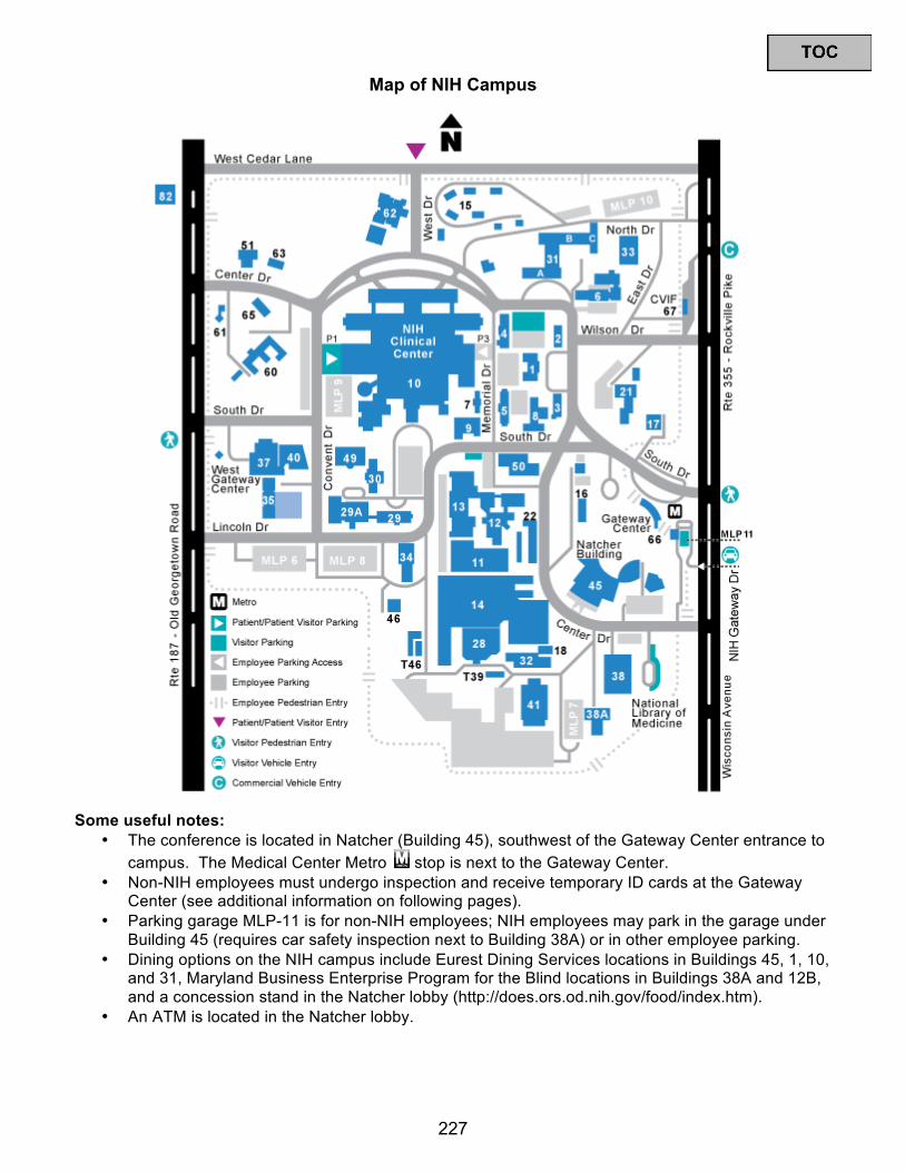
Map of NIH Campus
Some useful notes: • The conference is located in Natcher (Building 45), southwest of the Gateway Center entrance to
campus. The Medical Center Metro stop is next to the Gateway Center.• Non-NIH employees must undergo inspection and receive temporary ID cards at the Gateway
Center (see additional information on following pages).• Parking garage MLP-11 is for non-NIH employees;; NIH employees may park in the garage under
Building 45 (requires car safety inspection next to Building 38A) or in other employee parking.• Dining options on the NIH campus include Eurest Dining Services locations in Buildings 45, 1, 10,
and 31, Maryland Business Enterprise Program for the Blind locations in Buildings 38A and 12B,and a concession stand in the Natcher lobby (http://does.ors.od.nih.gov/food/index.htm).
• An ATM is located in the Natcher lobby.
227
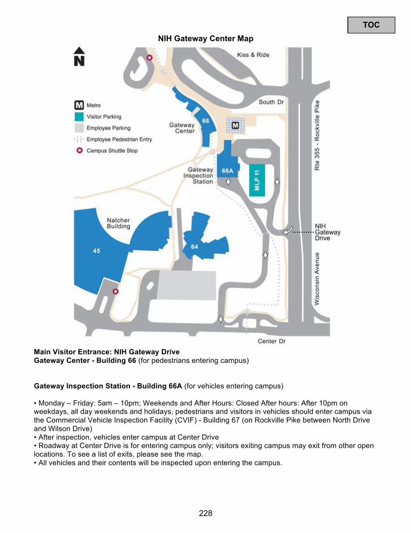
NIH Gateway Center Map
Main Visitor Entrance: NIH Gateway Drive Gateway Center - Building 66 (for pedestrians entering campus)
Gateway Inspection Station - Building 66A (for vehicles entering campus)
• Monday – Friday: 5am – 10pm;; Weekends and After Hours: Closed After hours: After 10pm onweekdays, all day weekends and holidays, pedestrians and visitors in vehicles should enter campus via the Commercial Vehicle Inspection Facility (CVIF) - Building 67 (on Rockville Pike between North Drive and Wilson Drive) • After inspection, vehicles enter campus at Center Drive• Roadway at Center Drive is for entering campus only;; visitors exiting campus may exit from other openlocations. To see a list of exits, please see the map. • All vehicles and their contents will be inspected upon entering the campus.
228

Multi-Level Parking Garage 11 – MLP-11 (for parking outside of campus) • Monday – Friday: 6am – 9pm (entrance) 6am – 11pm (exit) Weekends: Closed • When MLP-11 is closed, visitors can park in lots on the NIH Campus • Visitors parking in this garage should proceed to the Gateway Center (Bldg. 66) to get a visitor badge • All visitors traveling in a vehicle are highly encouraged to park in MLP-11 as there is limited visitor parking on the main campus • No vehicle inspection required to park in MLP-11 • Vehicles left in the MLP-11 parking garage after 11pm on weekdays or during any weekends are subject to ticketing and towing • Cost: $2 per hour for the first three hours, $12 maximum for the entire day Directions to NIH Gateway Drive from Rockville Pike/Wisconsin Avenue: Southbound: 1. Continue on Rockville Pike past South Drive 2. Turn right at NIH Gateway Drive Northbound – Option 1: 1. Continue on Rockville Pike past South Drive 2. Make a u-turn from the left turn lane at Wilson Drive 3. Continue southbound on Rockville Pike past South Drive 4. Turn right at NIH Gateway Drive Northbound – Option 2: 1. Continue on Rockville Pike 2. Turn left at Battery Lane 3. Turn right on Old Georgetown Road 4. Turn right on Cedar Lane 5. Turn right on Rockville Pike 6. Continue southbound on Rockville Pike past South Drive 7. Turn right at NIH Gateway Drive Northbound – Option 3: 1. Continue on Rockville Pike to South Drive 2. Make a u-turn from the left turn lane at South Drive 3. Continue southbound on Rockville Pike 4. Turn right at NIH Gateway Drive
229
Security Procedures for Entering the NIH Campus: * All visitors and patients—please be aware: Federal law prohibits the following items on Federal property: firearms, explosives, archery equipment, dangerous weapons, knives with blades over 2 ½ inches, alcoholic beverages and open containers of alcohol. * The NIH has implemented security measures to help ensure the safety of our patients, employees, guests and facilities. All visitors must enter through the NIH Gateway Center at Metro or the West Gateway Center. You will be asked to submit to a vehicle or personal inspection. * Visitors over 15 years of age must provide a form of government-issued ID such as a driver's license or passport. Visitors under 16 years of age must be accompanied by an adult. Vehicle Inspections – All vehicles and their contents will be inspected upon entering the campus. Additionally, all vehicles entering certain parking areas will be inspected, regardless of any prior inspection. Drivers will be required to present their driver’s license and may be asked to open the trunk and hood. If you are physically unable to perform this function, please inform the inspector and they will assist you. Vehicle inspection may consist of any combination of the following: Detection Dogs Teams (K-9), Electronic Detection Devices and Manual Inspection. After inspection, you will be issued a vehicle inspection pass. It must be displayed on your vehicle’s dashboard while you are on campus. The inspection pass is not a "parking permit." It only grants your vehicle access to enter the campus. You can only park in designated parking areas. Personal Inspections – All visitors should be prepared to submit to a personal inspection prior to entering the campus. These inspections may be conducted with a handheld monitoring device, a metal detector and by visible inspection. Additionally, your personal belongings may be inspected and passed through an x-ray machine. Visitor passes must be prominently displayed at all times while on the NIH campus. To learn more about visitor and security issues at the NIH, visit: http://www.nih.gov/about/visitor/index.htm. For questions about campus access, please contact the ORS Information Line at [email protected] or 301-594-6677, TTY - 301-435-1908.
230