Structural basis for KIT receptor tyrosine kinase ... · Structural basis for KIT receptor tyrosine...
6
Structural basis for KIT receptor tyrosine kinase inhibition by antibodies targeting the D4 membrane-proximal region Andrey V. Reshetnyak a,1 , Bryce Nelson b,1 , Xiarong Shi a , Titus J. Boggon a , Alevtina Pavlenco b , Elizabeth M. Mandel-Bausch c , Francisco Tome a , Yoshihisa Suzuki c , Sachdev S. Sidhu b,2 , Irit Lax a,2 , and Joseph Schlessinger a,2 a Department of Pharmacology, Yale University School of Medicine, New Haven, CT 06520; b Donnelly Centre for Cellular and Biomolecular Research, University of Toronto, Toronto, ON, Canada M5S 3E1; and c Kolltan Pharmaceuticals, Inc., New Haven, CT 06520 Contributed by Joseph Schlessinger, September 19, 2013 (sent for review August 15, 2013) Somatic oncogenic mutations in the receptor tyrosine kinase KIT function as major drivers of gastrointestinal stromal tumors and a subset of acute myeloid leukemia, melanoma, and other cancers. Although treatment of these cancers with tyrosine kinase inhib- itors shows dramatic responses and durable disease control, drug resistance followed by clinical progression of disease eventually occurs in virtually all patients. In this report, we describe inhibitory KIT antibodies that bind to the membrane-proximal Ig-like D4 of KIT with significant overlap with an epitope in D4 that mediates homotypic interactions essential for KIT activation. Crystal struc- tures of the anti-KIT antibody in complex with KIT D4 and D5 allowed design of affinity-matured libraries that were used to isolate variants with increased affinity and efficacy. Isolated antibodies showed KIT inhibition together with suppression of cell proliferation driven by ligand-stimulated WT or constitutively activated oncogenic KIT mutant. These antibodies represent a unique therapeutic approach and a step toward the development of “naked” or toxin-conjugated KIT antibodies for the treatment of KIT-driven cancers. phosphorylation | therapeutic antibodies | cancer therapy | cell signaling | protein kinase T he receptor tyrosine kinase (RTK) KIT is a transmembrane protein that plays crucial roles in mediating diverse cellular processes including cell differentiation, proliferation, and cell survival, among other activities. These processes occur through activation of KIT upon binding by stem cell factor (SCF), a li- gand found in membrane-anchored and soluble forms (1, 2) in a variety of cell types, including hematopoietic stem cells, germ cells, vascular endothelial cells, and the mesenchymal cells with uniquely neuromuscular differentiation known as the interstitial cells of Cajal (3, 4). KIT belongs to the type III subfamily of RTKs (5), a family composed of an extracellular region that includes five Ig-like domains (designated D1–D5), a single transmembrane domain (TM), a juxtamembrane region (JM), a tyrosine kinase domain split by a kinase insert, and a C-terminal tail (6) (Fig. 1A). Based on the determination of the crystal structure of the complete extracellular region of KIT before and after ligand stimulation (7), and the tyrosine kinase domain (8, 9), a mechanism has been proposed whereby activation of KIT is initiated through receptor dimerization (10). Dimerization of the KIT ectodomain is ligand-driven and initiated through high-affinity binding of an SCF dimer to the membrane distal Ig-like domains (D1–D3) of the receptor (11, 12). Cross-linking of the membrane distal Ig-like domains with their ligand dramatically increases local concen- tration of the membrane-proximal (D4 and D5) and TMs to en- able homotypic contacts by weak D4–D4 and D5–D5 interactions between neighboring KIT molecules. These homotypic associa- tions promote conformational rearrangements that permit cor- rect association between neighboring cytoplasmic regions of KIT dimers resulting in autophosphorylation, tyrosine kinase stimu- lation, recruitment of signaling proteins, and cell signaling. Dysregulation of KIT, by a variety of different somatic as well as rare germ-line mutations, has been associated with numerous hematopoietic and other cancers, including gastrointestinal stro- mal tumors (GIST), a subset of melanomas, systemic mastocytosis, and acute myeloid leukemia. Most reoccurring activating muta- tions map to the cytoplasmic JM region (exon 11) and to the membrane-proximal Ig-like domain D5 (exon 9) of the extra- cellular region (13, 14). Currently, initial treatment for patients with GIST involves the small-molecule kinase inhibitor Gleevec (imatinib), which can efficiently block kinase activity of JM and D5 mutants of KIT. Unfortunately, most patients with GIST develop resistance to the drug within 2 y of treatment by ac- quiring additional mutations usually mapped to exons 13 and 14 (V654A and V670I, respectively). Patients resistant to Gleevec are usually treated with the kinase inhibitor Sutent (sunitinib), which can more efficiently inhibit WT KIT protein as well as many of the mutations that confer imatinib resistance. In addi- tion to Gleevec-resistant mutations in exons 13 and 14, kinase domain mutants (exon 17) that are resistant to Gleevec and Sutent are also seen in patients with GIST (15). Several mAbs targeting RTKs have shown promising results as anticancer therapies, including mAbs against members of the Significance The receptor tyrosine kinase KIT is aberrantly activated pri- marily by somatic mutations in gastrointestinal stromal tumors and in a subset of acute myeloid leukemia, melanoma, and other cancers. Treatment of these cancers with tyrosine kinase inhibitors shows durable clinical response, but drug resistance and disease progression eventually occur in all patients. Here we describe monoclonal antibodies that block the activity of KIT and its oncogenic mutant. Structural and biochemical analyses of anti-KIT antibodies in complex with a KIT fragment demonstrated that KIT antibodies bind to a critical Achilles heel region that is essential for receptor activation. These anti- bodies may provide a potentially unique therapeutic approach for the treatment of tumors driven by WT or oncogenically mutated KIT. Author contributions: A.V.R., B.N., Y.S., S.S.S., I.L., and J.S. designed research; A.V.R., B.N., X.S., A.P., E.M.M.-B., F.T., and I.L. performed research; A.V.R., B.N., T.J.B., Y.S., I.L., and J.S. analyzed data; and A.V.R., B.N., S.S.S., I.L., and J.S. wrote the paper. The authors declare no conflict of interest. Data deposition: The atomic coordinates and structure factors have been deposited in the Protein Data Bank, www.pdb.org (PDB ID codes 4K94 and 4K9E). 1 A.V.R. and B.N. contributed equally to this work. 2 To whom correspondence may be addressed. E-mail: [email protected], irit. [email protected], or [email protected]. This article contains supporting information online at www.pnas.org/lookup/suppl/doi:10. 1073/pnas.1317118110/-/DCSupplemental. 17832–17837 | PNAS | October 29, 2013 | vol. 110 | no. 44 www.pnas.org/cgi/doi/10.1073/pnas.1317118110
Transcript of Structural basis for KIT receptor tyrosine kinase ... · Structural basis for KIT receptor tyrosine...
Structural basis for KIT receptor tyrosine kinaseinhibition by antibodies targeting the D4membrane-proximal regionAndrey V. Reshetnyaka,1, Bryce Nelsonb,1, Xiarong Shia, Titus J. Boggona, Alevtina Pavlencob,Elizabeth M. Mandel-Bauschc, Francisco Tomea, Yoshihisa Suzukic, Sachdev S. Sidhub,2, Irit Laxa,2,and Joseph Schlessingera,2
aDepartment of Pharmacology, Yale University School of Medicine, New Haven, CT 06520; bDonnelly Centre for Cellular and Biomolecular Research, Universityof Toronto, Toronto, ON, Canada M5S 3E1; and cKolltan Pharmaceuticals, Inc., New Haven, CT 06520
Contributed by Joseph Schlessinger, September 19, 2013 (sent for review August 15, 2013)
Somatic oncogenic mutations in the receptor tyrosine kinase KITfunction as major drivers of gastrointestinal stromal tumors anda subset of acute myeloid leukemia, melanoma, and other cancers.Although treatment of these cancers with tyrosine kinase inhib-itors shows dramatic responses and durable disease control, drugresistance followed by clinical progression of disease eventuallyoccurs in virtually all patients. In this report, we describe inhibitoryKIT antibodies that bind to the membrane-proximal Ig-like D4 ofKIT with significant overlap with an epitope in D4 that mediateshomotypic interactions essential for KIT activation. Crystal struc-tures of the anti-KIT antibody in complex with KIT D4 and D5allowed design of affinity-matured libraries that were usedto isolate variants with increased affinity and efficacy. Isolatedantibodies showed KIT inhibition together with suppression ofcell proliferation driven by ligand-stimulated WT or constitutivelyactivated oncogenic KIT mutant. These antibodies represent aunique therapeutic approach and a step toward the developmentof “naked” or toxin-conjugated KIT antibodies for the treatment ofKIT-driven cancers.
phosphorylation | therapeutic antibodies | cancer therapy | cell signaling |protein kinase
The receptor tyrosine kinase (RTK) KIT is a transmembraneprotein that plays crucial roles in mediating diverse cellular
processes including cell differentiation, proliferation, and cellsurvival, among other activities. These processes occur throughactivation of KIT upon binding by stem cell factor (SCF), a li-gand found in membrane-anchored and soluble forms (1, 2) ina variety of cell types, including hematopoietic stem cells, germcells, vascular endothelial cells, and the mesenchymal cells withuniquely neuromuscular differentiation known as the interstitialcells of Cajal (3, 4). KIT belongs to the type III subfamily ofRTKs (5), a family composed of an extracellular region thatincludes five Ig-like domains (designated D1–D5), a singletransmembrane domain (TM), a juxtamembrane region (JM), atyrosine kinase domain split by a kinase insert, and a C-terminaltail (6) (Fig. 1A).Based on the determination of the crystal structure of the
complete extracellular region of KIT before and after ligandstimulation (7), and the tyrosine kinase domain (8, 9), a mechanismhas been proposed whereby activation of KIT is initiated throughreceptor dimerization (10). Dimerization of the KIT ectodomainis ligand-driven and initiated through high-affinity binding of anSCF dimer to themembrane distal Ig-like domains (D1–D3) of thereceptor (11, 12). Cross-linking of the membrane distal Ig-likedomains with their ligand dramatically increases local concen-tration of the membrane-proximal (D4 and D5) and TMs to en-able homotypic contacts by weak D4–D4 and D5–D5 interactionsbetween neighboring KIT molecules. These homotypic associa-tions promote conformational rearrangements that permit cor-rect association between neighboring cytoplasmic regions of KIT
dimers resulting in autophosphorylation, tyrosine kinase stimu-lation, recruitment of signaling proteins, and cell signaling.Dysregulation of KIT, by a variety of different somatic as well
as rare germ-line mutations, has been associated with numeroushematopoietic and other cancers, including gastrointestinal stro-mal tumors (GIST), a subset of melanomas, systemic mastocytosis,and acute myeloid leukemia. Most reoccurring activating muta-tions map to the cytoplasmic JM region (exon 11) and to themembrane-proximal Ig-like domain D5 (exon 9) of the extra-cellular region (13, 14). Currently, initial treatment for patientswith GIST involves the small-molecule kinase inhibitor Gleevec(imatinib), which can efficiently block kinase activity of JM andD5 mutants of KIT. Unfortunately, most patients with GISTdevelop resistance to the drug within 2 y of treatment by ac-quiring additional mutations usually mapped to exons 13 and 14(V654A and V670I, respectively). Patients resistant to Gleevecare usually treated with the kinase inhibitor Sutent (sunitinib),which can more efficiently inhibit WT KIT protein as well asmany of the mutations that confer imatinib resistance. In addi-tion to Gleevec-resistant mutations in exons 13 and 14, kinasedomain mutants (exon 17) that are resistant to Gleevec andSutent are also seen in patients with GIST (15).Several mAbs targeting RTKs have shown promising results as
anticancer therapies, including mAbs against members of the
Significance
The receptor tyrosine kinase KIT is aberrantly activated pri-marily by somatic mutations in gastrointestinal stromal tumorsand in a subset of acute myeloid leukemia, melanoma, andother cancers. Treatment of these cancers with tyrosine kinaseinhibitors shows durable clinical response, but drug resistanceand disease progression eventually occur in all patients. Herewe describe monoclonal antibodies that block the activity ofKIT and its oncogenic mutant. Structural and biochemicalanalyses of anti-KIT antibodies in complex with a KIT fragmentdemonstrated that KIT antibodies bind to a critical Achilles heelregion that is essential for receptor activation. These anti-bodies may provide a potentially unique therapeutic approachfor the treatment of tumors driven by WT or oncogenicallymutated KIT.
Author contributions: A.V.R., B.N., Y.S., S.S.S., I.L., and J.S. designed research; A.V.R., B.N.,X.S., A.P., E.M.M.-B., F.T., and I.L. performed research; A.V.R., B.N., T.J.B., Y.S., I.L., and J.S.analyzed data; and A.V.R., B.N., S.S.S., I.L., and J.S. wrote the paper.
The authors declare no conflict of interest.
Data deposition: The atomic coordinates and structure factors have been deposited in theProtein Data Bank, www.pdb.org (PDB ID codes 4K94 and 4K9E).1A.V.R. and B.N. contributed equally to this work.2To whom correspondence may be addressed. E-mail: [email protected], [email protected], or [email protected].
This article contains supporting information online at www.pnas.org/lookup/suppl/doi:10.1073/pnas.1317118110/-/DCSupplemental.
17832–17837 | PNAS | October 29, 2013 | vol. 110 | no. 44 www.pnas.org/cgi/doi/10.1073/pnas.1317118110
EGFR family of RTKs, which were approved for clinical use,including cetuximab and panitumumab (anti-EGFR mAbs), aswell as trastuzumab and pertuzumab (anti-ErbB2 mAbs) (16,17). Antibodies can be highly specific for their targets, andtherefore off-target side effects are reduced compared withsmall-molecule kinase inhibitors. Specific targeting of oncogenicRTKs by inhibitory mAbs may allow the resistance that fre-quently occurs in patients treated with kinase inhibitors to besurmounted.We have shown previously that homotypic interactions be-
tween the membrane-proximal domains of KIT (D4 and D5) arecritical for receptor activation and that disruption of the D4–D4interface strongly compromises receptor activation (7). As KIToncogenic mutants located in D5 are dependent on homotypiccontacts between neighboring ectodomains, it is reasonable toexpect that activation of these mutants could be inhibited bymonoclonal antibodies directed against KIT domains D4 or D5.Here, anti-KIT antibodies were isolated from a naive, phage-displayed synthetic antibody library. Crystal structures of a frag-ment antigen-binding (Fab) in complex with KIT membrane-proximal domains D4 and D5 (KITD4-5) revealed binding toD4 that overlapped significantly with an epitope required forhomotypic interactions essential for SCF-dependent KIT acti-vation. Furthermore, information obtained from the structure ofantibody in complex with KIT was applied to guide the design ofaffinity maturation libraries, enabling isolation of antibody var-iants with increased binding affinity equating to increased effi-cacy. These antibodies were capable of efficient inhibition of KITactivity that led to suppression of cell proliferation and providea potentially unique therapeutic approach for the treatment oftumors driven by WT or aberrantly activated KIT mutants.
ResultsThe two membrane-proximal Ig-like domains of KIT (KITD4-5fragment; Fig. 1A) determined structurally to be critical for KITactivation (7) were used as an antigen to isolate binding Fabs
from a naive phage-displayed library (library F; Fig. S1) con-taining more than 1010 unique clones (18, 19). The bindingproperties of the phage-derived Fabs were compared with thebinding properties of a murine monoclonal anti-KIT antibodydesignated KTN37 that was obtained by immunization of micewith the same antigen. A variety of in vitro binding experimentsusing 3T3 cells ectopically expressing WT KIT demonstratedthat the phage-derived Fabs and the KTN37 mAb bind specifi-cally to D4 of recombinant isolated KIT or to native KIT mol-ecules expressed on the cell surface of live cells. The structure ofone of the most potent phage-derived Fab, designated Fab19(Fig. S1), in complex with KITD4-5 (Fig. 1A), was further ana-lyzed by X-ray crystallography.
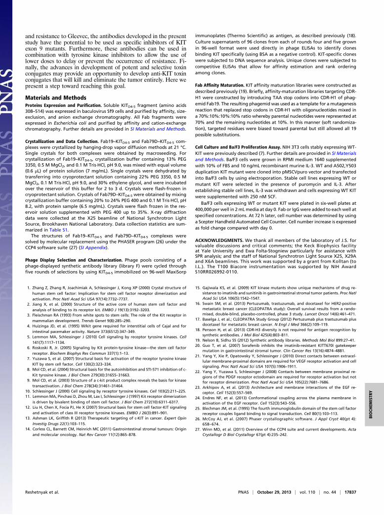
The Structure of Fab19–KITD4-5 Complex. A purified complex com-posed of Fab19 together with KITD4-5 was subjected to extensivescreening for crystal growth and further optimization. We obtainedcrystals that belong to the C2 space group with a single 1:1 com-plex of KITD4-5 and Fab19 in the asymmetric unit. The structure ofthis complex was determined to 2.4-Å resolution (SI Materials andMethods, Fig. 1B and Table S1).The overall structure of KITD4-5 bound to Fab19, is very
similar to the structures of these two Ig-like domains observedpreviously as part of the structures of full-length extracellularregion of KIT alone, or in complex with SCF [Protein Data Bank(PDB) ID codes 2EC8 and 2E9W; ref. 7]. Superposition of in-dividual D4 and D5 from Fab19–KITD4-5 complex structure withcorresponding domains of KIT ectodomain structure (PDB IDcode 2EC8) revealed rmsd values of 0.65 Å for 96 and 59 Cαresidues in D4 and D5, respectively. The structure revealedFab19 binding exclusively to D4 of KIT with a buried surface of1,029 Å2 on the D4 side of the interface (Fig. 1C and Table S2).Nearly the entire β-sheet of D4 (one of two β-sheets in Ig-likedomain), including βA, βB, β, and βD, as well as the AA′, A′B,EF, and DE loops, was buried under the Fab19 surface (Fig. 1Dand Fig. S2).The majority of the contacts were made by the heavy chain of
the Fab (800 Å2 vs. 283 Å2 for the light chain; Fig. 1D, Fig. S2,and Table S2), with most key interactions mediated by the com-plementarity-determining region (CDR) loops of the Fab, in-cluding all three CDRs of the heavy chain and L2 and L3 of thelight chain. The majority of the specificity-determining contactscame from CDRs H2 and H3 (additional details about Fab19–D4 interface is described in SI Appendix).
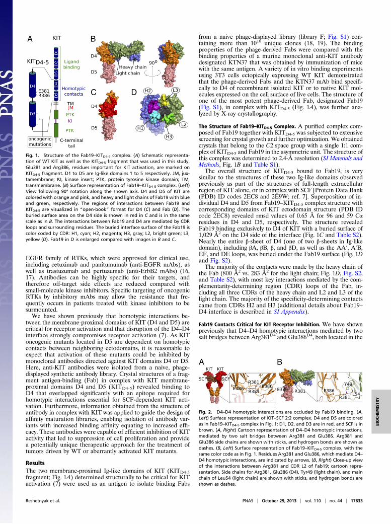
Fab19 Contacts Critical for KIT Receptor Inhibition. We have shownpreviously that D4–D4 homotypic interactions mediated by twosalt bridges between Arg381D4 and Glu386D4, both located in the
A B
C D
Fig. 1. Structure of the Fab19–KITD4-5 complex. (A) Schematic representa-tion of WT KIT as well as the KITD4-5 fragment that was used in this study.Glu381 and Arg386, residues important for KIT activation, are marked onKITD4-5 fragment. D1 to D5 are Ig-like domains 1 to 5 respectively. JM, jux-tamembrane; KI, kinase insert; PTK, protein tyrosine kinase domain; TM,transmembrane. (B) Surface representation of Fab19–KITD4-5 complex. (Left)View following 90° rotation along the shown axis. D4 and D5 of KIT arecolored with orange and pink, and heavy and light chains of Fab19 with blueand green, respectively. The regions of interactions between Fab19 andKITD4-5 are visualized in “open-book” format for D4 (C) and Fab (D). Theburied surface area on the D4 side is shown in red in C and is in the samescale as in B. The interactions between Fab19 and D4 are mediated by CDRloops and surrounding residues. The buried interface surface of the Fab19 iscolor coded by CDR: H1, cyan; H2, magenta; H3, gray; L2, bright green; L3,yellow (D). Fab19 in D is enlarged compared with images in B and C.
A B
Fig. 2. D4–D4 homotypic interactions are occluded by Fab19 binding. (A,Left) Surface representation of KIT–SCF 2:2 complex. D4 and D5 are coloredas in Fab19–KITD4-5 complex in Fig. 1; D1, D2, and D3 are in red, and SCF is inbrown. (A, Right) Cartoon representation of D4–D4 homotypic interactions,mediated by two salt bridges between Arg381 and Glu386. Arg381 andGlu386 side chains are shown with sticks, and hydrogen bonds are shown asdashes. (B, Left) Surface representation of Fab19–KITD4-5 complex, with thesame color code as in Fig. 1. Residues Arg381 and Glu386, which mediate D4–D4 homotypic interactions, are indicated by arrows. (B, Right) Close-up viewof the interactions between Arg381 and CDR L2 of Fab19; cartoon repre-sentation. Side chains for Arg381, Glu386 (D4), Tyr49 (light chain), and mainchain of Leu54 (light chain) are shown with sticks, and hydrogen bonds areshown as dashes.
Reshetnyak et al. PNAS | October 29, 2013 | vol. 110 | no. 44 | 17833
BIOCH
EMISTR
Y
EF loop (Fig. 2A), are critical for proper ligand dependent re-ceptor activation (7). Analysis of the Fab19–KITD4-5 complexstructure revealed that Arg381D4 makes contact with CDR L2 ofFab19 (Fig. 2B); the Arg381D4 side chain makes hydrogen bondswith the side chain of Tyr49L and the main chain of Leu54L. Inaddition, Tyr101H of CDR H3 makes contact with Thr380D4
(Fig. S3) of the EF loop, which is involved in mediating D4–D4homotypic interactions. The structure revealed that contactsbetween Fab19 and D4 of KIT occluded Arg381D4 from forminga salt bridge with Glu-386 of D4 from a neighboring KIT re-ceptor, thereby preventing proper lateral association betweenmembrane-proximal domains. As a consequence, homotypic con-tacts between the cytoplasmic domains, transphosphorylation andKIT activation were inhibited (as detailed later).
Affinity Maturation of Fab19. To improve the binding affinity ofFab19 to D4, affinity maturation was performed in a two-stepprocess. The first step was performed before solving of theFab19–KITD4-5 complex structure and involved soft randomiza-tion of individual CDRs wherein targeted residues were likely tobe kept parental, thereby minimizing the likelihood of changingepitopes during Fab maturation. The four CDRs into which di-versity was introduced in library F, CDR L3 and all heavy-chainCDRs, were targeting individually in affinity maturation librarydesign and resulted in the isolation of numerous variants from allfour libraries. Variants from the CDR H1 library showed strongimprovement over the parental Fab19, and, in particular, Fab12I,estimated to have one of the strongest binding affinity (Fig. S4A–C) and KIT receptor inhibition effect, was chosen for furtheraffinity maturation with the aid of the newly solved Fab19–KITD4-5 complex.The structure of the Fab19–KITD4-5 complex was used to
guide affinity maturation library design after analysis of the an-tibody–antigen interface revealed that, whereas the heavy chainof Fab19 makes extensive contacts with D4, light-chain inter-actions were few and weak (Fig. 1D). In particular, no contactswere observed between CDR L1 and D4 (Fig. 3A). Consideringthat the light chain makes most of the contact with Arg381D4,a residue that is important for formation of D4 homotypic con-tacts for receptor activation, affinity maturation libraries tar-geting CDR L1 were designed. Length variation between fourand seven residues was incorporated into library design in ad-dition to allowing for possible CDR H1 S31V and/or S33Msubstitutions found in the parental Fab12I CDR H1 template.Eight unique Fabs (Fab79A–H) were isolated with CDR L1lengths ranging from four to seven residues, suggesting that in-creased length allowed additional contacts with D4 (Fig. S4D).Interestingly, all clones were found to contain double S31VS33M substitutions in the CDR H1, indicating the importanceof these nucleophilic to hydrophobic substitutions (detailed inSI Appendix) (Fig. S9).
Binding of Anti-D4 Fabs to KITD4-5 Fragment. We used surfaceplasmon resonance (SPR) analysis to quantitatively characterizethe kinetics and dissociation constants of different generations ofaffinity-matured anti-D4 Fabs (Fab19, Fab12I, and Fab79D). Asa control, we used the Fab of the murine antibody KTN37 (asdetailed later). Purified KITD4-5 fragment, was covalently at-tached onto a CM5 biosensor chip and serial dilutions of eachFab were flowed over the biosensor surface to reveal the bindingkinetics (Table 1). Values for the association and dissociation
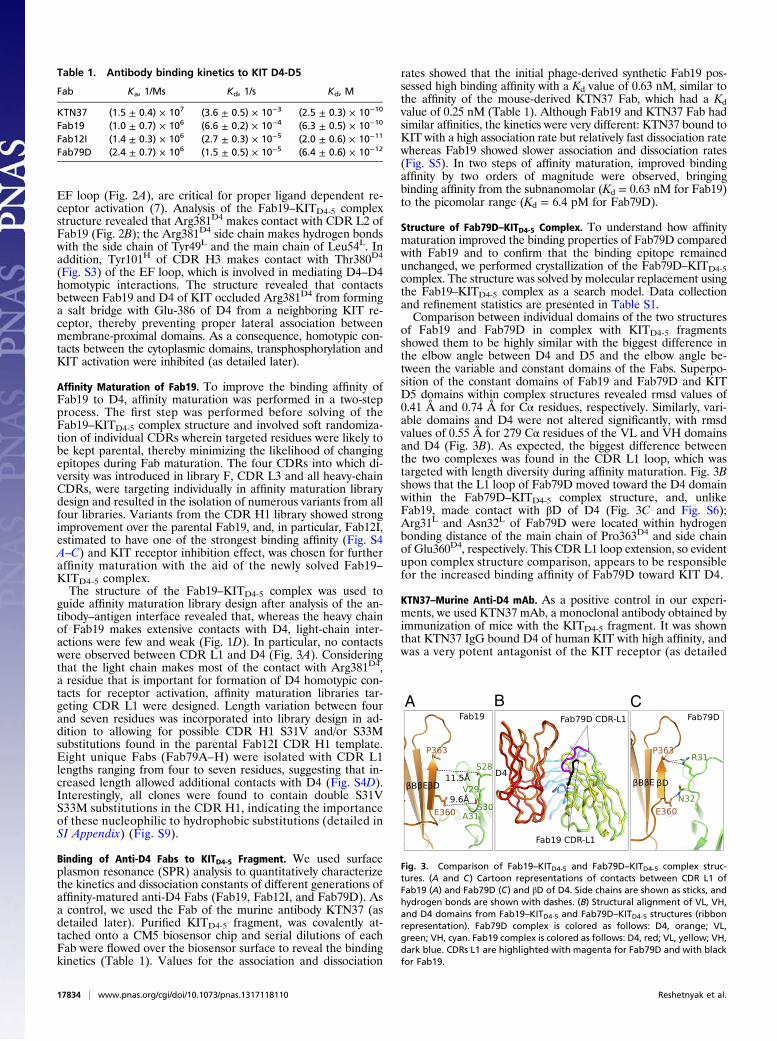
rates showed that the initial phage-derived synthetic Fab19 pos-sessed high binding affinity with a Kd value of 0.63 nM, similar tothe affinity of the mouse-derived KTN37 Fab, which had a Kdvalue of 0.25 nM (Table 1). Although Fab19 and KTN37 Fab hadsimilar affinities, the kinetics were very different: KTN37 bound toKIT with a high association rate but relatively fast dissociation ratewhereas Fab19 showed slower association and dissociation rates(Fig. S5). In two steps of affinity maturation, improved bindingaffinity by two orders of magnitude were observed, bringingbinding affinity from the subnanomolar (Kd = 0.63 nM for Fab19)to the picomolar range (Kd = 6.4 pM for Fab79D).
Structure of Fab79D–KITD4-5 Complex. To understand how affinitymaturation improved the binding properties of Fab79D comparedwith Fab19 and to confirm that the binding epitope remainedunchanged, we performed crystallization of the Fab79D–KITD4-5complex. The structure was solved by molecular replacement usingthe Fab19–KITD4-5 complex as a search model. Data collectionand refinement statistics are presented in Table S1.Comparison between individual domains of the two structures
of Fab19 and Fab79D in complex with KITD4-5 fragmentsshowed them to be highly similar with the biggest difference inthe elbow angle between D4 and D5 and the elbow angle be-tween the variable and constant domains of the Fabs. Superpo-sition of the constant domains of Fab19 and Fab79D and KITD5 domains within complex structures revealed rmsd values of0.41 Å and 0.74 Å for Cα residues, respectively. Similarly, vari-able domains and D4 were not altered significantly, with rmsdvalues of 0.55 Å for 279 Cα residues of the VL and VH domainsand D4 (Fig. 3B). As expected, the biggest difference betweenthe two complexes was found in the CDR L1 loop, which wastargeted with length diversity during affinity maturation. Fig. 3Bshows that the L1 loop of Fab79D moved toward the D4 domainwithin the Fab79D–KITD4-5 complex structure, and, unlikeFab19, made contact with βD of D4 (Fig. 3C and Fig. S6);Arg31L and Asn32L of Fab79D were located within hydrogenbonding distance of the main chain of Pro363D4 and side chainof Glu360D4, respectively. This CDR L1 loop extension, so evidentupon complex structure comparison, appears to be responsiblefor the increased binding affinity of Fab79D toward KIT D4.
KTN37–Murine Anti-D4 mAb. As a positive control in our experi-ments, we used KTN37 mAb, a monoclonal antibody obtained byimmunization of mice with the KITD4-5 fragment. It was shownthat KTN37 IgG bound D4 of human KIT with high affinity, andwas a very potent antagonist of the KIT receptor (as detailed
A B C
Fig. 3. Comparison of Fab19–KITD4-5 and Fab79D–KITD4-5 complex struc-tures. (A and C) Cartoon representations of contacts between CDR L1 ofFab19 (A) and Fab79D (C) and βD of D4. Side chains are shown as sticks, andhydrogen bonds are shown with dashes. (B) Structural alignment of VL, VH,and D4 domains from Fab19–KITD4-5 and Fab79D–KITD4-5 structures (ribbonrepresentation). Fab79D complex is colored as follows: D4, orange; VL,green; VH, cyan. Fab19 complex is colored as follows: D4, red; VL, yellow; VH,dark blue. CDRs L1 are highlighted with magenta for Fab79D and with blackfor Fab19.
Table 1. Antibody binding kinetics to KIT D4-D5
Fab Ka, 1/Ms Kd, 1/s Kd, M
KTN37 (1.5 ± 0.4) × 107 (3.6 ± 0.5) × 10−3 (2.5 ± 0.3) × 10−10
Fab19 (1.0 ± 0.7) × 106 (6.6 ± 0.2) × 10−4 (6.3 ± 0.5) × 10−10
Fab12I (1.4 ± 0.3) × 106 (2.7 ± 0.3) × 10−5 (2.0 ± 0.6) × 10−11
Fab79D (2.4 ± 0.7) × 106 (1.5 ± 0.5) × 10−5 (6.4 ± 0.6) × 10−12
17834 | www.pnas.org/cgi/doi/10.1073/pnas.1317118110 Reshetnyak et al.
later). As we were not able to obtain diffraction quality crystalsof KTN37 in complex with KIT D4 and D5 fragment, moleculardetails of the complex could not be obtained. However, to shedlight on the binding epitope of KTN37, we compared the KTN37IgG binding to the ectodomain of KIT from different species(Fig. S7A). Our results showed that this mAb was cross-reactivewith KIT from human, monkey, dog, and cat, but not mouse orrat. Sequence alignment of D4 of KIT for these six speciesrevealed one major spot with a few residues spread around D4,which are different in the rodent sequences (Fig. S7B). Wemapped all residue differences onto the D4 structure and foundout that most of these residues cover two continuous areas onthe surface of D4 (Fig. S8), suggesting that these two regions mayrepresent the binding epitope of KTN37 Fab to D4. It is worthnoting that these regions are located right on top of the D4homotypic contact interface (Fig. S8), and, most likely, bindingof KTN37 significantly overlaps with the region essential formediating D4–D4 homotypic interactions.
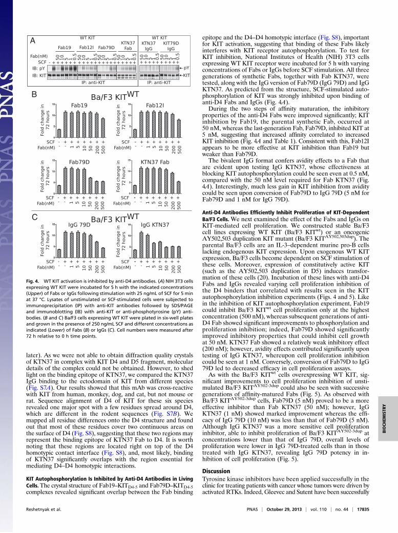
KIT Autophosphorylation Is Inhibited by Anti-D4 Antibodies in LivingCells.The crystal structure of Fab19–KITD4-5 and Fab79D–KITD4-5complexes revealed significant overlap between the Fab binding
epitope and the D4–D4 homotypic interface (Fig. S8), importantfor KIT activation, suggesting that binding of these Fabs likelyinterferes with KIT receptor autophosphorylation. To test forKIT inhibition, National Institutes of Health (NIH) 3T3 cellsexpressing WT KIT receptor were incubated for 5 h with varyingconcentrations of Fabs or IgGs before SCF stimulation. All threegenerations of synthetic Fabs, together with Fab KTN37, weretested, along with the IgG version of Fab79D (IgG 79D) and IgGKTN37. As predicted from the structure, SCF-stimulated auto-phosphorylation of KIT was strongly inhibited upon binding ofanti-D4 Fabs and IgGs (Fig. 4A).During the two steps of affinity maturation, the inhibitory
properties of the anti-D4 Fabs were improved significantly; KITinhibition by Fab19, the parental synthetic Fab, occurred at50 nM, whereas the last-generation Fab, Fab79D, inhibited KIT at5 nM, suggesting that increased affinity correlated to increasedKIT inhibition (Fig. 4A and Table 1). Consistent with this, Fab12Iappears to be more effective at KIT inhibition than Fab19 butweaker than Fab79D.The bivalent IgG format confers avidity effects to a Fab that
are evident upon testing IgG KTN37, whose effectiveness atblocking KIT autophosphorylation could be seen even at 0.5 nM,compared with the 50 nM level required for Fab KTN37 (Fig.4A). Interestingly, much less gain in KIT inhibition from aviditycould be seen upon conversion of Fab79D to IgG 79D (5 nM forFab79D and 1 nM for IgG 79D).
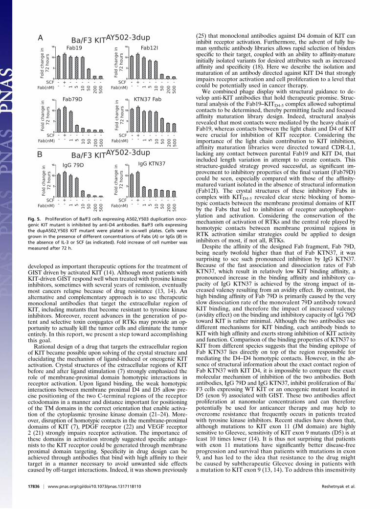
Anti-D4 Antibodies Efficiently Inhibit Proliferation of KIT-DependentBa/F3 Cells. We next examined the effect of the Fabs and IgGs onKIT-mediated cell proliferation. We constructed stable Ba/F3cell lines expressing WT KIT (Ba/F3 KITwt) or an oncogenicAY502,503 duplication KIT mutant (Ba/F3 KITAY502,503dup). Theparental Ba/F3 cells are an IL-3–dependent murine pro-B cellslacking endogenous KIT expression. Upon exogenous WT KITexpression, Ba/F3 cells become dependent on SCF stimulation ofthese cells. Moreover, expression of constitutively active KIT(such as the AY502,503 duplication in D5) induces transfor-mation of these cells (20). Incubation of these lines with anti-D4Fabs and IgGs revealed varying cell proliferation inhibition ofthe D4 binders that correlated with results seen in the KITautophosphorylation inhibition experiments (Figs. 4 and 5). Likein the inhibition of KIT autophosphorylation experiment, Fab19could inhibit Ba/F3 KITwt cell proliferation only at the highestconcentration (500 nM), whereas subsequent generations of anti-D4 Fab showed significant improvements to phosphorylation andproliferation inhibition; indeed, Fab79D showed significantlyimproved inhibitory properties that could inhibit cell growthat 50 nM. KTN37 Fab showed a relatively weak inhibitory effect(200 nM); however, avidity effects contributed significantly upontesting of IgG KTN37, whereupon cell proliferation inhibitioncould be seen at 1 nM. Conversely, conversion of Fab79D to IgG79D led to decreased efficacy in cell proliferation assays.As with the Ba/F3 KITwt cells overexpressing WT KIT, sig-
nificant improvements to cell proliferation inhibition of unsti-mulated Ba/F3 KITAY502-3dup could also be seen with successivegenerations of affinity-matured Fabs (Fig. 5). As observed withBa/F3 KITAY502-3dup cells, Fab79D (5 nM) proved to be a moreeffective inhibitor than Fab KTN37 (50 nM); however, IgGKTN37 (1 nM) showed marked improvement whereas the effi-cacy of IgG 79D (10 nM) was less than that of Fab79D (5 nM).Although IgG KTN37 was a more sensitive cell proliferationinhibitor, able to inhibit proliferation of Ba/F3 KITAY502-3dup atconcentrations lower than that of IgG 79D, overall levels ofproliferation were lower in IgG 79D-treated cells than in thosetreated with IgG KTN37, revealing IgG 79D potency in in-hibition of cell proliferation (Fig. 5).
DiscussionTyrosine kinase inhibitors have been applied successfully in theclinic for treating patients with cancer whose tumors were driven byactivated RTKs. Indeed, Gleevec and Sutent have been successfully
A
B
C
Fig. 4. WT KIT activation is inhibited by anti-D4 antibodies. (A) NIH 3T3 cellsexpressing WT KIT were incubated for 5 h with the indicated concentrations(Upper) of Fabs or IgGs following stimulation with 25 ng/mL of SCF for 5 minat 37 °C. Lysates of unstimulated or SCF-stimulated cells were subjected toimmunoprecipitation (IP) with anti-KIT antibodies followed by SDS/PAGEand immunoblotting (IB) with anti-KIT or anti-phosphotyrosine (pY) anti-bodies. (B and C) Ba/F3 cells expressing WT KIT were plated in six-well platesand grown in the presence of 250 ng/mL SCF and different concentrations asindicated (Lower) of Fabs (B) or IgGs (C). Cell numbers were measured after72 h relative to 0 h time points.
Reshetnyak et al. PNAS | October 29, 2013 | vol. 110 | no. 44 | 17835
BIOCH
EMISTR
Y
developed as important therapeutic options for the treatment ofGIST driven by activated KIT (14). Although most patients withKIT-driven GIST respond well when treated with tyrosine kinaseinhibitors, sometimes with several years of remission, eventuallymost cancers relapse because of drug resistance (13, 14). Analternative and complementary approach is to use therapeuticmonoclonal antibodies that target the extracellular region ofKIT, including mutants that become resistant to tyrosine kinaseinhibitors. Moreover, recent advances in the generation of po-tent and selective toxin conjugates of RTKs may provide an op-portunity to actually kill the tumor cells and eliminate the tumorentirely. In this report, we present a step toward accomplishingthis goal.Rational design of a drug that targets the extracellular region
of KIT became possible upon solving of the crystal structure andelucidating the mechanism of ligand-induced or oncogenic KITactivation. Crystal structures of the extracellular regions of KITbefore and after ligand stimulation (7) strongly emphasized therole of membrane-proximal domain homotypic interactions inreceptor activation. Upon ligand binding, the weak homotypicinteractions between membrane proximal D4 and D5 allow pre-cise positioning of the two C-terminal regions of the receptorectodomains in a manner and distance important for positioningof the TM domains in the correct orientation that enable activa-tion of the cytoplasmic tyrosine kinase domain (21–24). More-over, disruption of homotypic contacts in the membrane-proximaldomains of KIT (7), PDGF receptor (22) and VEGF receptor2 (21) strongly impairs receptor activation. The importance ofthese domains in activation strongly suggested specific antago-nists to the KIT receptor could be generated through membraneproximal domain targeting. Specificity in drug design can beachieved through antibodies that bind with high affinity to theirtarget in a manner necessary to avoid unwanted side effectscaused by off-target interactions. Indeed, it was shown previously
(25) that monoclonal antibodies against D4 domain of KIT caninhibit receptor activation. Furthermore, the advent of fully hu-man synthetic antibody libraries allows rapid selection of bindersspecific to their target, coupled with an ability to affinity-matureinitially isolated variants for desired attributes such as increasedaffinity and specificity (18). Here we describe the isolation andmaturation of an antibody directed against KIT D4 that stronglyimpairs receptor activation and cell proliferation to a level thatcould be potentially used in cancer therapy.We combined phage display with structural guidance to de-
velop anti-KIT antibodies that hold therapeutic promise. Struc-tural analysis of the Fab19–KITD4-5 complex allowed suboptimalcontacts to be determined, thereby permitting facile and focusedaffinity maturation library design. Indeed, structural analysisrevealed that most contacts were mediated by the heavy chain ofFab19, whereas contacts between the light chain and D4 of KITwere crucial for inhibition of KIT receptor. Considering theimportance of the light chain contribution to KIT inhibition,affinity maturation libraries were directed toward CDR-L1,lacking any contact between parental Fab19 and KIT D4, thatincluded length variation in attempt to create contacts. Thisstructure-guided strategy proved successful, as significant im-provement to inhibitory properties of the final variant (Fab79D)could be seen, especially compared with those of the affinity-matured variant isolated in the absence of structural information(Fab12I). The crystal structures of these inhibitory Fabs incomplex with KITD4-5 revealed clear steric blocking of homo-typic contacts between the membrane proximal domains of KITby the Fabs that led to inhibition of receptor autophosphor-ylation and activation. Considering the conservation of themechanism of activation of RTKs and the central role played byhomotypic contacts between membrane proximal regions inRTK activation similar strategies could be applied to designinhibitors of most, if not all, RTKs.Despite the affinity of the designed Fab fragment, Fab 79D,
being nearly twofold higher than that of Fab KTN37, it wassurprising to see such pronounced inhibition by IgG KTN37.Because of the fast association and dissociation rates of FabKTN37, which result in relatively low KIT binding affinity, apronounced increase in the binding affinity and inhibitory ca-pacity of IgG KTN37 is achieved by the strong impact of in-creased valency resulting from an avidity effect. By contrast, thehigh binding affinity of Fab 79D is primarily caused by the veryslow dissociation rate of the monovalent 79D antibody towardKIT binding, and therefore the impact of increased valency(avidity effect) on the binding and inhibitory capacity of IgG 79Dtoward KIT is rather minimal. Although the two antibodies usedifferent mechanisms for KIT binding, each antibody binds toKIT with high affinity and exerts strong inhibition of KIT activityand function. Comparison of the binding properties of KTN37 toKIT from different species suggests that the binding epitope ofFab KTN37 lies directly on top of the region responsible formediating the D4–D4 homotypic contacts. However, in the ab-sence of structural information about the exact contact region ofFab KTN37 with KIT D4, it is impossible to compare the exactmolecular mechanism of inhibition of the two antibodies. Bothantibodies, IgG 79D and IgG KTN37, inhibit proliferation of Ba/F3 cells expressing WT KIT or an oncogenic mutant located inD5 (exon 9) associated with GIST. These two antibodies affectproliferation at nanomolar concentrations and can thereforepotentially be used for anticancer therapy and may help toovercome resistance that frequently occurs in patients treatedwith tyrosine kinase inhibitors. Recent studies have shown that,although mutations to KIT exon 11 (JM domain) are highlysensitive to Gleevec, sensitivity of KIT exon 9 mutants (D5) is atleast 10 times lower (14). It is thus not surprising that patientswith exon 11 mutations have significantly better disease-freeprogression and survival than patients with mutations in exon9, and has led to the idea that resistance to the drug mightbe caused by subtherapeutic Gleevec dosing in patients witha mutation to KIT exon 9 (13, 14). To address this insensitivity
A
B
Fig. 5. Proliferation of Ba/F3 cells expressing A502,Y503 duplication onco-genic KIT mutant is inhibited by anti-D4 antibodies. Ba/F3 cells expressingthe dupA502,Y503 KIT mutant were plated in six-well plates. Cells weregrown in the presence of different concentrations of Fabs (A) or IgGs (B) inthe absence of IL-3 or SCF (as indicated). Fold increase of cell number wasmeasured after 72 h.
17836 | www.pnas.org/cgi/doi/10.1073/pnas.1317118110 Reshetnyak et al.
and resistance to Gleevec, the antibodies developed in the presentstudy have the potential to be used as specific inhibitors of KITexon 9 mutants. Furthermore, these antibodies can be used incombination with tyrosine kinase inhibitors to allow the use oflower doses to delay or prevent the occurrence of resistance. Fi-nally, the advances in development of potent and selective toxinconjugates may provide an opportunity to develop anti-KIT toxinconjugates that will kill and eliminate the tumor entirely. Here wepresent a step toward reaching this goal.
Materials and MethodsProteins Expression and Purification. Soluble KITD4-5 fragment (amino acids308–514) was expressed in baculovirus Sf9 cells and purified by affinity, size-exclusion, and anion exchange chromatography. All Fab fragments wereexpressed in Escherichia coli and purified by affinity and cation-exchangechromatography. Further details are provided in SI Materials and Methods.
Crystallization and Data Collection. Fab19–KITD4-5 and Fab79D–KITD4-5 com-plexes were crystallized by hanging-drop vapor diffusion methods at 21 °C.Single crystals for both complexes were obtained by macroseeding. Forcrystallization of Fab19–KITD4-5, crystallization buffer containing 13% PEG3350, 0.5 M MgCl2, and 0.1 M Tris·HCl, pH 9.0, was mixed with equal volume(0.6 μL) of protein solution (7 mg/mL). Single crystals were dehydrated bytransferring into cryoprotectant solution containing 22% PEG 3350, 0.5 MMgCl2, 0.1 M Tris·HCl, pH 9.0, and 30% ethylene glycol, and were incubatedover the reservoir of this buffer for 2 to 3 d. Crystals were flash-frozen incryoprotectant solution. Crystals of Fab79D–KITD4-5 were obtained by mixingcrystallization buffer containing 20% to 24% PEG 400 and 0.1 M Tris·HCl, pH8.2, with protein sample (6.5 mg/mL). Crystals were flash frozen in the res-ervoir solution supplemented with PEG 400 up to 35%. X-ray diffractiondata were collected at the X25 beamline of National Synchrotron LightSource, Brookhaven National Laboratory. Data collection statistics are sum-marized in Table S1.
The structures of Fab19–KITD4-5 and Fab79D–KITD4-5 complexes weresolved by molecular replacement using the PHASER program (26) under theCCP4 software suite (27) (SI Appendix).
Phage Display Selection and Characterization. Phage pools consisting of aphage-displayed synthetic antibody library (library F) were cycled throughfive rounds of selections by using KITD4-5 immobilized on 96-well MaxiSorp
immunoplates (Thermo Scientific) as antigen, as described previously (18).Culture supernatants of 96 clones from each of rounds four and five grownin 96-well format were used directly in phage ELISAs to identify clonesbinding KIT specifically (using BSA as a negative control). KIT-specific cloneswere subjected to DNA sequence analysis. Unique clones were subjected tocompetitive ELISAs that allow for affinity estimation and rank orderingamong clones.
Fab Affinity Maturation. KIT affinity maturation libraries were constructed asdescribed previously (19). Briefly, affinity-maturation libraries targeting CDR-H1 were constructed by introducing TAA stop codons into CDR-H1 of phag-emid Fab19. The resulting phagemid was used as a template for a mutagenesisreaction that replaced stop codons in CDR-H1 with oligonucleotides mixed ina 70%:10%:10%:10% ratio whereby parental nucleotides were represented at70% and the remaining nucleotides at 10%. In this manner (soft randomiza-tion), targeted residues were biased toward parental but still allowed all 19possible substitutions.
Cell Culture and Ba/F3 Proliferation Assay. NIH 3T3 cells stably expressing WT-KIT were previously described (7). Further details are provided in SI Materialsand Methods. Ba/F3 cells were grown in RPMI medium 1640 supplementedwith 10% of FBS and 10 ng/mL recombinant murine IL-3. WT and A502,Y503duplication KIT mutant were cloned into pMSCVpuro vector and transfectedinto Ba/F3 cells by using electroporation. Stable cell lines expressing WT ormutant KIT were selected in the presence of puromycin and IL-3. Afterestablishing stable cell lines, IL-3 was withdrawn and cells expressing WT KITwere supplemented with 250 nM SCF.
Ba/F3 cells expressing WT or mutant KIT were plated in six-well plates at400,000 per well in 2mLmedia at day 0. Fab or IgGwere added to eachwell atspecified concentrations. At 72 h later, cell number was determined by usinga Scepter Handheld Automated Cell Counter. Cell number increase is expressedas fold change compared with day 0.
ACKNOWLEDGMENTS. We thank all members of the laboratory of J.S. forvaluable discussions and critical comments; the Keck Biophysics facilityat Yale University and Ewa Folta-Stogniew particularly for assistance withSPR analysis; and the staff of National Synchrotron Light Source X25, X29Aand X6A beamlines. This work was supported by a grant from Kolltan (toI.L.). The T100 Biacore instrumentation was supported by NIH AwardS10RR026992-0110.
1. Zhang Z, Zhang R, Joachimiak A, Schlessinger J, Kong XP (2000) Crystal structure of
human stem cell factor: Implication for stem cell factor receptor dimerization and
activation. Proc Natl Acad Sci USA 97(14):7732–7737.2. Jiang X, et al. (2000) Structure of the active core of human stem cell factor and
analysis of binding to its receptor kit. EMBO J 19(13):3192–3203.3. Fleischman RA (1993) From white spots to stem cells: The role of the Kit receptor in
mammalian development. Trends Genet 9(8):285–290.4. Huizinga JD, et al. (1995) W/kit gene required for interstitial cells of Cajal and for
intestinal pacemaker activity. Nature 373(6512):347–349.5. Lemmon MA, Schlessinger J (2010) Cell signaling by receptor tyrosine kinases. Cell
141(7):1117–1134.6. Roskoski R, Jr. (2005) Signaling by Kit protein-tyrosine kinase—the stem cell factor
receptor. Biochem Biophys Res Commun 337(1):1–13.7. Yuzawa S, et al. (2007) Structural basis for activation of the receptor tyrosine kinase
KIT by stem cell factor. Cell 130(2):323–334.8. Mol CD, et al. (2004) Structural basis for the autoinhibition and STI-571 inhibition of c-
Kit tyrosine kinase. J Biol Chem 279(30):31655–31663.9. Mol CD, et al. (2003) Structure of a c-kit product complex reveals the basis for kinase
transactivation. J Biol Chem 278(34):31461–31464.10. Schlessinger J (2000) Cell signaling by receptor tyrosine kinases. Cell 103(2):211–225.11. LemmonMA, Pinchasi D, Zhou M, Lax I, Schlessinger J (1997) Kit receptor dimerization
is driven by bivalent binding of stem cell factor. J Biol Chem 272(10):6311–6317.12. Liu H, Chen X, Focia PJ, He X (2007) Structural basis for stem cell factor-KIT signaling
and activation of class III receptor tyrosine kinases. EMBO J 26(3):891–901.13. Ashman LK, Griffith R (2013) Therapeutic targeting of c-KIT in cancer. Expert Opin
Investig Drugs 22(1):103–115.14. Corless CL, Barnett CM, Heinrich MC (2011) Gastrointestinal stromal tumours: Origin
and molecular oncology. Nat Rev Cancer 11(12):865–878.
15. Gajiwala KS, et al. (2009) KIT kinase mutants show unique mechanisms of drug re-sistance to imatinib and sunitinib in gastrointestinal stromal tumor patients. Proc NatlAcad Sci USA 106(5):1542–1547.
16. Swain SM, et al. (2013) Pertuzumab, trastuzumab, and docetaxel for HER2-positivemetastatic breast cancer (CLEOPATRA study): Overall survival results from a rando-mised, double-blind, placebo-controlled, phase 3 study. Lancet Oncol 14(6):461–471.
17. Baselga J, et al.; CLEOPATRA Study Group (2012) Pertuzumab plus trastuzumab plusdocetaxel for metastatic breast cancer. N Engl J Med 366(2):109–119.
18. Persson H, et al. (2013) CDR-H3 diversity is not required for antigen recognition bysynthetic antibodies. J Mol Biol 425(4):803–811.
19. Nelson B, Sidhu SS (2012) Synthetic antibody libraries. Methods Mol Biol 899:27–41.20. Guo T, et al. (2007) Sorafenib inhibits the imatinib-resistant KITT670I gatekeeper
mutation in gastrointestinal stromal tumor. Clin Cancer Res 13(16):4874–4881.21. Yang Y, Xie P, Opatowsky Y, Schlessinger J (2010) Direct contacts between extracel-
lular membrane-proximal domains are required for VEGF receptor activation and cellsignaling. Proc Natl Acad Sci USA 107(5):1906–1911.
22. Yang Y, Yuzawa S, Schlessinger J (2008) Contacts between membrane proximal re-gions of the PDGF receptor ectodomain are required for receptor activation but notfor receptor dimerization. Proc Natl Acad Sci USA 105(22):7681–7686.
23. Arkhipov A, et al. (2013) Architecture and membrane interactions of the EGF re-ceptor. Cell 152(3):557–569.
24. Endres NF, et al. (2013) Conformational coupling across the plasma membrane inactivation of the EGF receptor. Cell 152(3):543–556.
25. Blechman JM, et al. (1995) The fourth immunoglobulin domain of the stem cell factorreceptor couples ligand binding to signal transduction. Cell 80(1):103–113.
26. McCoy AJ, et al. (2007) Phaser crystallographic software. J Appl Cryst 40(pt 4):658–674.
27. Winn MD, et al. (2011) Overview of the CCP4 suite and current developments. ActaCrystallogr D Biol Crystallogr 67(pt 4):235–242.
Reshetnyak et al. PNAS | October 29, 2013 | vol. 110 | no. 44 | 17837
BIOCH
EMISTR
Y