Expression of Functional TrkA Receptor Tyrosine Kinase in the
15
Expression of Functional TrkA Receptor Tyrosine Kinase in the HMC-1 Human Mast Cell Line and in Human Mast Cells By See-Ying Tam, Mindy Tsai, Masao Yamaguchi, Koji Yano, Joseph H. Butterfield, and Stephen J. Galli Nerve growth factor (NGF ) can influence mast cell develop- blood-derived mast cells expressed mRNAs for trkA and trkC, but not trkB. Moreover, preparations of human umbili- ment and function in murine rodents by interacting with its receptors on mast cells. We now report the identification of cal cord blood-derived immature mast cells not only ex- pressed mRNA transcript and protein for TrkA, but exhibited mRNA transcripts of full-length tyrosine kinase-containing trkA, trkB, and trkC neurotrophin receptor genes in HMC-1 significantly higher numbers of chymase-positive cells after the addition of NGF to their culture medium for 3 weeks. human mast cell leukemia cells. Although HMC-1 cells lacked p75 mRNA, they expressed transcripts for the exon-lacking In addition, HMC-1 cells expressed mRNAs for NGF, brain- derived neurotrophic factor (BDNF ), and neurotrophin-3 (NT- splice variant of trkA(trkAI), truncated trkB(trkB.T1), and truncated trkC. By flow cytometry, HMC-1 cells exhibited 3), the cognate ligands for TrkA, TrkB, and TrkC, whereas NGF and BDNF transcripts were detectable in human umbili- expression of TrkA, TrkB, and TrkC receptor proteins con- taining full-length tyrosine kinase domains. NGF stimulation cal cord blood mast cell preparations. Taken together, our findings show that human mast cells express a functional of HMC-1 cells induced tyrosine phosphorylation of TrkA protein, increased expression of the early response genes TrkA receptor tyrosine kinase and indicate that NGF may be able to promote certain aspects of mast cell development c-fos and NGF1-A, and activation of ERK-mitogen – activated protein (MAP) kinase, results which indicate that TrkA recep- and/or maturation in humans. Our studies also raise the possibility that human mast cells may represent a potential tors in HMC-1 cells are fully functional. Highly purified popu- lations of human lung mast cells expressed mRNAs for trkA, source for neurotrophins. q 1997 by The American Society of Hematology. trkB and trkC, whereas preparations of human umbilical cord N trophins, increasing the discrimination of ligand, and regulat- ing NGF-dependent apoptosis. 20 By contrast, the high-affin- ERVE GROWTH FACTOR (NGF) represents the pro- totype for a family of related neurotrophic factors known as the neurotrophins, which also includes brain-de- ity neurotrophin receptors have been identified as the Trk family of receptor tyrosine kinases. NGF, BDNF, and NT-3 rived neurotrophic factor (BDNF ), neurotrophin-3 (NT-3), and neurotrophin-4/5 (NT-4/5). 1 The neurotrophins are es- are primary ligands for TrkA, TrkB, and TrkC, respectively, whereas TrkA and TrkB also serve as receptors for NT-3 sential for the growth, differentiation, and survival of many neurons in the developing nervous system and for the mainte- and NT-4/5. 21 Mature rat mast cells, such as PMCs, express TrkA, but nance of some neurons in the mature nervous system. 2 Al- though neurotrophins are thought to exert their actions pre- not p75. 14,18 The TrkA detected in these cells is functional and appears to interact specifically with NGF to initiate rele- dominantly on neurons, some neurotrophins, particularly NGF, can also influence the development and activation of vant signaling responses, such as tyrosine autophosphoryla- tion and the induction of expression of early response many hematopoietic cell types, including basophils and mast cells. 3-6 genes. 13,14,18 On the other hand, cultured mouse mast cells maintained in interleukin-3 (IL-3) express gene transcripts Mast cells are derived from hematopoietic precursors and represent critical effector cells in allergic diseases and other for both TrkA and p75, 22 suggesting that the pattern of mRNA expression of these two types of NGF receptors can IgE-dependent responses. 7-9 Several lines of evidence indi- cate that NGF can influence rat and mouse mast cell develop- differ among distinct mast cell preparations, or according to species. ment and function. In neonatal rats, subcutaneous injection of NGF can induce mast cell hyperplasia and hypertrophy To begin to characterize the pattern of expression of neu- rotrophin receptors in human mast cells, we analyzed the in many tissues and organs, 6,10,11 and mast cell numbers can be greatly decreased in rats by antibodies to NGF. 11 In the human mast cell line, HMC-1. HMC-1 cells, which are de- presence of IL-3, NGF can promote colony formation and induce maturation of mouse bone marrow-derived cultured From the Departments of Pathology, Beth Israel Deaconess Medi- mast cells in vitro. 12 NGF can also promote the survival of rat cal Center and Harvard Medical School, Boston, MA; and the Divi- peritoneal mast cells (PMCs) by suppressing apoptosis. 13,14 sion of Allergic Diseases, Department of Internal Medicine, Mayo Moreover, in vitro studies have indicated that NGF can in- Clinic, Rochester, MN. duce degranulation and mediator release from rat PMCs. 15-18 Submitted November 15, 1996; accepted April 22, 1997. Supported in part by United States Public Health Service Grants Finally, a recent report has demonstrated that rat PMCs can No. CA/AI-72074, AI/CA-23990, and HL-56383 (Project 4) (to synthesize, store, and spontaneously release biologically ac- S.J.G.) and by the Beth Israel Hospital Pathology Foundation (Bos- tive NGF. 19 Taken together, these findings indicate that rat ton, MA). mast cells represent a potential source of NGF, and that Address reprint requests to See-Ying Tam, PhD, Department of neurotrophins may mediate autocrine and/or paracrine ef- Pathology, Research North, Beth Israel Deaconess Medical Center- fects on rat and mouse mast cell development and function. East, 330 Brookline Ave, Boston, MA 02215. All neurotrophins can interact with two distinct types of The publication costs of this article were defrayed in part by page cell surface receptors to exert their biologic effects. The charge payment. This article must therefore be hereby marked low-affinity neurotrophin receptor, known as p75, binds all ‘‘advertisement’’ in accordance with 18 U.S.C. section 1734 solely to neurotrophins and appears to mediate a number of diverse indicate this fact. functions in neurons, such as increasing the high-affinity q 1997 by The American Society of Hematology. 0006-4971/97/9005-0017$3.00/0 binding of NGF, promoting the retrograde transport of neuro- 1807 Blood, Vol 90, No 5 (September 1), 1997: pp 1807-1820 AID Blood 0028 / 5h3c$$$541 08-01-97 18:53:18 blda WBS: Blood For personal use only. on April 9, 2019. by guest www.bloodjournal.org From
Transcript of Expression of Functional TrkA Receptor Tyrosine Kinase in the
Expression of Functional TrkA Receptor Tyrosine Kinase in the HMC-1Human Mast Cell Line and in Human Mast Cells
By See-Ying Tam, Mindy Tsai, Masao Yamaguchi, Koji Yano, Joseph H. Butterfield, and Stephen J. Galli
Nerve growth factor (NGF) can influence mast cell develop- blood-derived mast cells expressed mRNAs for trkA andtrkC, but not trkB. Moreover, preparations of human umbili-ment and function in murine rodents by interacting with its
receptors on mast cells. We now report the identification of cal cord blood-derived immature mast cells not only ex-pressed mRNA transcript and protein for TrkA, but exhibitedmRNA transcripts of full-length tyrosine kinase-containing
trkA, trkB, and trkC neurotrophin receptor genes in HMC-1 significantly higher numbers of chymase-positive cells afterthe addition of NGF to their culture medium for 3 weeks.human mast cell leukemia cells. Although HMC-1 cells lacked
p75 mRNA, they expressed transcripts for the exon-lacking In addition, HMC-1 cells expressed mRNAs for NGF, brain-derived neurotrophic factor (BDNF), and neurotrophin-3 (NT-splice variant of trkA (trkAI), truncated trkB (trkB.T1), and
truncated trkC. By flow cytometry, HMC-1 cells exhibited 3), the cognate ligands for TrkA, TrkB, and TrkC, whereasNGF and BDNF transcripts were detectable in human umbili-expression of TrkA, TrkB, and TrkC receptor proteins con-
taining full-length tyrosine kinase domains. NGF stimulation cal cord blood mast cell preparations. Taken together, ourfindings show that human mast cells express a functionalof HMC-1 cells induced tyrosine phosphorylation of TrkA
protein, increased expression of the early response genes TrkA receptor tyrosine kinase and indicate that NGF may beable to promote certain aspects of mast cell developmentc-fos and NGF1-A, and activation of ERK-mitogen–activated
protein (MAP) kinase, results which indicate that TrkA recep- and/or maturation in humans. Our studies also raise thepossibility that human mast cells may represent a potentialtors in HMC-1 cells are fully functional. Highly purified popu-
lations of human lung mast cells expressed mRNAs for trkA, source for neurotrophins.q 1997 by The American Society of Hematology.trkB and trkC, whereas preparations of human umbilical cord
N trophins, increasing the discrimination of ligand, and regulat-ing NGF-dependent apoptosis.20 By contrast, the high-affin-
ERVE GROWTH FACTOR (NGF) represents the pro-totype for a family of related neurotrophic factors
known as the neurotrophins, which also includes brain-de- ity neurotrophin receptors have been identified as the Trkfamily of receptor tyrosine kinases. NGF, BDNF, and NT-3rived neurotrophic factor (BDNF), neurotrophin-3 (NT-3),
and neurotrophin-4/5 (NT-4/5).1 The neurotrophins are es- are primary ligands for TrkA, TrkB, and TrkC, respectively,whereas TrkA and TrkB also serve as receptors for NT-3sential for the growth, differentiation, and survival of many
neurons in the developing nervous system and for the mainte- and NT-4/5.21
Mature rat mast cells, such as PMCs, express TrkA, butnance of some neurons in the mature nervous system.2 Al-though neurotrophins are thought to exert their actions pre- not p75.14,18 The TrkA detected in these cells is functional
and appears to interact specifically with NGF to initiate rele-dominantly on neurons, some neurotrophins, particularlyNGF, can also influence the development and activation of vant signaling responses, such as tyrosine autophosphoryla-
tion and the induction of expression of early responsemany hematopoietic cell types, including basophils and mastcells.3-6 genes.13,14,18 On the other hand, cultured mouse mast cells
maintained in interleukin-3 (IL-3) express gene transcriptsMast cells are derived from hematopoietic precursors andrepresent critical effector cells in allergic diseases and other for both TrkA and p75,22 suggesting that the pattern of
mRNA expression of these two types of NGF receptors canIgE-dependent responses.7-9 Several lines of evidence indi-cate that NGF can influence rat and mouse mast cell develop- differ among distinct mast cell preparations, or according to
species.ment and function. In neonatal rats, subcutaneous injectionof NGF can induce mast cell hyperplasia and hypertrophy To begin to characterize the pattern of expression of neu-
rotrophin receptors in human mast cells, we analyzed thein many tissues and organs,6,10,11 and mast cell numbers canbe greatly decreased in rats by antibodies to NGF.11 In the human mast cell line, HMC-1. HMC-1 cells, which are de-presence of IL-3, NGF can promote colony formation andinduce maturation of mouse bone marrow-derived cultured
From the Departments of Pathology, Beth Israel Deaconess Medi-mast cells in vitro.12 NGF can also promote the survival of rat cal Center and Harvard Medical School, Boston, MA; and the Divi-peritoneal mast cells (PMCs) by suppressing apoptosis.13,14
sion of Allergic Diseases, Department of Internal Medicine, MayoMoreover, in vitro studies have indicated that NGF can in- Clinic, Rochester, MN.duce degranulation and mediator release from rat PMCs.15-18
Submitted November 15, 1996; accepted April 22, 1997.Supported in part by United States Public Health Service GrantsFinally, a recent report has demonstrated that rat PMCs can
No. CA/AI-72074, AI/CA-23990, and HL-56383 (Project 4) (tosynthesize, store, and spontaneously release biologically ac-S.J.G.) and by the Beth Israel Hospital Pathology Foundation (Bos-tive NGF.19 Taken together, these findings indicate that ratton, MA).mast cells represent a potential source of NGF, and that
Address reprint requests to See-Ying Tam, PhD, Department ofneurotrophins may mediate autocrine and/or paracrine ef-Pathology, Research North, Beth Israel Deaconess Medical Center-fects on rat and mouse mast cell development and function.East, 330 Brookline Ave, Boston, MA 02215.
All neurotrophins can interact with two distinct types of The publication costs of this article were defrayed in part by pagecell surface receptors to exert their biologic effects. The charge payment. This article must therefore be hereby markedlow-affinity neurotrophin receptor, known as p75, binds all ‘‘advertisement’’ in accordance with 18 U.S.C. section 1734 solely toneurotrophins and appears to mediate a number of diverse indicate this fact.functions in neurons, such as increasing the high-affinity q 1997 by The American Society of Hematology.
0006-4971/97/9005-0017$3.00/0binding of NGF, promoting the retrograde transport of neuro-
1807Blood, Vol 90, No 5 (September 1), 1997: pp 1807-1820
AID Blood 0028 / 5h3c$$$541 08-01-97 18:53:18 blda WBS: Blood
For personal use only.on April 9, 2019. by guest www.bloodjournal.orgFrom
TAM ET AL1808
assessed by counting in a hemocytometer after metachromatic stain-rived from a patient with mast cell leukemia,23 express manying with Kimura stain.29 In a typical experiment, mast cells obtainedof the features of immature mast cells, and in some respects,at this stage were Ç1.58 1 106/g of wet lung tissue and the purityresemble the mouse ‘‘pro-mastocyte.’’24 We found thatwas 9.7%. Mast cells were enriched by centrifugation through aHMC-1 mast cells expressed the gene transcripts and thePercoll gradient. The dispersed lung cell suspension was overlaidproteins of TrkA, TrkB, and TrkC receptors, but lacked ex-onto 30%/80% isotonic Percoll layers and was centrifugated at 500g,
pression of p75 mRNA. Moreover, the TrkA receptors pres- 47C for 20 minutes. The interface was harvested and washed withent in HMC-1 cells could function to initiate signaling re- four to five times volume excess of calcium- and magnesium-freesponses. In addition, we detected mRNA transcripts for trkA, Hanks’ Balanced Salt Solution (HBSS) supplemented with 2% FCS.trkB, and trkC in highly purified human lung mast cells. We Mast cells at this step represented a purity of 30.9% and 1.08 1 106
cells/g. Cells were cultured overnight in RPMI 1640 containing 10%also demonstrated that human umbilical cord blood-derivedFCS, 2 mmol/L L-glutamine, 100 U/mL penicillin, and 100 mg/mLmast cells can express the transcript and protein for TrkAstreptomycin.receptor, and showed that treatment with NGF in vitro can
The next day, cells were washed twice and were resuspended insignificantly increase the number of chymase-positive cells2 mL of phosphate-buffered saline (PBS) containing 5 mmol/Lin such mast cell population. Finally, we found that HMC-1EDTA and 0.5% bovine serum albumin (BSA) (PBS/BSA), 8% ratcells expressed mRNAs for NGF, BDNF, and NT-3, and thatserum, 50 mg/mL human IgG for 15 minutes at 47C (to help reduce
human umbilical cord blood-derived mast cell preparations any nonspecific binding of antibodies). The cells were then pelletedexpressed mRNAs for NGF and BDNF. Some of our results and resuspended in 15 mg/mL SR-1 monoclonal mouse IgG2a anti-have been previously reported in abstract form.25
body against human c-kit (provided by Dr Keith Langley) for 15minutes at 47C. After being washed, cells were incubated with 15
MATERIALS AND METHODS mg/mL of rat-antimouse-IgG2a / b antibody conjugated with colloidalmicrobeads (Miltenyi Biotec, Sunnyvale, CA) in PBS/BSA for 30Culture of human cell lines and sources of animal tissues. Theminutes at 47C. The c-kit-positive cells were recovered using MACShuman mast cell line HMC-1 was originally established from thecell separation system, according to the manufacturer’s specificationsperipheral blood of a patient with mast cell leukemia.23 HMC-1 cells(Miltenyi Biotec), yielding preparations of ú99% mast cells ac-were grown in RPMI medium supplemented with 10% fetal calfcording to staining with Kimura stain or with a mouse monoclonalserum (FCS) and L-glutamine. K-562, a human chronic myelogenousantibody against human mast cell tryptase (see below).leukemia cell line, and Hs294T, a human melanoma cell line, were
Stimulation of human mast cells with anti-IgE. Cells were sensi-obtained from American Type Culture Collection (ATCC) (Rock-tized overnight with 1 mg/mL of human myeloma IgE (Biodesignville, MD). K-562 cells were grown in RPMI medium supplementedInternational). They were then washed twice and stimulated withwith 10% FCS, whereas Hs294T cells were grown in Dulbecco’sgoat antihuman IgE (1 mg/mL, Sigma) or with vehicle alone for 2Modified Eagle’s Medium (DMEM) with 4.5 g/L glucose supple-hours at 377C (1.5 1 105 cells in 500 mL) in HBSS supplementedmented with 10% FCS. The fetal monkey mesencephalon was dis-with 0.4 mmol/L MgCl2 , 1.0 mmol/L CaCl2 , and 0.1% BSA. Thesected from coronal sections of a brain removed from an Africanspecimens were washed with ice-cold HBSS/0.1% BSA and resus-green monkey fetus (St Kitts Biochemical Research Foundation, Stpended in an adequate amount of buffer. Cells were pelleted againKitts) at 150 days of gestation.26
and stored at 0807C until RNA was extracted.Culture of human umbilical cord blood-derived mast cells. Nor-Immunocytochemical analysis of human cord blood-derived mastmal human mast cells were generated as previously described by
cells. Immunocytochemical identification of human mast cell tryp-Saito et al27 with slight modifications. Briefly, mononucleated cellstase and chymase was performed as reported by Saito et al withwere isolated from human umbilical cord blood (Advanced Biotech-slight modifications.27 Briefly, cytospin preparations were fixed withnologies, Columbia, MD) by centrifugation over Histopaque-10774% paraformaldehyde in PBS for 15 minutes at room temperature,(Sigma Chemical, St Louis, MO), and were then cultured in Iscove’sand washed in Tris-buffered saline (TBS). After incubation with 5%modified Dulbecco’s medium (Sigma) supplemented with 10% heat-rabbit serum in TBS for 20 minutes, cells were incubated with ainactivated FCS (Hyclone, Logan, UT), E coli–derived recombinantmouse monoclonal IgG1k antibody against either human mast cellhuman stem cell factor (SCF, at 80 ng/mL, generously provided bytryptase or human mast cell chymase (Chemicon International, Tem-Dr Keith E. Langley, AMGEN Inc, Thousand Oaks, CA), recombi-ecula, CA), or a control mouse IgG1k antibody (MOPC21, Sigma)nant human IL-6 (50 ng/mL, AMGEN), and prostaglandin E2 (PGE2)at 2 mg/mL for 1 hour at room temperature. Slides were then washed(1 mmol/L; Cayman Chemical, Ann Arbor, MI). The culture mediumand incubated with rabbit antimouse IgG (APAAP kit, Dako, Car-was changed weekly, and the cells were maintained for 12 to 23pinteria, CA) for 1 hour. After washing, slides were incubated withweeks. For sensitizing cells with IgE, cells were cultured in 24-wellalkaline phosphatase-antimouse alkaline phosphatase complexculture plate with human myeloma IgE (Biodesign International,(APAAP) (Dako) for 1 hour. Cells were then reacted with red sub-Kennebunk, ME) at a cell density of 2 1 105/mL.strate (Fast Red TR/Naphthol AS-MX with 1 mmol/L levamisole;Preparation of 99% pure human lung mast cells. Lung mastSigma) for 5 minutes, counterstained with hematoxylin, and coveredcell purification was performed using gradient centrifugation andwith CrystalMount (Biomeda, Foster City, CA). From each slide,magnetic cell separation systems.28 Briefly, lung mast cells were500 cells were analyzed for chymase using light microscopy by andispersed from Ç12.0 g of macroscopically normal human lungobserver who was unaware of the identity of the individual slides,tissue obtained from patients undergoing partial pneumonectomy forand the strength of chymase positivity was graded from negative (0)lung cancer. The lung tissue was cut into strips with scissors, washedto strongly positive (///). The distribution of mast cells exhibitingextensively in calcium- and magnesium-free Tyrode’s buffer, andvarious intensities of chymase positivity (0 to ///) in individualthen incubated in 0.5 mg/mL collagenase (type I, Sigma) in the samespecimens was compared using the x2 test.buffer for 45 minutes at 377C. Cells dispersed by this procedure
Cytokines and antibodies. Purified mouse 2.5S nerve growthwere separated from undissociated tissue by subsequent filtrationfactor was purchased from Upstate Biotechnology (Lake Placid,through 800 mm and 80 mm pore-sized nylon gauze and washedNY). For flow cytometry and Western blotting experiments, specifictwice with the buffer by centrifugation at 500g for 10 minutes at
room temperature. The numbers and percentage of mast cells were affinity-purified rabbit anti-TrkA (Product No. 763), anti-TrkB (794),
AID Blood 0028 / 5h3c$$$542 08-01-97 18:53:18 blda WBS: Blood
For personal use only.on April 9, 2019. by guest www.bloodjournal.orgFrom
FUNCTIONAL TrkA RECEPTORS IN HUMAN MAST CELLS 1809
and anti-TrkC (798) polyclonal antibodies and their respective con- NGF, BDNF, and NT-3. Human NGF, BDNF, and NT-3 geneslack any introns.37 Accordingly, it was crucial for valid RT-PCRtrol peptides were purchased from Santa Cruz Biotechnology (Santa
Cruz, CA). These three Trk antibodies were raised against three analysis of the corresponding transcripts that the total RNA isolatedfrom the HMC-1 cells and human umbilical cord blood-derived mastdifferent polypeptides, each comprised of 15 nonhomologous amino
acids present in the tyrosine kinase domain of each of the three Trk cells be treated with DNase I (RQ1 RNase-free DNase) (Promega,Madison, WI) at 377C for 30 minutes, at 1 U/10 mg total RNA, toreceptors. These antibodies recently have been used to characterize
Trk receptors in human transformed B lymphocytes by flow cytome- eliminate any contamination by genomic DNA. The DNase I-treatedRNA was subsequently reverse transcribed as described above (hu-try30; this study also showed that these antibodies could identify
TrkA, TrkB, and TrkC proteins in PC12 cells and rat embryonic man umbilical cord blood-derived mast cell RNA was subjected toan additional treatment with heparinase I before the reverse transcrip-dorsal root ganglia by Western blotting and in human dorsal root
ganglia and sural nerve biopsy specimens using immunohistochem- tion procedure), and the resulting cDNA was amplified by PCRusing native Pfu DNA polymerase (Stratagene) with three differentistry.30
Reverse transcriptase-polymerase chain reaction (RT-PCR) and sets of gene-specific primers as listed in the following37-39: NGF,Primer T: 5*-TCATCATCCCATCCCATCTTCCAC-3* (5* corre-Southern analysis. Total RNA was extracted from cultured cells
and from fetal monkey midbrain using the RNAzolB method ac- sponding to residue 1825); Primer U: 5*-CACAGCCTTCCTGCT-GAGCACACA-3* (3* corresponding to residue 2144). BDNF,cording to the manufacturer’s specifications (Biotecx Laboratories,
Houston, TX). Human substanta nigra poly A/ mRNA was pur- Primer V: 5*-GACCCTGCCCGCCGAGGGGAG-3* (5* correspond-ing to residue 466); Primer W: 5*-AGTGTCTATCCTTATGAA-chased from Clontech Laboratories (Palo Alto, CA). Approximately
5 mg of total RNA, or Ç0.1-0.5 mg of heparinase-treated total RNA TCGCC-3* (3* corresponding to residue 764). NT-3, Primer X: 5*-CGGTACGCGGAGCATAAGAGTCAC-3* (5* corresponding to(for cultured and purified human mast cells),31 or 0.5 mg of poly A/
mRNA, was reversed transcribed with random hexamers using the residue 487); Primer Y: 5*-TCCGATTTTTCTCGACAAGGCACA-3* (3* corresponding to residue 817).StrataScript RNaseH0 reverse transcriptase (Stratagene, La Jolla,
CA) at 377C for 60 minutes. The cDNA synthesized was subse- cDNA probes. The full-length human trkA (ATCC# 41055) andmouse zif/268 cDNA (ATCC# 63027) clones were obtained fromquently amplified in a 100-mL volume reaction mixture by a DNA
thermal cycler for 30 seconds at 967C, 2 minutes at 557C, and 3 ATCC. The c-fos probe is a 1.2-kb HindIII/EcoRI mouse cDNAfragment kindly provided by Dr Brent Cochran (Massachusetts Insti-minutes at 727C for 38 thermocycles followed by an additional 7
minutes at 727C using 2.5 U of AmpliTaq DNA polymerase (Perkin- tute of Technology, Cambridge). The partial cDNA probes for mon-key trkB, trkC, and p75 were generated by RT-PCR using fetalElmer Cetus, Norwalk, CT). PCR products were analyzed on 1.5%
agarose gels, and then transferred onto Zetabind nylon membrane monkey midbrain total RNA and primers A and B (trkB and trkC)or primers R and S (p75) as previously described.26 The authenticity(Cuno, Meriden, CT). Blots were hybridized at 427C and washed at
587C to a final stringency of 0.11 SSC. of the PCR-amplified monkey cDNAs was verified by subcloningthe DNA fragments into pGEM-T Vector (Promega) and subsequentFull-length isoforms of trk transcripts. To amplify simultane-
ously trkA, trkB, and trkC cDNAs that contain the full-length tyro- DNA sequencing analysis using Sequenase version 2.0 (U.S. Bio-chemical, Cleveland, OH).26 Similarly, the partial cDNA probes forsine kinase domains, we used two degenerate oligo primers (A and
B) corresponding to the highly conserved regions of the extracellular human NGF and NT-3 were generated by RT-PCR of human brainpoly A/ mRNA using Pfu DNA polymerase (Stratagene) and prim-domain [PTH(Y,M,V)NNG(N,D)YTL] and of the tyrosine kinase
domain (EGAFGKVFLAEC) shared among human TrkA, mouse ers T and U (NGF) or primers X and Y (NT-3). The human BDNFprobe was generated by RT-PCR of DNase I-treated RNA fromTrkB, and porcine TrkC receptor proteins.32-34 The sequences of the
primers are: Primer A, 5*-ATGGATCCCAC(CT)CA(CT)(TGA) HMC-1 cells using Pfu DNA polymerase and primers V and W.The amplified DNA fragments of the predicted sizes were subcloned(AT)(CG)AA(CT)AA(CT)GG(CA)(AG)ACTACAC(GAC)CT-3*
(sense); Primer B, 5*-TCTCGAGCACTC(GCA)GC(CTA)AGG- into pCR-Script SK(/) vector (Stratagene), and subsequent DNAsequencing showed that they contained authentic human cDNA se-AA(GA)AC(CT)TTCCCAAAGGC(TG)CC(CT)TC-3* (antisense).
To characterize the different splice variants of trkA gene transcripts, quences for NGF, BDNF, and NT-3. The cDNA probes were labeledwith [a-32P] deoxycytidine triphosphate (New England Nuclear, Bos-PCR was performed using a pair of primers (C and D) flanking the
putative alternative splicing region of human trkA mRNA.18 Primer ton, MA) using the exo(0) Klenow fragment of DNA polymeraseI with random nonamer primers (Stratagene).C: 5*-ATGGACAACCCTTTCGAGTTCAAC-3* (5* corresponding
to residue 1219); Primer D: 5*-GACCCCAAAAGGTGTTTCGTCC- Northern analysis. Total RNA (20 mg/lane) was isolated by theRNAzolB method, electrophoresed in 1% agarose-formaldehyde de-3* (3* corresponding to residue 1299).
Truncated isoforms of trkB and trkC transcripts. To amplify naturing gel, and transferred onto Zetabind nylon membrane. RNAblots were hybridized at 427C, and washed at 427C to a final strin-cDNAs encoding human truncated TrkB (TrkB.T1) by PCR, primers
E and F, which are specific for human truncated trkB.T1 sequence, gency of 0.21 SSC.Flow cytometry. Approximately 1.0 1 106 HMC-1 cells, or hu-were used.35 Primer E: 5*-ATGGGCTGGCCTGGAATTGACGAT-
3* (5* corresponding to residue 1249); Primer F: 5*-CATCAACCA- man umbilical cord blood-derived mast cells, were fixed with a 1:1mixture of methanol/ethanol for 30 minutes at 47C and subsequentlyACAAGCACCACAGCC-3* (3* corresponding to residue 1567 of
human trkB.T1). To amplify cDNAs encoding human truncated incubated with the primary anti-TrkA, anti-TrkB, or anti-TrkC anti-body at 10 mg/mL for 30 minutes at 47C. After three consecutiveTrkC by PCR, specific human trkC primers G and H were used.35
Primer G: 5*-AGATGCCATGGTTAAGAGGCTTGG-3* (5* corre- washes with PBS containing 1% BSA (pH 7.3) and 15% goat serum,cells were incubated with a fluorescein-conjugated goat F(ab*)2 anti-sponding to residue 1162); Primer H: 5*-CCACTGGGCACAGCC-
AACCAGACC-3* (3* corresponding to residue 1751 of human trun- rabbit IgG (10 mg/mL) (Santa Cruz Biotechnology). Cells were re-suspended in PBS and analyzed with a FACSCalibur flow cytometercated trkC).
p75. To detect human p75 mRNA transcripts by PCR, primers (Becton Dickinson, San Jose, CA). Negative controls were cellsincubated with a rabbit IgG preparation (10 mg/mL) (Sigma) insteadR and S, which are specific for human p75 cDNA sequence, were
used.36 Primer R: 5*-ACAGGCCTGTACACACACAGC-3* (5* cor- of a specific primary antibody. We also assessed the specificity ofstaining by preincubation each anti-Trk antibody for 10 minutesresponding to residue 213); Primer S: 5*-GCCAGGGATCTCCTC-
GCACTC-3* (3* corresponding to residue 672). before using it for flow cytometry analysis, with 0.02 to 20 mg/mL
AID Blood 0028 / 5h3c$$$542 08-01-97 18:53:18 blda WBS: Blood
For personal use only.on April 9, 2019. by guest www.bloodjournal.orgFrom
TAM ET AL1810
of either a peptide representing the appropriate Trk receptor or apeptide representing an inappropriate Trk receptor (Santa Cruz Bio-technology).
TrkA immunoprecipitation and Western blotting. Approximately2.0 1 107 cells were lysed in 1.0 mL of ice cold RIPA buffer (PBSwith 1% NP40, 0.5% sodium deoxycholate, 0.1% sodium dodecylsulfate [SDS], 0.1 mg/mL phenylmethylsulfonyl fluoride, 63 mg/mLaprotinin, 1 mmol/L sodium orthovanadate) by passage through 21-gauge needles. The lysate was cleared by centrifugation, and incu-bated with 2 mg of anti-TrkA antibody (763) for 3 hours at 47C, andsubsequently incubated with 20 mL of Protein A/G PLUS-Agarose(Santa Cruz Biotechnology) with gentle mixing overnight at 47C.Immunoprecipitates were washed four times with RIPA buffer, sub-jected to 6% SDS-polyacrylamide gel electrophoresis, and then elec-troblotted onto a Hybond-ECL nitrocellulose membrane (Amersham,Arlington Heights, IL). Membranes were incubated in blockingbuffer (0.01 mol/L Tris, pH 7.5, 0.1 mol/L NaCl, 0.1% Tween 20,1% BSA) at 377C for 20 minutes and then immunoblotted with ahorseradish peroxidase-conjugated recombinant antiphosphotyrosineantibody RC20 (Transduction Laboratories, Lexington, KY) at1:2,500 dilution at 377C for 20 minutes. The phosphorylated proteinswere detected using an enhanced chemiluminescent detectionmethod (Amersham).
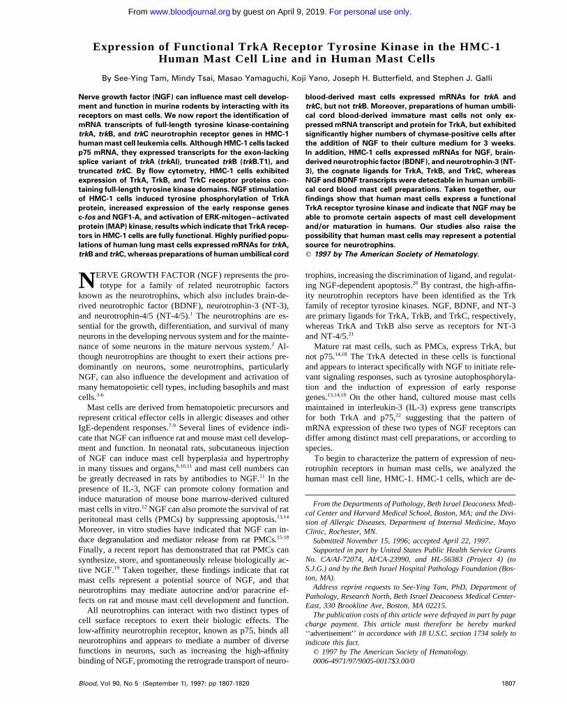
Fig 1. Expression of mRNA transcripts encoding trkA, trkB, andMitogen-activated protein (MAP) kinase activity. Previous stud-trkC genes in the HMC-1 mast cell line, the K-562 chronic myeloge-ies from our laboratory have shown that ERK1-pp44 and ERK2-nous leukemia cell line, and fetal monkey brain tissue. The cDNA waspp42 MAP kinases are coordinately regulated in mouse mast cellssynthesized from total RNA by reverse transcriptase and amplified by
that are stimulated with SCF or IgE and antigen.40 Therefore, only PCR using two degenerate oligonucleotide primers A and B. Thepp44 MAP kinase activity was measured in this study. ERK1-MAP identity of the PCR products was confirmed by Southern blot analysiskinase pp44 was partially purified from ERK1 ImmunoCruz System using 32P-labeled human trkA, monkey trkB, and monkey trkC probes.(Santa Cruz Biotechnology), which consists of an immunoaffinitycolumn containing ERK1 rabbit polyclonal IgG antibody coupled toan agarose matrix, as described in the instruction manual supplied in simultaneously amplifying partial trkA, trkB, and trkCby the manufacturer. The eluted antigen was assayed for phosphory- sequences by PCR with fetal monkey brain cDNAs.26 Thelation of myelin basic protein (Santa Cruz Biotechnology) using a partial monkey trkB and trkC sequences amplified are highlysubstrate peptide kinase assay as previously described.41 Briefly, 30 homologous to their human counterparts,26,35 and thus weremL of the eluted antigen was incubated with 25 mmol/L Tris-HCl used as cDNA probes in our subsequent Southern hybridiza-(pH 7.5), 1 mmol/L MgCl2 , 50 mmol/L adenosine triphosphate
tion studies. Southern analysis of the PCR products using(ATP), 2.5 mg of myelin basic protein, and 1 mCi of [g-32P]ATP inhuman trkA and monkey trkB and trkC cDNAs as probesa reaction volume of 50 mL. After incubation at 307C for 15 minutes,detected strong signals for the mRNA expression of trkA25 mL of the reaction was spotted onto a 1.5 1 1.5 cm P-81 cellulose(predicted size 525 bp) in HMC-1 cells and in fetal monkeyphosphate paper (Life Technologies, Gaithersburg, MD) and washed
twice with 1% phosphoric acid and then water before quantitation brain (Fig 1). Moreover, we could detect weaker, but consis-of radioactivity by liquid scintillation counting. The protein concen- tently reproducible, hybridization signals indicating the ex-tration for each eluted antigen sample was determined by Bio-Rad pression of mRNAs for trkB (predicted size 624 bp) and trkCProtein Assay (Bio-Rad, Hercules, CA), and used to calculate spe- (predicited size 576 bp) in HMC-1 cells. These hybridizationcific activities (cpm/mg protein). signals did not appear to result from nonspecific cross-hy-
bridization of different trk cDNA probes with the HMC-1RESULTS trk gene messages, as illustrated by the observation that the
human K-562 cell line, which is known to express exclu-HMC-1 cells express mRNA transcripts for trkA, trkB, andtrkC that contain the full-length tyrosine kinase domains. sively the TrkA receptor,32 exhibited no detectable expres-
sion of trkB and trkC mRNAs (Fig 1). Moreover, the trkBRecent studies have indicated that mRNA for trkA, but nottrkB or trkC, is detectable in rat and mouse mast cells.18,22 and trkC signals detected on the Southern blot exhibited the
expected difference in their molecular weights (trkB útrkC)To assess the pattern of expression of trk mRNAs in humanmast cells, we first searched for the presence of mRNA (Fig 1). The results shown in Fig 1 were obtained in at least
four separate experiments.transcripts encoding the tyrosine kinase-containing isoformsof different members of the Trk receptor family in HMC-1 It has been reported that two different splice variants of
trkA mRNA transcripts are expressed in PC12 cells and ratmast cells.RT-PCR was performed on HMC-1 RNA with two degen- PMC (trkAI and trkAII).18,42 Moreover, transfection studies
in PC12nnr5 cells show that, in contrast to TrkAI receptors,erate oligo primers, A and B, which correspond to highlyconserved regions of the extracellular domain and of the TrkAII receptors exhibit enhanced functional responses to
stimulation by the presumably noncognate ligand, NT-3.43tyrosine kinase domain shared among human TrkA, mouseTrkB, and porcine TrkC receptor polypeptides.32-34 These To assess which of the two trkA transcripts was expressed
in HMC-1 cells, PCR was performed using a pair of primerstwo primers have previously been shown to be effective
AID Blood 0028 / 5h3c$$$542 08-01-97 18:53:18 blda WBS: Blood
For personal use only.on April 9, 2019. by guest www.bloodjournal.orgFrom
FUNCTIONAL TrkA RECEPTORS IN HUMAN MAST CELLS 1811
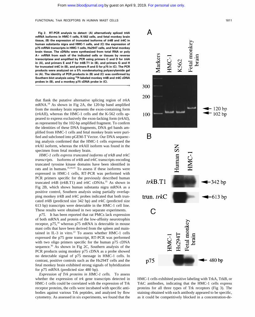
Fig 2. RT-PCR analysis to detect: (A) alternatively spliced trkAmRNA isoforms in HMC-1 cells, K-562 cells, and fetal monkey braintissue, (B) the expression of truncated isoforms of trkB and trkC inhuman substanta nigra and HMC-1 cells, and (C) the expression ofp75 mRNA transcripts in HMC-1 cells, Hs294T cells, and fetal monkeybrain tissue. The cDNAs were synthesized from total RNA or polyA" mRNA from each of the indicated cells or tissues by reversetranscriptase and amplified by PCR using primers C and D for trkAin (A), and primers E and F for trkB.T1 in (B), and primers G and Hfor truncated trkC in (B), and primers R and S for p75 in (C). The PCRproducts were analyzed on a 5% nondenaturing polyacrylamide gelin (A). The identity of PCR products in (B) and (C) was confirmed bySouthern blot analysis using 32P-labeled monkey trkB and trkC cDNAprobes in (B), and a monkey p75 cDNA probe in (C).
r
that flank the putative alternative splicing region of trkAmRNA.18 As shown in Fig 2A, the 120-bp band amplifiedfrom the monkey brain represents the exon-containing form(trkAII), whereas the HMC-1 cells and the K-562 cells ap-peared to express exclusively the exon-lacking form (trkAI),as represented by the 102-bp amplified fragment. To confirmthe identities of these DNA fragments, DNA gel bands am-plified from HMC-1 cells and fetal monkey brain were puri-fied and subcloned into pGEM-T Vector. Our DNA sequenc-ing analysis confirmed that the HMC-1 cells expressed thetrkAI isoform, whereas the trkAII isoform was found in thespecimen from fetal monkey brain.
HMC-1 cells express truncated isoforms of trkB and trkCtranscripts. Isoforms of trkB and trkC transcripts encodingtruncated tyrosine kinase domains have been identified inrats and in humans.35,44,45 To assess if these isoforms wereexpressed in HMC-1 cells, RT-PCR was performed withPCR primers specific for the previously described humantruncated trkB (trkB.T1) and trkC cDNAs.35 As shown inFig 2B, which shows human substanta nigra mRNA as apositive control, Southern analysis using partially overlap-ping monkey trkB and trkC probes indicated that both trun-cated trkB (predicted size 342 bp) and trkC (predicted size613 bp) transcripts were detectable in the HMC-1 cell line.These results were obtained in two separate experiments.
p75. It has been reported that rat PMCs lack expressionof both mRNA and protein of the low-affinity neurotrophinreceptor, p75,18 whereas p75 mRNA is detectable in mousemast cells that have been derived from the spleen and main-tained in IL-3 in vitro.22 To assess whether HMC-1 cellsexpressed the p75 gene transcript, RT-PCR was performedwith two oligo primers specific for the human p75 cDNAsequence.36 As shown in Fig 2C, Southern analysis of thePCR products using monkey p75 cDNA as a probe showedno detectable signal of p75 message in HMC-1 cells. Incontrast, positive controls such as the Hs294T cells and thefetal monkey brain exhibited strong signals of hybridizationfor p75 mRNA (predicted size 480 bp).
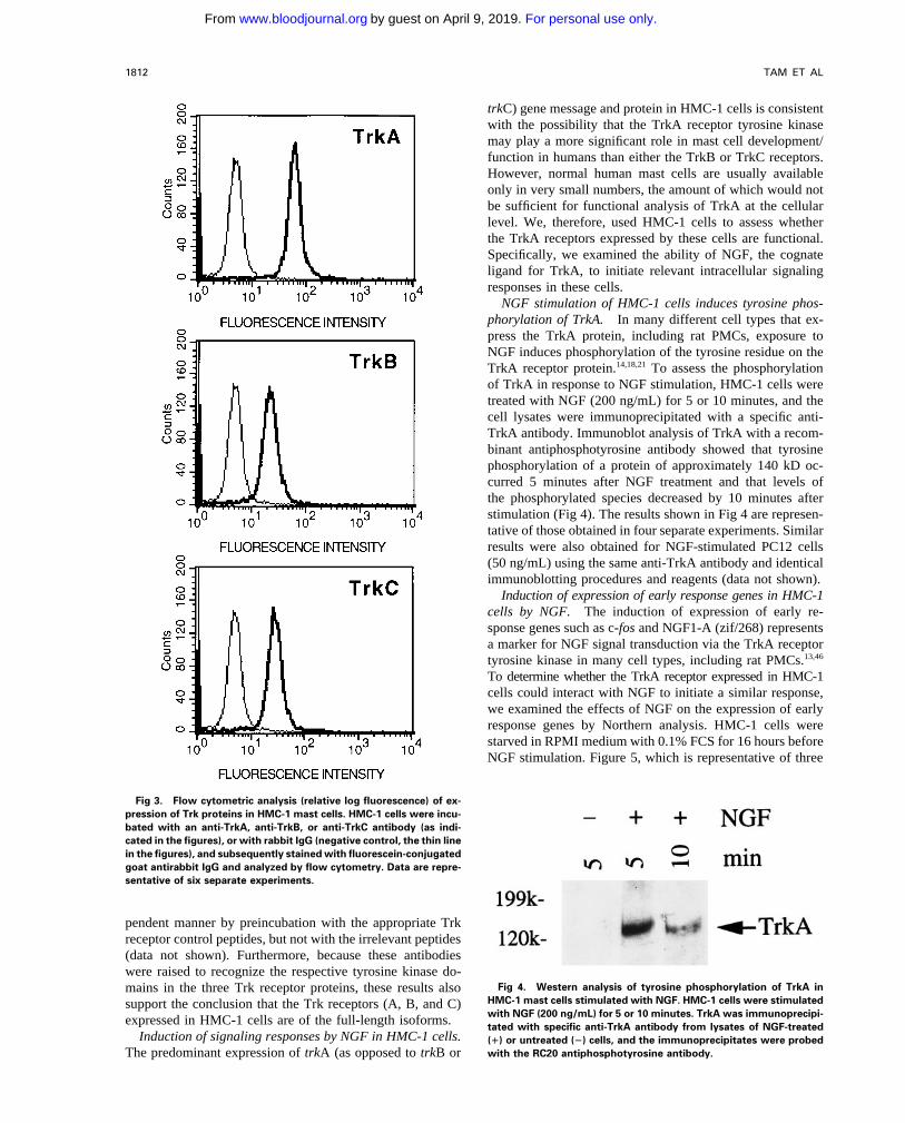
Expression of Trk proteins in HMC-1 cells. To assesswhether the expression of trk gene transcripts detected in HMC-1 cells exhibited positive labeling with TrkA, TrkB, or
TrkC antibodies, indicating that the HMC-1 cells expressHMC-1 cells could be correlated with the expression of Trkreceptor proteins, the cells were incubated with specific anti- proteins for all three types of Trk receptors (Fig 3). The
staining obtained with each antibody appeared to be specific,bodies against various Trk peptides, and analyzed by flowcytometry. As assessed in six experiments, we found that the as it could be competitively blocked in a concentration-de-
AID Blood 0028 / 5h3c$$$543 08-01-97 18:53:18 blda WBS: Blood
For personal use only.on April 9, 2019. by guest www.bloodjournal.orgFrom
TAM ET AL1812
trkC) gene message and protein in HMC-1 cells is consistentwith the possibility that the TrkA receptor tyrosine kinasemay play a more significant role in mast cell development/function in humans than either the TrkB or TrkC receptors.However, normal human mast cells are usually availableonly in very small numbers, the amount of which would notbe sufficient for functional analysis of TrkA at the cellularlevel. We, therefore, used HMC-1 cells to assess whetherthe TrkA receptors expressed by these cells are functional.Specifically, we examined the ability of NGF, the cognateligand for TrkA, to initiate relevant intracellular signalingresponses in these cells.
NGF stimulation of HMC-1 cells induces tyrosine phos-phorylation of TrkA. In many different cell types that ex-press the TrkA protein, including rat PMCs, exposure toNGF induces phosphorylation of the tyrosine residue on theTrkA receptor protein.14,18,21 To assess the phosphorylationof TrkA in response to NGF stimulation, HMC-1 cells weretreated with NGF (200 ng/mL) for 5 or 10 minutes, and thecell lysates were immunoprecipitated with a specific anti-TrkA antibody. Immunoblot analysis of TrkA with a recom-binant antiphosphotyrosine antibody showed that tyrosinephosphorylation of a protein of approximately 140 kD oc-curred 5 minutes after NGF treatment and that levels ofthe phosphorylated species decreased by 10 minutes afterstimulation (Fig 4). The results shown in Fig 4 are represen-tative of those obtained in four separate experiments. Similarresults were also obtained for NGF-stimulated PC12 cells(50 ng/mL) using the same anti-TrkA antibody and identicalimmunoblotting procedures and reagents (data not shown).
Induction of expression of early response genes in HMC-1cells by NGF. The induction of expression of early re-sponse genes such as c-fos and NGF1-A (zif/268) representsa marker for NGF signal transduction via the TrkA receptortyrosine kinase in many cell types, including rat PMCs.13,46
To determine whether the TrkA receptor expressed in HMC-1cells could interact with NGF to initiate a similar response,we examined the effects of NGF on the expression of earlyresponse genes by Northern analysis. HMC-1 cells werestarved in RPMI medium with 0.1% FCS for 16 hours beforeNGF stimulation. Figure 5, which is representative of three
Fig 3. Flow cytometric analysis (relative log fluorescence) of ex-pression of Trk proteins in HMC-1 mast cells. HMC-1 cells were incu-bated with an anti-TrkA, anti-TrkB, or anti-TrkC antibody (as indi-cated in the figures), or with rabbit IgG (negative control, the thin linein the figures), and subsequently stained with fluorescein-conjugatedgoat antirabbit IgG and analyzed by flow cytometry. Data are repre-sentative of six separate experiments.
pendent manner by preincubation with the appropriate Trkreceptor control peptides, but not with the irrelevant peptides(data not shown). Furthermore, because these antibodieswere raised to recognize the respective tyrosine kinase do-
Fig 4. Western analysis of tyrosine phosphorylation of TrkA inmains in the three Trk receptor proteins, these results alsoHMC-1 mast cells stimulated with NGF. HMC-1 cells were stimulatedsupport the conclusion that the Trk receptors (A, B, and C)with NGF (200 ng/mL) for 5 or 10 minutes. TrkA was immunoprecipi-expressed in HMC-1 cells are of the full-length isoforms. tated with specific anti-TrkA antibody from lysates of NGF-treated
Induction of signaling responses by NGF in HMC-1 cells. (") or untreated (Ï) cells, and the immunoprecipitates were probedwith the RC20 antiphosphotyrosine antibody.The predominant expression of trkA (as opposed to trkB or
AID Blood 0028 / 5h3c$$$543 08-01-97 18:53:18 blda WBS: Blood
For personal use only.on April 9, 2019. by guest www.bloodjournal.orgFrom
FUNCTIONAL TrkA RECEPTORS IN HUMAN MAST CELLS 1813
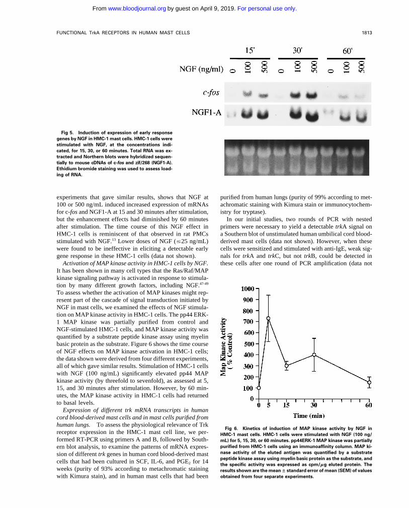
Fig 5. Induction of expression of early responsegenes by NGF in HMC-1 mast cells. HMC-1 cells werestimulated with NGF, at the concentrations indi-cated, for 15, 30, or 60 minutes. Total RNA was ex-tracted and Northern blots were hybridized sequen-tially to mouse cDNAs of c-fos and zif/268 (NGF1-A).Ethidium bromide staining was used to assess load-ing of RNA.
experiments that gave similar results, shows that NGF at purified from human lungs (purity of 99% according to met-achromatic staining with Kimura stain or immunocytochem-100 or 500 ng/mL induced increased expression of mRNAs
for c-fos and NGF1-A at 15 and 30 minutes after stimulation, istry for tryptase).In our initial studies, two rounds of PCR with nestedbut the enhancement effects had diminished by 60 minutes
after stimulation. The time course of this NGF effect in primers were necessary to yield a detectable trkA signal ona Southern blot of unstimulated human umbilical cord blood-HMC-1 cells is reminiscent of that observed in rat PMCs
stimulated with NGF.13 Lower doses of NGF (°25 ng/mL) derived mast cells (data not shown). However, when thesecells were sensitized and stimulated with anti-IgE, weak sig-were found to be ineffective in eliciting a detectable early
gene response in these HMC-1 cells (data not shown). nals for trkA and trkC, but not trkB, could be detected inthese cells after one round of PCR amplification (data notActivation of MAP kinase activity in HMC-1 cells by NGF.
It has been shown in many cell types that the Ras/Raf/MAPkinase signaling pathway is activated in response to stimula-tion by many different growth factors, including NGF.47-49
To assess whether the activation of MAP kinases might rep-resent part of the cascade of signal transduction initiated byNGF in mast cells, we examined the effects of NGF stimula-tion on MAP kinase activity in HMC-1 cells. The pp44 ERK-1 MAP kinase was partially purified from control andNGF-stimulated HMC-1 cells, and MAP kinase activity wasquantified by a substrate peptide kinase assay using myelinbasic protein as the substrate. Figure 6 shows the time courseof NGF effects on MAP kinase activation in HMC-1 cells;the data shown were derived from four different experiments,all of which gave similar results. Stimulation of HMC-1 cellswith NGF (100 ng/mL) significantly elevated pp44 MAPkinase activity (by threefold to sevenfold), as assessed at 5,15, and 30 minutes after stimulation. However, by 60 min-utes, the MAP kinase activity in HMC-1 cells had returnedto basal levels.
Expression of different trk mRNA transcripts in humancord blood-derived mast cells and in mast cells purified fromhuman lungs. To assess the physiological relevance of Trk
Fig 6. Kinetics of induction of MAP kinase activity by NGF inreceptor expression in the HMC-1 mast cell line, we per- HMC-1 mast cells. HMC-1 cells were stimulated with NGF (100 ng/formed RT-PCR using primers A and B, followed by South- mL) for 5, 15, 30, or 60 minutes. pp44ERK-1 MAP kinase was partially
purified from HMC-1 cells using an immunoaffinity column. MAP ki-ern blot analysis, to examine the patterns of mRNA expres-nase activity of the eluted antigen was quantified by a substratesion of different trk genes in human cord blood-derived mastpeptide kinase assay using myelin basic protein as the substrate, andcells that had been cultured in SCF, IL-6, and PGE2 for 14 the specific activity was expressed as cpm/mg eluted protein. The
weeks (purity of 93% according to metachromatic staining results shown are the meanÔ standard error of mean (SEM) of valuesobtained from four separate experiments.with Kimura stain), and in human mast cells that had been
AID Blood 0028 / 5h3c$$$543 08-01-97 18:53:18 blda WBS: Blood
For personal use only.on April 9, 2019. by guest www.bloodjournal.orgFrom
TAM ET AL1814
detectable in purified preparations of normal human mastcells. However, our results also raise the possibility that anti-IgE stimulation of human lung mast cells can downregulatethe cells’ expression of trkB mRNA. Thus, the extent ofspecific histamine release (% release from anti-IgE-stimu-lated cells minus % release from cells without anti-IgE)detected in response to anti-IgE challenge was higher inExperiment II (Ç18% specific release) than in ExperimentI (no specific release). This result suggests that the level ofmast cell activation achieved in the second experiment wassubstantially greater than that in Experiment I.
Human umbilical cord blood-derived mast cells expressfunctional TrkA receptor protein and respond to NGF stimu-lation by exhibiting increased expression of chymase. Toassess the physiologic relevance of the expression of func-tional TrkA receptor protein in HMC-1 cells, we examinedthe expression of TrkA protein in human umbilical cord
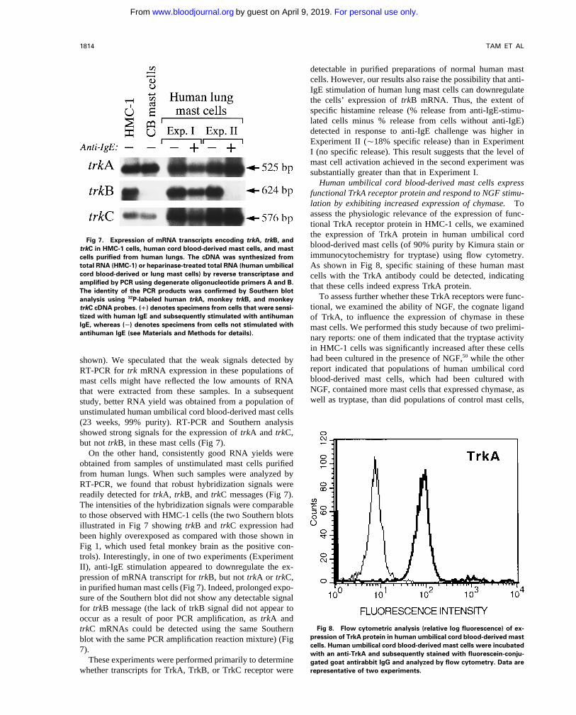
Fig 7. Expression of mRNA transcripts encoding trkA, trkB, andblood-derived mast cells (of 90% purity by Kimura stain ortrkC in HMC-1 cells, human cord blood-derived mast cells, and mastimmunocytochemistry for tryptase) using flow cytometry.cells purified from human lungs. The cDNA was synthesized from
total RNA (HMC-1) or heparinase-treated total RNA (human umbilical As shown in Fig 8, specific staining of these human mastcord blood-derived or lung mast cells) by reverse transcriptase and cells with the TrkA antibody could be detected, indicatingamplified by PCR using degenerate oligonucleotide primers A and B. that these cells indeed express TrkA protein.The identity of the PCR products was confirmed by Southern blot
To assess further whether these TrkA receptors were func-analysis using 32P-labeled human trkA, monkey trkB, and monkeytional, we examined the ability of NGF, the cognate ligandtrkC cDNA probes. (") denotes specimens from cells that were sensi-
tized with human IgE and subsequently stimulated with antihuman of TrkA, to influence the expression of chymase in theseIgE, whereas (Ï) denotes specimens from cells not stimulated with mast cells. We performed this study because of two prelimi-antihuman IgE (see Materials and Methods for details).
nary reports: one of them indicated that the tryptase activityin HMC-1 cells was significantly increased after these cellshad been cultured in the presence of NGF,50 while the othershown). We speculated that the weak signals detected byreport indicated that populations of human umbilical cordRT-PCR for trk mRNA expression in these populations ofblood-derived mast cells, which had been cultured withmast cells might have reflected the low amounts of RNANGF, contained more mast cells that expressed chymase, asthat were extracted from these samples. In a subsequentwell as tryptase, than did populations of control mast cells,study, better RNA yield was obtained from a population of
unstimulated human umbilical cord blood-derived mast cells(23 weeks, 99% purity). RT-PCR and Southern analysisshowed strong signals for the expression of trkA and trkC,but not trkB, in these mast cells (Fig 7).
On the other hand, consistently good RNA yields wereobtained from samples of unstimulated mast cells purifiedfrom human lungs. When such samples were analyzed byRT-PCR, we found that robust hybridization signals werereadily detected for trkA, trkB, and trkC messages (Fig 7).The intensities of the hybridization signals were comparableto those observed with HMC-1 cells (the two Southern blotsillustrated in Fig 7 showing trkB and trkC expression hadbeen highly overexposed as compared with those shown inFig 1, which used fetal monkey brain as the positive con-trols). Interestingly, in one of two experiments (ExperimentII), anti-IgE stimulation appeared to downregulate the ex-pression of mRNA transcript for trkB, but not trkA or trkC,in purified human mast cells (Fig 7). Indeed, prolonged expo-sure of the Southern blot did not show any detectable signalfor trkB message (the lack of trkB signal did not appear tooccur as a result of poor PCR amplification, as trkA and
Fig 8. Flow cytometric analysis (relative log fluorescence) of ex-trkC mRNAs could be detected using the same Southernpression of TrkA protein in human umbilical cord blood-derived mastblot with the same PCR amplification reaction mixture) (Figcells. Human umbilical cord blood-derived mast cells were incubated7). with an anti-TrkA and subsequently stained with fluorescein-conju-
These experiments were performed primarily to determine gated goat antirabbit IgG and analyzed by flow cytometry. Data arerepresentative of two experiments.whether transcripts for TrkA, TrkB, or TrkC receptor were
AID Blood 0028 / 5h3c$$$543 08-01-97 18:53:18 blda WBS: Blood
For personal use only.on April 9, 2019. by guest www.bloodjournal.orgFrom
FUNCTIONAL TrkA RECEPTORS IN HUMAN MAST CELLS 1815
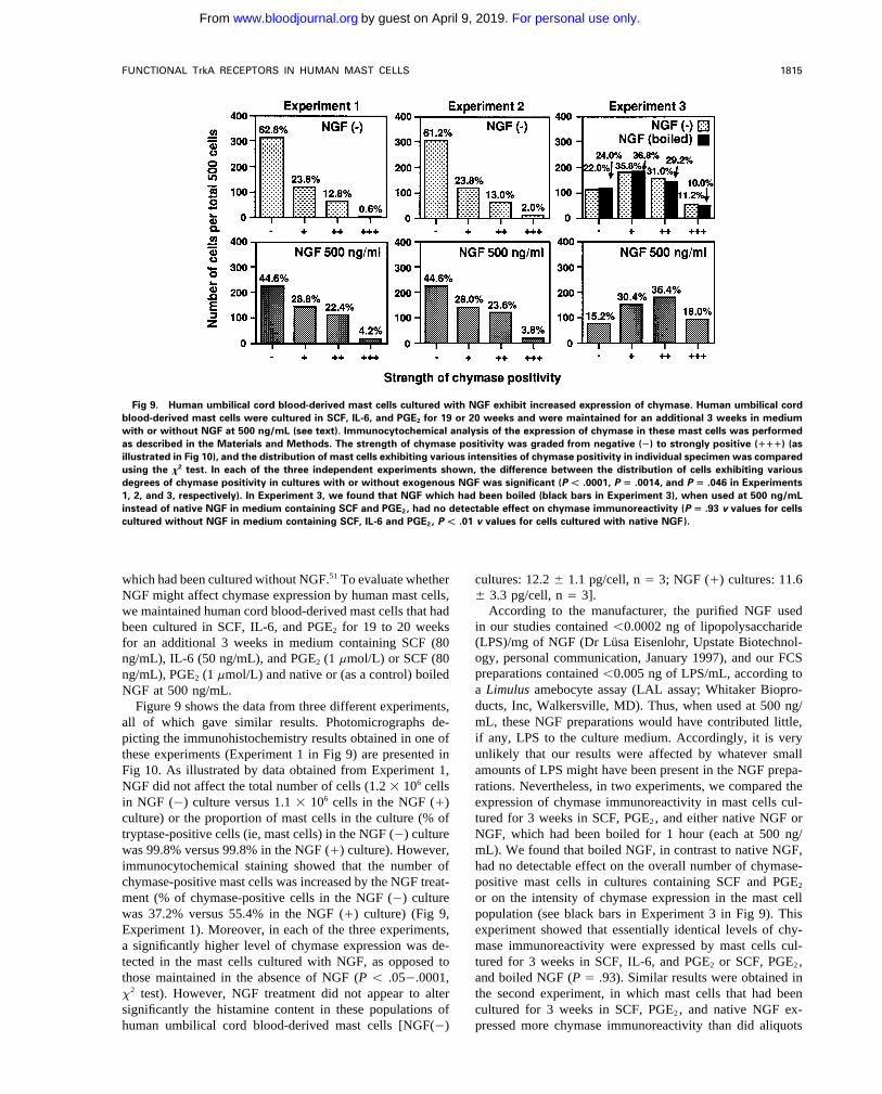
Fig 9. Human umbilical cord blood-derived mast cells cultured with NGF exhibit increased expression of chymase. Human umbilical cordblood-derived mast cells were cultured in SCF, IL-6, and PGE2 for 19 or 20 weeks and were maintained for an additional 3 weeks in mediumwith or without NGF at 500 ng/mL (see text). Immunocytochemical analysis of the expression of chymase in these mast cells was performedas described in the Materials and Methods. The strength of chymase positivity was graded from negative (Ï) to strongly positive (""") (asillustrated in Fig 10), and the distribution of mast cells exhibiting various intensities of chymase positivity in individual specimen was comparedusing the x2 test. In each of the three independent experiments shown, the difference between the distribution of cells exhibiting variousdegrees of chymase positivity in cultures with or without exogenous NGF was significant (P Ú .0001, P ! .0014, and P ! .046 in Experiments1, 2, and 3, respectively). In Experiment 3, we found that NGF which had been boiled (black bars in Experiment 3), when used at 500 ng/mLinstead of native NGF in medium containing SCF and PGE2, had no detectable effect on chymase immunoreactivity (P ! .93 v values for cellscultured without NGF in medium containing SCF, IL-6 and PGE2, P Ú .01 v values for cells cultured with native NGF).
which had been cultured without NGF.51 To evaluate whether cultures: 12.2 { 1.1 pg/cell, n Å 3; NGF (/) cultures: 11.6{ 3.3 pg/cell, n Å 3].NGF might affect chymase expression by human mast cells,
According to the manufacturer, the purified NGF usedwe maintained human cord blood-derived mast cells that hadin our studies contained õ0.0002 ng of lipopolysaccharidebeen cultured in SCF, IL-6, and PGE2 for 19 to 20 weeks(LPS)/mg of NGF (Dr Lusa Eisenlohr, Upstate Biotechnol-for an additional 3 weeks in medium containing SCF (80ogy, personal communication, January 1997), and our FCSng/mL), IL-6 (50 ng/mL), and PGE2 (1 mmol/L) or SCF (80preparations contained õ0.005 ng of LPS/mL, according tong/mL), PGE2 (1 mmol/L) and native or (as a control) boileda Limulus amebocyte assay (LAL assay; Whitaker Biopro-NGF at 500 ng/mL.ducts, Inc, Walkersville, MD). Thus, when used at 500 ng/Figure 9 shows the data from three different experiments,mL, these NGF preparations would have contributed little,all of which gave similar results. Photomicrographs de-if any, LPS to the culture medium. Accordingly, it is verypicting the immunohistochemistry results obtained in one ofunlikely that our results were affected by whatever smallthese experiments (Experiment 1 in Fig 9) are presented inamounts of LPS might have been present in the NGF prepa-Fig 10. As illustrated by data obtained from Experiment 1,
NGF did not affect the total number of cells (1.2 1 106 cells rations. Nevertheless, in two experiments, we compared theexpression of chymase immunoreactivity in mast cells cul-in NGF (0) culture versus 1.1 1 106 cells in the NGF (/)
culture) or the proportion of mast cells in the culture (% of tured for 3 weeks in SCF, PGE2, and either native NGF orNGF, which had been boiled for 1 hour (each at 500 ng/tryptase-positive cells (ie, mast cells) in the NGF (0) culture
was 99.8% versus 99.8% in the NGF (/) culture). However, mL). We found that boiled NGF, in contrast to native NGF,had no detectable effect on the overall number of chymase-immunocytochemical staining showed that the number of
chymase-positive mast cells was increased by the NGF treat- positive mast cells in cultures containing SCF and PGE2
or on the intensity of chymase expression in the mast cellment (% of chymase-positive cells in the NGF (0) culturewas 37.2% versus 55.4% in the NGF (/) culture) (Fig 9, population (see black bars in Experiment 3 in Fig 9). This
experiment showed that essentially identical levels of chy-Experiment 1). Moreover, in each of the three experiments,a significantly higher level of chymase expression was de- mase immunoreactivity were expressed by mast cells cul-
tured for 3 weeks in SCF, IL-6, and PGE2 or SCF, PGE2,tected in the mast cells cultured with NGF, as opposed tothose maintained in the absence of NGF (P õ .050.0001, and boiled NGF (P Å .93). Similar results were obtained in
the second experiment, in which mast cells that had beenx2 test). However, NGF treatment did not appear to altersignificantly the histamine content in these populations of cultured for 3 weeks in SCF, PGE2, and native NGF ex-
pressed more chymase immunoreactivity than did aliquotshuman umbilical cord blood-derived mast cells [NGF(0)
AID Blood 0028 / 5h3c$$$543 08-01-97 18:53:18 blda WBS: Blood
For personal use only.on April 9, 2019. by guest www.bloodjournal.orgFrom
TAM ET AL1816
Fig 10.
AID Blood 0028 / 5h3c$$0028 08-01-97 18:53:18 blda WBS: Blood
For personal use only.on April 9, 2019. by guest www.bloodjournal.orgFrom
FUNCTIONAL TrkA RECEPTORS IN HUMAN MAST CELLS 1817
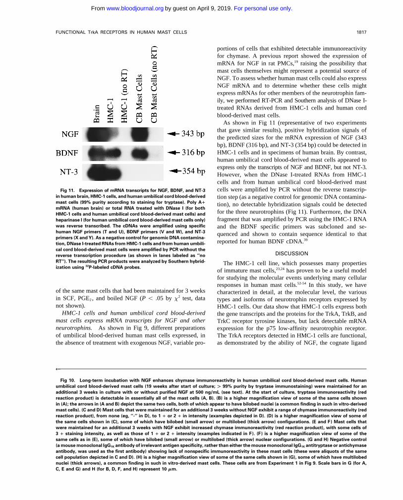
portions of cells that exhibited detectable immunoreactivityfor chymase. A previous report showed the expression ofmRNA for NGF in rat PMCs,19 raising the possibility thatmast cells themselves might represent a potential source ofNGF. To assess whether human mast cells could also expressNGF mRNA and to determine whether these cells mightexpress mRNAs for other members of the neurotrophin fam-ily, we performed RT-PCR and Southern analysis of DNase I-treated RNAs derived from HMC-1 cells and human cordblood-derived mast cells.
As shown in Fig 11 (representative of two experimentsthat gave similar results), positive hybridization signals ofthe predicted sizes for the mRNA expression of NGF (343bp), BDNF (316 bp), and NT-3 (354 bp) could be detected inHMC-1 cells and in specimens of human brain. By contrast,human umbilical cord blood-derived mast cells appeared toexpress only the transcripts of NGF and BDNF, but not NT-3.However, when the DNase I-treated RNAs from HMC-1cells and from human umbilical cord blood-derived mastcells were amplified by PCR without the reverse transcrip-Fig 11. Expression of mRNA transcripts for NGF, BDNF, and NT-3
in human brain, HMC-1 cells, and human umbilical cord blood-derived tion step (as a negative control for genomic DNA contamina-mast cells (99% purity according to staining for tryptase). Poly A" tion), no detectable hybridization signals could be detectedmRNA (human brain) or total RNA treated with DNase I (for both
for the three neurotrophins (Fig 11). Furthermore, the DNAHMC-1 cells and human umbilical cord blood-derived mast cells) andfragment that was amplified by PCR using the HMC-1 RNAheparinase I (for human umbilical cord blood-derived mast cells only)
was reverse transcribed. The cDNAs were amplified using specific and the BDNF specific primers was subcloned and se-human NGF primers (T and U), BDNF primers (V and W), and NT-3 quenced and shown to contain sequence identical to thatprimers (X and Y). As a negative control for genomic DNA contamina-
reported for human BDNF cDNA.39tion, DNase I-treated RNAs from HMC-1 cells and from human umbili-cal cord blood-derived mast cells were amplified by PCR without the
DISCUSSIONreverse transcription procedure (as shown in lanes labeled as ‘‘noRT’’). The resulting PCR products were analyzed by Southern hybrid- The HMC-1 cell line, which possesses many propertiesization using 32P-labeled cDNA probes.
of immature mast cells,23,24 has proven to be a useful modelfor studying the molecular events underlying many cellularresponses in human mast cells.52-54 In this study, we have
of the same mast cells that had been maintained for 3 weeks characterized in detail, at the molecular level, the variousin SCF, PGE2, and boiled NGF (P õ .05 by x2 test, data types and isoforms of neurotrophin receptors expressed bynot shown). HMC-1 cells. Our data show that HMC-1 cells express both
HMC-1 cells and human umbilical cord blood-derived the gene transcripts and the proteins for the TrkA, TrkB, andmast cells express mRNA transcripts for NGF and other TrkC receptor tyrosine kinases, but lack detectable mRNAneurotrophins. As shown in Fig 9, different preparations expression for the p75 low-affinity neurotrophin receptor.of umbilical blood-derived human mast cells expressed, in The TrkA receptors detected in HMC-1 cells are functional,
as demonstrated by the ability of NGF, the cognate ligandthe absence of treatment with exogenous NGF, variable pro-
R
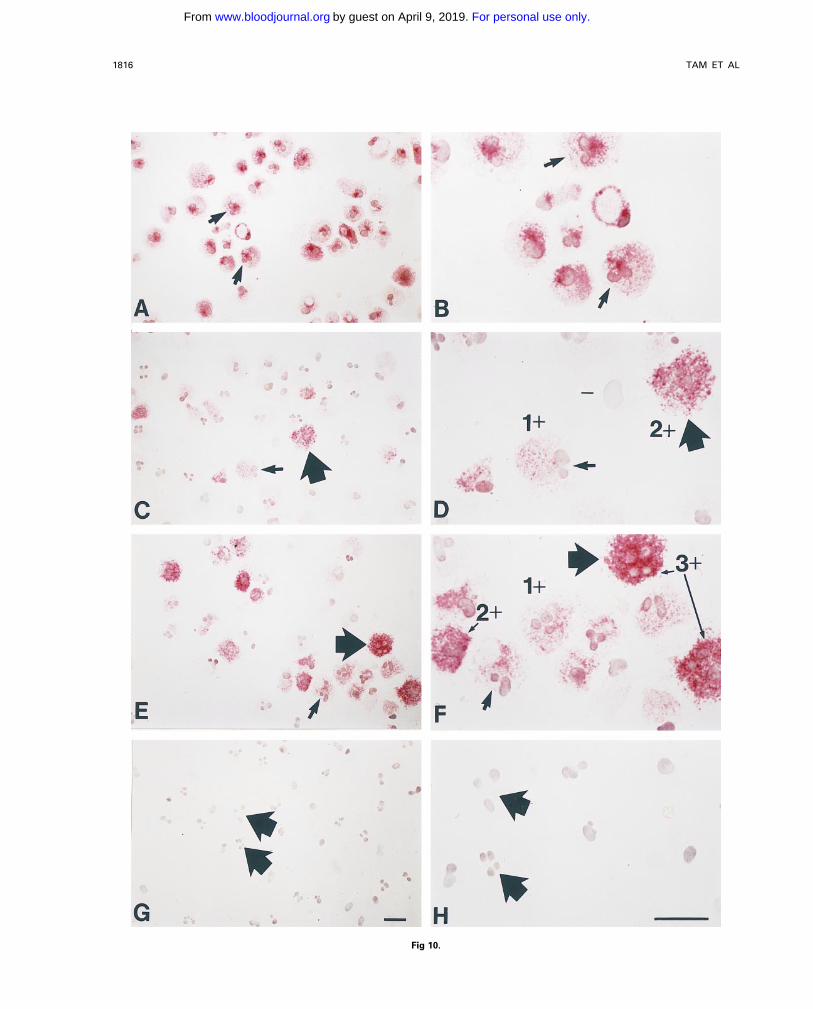
Fig 10. Long-term incubation with NGF enhances chymase immunoreactivity in human umbilical cord blood-derived mast cells. Humanumbilical cord blood-derived mast cells (19 weeks after start of culture; Û 99% purity by tryptase immunostaining) were maintained for anadditional 3 weeks in culture with or without purified NGF at 500 ng/mL (see text). At the start of culture, tryptase immunoreactivity (redreaction product) is detectable in essentially all of the mast cells (A, B). (B) is a higher magnification view of some of the same cells shownin (A); the arrows in (A and B) depict the same two cells, both of which appear to have bilobed nuclei (a common finding in such in vitro-derivedmast cells). (C and D) Mast cells that were maintained for an additional 3 weeks without NGF exhibit a range of chymase immunoreactivity (redreaction product), from none (eg, ‘‘-’’ in D), to 1 " or 2 " in intensity (examples depicted in D). (D) is a higher magnification view of some ofthe same cells shown in (C), some of which have bilobed (small arrow) or multilobed (thick arrow) configurations. (E and F) Mast cells thatwere maintained for an additional 3 weeks with NGF exhibit increased chymase immunoreactivity (red reaction product), with some cells of3 " staining intensity, as well as those of 1 " or 2 " intensity (examples indicated in F). (F) is a higher magnification view of some of thesame cells as in (E), some of which have bilobed (small arrow) or multilobed (thick arrow) nuclear configurations. (G and H) Negative control(a mouse monoclonal IgG1K antibody of irrelevant antigen specificity, rather than either the mouse monoclonal IgG1K antitryptase or antichymaseantibody, was used as the first antibody) showing lack of nonspecific immunoreactivity in these mast cells (these were aliquots of the samecell population depicted in C and D). (H) is a higher magnification view of some of the same cells shown in (G), some of which have multilobednuclei (thick arrows), a common finding in such in vitro-derived mast cells. These cells are from Experiment 1 in Fig 9. Scale bars in G (for A,C, E and G) and H (for B, D, F, and H) represent 10 mm.
AID Blood 0028 / 5h3c$$$543 08-01-97 18:53:18 blda WBS: Blood
For personal use only.on April 9, 2019. by guest www.bloodjournal.orgFrom
TAM ET AL1818
of TrkA, to initiate appropriate cellular signaling responses cells can be quite heterogeneous, depending both on thecells’ stage of development and species of origin.in these cells. Thus, NGF stimulation of HMC-1 cells was
shown to induce tyrosine autophosphorylation of TrkA, in- Despite the apparent heterogeneity of neurotrophin recep-tor expression in mouse and human mast cells, the interactioncreased expression of early response genes, and activation
of MAP kinase activity. of NGF with its receptors on different populations of mastcells appears to be consistently of the low-affinity type. AnIt is currently quite difficult to obtain normal human mast
cells in large numbers. Accordingly, the full extent to which earlier report indicated that NGF binds to its receptor on ratPMCs with a KD that is consistent with low-affinity bindingour observations with HMC-1 cells are relevant to normal
human mast cells can only be established by further studies. (4.0 1 1009 mol/L).56 Similarly, a recent study shows thatthe NGF binding detected in cultured mouse mast cells alsoHowever, we found that highly purified populations of both
cultured umbilical cord blood-derived human mast cells and exhibits a low-affinity (2.1 1 1009 mol/L).22 The dose re-sponse curves for NGF effects on serotonin release and onmast cells isolated from adult human lung tissue predomi-
nantly express trkA mRNA, and that human umbilical cord cell survival in rat PMCs are also consistent with a low-affinity interaction,13,18 although one recent apparently con-blood-derived mast cells also express TrkA protein. More-
over, the TrkA receptors expressed in human umbilical cord flicting report has indicated that NGF can promote survivalof rat PMCs via a high-affinity NGF receptor.14 In accordblood-derived mast cells appear to mediate at least one po-
tentially physiological response to NGF stimulation, the in- with most of these observations, we found that the doses ofNGF required to induce the expression of early responsecreased expression of chymase.
In contrast to mRNA for trkA, the expression of trkB and genes in HMC-1 cells are consistent with a low-affinity typeof interaction between NGF and the NGF receptors ex-trkC mRNAs in HMC-1 cells (and in populations of purified
human lung mast cells) was unexpected, particularly in light pressed in these cells.The TrkA receptor tyrosine kinase expressed by HMC-1of recent studies indicating a lack of expression of these trk
mRNAs in either rat PMCs or mouse mast cells derived in cells appears to be functional, as NGF stimulation of thesecells elicited signaling responses that are characteristic ofIL-3–containing media.13,22 Indeed, of all of the hematopoi-
etic cells examined to date, human mast cells, including the NGF/TrkA interaction in other NGF-responsive cell types.These responses include tyrosine phosphorylation of TrkA,HMC-1 cell line and populations of mature lung mast cells,
appear to be the only cell type that can express all three trk the induction of early response gene expression, and theactivation of MAP kinase activity. However, we found thatgene transcripts. However, unlike preparations of human
lung mast cells, populations of human umbilical cord blood- the kinetics of MAP kinase activation and expression ofearly response genes in HMC-1 cells stimulated with NGFderived mast cells, which have features of immature mast
cells, express transcripts for trkA and trkC, but not trkB. were more transient than those observed with other celltypes. In HMC-1 cells, these two NGF-induced signalingThese findings indicate that populations of human mast cells
can express all three Trk receptors, at least at the level of responses appeared to dissipate completely by 1 hour afterstimulation (Figs 5 and 6), whereas both MAP kinase activitymRNA, but that the patterns of expression of the three recep-
tors in individual human mast cell populations (eg, lung v and the expression of c-fos remained elevated 1 hour afterNGF-stimulation in PC12 cells and human B lympho-in vitro–derived cord blood mast cells) may vary.
Furthermore, we found that HMC-1 cells express mRNAs cytes.57,58 Very similar transient kinetics of MAP kinase acti-vation and early response gene expression were exhibited inencoding the truncated forms of human TrkB (TrkB.T1) and
TrkC receptors, as well as mRNAs for the full-length, tyro- rat PMCs that were treated with NGF,13 as well as in mousebone marrow-derived cultured mast cells, which had beensine kinase-containing, forms.35 While the exact function(s)
of these truncated TrkB and TrkC receptors have not been activated via c-kit (with SCF) or FceRI (with IgE and specificantigen).40,59 These findings are consistent with the widelyfully determined, there is speculation that they may act as
‘‘scavenger receptors’’ to maintain high levels of neuro- held view that distinct patterns or kinetics of activation ofcommon signaling elements may be used by different celltrophins at the cell surface, or may initiate distinct cellular
responses through ligand-dependent interactions with down- types to achieve specificity in their cellular responses toligand-dependent stimulation of Trk receptors.46 In particu-stream signaling elements.55
Our findings indicate that there may be significant differ- lar, it has recently been suggested that variation in the dura-tion of MAP kinase activation may represent a mechanismences in the expression of neurotrophin receptors by mast
cells of different species, as well as by different mast cell that can elicit differential cellular responses to stimulationvia receptor tyrosine kinases.60populations in the same species. Thus, recent studies show
that rat PMCs can express transcripts for both the TrkAI and Previous studies have indicated that NGF can promote thematuration of mouse bone marrow-derived cultured mastTrkAII isoforms,18 whereas we report here that the HMC-1
cell line expresses solely transcripts for the shorter TrkAI cells in vitro in the presence of IL-3.12 We show in thisstudy, in confirmation of results reported by Igarashi et al51isoform that lack the 18 nucleotide insert.42,43 In addition,
the p75 transcript is detectable in cultured mouse mast in abstract form that NGF can enhance the expression of thecytoplasmic granule-associated protease, chymase, in humancells,22 but in neither rat PMCs14,18 nor HMC-1 cells (this
study). Taken together, these findings and all of our other umbilical cord blood-derived mast cells cultured in the pres-ence of SCF. Previously, Furitsu et al61 reported that factorsdata are consistent with the suggestion of Jippo et al22 that
the patterns of expression of neurotrophin receptors by mast derived from mouse 3T3 fibroblasts can induce the mast cell
AID Blood 0028 / 5h3c$$$543 08-01-97 18:53:18 blda WBS: Blood
For personal use only.on April 9, 2019. by guest www.bloodjournal.orgFrom
FUNCTIONAL TrkA RECEPTORS IN HUMAN MAST CELLS 1819
5. Stanisz AM: Neuronal factors modulating immunity. Neuroim-precursors that are present in the human umbilical cord bloodmunomodulation 1:217, 1994mononuclear cell preparations to develop predominantly into
6. Levi-Montalcini R, Skaper SD, Dal Toso R, Petrelli L, Leonmast cells that contain both tryptase and chymase.61 BasedA: Nerve growth factor: From neurotrophin to neurokine. Trendson this characteristic, such in vitro-derived mast cells resem-Neurosci 19:514, 1996ble the mature mast cells that are present in human dermis
7. Bochner BS, Lichtenstein LM: Anaphylaxis. N Engl J Medand certain other sites.62 On the other hand, the human cord 324:1785, 1991blood-derived mast cells, which are generated in vitro in the 8. Paul WE, Seder RA, Plaut M: Lymphokine and cytokine pro-absence of 3T3 cells, but in the presence of SCF, exhibit duction by FceRI/ cells. Adv Immunol 53:1, 1993many phenotypic characteristics of immature mast cells, in- 9. Galli SJ: New concepts about the mast cell. N Engl J Med
328:257, 1993cluding a predominantly tryptase/ chymase0 or low pheno-10. Aloe L, Levi-Montalcini R: Mast cells increase in tissues oftype.27 The ability of NGF to enhance the expression of
neonatal rats injected with the nerve growth factor. Brain Reschymase in umbilical cord blood-derived human mast cells133:358, 1977thus provides evidence that NGF may represent one of the
11. Aloe L: The effects of nerve growth factor and its antibodymicroenvironmental factors, which can synergize with SCFon mast cells in vivo. J Neuroimmunol 18:1, 1988in promoting at least one of the phenotypic changes (ie,
12. Matsuda H, Kannan Y, Ushio H, Kiso Y, Kanemoto T, Suzukiincreased chymase expression) that is associated with the H, Kitamura Y: Nerve growth factor induces development of connec-maturation/differentiation of certain populations of human tive tissue-type mast cells in vitro from murine bone marrow cells.mast cells in vivo. On the other hand, treatment with NGF J Exp Med 174:7, 1991did not significantly increase the cells’ histamine content. 13. Horigome K, Bullock ED, Johnson EM: Effects of nerve
growth factor on rat peritoneal mast cells: Survival promotion andThis result suggests that NGF may have different effects onimmediate-early gene induction. J Biol Chem 269:2695, 1994different aspects of human mast cell phenotype.
14. Kawamoto K, Okada T, Kannan Y, Ushio H, Matsumoto M,It has been proposed that there are complex and bidirec-Matsuda H: Nerve growth factor prevents apoptosis of rat peritonealtional, developmental, and functional interactions betweenmast cells through the trk proto-oncogene receptor. Blood 86:4638,mast cells and the nervous system.4-6 Our finding that various1995populations of human mast cells can express TrkA and TrkC,
15. Bruni A, Bigon E, Boarato E, Mietto L, Leon A, Toffano G:or Trk A, TrkB and TrkC, offers further support for this Interaction between nerve growth factor and lysophosphatidylserinehypothesis. Moreover, a recent report showed that rat PMCs, on rat peritoneal mast cells. FEBS Lett 138:190, 1982which express TrkA receptors, can synthesize, store, and 16. Pearce FL, Thompson HL: Some characteristics of histaminespontaneously release biologically active NGF.19 In the pres- secretion from rat peritoneal mast cells stimulated with nerve growth
factor. J Physiol 372:379, 1986ent study, we found that HMC-1 mast cells and cultured17. Marshall JS, Stead RH, McSharry C, Nielsen L, Bienenstockhuman umbilical cord blood-derived mast cells can express
J: The role of mast cell degranulation products in mast cell hyperpla-the gene transcript for BDNF, as well as NGF mRNA. More-sia. I. Mechanism of action of nerve growth factor. J Immunolover, HMC-1 cells also express mRNA for NT-3. While the144:1886, 1990extent to which these findings pertain to other types of nor-
18. Horigome K, Pryor JC, Bullock ED, Johnson EM: Mediatormal human mast cells and the extent to which human mastrelease from mast cells by nerve growth factor: Neurotrophin speci-
cells can produce and secrete neurotrophins, remain to be ficity and receptor mediation. J Biol Chem 268:14881, 1993determined, our results raise the possibility that mast cell- 19. Leon A, Buriani A, Dal Toso R, Fabris M, Romanello S,derived neurotrophins in addition to NGF may mediate auto- Aloe L, Levi-Montalcini R: Mast cells synthesize, store, and releasecrine and/or paracrine effects on mast cell development and nerve growth factor. Proc Natl Acad Sci USA 91:3739, 1994
20. Chao MV, Hempstead BL: p75 and Trk: A two-receptor sys-function.tem. Trends Neurosci 18:321, 1995
ACKNOWLEDGMENT 21. Barbacid M: The Trk family of neurotrophin receptors. JNeurobiol 25:1386, 1994We thank Drs Claudia Cabral and Chris S. Lantz for assistance
22. Jippo T, Ushio H, Hirota S, Mizuno H, Yamatodani A, No-with the flow cytometry, Dr Jurg Boesiger for analysis of the hista-mura S, Matsuda H, Kitamura Y: Poor response of cultured mastmine content of human umbilical cord blood-derived mast cells, andcells derived from mi/mi mutant mice to nerve growth factor. BloodDr Keith Langley of AMGEN Inc for recombinant human SCF and84:2977, 1994IL-6 and the SR-1 antibody.
23. Butterfield JH, Weiler D, Dewald G, Gleich GJ: Establish-REFERENCES ment of an immature mast cell line from a patient with mast cell
leukemia. Leuk Res 12:345, 19881. Bothwell M: Functional interactions of neurotrophins and neu-24. Rodewald H-R, Dessing M, Dvorak AM, Galli SJ: Identifica-rotrophin receptors. Ann Rev Neurosci 18:223, 1995
tion of a committed precursor for the mast cell lineage. Science2. Snider WD: Functions of the neurotrophins during nervous271:818, 1996system development: What the knockouts are teaching us. Cell
25. Tam S-Y, Tsai M, Butterfield JH, Galli SJ: Expression of77:627, 1994functional TrkA receptor tyrosine kinase in human mast cell line3. Tsuda T, Wong D, Dolovich J, Bienenstock J, Marshall J,HMC-1. Soc Neurosci Abstr 20:34, 1994 (abstr)Denburg JA: Synergistic effects of nerve growth factor and granulo-
26. Tam S-Y, Elsworth JD, Sladek JR, Redmond DE, Roth RH:cyte-macrophage colony-stimulating factor on human basophilic cellIdentification of novel variants of trkC mRNA transcripts in braindifferentiation. Blood 77:971, 1991of African green monkeys. Exp Neurol 143:172, 19974. Bienenstock J, Stead RH, Marshall JS: Mast cells and the
27. Saito H, Ebisawa M, Sakaguchi N, Onda T, Iikura Y, Yana-nervous system, in Kaliner MA, Metcalfe DD (eds): The Mast Cellin Health and Disease. New York, NY, Dekker, 1993, p 687 gida M, Uzumaki H, Nakahata T: Characterization of cord-blood-
AID Blood 0028 / 5h3c$$$544 08-01-97 18:53:18 blda WBS: Blood
For personal use only.on April 9, 2019. by guest www.bloodjournal.orgFrom
TAM ET AL1820
derived human mast cells cultured in the presence of steel factor locus encodes multiple neurogenic receptors that exhibit differentialresponse to neurotrophin-3 in PC12 cells. Neuron 10:975, 1993and interleukin-6. Int Arch Allergy Immunol 107:63, 1995
28. Okayama Y, Hunt TC, Kassel O, Ashman LK, Church MK: 46. Arenander AT, Herschman HR: Primary response gene ex-pression in the nervous system, in Loughlin SE, Fallon JH (eds):Assessment of the anti-c-kit monoclonal antibody YB5.B8 in affinity
magnetic enrichment of human lung mast cells. J Immunol Methods Neurotrophic Factors. San Diego, CA, Academic, 1993, p 8947. Boulton TG, Nye SH, Robbins DJ, Ip NY, Radziejewska E,169:153, 1994
29. Kimura I, Moritani Y, Tanizaki Y: Basophils in bronchial Morgenbesser SD, DePinho RA, Panayotatos N, Cobb MH, Yanco-poulos GD: ERKs: A family of protein-serine/threonine kinases thatasthma with reference to reagin-type allergy. Clin Allergy 3:195,
1973 are activated and tyrosine phosphorylated in response to insulin andNGF. Cell 65:663, 199130. Schenone A, Gill JS, Zacharias DA, Windebank AJ: Expres-
sion of high- and low-affinity neurotrophin receptors on human trans- 48. Thomas SM, DeMarco M, D’Arcangelo G, Halegoua S,Brugge JS: Ras is essential for nerve-growth factor- and phorbolformed B lymphocytes. J Neuroimmunol 64:141, 1996
31. Tsai M, Miyamoto M, Tam S-Y, Wang Z-s, Galli SJ: Detec- ester-induced tyrosine phosphorylation of MAP kinases. Cell68:1031, 1992tion of mouse mast cell-associated protease mRNA: Heparinase
49. Wood KW, Sarnecki C, Roberts TM, Blenis J: ras mediatestreatment greatly improves RT-PCR of tissues containing mast cellnerve growth factor receptor modulation of three signal-transducingheparin. Am J Pathol 146:335, 1995protein kinases: MAP kinase, Raf-1, and RSK. Cell 68:1041, 199232. Martin-Zanca D, Oskam R, Mitra G, Copeland T, Barbacid
50. Welker P, Grabbe J, Grutzkau A, Henz BM: Influence of theM: Molecular and biochemical characterization of the human trkgrowth factors NGF and GM-CSF on the expression of mast cellprotooncogene. Mol Cell Biol 9:24, 1989characteristics. J Allergy Clin Immunol 266:334a, 1996 (abstr)33. Middlemas DS, Lindberg RA, Hunter T: trkB, a neural recep-
51. Igarashi Y, Kando N, Amano H, Kurosawa M, Miyachi Y,tor protein-tyrosine kinase: Evidence for a full-length and two trun-Saito H, Iikura Y, Nakahata T, Matsuda H: Effect of nerve growthcated receptors. Mol Cell Biol 11:143, 1991factor on subtypes of cultured human mast cells. Jpn J Allergology34. Lamballe F, Klein R, Barbacid M: trkC, a new member of44:1059, 1995 (abstr)the trk family of tyrosine protein kinases, is a receptor for neuro-
52. Sillaber C, Bevec D, Butterfield JH, Heppner C, Valenta R,trophin-3. Cell 66:967, 1991Scheiner O, Kraft D, Lechner K, Bettelheim P, Valent P: Tumor35. Shelton DL, Sutherland J, Gripp J, Camerato T, Armanininecrosis factor a and interleukin-1b mRNA expression in HMC-1MP, Phillips HS, Carroll K, Spencer SD, Levinson AD: Human trks:cells: Differential regulation of gene product expression by recombi-Molecular cloning, tissue distribution, and expression of extracellu-nant interleukin-4. Exp Hematol 21:1271, 1993lar domain immunoadhesions. J Neurosci 15:477, 1995
53. Furitsu T, Tsujimura T, Tono T, Ikeda H, Kitayama H, Ko-36. Johnson D, Lanahan A, Buck CR, Sehgal A, Morgan C, Mer-shimizu U, Sugahara H, Butterfield JH, Ashman LK, Kanayama Y,cer E, Bothwell M, Chao M: Expression and structure of the humanMatsuzawa Y, Kitamura Y, Kanakura Y: Identification of mutationsNGF receptor. Cell 47:545, 1986in the coding sequence of the proto-oncogene c-kit in a human mast37. Rosenthal A, Goeddel DV, Nguyen T, Lewis M, Shih A,cell leukemia cell line causing ligand-independent activation of c-Laramee GR, Nikolics K, Winslow JW: Primary structure and bio-kit product. J Clin Invest 92:1736, 1993logical activity of a novel human neurotrophic factor. Neuron 4:767,
54. Selvan RS, Butterfield JH, Krangel MS: Expression of multi-1990ple chemokine genes by a human mast cell leukemia. J Biol Chem
38. Ullrich A, Gray A, Berman C, Dull TJ: Human b-nerve269:13893, 1994
growth factor gene sequence highly homologous to that of mouse.55. Eide FF, Lowenstein DH, Reichardt LF: Neurotrophins and
Nature 303:821, 1983their receptors—Current concepts and implications for neurologic
39. Jones KR, Reichardt LF: Molecular cloning of a human genedisease. Exp Neurol 121:200, 1993
that is a member of the nerve growth factor family. Proc Natl Acad 56. Cattaneo A: Thymocytes as potential target cells for nerveSci USA 87:8060, 1990 growth factor, in Levi-Montalcini R, Calissano P, Kandel ER, Maggi
40. Tsai M, Chen R-H, Tam S-Y, Blenis J, Galli SJ: Activation A (eds): Molecular Aspects of Neurobiology. New York, NY,of MAP kinases, pp90rsk and pp70-S6 kinases in mouse mast cells Springer-Verlag, 1986, p 31by signaling through the c-kit receptor tyrosine kinase or FceRI. 57. Kruijer W, Schubert D, Verma IM: Induction of the proto-Rapamycin inhibits activation of pp70-S6 kinase and proliferation oncogene fos by nerve growth factor. Proc Natl Acad Sci USAin mouse mast cells. Eur J Immunol 23:3286, 1993 82:7330, 1985
41. Kharbanda S, Saleem A, Emoto Y, Stone R, Rapp U, Kufe 58. Franklin RA, Brodie C, Melamed I, Terada N, Lucas JJ,D: Activation of Raf-1 and mitogen-activated protein kinases during Gelfand EW: Nerve growth factor induces activation of MAP-kinasemonocytic differentiation of human myeloid leukemia cells. J Biol and p90rsk in human B lymphocytes. J Immunol 154:4965, 1995Chem 269:872, 1994 59. Tsai M, Tam S-Y, Galli SJ: Distinct patterns of early response
42. Barker PA, Lomen-Hoerth C, Gensch EM, Meakin SO, Glass gene expression and proliferation in mouse mast cells stimulated byDJ, Shooter EM: Tissue-specific alternative splicing generates two stem cell factor, interleukin-3, or IgE and antigen. Eur J Immunolisoforms of the trkA receptor. J Biol Chem 268:15150, 1993 23:867, 1993
43. Clary DO, Reichardt LF: An alternatively spliced form of the 60. Marshall CJ: Specificity of receptor tyrosine kinase signaling:nerve growth factor receptor TrkA confers an enhanced response to Transient versus sustained extracellular signal-regulated kinase acti-neurotrophin-3. Proc Natl Acad Sci USA 91:11133, 1994 vation. Cell 80:179, 1995
44. Valenzuela DM, Maisonpierre PC, Glass DJ, Rojas E, Nunez 61. Furitsu T, Saito H, Dvorak A, Schwartz LB, Irani AA, Bur-L, Kong Y, Gies DR, Stitt TN, Ip NY, Yancopoulos GD: Alternative dick JF, Ishizaka K, Ishizaka T: Development of human mast cellsforms of rat TrkC with different functional capabilities. Neuron in vitro. Proc Natl Acad Sci USA 86:10039, 198910:963, 1993 62. Schwartz LB: Heterogeneity of human mast cells, in Kaliner
45. Tsoulfas P, Soppet D, Escandon E, Tessarollo L, Mendoza- MA, Metcalfe DD (eds): The Mast Cell in Health and Disease. NewYork, NY, Dekker, 1993, p 219Ramirez JL, Rosenthal A, Nikolics K, Parada LF: The rat trkC
AID Blood 0028 / 5h3c$$$544 08-01-97 18:53:18 blda WBS: Blood
For personal use only.on April 9, 2019. by guest www.bloodjournal.orgFrom
1997 90: 1807-1820
See-Ying Tam, Mindy Tsai, Masao Yamaguchi, Koji Yano, Joseph H. Butterfield and Stephen J. Galli Human Mast Cell Line and in Human Mast CellsExpression of Functional TrkA Receptor Tyrosine Kinase in the HMC-1
http://www.bloodjournal.org/content/90/5/1807.full.htmlUpdated information and services can be found at:
(3560 articles)Hematopoiesis and Stem Cells Articles on similar topics can be found in the following Blood collections
http://www.bloodjournal.org/site/misc/rights.xhtml#repub_requestsInformation about reproducing this article in parts or in its entirety may be found online at:
http://www.bloodjournal.org/site/misc/rights.xhtml#reprintsInformation about ordering reprints may be found online at:
http://www.bloodjournal.org/site/subscriptions/index.xhtmlInformation about subscriptions and ASH membership may be found online at:
Copyright 2011 by The American Society of Hematology; all rights reserved.Hematology, 2021 L St, NW, Suite 900, Washington DC 20036.Blood (print ISSN 0006-4971, online ISSN 1528-0020), is published weekly by the American Society of
For personal use only.on April 9, 2019. by guest www.bloodjournal.orgFrom