Strong-LAMP Assay Based on a Strongyloides Partial ...downloads.hindawi.com › journals › dm ›...
10
Research Article Strong-LAMP Assay Based on a Strongyloides spp.-Derived Partial Sequence in the 18S rRNA as Potential Biomarker for Strongyloidiasis Diagnosis in Human Urine Samples Pedro Fernández-Soto , 1 Carmen T. Celis-Giraldo , 2 Coralina Collar-Fernández, 1 Óscar Gorgojo , 1 Milena Camargo , 3,4 José Muñoz, 5 Joaquín Salas-Coronas, 6 Manuel A. Patarroyo , 3,7 and Antonio Muro 1 1 Infectious and Tropical Diseases Research Group (e-INTRO), Biomedical Research Institute of Salamanca-Research Centre for Tropical Diseases at the University of Salamanca (IBSAL-CIETUS), Faculty of Pharmacy, University of Salamanca, Salamanca 37007, Spain 2 Animal Science Faculty, Universidad de Ciencias Aplicadas y Ambientales (U.D.C.A), Bogotá 111166, Colombia 3 Molecular Biology and Immunology Department, Fundación Instituto de Inmunología de Colombia (FIDIC), Bogotá 111321, Colombia 4 PhD Programme in Biomedical and Biological Sciences, School of Medicine and Health Sciences, Universidad del Rosario, Bogotá 112111, Colombia 5 ISGlobal, Barcelona Ctr. Int. Health Res. (CRESIB), Hospital Clínic-Universitat de Barcelona, Barcelona 08036, Spain 6 Unidad de Medicina Tropical, Hospital de Poniente, El Ejido 04700, Almería, Spain 7 Basic Sciences Department, School of Medicine and Health Sciences, Universidad del Rosario, Bogotá 112111, Colombia Correspondence should be addressed to Manuel A. Patarroyo; mapatarr.fi[email protected] and Antonio Muro; [email protected] Received 28 August 2019; Accepted 24 April 2020; Published 31 May 2020 Academic Editor: Lucio Castellano Copyright © 2020 Pedro Fernández-Soto et al. This is an open access article distributed under the Creative Commons Attribution License, which permits unrestricted use, distribution, and reproduction in any medium, provided the original work is properly cited. Human strongyloidiasis a soil-transmitted infection caused by Strongyloides stercoralis is one of the most neglected amongst the so-called Neglected Tropical Diseases (NTDs). S. stercoralis is a nematode, which is distributed worldwide; it has been estimated that it could affect millions of people, mainly in tropical and subtropical endemic regions. The difficulties of diagnosis lead to infection rates being underreported. Asymptomatic patients have chronic infections that can lead to severe hyperinfection syndrome or disseminated strongyloidiasis in immunocompromised patients. Strongyloidiasis can easily be misdiagnosed because conventional faecal-based techniques lack of sensitivity for the morphological identification of infective larvae in faeces. None of the currently used molecular methods have used urine samples as an alternative to faecal samples for diagnosing strongyloidiasis. This study was thus aimed at comparing, for the first time, the use of a new loop-mediated isothermal amplification (LAMP) molecular assay (Strong-LAMP) to traditional methods on patients’ urine samples. Twenty-four urine samples were taken from patients included in a study involving two Spanish hospitals for strongyloidiasis screening using parasitological and serological tests. Strongyloides larvae were found in 11 patients’ faecal samples, thereby ascertaining that they had the disease. Other patients had high antibody titres but no larvae were found in their faeces. All urine samples were analysed by PCR and Strong-LAMP assay. No amplification occurred when using PCR. Strong-LAMP led to detecting S. stercoralis DNA in urine samples from patients having previously confirmed strongyloidiasis by parasitological tests and/or a suspicion of being infected by serological ones. The Strong-LAMP assay is a useful molecular tool for research regarding strongyloidiasis in human urine samples. After further validation, the Strong-LAMP assay could also be used for complementary and effective diagnosis of strongyloidiasis in a clinical setting. Hindawi Disease Markers Volume 2020, Article ID 5265198, 10 pages https://doi.org/10.1155/2020/5265198
Transcript of Strong-LAMP Assay Based on a Strongyloides Partial ...downloads.hindawi.com › journals › dm ›...
Research ArticleStrong-LAMP Assay Based on a Strongyloides spp.-DerivedPartial Sequence in the 18S rRNA as Potential Biomarker forStrongyloidiasis Diagnosis in Human Urine Samples
Pedro Fernández-Soto ,1 Carmen T. Celis-Giraldo ,2 Coralina Collar-Fernández,1
Óscar Gorgojo ,1 Milena Camargo ,3,4 José Muñoz,5 Joaquín Salas-Coronas,6
Manuel A. Patarroyo ,3,7 and Antonio Muro 1
1Infectious and Tropical Diseases Research Group (e-INTRO), Biomedical Research Institute of Salamanca-Research Centre forTropical Diseases at the University of Salamanca (IBSAL-CIETUS), Faculty of Pharmacy, University of Salamanca,Salamanca 37007, Spain2Animal Science Faculty, Universidad de Ciencias Aplicadas y Ambientales (U.D.C.A), Bogotá 111166, Colombia3Molecular Biology and Immunology Department, Fundación Instituto de Inmunología de Colombia (FIDIC),Bogotá 111321, Colombia4PhD Programme in Biomedical and Biological Sciences, School of Medicine and Health Sciences, Universidad del Rosario,Bogotá 112111, Colombia5ISGlobal, Barcelona Ctr. Int. Health Res. (CRESIB), Hospital Clínic-Universitat de Barcelona, Barcelona 08036, Spain6Unidad de Medicina Tropical, Hospital de Poniente, El Ejido 04700, Almería, Spain7Basic Sciences Department, School of Medicine and Health Sciences, Universidad del Rosario, Bogotá 112111, Colombia
Correspondence should be addressed to Manuel A. Patarroyo; [email protected] and Antonio Muro; [email protected]
Received 28 August 2019; Accepted 24 April 2020; Published 31 May 2020
Academic Editor: Lucio Castellano
Copyright © 2020 Pedro Fernández-Soto et al. This is an open access article distributed under the Creative Commons AttributionLicense, which permits unrestricted use, distribution, and reproduction in any medium, provided the original work isproperly cited.
Human strongyloidiasis a soil-transmitted infection caused by Strongyloides stercoralis is one of the most neglected amongstthe so-called Neglected Tropical Diseases (NTDs). S. stercoralis is a nematode, which is distributed worldwide; it has been estimatedthat it could affect millions of people, mainly in tropical and subtropical endemic regions. The difficulties of diagnosis lead toinfection rates being underreported. Asymptomatic patients have chronic infections that can lead to severe hyperinfectionsyndrome or disseminated strongyloidiasis in immunocompromised patients. Strongyloidiasis can easily be misdiagnosedbecause conventional faecal-based techniques lack of sensitivity for the morphological identification of infective larvae in faeces.None of the currently used molecular methods have used urine samples as an alternative to faecal samples for diagnosingstrongyloidiasis. This study was thus aimed at comparing, for the first time, the use of a new loop-mediated isothermalamplification (LAMP) molecular assay (Strong-LAMP) to traditional methods on patients’ urine samples. Twenty-four urinesamples were taken from patients included in a study involving two Spanish hospitals for strongyloidiasis screening usingparasitological and serological tests. Strongyloides larvae were found in 11 patients’ faecal samples, thereby ascertaining that theyhad the disease. Other patients had high antibody titres but no larvae were found in their faeces. All urine samples wereanalysed by PCR and Strong-LAMP assay. No amplification occurred when using PCR. Strong-LAMP led to detecting S.stercoralis DNA in urine samples from patients having previously confirmed strongyloidiasis by parasitological tests and/or asuspicion of being infected by serological ones. The Strong-LAMP assay is a useful molecular tool for research regardingstrongyloidiasis in human urine samples. After further validation, the Strong-LAMP assay could also be used for complementaryand effective diagnosis of strongyloidiasis in a clinical setting.
HindawiDisease MarkersVolume 2020, Article ID 5265198, 10 pageshttps://doi.org/10.1155/2020/5265198
1. Introduction
Strongyloidiasis is an infection caused by the parasitic nema-todes from the genus Strongyloides: S. stercoralis and to alesser extent Strongyloides fuelleborni. Originally known as“anguilulosis” or “Cochinchina diarrhoea”, theWorld HealthOrganisation (WHO) now considers it a neglected tropicaldisease (NTD) [1, 2]. S. stercoralis has a cosmopolitan distri-bution in tropical and subtropical regions [3]. It can also befound in temperate areas, such as the Mediterranean region,southern USA, and Japan. Regarding S. fuelleborni, althoughprimarily affecting nonhuman primates, human cases havealso been described in Africa and Southeast Asia, mainly inPapua New Guinea [4, 5].
Strongyloidiasis worldwide has currently been calculatedas ranging from 30-100 million infected people, mainly inlow-income countries and those having poor sanitary condi-tions [6, 7]. Such variation regarding its estimation is largelydue to its asymptomatic clinical picture, the tremendous dif-ficulties regarding its diagnosis, and the affected people’s lackof access to a health system. The disease’s prevalence is con-sidered to be greatly underestimated [8]. S. stercoralis is anautochthonous parasite in Spain all along its Mediterraneancoastline, particularly in La Safor region within the provinceof Valencia, Spain, where it reaches 12.4% in high-riskgroups related to agricultural work [6, 9]; cases have alsobeen reported on the banks of the Ebro river [10]. MostEuropean cases have been concerned with parasitosisimported by immigrants from strongyloidiasis-endemicareas, to a lesser extent, cases of travellers visiting suchareas [11, 12] .
Strongyloidiasis clinical manifestations depend on para-site development and invasion stage, its self-infection capa-bility, and a patient’s immunological state. This may appearas an acute infection and chronic infection and produce ahyperinfection syndrome and/or a disseminated infection.Acute strongyloidiasis is not common and usually appearsin travellers returning from a highly-endemic area sufferingfrom pruritic dermatitis (due to the larvae penetrating theskin), pneumonitis accompanied by cough and expectoration(when the larvae enter the lungs), and fever. The parasitesproduce gastrointestinal pain accompanied by diarrhoea,nausea, and, occasionally, vomiting when they reach theintestines. Chronic (or low intensity) strongyloidiasis isusually asymptomatic, although it can have slight to mod-erate symptomatology, accompanied by gastrointestinal,pulmonary and cutaneous manifestations, and eosinophilia(in 75% of patients) [13].
It can produce the hyperinfection syndrome in immuno-suppressed individuals when the larvaemigrate, accompaniedby more severe intestinal and pulmonary manifestations,fever, weakness, and a greater amount of larvae in faecesand sputum. Immunosuppressive treatments involvingcorticosteroids, solid or haematopoietic organ transplants,cancer, and HTLV-1 infection are considered the mostimportant associated risk factors [4], along with malnutri-tion and associated infections in areas having high endemic-ity [14]. Anti-TNF therapies (stand alone or in combinationwith glucocorticoids) have favoured the development of
clinical pictures and hyperinfections as they affect Th2 cells’immune response [15, 16].
The larvae can cross the blood-brain barrier, producingencephalitis and up to 87% mortality rates. The treatmentusually used for strongyloidiasis is no longer effective at thispoint [17]; screening individuals suspected of having stron-gyloidiasis before immunosuppressive treatment is thusessential [4]. Ivermectin has been seen to be the most thera-peutically effective drug used in control strategy; it continuesbeing the drug of first choice regarding other options such asalbendazole, thiabendazole, or mebendazole which are lesseffective and less safe [6, 16, 18, 19].
However, diagnosis is undoubtedly the main problemregarding strongyloidiasis due to little knowledge beingavailable concerning the disease, its effects in nonendemicareas, current diagnostic techniques having little sensitivityand specificity, the parasitological methods requiring spe-cialised personnel, and centres and no gold standard fordiagnosis. This means that the case definition and the pos-sible validation of new diagnostic methods are enormouslyhampered [20].
Current parasitological and immunological S. stercoralisdiagnostic methods are thus being complemented by molec-ular methods [17, 21, 22]. Different approaches to the molec-ular detection of S. stercoralis in faecal samples have beendeveloped from the description of the Strongyloides spp.18S ribosomal subunit sequence, using polymerase chainreaction (PCR), both simple and nested techniques, and realtime-PCR (RT-PCR) [8, 23]. Another recent molecular alter-native method for diagnosing strongyloidiasis in patients’faecal samples [24] is the loop-mediated isothermal amplifi-cation (LAMP) of nucleic acids which has numerous advan-tages over other more complex molecular diagnosistechniques [25, 26].
LAMP is currently considered a technique having greatpotential for use in field conditions, mainly in endemic areas,as a future, highly effective, point-of-care testing method[27]. Fernández-Soto and colleagues [28] have developed anew LAMP method called Strong-LAMP for the moleculardetection of Strongyloides spp. in urine and faecal samplesin a murine model. It has also been used for analysing stoolsamples from patients previously diagnosed by parasitologi-cal and molecular (RT-PCR) methods, thereby making it ahighly efficient diagnosis technique [28]. In this work, theStrong-LAMP is used for the first time in human urine sam-ples from patients attending at two hospitals in Spain andcompared with parasitological and serological methods.
2. Materials and Methods
2.1. Obtaining the Samples. The urine samples used in thisstudy were obtained from patients (mostly immigrants)attending the Tropical Medicine Unit in Hospital Clínic deBarcelona (Barcelona, Spain) and Hospital de Poniente(El Ejido Almería, Spain) as part of a strongyloidiasis diag-nosis screening study. The inclusion criteria were being apatient having simultaneous blood, faeces, and urine sam-ples; having a positive parasitological and/or serologicaldiagnosis for Strongyloides who had been recruited for a
2 Disease Markers
multicentre study for strongyloidiasis diagnosis; a conveniencesampling was thus performed, considering the abovementionedcriteria. Each individual’s clinical picture was recorded. All thepatients included in the study were first analysed by S. stercora-lis serology using a Microwell ELISA kit (IVD Research, Inc.,Carlsbad, CA) (https://ivdresearch.com/elisa/strongyloides-serum-antibody-detection-microwell-elisa/) and coproparasito-logical analysis performed on only one sample (Ritchie tech-nique and agar-plate culture for patients attending theHospital de Poniente and just agar-plate culture for theHospitalClínic de Barcelona patients) for detecting S. stercoralis. Sampleswhose optical density (OD) values were ≥1 in the IVD-ELISAtest were considered positive (according to the manufacturers’specifications). The study involved 24 urine samples: 16 fromHospital Clínic (Barcelona) and 8 from Hospital de Poniente(Almería). All samples were sent to CIETUS in 10mL tubesand stored frozen until use.
2.2. Ethical Considerations. Both hospitals’ Ethics Commit-tees approved the study; the urine samples were obtainedafter the patients had signed informed consent forms regard-ing their analysis.
2.3. Obtaining and Preparing the DNA Samples
2.3.1. Obtaining and Preparing Strongyloides venezuelensisDNA. DNA from S. venezuelensis infective filiform larvae(L3) was used as amplification positive control for PCR andLAMP reactions; their biological cycle is routinely main-tained in experimentally-infected Wistar rats in the CIETUS,Universidad de Salamanca. A NucleoSpin Tissue kit(Machery-Nagel) was used for extracting DNA from the lar-vae, following the manufacturers’ instructions. The DNAconcentration was measured on a NanoDrop spectropho-tometer (ND-1000) and adjusted to the final 5 ng/μL concen-tration. This DNA (2μL) was then used as positive controlfor all subsequent PCR and LAMP reactions.
2.3.2. Obtaining and Preparing DNA from Patients’ Urine.This involved taking 2mL aliquots of patients’ urine fromthe unfrozen vials; the rest remained frozen at -20°C. Thevials to be analysed were spun at 4,000 rpm for 15min toobtain sediment for extracting the DNA using thei-genomic Urine DNA Extraction Mini Kit (Intron Biotech-nology), following the manufacturers’ instructions. A Nano-Drop spectrophotometer (ND-1000) was used for measuringthe DNA concentration from each urine sample (100μL elu-tion volume); they were then labelled and stored until use at-20°C in two vials (50μL in each).
2.4. Amplification Target and Specific Primers for LAMPAmplifying Strongyloides spp. DNA. The Strong-LAMPmethod previously validated and developed at CIETUS wasused for Strongyloides spp. DNA amplification [17]. Briefly,the selected amplification target was a Strongyloides venezue-lensis partial 18S rRNA gene (GenBank Accession number:AJ417026.1) 329 base pair (bp) sequence; Primer ExplorerV.4 software (https://primerexplorer.jp/e/) was used fordesigning a set of four primers (F3, B3, FIP, and BIP) on thissequence (Table 1 shows the selected primer sequences). AFisher Scientific synthesis kit was used, and purified productswere suspended in ultrapure water at 100 pmol/μL finalconcentration.
2.5. Molecular Analysis of Patients’ Urine Samples
2.5.1. PCR Analysis of F3 and B3 External Primers. The urinesamples were analysed by PCR using F3 and B3 externalprimers from the set of 4 primers for the LAMP assay; correctPCR functioning had already been verified with S. venezue-lensis DNA. A touchdown PCR (TD-PCR) was carried outconsisting of decreasing the annealing temperature by onedegree per each amplification cycle for guaranteeing a suit-able range of annealing temperatures for correct targetsequence amplification [29]; briefly, TD-PCR was performedas follows: 94°C for 60 seconds and a touchdown programinvolving 18 cycles; Table 2 shows the reaction mixture andamplification conditions used in the TD-PCR. A 96-wellthermal cycler (Gradient Mastercycler, Eppendorf) was usedfor all PCR reactions.
2.5.2. LAMP Analysis of Patients’Urine Samples.All the urinesamples were analysed by the Strong-LAMP previouslydescribed by Fernández-Soto et al. [28]. Table 3 describesthe reaction mixture used. All reactions were incubated for60min at 63°C in a heating block (K Dry-Bath) plus 10minat 80°C for deactivating the enzyme and stopping the reac-tion; 2μL DNA from each urine sample were used for ampli-fication; S. venezuelensis DNA (2μL) and ultrapure waterinstead of DNA (2μL) were used as positive and negativecontrols, respectively.
2.5.3. Detecting the Amplification Products
(1) PCR. PCR amplification products were detected on 1.5%agarose gels (100mL 0.5X TBE, 1.5 g agarose), stained withethidium bromide at 60V for 20min and then at 90-100Vfor one hour. The gels were visualised and photographedusing an ultraviolet imaging system (UVITECGel Documen-tation System, Cambridge, UK).
Table 1: Nucleotide sequences from a set of primers selected from the 329 bp sequence (GenBank Acc. num.: AJ417026.1) for LAMPamplification of Strongyloides spp. DNA [28].
Primer Length (bp) Sequence (5′-3′)F3 21 ACACGCTTTTTATACCACATT
B3 18 GTGGAGCCGTTTATCAGG
FIP 49 ACCAGATACACATACGGTATGTTTTGGATTTGATGAAACCATTTTTTCG
BIP 43 ATCAACTTTCGATGGTAGGGTATTGCCTATCCGGAGTCGAACC
3Disease Markers
(2) Strong-LAMP. LAMP amplification products were visu-ally detected by observing white turbidity at the bottom ofthe reaction tube and by colorimetric change on adding2μL SYBR Green I fluorescent dye (Invitrogen) (1 : 10;10,000X) to each reaction tube. Green indicated a positiveresult and orange a negative one (i.e., maintaining the dye’soriginal colour). Colorimetric results were verified by 1.5%
agarose electrophoresis for observing the characteristic pat-tern of bands which appears in positive LAMP results. Thegels were then photographed and the images saved in digitalformat for editing.
2.6. Statistical Analysis. Stata MP 14.0 statistical software wasused for analysing the data. A descriptive analysis was made;quantitative variables are shown as Medians or Means withtheir corresponding dispersion measures (Interquartilerange-IQR or Standard Deviation-SD); 5% significance con-fidence intervals were calculated for each test. Agreementbetween serological and coproparasitological screening testswith Strong-LAMP results in this study was quantified andanalysed using the kappa coefficient (κ), interpreted as:<0.00 poor, 0 ≤ κ ≤ 0:2 slight, 0:21 ≤ κ ≤ 0:40 fair, 0:41 ≤ κ≤ 0:60 moderate, 0:61 ≤ κ ≤ 0:80 substantial, and >0.80almost perfect [30].
3. Results
3.1. Serological and Parasitological Data. The study resultsshowed that 54% of the samples came from Latin-Americanpatients (mainly from Bolivia (9/24) and the remainder fromAfrica (specifically Gambia (4/24) and Guinea-Bissau(2/24)). The patients’ epidemiological data stated that62.5% (15/24) were male, most being aged from 30 to 60years old (75%), with a mean = 41:1 (SD = 12:6). Regardingtheir clinical pictures, 50% (12/24) of the target populationhad symptoms such as abdominal pain, diarrhoea, urticaria,and pruritus. Eosinophilia was observed in 45.8% (11/24) ofthe population, with a median = 870 (IQR = 680 − 1900),accompanied by high IgE levels in 52.15% of them (12/23),with amedian = 1320:5 (IQR = 542 − 2438) (Table 4). Stron-gyloidiasis diagnosis test revealed that 87.5% of the studypopulation had ≥1 titres for IVD-ELISA with a median =2:22 (IQR = 1:71 − 4:52). However, 37.5% (0.19-0.614 95%CI) of the population was positive by coproparasitologicalanalysis by agar culture plate method (individual resultsin Supplementary material Tables 1 and 2. Once diagnosed,95.8% (23/24) of the individuals were treated with iver-mectin; all but one patient were followed-up.
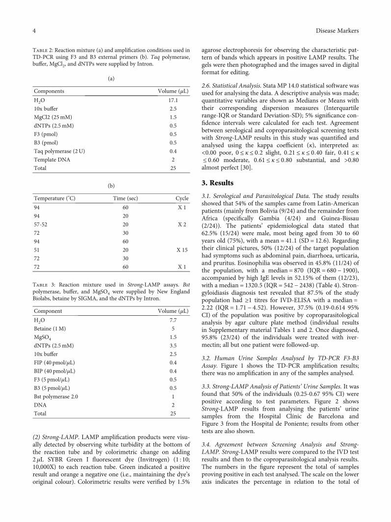
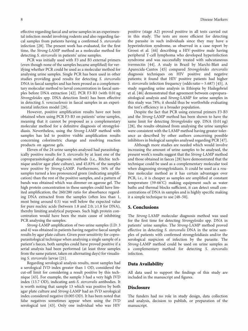
3.2. Human Urine Samples Analysed by TD-PCR F3-B3Assay. Figure 1 shows the TD-PCR amplification results;there was no amplification in any of the samples analysed.
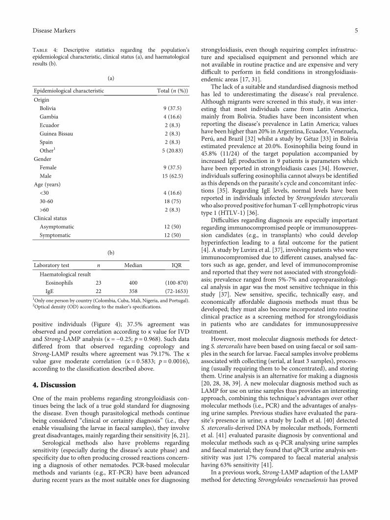
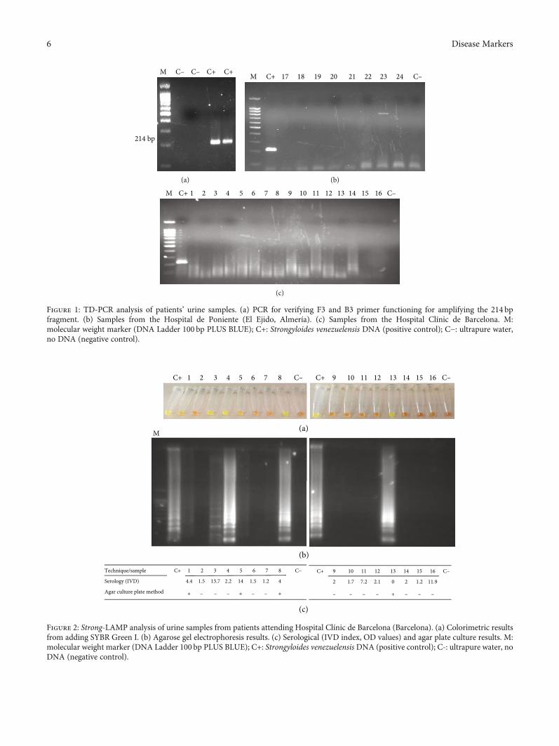
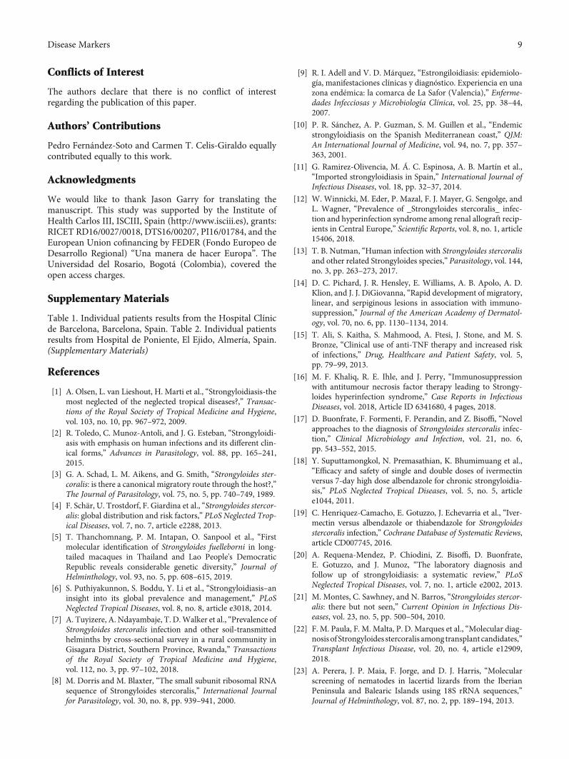
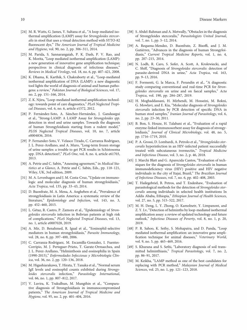
3.3. Strong-LAMP Analysis of Patients’ Urine Samples. It wasfound that 50% of the individuals (0.25-0.67 95% CI) werepositive according to test parameters. Figure 2 showsStrong-LAMP results from analysing the patients’ urinesamples from the Hospital Clínic de Barcelona andFigure 3 from the Hospital de Poniente; results from othertests are also shown.
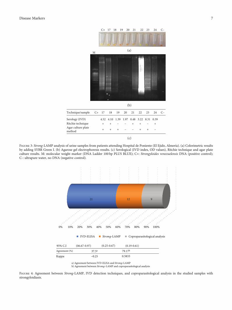
3.4. Agreement between Screening Analysis and Strong-LAMP. Strong-LAMP results were compared to the IVD testresults and then to the coproparasitological analysis results.The numbers in the figure represent the total of samplesproving positive in each test analysed. The scale on the loweraxis indicates the percentage in relation to the total of
Table 2: Reaction mixture (a) and amplification conditions used inTD-PCR using F3 and B3 external primers (b). Taq polymerase,buffer, MgCl2, and dNTPs were supplied by Intron.
(a)
Components Volume (μL)
H2O 17.1
10x buffer 2.5
MgCl2 (25mM) 1.5
dNTPs (2.5mM) 0.5
F3 (pmol) 0.5
B3 (pmol) 0.5
Taq polymerase (2U) 0.4
Template DNA 2
Total 25
(b)
Temperature (°C) Time (sec) Cycle
94 60 X 1
94 20
X 257-52 20
72 30
94 60
X 1551 20
72 30
72 60 X 1
Table 3: Reaction mixture used in Strong-LAMP assays. Bstpolymerase, buffer, and MgSO4 were supplied by New EnglandBiolabs, betaine by SIGMA, and the dNTPs by Intron.
Component Volume (μL)
H2O 7.7
Betaine (1M) 5
MgSO4 1.5
dNTPs (2.5mM) 3.5
10x buffer 2.5
FIP (40 pmol/μL) 0.4
BIP (40 pmol/μL) 0.4
F3 (5 pmol/μL) 0.5
B3 (5 pmol/μL) 0.5
Bst polymerase 2.0 1
DNA 2
Total 25
4 Disease Markers
positive individuals (Figure 4); 37.5% agreement wasobserved and poor correlation according to κ value for IVDand Strong-LAMP analysis (κ = −0:25; p = 0:968). Such datadiffered from that observed regarding coprology andStrong-LAMP results where agreement was 79.17%. The κvalue gave moderate correlation (κ = 0:5833; p = 0:0016),according to the classification described above.
4. Discussion
One of the main problems regarding strongyloidiasis con-tinues being the lack of a true gold standard for diagnosingthe disease. Even though parasitological methods continuebeing considered “clinical or certainty diagnosis” (i.e., theyenable visualising the larvae in faecal samples), they involvegreat disadvantages, mainly regarding their sensitivity [6, 21].
Serological methods also have problems regardingsensitivity (especially during the disease’s acute phase) andspecificity due to often producing crossed reactions concern-ing a diagnosis of other nematodes. PCR-based molecularmethods and variants (e.g., RT-PCR) have been advancedduring recent years as the most suitable ones for diagnosing
strongyloidiasis, even though requiring complex infrastruc-ture and specialised equipment and personnel which arenot available in routine practice and are expensive and verydifficult to perform in field conditions in strongyloidiasis-endemic areas [17, 31].
The lack of a suitable and standardised diagnosis methodhas led to underestimating the disease’s real prevalence.Although migrants were screened in this study, it was inter-esting that most individuals came from Latin America,mainly from Bolivia. Studies have been inconsistent whenreporting the disease’s prevalence in Latin America; valueshave been higher than 20% inArgentina, Ecuador, Venezuela,Perú, and Brazil [32] whilst a study by Gétaz [33] in Boliviaestimated prevalence at 20.0%. Eosinophilia being found in45.8% (11/24) of the target population accompanied byincreased IgE production in 9 patients is parameters whichhave been reported in strongyloidiasis cases [34]. However,individuals suffering eosinophilia cannot always be identifiedas this depends on the parasite’s cycle and concomitant infec-tions [35]. Regarding IgE levels, normal levels have beenreported in individuals infected by Strongyloides stercoraliswho also proved positive for humanT-cell lymphotropic virustype 1 (HTLV-1) [36].
Difficulties regarding diagnosis are especially importantregarding immunocompromised people or immunosuppres-sion candidates (e.g., in transplants) who could develophyperinfection leading to a fatal outcome for the patient[4]. A study by Luvira et al. [37], involving patients who wereimmunocompromised due to different causes, analysed fac-tors such as age, gender, and level of immunocompromiseand reported that they were not associated with strongyloidi-asis; prevalence ranged from 5%-7% and coproparasitologi-cal analysis in agar was the most sensitive technique in thisstudy [37]. New sensitive, specific, technically easy, andeconomically affordable diagnosis methods must thus bedeveloped; they must also become incorporated into routineclinical practice as a screening method for strongyloidiasisin patients who are candidates for immunosuppressivetreatment.
However, most molecular diagnosis methods for detect-ing S. stercoralis have been based on using faecal or soil sam-ples in the search for larvae. Faecal samples involve problemsassociated with collecting (serial, at least 3 samples), process-ing (usually requiring them to be concentrated), and storingthem. Urine analysis is an alternative for making a diagnosis[20, 28, 38, 39]. A new molecular diagnosis method such asLAMP for use on urine samples thus provides an interestingapproach, combining this technique’s advantages over othermolecular methods (i.e., PCR) and the advantages of analys-ing urine samples. Previous studies have evaluated the para-site’s presence in urine; a study by Lodh et al. [40] detectedS. stercoralis-derived DNA by molecular methods, Formentiet al. [41] evaluated parasite diagnosis by conventional andmolecular methods such as q-PCR analysing urine samplesand faecal material; they found that qPCR urine analysis sen-sitivity was just 17% compared to faecal material analysishaving 63% sensitivity [41].
In a previous work, Strong-LAMP adaption of the LAMPmethod for detecting Strongyloides venezuelensis has proved
Table 4: Descriptive statistics regarding the population’sepidemiological characteristic, clinical status (a), and haematologicalresults (b).
(a)
Epidemiological characteristic Total (n (%))
Origin
Bolivia 9 (37.5)
Gambia 4 (16.6)
Ecuador 2 (8.3)
Guinea Bissau 2 (8.3)
Spain 2 (8.3)
Other1 5 (20.83)
Gender
Female 9 (37.5)
Male 15 (62.5)
Age (years)
<30 4 (16.6)
30-60 18 (75)
>60 2 (8.3)
Clinical status
Asymptomatic 12 (50)
Symptomatic 12 (50)
(b)
Laboratory test n Median IQR
Haematological result
Eosinophils 23 400 (100-870)
IgE 22 358 (72-1653)1Only one person by country (Colombia, Cuba, Mali, Nigeria, and Portugal).2Optical density (OD) according to the maker’s specifications.
5Disease Markers
M C– C– C+ C+
214 bp
(a)
M C+ 24 C–17 18 19 20 21 22 23
(b)
M C+ C–31 42 5 76 11108 9 1312 1514 16
(c)
Figure 1: TD-PCR analysis of patients’ urine samples. (a) PCR for verifying F3 and B3 primer functioning for amplifying the 214 bpfragment. (b) Samples from the Hospital de Poniente (El Ejido, Almería). (c) Samples from the Hospital Clínic de Barcelona. M:molecular weight marker (DNA Ladder 100 bp PLUS BLUE); C+: Strongyloides venezuelensis DNA (positive control); C−: ultrapure water,no DNA (negative control).
(a)
(b)
(c)
M
1 2 3 4 5 6 7 8 C–C+ 9 10 11 12 13 14 15 16 C–C+
Technique/sample C+ 1 2 3 4 5 6 7 8 C–
Serology (IVD) 4.4 1.5 13.7 2.2 14 1.5 1.2 4
Agar culture plate method + – – – + – – +
C+ 9 10 11 12 13 14 15 16 C–
2 1.7 7.2 2.1 0 2 1.2 11.9
– – – – + – – –
Figure 2: Strong-LAMP analysis of urine samples from patients attending Hospital Clínic de Barcelona (Barcelona). (a) Colorimetric resultsfrom adding SYBR Green I. (b) Agarose gel electrophoresis results. (c) Serological (IVD index, OD values) and agar plate culture results. M:molecular weight marker (DNA Ladder 100 bp PLUS BLUE); C+: Strongyloides venezuelensis DNA (positive control); C-: ultrapure water, noDNA (negative control).
6 Disease Markers
(a)
(b)
(c)
Technique/sample C+ 17 18 19 20 21 22 23 24 C–
Serology (IVD) 4.52 4.10 1.39 1.97 0.48 3.22 8.31 0.39Ritchie technique + + – – + + – +Agar culture platemethod + + + – – + + –
C+
M
17 18 19 20 21 22 23 24 C–
Figure 3: Strong-LAMP analysis of urine samples from patients attending Hospital de Poniente (El Ejido, Almería). (a) Colorimetric resultsby adding SYBR Green I. (b) Agarose gel electrophoresis results. (c) Serological (IVD index, OD values), Ritchie technique and agar plateculture results. M: molecular weight marker (DNA Ladder 100 bp PLUS BLUE); C+: Strongyloides venezuelensis DNA (positive control);C-: ultrapure water, no DNA (negative control).
95% C.I (06.67-0.97) (0.25-0.67) (0.19-0.61)Agreement (%) 37.5a 79.17b
Kappa –0.25 0.5833
a) Agreement between IVD-ELISA and Strong-LAMPb) Agreement between Strong–LAMP and coproparasitological analysis
0% 10% 20% 30% 40% 50% 60% 70% 80% 90% 100%
21 12 9
IVD-ELISA Strong-LAMP Coproparasitological analysis
Figure 4: Agreement between Strong-LAMP, IVD detection techniques, and coproparasitological analysis in the studied samples withstrongyloidiasis.
7Disease Markers
effective regarding faecal and urine samples in an experimen-tal infection model involving rodents and also regarding fae-cal samples from patients having a confirmed S. stercoralisinfection [28]. The present work has evaluated, for the firsttime, the Strong-LAMP method as a molecular method fordetecting S. stercoralis in patients’ urine samples.
PCR was initially used with F3 and B3 external primers(even though none of the samples became amplified) for ver-ifying whether PCR could be used as a molecular method foranalysing urine samples. Single PCR has been used in otherstudies providing good results for detecting S. stercoralisDNA in faecal samples and has been prosed as a complemen-tary molecular method to larval concentration in faecal sam-ples before DNA extraction [42]. PCR F3-B3 (with 0.01 ngStrongyloides spp. DNA detection limit) has been effectivein detecting S. venezuelensis in faecal samples in an experi-mental infection model [28].
However, positive amplification results have not beenobtained when using PCR F3-B3 on patients’ urine samples,meaning that it cannot be proposed as a complementarymolecular method for a definitive diagnosis of strongyloi-diasis. Nevertheless, using the Strong-LAMP method withsamples has led to positive visible amplification resultsconcerning colorimetric change and resolving reactionproducts on agarose gels.
Eleven of the 24 urine samples analysed had parasitolog-ically positive results for S. stercoralis by at least one of thecoproparasitological diagnosis methods (i.e., Ritchie tech-nique and/or agar plate culture), and 45.83% of the sampleswere positive by Strong-LAMP. Furthermore, 16% of thesamples turned a less pronounced green (indicating amplifi-cation) than the rest of the positive samples, and a pattern ofbands was obtained which was dimmer on agarose gel. Thehigh protein concentration in these samples could have lim-ited amplification; the 260/280 ratio for absorbance regard-ing DNA extracted from the samples (often less than 1,most being around 0.5) was well below the expected valuefor pure nucleic acids (between 1.8 and 2.0; ≥1.8 for DNA),thereby limiting analytical purposes. Such high protein con-centration would have been the main cause of inhibitingPCR analysing the samples.
Strong-LAMP amplification of two urine samples (I.D: 3and 4) was obtained in patients having negative faecal sampleresults by agar plate culture. Given poor sensitivity for copro-parasitological technique when analysing a single sample of apatient’s faeces, both samples could have proved positive if aserial analysis had been performed (at least three samplesfrom the same patient, taken on alternating days) for visualis-ing S. stercoralis larvae [21].
Regarding serological analysis results, most samples hada serological IVD index greater than 1 OD, considered thecut-off limit for considering a result positive by this tech-nique [43]. For example, the sample 3 had a very high IVDindex (13.7 OD), indicating anti-S. stercoralis antibodies. Itis worth noting that sample 13 which was positive by bothagar plate culture and Strong-LAMP had an IVD serologicalindex considered negative (0.005 OD). It has been noted thatfalse negatives sometimes appear when using the IVDserological test [43]. Only one individual who was HIV
positive (stage A2) proved positive in all tests carried outin this study. The tests are more efficient for detectingthe parasite in such individuals since they may have ahyperinfection syndrome, as observed in a case report byGrossi et al. [44] describing a HIV-positive male havingperipheral T-cell lymphoma who developed hyperinfectionsyndrome and was successfully treated with subcutaneousivermectin [44]. A study in Brazil by Marchi-Blatt andAparecida-Cantos [45] compared Strongyloides stercoralisdiagnosis techniques on HIV positive and negativepatients; it found that HIV positive patients had higherS. stercoralis infection frequency (odds ratio = 5:687) [45]. Astudy regarding urine analysis in Ethiopia by Hailegebrielet al. [46] demonstrated that agreement between copropara-sitological analysis and Strong-LAMP for urine samples inthis study was 78%; it should thus be worthwhile evaluatingthe test’s efficiency in a broader population.
Despite the fact that PCR using external primers F3-B3and the Strong-LAMP method has been shown to have thesame limit for detecting Strongyloides spp. DNA (0.01 ng)[28], the results obtained from analysing the urine sampleswere consistent with the LAMP method having greater toler-ance as described by other authors concerning possibleinhibitors in biological samples analysed regarding PCR [47].
Although more studies are needed which would involveincreasing the amount of urine samples to be analysed, thepresent work’s results suggest that the Strong-LAMP methodand those obtained in faeces [28] have demonstrated that thetechnique could be used as a complementary molecular toolwhen diagnosing strongyloidiasis. It could be used as a rou-tine molecular method as it has certain advantages overPCR, i.e., it is cheaper as samples are amplified at constanttemperature (59-66°C) making equipment such as waterbaths and thermal blocks sufficient, it can detect small con-centrations of DNA in samples and is highly specific makingit a simple technique to use [48–50].
5. Conclusions
The Strong-LAMP molecular diagnosis method was usedfor the first time for detecting Strongyloides spp. DNA inpatients’ urine samples. The Strong-LAMP method provedeffective in detecting S. stercoralis DNA in the urine sam-ples of patients with confirmed strongyloidiasis and/or theserological suspicion of infection by the parasite. TheStrong-LAMP method could be used on urine samples asa complementary method for detecting S. stercoralisinfection.
Data Availability
All data used to support the findings of this study areincluded in the manuscript and figures.
Disclosure
The funders had no role in study design, data collectionand analysis, decision to publish, or preparation of themanuscript.
8 Disease Markers
Conflicts of Interest
The authors declare that there is no conflict of interestregarding the publication of this paper.
Authors’ Contributions
Pedro Fernández-Soto and Carmen T. Celis-Giraldo equallycontributed equally to this work.
Acknowledgments
We would like to thank Jason Garry for translating themanuscript. This study was supported by the Institute ofHealth Carlos III, ISCIII, Spain (http://www.isciii.es), grants:RICET RD16/0027/0018, DTS16/00207, PI16/01784, and theEuropean Union cofinancing by FEDER (Fondo Europeo deDesarrollo Regional) “Una manera de hacer Europa”. TheUniversidad del Rosario, Bogotá (Colombia), covered theopen access charges.
Supplementary Materials
Table 1. Individual patients results from the Hospital Clínicde Barcelona, Barcelona, Spain. Table 2. Individual patientsresults from Hospital de Poniente, El Ejido, Almería, Spain.(Supplementary Materials)
References
[1] A. Olsen, L. van Lieshout, H. Marti et al., “Strongyloidiasis-themost neglected of the neglected tropical diseases?,” Transac-tions of the Royal Society of Tropical Medicine and Hygiene,vol. 103, no. 10, pp. 967–972, 2009.
[2] R. Toledo, C. Munoz-Antoli, and J. G. Esteban, “Strongyloidi-asis with emphasis on human infections and its different clin-ical forms,” Advances in Parasitology, vol. 88, pp. 165–241,2015.
[3] G. A. Schad, L. M. Aikens, and G. Smith, “Strongyloides ster-coralis: is there a canonical migratory route through the host?,”The Journal of Parasitology, vol. 75, no. 5, pp. 740–749, 1989.
[4] F. Schär, U. Trostdorf, F. Giardina et al., “Strongyloides stercor-alis: global distribution and risk factors,” PLoS Neglected Trop-ical Diseases, vol. 7, no. 7, article e2288, 2013.
[5] T. Thanchomnang, P. M. Intapan, O. Sanpool et al., “Firstmolecular identification of Strongyloides fuelleborni in long-tailed macaques in Thailand and Lao People's DemocraticRepublic reveals considerable genetic diversity,” Journal ofHelminthology, vol. 93, no. 5, pp. 608–615, 2019.
[6] S. Puthiyakunnon, S. Boddu, Y. Li et al., “Strongyloidiasis–aninsight into its global prevalence and management,” PLoSNeglected Tropical Diseases, vol. 8, no. 8, article e3018, 2014.
[7] A. Tuyizere, A. Ndayambaje, T. D.Walker et al., “Prevalence ofStrongyloides stercoralis infection and other soil-transmittedhelminths by cross-sectional survey in a rural community inGisagara District, Southern Province, Rwanda,” Transactionsof the Royal Society of Tropical Medicine and Hygiene,vol. 112, no. 3, pp. 97–102, 2018.
[8] M. Dorris and M. Blaxter, “The small subunit ribosomal RNAsequence of Strongyloides stercoralis,” International Journalfor Parasitology, vol. 30, no. 8, pp. 939–941, 2000.
[9] R. I. Adell and V. D. Márquez, “Estrongiloidiasis: epidemiolo-gía, manifestaciones clínicas y diagnóstico. Experiencia en unazona endémica: la comarca de La Safor (Valencia),” Enferme-dades Infecciosas y Microbiología Clínica, vol. 25, pp. 38–44,2007.
[10] P. R. Sánchez, A. P. Guzman, S. M. Guillen et al., “Endemicstrongyloidiasis on the Spanish Mediterranean coast,” QJM:An International Journal of Medicine, vol. 94, no. 7, pp. 357–363, 2001.
[11] G. Ramirez-Olivencia, M. Á. C. Espinosa, A. B. Martín et al.,“Imported strongyloidiasis in Spain,” International Journal ofInfectious Diseases, vol. 18, pp. 32–37, 2014.
[12] W. Winnicki, M. Eder, P. Mazal, F. J. Mayer, G. Sengolge, andL. Wagner, “Prevalence of _Strongyloides stercoralis_ infec-tion and hyperinfection syndrome among renal allograft recip-ients in Central Europe,” Scientific Reports, vol. 8, no. 1, article15406, 2018.
[13] T. B. Nutman, “Human infection with Strongyloides stercoralisand other related Strongyloides species,” Parasitology, vol. 144,no. 3, pp. 263–273, 2017.
[14] D. C. Pichard, J. R. Hensley, E. Williams, A. B. Apolo, A. D.Klion, and J. J. DiGiovanna, “Rapid development of migratory,linear, and serpiginous lesions in association with immuno-suppression,” Journal of the American Academy of Dermatol-ogy, vol. 70, no. 6, pp. 1130–1134, 2014.
[15] T. Ali, S. Kaitha, S. Mahmood, A. Ftesi, J. Stone, and M. S.Bronze, “Clinical use of anti-TNF therapy and increased riskof infections,” Drug, Healthcare and Patient Safety, vol. 5,pp. 79–99, 2013.
[16] M. F. Khaliq, R. E. Ihle, and J. Perry, “Immunosuppressionwith antitumour necrosis factor therapy leading to Strongy-loides hyperinfection syndrome,” Case Reports in InfectiousDiseases, vol. 2018, Article ID 6341680, 4 pages, 2018.
[17] D. Buonfrate, F. Formenti, F. Perandin, and Z. Bisoffi, “Novelapproaches to the diagnosis of Strongyloides stercoralis infec-tion,” Clinical Microbiology and Infection, vol. 21, no. 6,pp. 543–552, 2015.
[18] Y. Suputtamongkol, N. Premasathian, K. Bhumimuang et al.,“Efficacy and safety of single and double doses of ivermectinversus 7-day high dose albendazole for chronic strongyloidia-sis,” PLoS Neglected Tropical Diseases, vol. 5, no. 5, articlee1044, 2011.
[19] C. Henriquez-Camacho, E. Gotuzzo, J. Echevarria et al., “Iver-mectin versus albendazole or thiabendazole for Strongyloidesstercoralis infection,” Cochrane Database of Systematic Reviews,article CD007745, 2016.
[20] A. Requena-Mendez, P. Chiodini, Z. Bisoffi, D. Buonfrate,E. Gotuzzo, and J. Munoz, “The laboratory diagnosis andfollow up of strongyloidiasis: a systematic review,” PLoSNeglected Tropical Diseases, vol. 7, no. 1, article e2002, 2013.
[21] M. Montes, C. Sawhney, and N. Barros, “Strongyloides stercor-alis: there but not seen,” Current Opinion in Infectious Dis-eases, vol. 23, no. 5, pp. 500–504, 2010.
[22] F. M. Paula, F. M. Malta, P. D. Marques et al., “Molecular diag-nosisofStrongyloides stercoralis among transplant candidates,”Transplant Infectious Disease, vol. 20, no. 4, article e12909,2018.
[23] A. Perera, J. P. Maia, F. Jorge, and D. J. Harris, “Molecularscreening of nematodes in lacertid lizards from the IberianPeninsula and Balearic Islands using 18S rRNA sequences,”Journal of Helminthology, vol. 87, no. 2, pp. 189–194, 2013.
9Disease Markers
[24] M. R. Watts, G. James, Y. Sultana et al., “A loop-mediated iso-thermal amplification (LAMP) assay for Strongyloides stercor-alis in stool that uses a visual detection method with SYTO-82fluorescent dye,” The American Journal of Tropical Medicineand Hygiene, vol. 90, no. 2, pp. 306–311, 2014.
[25] M. Parida, S. Sannarangaiah, P. K. Dash, P. V. Rao, andK. Morita, “Loop mediated isothermal amplification (LAMP):a new generation of innovative gene amplification technique;perspectives in clinical diagnosis of infectious diseases,”Reviews in Medical Virology, vol. 18, no. 6, pp. 407–421, 2008.
[26] K. Dhama, K. Karthik, S. Chakraborty et al., “Loop-mediatedisothermal amplification of DNA (LAMP): a new diagnostictool lights the world of diagnosis of animal and human patho-gens: a review,” Pakistan Journal of Biological Sciences, vol. 17,no. 2, pp. 151–166, 2014.
[27] Z. K. Njiru, “Loop-mediated isothermal amplification technol-ogy: towards point of care diagnostics,” PLoS Neglected Tropi-cal Diseases, vol. 6, no. 6, article e1572, 2012.
[28] P. Fernández-Soto, A. Sánchez-Hernández, J. Gandaseguiet al., “Strong-LAMP: A LAMP Assay for Strongyloides spp.detection in stool and urine samples. Towards the diagnosisof human Strongyloidiasis starting from a rodent model,”PLOS Neglected Tropical Diseases, vol. 10, no. 7, articlee0004836, 2016.
[29] P. Fernandez-Soto, V. Velasco Tirado, C. Carranza Rodriguez,J. L. Perez-Arellano, and A. Muro, “Long-term frozen storageof urine samples: a trouble to get PCR results in Schistosomaspp. DNA detection?,” PLoS One, vol. 8, no. 4, article e61703,2013.
[30] A. Petrie and C. Sabin, “Assessing agreement,” inMedical Sta-tistics at a Glance, A. Petrie and C. Sabin, Eds., pp. 118–121,Wiley, UK, 3rd edition, 2009.
[31] M. A. Levenhagen and J. M. Costa-Cruz, “Update on immuno-logic and molecular diagnosis of human strongyloidiasis,”Acta Tropica, vol. 135, pp. 33–43, 2014.
[32] D. Buonfrate, M. A. Mena, A. Angheben et al., “Prevalence ofstrongyloidiasis in Latin America: a systematic review of theliterature,” Epidemiology and Infection, vol. 143, no. 3,pp. 452–460, 2015.
[33] L. Gétaz, R. Castro, P. Zamora et al., “Epidemiology of Stron-gyloides stercoralis infection in Bolivian patients at high riskof complications,” PLoS Neglected Tropical Diseases, vol. 13,no. 1, article e0007028, 2019.
[34] A. Mir, D. Benahmed, R. Igual et al., “Eosinophil-selectivemediators in human strongyloidiasis,” Parasite Immunology,vol. 28, no. 8, pp. 397–400, 2006.
[35] C. Carranza-Rodriguez, M. Escamilla-Gonzalez, I. Fuentes-Corripio, M. J. Perteguer-Prieto, T. Garate-Ormaechea, andJ. L. Perez-Arellano, “Helminthosis and eosinophilia in Spain(1990-2015),” Enfermedades Infecciosas y Microbiología Clín-ica, vol. 36, no. 2, pp. 120–136, 2018.
[36] M. Higashiarakawa, T. Hirata, T. Tanaka et al., “Normal serumIgE levels and eosinophil counts exhibited during Strongy-loides stercoralis infection,” Parasitology International,vol. 66, no. 1, pp. 807–812, 2017.
[37] V. Luvira, K. Trakulhun, M. Mungthin et al., “Compara-tive diagnosis of Strongyloidiasis in immunocompromisedpatients,” The American Journal of Tropical Medicine andHygiene, vol. 95, no. 2, pp. 401–404, 2016.
[38] S. Abdel-Rahman and A. Metwally, “Obstacles in the diagnosisof Strongyloides stercoralis,” Parasitologists United Journal,vol. 7, no. 1, pp. 5–12, 2014.
[39] A. Requena-Mendez, D. Buonfrate, Z. Bisoffi, and J. M.Gutiérrez, “Advances in the diagnosis of human Strongyloi-diasis,” Current Tropical Medicine Reports, vol. 1, no. 4,pp. 207–215, 2014.
[40] N. Lodh, R. Caro, S. Sofer, A. Scott, A. Krolewiecki, andC. Shiff, “Diagnosis of Strongyloides stercoralis: detection ofparasite-derived DNA in urine,” Acta Tropica, vol. 163,pp. 9–13, 2016.
[41] F. Formenti, G. la Marca, F. Perandin et al., “A diagnosticstudy comparing conventional and real-time PCR for Stron-gyloides stercoralis on urine and on faecal samples,” ActaTropica, vol. 190, pp. 284–287, 2019.
[42] H. Moghaddassani, H. Mirhendi, M. Hosseini, M. Rokni,G. Mowlavi, and E. Kia, “Molecular diagnosis of Strongyloidesstercoralis infection by PCR detection of specific DNA inhuman stool samples,” Iranian Journal of Parasitology, vol. 6,no. 2, pp. 23–30, 2011.
[43] B. Bon, S. Houze, H. Talabani et al., “Evaluation of a rapidenzyme-linked immunosorbent assay for diagnosis of strongy-loidiasis,” Journal of Clinical Microbiology, vol. 48, no. 5,pp. 1716–1719, 2010.
[44] P. A. Grossi, D. Lombardi, A. Petrolo et al., “Strongyloides ster-coralis hyperinfection in an HIV-infected patient successfullytreated with subcutaneous ivermectin,” Tropical Medicineand Infectious Disease, vol. 3, no. 2, p. 46, 2018.
[45] J. Marchi Blatt and G. Aparecida Cantos, “Evaluation of tech-niques for the diagnosis of Strongyloides stercoralis in humanimmunodeficiency virus (HIV) positive and HIV negativeindividuals in the city of Itajaí, Brazil,” The Brazilian Journalof Infectious Diseases, vol. 7, no. 6, pp. 402–408, 2003.
[46] T. Hailegebriel, B. Petros, and T. Endeshaw, “Evaluation ofparasitological methods for the detection of Strongyloides ster-coralis among individuals in selected health institutions InAddis Ababa, Ethiopia,” Ethiopian Journal of Health Sciences,vol. 27, no. 5, pp. 515–522, 2017.
[47] M. H. Deng, L. Y. Zhong, O. Kamolnetr, Y. Limpanont, andZ. Y. Lv, “Detection of helminths by loop-mediated isothermalamplification assay: a review of updated technology and futureoutlook,” Infectious Diseases of Poverty, vol. 8, no. 1, p. 20,2019.
[48] P. R. Sahoo, K. Sethy, S. Mohapatra, and D. Panda, “Loopmediated isothermal amplification: an innovative gene ampli-fication technique for animal diseases,” Veterinary World,vol. 9, no. 5, pp. 465–469, 2016.
[49] S. Khurana and S. Sethi, “Laboratory diagnosis of soil trans-mitted helminthiasis,” Tropical Parasitology, vol. 7, no. 2,pp. 86–91, 2017.
[50] M. Keikha, “LAMP method as one of the best candidates forreplacing with PCR method,” Malaysian Journal of MedicalSciences, vol. 25, no. 1, pp. 121–123, 2018.
10 Disease Markers