Intestinal malabsorption in Strongyloides stercoralis ... · Intestinal malabsorption in...
8
Gut, 1965, 6, 574 Intestinal malabsorption in Strongyloides stercoralis infestation P. F. MILNER, R. A. IRVINE, C. J. BARTON, G. BRAS, AND R. RICHARDS From the Departments of Pathology and Medicine, University of the West Indies, Kingston, Jamaica EDITORIAL SYNOPSIS The number of conditions which can result in intestinal malabsorption increases steadily. The one described by the authors in this paper follows infestation with Strongyloides stercoralis which is widespread in the tropics. Features suggesting the diagnosis in a patient with malabsorption are, according to the authors, persistent vomiting due to duodenal involvement and characteristic radiographs of the duodenum. The diagnosis is proven when the duodenal mucosa is examined and the lesion can be treated, when the symptoms will disappear. The disease states that may be associated with intestinal malabsorption are numerous, and a recent review lists almost 40 of these (Jeffries, Weser, and Sleisenger, 1964). As the majority of publications on this subject originate in temperate countries, it is not surprising that there are few references to malabsorption resulting from parasitic infestation of the bowel. Steatorrhoea has, however, been described in giardiasis (Amini, 1963; Zamcheck, Hoskins, Winawer, Broitman, and Gottlieb, 1963) and in hookworm disease (Sheehy, Meroney, Cox, and Soler, 1962; Salem and Truelove, 1964). Infestation with the helminth Strongyloides stercoralis is widespread in tropical countries and, although in the majority of instances the infestation is asymptomatic, serious disease and even death may ensue (Bras, Richards, Irvine, Milner, and Ragbeer, 1964). Intestinal malabsorption resulting from the disease is rare (Stemmermann and Nakasone, 1960; Alcorn and Kotcher, 1961). The clinical picture of severe malnutrition with a bizarre duodenal lesion seen on radiographs and larvae of Strongyloides in duodenal biopsies as well as in the stools has not been previously reported. The greatly depressed absorption of iron salts demonstrated in these cases is also of interest. In the four cases reported here there was incontrovert- ible evidence of intestinal malabsorption resulting from strongyloidiasis. LABORATORY INVESTIGATIONS SERUM IRON AND IRON-BINDING CAPACITY These were estimated by the method of Beale, Bostrom, and Taylor (1961, 1962). IRON ABSORPTION TESTS The patients were fasting when two tablets (400 mg.) of ferrous sulphate (130 mg. of elemental iron) were given and blood specimens taken for serum iron and iron-binding capacity at hourly intervals for four hours. XYLOSE AND FOLIC ACID ABSORPTION TESTS These tests were done according to the method of Chanarin and Bennett (1962) using 0 4 g. of xylose and 40 ,ug. of folic acid per kilogram body weight in a single dose. Xylose was estimated in urine and serum by the method of Roe and Rice (1948) and folic acid was measured by micro- biological assay using Streptococcus faecalis (Chanarin, Anderson, and Mollin, 1958). DUODENAL AND JEJUNAL BIOPSIES Biopsies were taken under fluoroscopic control using a Crosby capsule (Crosby and Kugler, 1957). Specimens were examined under a dissecting microscope before fixation as well as in stained histological sections cut vertically. CASE REPORTS CASE 1 A 28-year-old Jamaican housewife was admitted to the University College Hospital of the West Indies in March 1964. She gave a history of vomiting, diarrhoea, and weight loss for three months. In the three weeks preceding admission she had experienced lower abdominal pain, fever, ankle oedema, and skin rash. She presented the classical picture of kwashiorkor in the adult; there was dehydration and cachexia, with generalized oedema and ascites; her body was covered with a scaly, purpuric rash associated with dyspigmentation and desquamation of the groins, perineum, and limbs (Fig. 1). The liver was enlarged and smooth but there was no splenomegaly. It is unusual to see severe protein malnutrition in adults in Jamaica without there being some predisposing underlying disease. Detailed investigations were therefore 574 on June 5, 2020 by guest. Protected by copyright. http://gut.bmj.com/ Gut: first published as 10.1136/gut.6.6.574 on 1 December 1965. Downloaded from
Transcript of Intestinal malabsorption in Strongyloides stercoralis ... · Intestinal malabsorption in...

Gut, 1965, 6, 574
Intestinal malabsorption in Strongyloidesstercoralis infestation
P. F. MILNER, R. A. IRVINE, C. J. BARTON, G. BRAS, AND R. RICHARDS
From the Departments ofPathology and Medicine, University of the West Indies,Kingston, Jamaica
EDITORIAL SYNOPSIS The number of conditions which can result in intestinal malabsorption increasessteadily. The one described by the authors in this paper follows infestation with Strongyloidesstercoralis which is widespread in the tropics. Features suggesting the diagnosis in a patient withmalabsorption are, according to the authors, persistent vomiting due to duodenal involvementand characteristic radiographs of the duodenum. The diagnosis is proven when the duodenalmucosa is examined and the lesion can be treated, when the symptoms will disappear.
The disease states that may be associated withintestinal malabsorption are numerous, and arecent review lists almost 40 of these (Jeffries,Weser, and Sleisenger, 1964). As the majority ofpublications on this subject originate in temperatecountries, it is not surprising that there are fewreferences to malabsorption resulting from parasiticinfestation of the bowel. Steatorrhoea has, however,been described in giardiasis (Amini, 1963; Zamcheck,Hoskins, Winawer, Broitman, and Gottlieb, 1963)and in hookworm disease (Sheehy, Meroney, Cox,and Soler, 1962; Salem and Truelove, 1964).
Infestation with the helminth Strongyloidesstercoralis is widespread in tropical countries and,although in the majority of instances the infestationis asymptomatic, serious disease and even death mayensue (Bras, Richards, Irvine, Milner, and Ragbeer,1964). Intestinal malabsorption resulting from thedisease is rare (Stemmermann and Nakasone, 1960;Alcorn and Kotcher, 1961).The clinical picture of severe malnutrition with a
bizarre duodenal lesion seen on radiographs andlarvae of Strongyloides in duodenal biopsies as wellas in the stools has not been previously reported.The greatly depressed absorption of iron salts
demonstrated in these cases is also of interest. Inthe four cases reported here there was incontrovert-ible evidence of intestinal malabsorption resultingfrom strongyloidiasis.
LABORATORY INVESTIGATIONS
SERUM IRON AND IRON-BINDING CAPACITY These wereestimated by the method of Beale, Bostrom, and Taylor(1961, 1962).
IRON ABSORPTION TESTS The patients were fasting whentwo tablets (400 mg.) of ferrous sulphate (130 mg. ofelemental iron) were given and blood specimens takenfor serum iron and iron-binding capacity at hourlyintervals for four hours.
XYLOSE AND FOLIC ACID ABSORPTION TESTS These testswere done according to the method of Chanarin andBennett (1962) using 0 4 g. of xylose and 40 ,ug. of folicacid per kilogram body weight in a single dose. Xylosewas estimated in urine and serum by the method of Roeand Rice (1948) and folic acid was measured by micro-biological assay using Streptococcus faecalis (Chanarin,Anderson, and Mollin, 1958).
DUODENAL AND JEJUNAL BIOPSIES Biopsies were takenunder fluoroscopic control using a Crosby capsule(Crosby and Kugler, 1957). Specimens were examinedunder a dissecting microscope before fixation as well asin stained histological sections cut vertically.
CASE REPORTS
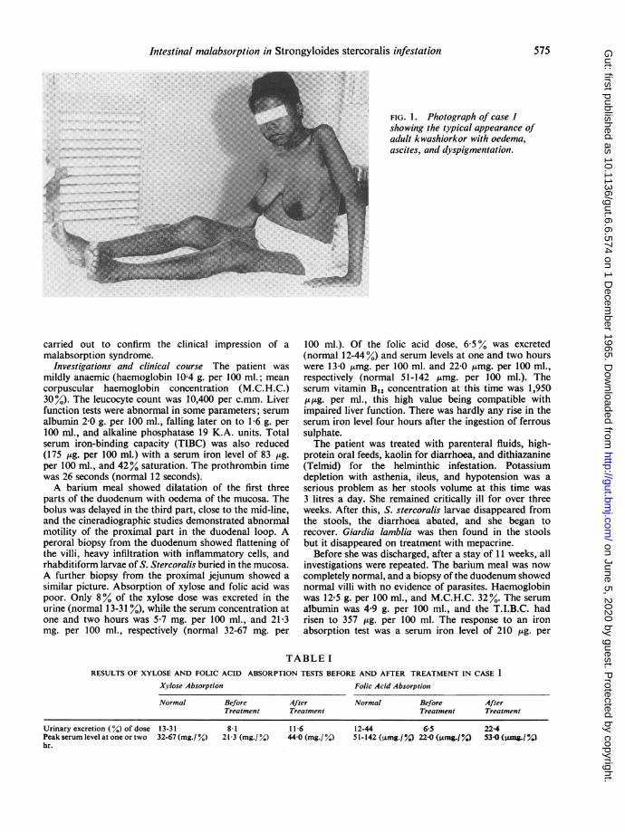
CASE 1 A 28-year-old Jamaican housewife was admittedto the University College Hospital of the West Indies inMarch 1964. She gave a history of vomiting, diarrhoea,and weight loss for three months. In the three weekspreceding admission she had experienced lower abdominalpain, fever, ankle oedema, and skin rash. She presentedthe classical picture of kwashiorkor in the adult; therewas dehydration and cachexia, with generalized oedemaand ascites; her body was covered with a scaly, purpuricrash associated with dyspigmentation and desquamationof the groins, perineum, and limbs (Fig. 1). The liver wasenlarged and smooth but there was no splenomegaly.
It is unusual to see severe protein malnutrition inadults in Jamaica without there being some predisposingunderlying disease. Detailed investigations were therefore
574
on June 5, 2020 by guest. Protected by copyright.
http://gut.bmj.com
/G
ut: first published as 10.1136/gut.6.6.574 on 1 Decem
ber 1965. Dow
nloaded from
Intestinal malabsorption in Strongyloides stercoralis infestation
FIG. 1. Photograph of case 1showing the typical appearance ofadult kwashiorkor with oedema,ascites, and dyspigmentation.
carried out to confirm the clinical impression of amalabsorption syndrome.
Investigations and clinical course The patient wasmildly anaemic (haemoglobin 10-4 g. per 100 ml.; meancorpuscular haemoglobin concentration (M.C.H.C.)30%). The leucocyte count was 10,400 per c.mm. Liverfunction tests were abnormal in some parameters; serumalbumin 2-0 g. per 100 ml., falling later on to 1-6 g. per100 ml., and alkaline phosphatase 19 K.A. units. Totalserum iron-binding capacity (TIBC) was also reduced(175 jig. per 100 ml.) with a serum iron level of 83 ,ug.per 100 ml., and 42% saturation. The prothrombin timewas 26 seconds (normal 12 seconds).A barium meal showed dilatation of the first three
parts of the duodenum with oedema of the mucosa. Thebolus was delayed in the third part, close to the mid-line,and the cineradiographic studies demonstrated abnormalmotility of the proximal part in the duodenal loop. Aperoral biopsy from the duodenum showed flattening ofthe villi, heavy infiltration with inflammatory cells, andrhabditiform larvae of S. Stercoralis buried in the mucosa.A further biopsy from the proximal jejunum showed asimilar picture. Absorption of xylose and folic acid waspoor. Only 8 % of the xylose dose was excreted in theurine (normal 13-31 %), while the serum concentration atone and two hours was 5-7 mg. per 100 ml., and 21-3mg. per 100 ml., respectively (normal 32-67 mg. per
100 ml.). Of the folic acid dose, 6 5% was excreted(normal 12-44%) and serum levels at one and two hourswere 13 0 t4mg. per 100 ml. and 22-0 prmg. per 100 ml.,respectively (normal 51-142 jsmg. per 100 ml.). Theserum vitamin B12 concentration at this time was 1,950iqsg. per ml., this high value being compatible withimpaired liver function. There was hardly any rise in theserum iron level four hours after the ingestion of ferroussulphate.The patient was treated with parenteral fluids, high-
protein oral feeds, kaolin for diarrhoea, and dithiazanine(Telmid) for the helminthic infestation. Potassiumdepletion with asthenia, ileus, and hypotension was aserious problem as her stools volume at this time was3 litres a day. She remained critically ill for over threeweeks. After this, S. stercoralis larvae disappeared fromthe stools, the diarrhoea abated, and she began torecover. Giardia lamblia was then found in the stoolsbut it disappeared on treatment with mepacrine.
Before she was discharged, after a stay of 11 weeks, allinvestigations were repeated. The barium meal was nowcompletely normal, and a biopsy of the duodenum showednormal villi with no evidence of parasites. Haemoglobinwas 12-5 g. per 100 ml., and M.C.H.C. 32%. The serumalbumin was 4-9 g. per 100 ml., and the T.I.B.C. hadrisen to 357 lsg. per 100 ml. The response to an ironabsorption test was a serum iron level of 210 jig. per
TABLE IRESULTS OF XYLOSE AND FOLIC ACID ABSORPTION TESTS BEFORE AND AFTER TREATMENT IN CASE 1
Xylose Absorption Folic Acid Absorption
Before AfterTreatment Treatment
Normal Before AfterTreatment Treatment
Urinary excretion ('%) of dose 13-31Peak serum level at one or two 32-67 (mg./ %)hr.
8-1 11*6 12-44 65 22-421 3 (mg./%) 440 (mg./%) 51-142(gmg./%.) 220(gntg./4Y) 53{0(imug-l%)
Normal
575
on June 5, 2020 by guest. Protected by copyright.
http://gut.bmj.com
/G
ut: first published as 10.1136/gut.6.6.574 on 1 Decem
ber 1965. Dow
nloaded from
P. F. Milner, R. A. Irvine, C. J. Barton, G. Bras, and R. Richards
I[(". 2a.FIG. 3a.
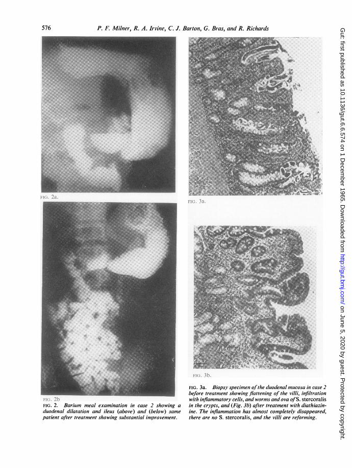
FIG. 2. Barium meal examination in case 2 showing a
duodenal dilatation and ileus (above) and (below) samepatient after treatment showing substantial improvement.
FIG. 3b.
FIG. 3a. Biopsy specimen ofthe duodenal mucosa in case 2before treatment showing flattening of the villi, infiltrationwith inflammatory cells, and worms and ova ofS. stercoralisin the crypts, and (Fig. 3b) after treatment with diathiazin-ine. The inflammation has almost completely disappeared,there are no S. stercoralis, and the villi are reforming.
576
on June 5, 2020 by guest. Protected by copyright.
http://gut.bmj.com
/G
ut: first published as 10.1136/gut.6.6.574 on 1 Decem
ber 1965. Dow
nloaded from
Intestinal malabsorption in Strongyloides stercoralis infestation
100 ml. at four hours (Fig. 5). Table I shows the improve-ment in xylose and folic acid absorption.
She had gained 4 lb. in weight, which was significantconsidering the loss of oedema and ascites.
CASE 2 A 17-year-old boy was first admitted to hospitalin October 1963. He gave a history of epigastric pain andvomiting for eight weeks with weight loss for the precedingsix months. Two months before admission he hadreported to hospital with the typical picture of pyloricstenosis: vomiting, dehydration, visible peristalsis in thestomach, and weight loss. At that time he refusedadmission and apparently recovered spontaneously.On admission he was not malnourished and there was
no diarrhoea. S. stercoralis larvae were found in severalstool specimens. A barium meal examination showeddelay in the third part of the duodenum, with an alteredmucosal pattern, areas of dilatation, and abnormalmotility. The appearance was similar to that seen in case 1.Radiographs taken before and after treatment are shownin Figure 2.The haemoglobin level was 9-6 g. per 100 ml., M.C.H.C.
300%, and white cell count 11,600 per c.mm., with 330%eosinophils. The serum albumin level was 4-1 g. per100 ml., the serum T.I.B.C. was 387 ,ug. per 100 ml.with a serum iron level of 44 ,(g. per 100 ml., andsaturation 11 %. Liver function tests were unremarkable
Peroral biopsy of the duodenum showed flattenedvilli, inflammatory infiltrate, and S. stercoralis worms.and ova in the crypts. Among the worms a pregnantfemale and rhabditiform larvae were seen (Fig. 3).
Xylose and folic acid absorption was within normallimits but an iron absorption test showed a flat curvedespite a normal transferrin level. The significance of thisflat iron absorption curve is better appreciated whencompared with the result of a repeat test performed aftertreatment and recovery (Fig. 5).The patient remained in hospital for six weeks and was
treated with dithiazanine. On discharge he had gained16 lb. in weight and the stools were free of S. stercoralislarvae. He has since remained symptom free.
CASE 3 A 13-year-old boy was first seen in May 1963.At that time he complained of vomiting and swollenlegs for four weeks, followed by diarrhoea for threeweeks. In addition he had had a generalized pruriticrash for at least one year. He was covered with aninfected papular rash; there was oedema of the eyelidsand swelling of the legs up to the knees. The tongue wasred, smooth and moist, and enlarged lymph nodes werepalpable in the neck, axillae, and groins. He was passingwatery stools up to six times a day, containing S. stercoralislarvae.
His haemoglobin level was 9-0 g. per 100 ml., M.C.H.C.30 %, and bone marrow examination showed normo-blastic erythropoiesis without convincing evidence of irondeficierncy. The serum iron level, however, was 34 ,ug.per 100 ml., with an unsaturated iron-binding capacityof only 66 ,ag. per 100 ml., T.I.B.C. 100 ,ug. per 100 ml.,saturation 34%. The serum albumin level was 1-0 g.per 100 ml. A glucose tolerance test showed a 'diabetic'curve with a fasting sugar of 105 mg. per 100 ml., and
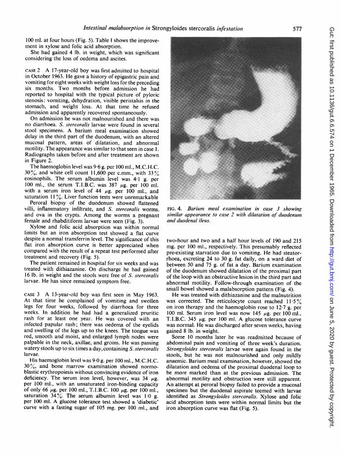
FIG. 4. Barium meal examination in case 3 showingsimilar appearance to case 2 with dilatation of duodenumand duodenal ileus.
two-hour and two and a half hour levels of 190 and 215mg. per 100 ml., respectively. This presumably reflectedpre-existing starvation due to vomiting. He had steator-rhoea, excreting 24 to 30 g. fat daily, on a ward diet ofbetween 50 and 75 g. of fat a day. Barium examinationof the duodenum showed dilatation of the proximal partof the loop with an obstructive lesion in the third part andabnormal motility. Follow-through examination of thesmall bowel showed a malabsorption pattern (Fig. 4).He was treated with dithiazanine and the malnutrition
was corrected. The reticulocyte count reached 11-50%on iron therapy and the haemoglobin rose to 12 7 g. per100 ml. Serum iron level was now 145 ,tg. per 100 ml.,T.I.B.C. 345 4g. per 100 ml. A glucose tolerance curvewas normal. He was discharged after seven weeks, havinggained 8 lb. in weight.Some 10 months later he was readmitted because of
abdominal pain and vomiting of three week's duration.Strongyloides stercoralis larvae were again found in thestools, but he was not malnourished and only mildlyanaemic. Barium meal examination, however, showed thedilatation and oedema of the proximal duodenal loop tobe more marked than at the previous admission. Theabnormal motility and obstruction were still apparent.An attempt at peroral biopsy failed to provide a mucosalspecimen but the duodenal aspirate teemed with larvaeidentified as Strongyloides stercoralis. Xylose and folicacid absorption tests were within normal limits but theiron absorption curve was flat (Fig. 5).
577
on June 5, 2020 by guest. Protected by copyright.
http://gut.bmj.com
/G
ut: first published as 10.1136/gut.6.6.574 on 1 Decem
ber 1965. Dow
nloaded from
P. F. Milner, R. A. Irvine, C. J. Barton, G. Bras, and R. Richards
400-
BEFORE TREATMENT AFTER TREATMENT
300- Fe S04 Fe S04400mg. 400 mg.
00
1-200-
w~~~~~~~~~
I)
L~~~~n ~~+
J
O i 3 4 O 2 3 4HOURS AFTER ORAL IRON
FIG. 2. Iron absorption testfor duodenalfunction (case 1 0 0, case 2, * -*, case 3, + +).Serum iron response after ingestion of400 mg. offerrous sulphate in the fasting subject, on admission and afterthe strongyloides infestation had been treated.
After three weeks' treatment with dithiazanine Strongy-loides larvae were still present in the stools and werefound again, six weeks later, in spite of further treatment.By this time, however, he looked quite well, was notmalnourished, and had a haemoglobin level of 11-3 g.per 100 ml., M.C.H.C. 33 %, and a serum albumin level
< ... x > t_ S of 3 9 g. per 100 ml. Absorption of oral iron was greatlym improved but the barium meal showed very little improve-ment in the appearance of the duodenum, and mucosalbiopsy showed flattened villi and inflammatory infiltratebut no parasites.He was given gentian violet, grains 1 (66 mg.) t.i.d.,
in enteric-coated capsules, together with dithiazanine,and the stools were finally cleared of Strongyloideslarvae. So far he has remained well.
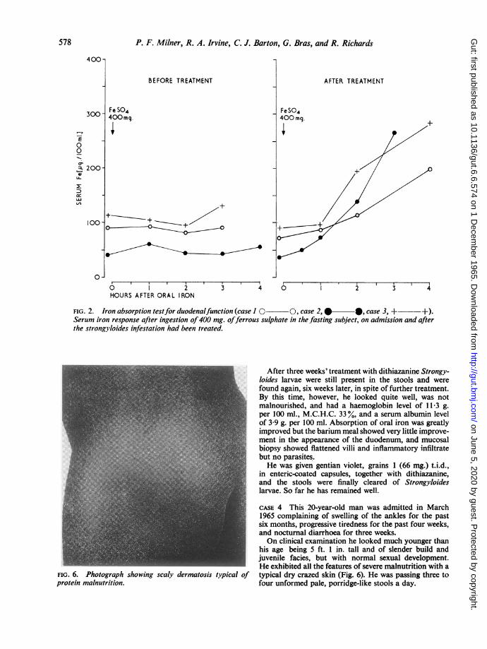
CASE 4 This 20-year-old man was admitted in March1965 complaining of swelling of the ankles for the pastsix months, progressive tiredness for the past four weeks,and nocturnal diarrhoea for three weeks.On clinical examination he looked much younger than
his age being 5 ft. 1 in. tall and of slender build andjuvenile facies, but with normal sexual development.He exhibited all the features of severe malnutrition with a
6. Photograph showing scaly dermatosis typical of typical dry crazed skin (Fig. 6). He was passing three torein malnutrition. four unformed pale, porridge-like stools a day.
FIG.prot
578
on June 5, 2020 by guest. Protected by copyright.
http://gut.bmj.com
/G
ut: first published as 10.1136/gut.6.6.574 on 1 Decem
ber 1965. Dow
nloaded from
Intestinal malabsorption in Strongyloides stercoralis infestation
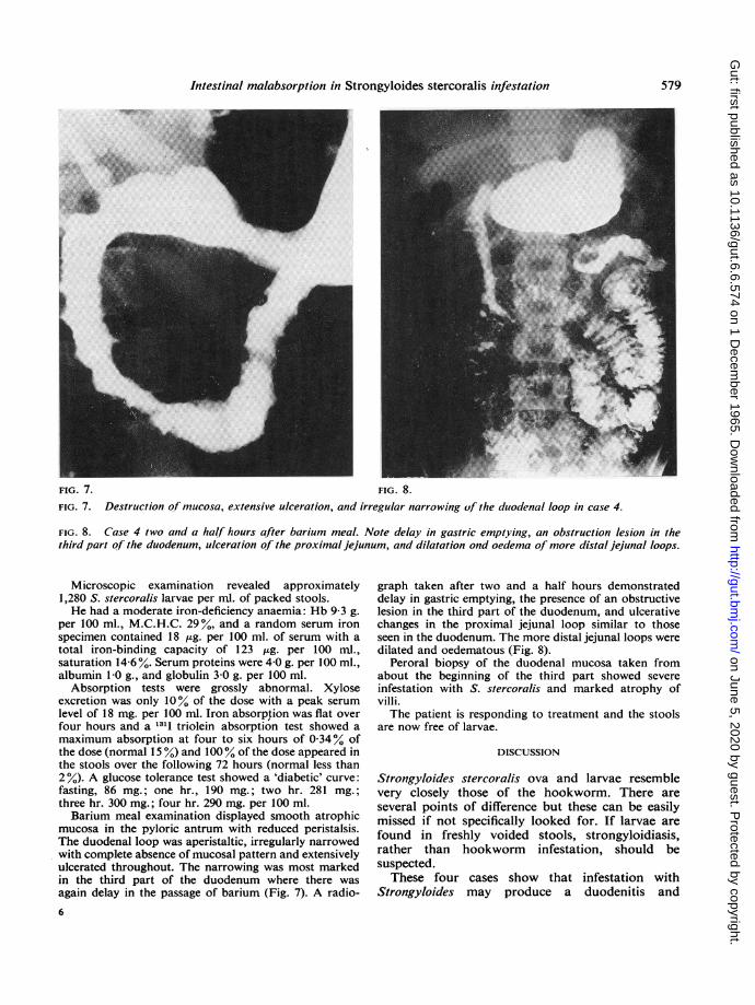
FIG. 7. FIG. 8.
FIG. 7. Destruction of mucosa, extensive ulceration, and irregular narrowing of the duodenal loop in case 4.
FIG. 8. Case 4 two and a half hours after barium meal. Note delay in gastric emptying, an obstruction lesion in thethird part of the duodenum, ulceration of the proximal jejunum, and dilatation ond oedema of more distal jejunal loops.
Microscopic examination revealed approximately1,280 S. stercoralis larvae per ml. of packed stools.He had a moderate iron-deficiency anaemia: Hb 9 3 g.
per 100 ml., M.C.H.C. 29%, and a random serum ironspecimen contained 18 ,g. per 100 ml. of serum with atotal iron-binding capacity of 123 ,ug. per 100 ml.,saturation 14-6 %. Serum proteins were 4.0 g. per 100 ml.,albumin 1 0 g., and globulin 3.0 g. per 100 ml.
Absorption tests were grossly abnormal. Xyloseexcretion was only 100% of the dose with a peak serumlevel of 18 mg. per 100 ml. Iron absorption was flat overfour hours and a 1311 triolein absorption test showed amaximum absorption at four to six hours of 0.34% ofthe dose (normal 15 %) and 100% of the dose appeared inthe stools over the following 72 hours (normal less than2%). A glucose tolerance test showed a 'diabetic' curve:fasting, 86 mg.; one hr., 190 mg.; two hr. 281 mg.;three hr. 300 mg.; four hr. 290 mg. per 100 ml.Barium meal examination displayed smooth atrophic
mucosa in the pyloric antrum with reduced peristalsis.The duodenal loop was aperistaltic, irregularly narrowedwith complete absence of mucosal pattern and extensivelyulcerated throughout. The narrowing was most markedin the third part of the duodenum where there wasagain delay in the passage of barium (Fig. 7). A radio-6
graph taken after two and a half hours demonstrateddelay in gastric emptying, the presence of an obstructivelesion in the third part of the duodenum, and ulcerativechanges in the proximal jejunal loop similar to thoseseen in the duodenum. The more distal jejunal loops weredilated and oedematous (Fig. 8).
Peroral biopsy of the duodenal mucosa taken fromabout the beginning of the third part showed severeinfestation with S. stercoralis and marked atrophy ofvilli.The patient is responding to treatment and the stools
are now free of larvae.
DISCUSSION
Strongyloides stercoralis ova and larvae resemblevery closely those of the hookworm. There areseveral points of difference but these can be easilymissed if not specifically looked for. If larvae arefound in freshly voided stools, strongyloidiasis,rather than hookworm infestation, should besuspected.
These four cases show that infestation withStrongyloides may produce a duodenitis and
579
on June 5, 2020 by guest. Protected by copyright.
http://gut.bmj.com
/G
ut: first published as 10.1136/gut.6.6.574 on 1 Decem
ber 1965. Dow
nloaded from
P. F. Milner, R. A. Irvine, C. J. Barton, G. Bras, and R. Richards
E
8
z
0
W
r
v
ULJ
++ '+ \
Fe S04400 mg.
o-Jfasting
2 3 4HOURS
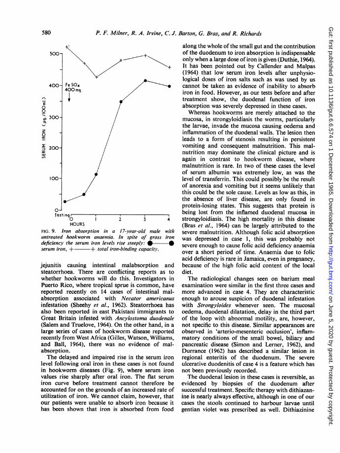
FIG. 9. Iron absorption in a 17-year-old male withuntreated hookworm anaemia. In spite of gross irondeficiency the serum iron levels rise steeply: *serum iron, +- + total iron-binding capacity.
jejunitis causing intestinal malabsorption andsteatorrhoea. There are conflicting reports as towhether hookworms will do this. Investigators inPuerto Rico, where tropical sprue is common, havereported recently on 14 cases of intestinal mal-absorption associated with Necator americanusinfestation (Sheehy et al., 1962). Steatorrhoea hasalso been reported in east Pakistani immigrants toGreat Britain infested with Ancylostoma duodenale(Salem and Truelove, 1964). On the other hand, in alarge series of cases of hookworm disease reportedrecently from West Africa (Gilles, Watson, Williams,and Ball, 1964), there was no evidence of mal-absorption.The delayed and impaired rise in the serum iron
level following oral iron in these cases is not foundin hookworm diseases (Fig. 9), where serum ironvalues rise sharply after oral iron. The flat serumiron curve before treatment cannot therefore beaccounted for on the grounds of an increased rate ofutilization of iron. We cannot claim, however, thatour patients were unable to absorb iron because ithas been shown that iron is absorbed from food
along the whole of the small gut and the contributionof the duodenum to iron absorption is indispensableonly when a large dose of iron is given (Duthie, 1964).It has been pointed out by Callender and Malpas(1964) that low serum iron levels after unphysio-logical doses of iron salts such as was used by uscannot be taken as evidence of inability to absorbiron in food. However, as our tests before and aftertreatment show, the duodenal function of ironabsorption was severely depressed in these cases.Whereas hookworms are merely attached to the
mucosa, in strongyloidiasis the worms, particularlythe larvae, invade the mucosa causing oedema andinflammation of the duodenal walls. The lesion thenleads to a form of stenosis resulting in persistentvomiting and consequent malnutrition. This mal-nutrition may dominate the clinical picture and isagain in contrast to hookworm disease, wheremalnutrition is rare. In two of these cases the levelof serum albumin was extremely low, as was thelevel of transferrin. This could possibly be the resultof anorexia and vomiting but it seems unlikely thatthis could be the sole cause. Levels as low as this, inthe absence of liver disease, are only found inprotein-losing states. This suggests that protein isbeing lost from the inflamed duodenal mucosa instrongyloidiasis. The high mortality in this disease(Bras et al., 1964) can be largely attributed to thesevere malnutrition. Although folic acid absorptionwas depressed in case 1, this was probably notsevere enough to cause folic acid deficiency anaemiaover a short period of time. Anaemia due to folicacid deficiency is rare in Jamaica, even in pregnancy,because of the high folic acid content of the localdiet.The radiological changes seen on barium meal
examination were similar in the first three cases andmore advanced in case 4. They are characteristicenough to arouse suspicion of duodenal infestationwith Strongyloides whenever seen. The mucosaloedema, duodenal dilatation, delay in the third partof the loop with abnormal motility, are, however,not specific to this disease. Similar appearances areobserved in 'arterio-mesenteric occlusion', inflam-matory conditions of the small bowel, biliary andpancreatic disease (Simon and Lerner, 1962), andDurrance (1962) has described a similar lesion inregional enteritis of the duodenum. The severeulcerative duodenitis of case 4 is a feature which hasnot been previously recorded.The duodenal lesion in these cases is reversible, as
evidenced by biopsies of the duodenum aftersuccessful treatment. Specific therapy with dithiazan-ine is nearly always effective, although in one of ourcases the stools continued to harbour larvae untilgentian violet was prescribed as well. Dithiazinine
580
on June 5, 2020 by guest. Protected by copyright.
http://gut.bmj.com
/G
ut: first published as 10.1136/gut.6.6.574 on 1 Decem
ber 1965. Dow
nloaded from
Intestinal malabsorption in Strongyloides stercoralis infestation 581
treatment is not without hazards, as was shown bycase 1, when profuse diarrhoea and severe potassiumdepletion forced us temporarily to discontinuetreatment.
We are grateful to Dr. K. Wood, Pathology Department,U.W.I., for performing the 131I triolein test on case 4;to the nursing staff of the University College Hospitalfor their cooperation in the investigation of these patients,and to the Clinical Photography Department for thephotographs.
REFERENCES
Alcorn, M. 0., and Kotcher, E. (1961). Secondary malabsorptionsyndrome produced by chronic strongyloidiasis. Sth. med. J.(Bgham, Ala.), 54, 193-197.
Amini, F. (1963). Giardiasis and steatorrhoea. J. trop. Med. Hyg., 66,190-192.
Beale, R. N., Bostrom, J. 0., and Taylor, R. F. (1961). Rapid incre-mental methods for the determination of serum iron andiron-binding capacity. J. clin. Path., 14, 488-495.
(1962). Improved rapid methods for the determinationof iron content and binding capacity ofserum. Ibid., 15, 156-160.
Bras, G., Richards, R. C., Irvine, R. A., Milner, F. P. A., and Ragbeer,M. M. S. (1964). Infection with Strongyloides stercoralis inJamaica. Lancet, 2, 1257-1260.
Callender, S. T. E., and Malpas, J. S. (1964). Anaemia and hiatushernia. Brit. med. J., 1, 1312.
Chanarin, I., Anderson, B. B., and Mollin, D. L. (1958). The absorp-tion of folic acid. Brit. J. Haemat., 4, 156-166.
, and Bennett, M. C. (1962). Absorption of folic acid and D-xylose as tests of small-intestinal function. Brit. med. J., 1,985-989.
Crosby, W. H., and Kugler, H. W. (1957). Intraluminal biopsy of thesmall intestine: the intestinal biopsy capcule. Amer. J. dig. Dis.,2, 236-241.
Durrance, F. Y. (1962). Regional enteritis of the duodenum. Amer. J.Roentgenol., 88, 658-661.
Duthie, H. L. (1964). The relative importance of the duodenum in theintestinal absorption of iron. Brit. J. Haemat., 10, 59-68.
Gilles, H. M., Watson Williams, E. J., and Ball, P. A. J. (1964).Hookworm infection and anaemia. Quart. J. Med., 33, 1-24.
Jeffries, G. H., Weser, E., and Sleisenger, M. H. (1964). Malabsorp-tion. Gastroenterology, 46, 434-466.
Roe, J. H., and Rice, E. W. (1948). A photometric method for thedetermination of free pentoses in animal tissues. J. biol. Chem.,173, 507-512.
Salem, S. N., and Truelove, S. C. (1964). Hookworm Disease inimmigrants. Brit. med. J., 1, 1074-1077.
Sheehy, T. W., Meroney, W. H., Cox, R. S., Jr., and Soler, J. E.(1962). Hookworm disease and malabsorption. Gastroenterology,42, 148-156.
Simon, M., and Lerner, M. A. (1962). Duodenal comparison by themesenteric root in acute pancreatitis and inflammatoryconditions of the bowel. Radiology, 79, 75-81.
Stemmermann, G. N., and Nakasone, N. (1960). Strongyloidesstercoralis infestation. Malabsorption defect with reaction todithiazanine iodide. J. Amer. med. Ass., 174, 1250-1253.
Zamcheck, N., Hoskins, L. C., Winawer, S. J., Broitman, S. A., andGottlieb, L. S. (1963). Histology and ultrastructure of theparasite and intestinal mucosa in human giardiasis: effects ofatabrine therapy. (Abstract). Gastroenterology, 44, 860.
on June 5, 2020 by guest. Protected by copyright.
http://gut.bmj.com
/G
ut: first published as 10.1136/gut.6.6.574 on 1 Decem
ber 1965. Dow
nloaded from


![Prevalence and risk factors of Strongyloides stercoralis ...Strongyloides stercoralis, a soil-transmitted nematode, is ar-guably the most neglected tropical disease [1], yet an esti-mated](https://static.fdocuments.in/doc/165x107/603910f33d86085b0845e0dd/prevalence-and-risk-factors-of-strongyloides-stercoralis-strongyloides-stercoralis.jpg)