Stress-responsive FKBP51 regulates AKT2-AS160 signaling ... · ARTICLE Stress-responsive FKBP51...
12
ARTICLE Stress-responsive FKBP51 regulates AKT2-AS160 signaling and metabolic function Georgia Balsevich 1 , Alexander S. Häusl 1 , Carola W. Meyer 2 , Stoyo Karamihalev 1 , Xixi Feng 3 , Max L. Pöhlmann 1 , Carine Dournes 1 , Andres Uribe-Marino 1 , Sara Santarelli 1 , Christiana Labermaier 1 , Kathrin Hafner 3 , Tianqi Mao 3 , Michaela Breitsamer 4 , Marily Theodoropoulou 3 , Christian Namendorf 3 , Manfred Uhr 3 , Marcelo Paez-Pereda 3 , Gerhard Winter 4 , Felix Hausch 5 , Alon Chen 1 , Matthias H. Tschöp 2 , Theo Rein 3 , Nils C. Gassen 3 & Mathias V. Schmidt 1 The co-chaperone FKBP5 is a stress-responsive protein-regulating stress reactivity, and its genetic variants are associated with T2D related traits and other stress-related disorders. Here we show that FKBP51 plays a role in energy and glucose homeostasis. Fkbp5 knockout (51KO) mice are protected from high-fat diet-induced weight gain, show improved glucose tolerance and increased insulin signaling in skeletal muscle. Chronic treatment with a novel FKBP51 antagonist, SAFit2, recapitulates the effects of FKBP51 deletion on both body weight regulation and glucose tolerance. Using shorter SAFit2 treatment, we show that glucose tolerance improvement precedes the reduction in body weight. Mechanistically, we identify a novel association between FKBP51 and AS160, a substrate of AKT2 that is involved in glucose uptake. FKBP51 antagonism increases the phosphorylation of AS160, increases glucose transporter 4 expression at the plasma membrane, and ultimately enhances glucose uptake in skeletal myotubes. We propose FKBP51 as a mediator between stress and T2D development, and potential target for therapeutic approaches. DOI: 10.1038/s41467-017-01783-y OPEN 1 Department of Stress Neurobiology and Neurogenetics, Max Planck Institute of Psychiatry, Kraepelinstraße 2-10, 80804 Munich, Germany. 2 Institute of Diabetes and Obesity, Helmholtz Zentrum München, Parkring 13, 85748 Garching, Germany. 3 Department of Translational Research in Psychiatry, Max Planck Institute of Psychiatry, Kraepelinstraße 2-10, 80804 Munich, Germany. 4 Ludwig Maximilians University, Butenandtstr. 5-13, 81377 Munich, Germany. 5 Technical University Darmstadt, Institute of Organic Chemistry and Biochemistry, Alarich-Weiss-Str. 4, 64287 Darmstadt, Germany. Georgia Balsevich and Alexander S. Häusl contributed equally to this work. Theo Rein, Nils C. Gassen and Mathias V. Schmidt jointly supervised this work. Correspondence and requests for materials should be addressed to G.B. (email: [email protected]) or to M.V.S. (email: [email protected]) NATURE COMMUNICATIONS | 8: 1725 | DOI: 10.1038/s41467-017-01783-y | www.nature.com/naturecommunications 1 1234567890
Transcript of Stress-responsive FKBP51 regulates AKT2-AS160 signaling ... · ARTICLE Stress-responsive FKBP51...
ARTICLE
Stress-responsive FKBP51 regulates AKT2-AS160signaling and metabolic functionGeorgia Balsevich1, Alexander S. Häusl1, Carola W. Meyer2, Stoyo Karamihalev1, Xixi Feng3, Max L. Pöhlmann1,
Carine Dournes1, Andres Uribe-Marino1, Sara Santarelli1, Christiana Labermaier1, Kathrin Hafner3,
Tianqi Mao3, Michaela Breitsamer4, Marily Theodoropoulou3, Christian Namendorf3, Manfred Uhr3,
Marcelo Paez-Pereda3, Gerhard Winter4, Felix Hausch5, Alon Chen1, Matthias H. Tschöp2, Theo Rein 3,
Nils C. Gassen3 & Mathias V. Schmidt 1
The co-chaperone FKBP5 is a stress-responsive protein-regulating stress reactivity, and its
genetic variants are associated with T2D related traits and other stress-related disorders.
Here we show that FKBP51 plays a role in energy and glucose homeostasis. Fkbp5 knockout
(51KO) mice are protected from high-fat diet-induced weight gain, show improved glucose
tolerance and increased insulin signaling in skeletal muscle. Chronic treatment with a novel
FKBP51 antagonist, SAFit2, recapitulates the effects of FKBP51 deletion on both body weight
regulation and glucose tolerance. Using shorter SAFit2 treatment, we show that glucose
tolerance improvement precedes the reduction in body weight. Mechanistically, we identify a
novel association between FKBP51 and AS160, a substrate of AKT2 that is involved in glucose
uptake. FKBP51 antagonism increases the phosphorylation of AS160, increases glucose
transporter 4 expression at the plasma membrane, and ultimately enhances glucose uptake in
skeletal myotubes. We propose FKBP51 as a mediator between stress and T2D development,
and potential target for therapeutic approaches.
DOI: 10.1038/s41467-017-01783-y OPEN
1 Department of Stress Neurobiology and Neurogenetics, Max Planck Institute of Psychiatry, Kraepelinstraße 2-10, 80804 Munich, Germany. 2 Institute ofDiabetes and Obesity, Helmholtz Zentrum München, Parkring 13, 85748 Garching, Germany. 3 Department of Translational Research in Psychiatry, MaxPlanck Institute of Psychiatry, Kraepelinstraße 2-10, 80804 Munich, Germany. 4 Ludwig Maximilians University, Butenandtstr. 5-13, 81377 Munich, Germany.5 Technical University Darmstadt, Institute of Organic Chemistry and Biochemistry, Alarich-Weiss-Str. 4, 64287 Darmstadt, Germany. Georgia Balsevich andAlexander S. Häusl contributed equally to this work. Theo Rein, Nils C. Gassen and Mathias V. Schmidt jointly supervised this work. Correspondence andrequests for materials should be addressed to G.B. (email: [email protected]) or to M.V.S. (email: [email protected])
NATURE COMMUNICATIONS |8: 1725 |DOI: 10.1038/s41467-017-01783-y |www.nature.com/naturecommunications 1
1234
5678
90
FK506-binding protein 51 (FKBP51) is an immunophilinprotein best known as a negative regulator of the gluco-corticoid receptor (GR) and consequently the physiological
stress response1. Specifically, in complex with FKBP51 (FKBP5gene), the GR displays reduced ligand affinity, reduced nucleartranslocation, and ultimately decreased GR sensitivity1–5.By contrast, in complex with its functional counter-playerFKBP52 (FKBP4 gene), GR activity is enhanced6. Single-nucleotide polymorphisms (SNPs) in the FKBP5 gene, whichare associated with increased expression of FKBP5 (high-induc-tion allele), have been fundamentally linked to stress-relateddisorders, and most notably in psychiatric disorders7. In thiscontext, it is possible that stress-induced FKBP5 may similarly beimplicated in additional stress-related pathophysiologies, such astype 2 diabetes (T2D).
Exposure to nutrient overload, including exposure to a high-fatdiet (HFD), is considered a metabolic stressor8. Interestingly, itwas reported that 8 weeks of HFD exposure in mice led toenhanced Fkbp5 expression in the hypothalamus9, suggesting thatFkbp5 is responsive to metabolic stressors and is able to sense thenutrient environment. Accordingly, a study examining foodrestricted-responsive genes reported an induction of Fkbp5 in thehypothalamus and ventral tegmental area10. This is in agreementwith an earlier study, which used a 24-h food restriction paradigmas a stressor to investigate stress-induced Fkbp5 expression acrossmultiple brain regions11.
There are several additional lines of evidence to support thepossibility that FKBP51 links stress to metabolic function. Humanadipocytes and skeletal muscle are among the tissues presentingthe strongest expression of FKBP512. Recently, the high-inductionFKBP5 risk allele was associated with reduced weight loss fol-lowing bariatric surgery13. A genome-wide association studyfurthermore demonstrated that SNPs within the FKBP5 gene lociare associated with T2D and markers of insulin resistance14.Preclinical studies in animal models have similarly demonstratedthat complete loss of FKBP51 protects against HFD-induced bodyweight gain and hepatic steatosis, which is, in part, explained byan increased expression of uncoupling protein 1 (UCP1), a spe-cific marker of browning, in white adipose tissue (WAT) andincreased thermogenesis15. Additionally, FKBP51 is a negativeregulator of all 3 isoforms of the serine/threonine protein kinaseAKT (AKT1, AKT2, and AKT3), and through this action, reg-ulates the response to chemotherapy16. AKT is also a central nodewithin the insulin signaling pathway, and deregulation of AKTactivation, most notably AKT2 activation, has been linked to thepathogenesis of diabetes and obesity17, 18. In this context, FKBP51may be an important regulator of insulin signaling and conse-quently energy and glucose homeostasis19. Nevertheless, whetherFKBP51 plays a critical role in whole body glucose metabolismremains to be elucidated. For this purpose, we aimed to char-acterize the role of FKBP51 in energy and glucose homeostasisusing a combination of Fkbp5 knockout (51KO) mice,
0 1 2 3 4 5 6 7 82024283236404448
WT chow
51KO chow51KO HFD
WT HFD
Weeks
Bod
y w
eigh
t (g)
b
WT 51KO0
10
20
30
40ChowHFD
#
+++
Bod
y w
eigh
t (g)
(8 w
eeks
on
diet
s)
c
WT 51KO0.0
0.5
1.0
1.5
2.0
2.5
ChowHFD
###+++
eWA
T (
g)
WT 51KO0.0
0.2
0.4
0.6
0.8
1.0 ###+++
iWA
T (
g)
WT 51KO0.0
0.1
0.2
0.3
0.4
0.5
0.6##++
pWA
T (
g)
WT 51KO0.00
0.05
0.10
0.15
0.20
0.25
BA
T (
g)
a
WT 51KO20
22
24
26
28
30
+++
Wei
ght a
djus
ted
lean
mas
s (g
)
WT 51KO0.0
0.5
1.0
1.5
+++
Fat
mas
s (g
)
WT 51KO0
20
24
28
32++
Initi
al b
ody
wei
ght (
g)
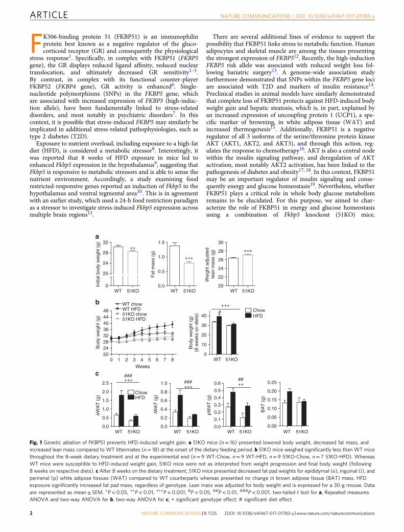
Fig. 1 Genetic ablation of FKBP51 prevents HFD-induced weight gain. a 51KO mice (n= 16) presented lowered body weight, decreased fat mass, andincreased lean mass compared to WT littermates (n= 18) at the onset of the dietary feeding period. b 51KO mice weighed significantly less than WT micethroughout the 8-week dietary treatment and at the experimental end (n= 9 WT-Chow, n= 9 WT-HFD, n= 9 51KO-Chow, n= 7 51KO-HFD). WhereasWT mice were susceptible to HFD-induced weight gain, 51KO mice were not as interpreted from weight progression and final body weight (following8 weeks on respective diets). c After 8 weeks on the dietary treatment, 51KO mice presented decreased fat pad weights for epididymal (e), inguinal (i), andperirenal (p) white adipose tissues (WAT) compared to WT counterparts whereas presented no change in brown adipose tissue (BAT) mass. HFDexposure significantly increased fat pad mass, regardless of genotype. Lean mass was adjusted for body weight and is expressed for a 30-g mouse. Dataare represented as mean± SEM. +P< 0.05, ++P< 0.01, +++P< 0.001; #P< 0.05, ##P< 0.01, ###P< 0.001, two-tailed t test for a, Repeated measuresANOVA and two-way ANOVA for b, two-way ANOVA for c; + significant genotype effect; # significant diet effect
ARTICLE NATURE COMMUNICATIONS | DOI: 10.1038/s41467-017-01783-y
2 NATURE COMMUNICATIONS | 8: 1725 |DOI: 10.1038/s41467-017-01783-y |www.nature.com/naturecommunications
pharmacological manipulations, and mechanistic studies. Wefound in this study that FKBP51 regulates glucose metabolism inmice, through it, regulation of AKT2-AS160 signaling, glucosetransporter expression, and glucose uptake in myotubes. Phar-macological antagonism of FKBP51 improves glucose tolerance,irrespective of body weight changes, which suggests an oppor-tunity to target FKBP51 for the treatment of T2D.
ResultsFKBP51 loss opposes obesity and improves glucose tolerance.In order to examine the role of FKBP51 in energy and glucosehomeostasis, we initially characterized the metabolic outcomesarising in 51KO mice. We found that 51KO mice fed with astandard chow diet showed a modest body weight reduction,reduced adiposity, and increased lean mass compared to WTlittermates (Fig. 1a). When challenged with HFD exposure for8 weeks, the 51KO mice were protected from both HFD-inducedweight gain and increased adiposity (Fig. 1b, c). Loss of FKBP51likewise counteracted diet-induced obesity under thermoneutralconditions (30 °C), arguing against a thermoregulatory basis ofthe phenotype (Supplementary Fig. 1). Indirect calorimetryrevealed that the body weight phenotype observed in 51KO miceunder standard chow conditions was accompanied by a modestincrease in total energy expenditure, as a result of an increasedresting metabolic rate (RMR) (Supplementary Fig. 2A). In
addition, 51KO mice presented a modest decrease in theirrespiratory exchange ratio (RER) and a slight increase in theirhome-cage activity (Supplementary Fig. 2B, C). By contrast,neither water nor food intakes were affected by loss of FKBP51(Supplementary Fig. 2D, E). To confirm a lack of FKBP51 effecton feeding behavior, a separate pair-feeding experiment wasperformed, in which a cohort of WT mice was pair-fed to 51KOmice. This experiment again revealed no genotype effect onenergy intake (Supplementary Fig. 2G). Cold-induced bodytemperature regulation was unaffected by FKBP51 genotype(Supplementary Fig. 2H).
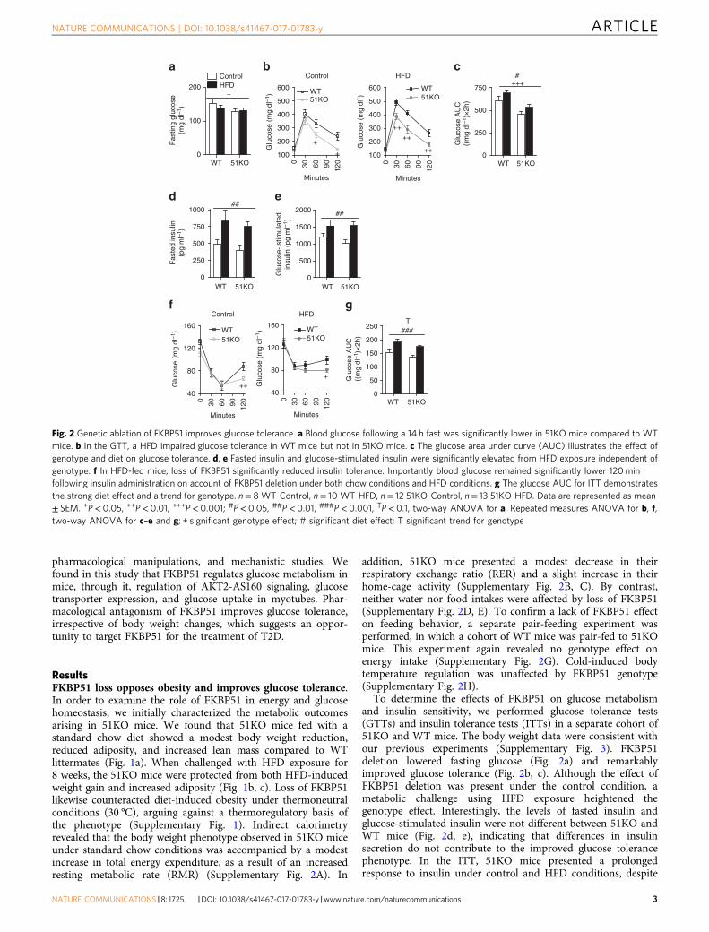
To determine the effects of FKBP51 on glucose metabolismand insulin sensitivity, we performed glucose tolerance tests(GTTs) and insulin tolerance tests (ITTs) in a separate cohort of51KO and WT mice. The body weight data were consistent withour previous experiments (Supplementary Fig. 3). FKBP51deletion lowered fasting glucose (Fig. 2a) and remarkablyimproved glucose tolerance (Fig. 2b, c). Although the effect ofFKBP51 deletion was present under the control condition, ametabolic challenge using HFD exposure heightened thegenotype effect. Interestingly, the levels of fasted insulin andglucose-stimulated insulin were not different between 51KO andWT mice (Fig. 2d, e), indicating that differences in insulinsecretion do not contribute to the improved glucose tolerancephenotype. In the ITT, 51KO mice presented a prolongedresponse to insulin under control and HFD conditions, despite
WT 51KO0
500
1000
1500
2000 ##
Glu
cose
- st
imul
ated
insu
lin (
pg m
l–1)
WT 51KO0
250
500
750
1000##
Fas
ted
insu
lin(p
g m
l–1)
d e
WT 51KO0
100
200
ControlHFD
+
Fas
ting
gluc
ose
(mg
dl–1
)
WT 51KO0
250
500
750
#+++
Glu
cose
AU
C((
mg
dl–1
)×2h
)
Control
100
200
300
400
500
600 WT
+
+
51KO
Minutes
Glu
cose
(m
g dl
–1)
HFD
100
200
300
400
500
600
++++
++Glu
cose
(m
g dl
1 )
a b c
51KOWT
1200 906030
Minutes
1200 906030
f g
WT 51KO0
50
100
150
200
250###
Glu
cose
AU
C((
mg
dl–1
)×2h
)
Control
40
80
120
160WT51KO
Glu
cose
(m
g dl
–1)
HFD
40
80
120
160
51KOWT
Glu
cose
(m
g dl
–1)
+++
T
Minutes
1200 906030
Minutes
1200 906030
Fig. 2 Genetic ablation of FKBP51 improves glucose tolerance. a Blood glucose following a 14 h fast was significantly lower in 51KO mice compared to WTmice. b In the GTT, a HFD impaired glucose tolerance in WT mice but not in 51KO mice. c The glucose area under curve (AUC) illustrates the effect ofgenotype and diet on glucose tolerance. d, e Fasted insulin and glucose-stimulated insulin were significantly elevated from HFD exposure independent ofgenotype. f In HFD-fed mice, loss of FKBP51 significantly reduced insulin tolerance. Importantly blood glucose remained significantly lower 120minfollowing insulin administration on account of FKBP51 deletion under both chow conditions and HFD conditions. g The glucose AUC for ITT demonstratesthe strong diet effect and a trend for genotype. n= 8 WT-Control, n= 10 WT-HFD, n= 12 51KO-Control, n= 13 51KO-HFD. Data are represented as mean± SEM. +P< 0.05, ++P< 0.01, +++P< 0.001; #P< 0.05, ##P< 0.01, ###P< 0.001, TP< 0.1, two-way ANOVA for a, Repeated measures ANOVA for b, f,two-way ANOVA for c–e and g; + significant genotype effect; # significant diet effect; T significant trend for genotype
NATURE COMMUNICATIONS | DOI: 10.1038/s41467-017-01783-y ARTICLE
NATURE COMMUNICATIONS |8: 1725 |DOI: 10.1038/s41467-017-01783-y |www.nature.com/naturecommunications 3
the fact that both 51KO and WT mice remained vulnerable toHFD-induced insulin intolerance (Fig. 2f, g).
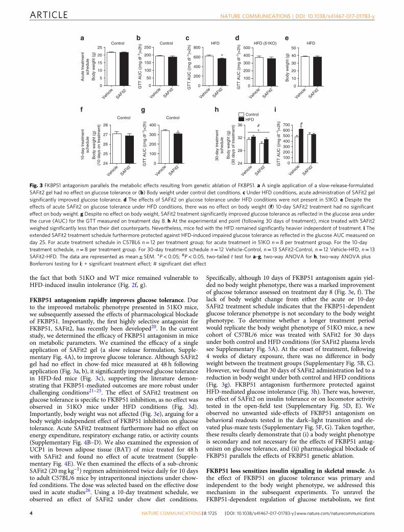
FKBP51 antagonism rapidly improves glucose tolerance. Dueto the improved metabolic phenotype presented in 51KO mice,we subsequently assessed the effects of pharmacological blockadeof FKBP51. Importantly, the first highly selective antagonist forFKBP51, SAFit2, has recently been developed20. In the currentstudy, we determined the efficacy of FKBP51 antagonism in miceon metabolic parameters. We examined the efficacy of a singleapplication of SAFit2 gel (a slow release formulation, Supple-mentary Fig. 4A), to improve glucose tolerance. Although SAFit2gel had no effect in chow-fed mice measured at 48 h followingapplication (Fig. 3a, b), it significantly improved glucose tolerancein HFD-fed mice (Fig. 3c), supporting the literature demon-strating that FKBP51-mediated outcomes are more robust underchallenging conditions21–25. The effect of SAFit2 treatment onglucose tolerance is specific to FKBP51 inhibition, as no effect wasobserved in 51KO mice under HFD conditions (Fig. 3d).Importantly, body weight was not affected (Fig. 3e), arguing for abody weight-independent effect of FKBP51 inhibition on glucosetolerance. Acute SAFit2 treatment furthermore had no effect onenergy expenditure, respiratory exchange ratio, or activity counts(Supplementary Fig. 4B–D). We also examined the expression ofUCP1 in brown adipose tissue (BAT) of mice treated for 48 hwith SAFit2 and found no effect of acute treatment (Supple-mentary Fig. 4E). We then examined the effects of a sub-chronicSAFit2 (20 mg kg−1) regimen administered twice daily for 10 daysto adult C57BL/6 mice by intraperitoneal injections under chow-fed conditions. The dose was selected based on the effective doseused in acute studies26. Using a 10-day treatment schedule, weobserved an effect of SAFit2 under chow diet conditions.
Specifically, although 10 days of FKBP51 antagonism again yiel-ded no body weight phenotype, there was a marked improvementof glucose tolerance assessed on treatment day 8 (Fig. 3e, f). Thelack of body weight change from either the acute or 10-daySAFit2 treatment schedule indicates that the FKBP51-dependentglucose tolerance phenotype is not secondary to the body weightphenotype. To determine whether a longer treatment periodwould replicate the body weight phenotype of 51KO mice, a newcohort of C57BL/6 mice was treated with SAFit2 for 30 daysunder both control and HFD conditions (for SAFit2 plasma levelssee Supplementary Fig. 5A). At the onset of treatment, following4 weeks of dietary exposure, there was no difference in bodyweight between the treatment groups (Supplementary Fig. 5B, C).However, we found that 30 days of SAFit2 administration led to areduction in body weight under both control and HFD conditions(Fig. 3g). FKBP51 antagonism furthermore protected againstHFD-mediated glucose intolerance (Fig. 3h). There was, however,no effect of SAFit2 on insulin tolerance or on locomotor activitytested in the open-field test (Supplementary Fig. 5D, E). Weobserved no unwanted side-effects of FKBP51 antagonism onbehavioral readouts tested in the dark–light transition and ele-vated plus-maze tests (Supplementary Fig. 5F, G). Taken together,these results clearly demonstrate that (i) a body weight phenotypeis secondary and not necessary for the effects of FKBP51 antag-onism on glucose tolerance, and (ii) pharmacological blockade ofFKBP51 parallels the effects of FKBP51 genetic ablation.
FKBP51 loss sensitizes insulin signaling in skeletal muscle. Asthe effect of FKBP51 on glucose tolerance was primary andindependent to the body weight phenotype, we addressed thismechanism in the subsequent experiments. To unravel theFKBP51-dependent regulation of glucose metabolism, we first
c ea b
SAFit224
25
26
27
28
Bod
y w
eigh
t (g)
(10
days
on
trea
tmen
t)
Vehicl
e0
100
200
300
400+
GT
T A
UC
((m
g dl
–1)×
2h)
SAFit2
Vehicl
e
Control
10-d
ay tr
eatm
ent
sche
dule
30-d
ay tr
eatm
ent
sche
dule
24
28
32
36 #+
Bod
y w
eigh
t (g)
(30
days
of t
reat
men
t)
SAFit2
Vehicl
e0
100200300400500600700 #
GT
T A
UC
((m
g dl
–1)×
2h)
SAFit2
Vehicl
e
ControlHFD
Control
0
50
100
150
200
GT
T A
UC
((m
g dl
–1)×
2h)
Control
SAFit2
Vehicl
e
HFD
0
200
400
600
800
+
GT
T A
UC
((m
g dl
–1)×
2h)
SAFit2
Vehicl
e
Acu
te tr
eatm
ent
sche
dule
gf
HFD
0
10
20
30
40
50
Bod
y w
eigh
t (g)
SAFit2
Vehicl
e
Control
0
5
10
15
20
25
Bod
y w
eigh
t (g)
250
SAFit2
Vehicl
e
ih
dHFD (51KO)
0
100
200
300
400
500
GT
T A
UC
((m
g dl
–1)×
2h)
SAFit2
Vehicl
e
Fig. 3 FKBP51 antagonism parallels the metabolic effects resulting from genetic ablation of FKBP51. a A single application of a slow-release-formulatedSAFit2 gel had no effect on glucose tolerance or (b) Body weight under control diet conditions. c Under HFD conditions, acute administration of SAFit2 gelsignificantly improved glucose tolerance. d The effects of SAFit2 on glucose tolerance under HFD conditions were not present in 51KO. e Despite theeffects of acute SAFit2 on glucose tolerance under HFD conditions, there was no effect on body weight (f) 10-day SAFit2 treatment had no significanteffect on body weight. g Despite no effect on body weight, SAFit2 treatment significantly improved glucose tolerance as reflected in the glucose area underthe curve (AUC) for the GTT measured on treatment day 8. h At the experimental end point (following 30 days of treatment), mice treated with SAFit2weighed significantly less than their diet counterparts. Nevertheless, mice fed with the HFD remained significantly heavier independent of treatment. i Theextended SAFit2 treatment schedule furthermore protected against HFD-induced impaired glucose tolerance as reflected in the glucose AUC measured onday 25. For acute treatment schedule in C57BL6 n= 12 per treatment group; for acute treatment in 51KO n= 8 per treatment group. For the 10-daytreatment schedule, n= 8 per treatment group. For 30-day treatment schedule n= 12 Vehicle-Control, n= 13 SAFit2-Control, n= 12 Vehicle-HFD, n= 13SAFit2-HFD. The data are represented as mean± SEM. +P< 0.05; #P< 0.05, two-tailed t test for a–g, two-way ANOVA for h, two-way ANOVA plusBonferroni testing for i; + significant treatment effect; # significant diet effect
ARTICLE NATURE COMMUNICATIONS | DOI: 10.1038/s41467-017-01783-y
4 NATURE COMMUNICATIONS | 8: 1725 |DOI: 10.1038/s41467-017-01783-y |www.nature.com/naturecommunications
examined FKBP51 protein expression across multiple peripheraltissues. Interestingly, FKBP51 was not ubiquitously expressedacross all tissues examined, but rather showed a defined expres-sion profile. FKBP51 was detected within skeletal muscle (soleusmuscle and EDL) eWAT and iWAT (Supplementary Fig. 6A). Bycontrast, FKBP51 was not detected in the liver, kidney, spleen,pancreas, gut, or BAT. In addition, HFD exposure (for 8 weeks)significantly increased levels of FKBP51 in EDL skeletal muscle(Supplementary Fig. 6B), and supports the notion that a high-fatdietary environment acts as a metabolic stressor8, 27.
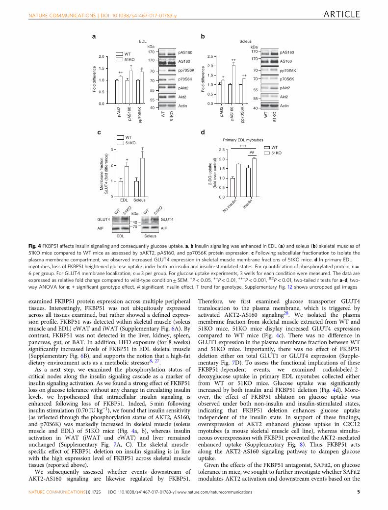
As a next step, we examined the phosphorylation status ofcritical nodes along the insulin signaling cascade as a marker ofinsulin signaling activation. As we found a strong effect of FKBP51loss on glucose tolerance without any change in circulating insulinlevels, we hypothesized that intracellular insulin signaling isenhanced following loss of FKBP51. Indeed, 5 min followinginsulin stimulation (0.70 IU kg−1), we found that insulin sensitivity(as reflected through the phosphorylation status of AKT2, AS160,and p70S6K) was markedly increased in skeletal muscle (soleusmuscle and EDL) of 51KO mice (Fig. 4a, b), whereas insulinactivation in WAT (iWAT and eWAT) and liver remainedunchanged (Supplementary Fig. 7A, C). The skeletal muscle-specific effect of FKBP51 deletion on insulin signaling is in linewith the high expression level of FKBP51 across skeletal muscletissues (reported above).
We subsequently assessed whether events downstream ofAKT2-AS160 signaling are likewise regulated by FKBP51.
Therefore, we first examined glucose transporter GLUT4translocation to the plasma membrane, which is triggered byactivated AKT2-AS160 signaling28. We isolated the plasmamembrane fraction from skeletal muscle extracted from WT and51KO mice. 51KO mice display increased GLUT4 expressioncompared to WT mice (Fig. 4c). There was no difference inGLUT1 expression in the plasma membrane fraction between WTand 51KO mice. Importantly, there was no effect of FKBP51deletion either on total GLUT1 or GLUT4 expression (Supple-mentary Fig. 7D). To assess the functional implications of theseFKBP51-dependent events, we examined radiolabeled-2-deoxyglucose uptake in primary EDL myotubes collected eitherfrom WT or 51KO mice. Glucose uptake was significantlyincreased by both insulin and FKBP51 deletion (Fig. 4d). More-over, the effect of FKBP51 ablation on glucose uptake wasobserved under both non-insulin and insulin-stimulated states,indicating that FKBP51 deletion enhances glucose uptakeindependent of the insulin state. In support of these findings,overexpression of AKT2 enhanced glucose uptake in C2C12myotubes (a mouse skeletal muscle cell line), whereas simulta-neous overexpression with FKBP51 prevented the AKT2-mediatedenhanced uptake (Supplementary Fig. 8). Thus, FKBP51 actsalong the AKT2-AS160 signaling pathway to dampen glucoseuptake.
Given the effects of the FKBP51 antagonist, SAFit2, on glucosetolerance in mice, we sought to further investigate whether SAFit2modulates AKT2 activation and downstream events based on the
a
c
b
++
+ ++
WT51KO
+
+++
pAS160
AS160
pp70S6K
p70S6K
pAkt2
Akt2
Actin
pAS160
AS160
p70S6K
pAkt2
Akt2
Actin
pp70S6K
EDL Soleus0
1
2
3
WT51KO
+
T
Mem
bran
e fr
actio
nG
LUT
4 (f
old
diffe
renc
e)
WT
51KO
51KO
GLUT4
AIF
Soleus
WT
GLUT4
AIF
EDL
dPrimary EDL myotubes
0.0
0.5
1.0
1.5
2.0
2.5 WT51KO##
+++
2-D
G u
ptak
e (f
old
over
con
trol
)
kDa
40
55
55
70
70
170
170kDa
40
55
55
70
70
170
170
40
kDa
70
Fol
d di
ffere
nce
2.0
1.5
1.0
0.5
0.0
pAkt
2
pAS
160
pp70
S6K
pAkt
2
pAS
160
pp70
S6K
51K
O
WT
51K
O
WT
EDL
Fol
d di
ffere
nce
2.5
2.0
1.5
1.0
0.5
0.0
Soleus
No ins
ulin
Insu
lin
Fig. 4 FKBP51 affects insulin signaling and consequently glucose uptake. a, b Insulin signaling was enhanced in EDL (a) and soleus (b) skeletal muscles of51KO mice compared to WT mice as assessed by pAKT2, pAS160, and pp70S6K protein expression. c Following subcellular fractionation to isolate theplasma membrane compartment, we observed increased GLUT4 expression in skeletal muscle membrane fractions of 51KO mice. d In primary EDLmyotubes, loss of FKBP51 heightened glucose uptake under both no insulin and insulin-stimulated states. For quantification of phosphorylated protein, n=6 per group. For GLUT4 membrane localization, n= 3 per group. For glucose uptake experiments, 3 wells for each condition were measured. The data areexpressed as relative fold change compared to wild-type condition± SEM. +P< 0.05, ++P< 0.01, +++P< 0.001, ##P< 0.01, two-tailed t tests for a–d, two-way ANOVA for e; + significant genotype effect, # significant insulin effect, T trend for genotype. Supplementary Fig. 12 shows uncropped gel images
NATURE COMMUNICATIONS | DOI: 10.1038/s41467-017-01783-y ARTICLE
NATURE COMMUNICATIONS |8: 1725 |DOI: 10.1038/s41467-017-01783-y |www.nature.com/naturecommunications 5
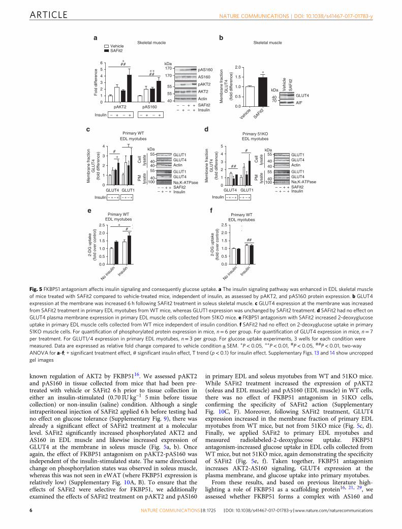
known regulation of AKT2 by FKBP5116. We assessed pAKT2and pAS160 in tissue collected from mice that had been pre-treated with vehicle or SAFit2 6 h prior to tissue collection ineither an insulin-stimulated (0.70 IU kg−1 5 min before tissuecollection) or non-insulin (saline) condition. Although a singleintraperitoneal injection of SAFit2 applied 6 h before testing hadno effect on glucose tolerance (Supplementary Fig. 9), there wasalready a significant effect of SAFit2 treatment at a molecularlevel. SAFit2 significantly increased phosphorylated AKT2 andAS160 in EDL muscle and likewise increased expression ofGLUT4 at the membrane in soleus muscle (Fig. 5a, b). Onceagain, the effect of FKBP51 antagonism on pAKT2-pAS160 wasindependent of the insulin-stimulated state. The same directionalchange on phosphorylation states was observed in soleus muscle,whereas this was not seen in eWAT (where FKBP51 expression isrelatively low) (Supplementary Fig. 10A, B). To ensure that theeffects of SAFit2 were selective for FKBP51, we additionallyexamined the effects of SAFit2 treatment on pAKT2 and pAS160
in primary EDL and soleus myotubes from WT and 51KO mice.While SAFit2 treatment increased the expression of pAKT2(soleus and EDL muscle) and pAS160 (EDL muscle) in WT cells,there was no effect of FKBP51 antagonism in 51KO cells,confirming the specificity of SAFit2 action (SupplementaryFig. 10C, F). Moreover, following SAFit2 treatment, GLUT4expression increased in the membrane fraction of primary EDLmyotubes from WT mice, but not from 51KO mice (Fig. 5c, d).Finally, we applied SAFit2 to primary EDL myotubes andmeasured radiolabeled-2-deoxyglucose uptake. FKBP51antagonism-increased glucose uptake in EDL cells collected fromWT mice, but not 51KO mice, again demonstrating the specificityof SAFit2 (Fig. 5e, f). Taken together, FKBP51 antagonismincreases AKT2-AS160 signaling, GLUT4 expression at theplasma membrane, and glucose uptake into primary myotubes.
From these results, and based on previous literature high-lighting a role of FKBP51 as a scaffolding protein16, 21, 29, weassessed whether FKBP51 forms a complex with AS160 and
Skeletal muscle
0
1
2
3
4
5
6
– + – + – + – +
##
##
++
+
Skeletal muscle
pAS160
AS160
pAKT2
AKT2
Actin
SAFit2Insulin
– – + +– ++ –
+
GLUT4
AIF
c dPrimary WT
EDL myotubes
GLUT1GLUT4Actin
GLUT4Na,K-ATPaseSAFit2Insulin
– – + +– ++ –
GLUT1
Cel
lly
sate
P
Mly
sate
Cel
lly
sate
P
Mly
sate
0
1
2
3
4
– + – +
+#
T
– + – +
GLUT1GLUT40
1
2
3
4
5
##
#
– + – + – + – +
GLUT1GLUT4
GLUT1GLUT4Actin
GLUT4Na,K-ATPaseSAFit2Insulin
+–– – +
++ –
GLUT1
0.0
0.5
1.0
1.5
2.0
2.5#
+
2-D
G u
ptak
e(f
old
over
con
trol
)
Primary WTEDL myotubes
##
ePrimary WT
EDL myotubes
f
kDa170
170
55
55
40
kDa
4070
kDa
40
40
40
55
55
100
kDa
40
40
40
55
55
100Mem
bran
e fr
actio
nG
LUT
4(f
old
diffe
renc
e)
Mem
bran
e fr
actio
nG
LUT
4(f
old
diffe
renc
e)
VehicleSAFit2
Fol
d di
ffere
nce
pAKT2 pAS160
Insulin
Mem
bran
e fr
actio
nG
LUT
4(f
old
diffe
renc
e)
2.0
1.5
1.0
0.5
0.0
SAFit2
Vehicl
e
Veh
icle
SA
Fit2
Insulin
No ins
ulin
Insu
lin
No ins
ulin
Insu
lin
2-D
G u
ptak
e(f
old
over
con
trol
)
2.5
2.0
1.5
1.0
0.5
0.0
a b
Primary 51KOEDL myotubes
Insulin
Fig. 5 FKBP51 antagonism affects insulin signaling and consequently glucose uptake. a The insulin signaling pathway was enhanced in EDL skeletal muscleof mice treated with SAFit2 compared to vehicle-treated mice, independent of insulin, as assessed by pAKT2, and pAS160 protein expression. b GLUT4expression at the membrane was increased 6 h following SAFit2 treatment in soleus skeletal muscle. c GLUT4 expression at the membrane was increasedfrom SAFit2 treatment in primary EDL myotubes fromWT mice, whereas GLUT1 expression was unchanged by SAFit2 treatment. d SAFit2 had no effect onGLUT4 plasma membrane expression in primary EDL muscle cells collected from 51KO mice. e FKBP51 antagonism with SAFit2 increased 2-deoxyglucoseuptake in primary EDL muscle cells collected from WT mice independent of insulin condition. f SAFit2 had no effect on 2-deoxyglucose uptake in primary51KO muscle cells. For quantification of phosphorylated protein expression in mice, n= 6 per group. For quantification of GLUT4 expression in mice, n= 7per treatment. For GLUT1/4 expression in primary EDL myotubes, n= 3 per group. For glucose uptake experiments, 3 wells for each condition weremeasured. Data are expressed as relative fold change compared to vehicle condition± SEM. +P< 0.05, ++P< 0.01, #P< 0.05, ##P< 0.01, two-wayANOVA for a–f; + significant treatment effect, # significant insulin effect, T trend (p< 0.1) for insulin effect. Supplementary Figs. 13 and 14 show uncroppedgel images
ARTICLE NATURE COMMUNICATIONS | DOI: 10.1038/s41467-017-01783-y
6 NATURE COMMUNICATIONS | 8: 1725 |DOI: 10.1038/s41467-017-01783-y |www.nature.com/naturecommunications

furthermore assessed whether SAFit2 modulates this interaction.Immunoprecipitation reactions, using protein extracts from 30-day vehicle-treated and SAFit2-treated mice, revealed that SAFit2treatment strengthened the binding between AKT2 and AS160(Fig. 6a, b, bottom). This is in line with increased glucose uptakegiven that AKT2 inactivates AS160, which in turn stimulates thetranslocation of GLUT4 to the plasma membrane28. Wefurthermore observed a novel interaction between FKBP51 andAS160, and confirmed that SAFit2 disrupts this interaction(Fig. 6a, b, top). SAFit2, by contrast, had no effect on the bindingstrength between AKT2 and PHLPP1 (the negative regulator ofAKT2). We confirmed that the phosphorylation state of AKT2and AS160 does not interfere with the interaction betweenFKBP51 and either AKT2 or AS160 by also examining pAKT2and pAS160. Co-immunoprecipitation does not discriminatebetween direct and indirect protein interactions, and it is possiblethat intermediate proteins are also involved in the interactionbetween FKBP51 and AS160. Regardless, these findings suggestthat FKBP51 associates (directly or indirectly) with AS160 toreduce glucose uptake. By contrast, SAFit2 antagonizes FKBP51and reduces the binding between FKBP51 and AS160 to promotea steric arrangement that favors glucose uptake.
We have shown that FKBP51 regulates insulin signalingselectively in skeletal muscle. Yet FKBP51 is nonethelessexpressed in select WAT depots, albeit at lower levels. In orderto account for the tissue-specific effects of FKBP51, wefurthermore investigated the expression profile of FKBP52, astructural homolog of FKBP51 that competes as a scaffoldingprotein, resulting in opposite functional effects30. Interestingly,the expression profile between FKBP51 and FKBP52 acrossWATs and skeletal muscle are very distinct and provide anexplanation for the FKBP51 skeletal muscle-selective effects.
Specifically, FKBP51 expression is relatively high in skeletalmuscle compared to WATs (Supplementary Fig. 6A). In starkcontrast, levels of FKBP52 are high in WAT depots relative toskeletal muscle. (Supplementary Fig. 11A). We additionallyperformed co-immunoprecipitation reactions with FKBP52 toinvestigate whether FKBP51 and FKBP52 compete for binding toAKT2. We demonstrate that similar to FKBP51, FKBP52 is incomplex with AKT2 and AS160 (Supplementary Fig. 11B).However, FKBP52 does not likewise interact with PHLPP1(Supplementary Fig. 11B), the negative regulator of AKT2, andconsequently has divergent effects on downstream AKT2signaling. Specifically, where ectopic overexpression of FKBP51significantly decreased pAKT2 and pAS160 expression in C2C12myotubes, simultaneous overexpression of FKBP52 abolished theeffects of ectopic FKBP51 (Supplementary Fig. 10C). Takentogether, FKBP51 and FKBP52 compete for binding with AKT2.The distinct expression profile and functional outcomes ofFKBP51 and FKBP52 are responsible for the skeletal muscle-specific effects of SAFit2, which selectively antagonizes FKBP51.
DiscussionHere we describe that loss of FKBP51 in mice markedly improvesmetabolism and especially improves glucose tolerance under bothcontrol and HFD conditions. This is in line with earlier pre-clinical and human studies, which identified an associationbetween FKBP51 ablation and FKBP5 SNPs on traits related tobody weight regulation and T2D, respectively14, 15. Although theeffects of FKBP51 loss are witnessed under dietary control con-ditions, the effects are significantly accentuated when mice arechallenged to an HFD, which acts as a metabolic stressor8. Thissupports a large body of literature, demonstrating that an
IgG
Vehicl
e
AS160
PHLPP
SAFit2
FKBP51
AKT2
FKBP51
AS160
AKT2
PHLPP
Inpu
tF
KB
P51
-IP
AKT2
AS160
FKBP51
PHLPP
AK
T2-
IP
IgG
AS160
PHLPP
FKBP51
AKT2
FKBP51
AS160
AKT2
PHLPP
Inpu
tF
KB
P51
-IP
AKT2
AS160
FKBP51
PHLPP
AK
T2-
IP
Soleus
IgG
Vehicl
e
pAS160
pAKT2
SAFit2
pAS160
pAKT2
Inpu
tF
KB
P51
IP
Vehicl
e
SAFit2
IgG
Vehicl
e
pAS160
pAKT2
SAFit2
pAS160
pAKT2
Inpu
tF
KB
P51
IP
++
+
EDL
+
kDa170
170
55
55
55170
55170
55
55
170
170
170
17055
55
kDa170
170
55
55
55
170
55
170
55
55
170
170
kDa 170
17055
55
kDa
AKT2
PHLPP1AS160
Vehicle SAFit2
1.5
1.0
0.5
0.0Fol
d bi
ndin
g to
FK
BP
51(c
ompa
red
to v
ehic
le)
FKBP51AS160PHLPP1
Fol
d bi
ndin
g to
Akt
2(c
ompa
red
to v
ehic
le) 2.0
0.0
1.5
1.0
0.5
SAFit2Vehicle
Akt2
PHLPP1AS160
1.5
1.0
0.5
0.0Fol
d bi
ndin
g to
FK
BP
51(c
ompa
red
to v
ehic
le)
Vehicle SAFit2
FKBP51AS160PHLPP1
Fol
d bi
ndin
g to
Akt
2(c
ompa
red
to v
ehic
le) 2.0
1.5
1.0
0.5
0.0Vehicle SAFit2
a b
Fig. 6 FKBP51 antagonism affects AKT2-AS160 signaling complex. Tissue lysates from 30-day vehicle-treated or SAFit2-treated mice exposed to HFD wereimmunoprecipitated with anti-AKT2 and anti-FKBP51 and then analyzed by Western blot using FKBP51, (p)AKT2, (p)AS160, and PHLPP1. a, bImmunoprecipitation reactions revealed that SAFit2 treatment increased binding between (p)AKT2 and (p)AS160 in soleus (a) and EDL (b) muscles, whilesimultaneously decreased binding between FKBP51 and AS160 in both muscle types. For co-immunoprecipitation experiments n= 3 per group. Data areexpressed as relative fold change compared to vehicle condition± SEM. +P< 0.05, two-tailed t tests for a, b; + significant SAFit2 treatment effect.Supplementary Fig. 15 shows uncropped gel images
NATURE COMMUNICATIONS | DOI: 10.1038/s41467-017-01783-y ARTICLE
NATURE COMMUNICATIONS |8: 1725 |DOI: 10.1038/s41467-017-01783-y |www.nature.com/naturecommunications 7
environmental challenge is a prerequisite for FKBP51-mediatedoutcomes. For example, early life trauma (i.e., environmentalchallenge) increases the risk of various psychiatric disordersselectively in FKBP5 risk allele carriers, which are associated withincreased FKBP51 protein levels7, 24, 25, 31–33. In rodent studies,51KO mice present no overt phenotype under basal conditions,yet show improved stress resilience following either acute stress23
or chronic stress22. Indeed, stressors induce FKBP5 expression,and this may underlie the more pronounced effects of FKBP51deletion on metabolic phenotypes seen in the current study. Wecertainly found that an HFD increases levels of FKBP51 in fat andskeletal muscle. Taken together, previous findings have found thathigher levels of FKBP51 are associated with poorer outcomes instress-related psychiatric disorders. The present study shows thathigher levels of FKBP51 are likewise detrimental to metabolichealth, especially when confronted with environmental challenges(i.e., an obesogenic environment).
Selective pharmacological antagonism of FKBP51 has onlyrecently been realized20. Selectivity for FKBP51 is especiallyimportant since its structural homolog, FKBP52, acts as a func-tional opponent. In fact, it was this structural similarity thatinitially hampered drug discovery for FKBP51. The current abilityto antagonize FKBP51 offers new opportunities for drug devel-opment. To date, only three studies have investigated the effectsof FKBP51 antagonism on functions related to FKBP51, and nostudy has yet been investigated for long-term (i.e., 30 day)applicability. These previous studies independently found thatFKBP51 antagonism induces anxiolytic effects26, reduces theseverity of pain symptoms34, and opposes the known ability ofFKBP51 to promote NFκB signaling35. Nevertheless, FKBP51 is amulti-domain protein36, and it remains unknown whether theFKBP51 antagonist, SAFit2, blocks all functions of FKBP51.Therefore, in order to address whether pharmacological antag-onism affects metabolic function, mice were treated with SAFit2once (slow release formula) or repeatedly for either 10 or 30 days.Administration of SAFit2 paralleled the metabolic phenotypearising from total genetic loss of FKBP51. Acute SAFit2 treatmentimproved glucose tolerance under metabolically challengingconditions (i.e., HFD conditions). Under metabolic control con-ditions (i.e., regular diet), FKBP51 blockade improved glucosetolerance as early as 8 days following treatment onset. Impor-tantly, the effects of FKBP51 modulation on glucose tolerancewere not secondary to changes in body weight since neither asingle nor a 10-day SAFit2 exposure had an effect on body weight.It is possible that our study was underpowered to detect themodest effects of SAFit2 treatment on body weight, since the 10-day SAFit2-treated group showed a lower (non-significant) bodyweight phenotype compared to the vehicle-treated counterparts.Regardless, these data support our studies in 51KO mice, whichcollectively indicate that FKBP51 is an integral component ofglucose homeostatic regulation, particularly in response tonutritional changes. In the context of body weight regulation, ourfindings that 30 days of SAFit2 treatment protects against HFD-induced weight gain supports the findings of a recently publishedpaper demonstrating that 51KO mice resist HFD-induced weightgain and present increased UCP1 in WAT15. One limitation ofour study is that we do not know the minimal effective dose ofSAFit2 required to improve glucose tolerance. Nevertheless, theimproved metabolic outcomes following systemic administrationof the selective FKBP51 antagonist clearly demonstrate theapplicability in a clinical setting.
It is well established that FKBP51 is able to regulate manysignaling pathways through direct protein–protein interactions16,21, 37, 38. Through these interactions, FKBP51 has been implicatedin various disease states (i.e., cancers, psychiatric disorders) andin the response to medications (i.e., chemotherapies,
antidepressants). For example, through the FKBP51-dependentregulation of AKT, Pei et al. (2009) reported that FKBP51 reducestumor growth16. FKBP51 binds to both AKT1 and AKT2 iso-forms and their corresponding negative regulators PHLPP2 andPHLPP1, to ultimately favor Akt inactivation. Interestingly, whileAkt1 is best known for its regulation of cell growth39, 40, AKT2 isbest known for its regulation of glucose homeostasis17, 41.Accordingly, in the present study, we determined that FKBP51 isalso important for glucose disposal through the regulation ofAKT2 and downstream AS160 (AKT substrate of 160 kDa), animportant signaling protein involved in insulin-stimulated glu-cose transport in skeletal muscle28, 42. 51KO mice exhibitenhanced insulin signaling, as interpreted from the increasedphosphorylation of AKT2, AS160, and p70S6K. Interestingly, theFKBP51-dependent effects on insulin signaling observed in micewere highly tissue-specific, in which FKBP51-dependent increasesin insulin signaling was limited to skeletal muscle. Indeed, skeletalmuscle accounts for an estimated 80% of postprandial glucosedisposal and is regarded as a principal site responsible for themaintenance of glucose homeostasis43, 44. In this context, we alsofound that primary EDL myotubes from 51KO mice exhibitedheightened glucose uptake compared to WT EDL myotubes.Ectopic FKBP51 overexpression furthermore completely reversedthe enhanced glucose uptake arising from AKT2 overexpressionin cultured myotubes, demonstrating that FKBP51 regulation ofinsulin signaling is critical for glucose uptake.
The FKBP51-dependent effects on the phosphorylation ofAKT2 and AS160 as well as on glucose uptake were evident underboth non-insulin- and insulin-stimulated states, suggesting thatFKBP51 acts independent of insulin to improve glucose uptakeand whole body glucose homeostasis. Follow-up studies shouldaddress whether FKBP51 is involved in insulin-independentglucose uptake through the regulation of auxiliary pathways. Forexample, AMP-activated protein kinase (AMPK) is a well-knownregulator of insulin-independent glucose uptake, leading toincreased AS160 phosphorylation and GLUT4 translocation inthe skeletal muscle45, 46. Furthermore, glucocorticoids are potentregulators of glucose homeostasis, and they have been shown toreduce insulin-stimulated glucose transport in muscle by blockingthe recruitment of GLUT4 to the cell surface47. Indeed, FKBP51is known to reduce glucocorticoid receptor sensitivity1, andtherefore glucocorticoid signaling is another strong candidatepathway by which FKBP51 may regulate whole body glucosehomeostasis. The data herein nevertheless provide unequivocalevidence that FKBP51 is a novel regulator of AKT2-AS160 sig-naling and glucose uptake.
Not only did FKBP51 antagonism parallel the metabolic phe-notype arising from FKBP51 deletion, but it furthermore paral-leled the molecular events induced by FKBP51 loss. SAFit2treatment strongly induced AKT2 and AS160 phosphorylationand led to increased GLUT4 expression at the plasma membrane.Although we suspect that enhanced plasma membrane GLUT4expression arises from pAKT2-pAS160-mediated increasedGLUT4 translocation to the membrane, it is possible that SAFit2treatment also increases the total expression level of GLUT4. At afunctional level, SAFit2 heightened glucose uptake assessed inprimary muscle cells. Importantly, SAFit2 action on glucose tol-erance and glucose uptake was highly specific for FKBP51 sinceneither 51KO mice nor primary muscle cells collected from 51KOmice responded to SAFit2 treatment. The robust effects ofFKBP51 antagonist SAFit2 on whole body glucose homeostasisand skeletal muscle glucose uptake led us to examine whetherSAFit2 disrupts the well-characterized interaction betweenFKBP51, AKT2, and PHLPP116. To our surprise we found noeffect of SAFit2. Rather, we discovered a novel interactionbetween FKBP51 and AS160, which can be disrupted by FKBP51
ARTICLE NATURE COMMUNICATIONS | DOI: 10.1038/s41467-017-01783-y
8 NATURE COMMUNICATIONS | 8: 1725 |DOI: 10.1038/s41467-017-01783-y |www.nature.com/naturecommunications
antagonism using SAFit2. This agrees nicely with our findingsthat SAFit2 treatment increases glucose uptake in primary myo-tubes and improves glucose tolerance in mice. Moreover thissupports accumulating evidence that FKBP51 has importantscaffolding properties to organize and concentrate various sig-naling complexes16, 21, 37.
An important question raised by this study is how exactlyFKBP51 acts within distinct cellular/tissue environments to affectwhole body energy and glucose homeostasis. Previous studieshave already alluded to the importance of tissue-specific actionsof FKBP5126, 48. Here we extend our current understanding of thetissue-specific actions of FKBP51 to include FKBP51-dependentregulation of insulin signaling exclusively within skeletal muscle.Despite microarray-based data demonstrating that skeletal muscleshows the second strongest expression profile of FKBP5 across alltissues examined12, 14, to our knowledge, the current study is thefirst to define a skeletal muscle-specific role for FKBP51. Thedistinct expression profiles of FKBP51 and FKBP52 provide anexplanation as to why FKBP51 affects glucose uptake exclusivelywithin skeletal muscle. Although FKBP52 shows strong
expression in WAT, it is expressed minimally in skeletal muscle.We found that FKBP52 competes with FKBP51 for binding toAKT2 in WAT, but this competition is minimal in skeletal musclebased on its expression profile. Importantly, although FKBP52 isfound in complex with AKT2 and AS160, it is not found tointeract with the negative regulator of AKT2, PHLPP1, and thusdoes not have the same functional implications on downstreamAKT2 signaling compared to FKBP51.
Our study focused on the effects of FKBP51 modulation onglucose homeostasis and pAKT2-pAS160 signaling, effects whichwere independent of body weight. Nevertheless, we also reportedthat 51KO mice are resistant to diet-induced obesity, present ahigher resting metabolic rate, and a slight increase in home-cageactivity. In support of these findings, a recent report demon-strated that UCP1 expression is enhanced in select WAT depot in51KO mice, which contributes to the improved body weightphenotype15. Our finding that 51KO mice also presentedincreased home-cage activity may furthermore underlie theimproved body weight phenotype. Although we found no dif-ference in activity-related energy expenditure, the increasedactivity in 51KO mice suggests that 51KO mice may haveincreased exercise efficacy. Follow-up studies are needed toaddress such effects of FKBP51 on exercise efficacy.
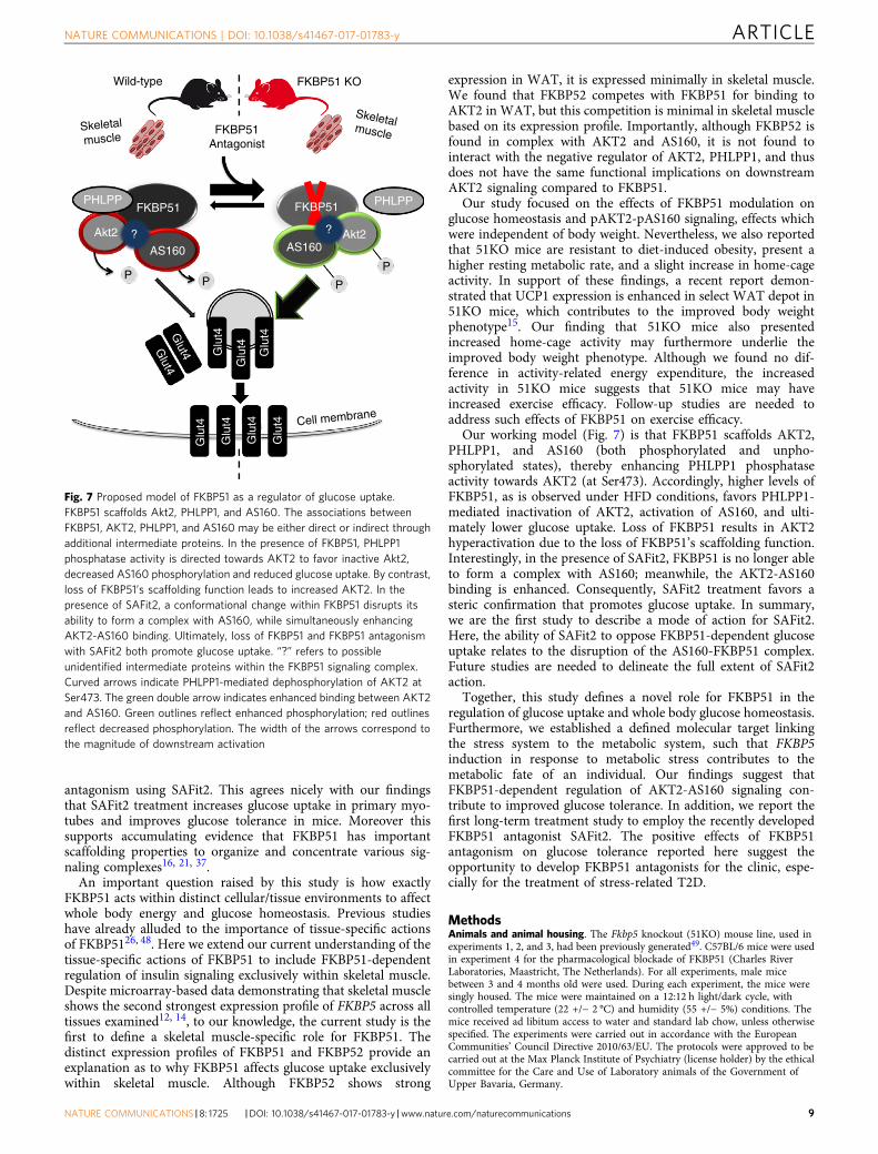
Our working model (Fig. 7) is that FKBP51 scaffolds AKT2,PHLPP1, and AS160 (both phosphorylated and unpho-sphorylated states), thereby enhancing PHLPP1 phosphataseactivity towards AKT2 (at Ser473). Accordingly, higher levels ofFKBP51, as is observed under HFD conditions, favors PHLPP1-mediated inactivation of AKT2, activation of AS160, and ulti-mately lower glucose uptake. Loss of FKBP51 results in AKT2hyperactivation due to the loss of FKBP51’s scaffolding function.Interestingly, in the presence of SAFit2, FKBP51 is no longer ableto form a complex with AS160; meanwhile, the AKT2-AS160binding is enhanced. Consequently, SAFit2 treatment favors asteric confirmation that promotes glucose uptake. In summary,we are the first study to describe a mode of action for SAFit2.Here, the ability of SAFit2 to oppose FKBP51-dependent glucoseuptake relates to the disruption of the AS160-FKBP51 complex.Future studies are needed to delineate the full extent of SAFit2action.
Together, this study defines a novel role for FKBP51 in theregulation of glucose uptake and whole body glucose homeostasis.Furthermore, we established a defined molecular target linkingthe stress system to the metabolic system, such that FKBP5induction in response to metabolic stress contributes to themetabolic fate of an individual. Our findings suggest thatFKBP51-dependent regulation of AKT2-AS160 signaling con-tribute to improved glucose tolerance. In addition, we report thefirst long-term treatment study to employ the recently developedFKBP51 antagonist SAFit2. The positive effects of FKBP51antagonism on glucose tolerance reported here suggest theopportunity to develop FKBP51 antagonists for the clinic, espe-cially for the treatment of stress-related T2D.
MethodsAnimals and animal housing. The Fkbp5 knockout (51KO) mouse line, used inexperiments 1, 2, and 3, had been previously generated49. C57BL/6 mice were usedin experiment 4 for the pharmacological blockade of FKBP51 (Charles RiverLaboratories, Maastricht, The Netherlands). For all experiments, male micebetween 3 and 4 months old were used. During each experiment, the mice weresingly housed. The mice were maintained on a 12:12 h light/dark cycle, withcontrolled temperature (22 +/− 2 °C) and humidity (55 +/− 5%) conditions. Themice received ad libitum access to water and standard lab chow, unless otherwisespecified. The experiments were carried out in accordance with the EuropeanCommunities’ Council Directive 2010/63/EU. The protocols were approved to becarried out at the Max Planck Institute of Psychiatry (license holder) by the ethicalcommittee for the Care and Use of Laboratory animals of the Government ofUpper Bavaria, Germany.
Wild-type
PHLPP PHLPPFKBP51 FKBP51
PP
PP
Glu
t4
Glu
t4
Glu
t4G
lut4
Glu
t4 Cell membrane
Glu
t4
Glu
t4
Glut4G
lut4
AS160 AS160Akt2 Akt2? ?
FKBP51Antagonist
FKBP51 KO
Skeletal
muscle
Skeletalmuscle
Fig. 7 Proposed model of FKBP51 as a regulator of glucose uptake.FKBP51 scaffolds Akt2, PHLPP1, and AS160. The associations betweenFKBP51, AKT2, PHLPP1, and AS160 may be either direct or indirect throughadditional intermediate proteins. In the presence of FKBP51, PHLPP1phosphatase activity is directed towards AKT2 to favor inactive Akt2,decreased AS160 phosphorylation and reduced glucose uptake. By contrast,loss of FKBP51’s scaffolding function leads to increased AKT2. In thepresence of SAFit2, a conformational change within FKBP51 disrupts itsability to form a complex with AS160, while simultaneously enhancingAKT2-AS160 binding. Ultimately, loss of FKBP51 and FKBP51 antagonismwith SAFit2 both promote glucose uptake. “?” refers to possibleunidentified intermediate proteins within the FKBP51 signaling complex.Curved arrows indicate PHLPP1-mediated dephosphorylation of AKT2 atSer473. The green double arrow indicates enhanced binding between AKT2and AS160. Green outlines reflect enhanced phosphorylation; red outlinesreflect decreased phosphorylation. The width of the arrows correspond tothe magnitude of downstream activation
NATURE COMMUNICATIONS | DOI: 10.1038/s41467-017-01783-y ARTICLE
NATURE COMMUNICATIONS |8: 1725 |DOI: 10.1038/s41467-017-01783-y |www.nature.com/naturecommunications 9

Indirect calorimetry and body composition. The direct effects of FKBP51 defi-ciency on metabolic parameters were investigated using 51KO (n = 16) and wild-type (WT) (n= 18) mice. Body composition (fat and lean mass) was assessed usingwhole body magnetic resonance imaging (Echo-MRI, Houston, TX). Thereafter,the mice were surgically implanted with a telemetric transponder (Respironics,Murrysville, PA) for the measurement of core body temperature. The mice wereallowed to recover for approximately 2 weeks before any metabolic recordings wereperformed. Indirect calorimetry and telemetry were performed on mice underchow conditions (TSE PhenoMaster, TSE Systems, Bad Homburg, Germany). Forexperimental details, see Supplemental Information. Each genotype group wassubsequently divided into a chow diet and high-fat diet (HFD) (58% kcal from fat,D12331, Research Diets, New Brunswick, NJ, USA) group, matched for bodyweight. Body weight was measured throughout the experiment. After 8 weeks onthe respective diets, 51KO and WT mice were killed. Epididymal (e), inguinal (i),and perirenal (p) WAT were collected and weighed; brown adipose tissue (BAT)was collected and weighed.
Thermoregulation. Body weight and body composition were examined in 51KOand WT mice (n = 8 per genotype) under HFD conditions at 30 °C to minimize theeffects of thermal stress. A separate cohort of 51KO and WT mice were exposed to6 h of cold exposure (4 °C) to assess cold-induced thermoregulation under bothcontrol and HFD conditions (see Supplemental Information).
Pair-feeding. To assess the contribution of food intake on body weight regulationin 51KO and WT mice, a pair-feeding experiment was performed. For experi-mental details, refer to Supplemental Information.
Glucose tolerance and insulin tolerance. Glucose tolerance and insulin tolerancewere investigated in 51KO (n= 25) and WT mice (n = 18). Briefly, 51KO and WTmice were initially divided into a control diet group and an HFD group, matchedfor body weight. After 8 weeks on the dietary treatment, the mice were subjected toa glucose tolerance test (GTT). Additionally, blood was collected to assess fastinginsulin and glucose-stimulated insulin levels. One week thereafter, an insulin tol-erance test (ITT) was performed. See Supplemental Information for more details.
SAFit2 administration. To determine whether antagonizing FKBP51 may be aneffective anti-obesity and/or diabetic therapeutic strategy, we treated mice with anantagonist of FKBP51, known as SAFit220. SAFit2 or vehicle was administeredeither acutely as a slow releasing vesicular phospholipid gel (VPG) (2 mg SAFit2 orvehicle) or repeatedly by intraperitoneal (i.p.) injections (20 mg kg−1 SAFit2 orvehicle) twice daily. For i.p. injections, SAFit2 was solubilized in vehicle containing4% ethanol, 5% Tween80, and 5% PEG400 in 0.9% saline. Body weight and foodintake were measured daily throughout the treatment periods. VPGs were com-posed of 50% (m/m) egg-lecithin containing at least 80% phosphatidylcholine(Lipoid E80, Lipoid GmbH, Ludwigshafen, Germany) and 10 mM phosphatebuffered saline (PBS), pH 7.4, and were prepared by a dual asymmetric cen-trifugation technique50. SAFit2 was encapsulated in the formulation by a directincorporation method. See Supplemental Information for more details.
Acute SAFit2 treatment. Male C57BL/6 mice were divided into vehicle-treatedand SAFit2-treated groups matched for body weight (n = 12 per group). Ontreatment day 1, mice were administered (subcutaneous injection between theshoulders) a slow release-formulated gel containing either SAFit2 or vehicle. After48 h, a GTT was performed following an overnight fast.
Sub-chronic SAFit2 treatment. One day before the treatment period, maleC57BL/6 mice were divided into a vehicle-treated group and a SAFit2-treatedgroup matched for body weight (n= 8 per group). On treatment day 7, locomotoractivity was assessed in the open field test. On treatment day 8, a GTT was per-formed. SAFit2 levels were assessed in plasma from blood taken at the time ofkilling. The animals were killed on day 10 following the treatment onset.
Chronic SAFit2 treatment. Four weeks before treatment onset, male C57BL/6mice were divided into a control diet group (n= 25) and an HFD group (n= 25)matched for body weight. One day before the treatment period, mice of eachdietary group were further subdivided into a vehicle-treated group and a SAFit2-treated group matched for body weight. SAFit2 or vehicle were administered twicedaily for 30 days. On treatment days 10 and 30, SAFit2 levels were assessed inplasma. The open field, dark–light transition, and elevated plus-maze behavioraltests were performed on treatment days 15, 16, and 17, respectively. The GTT wasperformed on treatment day 25 and the ITT on treatment day 29. The animalswere killed on day 31 following treatment onset; tissues were collected and stored at−80 °C for further analyses.
Tissue collection. Mice were anesthetized with isoflurane and immediately killedby decapitation. Basal trunk blood was collected and subsequently processed(plasma was collected and stored at −20 °C). Skeletal muscle (extensor digitorum
longus (EDL) and soleus), WAT (iWAT, eWAT, and pWAT), and liver werecollected and stored at −80 °C until used.
Cell lines. C2C12 myoblasts were maintained in Dulbecco’s modified Eagle’smedium (DMEM) supplemented with 10% fetal bovine serum and 1x penicillinstreptomycin antibiotics at 37 °C in a humidified atmosphere with 5% CO2. Oncethe cells reached ~90% confluency, C2C12 myoblasts were detached from the plateand 2 × 10⁶ cells were re-suspended in 100 µl of transfection buffer (50 mM HEPES[pH 7.3], 90 mM NaCl, 5 mM KCl, and 0.15 mM CaCl2). A total of 2.5 µg ofplasmid DNA was used per transfection. Plasmids expressing AKT2-HA andFKBP51-FLAG or GFP (control) have been described previously21, 51. Ectopicoverexpression of AKT2 and FKBP51 resulted in a 3.2-fold and 2.8-fold increase intheir expression, respectively. Electroporation was performed using the AmaxaNucleofector system (program #T-032). The cells were re-seeded onto 0.75%gelatin-coated 12-well plates at a density of ~ 105 cells per cm2. Transfected cellswere induced to differentiate once they reached 90% confluency. Differentiationwas induced by switching the growth medium to DMEM containing 2% horseserum for 3 days.
Primary EDL myotubes were prepared from satellite cells collected from soleusmuscle and EDL myofibers of 4- to 8-week old WT and 51KO mice as describedpreviously52. Briefly, dissected muscles were washed in warm 1 x PBS andsubsequently were digested in Collagenase 1 at 37 °C for 1.5 h. Thereafter, singlefibers were washed in DMEM with 1% P/S. After washing, the fibers weretransferred to a 60mm plate (coated with 0,75% gelatin) and were allowed toincubate at 37 °C for 3 days in growth medium (DMEM + 20% FBS + 1%P/S). Thesatellite cells were detached from the plate and were re-plated onto 24-well multi-well dishes. The medium was exchanged every 2–3 days and cells were split at90–100% confluency. Differentiation was induced by switching the growth mediumto DMEM containing 5% horse serum for 5 days.
Glucose uptake. Primary EDL myotubes or transfected C2C12 myotubes wereused for glucose uptake experiments. For SAFit2 experiments, a toxicity assay wasinitially performed to determine the appropriate SAFit2 concentration for sub-sequent experiments. Based on a lethal dose of 15 (LD 15), the cells were incubatedwith 0.6 µM SAFit2 or DMSO overnight before inducing glucose uptake.
Basal and insulin-stimulated glucose uptake in primary EDL muscle cells anddifferentiated C2C12 myotubes was examined. Briefly, the cells were serum-starvedin low glucose (1000 mg L−1) DMEM for 4 h, and then incubated in Krebs–Ringer-HEPES (KRH) buffer (136 mM NaCl, 4.7 mM KCl, 10 mM sodium phosphatebuffer, 1 mM MgSO4, 1 mM CaCl2, and 10 mM HEPES, pH 7.4, 0.2% BSA) for 10min. The cells were stimulated with insulin (100 nM) or left unstimulated for 1 h.Glucose uptake was induced by the addition of KRH buffer containing 100 µM 2-deoxy-D-[1,2-3 H]glucose, 2 µCi ml−1 (Perkin Elmer) to each well. After 4 min, thereactions were terminated by washing the cells with ice-cold PSB containing 10 µMcytochalasin B (inhibitor of membrane transporter-dependent glucose transport),and then 2 additional washes with ice-cold 1x PBS. Cells were lysed with 0.1 MNaOH for 30 min, and the incorporated radioactivity was determined by liquidscintillation counting. 2-deoxy-D-[1,2-3 H]glucose uptake was furthermorenormalized to total protein content assessed by the BCA assay (BCA Protein AssayKit, Life Technologies, Darmstadt, Germany).
Antibodies. Detailed information on antibodies and dilutions is provided inSupplementary Information.
GLUT4 membrane localization. Primary EDL myotubes were exposed to 0.6 µMSAFit2 or DMSO (vehicle) overnight. The following day, cells were serum-starvedin low glucose (1000 mg L−1) DMEM for 4 h with SAFit2 or DMSO, and weresubsequently collected for the rapid preparation of the plasma membrane fractionas described previously53. The membrane fraction was used in subsequent Westernblot assays for the detection of GLUT4. For quantification, GLUT4 was normalizedto both Na,K,ATPase (plasma membrane marker) and normalized total GLUT4, asdescribed previously54.
For GLUT4 membrane localization in 51KO (n= 6) and WT (n = 5) mice, themice were fasted for 6 h. Insulin was injected by i.p. administration (0.70 IU kg−1)5 min before mice were anesthetized with isoflurane, and were immediately killedby decapitation. Tissues were collected and stored at −80 °C until used. 2-waysubcellular fractionation was performed as described previously55.
Co-immunoprecipitation (coIP). Immunoprecipitations of endogenous proteinswere performed using protein extracts (n= 3 per group) from soleus muscle, EDL,and eWAT of vehicle-treated and SAFit2-treated mice that were fed HFD. TheCoIP experiments were performed with beads conjugated with rabbit IgG. Briefly,500 µg of lysate was incubated overnight with 2 µg of the appropriate IP-antibody(AKT2 (CST, #2964); FKBP5/FKBP51 (Bethyl, A301-430); and FKBP4/FKBP52(Bethyl, A301-427A) (See Supplementary Table 1) at 4 °C. 20 µL of protein Gdynabeads (Invitrogen, 100-03D) was blocked with bovine serum albumin andsubsequently added to the lysate-antibody mix and allowed to incubate at 4 °C for3 h in order to mediate binding between the dynabeads and the antibody-antigencomplex of interest. The beads were washed three times with ice-cold PBS. The
ARTICLE NATURE COMMUNICATIONS | DOI: 10.1038/s41467-017-01783-y
10 NATURE COMMUNICATIONS | 8: 1725 |DOI: 10.1038/s41467-017-01783-y |www.nature.com/naturecommunications

protein-antibody complexes were eluted with 60 µL Laemmli loading buffer.Thereafter, the eluate was boiled for 5 min at 95 °C. Then 2–5 µL of each immu-noprecipitate reaction product was separated by SDS-PAGE and electro-transferred onto nitrocellulose membranes. For assessing protein complexes,immunoblotting against AKT2, FKBP51, PHLPP1 (Millipore, #07-1341), andAS160 was performed. See Supplemental Information ‘Western blot analysis’ fordetails.
Statistical analysis. Data were analyzed using IBM SPSS Statistics 18 software(IBM SPSS Statistics, IBM, Chicago, IL, USA). The decomposition of total energyexpenditure (TEE) into activity-related energy expenditure (AEE) and restingmetabolic rate (RMR) was performed in MATLAB (The MathWorks, Natick, MA,USA) using a custom-designed toolbox graciously provided by JB van Klinken(Leiden University Medical Center, Leiden, The Netherlands). Body weight wasincluded as a covariate in the analyses of energy expenditure56. Statistical analysesfor all energy expenditure outcome variables, respiratory exchange ratio (RER),home-cage activity, food intake, water intake, and body temperature were per-formed on 24-hour averages. Statistical significance was set at p < 0.05; a statisticaltendency was set at p< 0.1. For interactions at p < 0.1, we also examined lowerorder main effects. Data are presented as the mean +/− SEM.
Data availability. The data herein are available from the corresponding authorsupon reasonable request.
Received: 28 June 2016 Accepted: 12 October 2017
References1. Ratajczak, T., Cluning, C. & Ward, B. K. Steroid receptor-associated
immunophilins: A gateway to steroid signalling. Clin. Biochem. Rev. 36, 31–52(2015).
2. Denny, W. B., Valentine, D. L., Reynolds, P. D., Smith, D. F. & Scammell, J. G.Squirrel monkey immunophilin {FKBP}51 is a potent inhibitor ofglucocorticoid receptor binding. Endocrinology 141, 4107–4113 (2000).
3. Wochnik, G. M. et al. {FK}506-binding proteins 51 and 52 differentiallyregulate dynein interaction and nuclear translocation of the glucocorticoidreceptor in mammalian cells. J. Biol. Chem. 280, 4609–4616 (2005).
4. Vermeer, H., Hendriks-Stegeman, B. I., van der Burg, B., van Buul-Offers, S. C.& Jansen, M. Glucocorticoid-induced increase in lymphocytic {FKBP}51messenger ribonucleic acid expression: a potential marker for glucocorticoidsensitivity, potency, and bioavailability. J. Clin. Endocrinol. Metab 88, 277–284(2003).
5. Jääskeläinen, T., Makkonen, H. & Palvimo, J. J. Steroid up-regulation ofFKBP51 and its role in hormone signaling. Curr. Opin. Pharmacol. 11, 326–331(2011).
6. Riggs, D. L. et al. The Hsp90-binding peptidylprolyl isomerase FKBP52potentiates glucocorticoid signaling in vivo. EMBO. J. 22, 1158–1167 (2003).
7. Zannas, A. S., Wiechmann, T., Gassen, N. C. & Binder, E. B.Gene–stress–epigenetic regulation of FKBP5: clinical and translationalimplications. Neuropsychopharmacology 41, 261–274 (2016).
8. Karalis, K. P. et al. Mechanisms of obesity and related pathology: linkingimmune responses to metabolic stress. FEBS. J. 276, 5747–5754 (2009).
9. Balsevich, G. et al. Interplay between diet-induced obesity and chronic stress inmice: potential role of FKBP51. J. Endocrinol. 222, 15–26 (2014).
10. Guarnieri, D. J. et al. Gene profiling reveals a role for stress hormones in themolecular and behavioral response to food restriction. Biol. Psychiatry 71,358–365 (2012).
11. Scharf, S. H., Liebl, C., Binder, E. B., Schmidt, M. V. & Muller, M. B. Expressionand regulation of the Fkbp5 gene in the adult mouse brain. PLoS ONE 6,e16883 (2011).
12. Su, A. I. et al. A gene atlas of the mouse and human protein-encodingtranscriptomes. Proc. Natl Acad. Sci. USA 101, 6062–6067 (2004).
13. Hartmann, I. B. et al. The FKBP5 polymorphism rs1360780 is associated withlower weight loss after bariatric surgery: 26 months of follow-up. Surg. Obes.Relat. Dis. 12, 1554–1560 (2016).
14. Pereira, M. J. et al. FKBP5 expression in human adipose tissue increasesfollowing dexamethasone exposure and is associated with insulin resistance.Metabolism 63, 1198–1208 (2014).
15. Stechschulte, L. A. et al. FKBP51 null mice are resistant to diet-induced obesityand the PPARγ agonist rosiglitazone. Endocrinology 157, 3888–3900 (2016).
16. Pei, H. et al. FKBP51 affects cancer cell response to chemotherapy by negativelyregulating Akt. Cancer Cell 16, 259–266 (2009).
17. Cho, H. et al. Insulin resistance and a diabetes mellitus-like syndrome in micelacking the protein kinase Akt2 (PKBbeta). Science 292, 1728–1731 (2001).
18. Taniguchi, C. M., Emanuelli, B. & Kahn, C. R. Critical nodes in signallingpathways: insights into insulin action. Nat. Rev. Mol. Cell Biol. 7, 85–96(2006).
19. Zannas, A. S., Balsevich, G. & Gassen, N. C. The emerging role of FKBP5 in theregulation of metabolism and body weight. Surg. Obes. Relat. Dis. 12,1560–1561 (2016).
20. Gaali, S. et al. Selective inhibitors of the FK506-binding protein 51 by inducedfit. Nat. Chem. Biol. 11, 33–37 (2015).
21. Gassen, N. C. et al. Association of FKBP51 with priming of autophagy pathwaysand mediation of antidepressant treatment response: evidence in cells, mice,and humans. PLoS Med. 11, e1001755 (2014).
22. Hartmann, J. et al. The involvement of FK506-binding protein 51 (FKBP5) inthe behavioral and neuroendocrine effects of chronic social defeat stress.Neuropharmacology 62, 332–339 (2012).
23. Touma, C. et al. FK506 binding protein 5 shapes stress responsiveness:modulation of neuroendocrine reactivity and coping behavior. Biol. Psychiatry70, 928–936 (2011).
24. Zimmermann, P. et al. Interaction of FKBP5 gene variants and adverse lifeevents in predicting depression onset: results from a 10-year prospectivecommunity study. Am. J. Psychiatry 168, 1107–1116 (2011).
25. Klengel, T. et al. Allele-specific FKBP5 DNA demethylation mediates gene-childhood trauma interactions. Nat. Neurosci. 16, 33–41 (2013).
26. Hartmann, J. et al. Pharmacological inhibition of the psychiatric risk factorFKBP51 Has anxiolytic properties. J. Neurosci. 35, 9007–9016 (2015).
27. Tamashiro, K. L., Sakai, R. R., Shively, C. A., Karatsoreos, I. N. & Reagan, L. P.Chronic stress, metabolism, and metabolic syndrome. Stress 14, 468–474(2011).
28. Sano, H. et al. Insulin-stimulated phosphorylation of a Rab GTPase-activatingprotein regulates GLUT4 translocation. J. Biol. Chem. 278, 14599–14602(2003).
29. Fabian, A. K. et al. InterAKTions with FKBPs-mutational and pharmacologicalexploration. PLoS ONE 8, e57508 (2013).
30. Storer, C. L., Dickey, C. A., Galigniana, M. D., Rein, T. & Cox, M. B. FKBP51and FKBP52 in signaling and disease. Trends Endocrinol. Metab. 22, 481–490(2011).
31. Roy, A., Gorodetsky, E., Yuan, Q., Goldman, D. & Enoch, M. A. Interaction ofFKBP5, a stress-related gene, with childhood trauma increases the risk forattempting suicide. Neuropsychopharmacology 35, 1674–1683 (2010).
32. Mehta, D. et al. Using polymorphisms in {FKBP}5 to define biologically distinctsubtypes of posttraumatic stress disorder: evidence from endocrine and geneexpression studies. Arch. Gen. Psychiatry 68, 901–910 (2011).
33. Binder, E. B. et al. Association of FKBP5 polymorphisms and childhood abusewith risk of posttraumatic stress disorder symptoms in adults. JAMA 299,1291–1305 (2008).
34. Maiarù, M. et al. The stress regulator FKBP51 drives chronic pain bymodulating spinal glucocorticoid signaling. Sci. Transl. Med. 8, 1–11 (2016).
35. Romano, S. et al. FKBP51 employs both scaffold and isomerase functions topromote NF-kappaB activation in melanoma. Nucleic Acids Res. 43, 6983–6993(2015).
36. Sinars, C. R. et al. Structure of the large FK506-binding protein FKBP51, anHsp90-binding protein and a component of steroid receptor complexes. Proc.Natl Acad. Sci. USA 100, 868–873 (2003).
37. Gassen, N. C. et al. FKBP51 inhibits GSK3β and augments the effects of distinctpsychotropic medications. Mol. Psychiatry 21, 277–289 (2016).
38. Jiang, W. et al. FK506 binding protein mediates glioma cell growth andsensitivity to rapamycin treatment by regulating NF-kappaB signaling pathway.Neoplasia 10, 235–243 (2008).
39. Chen, W. S. et al. Growth retardation and increased apoptosis in mice withhomozygous disruption of the Akt1 gene. Genes Dev. 15, 2203–2208 (2001).
40. Cho, H., Thorvaldsen, J. L., Chu, Q., Feng, F. & Birnbaum, M. J. Akt1/PKBalpha is required for normal growth but dispensable for maintenance ofglucose homeostasis in mice. J. Biol. Chem. 276, 38349–38352 (2001).
41. Garofalo, R. S. et al. Severe diabetes, age-dependent loss of adipose tissue, andmild growth deficiency in mice lacking Akt2/PKBb. J. Clin. Invest. 112, 197–208(2003).
42. Kramer, H. F. et al. AS160 regulates insulin- and contraction-stimulatedglucose uptake in mouse skeletal muscle. J. Biol. Chem. 281, 31478–31485(2006).
43. DeFronzo, R. A., Gunnarsson, R., Bjorkman, O., Olsson, M. & Wahren, J.Effects of insulin on peripheral and splanchnic glucose metabolism innoninsulin-dependent (type {II}) diabetes mellitus. J. Clin. Invest 76, 149–155(1985).
44. Zierath, J. R. & Wallberg-Henriksson, H. From receptor to effector: insulinsignal transduction in skeletal muscle from type {II} diabetic patients. Ann. N.Y. Acad. Sci. 967, 120–134 (2002).
45. Kurth-Kraczek, E. J., Hirshman, M. F., Goodyear, L. J. & Winder, W. W. 5′AMP-activated protein kinase activation causes GLUT4 translocation inskeletal muscle. Diabetes 48, 1667–1671 (1999).
NATURE COMMUNICATIONS | DOI: 10.1038/s41467-017-01783-y ARTICLE
NATURE COMMUNICATIONS |8: 1725 |DOI: 10.1038/s41467-017-01783-y |www.nature.com/naturecommunications 11
46. Treebak, J. T. et al. AMPK-mediated AS160 phosphorylation in skeletal muscleis dependent on AMPK catalytic and regulatory subunits. Diabetes 55,2051–2058 (2006).
47. Weinstein, S. P., Wilson, C. M., Pritsker, A. & Cushman, S. W. Dexamethasoneinhibits insulin-stimulated recruitment of {GLUT}4 to the cell surface in ratskeletal muscle. Metabolism 47, 3–6 (1998).
48. Toneatto, J. et al. Dynamic mitochondrial-nuclear redistribution of theimmunophilin FKBP51 is regulated by the PKA signaling pathway to controlgene expression during adipocyte differentiation. J. Cell Sci. 126, 5357–5368 (2013).
49. Tranguch, S. et al. Cochaperone immunophilin FKBP52 is critical to uterinereceptivity for embryo implantation. Proc. Natl Acad. Sci. USA 102,14326–14331 (2005).
50. Brandl, M., Drechsler, M., Bachmann, D. & Bauer, K. H. Morphology ofsemisolid aqueous phosphatidylcholine dispersions, a freeze fracture electronmicroscopy study. Chem. Phys. Lipids. 87, 65–72 (1997).
51. Kim, D. et al. A small molecule inhibits Akt through direct binding to Akt andpreventing Akt membrane translocation. J. Biol. Chem. 291, 22856 (2016).
52. Shefer, G. & Yablonka-Reuveni, Z. Isolation and culture of skeletal musclemyofibers as a means to analyze satellite cells. Methods Mol. Biol. 290, 281–304(2005).
53. Nishiumi, S. & Ashida, H. Rapid preparation of a plasma membrane fractionfrom adipocytes and muscle cells: application to detection of translocatedglucose transporter 4 on the plasma membrane. Biosci. Biotechnol. Biochem. 71,2343–2346 (2007).
54. Kong, D. et al. Overexpression of mitofusin 2 improves translocation of glucosetransporter 4 in skeletal muscle of high‑fat diet‑fed rats through AMP‑activatedprotein kinase signaling. Mol. Med. Rep. 8, 205–210 (2013).
55. Hwang, S.-I. & Han, D. K. Subcellular fractionation for identification ofbiomarkers: serial detergent extraction by subcellular accessibility andsolubility. Methods Mol. Biol. 1002, 25–35 (2013).
56. Arch, J. R., Hislop, D., Wang, S. J. & Speakman, J. R. Some mathematical andtechnical issues in the measurement and interpretation of open-circuit indirectcalorimetry in small animals. Int. J. Obes. 30, 1322–1331 (2006).
AcknowledgementsThe authors thank Marc Cox and Dave Smith (University of Texas at El Paso, El Paso,Texas, US) for originally sharing 51KO mice and MEFs and Lisa Tietze and JoseMonteserin (Max Planck Institute of Psychiatry, Munich, Germany) for their excellenttechnical assistance. N.C.G. was supported by a European Research Council startinggrant (grant# 281338, GxE molmech) within the FP7 framework.
Author contributionsG.B., A.S.H., N.C.G., M.V.S. and T.R.: Conceived the project and designed the experi-ments. G.B. and M.L.P.: Managed the colony/genotyping. G.B. and A.S.H.: Performedanimal experiments and glucose uptake experiments in cell culture. N.C.G. and K.H.:Performed CoIP experiments, western blot, and subcellular fractionation experiments. C.W.M.: Performed calorimetry experiments and analyses. S.K.: Helped with the calori-metry experiment analyses. M.L.P., C.D., S.S., C.L., A.U. and M.T.: Helped performanimal experiments. X.F. and F.H.: Synthesized SAFit2. M.B. and G.W.: Designed andprovided the SAFit2 slow release formulation. C.N. and M.U.: Performed LC/MS/MS. G.B.: Wrote initial version of the manuscript. M.‑P.P., A.C., M.H.T., T.R., N.C.G. and M.V.S.: Supervised the research and all authors revised the manuscript.
Additional informationSupplementary Information accompanies this paper at doi:10.1038/s41467-017-01783-y.
Competing interests: The authors declare no competing financial interests.
Reprints and permission information is available online at http://npg.nature.com/reprintsandpermissions/
Publisher's note: Springer Nature remains neutral with regard to jurisdictional claims inpublished maps and institutional affiliations.
Open Access This article is licensed under a Creative CommonsAttribution 4.0 International License, which permits use, sharing,
adaptation, distribution and reproduction in any medium or format, as long as you giveappropriate credit to the original author(s) and the source, provide a link to the CreativeCommons license, and indicate if changes were made. The images or other third partymaterial in this article are included in the article’s Creative Commons license, unlessindicated otherwise in a credit line to the material. If material is not included in thearticle’s Creative Commons license and your intended use is not permitted by statutoryregulation or exceeds the permitted use, you will need to obtain permission directly fromthe copyright holder. To view a copy of this license, visit http://creativecommons.org/licenses/by/4.0/.
© The Author(s) 2017
ARTICLE NATURE COMMUNICATIONS | DOI: 10.1038/s41467-017-01783-y
12 NATURE COMMUNICATIONS | 8: 1725 |DOI: 10.1038/s41467-017-01783-y |www.nature.com/naturecommunications