Protective Role of Akt2 in Salmonella enterica Serovar Typhimurium
13
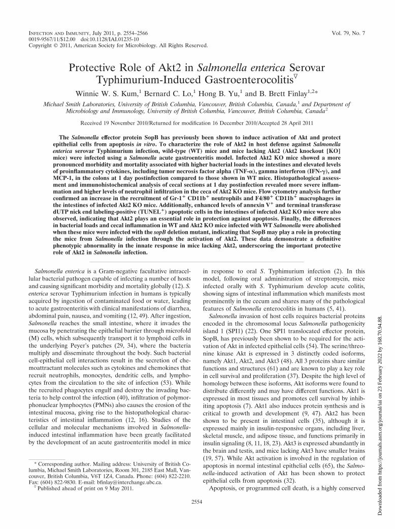
INFECTION AND IMMUNITY, July 2011, p. 2554–2566 Vol. 79, No. 7 0019-9567/11/$12.00 doi:10.1128/IAI.01235-10 Copyright © 2011, American Society for Microbiology. All Rights Reserved. Protective Role of Akt2 in Salmonella enterica Serovar Typhimurium-Induced Gastroenterocolitis Winnie W. S. Kum, 1 Bernard C. Lo, 1 Hong B. Yu, 1 and B. Brett Finlay 1,2 * Michael Smith Laboratories, University of British Columbia, Vancouver, British Columbia, Canada, 1 and Department of Microbiology and Immunology, University of British Columbia, Vancouver, British Columbia, Canada 2 Received 19 November 2010/Returned for modification 16 December 2010/Accepted 28 April 2011 The Salmonella effector protein SopB has previously been shown to induce activation of Akt and protect epithelial cells from apoptosis in vitro. To characterize the role of Akt2 in host defense against Salmonella enterica serovar Typhimurium infection, wild-type (WT) mice and mice lacking Akt2 (Akt2 knockout [KO] mice) were infected using a Salmonella acute gastroenteritis model. Infected Akt2 KO mice showed a more pronounced morbidity and mortality associated with higher bacterial loads in the intestines and elevated levels of proinflammatory cytokines, including tumor necrosis factor alpha (TNF-), gamma interferon (IFN-), and MCP-1, in the colons at 1 day postinfection compared to those shown in WT mice. Histopathological assess- ment and immunohistochemical analysis of cecal sections at 1 day postinfection revealed more severe inflam- mation and higher levels of neutrophil infiltration in the ceca of Akt2 KO mice. Flow cytometry analysis further confirmed an increase in the recruitment of Gr-1 CD11b neutrophils and F4/80 CD11b macrophages in the intestines of infected Akt2 KO mice. Additionally, enhanced levels of annexin V and terminal transferase dUTP nick end labeling-positive (TUNEL ) apoptotic cells in the intestines of infected Akt2 KO mice were also observed, indicating that Akt2 plays an essential role in protection against apoptosis. Finally, the differences in bacterial loads and cecal inflammation in WT and Akt2 KO mice infected with WT Salmonella were abolished when these mice were infected with the sopB deletion mutant, indicating that SopB may play a role in protecting the mice from Salmonella infection through the activation of Akt2. These data demonstrate a definitive phenotypic abnormality in the innate response in mice lacking Akt2, underscoring the important protective role of Akt2 in Salmonella infection. Salmonella enterica is a Gram-negative facultative intracel- lular bacterial pathogen capable of infecting a number of hosts and causing significant morbidity and mortality globally (12). S. enterica serovar Typhimurium infection in humans is typically acquired by ingestion of contaminated food or water, leading to acute gastroenteritis with clinical manifestations of diarrhea, abdominal pain, nausea, and vomiting (12, 49). After ingestion, Salmonella reaches the small intestine, where it invades the mucosa by penetrating the epithelial barrier through microfold (M) cells, which subsequently transport it to lymphoid cells in the underlying Peyer’s patches (29, 34), where the bacteria multiply and disseminate throughout the body. Such bacterial cell-epithelial cell interactions result in the secretion of che- moattractant molecules such as cytokines and chemokines that recruit neutrophils, monocytes, dendritic cells, and lympho- cytes from the circulation to the site of infection (53). While the recruited phagocytes engulf and destroy the invading bac- teria to help control the infection (40), infiltration of polymor- phonuclear lymphocytes (PMNs) also causes the erosion of the intestinal mucosa, giving rise to the histopathological charac- teristics of intestinal inflammation (12, 16). Studies of the cellular and molecular mechanisms involved in Salmonella- induced intestinal inflammation have been greatly facilitated by the development of an acute gastroenteritis model in mice in response to oral S. Typhimurium infection (2). In this model, following oral administration of streptomycin, mice infected orally with S. Typhimurium develop acute colitis, showing signs of intestinal inflammation which manifests most prominently in the cecum and shares many of the pathological features of Salmonella enterocolitis in humans (5, 41). Salmonella invasion of host cells requires bacterial proteins encoded in the chromosomal locus Salmonella pathogenicity island 1 (SPI1) (22). One SPI1 translocated effector protein, SopB, has previously been shown to be required for the acti- vation of Akt in infected epithelial cells (54). The serine/threo- nine kinase Akt is expressed in 3 distinctly coded isoforms, namely Akt1, Akt2, and Akt3 (48). All 3 proteins share similar functions and structures (61) and are known to play a key role in cell survival and proliferation (37). Despite the high level of homology between these isoforms, Akt isoforms were found to distribute differently and may have different functions. Akt1 is expressed in most tissues and promotes cell survival by inhib- iting apoptosis (7). Akt1 also induces protein synthesis and is critical to growth and development (9, 47). Akt2 has been shown to be present in intestinal cells (35), although it is expressed mainly in insulin-responsive organs, including liver, skeletal muscle, and adipose tissue, and functions primarily in insulin signaling (8, 11, 18, 23). Akt3 is expressed abundantly in the brain and testis, and mice lacking Akt3 have smaller brains (19, 57). While Akt activation is involved in the regulation of apoptosis in normal intestinal epithelial cells (65), the Salmo- nella-induced activation of Akt has been shown to protect epithelial cells from apoptosis (32). Apoptosis, or programmed cell death, is a highly conserved * Corresponding author. Mailing address: University of British Co- lumbia, Michael Smith Laboratories, Room 301, 2185 East Mall, Van- couver, British Columbia, V6T 1Z4, Canada. Phone: (604) 822-2210. Fax: (604) 822-9830. E-mail: bfi[email protected]. Published ahead of print on 9 May 2011. 2554 Downloaded from https://journals.asm.org/journal/iai on 23 February 2022 by 168.70.94.88.
Transcript of Protective Role of Akt2 in Salmonella enterica Serovar Typhimurium
INFECTION AND IMMUNITY, July 2011, p. 2554–2566 Vol. 79, No. 70019-9567/11/$12.00 doi:10.1128/IAI.01235-10Copyright © 2011, American Society for Microbiology. All Rights Reserved.
Protective Role of Akt2 in Salmonella enterica SerovarTyphimurium-Induced Gastroenterocolitis�
Winnie W. S. Kum,1 Bernard C. Lo,1 Hong B. Yu,1 and B. Brett Finlay1,2*Michael Smith Laboratories, University of British Columbia, Vancouver, British Columbia, Canada,1 and Department of
Microbiology and Immunology, University of British Columbia, Vancouver, British Columbia, Canada2
Received 19 November 2010/Returned for modification 16 December 2010/Accepted 28 April 2011
The Salmonella effector protein SopB has previously been shown to induce activation of Akt and protectepithelial cells from apoptosis in vitro. To characterize the role of Akt2 in host defense against Salmonellaenterica serovar Typhimurium infection, wild-type (WT) mice and mice lacking Akt2 (Akt2 knockout [KO]mice) were infected using a Salmonella acute gastroenteritis model. Infected Akt2 KO mice showed a morepronounced morbidity and mortality associated with higher bacterial loads in the intestines and elevated levelsof proinflammatory cytokines, including tumor necrosis factor alpha (TNF-�), gamma interferon (IFN-�), andMCP-1, in the colons at 1 day postinfection compared to those shown in WT mice. Histopathological assess-ment and immunohistochemical analysis of cecal sections at 1 day postinfection revealed more severe inflam-mation and higher levels of neutrophil infiltration in the ceca of Akt2 KO mice. Flow cytometry analysis furtherconfirmed an increase in the recruitment of Gr-1� CD11b� neutrophils and F4/80� CD11b� macrophages inthe intestines of infected Akt2 KO mice. Additionally, enhanced levels of annexin V� and terminal transferasedUTP nick end labeling-positive (TUNEL�) apoptotic cells in the intestines of infected Akt2 KO mice were alsoobserved, indicating that Akt2 plays an essential role in protection against apoptosis. Finally, the differencesin bacterial loads and cecal inflammation in WT and Akt2 KO mice infected with WT Salmonella were abolishedwhen these mice were infected with the sopB deletion mutant, indicating that SopB may play a role in protectingthe mice from Salmonella infection through the activation of Akt2. These data demonstrate a definitivephenotypic abnormality in the innate response in mice lacking Akt2, underscoring the important protectiverole of Akt2 in Salmonella infection.
Salmonella enterica is a Gram-negative facultative intracel-lular bacterial pathogen capable of infecting a number of hostsand causing significant morbidity and mortality globally (12). S.enterica serovar Typhimurium infection in humans is typicallyacquired by ingestion of contaminated food or water, leadingto acute gastroenteritis with clinical manifestations of diarrhea,abdominal pain, nausea, and vomiting (12, 49). After ingestion,Salmonella reaches the small intestine, where it invades themucosa by penetrating the epithelial barrier through microfold(M) cells, which subsequently transport it to lymphoid cells inthe underlying Peyer’s patches (29, 34), where the bacteriamultiply and disseminate throughout the body. Such bacterialcell-epithelial cell interactions result in the secretion of che-moattractant molecules such as cytokines and chemokines thatrecruit neutrophils, monocytes, dendritic cells, and lympho-cytes from the circulation to the site of infection (53). Whilethe recruited phagocytes engulf and destroy the invading bac-teria to help control the infection (40), infiltration of polymor-phonuclear lymphocytes (PMNs) also causes the erosion of theintestinal mucosa, giving rise to the histopathological charac-teristics of intestinal inflammation (12, 16). Studies of thecellular and molecular mechanisms involved in Salmonella-induced intestinal inflammation have been greatly facilitatedby the development of an acute gastroenteritis model in mice
in response to oral S. Typhimurium infection (2). In thismodel, following oral administration of streptomycin, miceinfected orally with S. Typhimurium develop acute colitis,showing signs of intestinal inflammation which manifests mostprominently in the cecum and shares many of the pathologicalfeatures of Salmonella enterocolitis in humans (5, 41).
Salmonella invasion of host cells requires bacterial proteinsencoded in the chromosomal locus Salmonella pathogenicityisland 1 (SPI1) (22). One SPI1 translocated effector protein,SopB, has previously been shown to be required for the acti-vation of Akt in infected epithelial cells (54). The serine/threo-nine kinase Akt is expressed in 3 distinctly coded isoforms,namely Akt1, Akt2, and Akt3 (48). All 3 proteins share similarfunctions and structures (61) and are known to play a key rolein cell survival and proliferation (37). Despite the high level ofhomology between these isoforms, Akt isoforms were found todistribute differently and may have different functions. Akt1 isexpressed in most tissues and promotes cell survival by inhib-iting apoptosis (7). Akt1 also induces protein synthesis and iscritical to growth and development (9, 47). Akt2 has beenshown to be present in intestinal cells (35), although it isexpressed mainly in insulin-responsive organs, including liver,skeletal muscle, and adipose tissue, and functions primarily ininsulin signaling (8, 11, 18, 23). Akt3 is expressed abundantly inthe brain and testis, and mice lacking Akt3 have smaller brains(19, 57). While Akt activation is involved in the regulation ofapoptosis in normal intestinal epithelial cells (65), the Salmo-nella-induced activation of Akt has been shown to protectepithelial cells from apoptosis (32).
Apoptosis, or programmed cell death, is a highly conserved
* Corresponding author. Mailing address: University of British Co-lumbia, Michael Smith Laboratories, Room 301, 2185 East Mall, Van-couver, British Columbia, V6T 1Z4, Canada. Phone: (604) 822-2210.Fax: (604) 822-9830. E-mail: [email protected].
� Published ahead of print on 9 May 2011.
2554
Dow
nloa
ded
from
http
s://j
ourn
als.
asm
.org
/jour
nal/i
ai o
n 23
Feb
ruar
y 20
22 b
y 16
8.70
.94.
88.
mechanism for intracellular disassembly without changingmembrane integrity (20, 32, 62). This cellular process can becharacterized by the externalization of phosphatidylserine tothe cell surface, DNA fragmentation, chromatin condensation,and release of apoptotic bodies (32, 62). Bacterial and viralpathogens can manipulate the host cell suicide mechanisms toenhance its pathogenicity (20, 32). Depending on the host celltype and stage of infection, Salmonella can induce differentapoptotic responses (31, 32). Various mechanisms of theseSalmonella-induced apoptotic events in different cell typeshave been identified, but their role in pathogenesis remainsunclear (32). Previously, the sustained activation of Akt hasbeen identified as an important prerequisite for the SopB-dependent antiapoptotic pathway in Salmonella-infected epi-thelial cells in vitro (32). It is believed that SopB is involved inthe delay of apoptosis, although the role of apoptosis in gas-troenteritis is still unclear (32).
Although the role of the prosurvival molecule Akt in infec-tious colitis has not yet been explored, Akt2 has been shown toplay a role in enterocyte differentiation (35). In the presentstudy, we used a Salmonella-induced intestinal inflammationmodel to examine the role of Akt2 in host defense againstSalmonella infection. During Salmonella infection, we foundthat in mice lacking the functional protein kinase Akt2, hostcells were more prone to apoptosis and more susceptible toinfection.
MATERIALS AND METHODS
Bacterial culture. S. enterica serovar Typhimurium SL1344 (26) and the sopBdeletion mutant (�sopB mutant) (30, 54) were grown overnight with shaking (200rpm) in Luria-Bertani (LB) broth supplemented with 50 �g/ml streptomycin at37°C for 18 h.
Mice. Wild-type (WT) 129S and homozygous 129S Akt2-deficient (Akt2knockout [KO]) mice, originally from the Wellcome Trust Sanger Institute(Hinxton, Cambridge, United Kingdom), were bred at the Vaccine and Infec-tious Disease Organization, University of Saskatchewan. Adult mice (8 to 10weeks) were transported and maintained under specific-pathogen-free condi-tions at the animal facility, University of British Columbia. All animal experi-ments were done according to institutional guidelines and were approved by theAnimal Care Committee of the University of British Columbia.
Mouse model of Salmonella-induced intestinal inflammation. The protocol forS. Typhimurium-induced enterocolitis was used as described previously (2, 13).Briefly, mice aged 8 to 10 weeks were given 20 mg of streptomycin by oral gavagewith a 21-gauge feeding needle. Twenty-four hours later, 3 � 106 S. Typhimu-rium bacteria were administered by oral gavage. Mice were assigned randomly tosurvival groups to monitor for survival and to organ harvest groups to assess forintestinal inflammation. To allow a humane endpoint, mice in the survival groupswere monitored twice daily for 18 days after bacterial inoculation for signs ofmorbidity (reduced level of motion, piloerection, labored breathing, weight loss).Mice that showed distress of these signs or became moribund were euthanizedwith CO2 and considered nonsurvival. For other experiments, mice were eutha-nized at designated time points postinfection. Colons and ceca were harvestedaseptically for bacterial enumeration, and ceca were collected for histopathology.
Bacterial enumeration. Colons and ceca were harvested at designated timepoints postinfection with S. Typhimurium or the �sopB mutant and weighed, andeach sample was collected into 1 ml of sterile phosphate-buffered saline (PBS)and homogenized with a Mixer Mill 301 (Retsch, Newtown, PA). Serial dilutionsof the resulting homogenates were plated on LB agar plates containing 100 �g/mlstreptomycin. Plates were incubated at 37°C for 24 h. Colony counts were ex-pressed as the numbers of CFU per ml.
Histopathology. Ceca of experimental animals were fixed in 10% formalin for18 h, followed by 18 h in 70% ethanol prior to being embedded in paraffin,sectioned, and stained with hematoxylin and eosin (H&E) by Wax-it HistologyServices (Vancouver, Canada). Stained sections were examined in a blindedfashion for signs of inflammation, PMN infiltration, edema, crypt abscess for-mation, regenerative changes, and necrosis. Pathological scores were determined
by grading the histopathologic findings from 0 (none) to 1� (mild), 2� (mod-erate), and 3� (severe) and averaging six fields/sample, according to a scoringsystem previously described (13).
Colonic cytokine measurements. Colon samples collected at 1 day postinfec-tion were assayed for the presence of cytokines. Colon homogenates collected asdescribed above were spun twice at 13,200 rpm in an Eppendorf benchtopcentrifuge (5415R) for 30 min each at 4°C to remove insoluble matters. Colonsupernatants were frozen at �80°C until assayed for cytokines using the mouseinflammation cytometric bead array (CBA) assay kit (BD Biosciences), accord-ing to the manufacturer’s instructions.
MPO staining. For myeloperoxidase (MPO) immunofluorescent staining forthe detection of neutrophils in the ceca of naïve and infected mice, paraffin-embedded tissues were deparaffinized in xylene (twice for 5 min each); rehy-drated in 100%, 95%, and 70% ethanol (5 min each); and blocked with 2%normal goat serum in TPBS-BSA (PBS containing 0.05% Tween 20 and 0.1%bovine serum albumin; BSA was obtained from Sigma) for 40 min at roomtemperature. They were then incubated with the primary antibody, a polyclonalrabbit anti-MPO antibody (Thermo Scientific) diluted 1:200, overnight at 4°C.The sections were washed with TPBS-BSA and then incubated for 1 h at roomtemperature with Alexa 488-conjugated goat anti-rabbit IgG (diluted 1:1,000) asthe secondary antibody. Subsequently, after being rinsed with TPBS-BSA, cov-erslips were mounted using Prolong Gold antifade reagent containing 4�,6-diamidino-2-phenylindole (DAPI) (Invitrogen). Digital images were capturedand processed using a Zeiss Axiophot epifluorescence microscope.
Flow cytometry analysis. Single-cell suspensions of colonic and cecal laminapropria cells were prepared by following a standard protocol (63). Briefly, colonsand ceca from naïve or infected mice were rinsed in Hank’s buffered salinesolution (HBSS) to remove fecal contents. Epithelial cells were removed byshaking for 30 min at 37°C in HBSS containing 5 mM EDTA (Sigma), 5% fetalcalf serum (HyClone), and 1 mM dithiothreitol (Sigma). The tissues were washedseveral times with HBSS, cut into small pieces, and digested with a mixture ofcollagenase-dispase (Roche) for 1 h at 37°C. The digested fragments were passedthrough a 70-mm nylon mesh. Supernatant containing endothelial cells wascollected, and the pellet was further purified by centrifugation over a discontin-uous Percoll gradient (40%/70%) for 20 min at 2,000 rpm to obtain laminapropria leukocytes. Purified cells from the colonic and cecal lamina propria weresurface stained with fluorochrome-conjugated antibodies against CD11b (cloneM1/70; BD Biosciences), Gr-1 (clone RB6-8C5; BD Biosciences), F4/80 (cloneBM8; eBioscience), and annexin V (recombinant protein from Escherichia coli;eBioscience) before being subjected to flow cytometry analysis with FACSCalibur(Becton Dickinson, San Jose, CA) using CellQuest software. 7-Aminoactinomy-cin D (7-AAD; Sigma) was included in all staining to define viable cells.
TUNEL staining. Ceca of experimental animals were fixed in 4% formalde-hyde for 3 h at room temperature, followed by being washed with PBS. Thetissues were then frozen and sectioned (10 �m). Cecal sections were then as-sessed for apoptotic features of damaged cells by terminal transferase dUTP nickend labeling (TUNEL) staining using the Apo-BrdU in situ DNA fragmentationassay kit (BioVision, Inc., Mountain View, CA), according to the manufacturer’sinstructions. Transferred bromolated dUTP nucleotides (Br-dUTP) to the free3�-OH of cleaved DNA by terminal deoxynucleotide transferase (TdT) weredetected by the anti-BrdU-fluorescein isothiocyanate (FITC) antibody usingFITC and rhodamine filters on an Olympus 10i confocal microscope. Propidiumiodide was used for counterstaining.
Statistical analysis. The survival curves of infected mice were comparedusing Kaplan-Meier analysis followed by the log rank test. Bacterial loads,total pathological scores of infected ceca, and CBA assays for cytokines in thecolons of infected mice were compared using the two-tailed, unpaired t test.All analyses were performed with a 95% confidence interval using GraphPadPrism version 4.0.
RESULTS
Akt2 is critical for host survival in S. Typhimurium infec-tions. Although Salmonella-induced activation of Akt has beenshown to protect epithelial cells from apoptosis in vitro (32),the role of Akt in Salmonella-induced gastroenteritis remainsunclear. We hypothesized that deficiency of Akt would impairthe host’s defense mechanisms against Salmonella infection,leading to increased morbidity and mortality. To address this,we orally infected mice deficient in the antiapoptotic kinase
VOL. 79, 2011 ROLE OF Akt2 IN SALMONELLA INFECTION 2555
Dow
nloa
ded
from
http
s://j
ourn
als.
asm
.org
/jour
nal/i
ai o
n 23
Feb
ruar
y 20
22 b
y 16
8.70
.94.
88.
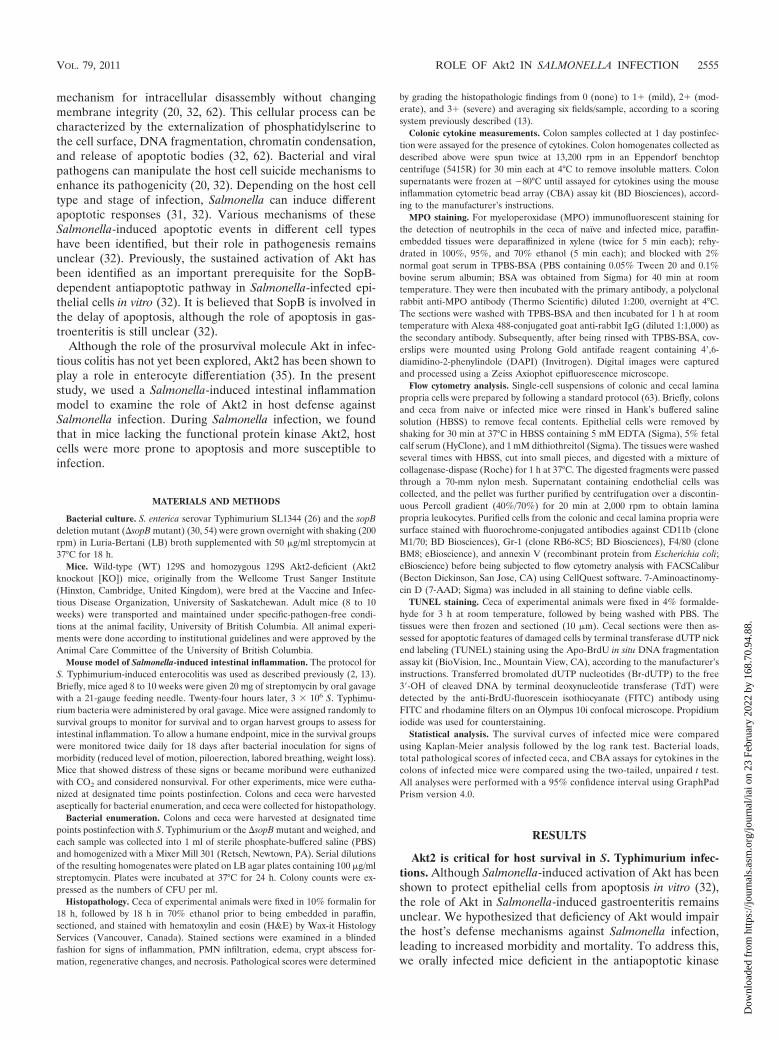
Akt2 with S. Typhimurium. A significantly higher mortalityrate (P � 0.0247) in Akt2 KO mice than that in 129S WT micewas noted throughout the period of observation (Fig. 1), indi-cating that Akt2 may play an important role in host survivalduring Salmonella infection.
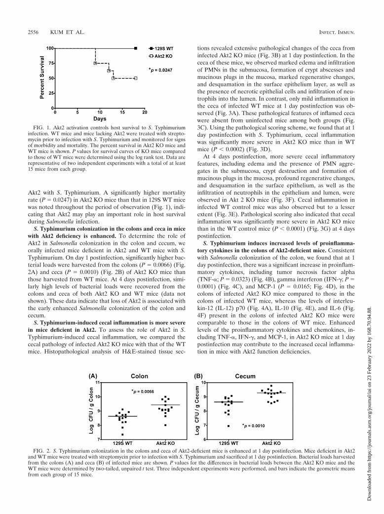
S. Typhimurium colonization in the colons and ceca in micewith Akt2 deficiency is enhanced. To determine the role ofAkt2 in Salmonella colonization in the colon and cecum, weorally infected mice deficient in Akt2 and WT mice with S.Typhimurium. On day 1 postinfection, significantly higher bac-terial loads were harvested from the colons (P � 0.0066) (Fig.2A) and ceca (P � 0.0010) (Fig. 2B) of Akt2 KO mice thanthose harvested from WT mice. At 4 days postinfection, simi-larly high levels of bacterial loads were recovered from thecolons and ceca of both Akt2 KO and WT mice (data notshown). These data indicate that loss of Akt2 is associated withthe early enhanced Salmonella colonization of the colon andcecum.
S. Typhimurium-induced cecal inflammation is more severein mice deficient in Akt2. To assess the role of Akt2 in S.Typhimurium-induced cecal inflammation, we compared thececal pathology of infected Akt2 KO mice with that of the WTmice. Histopathological analysis of H&E-stained tissue sec-
tions revealed extensive pathological changes of the ceca frominfected Akt2 KO mice (Fig. 3B) at 1 day postinfection. In thececa of these mice, we observed marked edema and infiltrationof PMNs in the submucosa, formation of crypt abscesses andmucinous plugs in the mucosa, marked regenerative changes,and desquamation in the surface epithelium layer, as well asthe presence of necrotic epithelial cells and infiltration of neu-trophils into the lumen. In contrast, only mild inflammation inthe ceca of infected WT mice at 1 day postinfection was ob-served (Fig. 3A). These pathological features of inflamed cecawere absent from uninfected mice among both groups (Fig.3C). Using the pathological scoring scheme, we found that at 1day postinfection with S. Typhimurium, cecal inflammationwas significantly more severe in Akt2 KO mice than in WTmice (P � 0.0002) (Fig. 3D).
At 4 days postinfection, more severe cecal inflammatoryfeatures, including edema and the presence of PMN aggre-gates in the submucosa, crypt destruction and formation ofmucinous plugs in the mucosa, profound regenerative changes,and desquamation in the surface epithelium, as well as theinfiltration of neutrophils in the epithelium and lumen, wereobserved in Akt 2 KO mice (Fig. 3F). Cecal inflammation ininfected WT control mice was also observed but to a lesserextent (Fig. 3E). Pathological scoring also indicated that cecalinflammation was significantly more severe in Akt2 KO micethan in the WT control mice (P � 0.0001) (Fig. 3G) at 4 dayspostinfection.
S. Typhimurium induces increased levels of proinflamma-tory cytokines in the colons of Akt2-deficient mice. Consistentwith Salmonella colonization of the colon, we found that at 1day postinfection, there was a significant increase in proinflam-matory cytokines, including tumor necrosis factor alpha(TNF-; P � 0.0323) (Fig. 4B), gamma interferon (IFN-; P �0.0001) (Fig. 4C), and MCP-1 (P � 0.0165; Fig. 4D), in thecolons of infected Akt2 KO mice compared to those in thecolons of infected WT mice, whereas the levels of interleu-kin-12 (IL-12) p70 (Fig. 4A), IL-10 (Fig. 4E), and IL-6 (Fig.4F) present in the colons of infected Akt2 KO mice werecomparable to those in the colons of WT mice. Enhancedlevels of the proinflammatory cytokines and chemokines, in-cluding TNF-, IFN-, and MCP-1, in Akt2 KO mice at 1 daypostinfection may contribute to the increased cecal inflamma-tion in mice with Akt2 function deficiencies.
FIG. 1. Akt2 activation controls host survival to S. Typhimuriuminfection. WT mice and mice lacking Akt2 were treated with strepto-mycin prior to infection with S. Typhimurium and monitored for signsof morbidity and mortality. The percent survival in Akt2 KO mice andWT mice is shown. P values for survival curves of KO mice comparedto those of WT mice were determined using the log rank test. Data arerepresentative of two independent experiments with a total of at least15 mice from each group.
FIG. 2. S. Typhimurium colonization in the colons and ceca of Akt2-deficient mice is enhanced at 1 day postinfection. Mice deficient in Akt2and WT mice were treated with streptomycin prior to infection with S. Typhimurium and sacrificed at 1 day postinfection. Bacterial loads harvestedfrom the colons (A) and ceca (B) of infected mice are shown. P values for the differences in bacterial loads between the Akt2 KO mice and theWT mice were determined by two-tailed, unpaired t test. Three independent experiments were performed, and bars indicate the geometric meansfrom each group of 15 mice.
2556 KUM ET AL. INFECT. IMMUN.
Dow
nloa
ded
from
http
s://j
ourn
als.
asm
.org
/jour
nal/i
ai o
n 23
Feb
ruar
y 20
22 b
y 16
8.70
.94.
88.

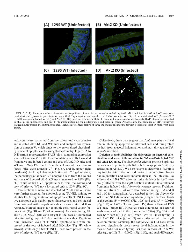
S. Typhimurium-induced neutrophil recruitment is morepronounced in the ceca of Akt2-deficient mice. Histopatholog-ical analysis of the H&E-stained sections revealed more pro-nounced neutrophil infiltration in the ceca of S. Typhimurium-
infected Akt2 KO mice than that in the ceca of WT mice at 1day postinfection. To confirm this, we stained tissue sections ofinfected ceca harvested at 1 day postinfection with antibodiesagainst MPO, which present most abundantly in neutrophils.
FIG. 3. Histology of ceca from S. Typhimurium-infected mice showing severe inflammation in Akt2-deficient mice at 1 day and 4 dayspostinfection. Mice defective in Akt2 and WT mice were treated with streptomycin prior to infection with S. Typhimurium and sacrificed at 1 dayand 4 days postinfection. Representative H&E staining of the ceca of one mouse from each group of at least 4 mice is shown. At 1 day postinfection,severe inflammation in the lumina of the ceca of infected Akt2 KO mice was observed (B), while only mild inflammation in the WT mice wasobserved (A). At 4 days postinfection, more severe cecal inflammatory features in Akt2 KO mice were detected (F), and to a lesser extent, cecalinflammation in infected WT mice was also observed (E). (C) These pathological features of inflamed ceca were absent from uninfected mice forboth groups. Images are shown at magnifications of �50 and �200. Pathological scoring for the cecal samples harvested from Akt2 KO mice andWT mice at 1 day postinfection (D) and at 4 days postinfection (G) is shown. P values for the differences in pathological scoring of the ceca betweenthe Akt2 KO mice and the WT mice were determined by two-tailed, unpaired t test. Data are representative of three independent experiments.
VOL. 79, 2011 ROLE OF Akt2 IN SALMONELLA INFECTION 2557
Dow
nloa
ded
from
http
s://j
ourn
als.
asm
.org
/jour
nal/i
ai o
n 23
Feb
ruar
y 20
22 b
y 16
8.70
.94.
88.

As shown in Fig. 5, infiltration of MPO-positively stained neu-trophils was observed in the submucosa of the ceca of infectedAkt2 KO mice (Fig. 5D) but was absent in the ceca of infectedWT mice (Fig. 5C) and the uninfected mice among bothgroups (Fig. 5A and B).
In addition to the microscopic staining of neutrophils oncross sections of fixed cecal tissues using anti-MPO antibody,we also performed fluorescence-activated cell sorter (FACS)staining to stain for live cells prepared from the entire intes-tinal tract using other markers to further characterize the phe-notype of the neutrophil populations. Lamina propria leuko-cytes were harvested from the colons and ceca of naïve andinfected Akt2 KO and WT mice and analyzed for the expres-sion of several surface markers for neutrophils (Gr-1 andCD11b) and macrophages (F4/80 and CD11b) using flow cy-tometry.
Figure 6A to D illustrate representative FACS plots com-paring the expression levels of CD11b and Gr-1 on neutrophilsharvested from naïve and infected colons and ceca of Akt2 KOand WT mice. Only 1% of cells from uninfected mice weremature neutrophils expressing CD11b� Gr-1� (Fig. 6A and B,upper right quadrants). At 1 day postinfection with Salmonella,the percentage of CD11b� Gr-1� cells from the colons andceca of infected Akt2 KO mice (Fig. 6D) increased to 33%,while the percentage of CD11b� Gr-1� cells from the colonsand ceca of infected WT mice (Fig. 6C) only increased to 15%.The percentage of immature neutrophils expressing CD11b�
Gr-1� (upper left quadrants) from the colons and ceca of
infected Akt2 KO mice and WT mice were similar (19% and22%, respectively).
Figure 7A to D show representative FACS plots comparingthe expression levels of CD11b and F4/80 on macrophagesharvested from naïve and infected colons and ceca of Akt2 KOand WT mice. Only 1% of cells from uninfected mice weremacrophages expressing CD11b� F4/80� (Fig. 7A and B, up-per right quadrants). At 1 day following infection with Salmo-nella, the percentage of CD11b� F4/80� cells from infectedAkt2 KO mice (Fig. 7D) increased to 35%, while the percent-age of CD11b� F4/80� cells from the colons and ceca ofinfected WT mice (Fig. 7C) increased to 8%.
These data indicate that loss of Akt2 facilitates transmigra-tion of neutrophils and macrophages to the sites of infection.The excessive accumulation of granulocytes and the subse-quent release of enzymes and oxygen radicals from these cellsin the colons and ceca of infected Akt2 KO mice may result inthe enhanced mucosal inflammatory reaction.
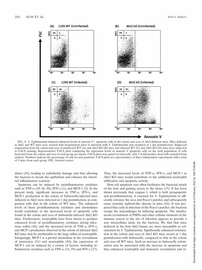
S. Typhimurium induces enhanced levels of annexin V� andTUNEL� apoptotic cells in the colons and ceca of Akt2-defi-cient mice. Our data mentioned above suggest that Akt2 KOmice are more susceptible to Salmonella infection. It has beenshown that Salmonella protects epithelial cells from camptoth-ecin-induced apoptosis in vitro by activation of the Akt path-way (32). We next determined if mice lacking Akt2 are unableto inhibit apoptosis of intestinal epithelial cells and thus con-tribute to mucosal inflammation and mortality during Salmo-nella infection. Intestinal epithelial cells and lamina propria
FIG. 4. S. Typhimurium induced enhanced levels of TNF-, IFN-, and MCP-1 in the colons of Akt2-deficient mice. Mice lacking Akt2 andWT mice were treated with streptomycin prior to infection with S. Typhimurium and sacrificed at 1 day postinfection. Cytokines present in thecolon extracts were determined by CBA assay. P values for the differences in colonic cytokines between the infected Akt2 KO mice and the infectedWT mice were determined by two-tailed Student’s t test. (U) � colon cytokines from uninfected mice; (I) � colon cytokines from infected mice.Results (means � standard errors of the means [SEM]) were obtained from 5 mice from each group. Data are representative of three independentexperiments with a total of at least 15 animals from each group.
2558 KUM ET AL. INFECT. IMMUN.
Dow
nloa
ded
from
http
s://j
ourn
als.
asm
.org
/jour
nal/i
ai o
n 23
Feb
ruar
y 20
22 b
y 16
8.70
.94.
88.
leukocytes were harvested from the colons and ceca of naïveand infected Akt2 KO and WT mice and analyzed for expres-sion of annexin V, which binds to the externalized phosphati-dylserine of apoptotic cells, using flow cytometry. Figure 8A toD illustrate representative FACS plots comparing expressionlevels of annexin V on the total population of cells harvestedfrom naïve and infected colons and ceca of Akt2 KO mice andWT mice. Only 1% of cells from the colons and ceca of unin-fected mice were annexin V� (Fig. 8A and B, upper rightquadrants). At 1 day following infection with S. Typhimurium,the percentage of annexin V� apoptotic cells from the colonsand ceca of infected Akt2 KO mice increased to 81% (Fig.8D), while annexin V� apoptotic cells from the colons andceca of infected WT mice increased only to 20% (Fig. 8C).
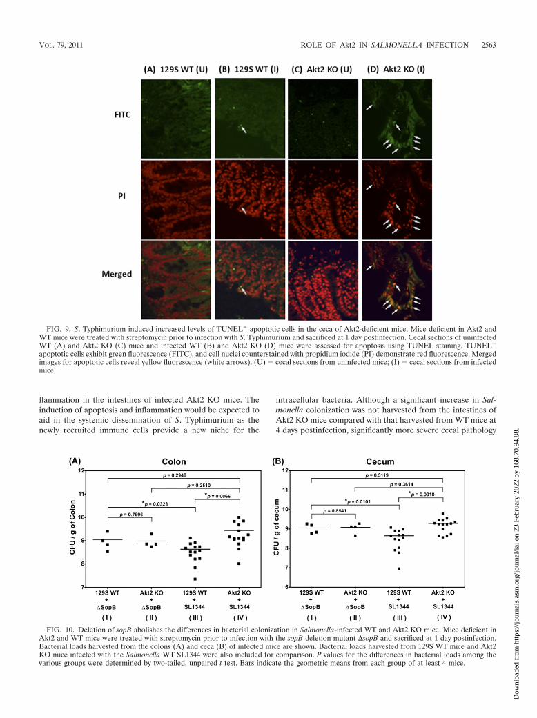
Cecal sections of naïve and infected Akt2 KO and WT micewere further assessed for apoptosis using TUNEL staining toidentify DNA fragmentation of apoptotic cells. TUNEL-posi-tive apoptotic cells exhibit green fluorescence, and cell nucleicounterstained with propidium iodide demonstrate red fluo-rescence. Merged images for apoptotic cells reveal yellow flu-orescence (Fig. 9B and D, white arrows). As shown in Fig. 9Aand C, TUNEL� cells were absent in the ceca of uninfectedmice for both groups. At 1 day postinfection with S. Typhimu-rium, increased levels of TUNEL� apoptotic cells were ob-served in the ceca of infected Akt2 KO mice (Fig. 9D, whitearrows), while only a few TUNEL� cells were present in thececa of infected WT mice (Fig. 9B).
Collectively, these data suggest that Akt2 may play a criticalrole in inhibiting apoptosis of intestinal cells and thus protectthe host from mucosal inflammation and mortality against Sal-monella infection.
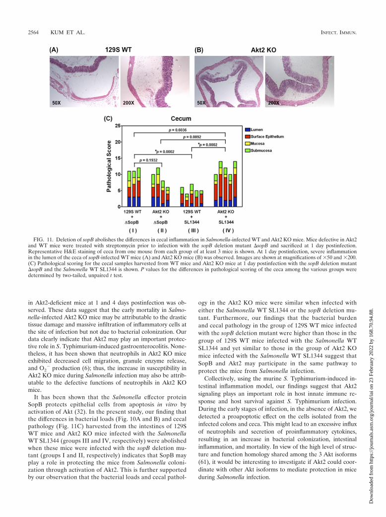
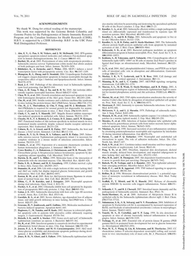
Deletion of sopB abolishes the differences in bacterial colo-nization and cecal inflammation in Salmonella-infected WTand Akt2 KO mice. The Salmonella effector protein SopB hasbeen shown to protect epithelial cells from apoptosis in vitro byactivation of Akt (32). We next sought to determine if SopB isrequired for Akt activation and protects the mice from bacte-rial colonization and cecal inflammation in the intestine. Toaddress this, 129S WT mice and mice deficient in Akt2 wereorally infected with the sopB deletion mutant. Data obtainedfrom mice infected with Salmonella enterica serovar Typhimu-rium WT strain SL1344 were also included in Fig. 10A and Band 11C for comparison. As described earlier, the SalmonellaWT strain SL1344 induced significantly higher bacterial loadsin the colons (P � 0.0066) (Fig. 10A) and ceca (P � 0.0010)(Fig. 10B) of Akt2 KO mice (group IV) than in those of 129SWT mice (group III). However, such differences in bacterialloads were abolished in the colons (P � 0.7996) (Fig. 10A) andceca (P � 0.8541) (Fig. 10B) when 129S WT mice (group I)and Akt2 KO mice (group II) were infected with the sopBdeletion mutant. Similarly, the Salmonella WT SL1344 straininduced significantly more severe cecal inflammation in thececa of Akt2 KO mice (group IV) than in those of 129S WTmice (group III) (P � 0.0002) (Fig. 11C), and such differences
FIG. 5. S. Typhimurium induced increased neutrophil recruitment in the ceca of mice lacking Akt2. Mice deficient in Akt2 and WT mice weretreated with streptomycin prior to infection with S. Typhimurium and sacrificed at 1 day postinfection. Ceca from uninfected WT (A) and Akt2KO (B) mice and infected WT (C) and Akt2 KO (D) mice were stained with MPO immunofluorescence for neutrophils. DAPI staining is indicatedin blue in the submucosa, and anti-MPO immunostaining for neutrophils is indicated in green. Arrows show the presence of MPO-positivelystained neutrophils in the submucosal area. Pictures are representative of three independent experiments with a total of at least 15 mice from eachgroup.
VOL. 79, 2011 ROLE OF Akt2 IN SALMONELLA INFECTION 2559
Dow
nloa
ded
from
http
s://j
ourn
als.
asm
.org
/jour
nal/i
ai o
n 23
Feb
ruar
y 20
22 b
y 16
8.70
.94.
88.

in cecal inflammation were abolished when 129S WT mice(group I) and Akt2 KO mice (group II) were infected with thesopB deletion mutant (P � 0.1932) (Fig. 11C).
Additionally, similar bacterial loads from the colons (P �0.2510) (Fig. 10A) and ceca (P � 0.3614) (Fig. 10B) as well ascomparable cecal pathology (P � 0.0892) (Fig. 11C) wereobserved in the Akt2 KO mice when these mice were infectedwith either the Salmonella WT SL1344 (group IV) or the sopBdeletion mutant (group II), suggesting that SopB and Akt2may be involved in the same signaling pathway to protect themice from Salmonella infection. This is further supported byour finding that in the group of 129S WT mice infected withthe sopB deletion mutant (group I) (Fig. 10A and B and 11C),higher bacterial loads in the colons (P � 0.0323) (Fig. 10A)and ceca (P � 0.0101) (Fig. 10B) as well as more severe cecalpathology (P � 0.0002) (Fig. 11C) were observed compared tothose in the 129S WT mice infected with the Salmonella WTSL1344 (group III) (Fig. 10A and B and 11C). Furthermore,our data also revealed that similar bacterial loads in the colons(P � 0.2948) (Fig. 10A) and ceca (P � 0.3119) (Fig. 10B) aswell as similar cecal pathology (P � 0.6036) (Fig. 11C) wereharvested in both the 129S WT mice infected with the sopB
deletion mutant (group I) (Fig. 10A and B and 11C) and theAkt2 KO mice infected with the Salmonella WT SL1344(group IV) (Fig. 10A and B and 11C). These data suggest thatSopB may play a role in protecting the mice from Salmonellacolonization and cecal inflammation through activation ofAkt2.
DISCUSSION
The prosurvival Akt protein kinase is critical in preventingcells from undergoing apoptosis (17). Akt activation has beenshown to be essential for SopB-mediated protection againstapoptosis in epithelial cells following Salmonella invasion (32,38). In S. Typhimurium-induced colitis, the role of Akt has notbeen explored. To address this, we analyzed S. Typhimurium-induced intestinal inflammation in Akt2-deficient mice. Anincrease in apoptotic cells associated with enhanced mortalityand morbidity, including higher bacterial loads and more se-vere proinflammatory responses in the colons and ceca in micedeficient in Akt2 following infection with S. Typhimurium,indicates functional involvement of Akt2 in this disease model.
Bacterial pathogens can either induce or prevent apoptosis
FIG. 6. S. Typhimurium infection is associated with an elevated influx of Gr-1� CD11b� and Gr-1� CD11b� cells in the colons and ceca ofmice lacking Akt2. Mice deficient in Akt2 and WT mice were treated with streptomycin prior to infection with S. Typhimurium and sacrificed at1 day postinfection. Single-cell suspensions from the colons and ceca of uninfected WT (A) and Akt2 KO (B) mice and infected WT (C) and Akt2KO (D) mice were subjected to FACS staining. Representative FACS plots comparing the expression levels of CD11b and GR-1 neutrophilsharvested from the colons and ceca of each group are shown. FACS plots were gated on live cells, with 7-AAD-positive dead cells excluded fromanalysis. Numbers indicate the percentage of cells in each quadrant. FACS plots are representative of four independent experiments with a totalof 12 mice from each group.
2560 KUM ET AL. INFECT. IMMUN.
Dow
nloa
ded
from
http
s://j
ourn
als.
asm
.org
/jour
nal/i
ai o
n 23
Feb
ruar
y 20
22 b
y 16
8.70
.94.
88.

to gain survival advantage in the host and to enhance infection.Many bacterial pathogens that cause apoptosis target immunecells such as macrophages (66) and neutrophils (4) becausethese cells would otherwise kill the bacteria (24, 33). Alterna-tively, bacterial pathogens could inhibit apoptosis during infec-tion, which provides a survival benefit for the bacteria to sur-vive and replicate inside host cells (10, 21, 32, 38, 43, 55). In thepresent study, we have demonstrated that Salmonella-inducedapoptosis at the sites of infection in mice lacking Akt2 andAkt2 deficiency renders these mice more susceptible to infec-tion, suggesting that apoptosis may play an important role inthe pathogenesis of Salmonella-induced colitis. Although Akt2has been shown to play an antiapoptotic role (17), the en-hanced level of apoptosis observed in Salmonella-infectedAkt2 KO mice may also be due to the decrease in glucoseavailability, as Akt2 modulates glucose homeostasis and down-stream apoptotic pathways during development (28) and Akt2knockout mice have been shown to develop a type 2 diabeticphenotype (8).
It has been suggested that the principal event leading toinflammatory diseases is cell damage as a result of apoptosis or
necrosis (36, 52, 56). Increased permeability of the epithelialbarrier and increased apoptotic rates of epithelial cells havebeen implicated to be major factors in the pathogenesis ofintestinal inflammation (25, 64). Our observation that cecalinflammation is more pronounced in Akt2 KO mice duringSalmonella infection may be associated with the lack of theantiapoptotic molecule Akt2. In the current study, a largenumber of annexin V� and TUNEL� apoptotic cells in theintestines of Salmonella-infected Akt2-deficient mice were de-tected (Fig. 8D and 9D). The massive cell damage would causeleakage of cellular contents into the adjacent tissues, resultingin the transmigration of granulocytes into the injured tissue ofinfected Akt2 KO mice (Fig. 5D, 6D, and 7D). Furthermore,the release of Salmonella from infected apoptotic cells intoadjacent tissues would contribute to the additional neutrophilinfiltration to the sites of infection. Although neutrophils havebeen shown to contribute to host protection from S. Typhimu-rium infection since they can target the virulence factors andeffectively eliminate the bacteria (60), excessive accumulationof neutrophils and monocytes would result in the release oftoxic products such as proteases and reactive oxygen interme-
FIG. 7. S. Typhimurium infection is associated with an elevated influx of CD11b� F4/80� macrophages in the colons and ceca of mice lackingAkt2. Mice deficient in Akt2 and WT mice were treated with streptomycin prior to infection with S. Typhimurium and sacrificed at 1 daypostinfection. Single-cell suspensions from the colons and ceca of uninfected WT (A) and Akt2 KO (B) mice and infected WT (C) and Akt2 KO(D) mice were subjected to FACS staining. Representative FACS plots comparing the expression levels of CD11b and F4/80 macrophagesharvested from the colons and ceca of each group are shown. FACS plots were gated on live cells, with 7-AAD-positive dead cells excluded fromanalysis. Numbers indicate the percentage of cells in each quadrant. FACS plots are representative of four independent experiments with a totalof 12 mice from each group.
VOL. 79, 2011 ROLE OF Akt2 IN SALMONELLA INFECTION 2561
Dow
nloa
ded
from
http
s://j
ourn
als.
asm
.org
/jour
nal/i
ai o
n 23
Feb
ruar
y 20
22 b
y 16
8.70
.94.
88.
diates (45), leading to endothelial damage and thus allowingthe bacteria to invade the epithelium and enhance the intesti-nal inflammatory reaction.
Apoptosis can be induced by proinflammatory cytokinessuch as TNF- (39, 46, 50), IFN- (1), and MCP-1 (3). In thepresent study, significant increases in TNF-, IFN-, andMCP-1 production in the colons of Salmonella-infected micedeficient in Akt2 were detected at 1 day postinfection, in com-parison with that in the colons of WT mice. The enhancedlevels of these proinflammatory cytokines and chemokineswould contribute to the increased levels of apoptotic cellsfound in the colons and ceca of Salmonella-infected Akt2 KOmice. Furthermore, neutrophils have been shown to produceenhanced levels of proinflammatory cytokines during infec-tious colitis (44), and the increased levels of TNF-, IFN-,and MCP-1 production observed in the colons of infected Akt2KO mice may be attributable to the large influx of neutrophils.Intriguingly, MCP-1 is a pivotal chemokine in the recruitmentof monocytes (51) and neutrophils (58); the expression ofMCP-1 can be induced by a variety of factors, including in-flammatory cytokines such as TNF- (14, 59) and IFN- (27).
Thus, the increased levels of TNF-, IFN-, and MCP-1 inAkt2 KO mice would contribute to the additional neutrophilinfiltration and apoptotic activity.
Host cell apoptosis very often facilitates the bacterial attackof the host and gaining access to the tissue (15). It has beenshown previously that caspase-1, which is both proapoptoticand proinflammatory, is essential for S. Typhimurium to effi-ciently colonize the ceca and Peyer’s patches and subsequentlycause systemic typhoid-like disease in mice (42). It was pro-posed that early in infection of the Peyer’s patches, the bacteriaescape the macrophages by inducing apoptosis. The simulta-neous recruitment of PMNs and other cellular elements of theimmune system to the site of infection appears to provide anew intracellular niche for the bacteria. We find that micedeficient in the host Akt2 kinase are more susceptible to col-onization by S. Typhimurium. Significantly enhanced coloniza-tion in the colons and ceca of Akt2 KO mice occurs at 1 daypostinfection with Salmonella, compared to that in the colonsand ceca off WT mice. Such an increase in Salmonella coloni-zation may be associated with the increase in apoptosis andthus enhanced neutrophil and monocyte recruitment and in-
FIG. 8. S. Typhimurium induced enhanced levels of annexin V� apoptotic cells in the colons and ceca of Akt2-deficient mice. Mice deficientin Akt2 and WT mice were treated with streptomycin prior to infection with S. Typhimurium and sacrificed at 1 day postinfection. Single-cellsuspensions from the colons and ceca of uninfected WT (A) and Akt2 KO (B) mice and infected WT (C) and Akt2 KO (D) mice were subjectedto FACS staining. Representative FACS plots comparing the expression levels of annexin V apoptotic cells on the total population of cellsharvested from the colons and ceca of each group are shown. FACS plots were gated on total cells, with 7-AAD-positive dead cells excluded fromanalysis. Numbers indicate the percentage of cells in each quadrant. FACS plots are representative of three independent experiments with a totalof 9 mice from each group. FSC, forward scatter.
2562 KUM ET AL. INFECT. IMMUN.
Dow
nloa
ded
from
http
s://j
ourn
als.
asm
.org
/jour
nal/i
ai o
n 23
Feb
ruar
y 20
22 b
y 16
8.70
.94.
88.
flammation in the intestines of infected Akt2 KO mice. Theinduction of apoptosis and inflammation would be expected toaid in the systemic dissemination of S. Typhimurium as thenewly recruited immune cells provide a new niche for the
intracellular bacteria. Although a significant increase in Sal-monella colonization was not harvested from the intestines ofAkt2 KO mice compared with that harvested from WT mice at4 days postinfection, significantly more severe cecal pathology
FIG. 9. S. Typhimurium induced increased levels of TUNEL� apoptotic cells in the ceca of Akt2-deficient mice. Mice deficient in Akt2 andWT mice were treated with streptomycin prior to infection with S. Typhimurium and sacrificed at 1 day postinfection. Cecal sections of uninfectedWT (A) and Akt2 KO (C) mice and infected WT (B) and Akt2 KO (D) mice were assessed for apoptosis using TUNEL staining. TUNEL�
apoptotic cells exhibit green fluorescence (FITC), and cell nuclei counterstained with propidium iodide (PI) demonstrate red fluorescence. Mergedimages for apoptotic cells reveal yellow fluorescence (white arrows). (U) � cecal sections from uninfected mice; (I) � cecal sections from infectedmice.
FIG. 10. Deletion of sopB abolishes the differences in bacterial colonization in Salmonella-infected WT and Akt2 KO mice. Mice deficient inAkt2 and WT mice were treated with streptomycin prior to infection with the sopB deletion mutant �sopB and sacrificed at 1 day postinfection.Bacterial loads harvested from the colons (A) and ceca (B) of infected mice are shown. Bacterial loads harvested from 129S WT mice and Akt2KO mice infected with the Salmonella WT SL1344 were also included for comparison. P values for the differences in bacterial loads among thevarious groups were determined by two-tailed, unpaired t test. Bars indicate the geometric means from each group of at least 4 mice.
VOL. 79, 2011 ROLE OF Akt2 IN SALMONELLA INFECTION 2563
Dow
nloa
ded
from
http
s://j
ourn
als.
asm
.org
/jour
nal/i
ai o
n 23
Feb
ruar
y 20
22 b
y 16
8.70
.94.
88.
in Akt2-deficient mice at 1 and 4 days postinfection was ob-served. These data suggest that the early mortality in Salmo-nella-infected Akt2 KO mice may be attributable to the drastictissue damage and massive infiltration of inflammatory cells atthe site of infection but not due to bacterial colonization. Ourdata clearly indicate that Akt2 may play an important protec-tive role in S. Typhimurium-induced gastroenterocolitis. None-theless, it has been shown that neutrophils in Akt2 KO miceexhibited decreased cell migration, granule enzyme release,and O2
� production (6); thus, the increase in susceptibility inAkt2 KO mice during Salmonella infection may also be attrib-utable to the defective functions of neutrophils in Akt2 KOmice.
It has been shown that the Salmonella effector proteinSopB protects epithelial cells from apoptosis in vitro byactivation of Akt (32). In the present study, our finding thatthe differences in bacterial loads (Fig. 10A and B) and cecalpathology (Fig. 11C) harvested from the intestines of 129SWT mice and Akt2 KO mice infected with the SalmonellaWT SL1344 (groups III and IV, respectively) were abolishedwhen these mice were infected with the sopB deletion mu-tant (groups I and II, respectively) indicates that SopB mayplay a role in protecting the mice from Salmonella coloni-zation through activation of Akt2. This is further supportedby our observation that the bacterial loads and cecal pathol-
ogy in the Akt2 KO mice were similar when infected witheither the Salmonella WT SL1344 or the sopB deletion mu-tant. Furthermore, our findings that the bacterial burdenand cecal pathology in the group of 129S WT mice infectedwith the sopB deletion mutant were higher than those in thegroup of 129S WT mice infected with the Salmonella WTSL1344 and yet similar to those in the group of Akt2 KOmice infected with the Salmonella WT SL1344 suggest thatSopB and Akt2 may participate in the same pathway toprotect the mice from Salmonella infection.
Collectively, using the murine S. Typhimurium-induced in-testinal inflammation model, our findings suggest that Akt2signaling plays an important role in host innate immune re-sponse and host survival against S. Typhimurium infection.During the early stages of infection, in the absence of Akt2, wedetected a proapoptotic effect on the cells isolated from theinfected colons and ceca. This might lead to an excessive influxof neutrophils and secretion of proinflammatory cytokines,resulting in an increase in bacterial colonization, intestinalinflammation, and mortality. In view of the high level of struc-ture and function homology shared among the 3 Akt isoforms(61), it would be interesting to investigate if Akt2 could coor-dinate with other Akt isoforms to mediate protection in miceduring Salmonella infection.
FIG. 11. Deletion of sopB abolishes the differences in cecal inflammation in Salmonella-infected WT and Akt2 KO mice. Mice defective in Akt2and WT mice were treated with streptomycin prior to infection with the sopB deletion mutant �sopB and sacrificed at 1 day postinfection.Representative H&E staining of ceca from one mouse from each group of at least 3 mice is shown. At 1 day postinfection, severe inflammationin the lumen of the ceca of sopB-infected WT mice (A) and Akt2 KO mice (B) was observed. Images are shown at magnifications of �50 and �200.(C) Pathological scoring for the cecal samples harvested from WT mice and Akt2 KO mice at 1 day postinfection with the sopB deletion mutant�sopB and the Salmonella WT SL1344 is shown. P values for the differences in pathological scoring of the ceca among the various groups weredetermined by two-tailed, unpaired t test.
2564 KUM ET AL. INFECT. IMMUN.
Dow
nloa
ded
from
http
s://j
ourn
als.
asm
.org
/jour
nal/i
ai o
n 23
Feb
ruar
y 20
22 b
y 16
8.70
.94.
88.
ACKNOWLEDGMENTS
We thank W. Deng for critical reading of the manuscript.This work was supported by the Genome British Columbia and
Genome Prairie for the Pathogenomics of Innate Immunity ResearchProgram and the Canadian Institutes for Health Research (CIHR).H.B.Y. is supported by a CIHR fellowship. B.B.F. is the UBC PeterWall Distinguished Professor.
REFERENCES
1. Ahn, E. Y., G. Pan, S. M. Vickers, and J. M. McDonald. 2002. IFN-gammaupregulates apoptosis-related molecules and enhances Fas-mediated apop-tosis in human cholangiocarcinoma. Int. J. Cancer 100:445–451.
2. Barthel, M., et al. 2003. Pretreatment of mice with streptomycin provides aSalmonella enterica serovar Typhimurium colitis model that allows analysisof both pathogen and host. Infect. Immun. 71:2839–2858.
3. Bidzhekov, K., A. Zernecke, and C. Weber. 2006. MCP-1 induces a noveltranscription factor with proapoptotic activity. Circ. Res. 98:1107–1109.
4. Blomgran, R., L. Zheng, and O. Stendahl. 2004. Uropathogenic Escherichiacoli triggers oxygen-dependent apoptosis in human neutrophils through thecooperative effect of type 1 fimbriae and lipopolysaccharide. Infect. Immun.72:4570–4578.
5. Boyd, J. F. 1985. Pathology of the alimentary tract in Salmonella typhimu-rium food poisoning. Gut 26:935–944.
6. Chen, J., H. Tang, N. Hay, J. Xu, and R. D. Ye. 2010. Akt isoforms differ-entially regulate neutrophil functions. Blood 115:4237–4246.
7. Chen, W. S., et al. 2001. Growth retardation and increased apoptosis in micewith homozygous disruption of the Akt1 gene. Genes Dev. 15:2203–2208.
8. Cho, H., et al. 2001. Insulin resistance and a diabetes mellitus-like syndromein mice lacking the protein kinase Akt2 (PKB beta). Science 292:1728–1731.
9. Cho, H., J. L. Thorvaldsen, Q. Chu, F. Feng, and M. J. Birnbaum. 2001.Akt1/PKBalpha is required for normal growth but dispensable for mainte-nance of glucose homeostasis in mice. J. Biol. Chem. 276:38349–38352.
10. Clark, C. S., and A. T. Maurelli. 2007. Shigella flexneri inhibits staurospo-rine-induced apoptosis in epithelial cells. Infect. Immun. 75:2531–2539.
11. Cleasby, M. E., T. A. Reinten, G. J. Cooney, D. E. James, and E. W. Kraegen.2007. Functional studies of Akt isoform specificity in skeletal muscle in vivo;maintained insulin sensitivity despite reduced insulin receptor substrate-1expression. Mol. Endocrinol. 21:215–228.
12. Coburn, B., G. A. Grassl, and B. B. Finlay. 2007. Salmonella, the host anddisease: a brief review. Immunol. Cell Biol. 85:112–118.
13. Coburn, B., Y. Li, D. Owen, B. A. Vallance, and B. B. Finlay. 2005. Salmo-nella enterica serovar Typhimurium pathogenicity island 2 is necessary forcomplete virulence in a mouse model of infectious enterocolitis. Infect.Immun. 73:3219–3227.
14. Colotta, F., et al. 1992. Expression of a monocyte chemotactic cytokine byhuman mononuclear phagocytes. J. Immunol. 148:760–765.
15. Cywes Bentley, C., A. Hakansson, J. Christianson, and M. R. Wessels. 2005.Extracellular group A Streptococcus induces keratinocyte apoptosis by dys-regulating calcium signalling. Cell. Microbiol. 7:945–955.
16. Darwin, K. H., and V. L. Miller. 1999. Molecular basis of the interaction ofSalmonella with the intestinal mucosa. Clin. Microbiol. Rev. 12:405–428.
17. Datta, S. R., A. Brunet, and M. E. Greenberg. 1999. Cellular survival: a playin three Akts. Genes Dev. 13:2905–2927.
18. Dummler, B., et al. 2006. Life with a single isoform of Akt: mice lacking Akt2and Akt3 are viable but display impaired glucose homeostasis and growthdeficiencies. Mol. Cell. Biol. 26:8042–8051.
19. Easton, R. M., et al. 2005. Role for Akt3/protein kinase Bgamma in attain-ment of normal brain size. Mol. Cell. Biol. 25:1869–1878.
20. Elbim, C., P. D. Katsikis, and J. Estaquier. 2009. Neutrophil apoptosisduring viral infections. Open Virol. J. 3:52–59.
21. Fischer, S. F., et al. 2004. Chlamydia inhibit host cell apoptosis by degrada-tion of proapoptotic BH3-only proteins. J. Exp. Med. 200:905–916.
22. Galan, J. E. 2001. Salmonella interactions with host cells: type III secretionat work. Annu. Rev. Cell Dev. Biol. 17:53–86.
23. Garofalo, R. S., et al. 2003. Severe diabetes, age-dependent loss of adiposetissue, and mild growth deficiency in mice lacking Akt2/PKB beta. J. Clin.Invest. 112:197–208.
24. Grassme, H., V. Jendrossek, and E. Gulbins. 2001. Molecular mechanisms ofbacteria induced apoptosis. Apoptosis 6:441–445.
25. Hagiwara, C., M. Tanaka, and H. Kudo. 2002. Increase in colorectal epithe-lial apoptotic cells in patients with ulcerative colitis ultimately requiringsurgery. J. Gastroenterol. Hepatol. 17:758–764.
26. Hoiseth, S. K., and B. A. Stocker. 1985. Genes aroA and serC of Salmonellatyphimurium constitute an operon. J. Bacteriol. 163:355–361.
27. Ito, R., et al. 2006. Interferon-gamma is causatively involved in experimentalinflammatory bowel disease in mice. Clin. Exp. Immunol. 146:330–338.
28. Jensen, P. J., L. B. Gunter, and M. O. Carayannopoulos. 2010. Akt2 mod-ulates glucose availability and downstream apoptotic pathways during devel-opment. J. Biol. Chem. 285:17673–17680.
29. Jones, B. D., N. Ghori, and S. Falkow. 1994. Salmonella typhimurium initi-
ates murine infection by penetrating and destroying the specialized epithelialM cells of the Peyer’s patches. J. Exp. Med. 180:15–23.
30. Knodler, L. A., et al. 2002. Salmonella effectors within a single pathogenicityisland are differentially expressed and translocated by separate type IIIsecretion systems. Mol. Microbiol. 43:1089–1103.
31. Knodler, L. A., and B. B. Finlay. 2001. Salmonella and apoptosis: to live orlet die? Microbes Infect. 3:1321–1326.
32. Knodler, L. A., B. B. Finlay, and O. Steele-Mortimer. 2005. The Salmonellaeffector protein SopB protects epithelial cells from apoptosis by sustainedactivation of Akt. J. Biol. Chem. 280:9058–9064.
33. Kobayashi, S. D., et al. 2003. Bacterial pathogens modulate an apoptosisdifferentiation program in human neutrophils. Proc. Natl. Acad. Sci. U. S. A.100:10948–10953.
34. Kohbata, S., H. Yokoyama, and E. Yabuuchi. 1986. Cytopathogenic effect ofSalmonella typhi GIFU 10007 on M cells of murine ileal Peyer’s patches inligated ileal loops: an ultrastructural study. Microbiol. Immunol. 30:1225–1237.
35. Li, X., et al. 2004. Akt2, phosphatidylinositol 3-kinase, and PTEN are in lipidrafts of intestinal cells: role in absorption and differentiation. Gastroent-erology 126:122–135.
36. Mackay, I. R., N. V. Leskovsek, and N. R. Rose. 2008. Cell damage andautoimmunity: a critical appraisal. J. Autoimmun. 30:5–11.
37. Manning, B. D., and L. C. Cantley. 2007. AKT/PKB signaling: navigatingdownstream. Cell 129:1261–1274.
38. Marcus, S. L., M. R. Wenk, O. Steele-Mortimer, and B. B. Finlay. 2001. Asynaptojanin-homologous region of Salmonella typhimurium SigD is essen-tial for inositol phosphatase activity and Akt activation. FEBS Lett. 494:201–207.
39. Marini, M., et al. 2003. TNF-alpha neutralization ameliorates the severity ofmurine Crohn’s-like ileitis by abrogation of intestinal epithelial cell apopto-sis. Proc. Natl. Acad. Sci. U. S. A. 100:8366–8371.
40. Mastroeni, P. 2002. Immunity to systemic Salmonella infections. Curr. Mol.Med. 2:393–406.
41. McGovern, V. J., and L. J. Slavutin. 1979. Pathology of salmonella colitis.Am. J. Surg. Pathol. 3:483–490.
42. Monack, D. M., et al. 2000. Salmonella exploits caspase-1 to colonize Peyer’spatches in a murine typhoid model. J. Exp. Med. 192:249–258.
43. Morales, P., et al. 2006. Infection of human fallopian tube epithelial cellswith Neisseria gonorrhoeae protects cells from tumor necrosis factor alpha-induced apoptosis. Infect. Immun. 74:3643–3650.
44. Nikolaus, S., et al. 1998. Increased secretion of pro-inflammatory cytokinesby circulating polymorphonuclear neutrophils and regulation by interleukin10 during intestinal inflammation. Gut 42:470–476.
45. Parent, C., and P. Q. Eichacker. 1999. Neutrophil and endothelial cellinteractions in sepsis. The role of adhesion molecules. Infect. Dis. Clin.North Am. 13:427–447.
46. Park, S. W., et al. 2011. Cytokines induce small intestine and liver injury afterrenal ischemia or nephrectomy. Lab. Invest. 91:63–84.
47. Peng, X. D., et al. 2003. Dwarfism, impaired skin development, skeletalmuscle atrophy, delayed bone development, and impeded adipogenesis inmice lacking Akt1 and Akt2. Genes Dev. 17:1352–1365.
48. Plas, D. R., and C. B. Thompson. 2005. Akt-dependent transformation: thereis more to growth than just surviving. Oncogene 24:7435–7442.
49. Rabsch, W., H. Tschape, and A. J. Baumler. 2001. Non-typhoidal salmonel-losis: emerging problems. Microbes Infect. 3:237–247.
50. Rath, P. C., and B. B. Aggarwal. 1999. TNF-induced signaling in apoptosis.J. Clin. Immunol. 19:350–364.
51. Rollins, B. J. 1996. Monocyte chemoattractant protein 1: a potential regu-lator of monocyte recruitment in inflammatory disease. Mol. Med. Today2:198–204.
52. Scaffidi, P., T. Misteli, and M. E. Bianchi. 2002. Release of chromatinprotein HMGB1 by necrotic cells triggers inflammation. Nature 418:191–195.
53. Srikanth, C. V., and B. J. Cherayil. 2007. Intestinal innate immunity and thepathogenesis of Salmonella enteritis. Immunol. Res. 37:61–78.
54. Steele-Mortimer, O., et al. 2000. Activation of Akt/protein kinase B inepithelial cells by the Salmonella typhimurium effector sigD. J. Biol. Chem.275:37718–37724.
55. Sukumaran, S. K., S. K. Selvaraj, and N. V. Prasadarao. 2004. Inhibition ofapoptosis by Escherichia coli K1 is accompanied by increased expression ofBclXL and blockade of mitochondrial cytochrome c release in macrophages.Infect. Immun. 72:6012–6022.
56. Tonetti, M. S., D. Cortellini, and N. P. Lang. 1998. In situ detection ofapoptosis at sites of chronic bacterially induced inflammation in humangingiva. Infect. Immun. 66:5190–5195.
57. Tschopp, O., et al. 2005. Essential role of protein kinase B gamma (PKBgamma/Akt3) in postnatal brain development but not in glucose homeosta-sis. Development 132:2943–2954.
58. Wan, M. X., Y. Wang, Q. Liu, R. Schramm, and H. Thorlacius. 2003. CCchemokines induce P-selectin-dependent neutrophil rolling and recruit-ment in vivo: intermediary role of mast cells. Br. J. Pharmacol. 138:698–706.
VOL. 79, 2011 ROLE OF Akt2 IN SALMONELLA INFECTION 2565
Dow
nloa
ded
from
http
s://j
ourn
als.
asm
.org
/jour
nal/i
ai o
n 23
Feb
ruar
y 20
22 b
y 16
8.70
.94.
88.
59. Watanabe, T., et al. 2004. Monocyte chemotactic protein-1 regulates leuko-cyte recruitment during gastric ulcer recurrence induced by tumor necrosisfactor-alpha. Am. J. Physiol. Gastrointest. Liver Physiol. 287:G919–G928.
60. Weinrauch, Y., D. Drujan, S. D. Shapiro, J. Weiss, and A. Zychlinsky. 2002.Neutrophil elastase targets virulence factors of enterobacteria. Nature 417:91–94.
61. Yang, Z. Z., et al. 2004. Physiological functions of protein kinase B/Akt.Biochem. Soc. Trans. 32:350–354.
62. Yrlid, U., and M. J. Wick. 2000. Salmonella-induced apoptosis of infectedmacrophages results in presentation of a bacteria-encoded antigen afteruptake by bystander dendritic cells. J. Exp. Med. 191:613–624.
63. Zaph, C., et al. 2007. Epithelial-cell-intrinsic IKK-beta expression regulatesintestinal immune homeostasis. Nature 446:552–556.
64. Zeissig, S., et al. 2004. Downregulation of epithelial apoptosis and barrierrepair in active Crohn’s disease by tumour necrosis factor alpha antibodytreatment. Gut 53:1295–1302.
65. Zhang, H. M., et al. 2004. Akt kinase activation blocks apoptosis in intestinalepithelial cells by inhibiting caspase-3 after polyamine depletion. J. Biol.Chem. 279:22539–22547.
66. Zhang, Y., A. T. Ting, K. B. Marcu, and J. B. Bliska. 2005. Inhibition ofMAPK and NF-kappa B pathways is necessary for rapid apoptosis in mac-rophages infected with Yersinia. J. Immunol. 174:7939–7949.
Editor: B. A. McCormick
2566 KUM ET AL. INFECT. IMMUN.
Dow
nloa
ded
from
http
s://j
ourn
als.
asm
.org
/jour
nal/i
ai o
n 23
Feb
ruar
y 20
22 b
y 16
8.70
.94.
88.