STOCK CATALOG - gpianatomicals.com · 2 #2251 FULL SIZE NORMAL CLEAR EAR Clear full size model of...
40
STOCK CATALOG DISTRIBUTOR RESOURCE THE EXPERTS IN ANATOMICAL MODELS www.gpianatomicals.com
-
Upload
phungquynh -
Category
Documents
-
view
214 -
download
0
Transcript of STOCK CATALOG - gpianatomicals.com · 2 #2251 FULL SIZE NORMAL CLEAR EAR Clear full size model of...
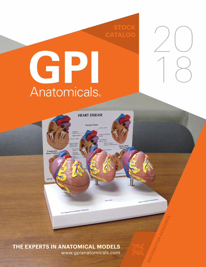
STOCKCATALOG
DIS
TRIB
UTO
R R
ESO
UR
CE
THE EXPERTS IN ANATOMICAL MODELSwww.gpianatomicals.com
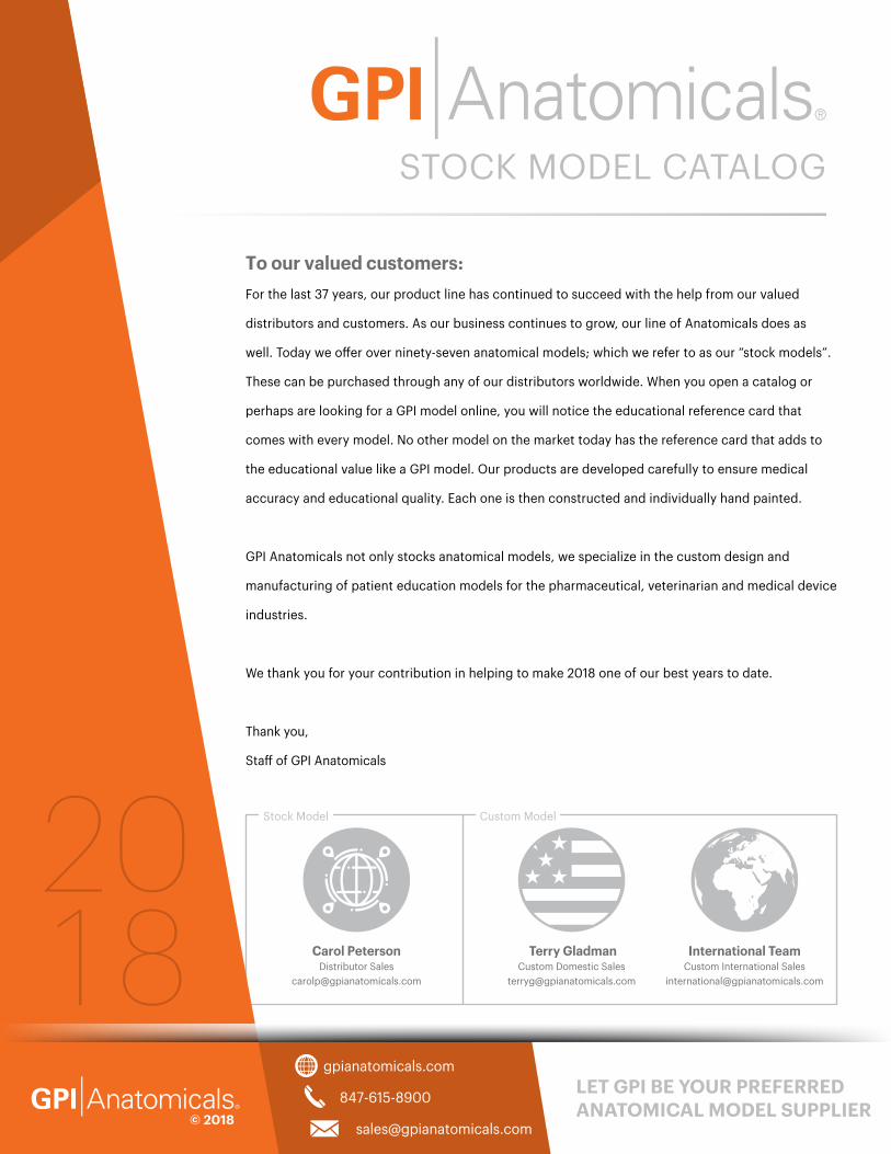
To our valued customers:
For the last 37 years, our product line has continued to succeed with the help from our valued
distributors and customers. As our business continues to grow, our line of Anatomicals does as
well. Today we offer over ninety-seven anatomical models; which we refer to as our “stock models”.
These can be purchased through any of our distributors worldwide. When you open a catalog or
perhaps are looking for a GPI model online, you will notice the educational reference card that
comes with every model. No other model on the market today has the reference card that adds to
the educational value like a GPI model. Our products are developed carefully to ensure medical
accuracy and educational quality. Each one is then constructed and individually hand painted.
GPI Anatomicals not only stocks anatomical models, we specialize in the custom design and
manufacturing of patient education models for the pharmaceutical, veterinarian and medical device
industries.
We thank you for your contribution in helping to make 2018 one of our best years to date.
Thank you,
Staff of GPI Anatomicals
847-615-8900
gpianatomicals.com
© 2018
LET GPI BE YOUR PREFERRED ANATOMICAL MODEL SUPPLIER
STOCK MODEL CATALOG
Carol PetersonDistributor Sales
Terry GladmanCustom Domestic Sales
International TeamCustom International Sales
Stock Model Custom Model
WELCOME
2
5
6
7
8-9
10-12
13
14
15-16
19
17-18
20-21
22-27
28-29
30-34
Ear
Eye
Sinus & Thyroid
Brain
Cardiovascular
Digestive
Obesity & Metabolic Syndrome
Diabetes
Respiratory
Female Health
Male Health
Skin
Joints & Bone Conditions
Vertebrae
Veterinary
Teeth & TMJ
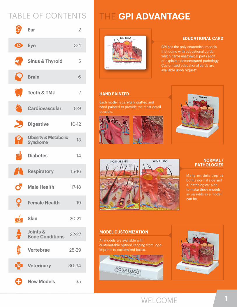
THE GPI ADVANTAGE
EDUCATIONAL CARD
GPI has the only anatomical models that come with educational cards, which name anatomical parts and/or explain a demonstrated pathology. Customized educational cards are available upon request.
HAND PAINTED
Each model is carefully crafted and hand painted to provide the most detail possible.
NORMAL / PATHOLOGIES
Many models depict both a normal side and a “pathologies” side to make these models as versatile as a model can be.
MODEL CUSTOMIZATION
All models are available with customizable options ranging from logo imprints to customized bases.
SKIN BURNSBlister
Epidermis
Dermis
Subcutaneous tissue
1st degree burnEpidermis damage
2nd degree burnEpidermis and
dermis damage
3rd degree burnEpidermis destroyed, dermis
destroyed, subcutaneous
tissue damage
GPI ©2005Vers. 2 PO 102374 www.gpianatomicals.comGPI Anatomicals®
YOUR LOGO
35New Models
3-4
1
2
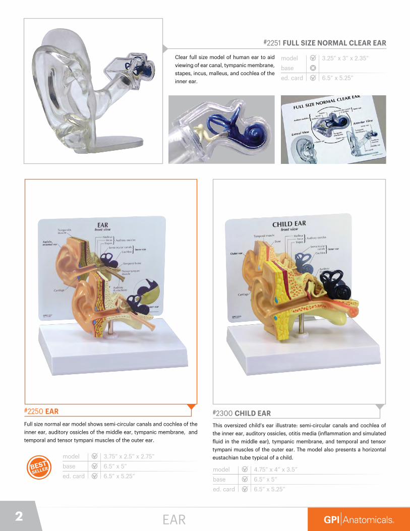
#2251 FULL SIZE NORMAL CLEAR EAR
Clear full size model of human ear to aid
viewing of ear canal, tympanic membrane,
stapes, incus, malleus, and cochlea of the
inner ear.
model
base
ed. card
3.25” x 3” x 2.35”
6.5” x 5.25”
#2250 EAR
Full size normal ear model shows semi-circular canals and cochlea of the
inner ear, auditory ossicles of the middle ear, tympanic membrane, and
temporal and tensor tympani muscles of the outer ear.
model
base
ed. card
3.75” x 2.5” x 2.75”
6.5” x 5”
6.5” x 5.25”
#2300 CHILD EAR
This oversized child’s ear illustrate: semi-circular canals and cochlea of
the inner ear, auditory ossicles, otitis media (inflammation and simulated
fluid in the middle ear), tympanic membrane, and temporal and tensor
tympani muscles of the outer ear. The model also presents a horizontal
eustachian tube typical of a child.
model
base
ed. card
4.75” x 4” x 3.5”
6.5” x 5”
6.5” x 5.25”
EAR

3847-615-8900 [email protected]
#2750 EYE
Oversized normal eye model with cut-away to
show inner anatomy: optic nerve, disc, macula,
retina, central retinal artery and vein. Lens and
cornea are removable.
model
base
ed. card
5” x 3” x 4”
6.5” x 5”
6.5” x 5.25”
#2780 CORNEA EYE
Oversized normal eye model with cut-away to show inner
anatomy. Includes four interchangeable corneas that show
various cornea conditions including bullous keratopathy, Fuch’s
endothelial dystrophy, keratoconus and normal. Lens and cornea
are removable.
model
base
ed. card
5” x 3” x 4”
6.5” x 5”
6.5” x 5.25”
EYE
Normal Keratoconus
Bullous Keratopathy Fuch’s Endothelial Dystrophy

4
#2800 CATARACT EYE
Oversized normal eye model with cut-away to show inner
anatomy. Includes five interchangeable lenses that show various
types of cataract conditions including subcapsular, capsular,
mature, cortical and nuclear. Lens and cornea are removable.
model
base
ed. card
5” x 3” x 4”
6.5” x 5”
6.5” x 5.25”
NuclearCortical Capsular
MatureSubcapsular
#2751 FULL EYE
Oversized normal eye model with split shell construction
to allow for viewing inner anatomy including optic nerve,
disc, macula, retina, and central retinal artery and vein.
Lens and cornea are removable.
model
base
ed. card
5” x 3” x 4”
6.5” x 5”
6.5” x 5.25”
EYE

5847-615-8900 [email protected]
#2851 CLEAR SINUS
Full size model demonstrating sinus cavities in the head. Clear face
shell allows for orientation of the sinus cavities: sphenoid, frontal,
ethmoid, maxillary, and mastoid.
modelbase
ed. card
7” x 5.5” x 5”
8.25” x 6.25”
#2850 SINUS
Full size, cut-away, normal model depicts a near median section through
the nose and nasal passages. Details include nasal cavity, soft and hard
palate, uvula, eustachian tube, and pharyngeal tonsil. Reverse side
shows ethmoid and maxillary sinus cavities.
modelbase
ed. card
4.5” x .625” x 5.25”
6.5” x 5.25”
6.5” x 5”
#3150 THYROID
Set of four average size thyroids and one larynx. Models show normal
thyroid, Hashimoto’s thyroiditis (lymphocytic thyroiditis), Graves’ disease,
papillary carcinoma, and the following structures: hyoid bone, thyroid
membrane, thyroid cartilage, cricoid cartilage, and trachea.
model
base
ed. card
3.5” x 2” x 5.5”
8.75” x 6.25”
8.25” x 6.25”
thyroid model details
Hashimoto’s Thyroiditis
3” x 1.5” x 2”
Graves’ Disease
2.75” x 1.5” x 2.25”
Papillary Carcinoma
2” x 1.25” x 2”
NormalThyroid
1.75” x 1” x 2”
SINUS & THYROID

6
#2900 BRAIN (HEALTHY/PATHOLOGIES)
Full size, segmented brain features half normal side and
three-piece sectioned pathologies side, as well as Circle of
Willis with aneurism. The brain, which sits inside a partial
skull, features the following pathologies which are also
illustrated on a two-sided education card: alcoholism,
Alzheimer’s, aneurism, depression related tumor, seizure
related tumor, migraine, multiple sclerosis, Parkinson’s
disease, stroke, and subdural hematoma.
modelbase
ed. card
5” x 6.75” x 5”
8.25” x 6.25”
Full size, normal, right-half brain features frontal, parietal, occipital
and temporal lobes; cerebellum, interthalmic adhesion, corpus
callosum, pons, midbrain-central peduncle, olfactory bulb, optic
nerve, optic chiasm, mammillary body, and medulla oblongata.
modelbase
ed. card
6.5” x 5” x 2.5”
6.5” x 5.25”
6.5” x 5”
#2950 HALF BRAIN (SENSORY/MOTOR)
Reverse side of model
BRAIN
7847-615-8900 [email protected]
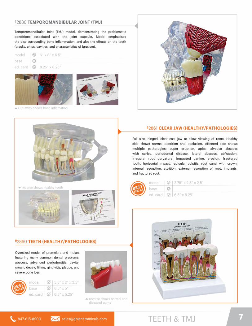
#2861 CLEAR JAW (HEALTHY/PATHOLOGIES)
Full size, hinged, clear cast jaw to allow viewing of roots. Healthy
side shows normal dentition and occlusion. Affected side shows
multiple pathologies: super eruption, apical alveolar abscess
with caries, periodontal disease, lateral abscess, abfraction,
irregular root curvature, impacted canine, erosion, fractured
tooth, horizontal impact, radicular pulpitis, root canal with crown,
internal resorption, attrition, external resorption of root, implants,
and fractured root.
model
base
ed. card 6.5” x 5.25”
2.75” x 2.5” x 2.5”
#2860 TEETH (HEALTHY/PATHOLOGIES)
Oversized model of premolars and molars
featuring many common dental problems:
abscess, advanced periodontitis, cavity,
crown, decay, filling, gingivitis, plaque, and
severe bone loss.
model
base
ed. card
5.5” x 2” x 3.5”
6.5” x 5”
6.5” x 5.25”reverse shows normal and diseased gums
reverse shows healthy teeth
#2880 TEMPOROMANDIBULAR JOINT (TMJ)
Temporomandibular Joint (TMJ) model, demonstrating the problematic
conditions associated with the joint capsule. Model emphasises
the disc surrounding bone inflammation, and also the effects on the teeth
(cracks, chips, cavities, and characteristics of bruxism).
model
base
ed. card
6” x 6” x 6.5”
8.25” x 6.25”
Cut-away shows bone inflamation
TEETH & TMJ
8
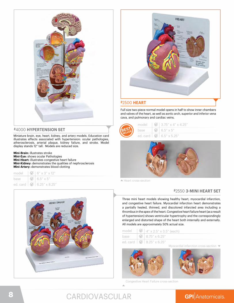
#4000 HYPERTENSION SETMiniature brain, eye, heart, kidney, and artery models. Education card illustrates effects associated with hypertension: ocular pathologies, atherosclerosis, arterial plaque, kidney failure, and stroke. Model display stands 12” tall. Models are reduced size.
Mini-Brain: illustrates strokeMini-Eye: shows ocular PathologiesMini-Heart: illustrates congestive heart failureMini-Kidney: demonstrates the qualities of nephrosclerosisMini-Artery: demonstrates blood clotting
model
base
ed. card
6” x 3” x 12”
6.5” x 5”
6.25” x 8.25”
#2500 HEART
Full size two-piece normal model opens in half to show inner chambers and valves of the heart, as well as aortic arch, superior and inferior vena cava, and pulmonary and cardiac veins.
model
base
ed. card
3.75” x 4” x 4.25”
6.5” x 5”
6.5” x 5.25”
Heart cross-section
#2550 3-MINI HEART SET
Three mini heart models showing healthy heart, myocardial infarction, and congestive heart failure. Myocardial infarction heart demonstrates a partially healed, thinned, and discolored infarcted area including a thrombus in the apex of the heart. Congestive heart failure heart (as a result of hypertension) shows ventricular hypertrophy and the correspondingly enlarged and distorted shape of the heart both internally and externally. All models are approximately 50% actual size.
model
base
ed. card
4” x 2.5” x 3.5” (each)
8.75” x 6.25”
8.25” x 6.25”Myocardial infarction cross-section
Congestive Heart Failure cross-section
CARDIOVASCULAR
9847-615-8900 [email protected]
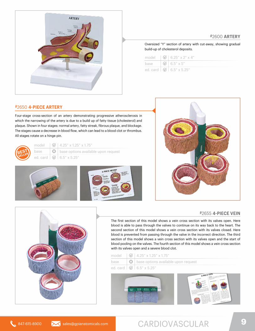
#2600 ARTERY
Oversized “Y” section of artery with cut-away, showing gradual
build-up of cholesterol deposits.
model
base
ed. card
6.25” x 2” x 4”
6.5” x 5”
6.5” x 5.25”
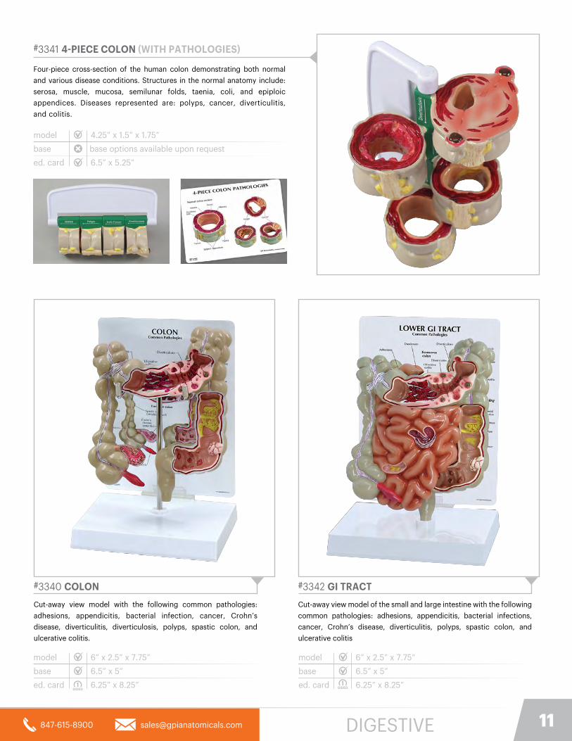
Four-stage cross-section of an artery demonstrating progressive atherosclerosis in
which the narrowing of the artery is due to a build up of fatty tissue (cholesterol) and
plaque. Shown in four stages: normal artery, fatty streak, fibrous plaque, and blockage.
The stages cause a decrease in blood flow, which can lead to a blood clot or thrombus.
All stages rotate on a hinge pin.
model
base
ed. card 6.5” x 5.25”
4.25” x 1.25” x 1.75”
#2650 4-PIECE ARTERY
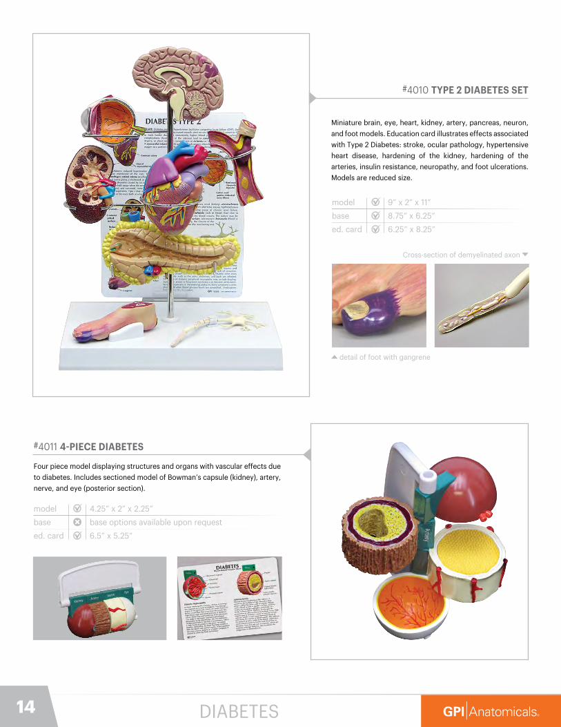
The first section of this model shows a vein cross section with its valves open. Here blood is able to pass through the valves to continue on its way back to the heart. The second section of this model shows a vein cross section with its valves closed. Here blood is prevented from passing through the valve in the incorrect direction. The third section of this model shows a vein cross section with its valves open and the start of blood pooling on the valves. The fourth section of this model shows a vein cross section with its valves open and a severe blood clot.
4-PIECE VEIN
©2016 GPIVers. 1 PO 102879
GPI Anatomicals® | www.gpianatomicals.com
valvulae venosae
(venous valves)
tunica intima
(endothelium cells)
lumen(empty space) tunica media
(smooth muscle)
tunica externa
(collagen)
BLO
OD
FLO
W Moderate ThrombosisSevere ThrombosisNormal Vein (open)
The third section of this
model shows a vein
cross section with its
valves open and the start
of blood pooling on the
valves. When blood starts
pooling on the valves
clots are formed when the
blood coagulates. Blood
pooling may occur from
inactivity or damage to
the veins.
The fourth section of
this model shows a vein
cross section with its
valves open and a severe
blood clot. Blood clots
can break away from the
valves and cause serious
damage to the body the
most common of which
is a pulmonary embolism.
The first section of this model shows a vein cross section
with its valves open. Here blood is able to pass through
the valves to continue on its way back to the heart. Staying
active through exercise is the best way too keep your veins
operating properly.
The second section of this
model shows a vein cross
section with its valves closed.
The veins operate under low
pressure and require these
one-way valves to prevent
the blood from flowing in the
reverse direction. Here blood
is prevented from passing
through the valve in the
incorrect direction.
Normal Vein (closed)
Thrombosis
Thrombosis is the formation of a blood clot inside a
blood vessel, obstructing the flow of blood through the
circulatory system.
model
base
ed. card 6.5” x 5.25”
4.25” x 1.25” x 1.75”
#2655 4-PIECE VEIN
CARDIOVASCULAR
base options available upon request
base options available upon request

10
#2010 GASTROESOPHAGEAL REFLUX DISEASE (GERD)
Four-piece model of progressive stages of GERD.
Conditions include: normal, sliding hiatal hernia and
acid reflux; chronic acid reflux/Barrett’s esophagus;
Barrett’s esophagus/ adeno carcinoma.
model
base
ed. card
4.75” x 3” x 3”
6.5” x 5.25”
ESOPHAGUS
Slidinghiatalhernia
Erosion of
esophageal
wall
Stomach acid
Tumor in
esophageal wall Barrett’sesophagus
Stomach and Esophagus Cross Section
Hiatal Hernia Barrett’s esophagus
with tumor
Stomach acid
Interior of
stomach
Irritation of
esophageal
wall caused
by acid reflux
GPI ©2008Vers. 2 PO 123456
www.gpianatomicals.com
GPI Anatomicals®
#3300 LIVER/GALLBLADDER (WITH GALLSTONES)
Full size liver and gallbladder with cut-away section showing inner
anatomy of gallbladder, including gallstones.
#3310 LIVER WITH PATHOLOGIES
Full size liver model with the following common pathologies: cirrhosis
(septal and nodular), biliary obstruction, gallstones, and tumors.
model
base
ed. card
8” x 4.5” x 5.5”
6.5” x 5”
8.25” x 6.25”
#2000 STOMACH WITH ULCERS
Full size cut-away section of stomach shows gastric ulcer, duodenal
ulcer, and esophageal inflammation.
#2001 STOMACH CANCER
Full size cut-away section of stomach showing a carcinoma tumor.
model
base
ed. card
7.75” x 1.25” x 6.25”
6.5” x 5”
6.5” x 5.25”
model
base
ed. card
7.75” x 1.25” x 6.25”
6.5” x 5”
6.5” x 5.25”
model
base
ed. card
8” x 4.5” x 5.5”
6.5” x 5”
6.5” x 5.25”
DIGESTIVE
stand available upon request
11847-615-8900 [email protected]
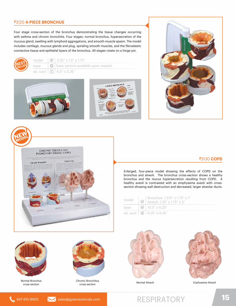
#3341 4-PIECE COLON (WITH PATHOLOGIES)
Four-piece cross-section of the human colon demonstrating both normal
and various disease conditions. Structures in the normal anatomy include:
serosa, muscle, mucosa, semilunar folds, taenia, coli, and epiploic
appendices. Diseases represented are: polyps, cancer, diverticulitis,
and colitis.
model
base
ed. card 6.5” x 5.25”
4.25” x 1.5” x 1.75”
#3340 COLON
Cut-away view model with the following common pathologies:
adhesions, appendicitis, bacterial infection, cancer, Crohn’s
disease, diverticulitis, diverticulosis, polyps, spastic colon, and
ulcerative colitis.
model
base
ed. card
6” x 2.5” x 7.75”
6.5” x 5”
6.25” x 8.25”
#3342 GI TRACT
Cut-away view model of the small and large intestine with the following
common pathologies: adhesions, appendicitis, bacterial infections,
cancer, Crohn’s disease, diverticulitis, polyps, spastic colon, and
ulcerative colitis
model
base
ed. card
6” x 2.5” x 7.75”
6.5” x 5”
6.25” x 8.25”
DIGESTIVE
base options available upon request

12
#3250 KIDNEY (NORMAL)
Oversized, longitudinal section of right kidney and adrenal gland. Model
highlights renal artery and vein, major and minor calyx, interlobular artery
and vein, and ureter.
#3260 KIDNEY (NORMAL/PATHOLOGIES)
This oversized, two-sided kidney model has a normal anatomy cutaway
on one side and a diseased anatomy cutaway on the other side depicting
infection, scarring, atrophy, urinary (kidney) stones, tumor, polycystic
disease, hypertension effects.
model
base
ed. card
3.75” x 2.5” x 6.5”
6.5” x 5”
5.25” x 6.5”
#3350 RECTUM
One and one half times life size cut-away model displaying ulcerative
colitis, internal and external fistula, internal and external hemorrhoids,
annular cancer, sessile polyp, submucosal abscess, skin tag, pedunculated
polyp, supralevator abscess, ischiorectal abscess, cryptitis, diverticulum,
condyloma acuminatum, fissure, and condyloma latum.
model
base
ed. card
5.5” x 2.5” x 7”
6.5” x 5”
6.5” x 5.25”
#3330 PANCREAS
Full size model shows pancreatic cancer, the gallbladder with stones,
a ruptured spleen, and a duodenal ulcer.
model
base
ed. card
7.75” x 2.5” x 5.75”
6.5” x 5”
6.5” x 5.25”
model
base
ed. card
3.75” x 2.5” x 6.5”
6.5” x 5”
6.5” x 5.25”
DIGESTIVE

13847-615-8900 [email protected]
#4020 METABOLIC SYNDROME SET
Miniature brain, heart, kidney, liver, artery, pancreas. Education
card illustrates effects associated with Metabolic Syndrome:
stroke, congestive heart failure, coronary artery disease,
angina, myocardial infarction, non-alcoholic fatty liver disease,
atherosclerosis, renal arteriosclerosis, and nephrosclerosis.
Mini-pancreas model demonstrates ocular pathologies. Models
are reduced size.
model
base
ed. card
9” x 2” x 13.5”
8.75” x 6.25”
6.25” x 8.25”
#3600 OBESITY
Pear and Apple shaped bodies in two cross-sections (one left, one right)
of the hip area. The Pear shape is referred to as “Pear body fat distribution
pattern” or “lower body fat.” This is mainly composed of subcutaneous fat.
The Apple shape is referred to as “Apple body fat distribution pattern” or
“intra-abdominal fat”. Intra-abdominal fat can be composed of visceral and
subcutaneous fat. Illustrating effects from obesity such as compression
from visceral fat on the colon, common iliac artery and veins, ureter, small
intestine, femoral nerve, etc. The education card depicts an Apple, Pear, and
a normal section of the same areas with callouts.
model
base
ed. card 6.25” x 8.25”
10” x 5.75” x 3.5”
10.5” x 6.5”
pear body shape apple body shape
METABOLIC SYNDROME
14
#4010 TYPE 2 DIABETES SET
Miniature brain, eye, heart, kidney, artery, pancreas, neuron,
and foot models. Education card illustrates effects associated
with Type 2 Diabetes: stroke, ocular pathology, hypertensive
heart disease, hardening of the kidney, hardening of the
arteries, insulin resistance, neuropathy, and foot ulcerations.
Models are reduced size.
model
base
ed. card
9” x 2” x 11”
8.75” x 6.25”
6.25” x 8.25”
#4011 4-PIECE DIABETES
Four piece model displaying structures and organs with vascular effects due
to diabetes. Includes sectioned model of Bowman’s capsule (kidney), artery,
nerve, and eye (posterior section).
model
base
ed. card 6.5” x 5.25”
4.25” x 2” x 2.25”
detail of foot with gangrene
Cross-section of demyelinated axon
DIABETES
base options available upon request
15847-615-8900 [email protected]
#3120 4-PIECE BRONCHUS
Four stage cross-section of the bronchus demonstrating the tissue changes occurring
with asthma and chronic bronchitis. Four stages: normal bronchus, hypersecretion of the
mucous gland, swelling with lymphoid aggregations, and smooth-muscle spasm. The model
includes cartilage, mucous glands and plug, spiraling smooth muscles, and the fibroelastic
connective tissue and epithelial layers of the bronchus. All stages rotate on a hinge pin.
model
base
ed. card 6.5” x 5.25”
4.25” x 1.5” x 1.75”
RESPIRATORY
Enlarged, four-piece model showing the effects of COPD on the bronchus and alveoli. The bronchus cross-section shows a healthy bronchus and the mucus hypersecretion resulting from COPD. A healthy aveoli is contrasted with an emphysema aveoli with cross-section showing wall destruction and decreased, larger alveolar ducts.
model
base
ed. card
Bronchus: 1.375” x 1.75” x 1”Alveoli: 1.75” x 1.75” x 2”
10.5” x 6.25”
8.25” x 6.25”
#3130 COPD
Normal Bronchuscross-section
Chronic Bronchituscross-section
Normal Alveoli Enphysema Alveoli
base options available upon request

16
#3100 LUNG (NORMAL)
Full size normal cut-away of right lung shows bronchus, arteries, vein,
two lymph nodes, bronchial passages, and trachea bifurcation.
model
base
ed. card
4.25” x 5.5” x 8.25”
6.5” x 5”
5.25” x 6.5”
#3110 LUNG SET (WITH PATHOLOGIES)
Set of two separate, full-size, two-sided lung models with
cutaway sections illustrating normal anatomy on one side and the
effects of COPD (Chronic Obstructive Pulmonary Disease),
cancer, and asthma on the opposite side.
model
base
ed. card
4.25” x 5.5” x 8.25” (each)
10.5” x 6.5”
8.25” x 6.25”
CANCER ASTHMA
COPD NORMAL
Reverse side of model
RESPIRATORY
17847-615-8900 [email protected]
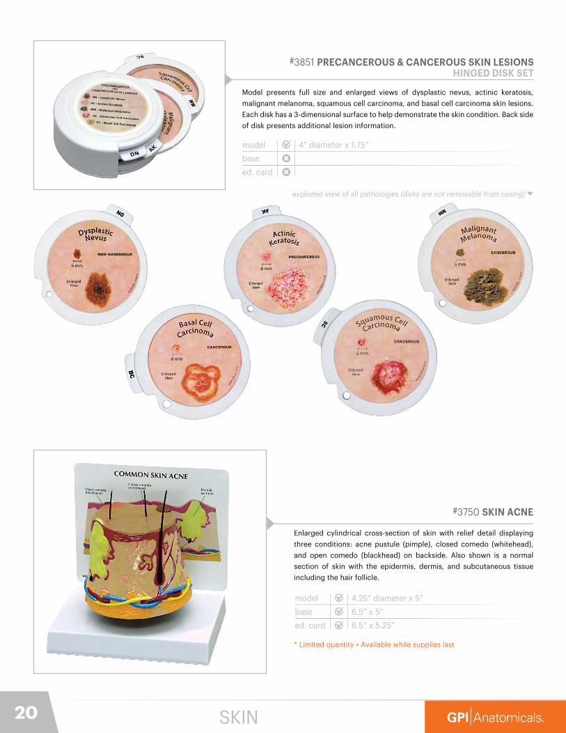
#3551 MALE PELVIS WITH 3D PROSTATE FRAME
Reduced size, mid sagittal section of the male pelvis. Base displays three
3-dimensional cross-sections of the prostate gland. Sections represent
stages (normal, moderate, advanced) of BPH (Benign Prostatic Hyperplasia/
Enlarged Prostate).
model
base
ed. card 6.5” x 5.25”
7.25” x 1” x 4.75”
8.75” x 6.25”
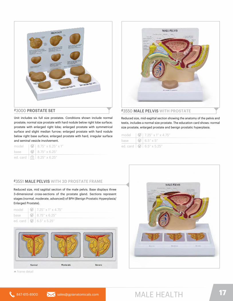
#3000 PROSTATE SET
Unit includes six full size prostates. Conditions shown include normal
prostate; normal size prostate with hard nodule below right lobe surface;
prostate with enlarged right lobe; enlarged prostate with symmetrical
surface and slight median furrow; enlarged prostate with hard nodule
below right base surface; enlarged prostate with hard, irregular surface
and seminal vesicle involvement.
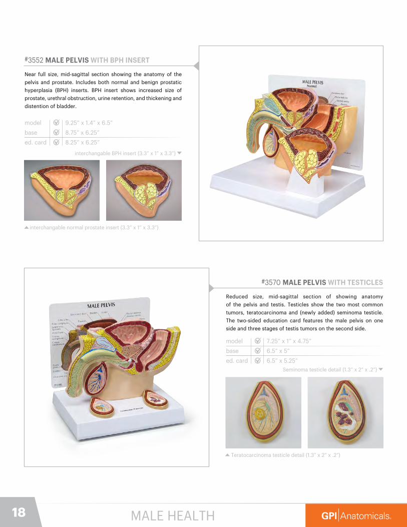
#3550 MALE PELVIS WITH PROSTATE
Reduced size, mid-sagittal section showing the anatomy of the pelvis and
testis, includes a normal size prostate. The education card shows: normal
size prostate, enlarged prostate and benign prostatic hyperplasia.
model
base
ed. card
7.25” x 1” x 4.75”
6.5” x 5”
6.5” x 5.25”model
base
ed. card
8.75” x 6.25” x 1”
8.75” x 6.25”
8.25” x 6.25”
frame detail
MALE HEALTH
18
#3552 MALE PELVIS WITH BPH INSERT
Near full size, mid-sagittal section showing the anatomy of the
pelvis and prostate. Includes both normal and benign prostatic
hyperplasia (BPH) inserts. BPH insert shows increased size of
prostate, urethral obstruction, urine retention, and thickening and
distention of bladder.
model
base
ed. card 8.25” x 6.25”
9.25” x 1.4” x 6.5”
interchangable normal prostate insert (3.3” x 1” x 3.3”)
interchangable BPH insert (3.3” x 1” x 3.3”)
#3570 MALE PELVIS WITH TESTICLES
Reduced size, mid-sagittal section of showing anatomy
of the pelvis and testis. Testicles show the two most common
tumors, teratocarcinoma and (newly added) seminoma testicle.
The two-sided education card features the male pelvis on one
side and three stages of testis tumors on the second side.
model
base
ed. card 6.5” x 5.25”
7.25” x 1” x 4.75”
Teratocarcinoma testicle detail (1.3” x 2” x .2”)
Seminoma testicle detail (1.3” x 2” x .2”)
8.75” x 6.25”
6.5” x 5”
MALE HEALTH

19847-615-8900 [email protected]
#3401 LEFT BREAST WITH IRREGULAR MASSES
Full size model is made of durable life-like material with embedded
lumps that simulate a fibrocystic mass and typical tumor. Model also
contains a lactiferous duct. Back side of education card shows locations
of irregular masses. Base art shows common self-inspection patterns.
#3450 BREAST CROSS-SECTION
Full size cross-section model depicts common pathologies such as
adenocarcinoma, cysts, fibroadenoma, and infiltrating scirrhous
carcinoma. Model also shows breast structures such as suspensory
ligaments, fat tissue, lymph nodes, muscles, and ribs.
model
base
ed. card
6.5” x 4.5” x 3”
8.75” x 6.25”
8.25” x 6.25”
#3480 UTERUS-OVARY
Full size cross-section model illustrates multiple pathologies,
some of which include: adhesions, carcinoma in four common
areas, cysts, endometriosis, fibroids, pedunculated fibroid tumor,
polyps, and salpingitis.
model
base
ed. card
8.75” x 6.25” x 2.5”
8.75” x 6.25”
8.25” x 6.25”
#3500 FEMALE PELVIS
Reduced size, mid-sagittal cross-section of the pelvis showing female anatomy, including the ovary and fallopian tube.
model
base
ed. card
6.25” x 2.75” x 5.5”
6.5” x 5”
6.5” x 5.25”
model
base
ed. card
8.75” x 6.25” x 3”
8.75” x 6.25”
8.25” x 6.25”
FEMALE HEALTH
20
#3851 PRECANCEROUS & CANCEROUS SKIN LESIONSHINGED DISK SET
Model presents full size and enlarged views of dysplastic nevus, actinic keratosis,
malignant melanoma, squamous cell carcinoma, and basal cell carcinoma skin lesions.
Each disk has a 3-dimensional surface to help demonstrate the skin condition. Back side
of disk presents additional lesion information.
model
base
ed. card
4” diameter x 1.75”
exploded view of all pathologies (disks are not removable from casing)
#3750 SKIN ACNE
Enlarged cylindrical cross-section of skin with relief detail displaying
three conditions: acne pustule (pimple), closed comedo (whitehead),
and open comedo (blackhead) on backside. Also shown is a normal
section of skin with the epidermis, dermis, and subcutaneous tissue
including the hair follicle.
model
base
ed. card
4.25” diameter x 5”
6.5” x 5”
6.5” x 5.25”
* Limited quantity • Available while supplies last
SKIN

21847-615-8900 [email protected]
NORMAL SKINNormal Skin side is common to all 2-sided skin models(#3751 Skin Acne, #3800 Skin Burn, and #3900 Aging Skin)
#3800 SKIN BURN & NORMAL SKIN
Enlarged, two-sided skin cross-section. The burn side of the model
illustrates first, second and third degree burns. The reverse side
illustrates normal skin anatomy.
model
base
ed. card
6” x 2.25” x 4”
6.5” x 5”
6.5” x 5.25”
#3751 SKIN ACNE & NORMAL SKINEnlarged two-sided skin cross-section. The “acne” side displays three
conditions: whitehead, blackhead and pustule/cyst. The reverse side
illustrates normal skin anatomy.
model
base
ed. card
6” x 2.25” x 4”
6.5” x 5”
6.5” x 5.25”
#3900 AGING SKIN / HAIR LOSS & NORMAL SKIN
Enlarged, two-sided skin cross-section. The “aging” side shows
increasing age in three stages from left to right; decreasing
elatin and collagen, and increasing wrinkles and age spots. Also
portrays decreased blood flow due to shrinking blood vessels,
and thinning hair follicle length and width associated with the
balding process. Graying hair, shrinking fat cells, and smaller
sweat glands are also shown.
model
base
ed. card
6” x 2.25” x 4”
6.5” x 5”
6.5” x 5.25”
SKIN

22
#1010 MENISCUS KNEE (6 TEARS)
Full size normal right knee demonstrating common
meniscus tears. Conditions shown include horizontal
tear, flap tear, bucket handle tear, degenerative,
radial tear, and longitudinal tear.
detailed view of all 6 tears
#1000 BASIC KNEE
Full size normal right knee includes femur, fibula, patella and tibia
bones; lateral and medial meniscus; quadriceps femoris tendon;
anterior cruciate, fibular and tibial collateral, and patellar and posterior
meniscofemoral ligaments.
model
base
ed. card
3.5” x 2.75” x 6”
6.5” x 5”
5.25” x 6.5”
#1060 MUSCLED KNEE
Full size normal right knee includes rectus femoris, vastus lateralis and
vastus medialis muscles; femur, fibula, patella and tibia bones; anterior
cruciate ligament, quadriceps femoris tendon plus six more ligaments
and tendons.
model
base
ed. card
3.5” x 3” x 10”
6.5” x 5”
5.25” x 6.5”
model
base
ed. card 6.25” x 8.25”
3.5” x 2.75” x 6”
8.75” x 6.25”
Horizontal Tear Bucket Handle TearFlap Tear Degenerative
Radial Tear Longitudinal Tear
JOINTS

23847-615-8900 [email protected]
#1311 MUSCLED HIP WITH SCIATIC NERVE
Full size normal right hip with proximal femur and lower lumbar
vertebrae including sciatic nerve, gluteus medius, gluteus
minimus, iliacus, inferior and superior gemellus, obturator
internus, piriformis and psoas muscles, articular capsule
ligaments, and L4-L5 with sacrum.
model
base
ed. card 6.25” x 8.25”
7.5” x 4.75” x 10”
posterior detail showing sciatic nerve
8.75” x 6.25”
#1260 BASIC HIP
Full size normal right hip with femur includes articular capsule of the
hip joint: greater and lesser trochanter; ilium, ischial spine, ischial
tuberosity, ischium and pubis bones.
#1310 MUSCLED HIP
Full size normal right hip with femur portion includes gluteus medius,
gluteus minimus, iliacus, inferior and superior gemellus, obturator
internus, piriformis and psoas muscles, and articular capsule ligaments.
model
base
ed. card
5” x 4.75” x 8.25”
6.5” x 5”
5.25” x 6.5”
model
base
ed. card
5” x 4.75” x 8.25”
6.5” x 5”
5.25” x 6.5”
JOINTS
24
#1900 MINI-MUSCLED JOINT SET
Set of four, reduced size (approximately 50% of stock models), joint models of the hip,
elbow, shoulder and knee. See model descriptions for model numbers 1060, 1310, 1810
and 1850 (elbow model does not contain the structures of the hand and wrist).
detailed look at all 4 detachable models
base
ed. card
10.5” x 6.25”
8.25” x 6.25”
Detachable Hip Size: 2.5” x 2.75” x 5”
Detachable Knee Size: 1.5” x 1.75” x 4.5”
Detachable Elbow Size: 1-1/2” x 5-1/4” x 3”
Detachable Shoulder Size: 3” x 3” x 3.75”
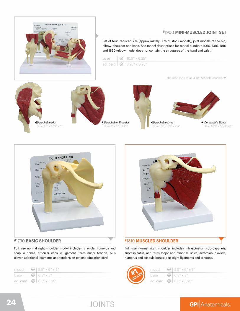
#1810 MUSCLED SHOULDER
Full size normal right shoulder includes infraspinatus, subscapularis,
supraspinatus, and teres major and minor muscles; acromion, clavicle,
humerus and scapula bones; plus eight ligaments and tendons.
model
base
ed. card
5.5” x 6” x 6”
6.5” x 5”
6.5” x 5.25”
#1790 BASIC SHOULDER
Full size normal right shoulder model includes: clavicle, humerus and
scapula bones; articular capsule ligament; teres minor tendon; plus
eleven additional ligaments and tendons on patient education card.
model
base
ed. card
5.5” x 6” x 6”
6.5” x 5”
6.5” x 5.25”
JOINTS
25847-615-8900 [email protected]
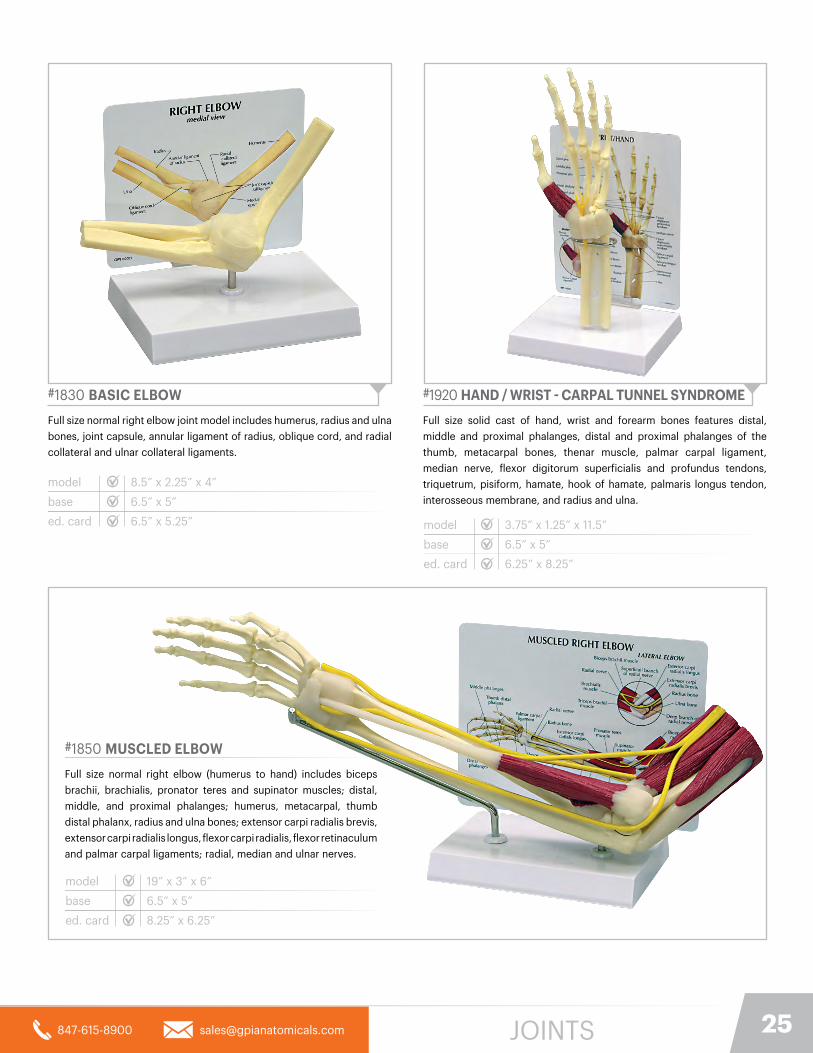
#1920 HAND / WRIST - CARPAL TUNNEL SYNDROME
Full size solid cast of hand, wrist and forearm bones features distal,
middle and proximal phalanges, distal and proximal phalanges of the
thumb, metacarpal bones, thenar muscle, palmar carpal ligament,
median nerve, flexor digitorum superficialis and profundus tendons,
triquetrum, pisiform, hamate, hook of hamate, palmaris longus tendon,
interosseous membrane, and radius and ulna.
model
base
ed. card
3.75” x 1.25” x 11.5”
6.5” x 5”
6.25” x 8.25”
#1830 BASIC ELBOW
Full size normal right elbow joint model includes humerus, radius and ulna
bones, joint capsule, annular ligament of radius, oblique cord, and radial
collateral and ulnar collateral ligaments.
model
base
ed. card
8.5” x 2.25” x 4”
6.5” x 5”
6.5” x 5.25”
#1850 MUSCLED ELBOW
Full size normal right elbow (humerus to hand) includes biceps
brachii, brachialis, pronator teres and supinator muscles; distal,
middle, and proximal phalanges; humerus, metacarpal, thumb
distal phalanx, radius and ulna bones; extensor carpi radialis brevis,
extensor carpi radialis longus, flexor carpi radialis, flexor retinaculum
and palmar carpal ligaments; radial, median and ulnar nerves.
model
base
ed. card
19” x 3” x 6”
6.5” x 5”
8.25” x 6.25”
JOINTS

26
#1985 GOUT
Full size ankle illustrates gouty tophi at the first metatarsal-phalangeal joint,
in the ankle, and around the Achilles tendon. Also shows inflammation,
redness and swelling of surrounding tissue, displaced tendons, and bone
erosion. Cross sections of the leg, ankle, talus, and heel show vasculature,
nerves, bones, tendons, cartilage, and joints of the foot.
model
base
ed. card
8.75” x 4” x 6”
8.75” x 6.25”
8.25” x 6.25”
#1980 FOOT /ANKLE - PLANTAR FASCIITIS
Full size solid cast of ankle and foot bones features the plantar
calcaneonavicular (spring) ligament with plantar fasciitis. Foot/ankle
anatomy also includes tibia, fibula, calcaneus, calcaneal (Achilles) tendon,
deltoid ligament, lateral (collateral) ligament, plantar aponeurosis,
cuneiform, phalanges, cuboid, navicular, and metatarsal bones.
model
base
ed. card
9” x 2.75” x 4”
6.5” x 5”
6.5” x 5.25”
#1931 RHEUMATOID ARTHRITIS HAND (RA)
Full size right hand with cutaway views to reveal the effects of rheumatoid arthritis on ligaments, flexor and extensor tendons, muscle, cartilage, bones, synovial membrane, and joint spaces. A cross-section of the metacarpophalangeal joint capsules shows progression of the disease, including synovial swelling, erosion, and degeneration. Swan neck and boutonniere deformities are depicted on the second and third phalanges, respectively. Flexor tendons can be lifted for better viewing of carpals.
model
base
ed. card
6” x 3.25” x 7.75”
6.5” x 5”
6.25” x 8.25”
#1930 OSTEOARTHRITIS HAND (OA)
Full size right hand with cutaway views to reveal effects of steoarthritis
including osteophytes (bone spurs), Heberden’s nodes Bouchard’s
nodes, and swan neck deformity of the thumb. Shows other anatomy
affected by osteoarthritis: ligaments, tendons, muscle, and cartilage. For comparison, normal anatomy is also shown.
model
base
ed. card
5” x 1.75” x 7.5”
6.5” x 5”
6.25” x 8.25”
JOINTS & BONE CONDITIONS

27847-615-8900 [email protected]
view of sciatic nerve
#1800 4-STAGE OSTEOARTHRITIS (SHOULDER)
Set of four shoulder models, reduced size, illustrating degenerative
joint disease (osteoarthritis) of glenohumeral joint: erosion to joint
articular cartilage, progression of degenerative disease, osteophyte
(bone spur) formation at the articular surfaces, and humeral head
flattening. Normal stage includes a full scapula. Advanced stage shows
acromioclavicular joint osteoarthritis and ligaments
modelbase
ed. card
4.5” x 2” x 4” (each)
8.25” x 6.25”
#1100 4-STAGE OSTEOARTHRITIS (KNEE)Set of four knee models, reduced size, illustrating degenerative
joint disease (osteoarthritis), erosion to joint articular cartilage,
progression of degenerative joint disease, and osteophytes (bone
spurs) at the articular surfaces. Advanced stage knee is articulating
for additional view of joint.
#1650 OSTEOPOROSIS HINGED DISK SETEnlarged cross-sections of bone illustrating the progressive thinning of the trabeculae that occurs due to osteoporosis.
modelbase
ed. card
4” diameter x 1.75”
6.5” x 5.25”
#1320 4-STAGE DEGENERATIVE HIPSet of four hip models, reduced size, illustrating degenerative joint diseases (osteoarthritis and osteoporosis) and fractures, erosion to joint articular cartilage, progression of degenerative joint disease, osteophytes (bone spurs) at the articular surfaces.
modelbase
ed. card
3.5” x 5” x 4” (each)
8.25” x 6.25”
10.5” x 6.25”
modelbase
ed. card
3” x 1.75” x 5.5” (each)
8.25” x 6.25”
10.5” x 6.25”
10.5” x 6.25”
humeral head detail
JOINTS & BONE CONDITIONS
28
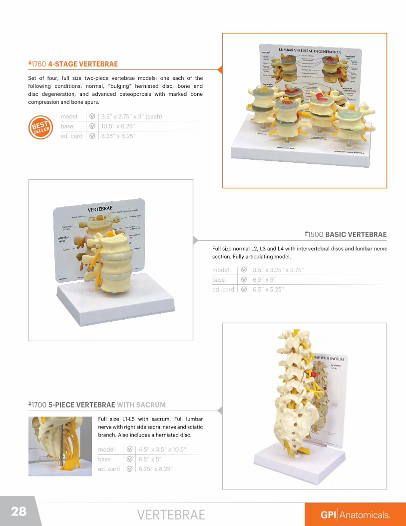
#1760 4-STAGE VERTEBRAE
Set of four, full size two-piece vertebrae models; one each of the
following conditions: normal, “bulging” herniated disc, bone and
disc degeneration, and advanced osteoporosis with marked bone
compression and bone spurs.
model
base
ed. card 8.25” x 6.25”
3.5” x 2.75” x 3” (each)
10.5” x 6.25”
#1700 5-PIECE VERTEBRAE WITH SACRUM
Full size L1-L5 with sacrum. Full lumbar
nerve with right side sacral nerve and sciatic
branch. Also includes a herniated disc.
model
base
ed. card 6.25” x 8.25”
4.5” x 3.5” x 10.5”
6.5” x 5”
#1500 BASIC VERTEBRAE
Full size normal L2, L3 and L4 with intervertebral discs and lumbar nerve
section. Fully articulating model.
model
base
ed. card 6.5” x 5.25”
3.5” x 3.25” x 3.75”
6.5” x 5”
VERTEBRAE
29847-615-8900 [email protected]
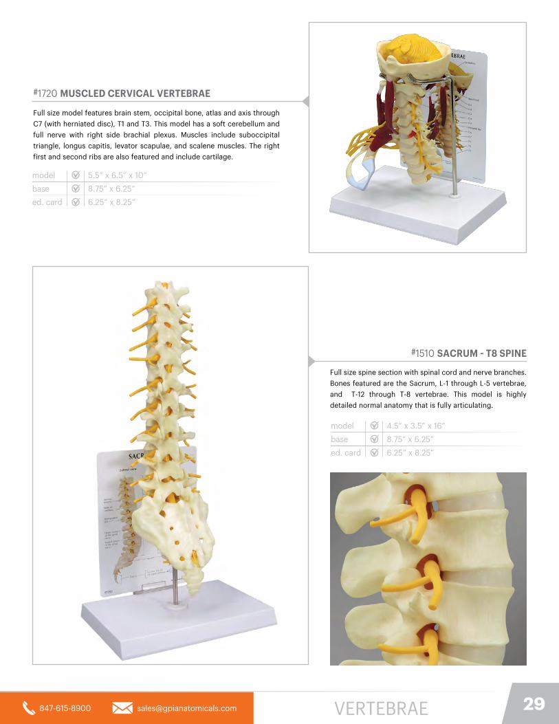
#1510 SACRUM - T8 SPINE
Full size spine section with spinal cord and nerve branches.
Bones featured are the Sacrum, L-1 through L-5 vertebrae,
and T-12 through T-8 vertebrae. This model is highly
detailed normal anatomy that is fully articulating.
model
base
ed. card
4.5” x 3.5” x 16”
8.75” x 6.25”
6.25” x 8.25”
#1720 MUSCLED CERVICAL VERTEBRAE
Full size model features brain stem, occipital bone, atlas and axis through
C7 (with herniated disc), T1 and T3. This model has a soft cerebellum and
full nerve with right side brachial plexus. Muscles include suboccipital
triangle, longus capitis, levator scapulae, and scalene muscles. The right
first and second ribs are also featured and include cartilage.
model
base
ed. card
5.5” x 6.5” x 10”
8.75” x 6.25”
6.25” x 8.25”
VERTEBRAE

VET
ERIN
ARY
CA
TALO
G

31847-615-8900 [email protected]
#9095 SKIN PARASITES
A parasite is an organism that grows, feeds, and is sheltered on or in another
type of organism while contributing nothing to the survival of its host. It is
fairly common for our pets to become the unknowing hosts to a number
of parasites. This model here shows in great detail the 3 most common
external parasites; mite, flea, and tick.
base
ed. card 8.25” x 6.25”
#9141 FELINE HEART / LUNG
Average feline heart and lungs infested with a single heartworm
(dirofilaria immitis). Cut-away view demonstrates structures in the heart
(right ventricle, pulmonary trunk, pulmonary arteries) and lungs where
adult-stage heartworms are normally found. The cut section of the lung
indicates patches of heartworm- induced irritation.
modelbase
ed. card
3.75” x 2.75” x 2.5”
6.5” x 5.25”
#9151 CANINE HEART / LUNG
Average canine heart and lungs infested with heartworms (dirofilaria
immitis). Cut-away view demonstrates structures in the heart (right
ventricle, pulmonary trunk, pulmonary arteries) and lungs where adult-
stage heartworms are normally found.
model
base
ed. card
3.75” x 2.75” x 2.5”
6.5” x 5”
6.5” x 5.25”
10.5” x 6.25”
Mite Size: 4” x 2.25” x 2.5”Model enlarged 330x
Flea Size: 4” x 2.25” x 2.5” Model enlarged 50x
Tick Size: 4” x 2.25” x 2.5” Model enlarged 32x
6.5” x 5”
VETERINARY

32 VETERINARY
#9190 FELINE JAW
Average size feline jaw depicts healthy teeth on the right side and
diseased and damaged teeth on the left. Featured pathologies: fractured
canine, periodontal disease, tartar accumulation, plaque, gingivitis, worn
incisors, retained deciduous tooth, missing premolar and (canine) gingival
recession. Jaws open, close, and separate for closer study.
modelbase
ed. card
2.75” x 2” x 1.25”
6.5” x 5.25”
#9191 CLEAR FELINE JAW
Healthy feline hinged jaw showing the roots of all teeth (incisors, canine,
premolars, molars). Jaws open, close, and separate for closer study.
model
base
ed. card
2.75” x 2” x 1.25”
6.5” x 5.25”
#9196 CLEAR CANINE JAW
Healthy canine hinged jaw showing the roots of all teeth (incisors, canine,
premolars, molars). Jaws open, close, and separate for closer study.
model
base
ed. card
4.25” x 3” x 2”
6.5” x 5.25”
#9195 CANINE JAW
Average size canine jaw depicts healthy teeth on the right side and
diseased and damaged teeth on the left. Featured pathologies: fractured
canine, periodontal disease, tartar accumulation, plaque, gingivitis, worn
incisors, retained deciduous tooth, missing premolar and (canine) gingival
recession. Jaws open, close, and separate for closer study.
modelbase
ed. card
4.75” x 2.75” x 2”
6.5” x 5.25”
33847-615-8900 [email protected]
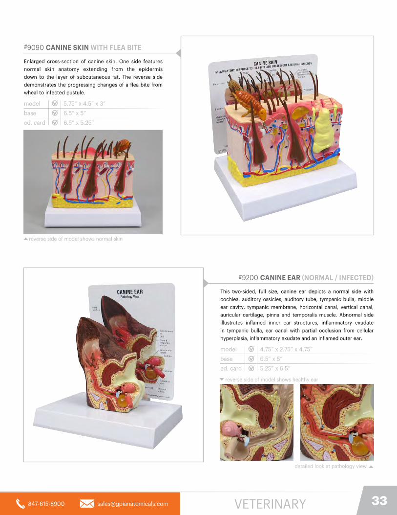
#9090 CANINE SKIN WITH FLEA BITE
Enlarged cross-section of canine skin. One side features
normal skin anatomy extending from the epidermis
down to the layer of subcutaneous fat. The reverse side
demonstrates the progressing changes of a flea bite from
wheal to infected pustule.
model
base
ed. card 6.5” x 5.25”
5.75” x 4.5” x 3”
reverse side of model shows normal skin
#9200 CANINE EAR (NORMAL / INFECTED)
This two-sided, full size, canine ear depicts a normal side with
cochlea, auditory ossicles, auditory tube, tympanic bulla, middle
ear cavity, tympanic membrane, horizontal canal, vertical canal,
auricular cartilage, pinna and temporalis muscle. Abnormal side
illustrates inflamed inner ear structures, inflammatory exudate
in tympanic bulla, ear canal with partial occlusion from cellular
hyperplasia, inflammatory exudate and an inflamed outer ear.
model
base
ed. card 5.25” x 6.5”
4.75” x 2.75” x 4.75”
reverse side of model shows healthy ear
6.5” x 5”
detailed look at pathology view
6.5” x 5”
VETERINARY
34 VETERINARY
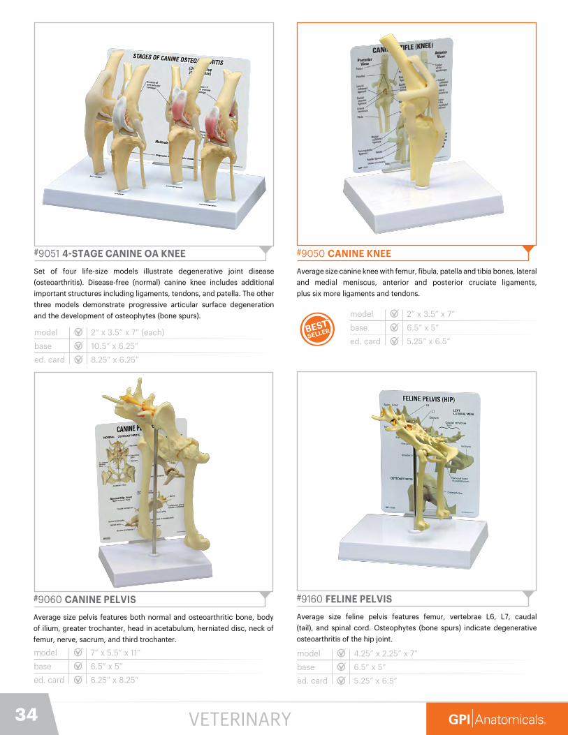
#9051 4-STAGE CANINE OA KNEE
Set of four life-size models illustrate degenerative joint disease
(osteoarthritis). Disease-free (normal) canine knee includes additional
important structures including ligaments, tendons, and patella. The other
three models demonstrate progressive articular surface degeneration
and the development of osteophytes (bone spurs).
model
base
ed. card
2” x 3.5” x 7” (each)
10.5” x 6.25”
8.25” x 6.25”
#9160 FELINE PELVIS
Average size feline pelvis features femur, vertebrae L6, L7, caudal
(tail), and spinal cord. Osteophytes (bone spurs) indicate degenerative
osteoarthritis of the hip joint.
model
base
ed. card
4.25” x 2.25” x 7”
6.5” x 5”
5.25” x 6.5”
#9060 CANINE PELVIS
Average size pelvis features both normal and osteoarthritic bone, body
of ilium, greater trochanter, head in acetabulum, herniated disc, neck of
femur, nerve, sacrum, and third trochanter.
model
base
ed. card
7” x 5.5” x 11”
6.5” x 5”
6.25” x 8.25”
#9050 CANINE KNEE
Average size canine knee with femur, fibula, patella and tibia bones, lateral
and medial meniscus, anterior and posterior cruciate ligaments,
plus six more ligaments and tendons.
model
base
ed. card
2” x 3.5” x 7”
6.5” x 5”
5.25” x 6.5”

#9070 CANINE ELBOW
Healthy left elbow of average size includes humerus, radius and ulna
bones, plus six ligaments.
model
base
ed. card
9.5” x 1.75” x 7.5”
8.75” x 6.25”
6.25” x 8.25”
#9075 CANINE SHOULDER
Average size canine shoulder with scapula, humerus, biceps brachii
tendon, coracobrachialis tendon, lateral glenohumeral ligament, medial
glenohumeral ligament, and transverse humeral ligament.
model
base
ed. card
3” x 2” x 11”
6.5” x 5”
6.25” x 8.25”
#9170 FELINE ELBOW / SHOULDER
Average size feline elbow and shoulder features normal bone and osteoarthritic changes to joints. Model includes the following bones: scapula, humerus, radius, ulna, and nine important ligaments and tendons.
model
base
ed. card
3.75” x .5” x 7.25”
6.5” x 5”
5.25” x 6.5”
#9080 CANINE VERTEBRAL COLUMN
Vertebral column of average size features five lumbar vertebrae and
discs, caudal (tail) vertebrae, and sacrum.
model
base
ed. card
8” x 2” x 2”
6.5” x 5”
6.5” x 5.25”
35847-615-8900 [email protected]

MODEL INDEX
36
Artery #2600. . . . . . . . . . . . . . . . . . . . . . . . . . . . . . . . . . . . . . . . . 9
Artery (4-pc) #2650 . . . . . . . . . . . . . . . . . . . . . . . . . . . . . . . . . . . 9
Bone Diseases-Hip (4-stage) #1320 . . . . . . . . . . . . . . . . . . . . . 27
Brain #2900 . . . . . . . . . . . . . . . . . . . . . . . . . . . . . . . . . . . . . . . . . 6
Brain (Sensory/Motor) #2950 . . . . . . . . . . . . . . . . . . . . . . . . . . . 6
Breast (Cross-Section) #3450 . . . . . . . . . . . . . . . . . . . . . . . . . . 19
Breast w/ Irregular Masses #3401. . . . . . . . . . . . . . . . . . . . . . . 19
Bronchus (4-pc) #3120. . . . . . . . . . . . . . . . . . . . . . . . . . . . . . . . 15
Canine Ear #9200. . . . . . . . . . . . . . . . . . . . . . . . . . . . . . . . . . . . 33
Canine Knee (4-Stage) #9051 . . . . . . . . . . . . . . . . . . . . . . . . . . 34
Canine Elbow #9070 . . . . . . . . . . . . . . . . . . . . . . . . . . . . . . . . . 35
Canine Heart / Lung #9151. . . . . . . . . . . . . . . . . . . . . . . . . . . . . 31
Canine Jaw #9195. . . . . . . . . . . . . . . . . . . . . . . . . . . . . . . . . . . . 32
Canine Jaw (clear) #9196. . . . . . . . . . . . . . . . . . . . . . . . . . . . . . 32
Canine Knee #9050 . . . . . . . . . . . . . . . . . . . . . . . . . . . . . . . . . . 34
Canine Pelvis #9060 . . . . . . . . . . . . . . . . . . . . . . . . . . . . . . . . . 34
Canine Shoulder #9075 . . . . . . . . . . . . . . . . . . . . . . . . . . . . . . . 35
Canine Skin #9090. . . . . . . . . . . . . . . . . . . . . . . . . . . . . . . . . . . 33
Canine Vertebral #9080 . . . . . . . . . . . . . . . . . . . . . . . . . . . . . . 35
Cataract Eye #2800 . . . . . . . . . . . . . . . . . . . . . . . . . . . . . . . . . . . 4
Colon #3340 . . . . . . . . . . . . . . . . . . . . . . . . . . . . . . . . . . . . . . . . .11
Colon (4-pc) #3341. . . . . . . . . . . . . . . . . . . . . . . . . . . . . . . . . . . .11
COPD #3130 . . . . . . . . . . . . . . . . . . . . . . . . . . . . . . . . . . . . . . . . 15
Cornea #2780 . . . . . . . . . . . . . . . . . . . . . . . . . . . . . . . . . . . . . . . . 3
Diabetes (4-pc) #4011 . . . . . . . . . . . . . . . . . . . . . . . . . . . . . . . . 14
Diabetes (set) #4010 . . . . . . . . . . . . . . . . . . . . . . . . . . . . . . . . . 14
Ear #2250 . . . . . . . . . . . . . . . . . . . . . . . . . . . . . . . . . . . . . . . . . . . 2
Ear (Child) #2300 . . . . . . . . . . . . . . . . . . . . . . . . . . . . . . . . . . . . . 2
Ear (Full Size - Clear) #2251 . . . . . . . . . . . . . . . . . . . . . . . . . . . . . 2
Elbow #1830 . . . . . . . . . . . . . . . . . . . . . . . . . . . . . . . . . . . . . . . . 25
Eye #2750 . . . . . . . . . . . . . . . . . . . . . . . . . . . . . . . . . . . . . . . . . . . 3
Eye (full) #2751 . . . . . . . . . . . . . . . . . . . . . . . . . . . . . . . . . . . . . . . 4
Feline Jaw (clear) #9191 . . . . . . . . . . . . . . . . . . . . . . . . . . . . . . . 32
Feline Elbow / Shoulder #9170 . . . . . . . . . . . . . . . . . . . . . . . . . 35
Feline Heart / Lung #9141 . . . . . . . . . . . . . . . . . . . . . . . . . . . . . 32
Feline Jaw – Model #9190 . . . . . . . . . . . . . . . . . . . . . . . . . . . . . 32
Feline Pelvis – Model #9160 . . . . . . . . . . . . . . . . . . . . . . . . . . . 34
Female Pelvis – Model #3500. . . . . . . . . . . . . . . . . . . . . . . . . . 19
Foot/Ankle – Plantar Fasciitis #1980 . . . . . . . . . . . . . . . . . . . . 26
GERD #2010 . . . . . . . . . . . . . . . . . . . . . . . . . . . . . . . . . . . . . . . . 10
GI Tract #3342 . . . . . . . . . . . . . . . . . . . . . . . . . . . . . . . . . . . . . . .11
Gout #1985 . . . . . . . . . . . . . . . . . . . . . . . . . . . . . . . . . . . . . . . . . 26
Hand/Wrist Carpal Tunnel #1920 . . . . . . . . . . . . . . . . . . . . . . . 25
Heart #2500 . . . . . . . . . . . . . . . . . . . . . . . . . . . . . . . . . . . . . . . . . 8
Heart (3-Mini Set) #2550 . . . . . . . . . . . . . . . . . . . . . . . . . . . . . . . 1
Hip #1260 . . . . . . . . . . . . . . . . . . . . . . . . . . . . . . . . . . . . . . . . . . 23
Hip (Muscled) #1310 . . . . . . . . . . . . . . . . . . . . . . . . . . . . . . . . . . 23
Hip (muscled w/ Sciatic Nerve) #1311. . . . . . . . . . . . . . . . . . . . 23
Hypertension (set) #4000 . . . . . . . . . . . . . . . . . . . . . . . . . . . . . 13
Jaw/Teeth (Human) #2861 . . . . . . . . . . . . . . . . . . . . . . . . . . . . . . 7
Joint Set (Mini-Muscled) #1900. . . . . . . . . . . . . . . . . . . . . . . . . 24
Kidney Normal #3250 . . . . . . . . . . . . . . . . . . . . . . . . . . . . . . . . 12
Kidney w/ Pathology #3260 . . . . . . . . . . . . . . . . . . . . . . . . . . . 12
Knee #1000. . . . . . . . . . . . . . . . . . . . . . . . . . . . . . . . . . . . . . . . . 22
Knee (muscled) #1060 . . . . . . . . . . . . . . . . . . . . . . . . . . . . . . . . 22
Liver/Gallbladder w/ Gallstones #3300 . . . . . . . . . . . . . . . . . . 10
Liver w/ Pathologies #3310 . . . . . . . . . . . . . . . . . . . . . . . . . . . . 10
Lung #3100 . . . . . . . . . . . . . . . . . . . . . . . . . . . . . . . . . . . . . . . . . 16
Lung Set w/ Pathologies #3110 . . . . . . . . . . . . . . . . . . . . . . . . . 16
Male Pelvis BPH #3552. . . . . . . . . . . . . . . . . . . . . . . . . . . . . . . . 18
Male Pelvis w/ Prostate #3551. . . . . . . . . . . . . . . . . . . . . . . . . . 17
Male Pelvis w/ Prostate #3550 . . . . . . . . . . . . . . . . . . . . . . . . . 17
Male Pelvis w/ Testicles #3570 . . . . . . . . . . . . . . . . . . . . . . . . . 18
Meniscus Tears #1010 . . . . . . . . . . . . . . . . . . . . . . . . . . . . . . . . 22
Metabolic Syndrome #4020 . . . . . . . . . . . . . . . . . . . . . . . . . . . 13
Muscled Elbow #1850 . . . . . . . . . . . . . . . . . . . . . . . . . . . . . . . . 25
Obesity #3600 . . . . . . . . . . . . . . . . . . . . . . . . . . . . . . . . . . . . . . 13
Osteoarthritis Hand #1930 . . . . . . . . . . . . . . . . . . . . . . . . . . . . 26
Osteoarthritic Knee (4-stage) #1100. . . . . . . . . . . . . . . . . . . . . 27
Osteoarthritis Shoulder (4-stage) #1800 . . . . . . . . . . . . . . . . . 27
Osteoporosis (Hinged Disk Set) #1650. . . . . . . . . . . . . . . . . . . 27
Pancreas #3330 . . . . . . . . . . . . . . . . . . . . . . . . . . . . . . . . . . . . . 12
Prostate Set #3000 . . . . . . . . . . . . . . . . . . . . . . . . . . . . . . . . . . 17
Rectum #3350 . . . . . . . . . . . . . . . . . . . . . . . . . . . . . . . . . . . . . . 12
Rheumatoid Arthritis (Hand) #1931 . . . . . . . . . . . . . . . . . . . . . . 26
Sacrum T8 Spine #1510 . . . . . . . . . . . . . . . . . . . . . . . . . . . . . . . 29
Shoulder #1790. . . . . . . . . . . . . . . . . . . . . . . . . . . . . . . . . . . . . . 24
Shoulder (muscled) #1810 . . . . . . . . . . . . . . . . . . . . . . . . . . . . . 24
Skin Acne #3750. . . . . . . . . . . . . . . . . . . . . . . . . . . . . . . . . . . . . 20
Skin Acne #3751 . . . . . . . . . . . . . . . . . . . . . . . . . . . . . . . . . . . . . 21
Skin (Aging - Hair loss) #3900 . . . . . . . . . . . . . . . . . . . . . . . . . 21
Skin Burn #3800. . . . . . . . . . . . . . . . . . . . . . . . . . . . . . . . . . . . . 21
Skin (Cancerous) #3851 . . . . . . . . . . . . . . . . . . . . . . . . . . . . . . . 20
Skin Parasites #9095 . . . . . . . . . . . . . . . . . . . . . . . . . . . . . . . . . 31
Sinus #2850 . . . . . . . . . . . . . . . . . . . . . . . . . . . . . . . . . . . . . . . . . 5
Sinus (Clear) #2851. . . . . . . . . . . . . . . . . . . . . . . . . . . . . . . . . . . . 5
Stomach Cancer #2001 . . . . . . . . . . . . . . . . . . . . . . . . . . . . . . . 10
Stomach w/ Ulcers #2000 . . . . . . . . . . . . . . . . . . . . . . . . . . . . 10
Teeth #2860 . . . . . . . . . . . . . . . . . . . . . . . . . . . . . . . . . . . . . . . . . 7
Thyroid #3150 . . . . . . . . . . . . . . . . . . . . . . . . . . . . . . . . . . . . . . . . 5
TMJ #2880. . . . . . . . . . . . . . . . . . . . . . . . . . . . . . . . . . . . . . . . . . . 7
Uterus-Ovary #3480. . . . . . . . . . . . . . . . . . . . . . . . . . . . . . . . . . 19
Vein (4-pc) #2655 . . . . . . . . . . . . . . . . . . . . . . . . . . . . . . . . . . . . . 9
Vertebrae #1500. . . . . . . . . . . . . . . . . . . . . . . . . . . . . . . . . . . . . 28
Vertebrae (4-stage) #1760 . . . . . . . . . . . . . . . . . . . . . . . . . . . . . 28
Vertebrae (Muscled Cervical) #1720. . . . . . . . . . . . . . . . . . . . . 29
Vertebrae w/ Sacrum #1700 . . . . . . . . . . . . . . . . . . . . . . . . . . . 29
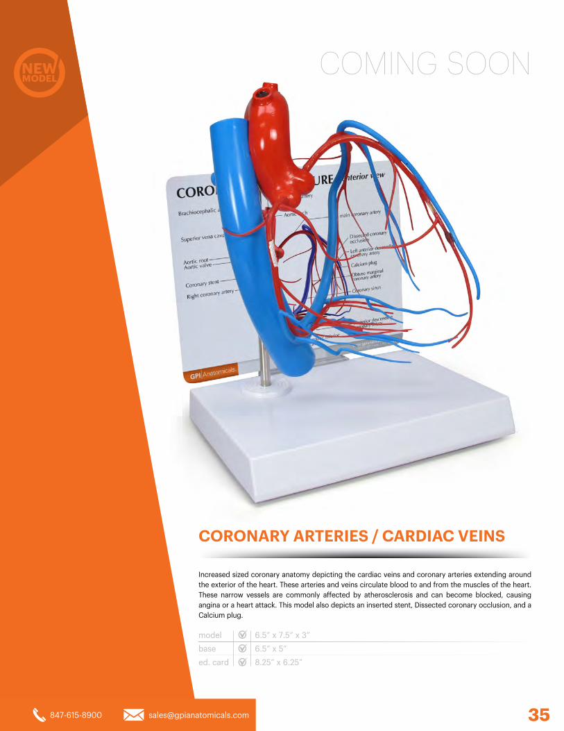
COMING SOON
CORONARY ARTERIES / CARDIAC VEINS
model
base
ed. card
6.5” x 7.5” x 3”
6.5” x 5”
8.25” x 6.25”
Increased sized coronary anatomy depicting the cardiac veins and coronary arteries extending around the exterior of the heart. These arteries and veins circulate blood to and from the muscles of the heart. These narrow vessels are commonly affected by atherosclerosis and can become blocked, causing angina or a heart attack. This model also depicts an inserted stent, Dissected coronary occlusion, and a Calcium plug.
35847-615-8900 [email protected]
847-615-8900 [email protected]
Visit our website to find a local dealer. ©2018 GPI Anatomicals
940 North Shore Drive • Lake Bluff, IL 60044
2018
STO
CK
MO
DEL
CAT
ALO
G
THE EXPERTS IN ANATOMICAL MODELSwww.gpianatomicals.com