Spondylolisthesis · Spondylolisthesis does not exist at birth Spondylolysis 4.4% at age 6, 6% in...
36
SPONDYLOLISTHESIS WAYNE CHENG, MD Bones and Spine
Transcript of Spondylolisthesis · Spondylolisthesis does not exist at birth Spondylolysis 4.4% at age 6, 6% in...

SPONDYLOLISTHESIS
WAYNE CHENG, MD
Bones and Spine

OUTLINE Definition
Classification
Clinical presentation
Imaging-measurement
Natural history
Treatment-
Non Surg Vs. Surg Decomp with fusion Vs. without fusion
Fusion with instrumentation Vs. without
Reduction Vs. In-situ fusion
High grade

SPONDYLOLYSIS VS. SPONDYLOLISTHESIS
Greek roots:
Spondylo = spine or vertebra
Lysis = to dissolve
Listhesis = to slide or slip

CLASSIFICATION
WILTSE,NEWMAN,MCNAB 1976
dysplastic isthmic degenerative
traumaticpathologic
20% listhesis
2F : 1 M
Cause
L5-S1
Most common
2M : 1F
Cause
L5-S16F : 1M
Age >40
10%F>60
cause

CLINICAL PRESENTATION
Mostly asymptomatic
Back pain
L5 root
Claudication
Vespers curse
Tight Hamstrings(80%)
High slip: L/S kyphosis
flattening of buttocks
forward thrust of Abd.
Absence of waistline

ASSOCIATED CONDITIONS
Spinal bifida occulta (24-70%)
Scoliosis (5-7%)
Disk Degeneration (50%)
Lumbarization/sacralization(7-9%)
Osteoarthritis (11-17%)

RADIOGRAPHIC STUDY
• Standing AP/Lateral
• Inc. slip 17%
• Inc slip angle 5 degree
• Oblique views
• Scottie dog’s neck
• Bone scan-cold/hot
• SPECT bone scan (single photon emission CT)
• MRI/CT myelogram

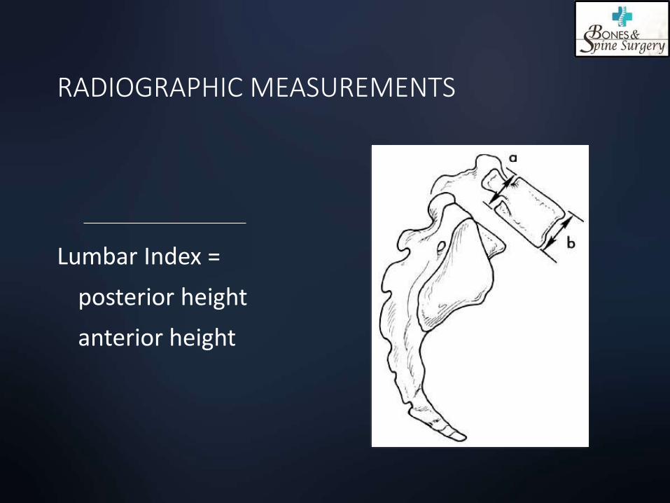
RADIOGRAPHIC MEASUREMENTS
Percentage slip
Meyerding
I 0-25%
II26-50%
III 51-75%
IV 76-100%
V > 100%

RADIOGRAPHIC MEASUREMENTS
Slip Angle:
Angle between L5 inf. Endplate to line perpendicular to post surface of S1.

RADIOGRAPHIC MEASUREMENTS
Sacral Inclination:
Angle between vertical line and back of S12

RADIOGRAPHIC MEASUREMENTS
Rounding ratio:
% of round shape of sacrum
RADIOGRAPHIC MEASUREMENTS
Lumbar Index =
posterior height
anterior height

PELVIC INCIDENCE

NATURAL HISTORY
Isthmic
Spondylolisthesis does not exist at birth
Spondylolysis 4.4% at age 6, 6% in adult
Development of pars defect does not cause pain in most patients
Progression is unusual.
Degenerative
Less understood
Progression of slip 30%
Clinical deterioration 10%
No correlation between slip progression and deterioration of Sx.
15% patients require surgery
Fredrickson, JBJS, 1984Fitzgerald,JBJS, 1976
Frymoyer,JAAOS, 1994

NON SURGICAL TREATMENT
Children
Asymptomatic:
no activity restriction
Frequency of x-ray
<10 YO q4month
11-15YO q6month
>15 YO q1-2years
Stop aggravating activities
Period of brace
Trunk strengthening
Adult
Mild analgesics/NSAID
Weight control
Aerobic exercise
Bracing
Epidural steroids

SURGICAL INDICATIONS
Persistence or recurrence of major symptoms for at least one year despite conservative treatment (incapacitating radicular pain or claudication) Quality of life
Progressive neurologic deficit (cauda equina, motor weakness)
Progressive slipping beyond 50% or high slip angle above 50 degree in a growing child(even if child is asymptomatic)
Gait or postural deformity unrelieved by therapy

SURGERY
Decompression alone without fusion.
Fusion
With decompression, without decompression.
Levels
Anterior vs. posterior vs. front&back
In situ vs. Reduction
Instrumentation Vs. no instrumentation

DECOMPRESSIONWITH FUSION VS. WITHOUT FUSION
(DEG. SPONDYLOLISTHESIS+STENOSIS)
Herkowitz,JBJS,1991
Prospective/random.
50 pts
3 year f/u
Post op listhesis: 96% non fused group
28% fused group
Op results: 96% good or excelnt. (fused
group)
44% good or excelnt (nonfusedgroup)
Epstein,J.spinal disord, 1998
Retrospective
290 pts with decomp. Only (<4mm, 10 degree)
10 year f/u
69%excellent, 13% good.
Only 2.7% required secondary fusion

INSTRUMENTED VS. NON-INSTRUMENTED FUSION
Zdeblick, Spine, 1993
Prospective, randomized.
124 pts.
F/u 16 month
Fusion rate 95% for rigid instrumt group vs. 65% for non instrumt group
95% good/excell. Result with Vs. 71% good/excell result without.
Herkowitz,Spine, 1997
Prospective, randomized
76 pts
F/u 24 month
Fusion rate 82% with instrumentation, 45% without.
76% good/excell. With instrumentation Vs. 85% without.

IN SITU FUSION VS. REDUCTION/FIXATION FOR HIGH GRADE SLIP
Wiltse,JBJS, 1989
8 young adults with grade III or IV with marked pre-op sciatica undergone In Situ fusion without decompression
F/u 5.5 years
All healed. Excellent results with resolution of marked pre-op sciatica
No neurologic complication
Edward & Spinal fixation Study group (Rothman-Simeone)
25 young adults with grade II to V undergone one stage post. Reduction /fixation.
F/U 2 years
91% slip correction,88% kyphosis correction
One nonunion. No long term neurologic complication

RISK FACTORS FOR PROGRESSION
Slip angle > 25 degree
Lumbar index (wedging ratio) < 75%
Rounded sacral end plate
Slip > 50%
Hyperlordosis (> 50 degree) L2-S1 or vertical sacral inclination
Female adolescents
Lumbosacral hypermobility ( > 4mm,10 degree deff. In flex. And ext. xray)
Pelvic incidence > 68degree (low grade); 79degree (high grade)

ADVANTAGES OF REDUCTION
Restore body posture and mechanics.
Decreases 30% chance of progression despite good in situ fusion
Permits full nerve decompression.
Limits fusion length.

INDICATIONS FOR REDUCTION
Cauda Equina Syndrome
Progressive Slip surpassing 50%
Severe deformity causing decompensation or distress
Major pain plus two or more risk factors

SPONDYLOPTOSIS
Posterior gradual instrumented reduction/fixation
Anterior resection + posterior fixation (Gaines procedure)
Fibula Strut graft

FIBULA STRUT GRAFT
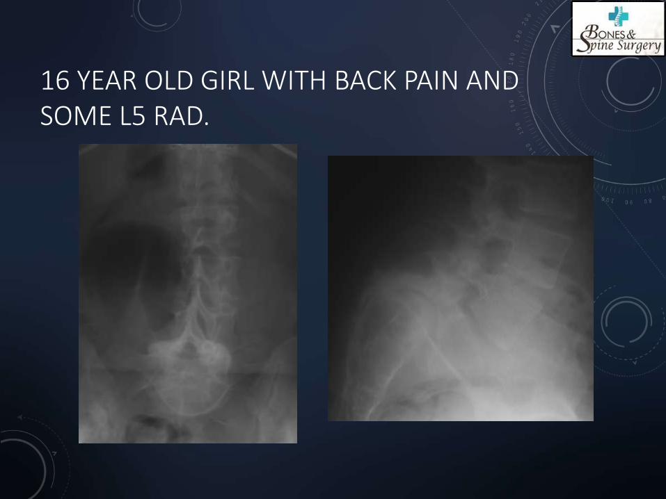
16 YEAR OLD GIRL WITH BACK PAIN AND SOME L5 RAD.

INTRA-OP

INTRA-OP REDUCTION

1 YEAR POST OP

SPONDYLOPTOSIS


CONCLUSION
PERSONAL PREFERENCE
80% of patients: Good trial of conservative treatment.
20% of patients: Adolescent without neuro. Deficit
In situ fusion with or without instrumentation
Adults with unstable degenerative spondylolisthesis
Post. Decompression, in situ fusion + instrumentation
High Grade Slip
INDIVIDUALIZE TO EACH PT’S NEEDS

2006 OSAE
A healthy 70 YO man has back and leg pain in an L5 distribution that is increased with standing and walking, relief by sitting. Neurological and pulse exam Normal. X-ray reveals spondylolisthesis, MRI with stenosis. Management should be:
A. Laminectomy B. Hemilaminectomy C. Laminectomy and fusion D. Anterior interbody fusion E. Posterior fusion

CASE DISCUSSION

CASE DISCUSSION