SPIKE1 Activates ROP GTPase to Modulate Petal … Activates ROP GTPase to Modulate Petal Growth and...
14
SPIKE1 Activates ROP GTPase to Modulate Petal Growth and Shape 1 Huibo Ren 2 , Xie Dang 2 , Yanqiu Yang, Dingquan Huang, Mengting Liu, Xiaowei Gao, and Deshu Lin* Basic Forestry and Proteomics Center (H.R., X.D., Y.Y., D.H., M.L., D.L.), Haixia Institute of Science and Technology (H.R., X.D., Y.Y., D.H., M.L., X.G., D.L.), and Horticultural Plant Biology and Metabolomics Center (X.G.), Fujian Agriculture and Forestry University, Fuzhou, Fujian 350002, China Plant organ growth and final shape rely on cell proliferation and, particularly, on cell expansion that largely determines the visible growth of plant organs. Arabidopsis (Arabidopsis thaliana) petals serve as an excellent model for dissecting the coordinated regulation of patterns of cell expansion and organ growth, but the molecular signaling mechanisms underlying this regulation remain largely unknown. Here, we demonstrate that during the late petal development stages, SPIKE1 (SPK1), encoding a guanine nucleotide exchange factor, activates Rho of Plants (ROP) GTPase proteins (ROP2, ROP4, and ROP6) to affect anisotropic expansion of epidermal cells in both petal blades and claws, thereby affecting anisotropic growth of the petal and the final characteristic organ shape. The petals of SPK1 knockdown mutants were significantly longer but narrower than those of the wild type, associated with increased anisotropic expansion of epidermal cells at late development stages. In addition, ROP2, ROP4, and ROP6 are activated by SPK1 to promote the isotropic organization of cortical microtubule arrays and thus inhibit anisotropic growth in the petal. Both knockdown of SPK1 and multiple rop mutants caused highly ordered cortical microtubule arrays that were transversely oriented relative to the axis of cell elongation after development stage 11. Taken together, our results suggest a SPK1-ROP-dependent signaling module that influences anisotropic growth in the petal and defines the final organ shape. A central question in plant organogenesis asks how gene activities contribute to organ growth and the final characteristic shape. Regulation of the growth and shape of plant organs is largely determined by the co- ordinated regulation of cell proliferation and cell ex- pansion (Tsukaya, 2006; Barkoulas et al., 2007; Powell and Lenhard, 2012; Peaucelle et al., 2015). Petals are fascinating organs in flowering plants, with their fra- grance and diverse color and shapes, which are im- portant for attracting pollinators to ensure successful pollination (Willmer et al., 2009; Yuan et al., 2013). The Arabidopsis (Arabidopsis thaliana) petal has a laminar structure with epidermal cells overlying the mesophyll and vasculature; therefore, it has emerged as an excel- lent model for dissecting the mechanisms underlying organ growth and cell expansion (Irish, 2008; Huang and Irish, 2016). In Arabidopsis, petal organogenesis is divided into several stages and depends on cell prolif- eration and cell expansion (Smyth et al., 1990; Huang and Irish, 2016). During the past decade, large numbers of Arabidopsis genes that play crucial roles in regulat- ing various aspects of cellular patterns and petal orga- nogenesis have been identified (Dinneny et al., 2004; Takeda et al., 2004; Szécsi et al., 2006; Irish, 2008; Li et al., 2008b; Nag et al., 2009; Varaud et al., 2011; Sauret-Güeto et al., 2013; Fujikura et al., 2014; Schiessl et al., 2014; Huang and Irish, 2015, 2016). For example, Arabidopsis JAGGED, which encodes a zinc finger transcription factor expressed in the distal domain of the petal, functions in the regulation of anisotropic growth of the petal (Sauret-Güeto et al., 2013; Schiessl et al., 2014). However, regulators that function in co- ordinating cell expansion and anisotropic growth of the petal remain largely unknown. Plant Rho-like small GTPase, usually termed Rho of Plants (ROP), belongs to a specific subfamily of the Rho GTPase family (Etienne-Manneville and Hall, 2002; Yang, 2002). In Arabidopsis, ROP proteins function as molecular signaling switches involved in a number of cellular processes, such as the regulation of cytoskeletal organization, cell wall patterning, the tip growth of pollen tubes, the interdigitated growth of leaf pavement cells, and the intracellular trafficking of PIN auxin ef- flux transporters (Lavy et al., 2007; Fu et al., 2009; Hazak et al., 2010; Qin and Yang, 2011; Wu et al., 2011; Craddock et al., 2012; Oda and Fukuda, 2012, 2013; Huang et al., 2014; Lin et al., 2015). As a molecular switch, ROP can shuttle between a GTP-bound active 1 This work was supported by the National Natural Science Foun- dation of China (grant nos. 31570278 and 31500160), the Natural Sci- ence Foundation of Fujian Province (grant no. 2015J01093), and the Fujian-Taiwan Joint Innovative Center for Germplasm Resources and Cultivation of Crops (FJ 2011 Program, grant no. 2015–75). 2 These authors contributed equally to the article. * Address correspondence to [email protected]. The author responsible for distribution of materials integral to the findings presented in this article in accordance with the policy de- scribed in the Instructions for Authors (www.plantphysiol.org) is: Deshu Lin ([email protected]). D.L. conceived and designed the experiments; H.R., X.D., Y.Y., D.H., M.L., and X.G. performed the experiments; D.L., H.R., and X.D. analyzed the data; D.L. wrote the article. www.plantphysiol.org/cgi/doi/10.1104/pp.16.00788 358 Plant Physiology Ò , September 2016, Vol. 172, pp. 358–371, www.plantphysiol.org Ó 2016 American Society of Plant Biologists. All rights reserved. www.plantphysiol.org on May 19, 2018 - Published by Downloaded from Copyright © 2016 American Society of Plant Biologists. All rights reserved.
Transcript of SPIKE1 Activates ROP GTPase to Modulate Petal … Activates ROP GTPase to Modulate Petal Growth and...
SPIKE1 Activates ROP GTPase to Modulate PetalGrowth and Shape1
Huibo Ren2, Xie Dang2, Yanqiu Yang, Dingquan Huang, Mengting Liu, Xiaowei Gao, and Deshu Lin*
Basic Forestry and Proteomics Center (H.R., X.D., Y.Y., D.H., M.L., D.L.), Haixia Institute of Science andTechnology (H.R., X.D., Y.Y., D.H., M.L., X.G., D.L.), and Horticultural Plant Biology and MetabolomicsCenter (X.G.), Fujian Agriculture and Forestry University, Fuzhou, Fujian 350002, China
Plant organ growth and final shape rely on cell proliferation and, particularly, on cell expansion that largely determines thevisible growth of plant organs. Arabidopsis (Arabidopsis thaliana) petals serve as an excellent model for dissecting the coordinatedregulation of patterns of cell expansion and organ growth, but the molecular signaling mechanisms underlying this regulationremain largely unknown. Here, we demonstrate that during the late petal development stages, SPIKE1 (SPK1), encoding aguanine nucleotide exchange factor, activates Rho of Plants (ROP) GTPase proteins (ROP2, ROP4, and ROP6) to affectanisotropic expansion of epidermal cells in both petal blades and claws, thereby affecting anisotropic growth of the petal andthe final characteristic organ shape. The petals of SPK1 knockdown mutants were significantly longer but narrower than those ofthe wild type, associated with increased anisotropic expansion of epidermal cells at late development stages. In addition, ROP2,ROP4, and ROP6 are activated by SPK1 to promote the isotropic organization of cortical microtubule arrays and thus inhibitanisotropic growth in the petal. Both knockdown of SPK1 and multiple rop mutants caused highly ordered cortical microtubulearrays that were transversely oriented relative to the axis of cell elongation after development stage 11. Taken together, ourresults suggest a SPK1-ROP-dependent signaling module that influences anisotropic growth in the petal and defines the finalorgan shape.
A central question in plant organogenesis asks howgene activities contribute to organ growth and the finalcharacteristic shape. Regulation of the growth andshape of plant organs is largely determined by the co-ordinated regulation of cell proliferation and cell ex-pansion (Tsukaya, 2006; Barkoulas et al., 2007; Powelland Lenhard, 2012; Peaucelle et al., 2015). Petals arefascinating organs in flowering plants, with their fra-grance and diverse color and shapes, which are im-portant for attracting pollinators to ensure successfulpollination (Willmer et al., 2009; Yuan et al., 2013). TheArabidopsis (Arabidopsis thaliana) petal has a laminarstructure with epidermal cells overlying the mesophylland vasculature; therefore, it has emerged as an excel-lent model for dissecting the mechanisms underlyingorgan growth and cell expansion (Irish, 2008; Huang
and Irish, 2016). In Arabidopsis, petal organogenesis isdivided into several stages and depends on cell prolif-eration and cell expansion (Smyth et al., 1990; Huangand Irish, 2016). During the past decade, large numbersof Arabidopsis genes that play crucial roles in regulat-ing various aspects of cellular patterns and petal orga-nogenesis have been identified (Dinneny et al., 2004;Takeda et al., 2004; Szécsi et al., 2006; Irish, 2008; Liet al., 2008b; Nag et al., 2009; Varaud et al., 2011;Sauret-Güeto et al., 2013; Fujikura et al., 2014; Schiesslet al., 2014; Huang and Irish, 2015, 2016). For example,Arabidopsis JAGGED, which encodes a zinc fingertranscription factor expressed in the distal domain ofthe petal, functions in the regulation of anisotropicgrowth of the petal (Sauret-Güeto et al., 2013; Schiesslet al., 2014). However, regulators that function in co-ordinating cell expansion and anisotropic growth ofthe petal remain largely unknown.
Plant Rho-like small GTPase, usually termed Rho ofPlants (ROP), belongs to a specific subfamily of the RhoGTPase family (Etienne-Manneville and Hall, 2002;Yang, 2002). In Arabidopsis, ROP proteins function asmolecular signaling switches involved in a number ofcellular processes, such as the regulation of cytoskeletalorganization, cell wall patterning, the tip growth ofpollen tubes, the interdigitated growth of leaf pavementcells, and the intracellular trafficking of PIN auxin ef-flux transporters (Lavy et al., 2007; Fu et al., 2009;Hazak et al., 2010; Qin and Yang, 2011; Wu et al., 2011;Craddock et al., 2012; Oda and Fukuda, 2012, 2013;Huang et al., 2014; Lin et al., 2015). As a molecularswitch, ROP can shuttle between a GTP-bound active
1 This work was supported by the National Natural Science Foun-dation of China (grant nos. 31570278 and 31500160), the Natural Sci-ence Foundation of Fujian Province (grant no. 2015J01093), and theFujian-Taiwan Joint Innovative Center for Germplasm Resources andCultivation of Crops (FJ 2011 Program, grant no. 2015–75).
2 These authors contributed equally to the article.* Address correspondence to [email protected] author responsible for distribution of materials integral to the
findings presented in this article in accordance with the policy de-scribed in the Instructions for Authors (www.plantphysiol.org) is:Deshu Lin ([email protected]).
D.L. conceived and designed the experiments; H.R., X.D., Y.Y.,D.H., M.L., and X.G. performed the experiments; D.L., H.R., andX.D. analyzed the data; D.L. wrote the article.
www.plantphysiol.org/cgi/doi/10.1104/pp.16.00788
358 Plant Physiology�, September 2016, Vol. 172, pp. 358–371, www.plantphysiol.org � 2016 American Society of Plant Biologists. All rights reserved. www.plantphysiol.orgon May 19, 2018 - Published by Downloaded from
Copyright © 2016 American Society of Plant Biologists. All rights reserved.

form and a GDP-bound inactive form, depending on itsactivating protein (ROPGAP) or guanine nucleotide ex-change factor (ROPGEF). Once activated by upstreamsignals, active ROP interacts with effector proteins torelay signals into downstream cellular components andthus induce cellular responses (Yang, 2002).ROPGEF proteins in the Arabidopsis genome have
been classified as two types: one is named SPIKE1(SPK1; Qiu et al., 2002; Basu et al., 2008; Zhang et al.,2010), a homolog of the animal single dock homologyregion; the other is a plant-specific ROPGEF family with14 members that contain a plant-specific ROP nucleo-tide exchange (PRONE) domain (Berken et al., 2005; Guet al., 2006). SPK1 loss-of-function mutation leads todwarf plant organs and severe defective leaf epidermis,such as reduced cell-cell adhesion, loss of pavement cellinterdigitation, and fewer trichome branches (Qiu et al.,2002; Basu et al., 2008; Zhang et al., 2010). SPK1 is in-volved in the activation of the ROP6-RIC1 pathway toregulate PIN2 internalization in root cells and, conse-quently, affects lateral root development (Lin et al.,2012). However, the molecular mechanisms by whichSPK1 regulates organ growth remain to be determined.Cortical microtubule (CMT) arrays orient the direc-
tion and deposition of cellulose microfibrils around thecell to build the cell wall and thus contribute to direc-tional cell expansion (i.e. anisotropy; Baskin, 2001, 2005;Wasteneys and Galway, 2003; Wasteneys, 2004; Smithand Oppenheimer, 2005; Ehrhardt and Shaw, 2006;Paredez et al., 2006; Crowell et al., 2009; Bringmannet al., 2012; Wolf et al., 2012). Previous work has shownthat, in the shoot apical meristem (SAM), CMT arrays inthe peripheral SAM cells display ordered circumferen-tial alignment to regulate growth anisotropy at bothcellular and tissue levels (Hamant et al., 2008; Uyttewaalet al., 2012). Loss of function of the microtubule-severingprotein katanin, a downstream component of the ROP6-RIC1 pathway (Lin et al., 2013), decreases the anisotropyof the CMT arrays in the SAM cells, which in turn affectsthe anisotropic growth of the SAM (Uyttewaal et al.,2012). A recent work has shown that the plant hormoneauxin regulates growth anisotropy of the SAM by af-fecting the activity of ROP6 and its downstream com-ponents that control circumferential CMT alignment atthe SAM (Sassi et al., 2014). In the absence of auxin ac-cumulation, the ROP6-dependent signaling keeps CMTarrays at the SAM in an ordered circumferential state toinhibit spontaneous lateral outgrowth, which leads tothe formation of a pin-like stem (Sassi et al., 2014).To further consolidate our understanding of ROP-
dependent signaling in the regulation of organ growth,we report here that SPK1 activates ROP proteins to in-hibit anisotropic growth of petals at late developmentstages and thus affect the final characteristic shape.Consistent with this, petal blades and claws of SPK1knockdown mutants have increased growth anisot-ropy, with significantly longer but narrower shape thanthose of the wild type, correlated with increased cellelongation and suppressed cell lateral expansion at latedevelopment stages. We also demonstrate that ROP
proteins are activated by SPK1 to affect anisotropicgrowth of the petal by promoting isotropic expansion ofepidermal cells in petal blades that is associated withthe isotropic organization of CMT arrays. Thus, wesuggest a ROP protein-dependent signaling modulethat contributes to anisotropic growth of the petalduring late development stages.
RESULTS
SPK1 Regulates Petal Anisotropic Growth
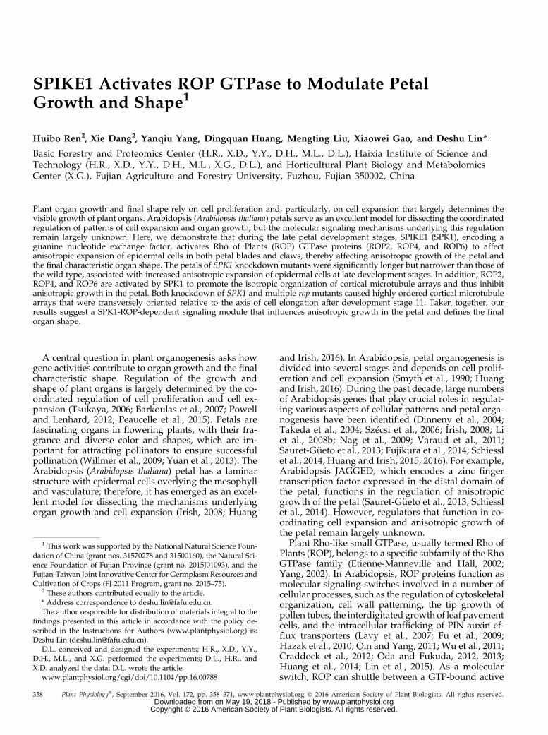
Tounderstandwhether SPK1 functions in petal growth,we first investigated SPK1 expression patterns andfound that SPK1 was highly expressed during petaldevelopment (Supplemental Fig. S1). Previous studieshave shown that SPK1 loss of function caused seedlinglethality when grown in soil at 30% to 50% humidity(Qiu et al., 2002; Lin et al., 2012). To better understandthe role of SPK1 in petal growth, we used SPK1knockdown mutants for the analysis of petal pheno-type. In an earlier study, we isolated a SPK1 weak-allele mutant (spk1-4) that carries a mutation causingmissplicing of the SPK1 pre-mRNA (Lin et al., 2012).We next examined the petal phenotype of the spk1-4mutant. At development stage 14, the mature petalblades of the spk1-4 mutant had significantly longerbut narrower shape and reduced petal blade areascompared with those of the wild type (Fig. 1), leadingto an increased petal index (the ratio of length towidth), a description of petal shape (Fig. 1D). More-over, the spk1-4 mutant petal showed a significant in-crease in claw length and a decrease in claw width(Supplemental Fig. S2). Expressing SPK1 in the spk1-4mutant by transforming pSPK1::SPK1 into the spk1-4mutant rescued the mutant petal phenotype in six in-dividual lines (two displayed in Supplemental Fig. S3).Thus, SPK1 knockdown mutants resulted in increasedanisotropic growth of petals.
To further confirm the role of SPK1 in the regulationof anisotropic growth in petals, we suppressed SPK1expression by transforming an RNA interference(RNAi) construct, containing 309 bp of the SPK1 codingsequence designed to create a double-stranded RNA,into wild-type plants. Twenty SPK1 RNAi lines wereisolated, and three lines with significantly reducedSPK1 transcriptional levels (Supplemental Fig. S4) werechosen for phenotype analysis. Compared with thewild type, at development stage 14, all three SPK1RNAi lines had increased anisotropic growth withmore elongated and narrower mature petals, as didthe spk1-4 mutant (Fig. 1).
The morphological events of petal development arewell described in Arabidopsis (Hill and Lord, 1989;Smyth et al., 1990). Before development stage 8, petalsgrow slowly depending on cell division, while petalsundergo a rapid lengthening process from develop-ment stage 9 until full flower opening (Smyth et al.,1990; Pyke and Page, 1998; Dinneny et al., 2004). Wenext asked how SPK1 influenced petal growth during
Plant Physiol. Vol. 172, 2016 359
SPIKE1 Regulation of Petal Anisotropic Growth
www.plantphysiol.orgon May 19, 2018 - Published by Downloaded from Copyright © 2016 American Society of Plant Biologists. All rights reserved.
late development stages by comparing petal phenotypebetween the wild type and the spk1-4 mutant at devel-opment stages 10 to 14. Measuring petal blade lengthand width at development stages 10 and 11 showedthat the spk1-4mutant had similar petal sizes to thewildtype (Fig. 2), while at development stage 12 and be-yond, the spk1-4 mutant had increased anisotropicgrowth with significantly longer and narrower petalsthan those of the wild type (Fig. 2).
Taken together, these results show that SPK1 partic-ipates in the inhibition of growth anisotropy of petalsduring late development stages, thus influencing thefinal characteristic shape.
SPK1 Regulates the Cell Expansion of PetalEpidermal Cells
To examine whether the petal phenotype observed inthe spk1mutant lines was associated with alterations incell proliferation and/or cell expansion, we first ana-lyzed cell numbers in mature spk1-4 petals (stage 14)and compared the results with those of the wild type.Our results demonstrated that cell numbers along thelength and width directions of the petals and total cellnumbers of petal blades in the spk1-4 mutant were
similar to those of the wild type (Supplemental Fig.S5), which demonstrated that SPK1 might not partic-ipate in controlling cell proliferation. Previous reportshave shown that loss-of-function SPK1 mutants ex-hibit severe defects in the interdigitated growth of leafepidermal cells (Qiu et al., 2002). Thus, we expectedthat the petal phenotype observed in the spk1 mutantlines was correlated with changes in cell expansionpatterns. We next analyzed the morphological phe-notypes of both adaxial and abaxial epidermal cells inwild-type and mutant petals, respectively.
Epidermal cells of the adaxial and abaxial surfacesof Arabidopsis mature petal blades can be easilydistinguished, as the adaxial epidermal cells have aconical shape with a pentagonal or hexagonal base,whereas the abaxial epidermal cells are flattened andshaped with interdigitated lobes (Irish, 2008). Anal-ysis of the two-dimensional ground area of the cellsin the middle part of the petal blades showed that, atdevelopment stage 14, adaxial epidermal cells in thespk1-4 mutant and the SPK1 RNAi lines showed in-creased anisotropic growth with increased cell elon-gation and suppressed cell lateral expansion comparedwith the wild type (Fig. 3), which was correlated withthemorphological phenotype of petal blades in spk1-4 and
Figure 1. Knockdown of SPK1 results in longer and narrower mature petals. A, Fully expanded flowers and petals at developmentstage 14 in the wild type, spk1-4, and SPK1 RNAi lines. Bar = 1 mm. B to E, Quantitative analyses of petal parameters for theindicated genotypes. Measurements are shown for petal blade length (B), petal blade width (C), petal blade index (i.e. lengthdivided by width; D), and petal blade area (E). The spk1-4mutant and the SPK1 RNAi lines had longer and narrower petals andreduced petal blade area than the wild type. Asterisks indicate significant differences from the wild type (**, P, 0.01, Student’st test). Values are given as means 6 SD of 16 petals.
360 Plant Physiol. Vol. 172, 2016
Ren et al.
www.plantphysiol.orgon May 19, 2018 - Published by Downloaded from Copyright © 2016 American Society of Plant Biologists. All rights reserved.

the SPK1 RNAi lines (Fig. 1A). Similarly, abaxial epider-mal cells in the spk1-4 mutant had increased growth an-isotropy compared with the wild type (Fig. 3).
Interestingly, the spk1 mutant lines exhibited severedefects in the interdigitated growth of abaxial epider-mal cells of petal blades (Fig. 3A), similar to the leaf
Figure 2. Petal phenotypes and quantitative analysis of the wild type and spk1-4 at development stages 10 to 14. A, Flowers and petals atdevelopment stages10 to14of thewild typeand spk1-4.Bar=1mm.B toE,Quantitativeanalysesofpetalbladeparameters for thewild typeandspk1-4 from development stages 10 to 14. The spk1-4mutant displayed similar petal blade length, width, and area to those of the wild type atstages 10 and 11 (Student’s t test, P. 0.05), whereas the spk1-4mutant had significantly longer and narrower petal blades and increased size inbladearea than thewild typeat development stage12andbeyond (Student’s t test,P,0.05).All dataare representedasmeans6 SDof 16petals.
Plant Physiol. Vol. 172, 2016 361
SPIKE1 Regulation of Petal Anisotropic Growth
www.plantphysiol.orgon May 19, 2018 - Published by Downloaded from Copyright © 2016 American Society of Plant Biologists. All rights reserved.

pavement cell defects induced by SPK1 mutation (Qiuet al., 2002). In addition, epidermal cells in the middlepart of petal claws of the spk1-4 mutant also had in-creased growth anisotropy (Supplemental Fig. S6),associated with the increased anisotropic shape ofpetal claws in the spk1-4 mutant (Supplemental Fig.S2). In the spk1-4mutant, cell areas in both petal bladesand claws were reduced in size (Fig. 3, B and C;Supplemental Fig. S6), correlated with changes in thepetal size of the mutant.
To determine whether the SPK1-regulated cell ex-pansion is associated with anisotropic growth of petals
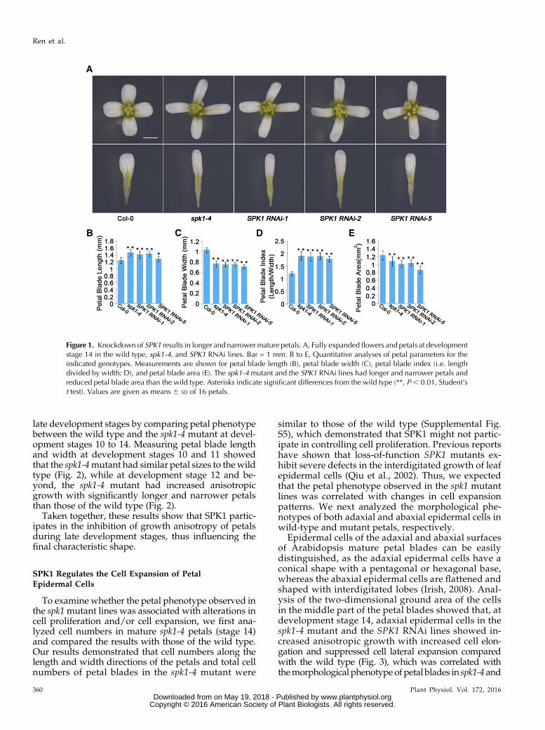
during late development stages, we next performeddetailed phenotype analyses in the spk1-4 mutant afterdevelopment stage 10. We showed that, in wild-typepetal blades, both adaxial and abaxial epidermal cellsunderwent both longitudinal and radial expansion atdevelopment stages 10 to 14 (Fig. 4; Supplemental Fig.S7). At development stages 10 and 11, the length andwidth of adaxial and abaxial epidermal cells in the spk1-4 mutant were similar to those of the wild type (Fig. 4;Supplemental Fig. S7). In striking contrast to the wildtype, after development stage 12, abaxial epidermalcells in the spk1-4 mutant expanded less radially and
Figure 3. Adaxial/abaxial epidermal cell shape of the wild type, spk1-4, and SPK1 RNAi lines at development stage 14. A,Epidermal cell shape in the middle part of petal blades from the wild type, spk1-4, and SPK1 RNAi lines at development stage 14.Epidermal cells of the spk1-4mutant and the SPK1 RNAi lines had increased growth anisotropy with longer and narrower shapethan the wild type. Bars = 10 mm. B and C, Quantitative analyses of adaxial/abaxial epidermal cell parameters for the indicatedgenotypes at development stage 14. The spk1-4 mutant and the SPK1 RNAi lines displayed significantly increased cell length,reduced cell width, and reduced cell area compared with the wild type. Asterisks indicate significant differences from the wildtype at P , 0.05 (Student’s t test). Values are given as means 6 SD of more than 500 cells from 12 petals.
362 Plant Physiol. Vol. 172, 2016
Ren et al.
www.plantphysiol.orgon May 19, 2018 - Published by Downloaded from Copyright © 2016 American Society of Plant Biologists. All rights reserved.
more longitudinally than in the wild type throughout thelate development stages 12 to 14, and mutant cells weremarked by a cessation of radial expansion at stage 12 anda dramatic increase in longitudinal expansion after stage12 (Fig. 4). In addition, adaxial epidermal cells in the spk1-4 mutant petal blades also showed increases in cell elon-gation after development stage 12 (Supplemental Fig. S7).Taken together, these observations suggest that SPK1
function is required mainly in the later phase during latedevelopment stages to influence anisotropic expansionof petal epidermal cells, correlating with growth anisot-ropy in both the distal and basal parts of petals, whichcontributes to the final characteristic shape of the petal.
ROP2, ROP4, and ROP6 Act Redundantly to RegulateAnisotropic Growth in Petals
We next addressed the downstream mechanism bywhich SPK1 mediates the effect on growth anisotropy
in petals. We speculated that, as a GEF, SPK1 must di-rectly activate ROP GTPase proteins and transmit thedevelopmental signals to downstream targets (Basuet al., 2008; Lin et al., 2012). We analyzed available ropknockout mutants for petal phenotype. However, norop single mutant displayed an obvious defect in petalmorphology (data not shown), suggesting that ROPgenes may have overlapping functions during petalmorphogenesis. Although neither rop2 nor rop6 singlemutants showed significant alterations in petal bladelength and width (Supplemental Fig. S8), the rop2 rop6double mutant at development stage 14 displayed anincrease in growth anisotropy with longer and nar-rower petal blades (Fig. 5), associated with increasedepidermal cell length and reduced epidermal cell width(Fig. 6; Supplemental Fig. S9). Expressing ROP2 orROP6 by transformation of pROP2::GFP-ROP2 orpROP6::GFP-ROP6 into the rop2 rop6 double mutant res-cued the petal blade phenotype (Supplemental Fig. S10),
Figure 4. Abaxial epidermal cell shape of the wild type and spk1-4 at development stages 10 to 14. A, Abaxial epidermal cell shapeof wild-type and spk1-4 petal blades at development stages 10 to 14. Bar = 10 mm. B to E, Measurements for abaxial epidermal cellparameters of the wild type and spk1-4 at development stages 10 to 14. B, Cell length measurements at various stages showed thatthe spk1-4mutant had similar cell length to the wild type at stages 10 and 11 (P. 0.05, Student’s t test) but displayed significantlyincreased cell length at stages 12 to 14 comparedwith thewild type (P, 0.05, Student’s t test). C, Cell widthmeasurements showedthat the spk1-4 mutant had similar cell width to the wild type at stages 10 and 11 (P . 0.05, Student’s t test) but displayed signif-icantly increased cell width at stages 12 to 14 comparedwith thewild type (P, 0.05, Student’s t test). D, The spk1-4mutant showedsignificantly increased cell index (cell length divided by cell width) at stage 11 and beyond. E, The spk1-4mutant had similar size incell area to the wild type before stage 11 (P . 0.05, Student’s t test) but had significantly reduced size in cell area at stages 11 to14 (P , 0.05, Student’s t test). Values are given as means 6 SD of more than 500 cells from 12 petals.
Plant Physiol. Vol. 172, 2016 363
SPIKE1 Regulation of Petal Anisotropic Growth
www.plantphysiol.orgon May 19, 2018 - Published by Downloaded from Copyright © 2016 American Society of Plant Biologists. All rights reserved.

which confirmed that the petal phenotype of the rop2rop6 double mutant was caused by the loss of bothROP2 and ROP6 functions.
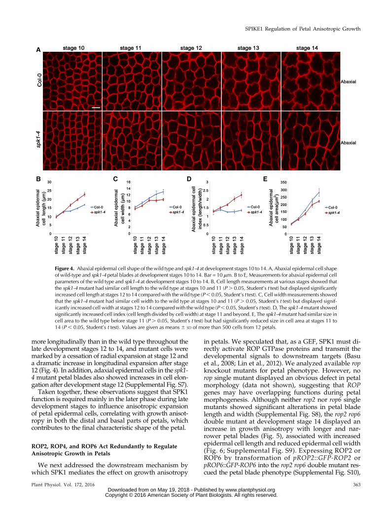
The mature petal blade phenotype of the rop2 rop6double mutant was milder than that of the spk1-4 mu-tant, which could be caused by the overlapping func-tion of ROP2 and ROP4, which share 97% amino acididentity (Fu et al., 2005). Next, we suppressed ROP4expression by transferring a ROP4 RNAi construct intorop2 rop6 double mutant plants. Multiple rop2 rop6ROP4 RNAi lines (referred as rop2 rop6 ROP4i) wereidentified, and three lines with significantly reducedROP4 transcriptional levels were chosen for phenotypeanalysis (Supplemental Fig. S11). Consistent with theoverlapping roles of ROP4 and ROP2, the phenotypicdefects (stage 14) of petal blades and epidermal cellsin the rop2 rop6 ROP4i lines were more severe thanthose of the rop2 rop6 double mutant (Figs. 5 and 6;Supplemental Fig. S9). Moreover, a mutant line, re-ferred to as rop2 rop6 ROP4i-4, which displayed thelowest expression level of ROP4 (Supplemental Fig.S11), had a similar phenotype to the spk1-4 mutant and
the SPK1 RNAi lines (Figs. 5 and 6; Supplemental Fig.S9). Furthermore, cell numbers along the length andwidth directions of the petals in the rop2 rop6 doublemutant and the rop2 rop6 ROP4i-4 mutant were similarto those of the wild type (Supplemental Fig. S5), whichdemonstrated that ROP protein-regulated growth an-isotropy was correlated with cell expansion rather thancell proliferation.
Moreover, the lengths of petal claws and claw cells inboth the rop2 rop6 mutant and the rop2 rop6 ROP4i-4mutant showed significant increases compared withthose of the wild type (Supplemental Figs. S12 and S13).Thewidths of petal claws and claw cells were normal inthe rop2 rop6 mutant but were reduced significantly inthe rop2 rop6 ROP4i-4 mutant (Supplemental Figs. S12and S13). Therefore, we conclude that ROP2, ROP4, andROP6 act redundantly to inhibit growth anisotropy atboth the distal and basal parts of petals.
To determine whether ROP proteins function re-dundantly in the regulation of growth anisotropy at latedevelopment stages, we performed detailed phenotypeanalyses from development stages 10 to 14. Our results
Figure 5. ROP2, ROP4, and ROP6 redundantly regulate growth anisotropy of petal blades. A, Fully expanded flowers and petalsat development stage 14 in the wild type, the rop2 rop6 double mutant, and the rop2 rop6 ROP4 RNAi lines. Bar = 1 mm. B to E,Quantitative analyses of petal parameters for the indicated genotypes. Measurements are shown for petal blade length (B), petalbladewidth (C), petal blade index (i.e. length divided bywidth; D), and petal blade area (E). The rop2 rop6 doublemutant and therop2 rop6 ROP4 RNAi lines had longer and narrower petals than the wild type (P , 0.05, Student’s t test) but displayed similarpetal blade area to thewild type (P. 0.05, Student’s t test). Asterisks indicate significant differences from thewild type at P, 0.05(Student’s t test). Values are given as means 6 SD of 16 petals.
364 Plant Physiol. Vol. 172, 2016
Ren et al.
www.plantphysiol.orgon May 19, 2018 - Published by Downloaded from Copyright © 2016 American Society of Plant Biologists. All rights reserved.
demonstrated that the rop2 rop6 mutant at develop-ment stages 13 and 14, and the rop2 rop6 ROP4i-4mutant at development stage 12 and beyond, hadsignificantly increased anisotropic growth with moreelongated and narrower petals (Supplemental Fig.S14), which were associated with increased cell lengthand reduced cell width, than those of the wild type(Fig. 6; Supplemental Fig. S9).
SPK1 Is Required for the Activation of Both ROP2 andROP6 in Floral Organs
We next tested the hypothesis that SPK1 is the directactivator of ROP proteins in the regulation of aniso-tropic growth in petals. To determine whether SPK1 isrequired for the activation of ROP2 and ROP6 duringpetal growth, we examined ROP activities in the wildtype and the spk1 mutant lines. The active form of a
Figure 6. Abaxial epidermal cell shape of the wild type, the rop2 rop6 mutant, and the rop2 rop6 RON4i-4 mutant at devel-opment stages 10 to 14. A to C, Abaxial epidermal cell shape of the indicated genotypes. The wild type (A), rop2 rop6 (B), androp2 rop6 rop4i-4 (C) are shown at development stages 10 to 14. Bar = 10 mm. D to G, Quantitative analyses of parameters ofabaxial epidermal cells for the wild type, the rop2 rop6mutant, and the rop2 rop6 rop4i-4mutant at development stages 10 to 14.D, Cell lengthmeasurements at various stages showed that both the rop2 rop6mutant and rop2 rop6 rop4i-4 displayed similar celllength to the wild type at stages 10 and 11 (P . 0.05, Student’s t test). The rop2 rop6 mutant displayed similar cell length to thewild type at stage 12 (P. 0.05, Student’s t test) but had significantly increased cell length at stages 13 and 14 compared with thewild type (P , 0.05, Student’s t test), while the rop2 rop6 rop4i-4 mutant displayed significantly increased cell length at stage12 and beyond compared with the wild type (P , 0.05, Student’s t test). E, Cell width measurements showed that both the rop2rop6mutant and rop2 rop6 rop4i-4 displayed similar cell width to the wild type at stages 10 to 12 (P. 0.05, Student’s t test) butdisplayed significantly increased cell width at stages 13 and 14 compared with the wild type (P , 0.05, Student’s t test). F,Quantitative analysis of cell index (cell length divided by cell width) for the indicated genotypes. G, Quantitative analysis of cellarea for the indicated genotypes. The rop2 rop6mutant and rop2 rop6 rop4i-4 displayed similar size in cell area to thewild type atstages 10 to 14 (P . 0.05, Student’s t test). All values are given as means 6 SD of more than 500 cells from 12 petals.
Plant Physiol. Vol. 172, 2016 365
SPIKE1 Regulation of Petal Anisotropic Growth
www.plantphysiol.orgon May 19, 2018 - Published by Downloaded from Copyright © 2016 American Society of Plant Biologists. All rights reserved.

ROP protein (GTP-bound ROP) can bind its effectorprotein, such as RIC1 (Fu et al., 2009); therefore, bac-terially expressed recombinant MBP-RIC1 proteins canbe used for the effector binding-based pull-down assay(Tao et al., 2002; Duan et al., 2010; Lin et al., 2012) tomeasure the active form of ROP2 or ROP6 by westernblotting with a ROP2- or ROP6-specific antibody, re-spectively. We then used this method for the quantita-tive analysis of ROP2 or ROP6 activity in the floralorgans of the wild type, the spk1-4 mutant, and theSPK1 RNAi lines. If SPK1 activates both ROP2 andROP6, we would expect both ROP2 and ROP6 activitiesto decrease in the spk1-4 mutant and the SPK1 RNAilines. Indeed, the pull-down experiments based on theeffector-binding assay demonstrated that, in spk1-4 andthe SPK1 RNAi lines, both ROP2 and ROP6 activitieswere greatly decreased (Fig. 7, A–C), suggesting thatSPK1 is required for the activation of both ROP2 andROP6 in the floral organs.
SPK1 Regulates the Organization of CMT Arrays inPetal Blades
Previous reports have shown that the isotropy oranisotropy of cells in the SAM depend on the presenceof cellulose microfibrils in the extracellular matrix thatare arranged in a highly orderedmanner (Hamant et al.,2008; Corson et al., 2009; Uyttewaal et al., 2012). Theorientation of these cellulose fibrils depends in turn onthe organization of CMT arrays (Baskin, 2001; Ehrhardtand Shaw, 2006). It is well established that ROPGTPase-dependent signaling functions in the pattern-ing of the cell wall (Oda and Fukuda, 2012, 2013). Well-ordered CMT arrays arranged transversely relative tothe cell axis are associated with increased cell elonga-tion but suppressed radial cell expansion (Wasteneysand Galway, 2003; Smith and Oppenheimer, 2005).Therefore, we hypothesized that the SPK1-ROP proteinpathway functions in the orientation of CMT arrays,which contributes to the anisotropic expansion of petalcells and to the growth anisotropy of petals during latedevelopment stages.
Because the adaxial epidermal cell displays a three-dimensional geometric shape with a conical tip and apentagonal or hexagonal base (Irish, 2008), we chose theflattened abaxial epidermal cell to study the correlationbetween CMT arrangements and cell expansion pat-terns.We next analyzed the organization of CMT arraysin a petal blade’s abaxial epidermal cells during latestages of petal development. We used a transgenic lineexpressing GFP-tagged a-tubulin (Ueda et al., 1999) toobserve CMT arrays and crossed the spk1-4 mutant tothe GFP-Tubulin6 line. We then compared the organi-zation of CMT arrays in petal abaxial cells throughoutlate development stages between the GFP-Tubulin6control line and a spk1-4 GFP-Tubulin6 line. In bothwild-type and spk1-4 mutant petals at stage 10, theyoung abaxial epidermal cells displaying a polygonalshape exhibited a network of CMT arrays that were
randomly oriented (Fig. 7, D, E, and J). Starting at stage11, wild-type abaxial epidermal cells with small lobesthat were beginning to extend retainedmany randomlyoriented CMTs and a few transverse CMTs, which wereonly associated with the indentation regions of the cells(Fig. 7, D and F–I). When spk1-4 mutant abaxial epi-dermal cells started to elongate after stage 11, theygenerally contained more transversely arranged CMTarrays throughout the fast cell elongation stages (stages11–13; Fig. 7, D and K–M). In mature petals at stage 14,CMT arrays in wild-type abaxial epidermal cells wereoriented randomly (Fig. 7I), whereas the spk1-4 mutantcells that were longer and narrower than the wild-typecells exhibited highly ordered CMT arrays orientedtransversely relative to the axis of cell elongation (Fig.7N). This finding, together with the petal cell pheno-type in the spk1-4 mutant, is consistent with previousobservations that transverse CMT arrays are associatedwith increased cell elongation but suppressed radialcell expansion (Wasteneys and Galway, 2003; Smithand Oppenheimer, 2005). Moreover, our immunofluo-rescence analysis further confirmed that mutation ofSPK1 caused highly ordered CMT arrays in petal epi-dermal cells after stage 11 (Fig. 8).
Taken together, our results show that mutation ofSPK1 promotes a transition of microtubule reorientationfrom random to transverse at stage 11, which is consis-tent with the inhibition of lateral expansion and thepromotion of longitudinal expansion in petal epidermalcells of the spk1-4mutant during late petal developmentstages. The SPK1-promoted isotropic organization ofCMT arrays contributes to the growth isotropy of epi-dermal cells likely by affecting cellulose microfibril ar-rangements and cell wall patterns.
ROP2, ROP4, and ROP6 Act Redundantly to Regulate theOrganization of CMT Arrays
Wenext investigated the contribution of downstreamsignaling components of SPK1, the ROP proteins, to theorganization of CMT arrays in petal abaxial epidermalcells. We visualized CMT arrays in the rop2 rop6and rop2 rop6 ROP4i-4 mutant petal cells using immu-nofluorescence. Starting at development stage 12, wild-type petal cells formed randomly oriented CMT arrays,whereas cells in the rop2 rop6 or rop2 rop6 ROP4imutantgenerally displayed transversely arranged CMT arraysat late stages of petal development (stages 12–14; Fig. 8),as did the spk1-4mutant. These well-ordered transverseCMT arrays induced by loss of function of ROP proteinswere correlated with reduced lateral expansion andincreased longitudinal expansion in petal epidermalcells of the rop2 rop6 mutant or the rop2 rop6 ROP4i-4mutant (Fig. 6; Supplemental Fig. S9). Taken together,these results suggest that ROP2, ROP4, and ROP6 actredundantly at late development stages to promote theformation of randomly oriented CMT arrays in petalabaxial epidermal cells, which contributes to cell ex-pansion and the final characteristic shape of petals.
366 Plant Physiol. Vol. 172, 2016
Ren et al.
www.plantphysiol.orgon May 19, 2018 - Published by Downloaded from Copyright © 2016 American Society of Plant Biologists. All rights reserved.

DISCUSSION
Our findings presented here have established aSPK1-ROP GTPase-dependent signaling module thatfunctions in petals to regulate growth anisotropy andthus defines the final characteristic shape. This signal-ing module regulates the organization of CMT arrays,which affects the expansion patterns of petal epidermalcells, possibly through the arrangement and depositionof cellulose microfibrils. Previous results have shownthat most anisotropically growing cells undergo twodistinct expansion phases: cells expand both longitu-dinally and radially in the first phase, while cells ex-pand only longitudinally in the second phase (Beemster
and Baskin, 1998). Interestingly, our results suggest thatpetal epidermal cells undergo a homogenous expansionthat requires both longitudinal and radial expansionthroughout late development stages, which is crucialfor the growth isotropy of petals and, consequently, togenerate the characteristic shape of petals with lengthand width in proportion. A previous study has shownthat loss of SPK1 causes abnormal cell expansion inleaves (Qiu et al., 2002); therefore, it remains an openquestion whether SPIKE1 has a specific role in regu-lating anisotropic cell expansion in petals.
Our results suggest that increased well-ordered CMTarrays result in the greater anisotropic growth of petals
Figure 7. SPK1 activates both ROP2 and ROP6 and is required for the organization of CMTs in petals. A to C, Knockdown of SPK1reduced both ROP2 and ROP6 activities by in vivo pull-down assays. A, Data shown at both top and bottom represent one of thethree replicates. B and C, Quantification of active ROP level (amount of GTP-bound ROP divided by the amount of total ROP)relative to the wild-type control (as 1). Quantification data demonstrated that both ROP2 (B) and ROP6 activities (C) in SPK1knockdown lines were reduced significantly in comparison with the wild type (**, P , 0.01, Student’s t test). Wild-type andmutant inflorescences were collected and used for protein extraction. Twenty micrograms of MBP-RIC1-conjugated agarosebeads were added to the protein extracts for pull-down assays. Activated forms of GTP-bound ROP proteins that were associatedwith the MBP-RIC1 beads were boiled and used for analysis by western blotting with a ROP2- or ROP6-specific antibody (fordetails, see “Materials andMethods”). D, Quantitative analysis of the percentage of transverse CMTs. Asterisks indicate significantdifferences from the control (**, P, 0.01, Student’s t test). E to N, Knockdown of SPK1 resulted in well-ordered transverse CMTarrays in petal abaxial epidermal cells of the middle part of the petal blade. The organization of CMT arrays was analyzed atvarious development stages in the wild type and spk1-4 using GFP-tagged Tubulin6 (TUA) as described in the text. At stage 10,CMT arrays were oriented randomly in wild-type petal abaxial epidermal cells (E), and mutation of SPK1 did not affect the or-ganization of CMTarrays at this stage (J). At stages 11 to 14, wild-type cells retainedmany randomly oriented CMTarrays and a fewtransverse CMTs that were only associated with the indentation regions of the cells (F–I), whereas the spk1-4 mutant generallycontained more transversely arranged CMT arrays throughout stages 11 to 14 (K–N). Bar = 5 mm.
Plant Physiol. Vol. 172, 2016 367
SPIKE1 Regulation of Petal Anisotropic Growth
www.plantphysiol.orgon May 19, 2018 - Published by Downloaded from Copyright © 2016 American Society of Plant Biologists. All rights reserved.

in the spk1 and rop mutants. However, the connectionbetween microtubule orientation and anisotropicgrowth needs to be explored further. We cannot ruleout the possibility that this connection is indirect. Forexample, perhaps the inability to form lobes in abaxialepidermal cells of the spk1 and ropmutants, which maybe regulated by actin filaments, leads to a default ani-sotropic elongation mode and transverse microtubules.
Our previous study has demonstrated that SPK1 isrequired in roots for the auxin-activated ROP6-RIC1pathway to inhibit PIN2 internalization by stabilizingactin microfilaments and, consequently, affect auxindistribution and lateral root development (Lin et al.,2012). In addition, SPK1 has been implicated in acti-vating ROP signaling to regulate actin polymerizationvia WAVE and ARP2/3 complexes in leaves (Qiu et al.,2002; Basu et al., 2008; Zhang et al., 2010). We cannotrule out the possibility that SPK1 plays an additional
role in other signaling mechanisms in the organizationof actin microfilaments during cell expansion of petaladaxial or abaxial epidermis, especially in the forma-tion of lobes of abaxial epidermal cells. Further researchis required to investigate whether the SPK1-ROP path-way affects the organization of actin microfilaments tocontrol the interdigitated growth of petal abaxial epi-dermal cells and the formation of the conical shape ofpetal adaxial epidermal cells.
It is well established that ROP2/4 and ROP6 functionantagonistically in leaves (Fu et al., 2005, 2009; Xu et al.,2010); however, in this study, our results show thatROP2/4 and ROP6 act redundantly in petals to regulatecell shape and petal growth, which suggests that ROPproteins may have distinct roles in specific organs. TheROP GTPase proteins ROP2 and ROP6 function an-tagonistically in leaves to create the jigsaw puzzle pieceshape of leaf pavement cells (Fu et al., 2005, 2009; Xuet al., 2010). ROP2 activates its effector protein RIC4 topromote the assembly of cortical actin microfilamentsrequired for localized outgrowth, thus leading to lobeformation, while ROP6 activates its effector proteinRIC1 to produce well-ordered transverse CMTs in neckregions, leading to the inhibition of neck outgrowth. Inleaves, the rop2 rop4 RNAi double mutant had pave-ment cells with fewer lobes and narrow necks associ-ated with transversely ordered CMT arrays (Fu et al.,2005), whereas loss of ROP6 function results in leafpavement cells with wider necks but normal lobes as-sociated with randomly oriented CMT arrays com-pared with the wild type (Fu et al., 2009). In this study,our results showed that, in petal blades at stage 12 andbeyond, the rop2 rop6 and rop2 rop6 ROP4i-4 mutantshad fewer lobed abaxial epidermal cells associatedwithtransversely ordered CMT arrays (Fig. 6), which dem-onstrates that ROP2, ROP4, and ROP6 act redundantlyto promote the isotropic organization of CMT arraysthat are required for isotropic expansion of abaxial epi-dermal cells in petal blades. Previous studies haveshown that the rop6 mutation alone decreases growthanisotropy in different organs, including hypocotyl andmeristem (Fu et al., 2009; Sassi et al., 2014), whereas ourstudy shows that the double rop2 rop6 mutation causesan increase in growth anisotropy in the petal. The de-tailed mechanisms underlying the regulation of growthanisotropy and CMT arrangements by ROP6 appear todiffer between cell types and tissues (Fu et al., 2009;Chen et al., 2014), which may reflect the diverse de-velopmental signals required for the activation of ROPsignaling and the fact that ROP proteins may use di-verse effectors (Lavy et al., 2007; Li et al., 2008a; Fu et al.,2009; Mucha et al., 2010).
Given that petal phenotypes of the rop mutants werenot as strong as the spk1mutants, this seems to suggestthat other ROP proteins or regulators that functiondownstream of SPK1 also may be involved in the SPK1pathway in the regulation of petal growth; therefore,future studies should identify novel components thatfunction in the SPK1 pathway. In addition, futurestudies should determine the developmental signals
Figure 8. Immunostaining of CMTs in petal abaxial epidermal cells. A,Immunostaining of CMTs in petal abaxial epidermal cells from the wildtype, the rop2 rop6 double mutant, the rop2 rop6 ROP4i-4 line, and thespk1-4 mutant at development stages 12 to 14. Immunofluorescencewas used for visualization of CMTorganization. Starting at developmentstage 12, wild-type petal cells formed randomly oriented CMT arrays,whereas cells in rop2 rop6, rop2 rop6 ROP4i-4, and spk1-4 generallydisplayed transversely arranged CMT arrays at late stages of petal de-velopment (stages 12–14). Bar = 10 mm. B, Quantitative analysis of thepercentage of transverse CMTs. Asterisks indicate significant differencesfrom the control (***, P , 0.001, Student’s t test).
368 Plant Physiol. Vol. 172, 2016
Ren et al.
www.plantphysiol.orgon May 19, 2018 - Published by Downloaded from Copyright © 2016 American Society of Plant Biologists. All rights reserved.

required for the activation of SPK1-ROP signaling andidentify the downstream targets of ROP proteins in theorganization of CMT arrays during petal growth andshould investigate the role of phytohormones, such asauxin and jasmonates, in the regulation of SPK1 activ-ity. In addition, further studies should illustrate howthe SPK1-ROP pathway influences the arrangement ofcellulose microfibrils and cell wall patterning duringpetal growth and development.
MATERIALS AND METHODS
Plant Materials and Growth Conditions
Arabidopsis (Arabidopsis thaliana) Columbia-0 was used as the wild type.The spk1-4 mutant used in this study was described previously (Lin et al.,2012). The T-DNA insertion mutants of ROP2 and ROP6, named rop2(SALK_055328C) and rop6 (SALK_091737C), respectively, were obtained fromthe Arabidopsis Biological Resource Center. The rop2 and rop6 homozygousmutants were identified with PCR amplification to confirm the presence ofT-DNA insertions. The rop2 rop6 double mutant was generated by crossingparental single homozygous lines. The resulting F2 segregating progeny weregenotyped to identify plants homozygous for each locus. Plants were grownin nutrition soil or in Murashige and Skoog agar petri dishes supplementedwith 1% (w/v) Suc. Controlled environmental conditions were provided inthe growth room at 22°C under a 16-h-light/8-h-dark cycle.
DNA Constructs and Plant Transformation
All constructs were made using the primers listed in Supplemental Table S1.For complementation experiments, full-length genomic DNAs (including itsnative promoter) of SPK1, ROP2, and ROP6 were amplified and cloned intovector PH35S-GW or PH35S-GFP-GW. The resulting pSPK1::SPK1 was intro-duced into the spk1-4 mutant by Agrobacterium tumefaciens-mediated transfor-mation for the complementation experiments. The resulting pROP2::GFP-ROP2and pROP6::GFP-ROP6 were introduced into the rop2 rop6 double mutant. Tomake the SPK1-RNAi and ROP4-RNAi constructs, cDNA fragments specific toSPK1 and ROP4 were amplified, respectively, using the PCR primers listed inSupplemental Table S1, and the fragments were further introduced into themodified binary vector pFGC5941. The SPK1-RNAi construct used the 35Spromoter, but the ROP4-RNAi construct used the native promoter of ROP4 toreplace the 35S promoter. The resulting construct, SPK1-RNAi, was thentransformed into Columbia-0 to generate the SPK1-RNAi lines. And the resultingconstruct, ROP4-RNAi, was then transformed into the rop2 rop6 double mutant togenerate the rop2 rop6 ROP4 RNAi lines.
Imaging of Petal Epidermal Cells and Measurements ofCell Phenotypes
Weusedyoungplants (wild-type andmutant plantswith nomature siliques)grown under the same conditions. Petals from flowers at stages 10 to 14 werecarefully dissected and then staining in a solution containing 10 mg mL21 pro-pidium iodide for 1 h. Then, petal adaxial epidermis or abaxial epidermis wasimaged by confocal microscopy. For all mutants and the wild type, 12 to16 petals from four individual plants were used to scan the epidermal cells.Petal blade cell length, cell width (distal region of the petal blade), and cell areawere measuredmanually using ImageJ. More than 500 cells from 12 to 16 petalswere measured. Student’s t test was used for statistical analysis.
Confocal Microscopy
Confocal images were collected using a Zeiss LSM 880 confocal microscope.To visualize petal epidermal cells, petal adaxial epidermis or abaxial epidermiswas stained in propidium iodide solution and then imaged by confocal scanningusing a 543-nm laser. To visualize CMT arrays, samples that express GFP-Tubulin6 were imaged by confocal scanning using a 488-nm laser, and a se-ries of optical sections were taken at 0.6-mm increments with a 403 water lensand then three-dimensional projections were used.
ROP2 and ROP6 Activity Assays
For ROP2 and ROP6 activity assays in the spk1-4mutant and the SPK1 RNAilines, the assay was conducted as described previously (Lin et al., 2012). Briefly,0.1 g of wild-type and mutant inflorescences was collected and frozen in liquidnitrogen. Total proteins were extracted using extraction buffer (25 mM HEPES,pH 7.4, 10 mM MgCl2, 10 mM KCl, 5 mM dithiothreitol, 5 mM Na3VO4, 5 mM NaF,1 mM phenylmethylsulfonyl fluoride, 1% Triton X-100, and protease inhibitorcocktail). Twenty micrograms (20 mL) of MBP-RIC1-conjugated agarose beads wasadded to the protein extracts and incubated at 4°C for 2 h on a rocker. The beadswerewashed four times in wash buffer (25 mMHEPES, pH 7.4, 1 mM EDTA, 5 mM MgCl2,1mMdithiothreitol, and0.5%TritonX-100) at 4°C.GTP-boundROPproteins thatwereassociated with the MBP-RIC1 beads were boiled and used for analysis by westernblotting with a ROP2- or ROP6-specific antibody (Xu et al., 2014). ROP2 and ROP6polyclonal antibodies were generated against the peptides QFFIDHPGAVPITTNQGand LIGAPAYIECSAKTQQ, respectively (Abicode). Prior to the pull-down assay, afraction of total proteins was analyzed by immunoblot assay to determine total ROP2or ROP6 (GDPbound andGTPbound). The amount of theGTP-bound active formofROPs was normalized to that of total ROPs.
Immunofluorescence
Immunostaining experimentswere performed according to the proceduredescribed previously (Wasteneys et al., 1997). Briefly, petals were fixed,frozen, shattered, and permeabilized. The fixed tissues were incubated withmonoclonal anti-a-tubulin-fluorescein isothiocyanate antibody produced inmouse (1:300; Sigma F2168) The samples were analyzed using the Zeiss LSM880 confocal microscopy system.
Accession Numbers
Sequence data from this article can be found in the Arabidopsis Genome Ini-tiative or GenBank/EMBL databases under the following accession numbers: SPK1(AT4G16340), ROP2 (AT1G20090), ROP4 (AT1G75840), and ROP6 (AT4G35020).
Supplemental Data
The following supplemental materials are available.
Supplemental Figure S1. Expression analysis of SPK1 in floral organs.
Supplemental Figure S2. Phenotype and quantification of petal claws ofthe spk1-4 mutant.
Supplemental Figure S3. Complementation of the spk1-4 mutant.
Supplemental Figure S4. RT-PCR and qRT-PCR analysis of SPK1 RNAilines.
Supplemental Figure S5. Quantitative analysis of cell numbers.
Supplemental Figure S6. Phenotype and quantification of epidermal cellsin petal claws of the spk1-4 mutant.
Supplemental Figure S7. Adaxial epidermal cell shapes of the wild typeand spk1-4 at development stages 10 to 13.
Supplemental Figure S8. Analysis of petal phenotypes of the rop2 and rop6single mutants.
Supplemental Figure S9. Adaxial epidermal cell shapes of the wild type,the rop2 rop6 mutant, and the rop2 rop6 ROP4 RNAi lines.
Supplemental Figure S10. Complementation of the rop2 rop6 mutant.
Supplemental Figure S11. RT-PCR analysis of the rop2 rop6 mutant andthe rop2 rop6 ROP4 RNAi lines.
Supplemental Figure S12. Phenotypes and quantification of petal claws inthe rop2 rop6 mutant and the rop2 rop6 ROP4i-4 mutant.
Supplemental Figure S13. Phenotypes and quantification of epidermal cellsin petal claws of the rop2 rop6 mutant and the rop2 rop6 ROP4i-4 mutant.
Supplemental Figure S14. Quantitative analysis of petal phenotypes forthe wild type, the rop2 rop6 double mutant, and the rop2 rop6 ROP4i-4line from development stages 10 to 14.
Supplemental Table S1. Primers used in this study.
Plant Physiol. Vol. 172, 2016 369
SPIKE1 Regulation of Petal Anisotropic Growth
www.plantphysiol.orgon May 19, 2018 - Published by Downloaded from Copyright © 2016 American Society of Plant Biologists. All rights reserved.

ACKNOWLEDGMENTS
We thank Zhenbiao Yang (University of California, Riverside) for helpfuldiscussions and generously sharing seeds of Arabidopsis mutants, Lei Shi (CellBiology Core Facility, Horticultural Plant Biology and Metabolomics Center,Fujian Agriculture and Forestry University) for technical assistance, and theArabidopsis Biological Resource Center at Ohio State University.
Received May 16, 2016; accepted July 19, 2016; published July 20, 2016.
LITERATURE CITED
Barkoulas M, Galinha C, Grigg SP, Tsiantis M (2007) From genes toshape: regulatory interactions in leaf development. Curr Opin Plant Biol10: 660–666
Baskin TI (2001) On the alignment of cellulose microfibrils by corticalmicrotubules: a review and a model. Protoplasma 215: 150–171
Baskin TI (2005) Anisotropic expansion of the plant cell wall. Annu RevCell Dev Biol 21: 203–222
Basu D, Le J, Zakharova T, Mallery EL, Szymanski DB (2008) A SPIKE1signaling complex controls actin-dependent cell morphogenesis throughthe heteromeric WAVE and ARP2/3 complexes. Proc Natl Acad Sci USA105: 4044–4049
Beemster GT, Baskin TI (1998) Analysis of cell division and elongationunderlying the developmental acceleration of root growth in Arabidopsisthaliana. Plant Physiol 116: 1515–1526
Berken A, Thomas C, Wittinghofer A (2005) A new family of RhoGEFsactivates the Rop molecular switch in plants. Nature 436: 1176–1180
Bringmann M, Li E, Sampathkumar A, Kocabek T, Hauser MT, Persson S(2012) POM-POM2/cellulose synthase interacting1 is essential for thefunctional association of cellulose synthase and microtubules in Arabi-dopsis. Plant Cell 24: 163–177
Chen X, Grandont L, Li H, Hauschild R, Paque S, Abuzeineh A, Rakusová H,Benkova E, Perrot-Rechenmann C, Friml J (2014) Inhibition of cell ex-pansion by rapid ABP1-mediated auxin effect on microtubules. Nature516: 90–93
Corson F, Hamant O, Bohn S, Traas J, Boudaoud A, Couder Y (2009)Turning a plant tissue into a living cell froth through isotropic growth.Proc Natl Acad Sci USA 106: 8453–8458
Craddock C, Lavagi I, Yang Z (2012) New insights into Rho signaling fromplant ROP/Rac GTPases. Trends Cell Biol 22: 492–501
Crowell EF, Bischoff V, Desprez T, Rolland A, Stierhof YD, SchumacherK, Gonneau M, Höfte H, Vernhettes S (2009) Pausing of Golgi bodieson microtubules regulates secretion of cellulose synthase complexes inArabidopsis. Plant Cell 21: 1141–1154
Dinneny JR, Yadegari R, Fischer RL, Yanofsky MF, Weigel D (2004) Therole of JAGGED in shaping lateral organs. Development 131: 1101–1110
Duan Q, Kita D, Li C, Cheung AY, Wu HM (2010) FERONIA receptor-likekinase regulates RHO GTPase signaling of root hair development. ProcNatl Acad Sci USA 107: 17821–17826
Ehrhardt DW, Shaw SL (2006) Microtubule dynamics and organization inthe plant cortical array. Annu Rev Plant Biol 57: 859–875
Etienne-Manneville S, Hall A (2002) Rho GTPases in cell biology. Nature420: 629–635
Fu Y, Gu Y, Zheng Z, Wasteneys G, Yang Z (2005) Arabidopsis interdig-itating cell growth requires two antagonistic pathways with opposingaction on cell morphogenesis. Cell 120: 687–700
Fu Y, Xu T, Zhu L, Wen M, Yang Z (2009) A ROP GTPase signalingpathway controls cortical microtubule ordering and cell expansion inArabidopsis. Curr Biol 19: 1827–1832
Fujikura U, Elsaesser L, Breuninger H, Sánchez-Rodríguez C, Ivakov A,Laux T, Findlay K, Persson S, Lenhard M (2014) Atkinesin-13A mod-ulates cell-wall synthesis and cell expansion in Arabidopsis thaliana viathe THESEUS1 pathway. PLoS Genet 10: e1004627
Gu Y, Li S, Lord EM, Yang Z (2006) Members of a novel class of ArabidopsisRho guanine nucleotide exchange factors control Rho GTPase-dependentpolar growth. Plant Cell 18: 366–381
Hamant O, Heisler MG, Jönsson H, Krupinski P, Uyttewaal M, Bokov P,Corson F, Sahlin P, Boudaoud A, Meyerowitz EM, et al (2008) Developmentalpatterning by mechanical signals in Arabidopsis. Science 322: 1650–1655
Hazak O, Bloch D, Poraty L, Sternberg H, Zhang J, Friml J, Yalovsky S(2010) A rho scaffold integrates the secretory system with feedbackmechanisms in regulation of auxin distribution. PLoS Biol 8: e1000282
Hill JP, Lord EM (1989) Floral development in Arabidopsis thaliana: acomparison of the wild type and the homeotic pistillata mutant. Can JBot 67: 2922–2936
Huang JB, Liu H, Chen M, Li X, Wang M, Yang Y, Wang C, Huang J, LiuG, Liu Y, et al (2014) ROP3 GTPase contributes to polar auxin transportand auxin responses and is important for embryogenesis and seedlinggrowth in Arabidopsis. Plant Cell 26: 3501–3518
Huang T, Irish VF (2015) Temporal control of plant organ growth by TCPtranscription factors. Curr Biol 25: 1765–1770
Huang T, Irish VF (2016) Gene networks controlling petal organogenesis. JExp Bot 67: 61–68
Irish VF (2008) The Arabidopsis petal: a model for plant organogenesis.Trends Plant Sci 13: 430–436
Lavy M, Bloch D, Hazak O, Gutman I, Poraty L, Sorek N, Sternberg H,Yalovsky S (2007) A novel ROP/RAC effector links cell polarity,root-meristem maintenance, and vesicle trafficking. Curr Biol 17:947–952
Li S, Gu Y, Yan A, Lord E, Yang ZB (2008a) RIP1 (ROP Interactive Partner1)/ICR1 marks pollen germination sites and may act in the ROP1pathway in the control of polarized pollen growth. Mol Plant 1: 1021–1035
Li Y, Zheng L, Corke F, Smith C, Bevan MW (2008b) Control of final seedand organ size by the DA1 gene family in Arabidopsis thaliana. GenesDev 22: 1331–1336
Lin D, Cao L, Zhou Z, Zhu L, Ehrhardt D, Yang Z, Fu Y (2013) Rho GTPasesignaling activates microtubule severing to promote microtubule or-dering in Arabidopsis. Curr Biol 23: 290–297
Lin D, Nagawa S, Chen J, Cao L, Chen X, Xu T, Li H, Dhonukshe P, YamamuroC, Friml J, et al (2012) A ROP GTPase-dependent auxin signaling pathwayregulates the subcellular distribution of PIN2 in Arabidopsis roots. Curr Biol22: 1319–1325
Lin D, Ren H, Fu Y (2015) ROP GTPase-mediated auxin signaling regulatespavement cell interdigitation in Arabidopsis thaliana. J Integr Plant Biol57: 31–39
Mucha E, Hoefle C, Hückelhoven R, Berken A (2010) RIP3 and AtKinesin-13A: a novel interaction linking Rho proteins of plants to microtubules.Eur J Cell Biol 89: 906–916
Nag A, King S, Jack T (2009) miR319a targeting of TCP4 is critical for petalgrowth and development in Arabidopsis. Proc Natl Acad Sci USA 106:22534–22539
Oda Y, Fukuda H (2012) Initiation of cell wall pattern by a Rho- andmicrotubule-driven symmetry breaking. Science 337: 1333–1336
Oda Y, Fukuda H (2013) Rho of plant GTPase signaling regulates the be-havior of Arabidopsis kinesin-13A to establish secondary cell wall pat-terns. Plant Cell 25: 4439–4450
Paredez AR, Somerville CR, Ehrhardt DW (2006) Visualization of cellulosesynthase demonstrates functional association with microtubules. Sci-ence 312: 1491–1495
Peaucelle A, Wightman R, Höfte H (2015) The control of growth symmetrybreaking in the Arabidopsis hypocotyl. Curr Biol 25: 1746–1752
Powell AE, Lenhard M (2012) Control of organ size in plants. Curr Biol 22:R360–R367
Pyke KA, Page AM (1998) Plastid ontogeny during petal development inArabidopsis. Plant Physiol 116: 797–803
Qin Y, Yang Z (2011) Rapid tip growth: insights from pollen tubes. SeminCell Dev Biol 22: 816–824
Qiu JL, Jilk R, Marks MD, Szymanski DB (2002) The Arabidopsis SPIKE1 gene isrequired for normal cell shape control and tissue development. Plant Cell 14:101–118
Sassi M, Ali O, Boudon F, Cloarec G, Abad U, Cellier C, Chen X, Gilles B,Milani P, Friml J, et al (2014) An auxin-mediated shift toward growthisotropy promotes organ formation at the shoot meristem in Arabidopsis.Curr Biol 24: 2335–2342
Sauret-Güeto S, Schiessl K, Bangham A, Sablowski R, Coen E (2013)JAGGED controls Arabidopsis petal growth and shape by interactingwith a divergent polarity field. PLoS Biol 11: e1001550
Schiessl K, Muiño JM, Sablowski R (2014) Arabidopsis JAGGED linksfloral organ patterning to tissue growth by repressing Kip-related cellcycle inhibitors. Proc Natl Acad Sci USA 111: 2830–2835
Smith LG, Oppenheimer DG (2005) Spatial control of cell expansion by theplant cytoskeleton. Annu Rev Cell Dev Biol 21: 271–295
Smyth DR, Bowman JL, Meyerowitz EM (1990) Early flower developmentin Arabidopsis. Plant Cell 2: 755–767
370 Plant Physiol. Vol. 172, 2016
Ren et al.
www.plantphysiol.orgon May 19, 2018 - Published by Downloaded from Copyright © 2016 American Society of Plant Biologists. All rights reserved.
Szécsi J, Joly C, Bordji K, Varaud E, Cock JM, Dumas C, Bendahmane M(2006) BIGPETALp, a bHLH transcription factor is involved in the controlof Arabidopsis petal size. EMBO J 25: 3912–3920
Takeda S, Matsumoto N, Okada K (2004) RABBIT EARS, encoding aSUPERMAN-like zinc finger protein, regulates petal development inArabidopsis thaliana. Development 131: 425–434
Tao LZ, Cheung AY, Wu HM (2002) Plant Rac-like GTPases are activated byauxin and mediate auxin-responsive gene expression. Plant Cell 14: 2745–2760
Tsukaya H (2006) Mechanism of leaf-shape determination. Annu Rev PlantBiol 57: 477–496
Ueda K, Matsuyama T, Hashimoto T (1999) Visualization of microtubules inliving cells of transgenic Arabidopsis thaliana. Protoplasma 206: 201–206
Uyttewaal M, Burian A, Alim K, Landrein B, Borowska-Wykręt D,Dedieu A, Peaucelle A, Ludynia M, Traas J, Boudaoud A, et al (2012)Mechanical stress acts via katanin to amplify differences in growth ratebetween adjacent cells in Arabidopsis. Cell 149: 439–451
Varaud E, Brioudes F, Szécsi J, Leroux J, Brown S, Perrot-Rechenmann C,Bendahmane M (2011) AUXIN RESPONSE FACTOR8 regulates Arabi-dopsis petal growth by interacting with the bHLH transcription factorBIGPETALp. Plant Cell 23: 973–983
Wasteneys GO (2004) Progress in understanding the role of microtubulesin plant cells. Curr Opin Plant Biol 7: 651–660
Wasteneys GO, GalwayME (2003) Remodeling the cytoskeleton for growth andform: an overview with some new views. Annu Rev Plant Biol 54: 691–722
Wasteneys GO, Willingale-Theune J, Menzel D (1997) Freeze shattering: asimple and effective method for permeabilizing higher plant cell walls. JMicrosc 188: 51–61
Willmer P, Stanley DA, Steijven K, Matthews IM, Nuttman CV (2009)Bidirectional flower color and shape changes allow a second opportu-nity for pollination. Curr Biol 19: 919–923
Wolf S, Hématy K, Höfte H (2012) Growth control and cell wall signalingin plants. Annu Rev Plant Biol 63: 381–407
Wu HM, Hazak O, Cheung AY, Yalovsky S (2011) RAC/ROP GTPases andauxin signaling. Plant Cell 23: 1208–1218
Xu T, Dai N, Chen J, Nagawa S, Cao M, Li H, Zhou Z, Chen X, De RyckeR, Rakusová H, et al (2014) Cell surface ABP1-TMK auxin-sensingcomplex activates ROP GTPase signaling. Science 343: 1025–1028
Xu T, Wen M, Nagawa S, Fu Y, Chen JG, Wu MJ, Perrot-Rechenmann C,Friml J, Jones AM, Yang Z (2010) Cell surface- and rho GTPase-basedauxin signaling controls cellular interdigitation in Arabidopsis. Cell 143:99–110
Yang Z (2002) Small GTPases: versatile signaling switches in plants. PlantCell (Suppl) 14: S375–S388
Yuan YW, Byers KJ, Bradshaw HD Jr (2013) The genetic control of flower-pollinator specificity. Curr Opin Plant Biol 16: 422–428
Zhang C, Kotchoni SO, Samuels AL, Szymanski DB (2010) SPIKE1 signalsoriginate from and assemble specialized domains of the endoplasmicreticulum. Curr Biol 20: 2144–2149
Plant Physiol. Vol. 172, 2016 371
SPIKE1 Regulation of Petal Anisotropic Growth
www.plantphysiol.orgon May 19, 2018 - Published by Downloaded from Copyright © 2016 American Society of Plant Biologists. All rights reserved.