Sphingosine kinase 1 in murine dorsal root ganglia
12
Manuscript submitted to: Volume 2, Issue 1, 22-33. AIMS Molecular Science DOI: 10.3934/molsci.2015.1.22 Received date 11 August 2014, Accepted date 31 January 2015, Published date 8 February 2015 Research article Sphingosine kinase 1 in murine dorsal root ganglia Dimitra Beroukas 1 , Maurice Selhorst 2 , Stuart M. Pitson 3 , Dusan Matusica 4 , Ian L. Gibbins 4 , Michaela Kress 2 and Rainer V. Haberberger 4, * 1 Immunology and SA Pathology, Centre for Neuroscience, Flinders University, 5001 Adelaide, Australia 2 Department of Physiology and Medical Physics, Division of Physiology, Innsbruck Medical University, 6020 Innsbruck, Austria 3 Centre for Cancer Biology, University of South Australia and SA Pathology, 5000 Adelaide, Australia 4 Anatomy & Histology, Centre for Neuroscience, Flinders University, 5001 Adelaide, Australia * Correspondence: Email: [email protected]; Tel: +61-8-82045271. Abstract: The bioactive sphingolipid, sphingosine 1-phosphate (S1P), is a multifunctional mediator that regulates a multitude of processes such as proliferation and differentiation, immune responses, airway constriction and nociception. S1P is synthesized by two sphingosine kinase isoforms, Sphk1 and Sphk2, which are expressed ubiquitously, but exhibit differential tissue expression patterns among organs. S1P has been shown to be involved in sensory neuron nociceptive signalling. However, the presence and regulation of Sphk expression in sensory neurons under conditions of persistent inflammatory pain are currently unknown. We therefore assessed the expression levels of Sphk in murine dorsal root ganglion (DRG) neurons, explored the localisation of Sphk mRNA using In-Situ-Hybridization and used mice with a global null mutation for Sphk1 to investigate the response of sensory neurons in a model of persistent inflammation. Here we showed the expression of both Sphk isoforms in mouse primary sensory neurons. The relative mRNA expression levels for markers of inflammation and nociceptive activity, TNFα and NPY, increased whereas mRNA expression levels for Sphk1 but not Sphk2 decreased in ipsilateral DRG in response to peripheral inflammation. Mice with a global deletion of Sphk1 showed a substantial reduction in Sphk1- but not Sphk2-activity in spinal cord but responded to CFA inflammation in a similar way to control mice, with increased sensitivity to mechanical and thermal stimuli, although the degree of inflammation- induced paw swelling was slightly increased in the Sphk1 −/− mice. In summary, Sphk1 mRNA was expressed in virtually all sensory DRG neurons and its expression changed in response to peripheral inflammation. However, deficiency of Sphk1did not impact on the inflammation-dependent changes in the expression of pro-inflammatory markers in DRGs, nor did it significantly change nocifensive behaviour. Keywords: Sphingosine 1-phosphate; mRNA; DRG; CFA; inflammation; Sphk1 −/− ; mouse; In-Situ- Hybridisation
Transcript of Sphingosine kinase 1 in murine dorsal root ganglia
Manuscript submitted to: Volume 2, Issue 1, 22-33.
AIMS Molecular Science DOI: 10.3934/molsci.2015.1.22
Received date 11 August 2014, Accepted date 31 January 2015, Published date 8 February 2015
Research article
Sphingosine kinase 1 in murine dorsal root ganglia
Dimitra Beroukas 1, Maurice Selhorst 2, Stuart M. Pitson 3, Dusan Matusica4, Ian L. Gibbins 4, Michaela Kress 2 and Rainer V. Haberberger 4, *
1 Immunology and SA Pathology, Centre for Neuroscience, Flinders University, 5001 Adelaide, Australia
2 Department of Physiology and Medical Physics, Division of Physiology, Innsbruck Medical University, 6020 Innsbruck, Austria
3 Centre for Cancer Biology, University of South Australia and SA Pathology, 5000 Adelaide, Australia
4 Anatomy & Histology, Centre for Neuroscience, Flinders University, 5001 Adelaide, Australia
* Correspondence: Email: [email protected]; Tel: +61-8-82045271.
Abstract: The bioactive sphingolipid, sphingosine 1-phosphate (S1P), is a multifunctional mediator that regulates a multitude of processes such as proliferation and differentiation, immune responses, airway constriction and nociception. S1P is synthesized by two sphingosine kinase isoforms, Sphk1 and Sphk2, which are expressed ubiquitously, but exhibit differential tissue expression patterns among organs. S1P has been shown to be involved in sensory neuron nociceptive signalling. However, the presence and regulation of Sphk expression in sensory neurons under conditions of persistent inflammatory pain are currently unknown. We therefore assessed the expression levels of Sphk in murine dorsal root ganglion (DRG) neurons, explored the localisation of Sphk mRNA using In-Situ-Hybridization and used mice with a global null mutation for Sphk1 to investigate the response of sensory neurons in a model of persistent inflammation. Here we showed the expression of both Sphk isoforms in mouse primary sensory neurons. The relative mRNA expression levels for markers of inflammation and nociceptive activity, TNFα and NPY, increased whereas mRNA expression levels for Sphk1 but not Sphk2 decreased in ipsilateral DRG in response to peripheral inflammation. Mice with a global deletion of Sphk1 showed a substantial reduction in Sphk1- but not Sphk2-activity in spinal cord but responded to CFA inflammation in a similar way to control mice, with increased sensitivity to mechanical and thermal stimuli, although the degree of inflammation-induced paw swelling was slightly increased in the Sphk1−/− mice. In summary, Sphk1 mRNA was expressed in virtually all sensory DRG neurons and its expression changed in response to peripheral inflammation. However, deficiency of Sphk1did not impact on the inflammation-dependent changes in the expression of pro-inflammatory markers in DRGs, nor did it significantly change nocifensive behaviour.
Keywords: Sphingosine 1-phosphate; mRNA; DRG; CFA; inflammation; Sphk1−/−; mouse; In-Situ-Hybridisation
23
AIMS Molecular Science Volume 2, Issue 1, 22-33.
1. Introduction
Bioactive sphingolipid metabolites like ceramide, lysophosphatic acid and sphingosine 1-phosphate (S1P) are products of the sphingomyelin degradation pathway. S1P is a multifunctional mediator that regulates several processes such as proliferation and differentiation of diverse cell types, immune responses, airway constriction and vascular permeability [1]. S1P is synthesized by two sphingosine kinase (Sphk) isoforms, Sphk1 and Sphk2, which are expressed ubiquitously, but exhibit differential tissue expression patterns among organs. Sphk1 and Sphk2 originate from different genes, and differ in size, activity and subcellular localisation [2]. Cellular Sphk activity is essential since mice deficient in both Sphk1 and 2 are not viable, although single knock-out mice do not show a major phenotype under non-pathological conditions [3]. Sphk1 is a regulated enzyme that is part of several signalling pathways including the cellular response to inflammation [4]. In vitro data suggest a potential role of Sphk1 in TNF-mediated inflammation such as inflammatory arthritis [5]. Activation of Sphk’s is also integral part of the signalling response of PC12 cells and sensory dorsal root ganglion (DRG) neurons to pro-nociceptive growth factors NGF and BDNF [6,7,8]. Nociceptive stimuli lead in DRG neurons to increased transcription and translation of pro-inflammatory and anti-inflammatory molecules such as TNF or neuropeptide tyrosine (NPY) in response to nociceptive stimuli [9,10]. Recent data from our group indicate that Sphk generated S1P is part of pain processing in DRGs by interaction with S1P1 and S1P3 receptors on peripheral nociceptors [11,12,13].
However, the presence and regulation of Sphk expression in sensory DRG neurons under conditions of persistent inflammatory pain are currently unknown. We therefore assessed the expression levels of Sphk in murine neurons, determined the localisation of Sphk mRNA using In-Situ-Hybridization and used mice deficient in Sphk1 to investigate the response of sensory neurons in a model of persistent inflammation. In addition we analysed mechanical and thermal sensitivity and enzyme activity.
2. Material & Methods
2.1. Animals
For the experiments, 6–7 week old, male, C57BL/6 (wild-type, wt), Sphk1−/− mice [14] were used. The experiments were approved from the Flinders University Animal Welfare Committee. Mice were housed on a 12 h light/dark cycle with free access to mouse chow and water.
2.2. Complete Freund’s Adjuvant injection
Mice underwent short anaesthesia using isoflurane. Chronic inflammatory pain was induced in male, 6 week old wt and Sphk1−/− mice by subcutaneous injection of 20 µL Complete Freund’s Adjuvant (CFA, 1 mg/mL Mycobacterium tuberculosis) solution into the plantar side of left hind paw. After 48 hours, mice were euthanized and ipsi- and contralateral DRG L3-5 were removed, snap frozen and stored at − 80˚C in Trizol (Invitrogen).
24
AIMS Molecular Science Volume 2, Issue 1, 22-33.
2.3. Determination of Sphk1 activity and S1P content in spinal cord tissue
Spinal cord tissue was dissected from C57BL/6 and Sphk1−/− mice. Lysis buffer composed of 50 mM Tris/HCl (pH 7.4), 150 mM NaCl, 2 mM activated Na2VO3, 10 mM NaF, 10 mM β-glycerophosphate, 1mM EDTA, 1 mM DTT, 10% glycerol, 0.05% Triton X-100 and Complete™ protease inhibitor cocktail (Roche) was added to spinal cord tissue in an approximate 1:1 ratio. The tissue was then homogenised with a microtube pestle (Axygen), followed by 4 × 30 s cycles of sonication on ice in a Bioruptor bath sonicator (Diagenode, NY). Protein content was normalized and SphK1 activity determined by [γ32P]ATP transfer toD-erythro-sphingosine under conditions [0.3 % (v/v) Triton X-100] [15]. Protein concentrations in the lysates were determined using the Bio-Rad protein assay reagent and bovine serum albumin as standard.
2.4. Quantitative real-time PCR (qPCR)
The total RNA was extracted from the DRGs previously stored in Trizol according to the manufacturers’ instructions (Invitrogen, Australia). The concentration of total RNA was determined using standard photospectrometry (Nanodrop2000, Thermo Scientific, Australia) and cDNA synthesised using the iScript cDNA Synthesis Kit (Bio-RAD, Australia) and stored at − 20 °C.
qPCR analysis of the relative mRNA expression levels in the DRG samples was performed using the StepOne cycler (Life Technologies) with primer-pairs directed against Sphk1 and Sphk2, neuropeptide tyrosine (NPY) and tumour necrosis factor alpha (TNFHypoxanthine guanine phosphoribosyl transferase (HPRT) was used as a reference gene. The primers were chosen with Primer-BLAST (http://www.ncbi.nlm.nih.gov/tools/primer-blast/), assessed with BLAST (http://blast.ncbi.nlm.nih.gov/) and tested for their respective efficiencies. TaqMan primers for NPY and HPRT (Life Technologies) were used for the detection of the peptide. The efficiencies of all primer-pairs were determined by 1/5 to 1/625 dilutions in a qPCR and calculated with the Relative Expression Software Tool 2009 (REST 2009, Qiagen). The primer specifications and characteristics are listed (Table 1). The final volume for each sample in the qPCR was 20 µL of which 6.5 µL were H2O, 10 µL Sybr-Green mastermix (Life Technologies), 2.5 µL primer-mix and 1 µL cDNA. For TaqMan assays the qPCR was 20 µL of which 8 µL were H2O, 10 µL mastermix (Life Technologies), 1 µL assay-mix (Life Technologies) and 1 µL cDNA. Each qPCR was done in duplicate. Amplifications were performed starting with 10 min of denaturation of the template at 95 °C, then 45 cycles of 15 s denaturation at 95 °C, 20 s primer specific annealing at 60 °C and 20 s primer specific extension at 72 °C. The Ct values were determined for each product and normalised against the Ct value of the reference gene. The ΔCt values were then subtracted from 50, so that higher values reflected higher relative mRNA expression levels. The procedures and terminologies used for the qPCRs, were in accordance to the MIQE guidelines.
2.5. In-Situ-Hybridization
In-Situ-Hybridization (ISH) probes were generated from PCR products obtained by using primer with SP6 and T7 binding sites and labelled with digoxigenin (DIG, Roche, Basel, Switzerland, Table 2). Cryostat sections of Zamboni’s fixative fixed DRGs were washed in phosphate buffered saline (PBS), acetylated (triethanolamine/HCl/acetic anhydride), digested with proteinase K (2 µg/mL) and incubated in prehybridisation buffer. Target mRNA was hybridised overnight at 56 °C with specific antisense DIG labelled probes or sense probes diluted in hybridization buffer. Washing with
25
AIMS Molecular Science Volume 2, Issue 1, 22-33.
SSC was followed by blocking and detection of the DIG labelling using alkaline phosphatase coupled anti-DIG antiserum and BCIP/NBT (Roche).
Table 1. Primer characteristics and efficiencies.
Gene Sequence (5’ to 3’) Prod. (bp)
Accession No. Tm
(°C) Sphk1 f
r
TGTCACCCATGAACCTGCTGTCCCTGCACA AGAAGGCACTGGCTCCTCCAGAGGAACAAG(not intronspanning)
298 NM_025367.5 NM_011451.2
63
Sphk2 f r
GGCATTGTCACTGTGTCTGG GCAGAGAAGAAGCGAGCAGT (intronspanning)
210 NM_203280.1 NM_020011.3
60
TNF f r
TTCGGGGTGATCGGTCCCCAAAG AGCTGCTCCTCCACTTGGTGGTT (intronspanning)
158 NM_013693.2 60
HPRT f r
GCCCCAAAATGGTTAAGGTT TTGCGCTCA TCTTAGGCTTT (intronspanning)
208 NM_013556 60
NPY TaqMan primer Life Technologies
107 Mm03048253_m1 60
HPRT TaqMan primer Life Technologies
89 Mm01545399_m1 60
Tm = melting temperature, f = forward, r = reverse
Table 2. Primers used to generate ISH probes.
Gene Sequence (5’ to 3’) Prod. (bp)
Accession No.
SphK1 f r
TAATACGACTCACTATAGGGAGGACTTCGTCCTGGTGCT ATTTAGGTGACACTATAGAACTCTGCTGCCACAGACCAT
336 NM_025367.5NM_011451.2
In bold f = primer containing the T7 binding site, r = primer containing the SP6 binding site
2.6. Behavioural tests
C57BL/6J mice of either sex (8–12 weeks old) were used in the experiments. Standard testing procedures were used to quantify nocifensive reflex behavior in response to mechanical and heat stimuli. For assessment of von Frey mechanical thresholds, mice were placed in a plastic chamber with metal mesh floor and allowed to habituate for at least one hour. Stimulation of the plantar side of the hind paw was performed with a calibrated set of von Frey hairs by using the up and down method as previously published [15]. CFA in a total volume of 20 µL was injected subcutaneously into the center of the plantar side of the hind paw. Baseline measurements were taken 24 h before and 6 h, 24 h and 48 h after injection to assess acute changes in mechanical sensitivity.
For assessment of heat sensitivity with the Hargreaves test [16], animals were placed into a plastic chamber and allowed to habituate for one hour. A feedback controlled heat source was focused to the plantar side of the hind paw (Algesiometer, Hugo Basile) and withdrawal latencies
26
AIMS Molecular Science Volume 2, Issue 1, 22-33.
were monitored 24 h before and 6 h, 24 h and 48 h after CFA injection to assess acute changes in thermal sensitivity.
2.7. Statistics
The paired t-test was used for data regarding the relative mRNA expression in ipsi- and contralateral DRG. Two-way ANOVA with Bonferroni multiple comparison was used for the analysis of animal behaviour.
3. Results
3.1. Sphk1 is expressed in sensory neurons of C57BL/6 but not in Sphk1−/− mice
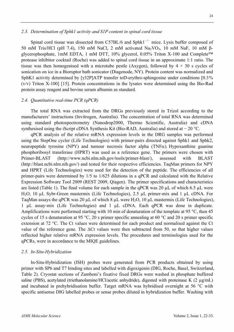
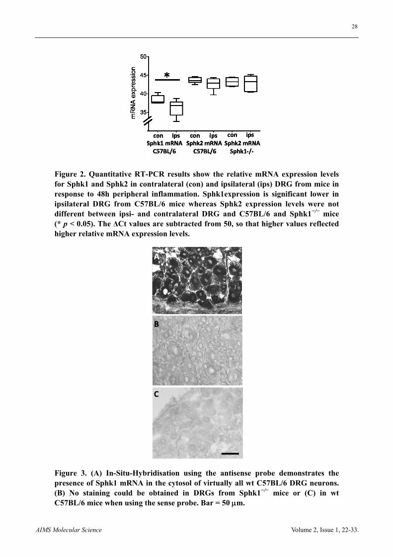
PCR and qRT-PCR showed the presence of Sphk1- and Sphk2-mRNA in DRG with higher relative expression levels for Sphk2 (Figure 1 and Figure 2). Deletion of Sphk1 abolished Sphk1 activity in the spinal cord but did not increase the activity of the Sphk2 isoform in spinal cord samples (n = 5 mice, t-test, Figure 1). qRT-PCR showed the mRNA expression of Sphk1 and Sphk2 ipsi- and contralateral DRG from mice in response to 48 h peripheral inflammation. The relative Sphk1 mRNA expression was significant lower in ipsilateral DRG from C57BL/6 mice compared to contralateral DRG (n = 6 mice, p < 0.05, t-test) whereas Sphk2 expression levels were similar in ipsi- and contralateral DRG and between C57BL/6 and Sphk1−/− mice (n = 6 mice, Figure 2).
ISH showed the presence of Sphk1-mRNA in virtually all sensory neurons. The cytosolic staining was present in DRGs of wt mice but absent in Sphk1−/− neurons. The negative control sense probe showed no unspecific staining in DRG of wt or gene-deficient mice (n = 3 mice, Figure 3).
3.2. NPY and TNF mRNA expression in DRG in response to peripheral inflammation
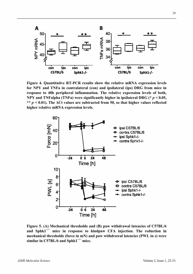
Expression levels of neuropeptide tyrosine (NPY) and TNF have been shown to increase in DRG in response to peripheral nerve damage and inflammation. We quantified mRNA expression of NPY, TNF and Sphk1 in ipsi- and contralateral DRG in C57BL/6 wt and Sphk1−/− mice in response to CFA injection. Peripheral CFA inflammation increased the mRNA expression levels for NPY in ipsilateral DRG of C57BL/6 and Sphk1−/− mice after two days with no difference between strains (Figure 4, C57BL/6, n = 9 mice, p < 0.05, Sphk1−/−, n = 6 mice, p < 0.01, paired t-test). Similarly, the mRNA expression levels of the inflammatory mediator TNFa were significantly higher in ipsilateral DRGs compared to contralateral DRG in both strains (Figure 4, C57BL/6, n = 6 mice, p < 0.05, Sphk1−/−, n = 5 mice, p < 0.01, paired t-test).
3.3. CFA-induced mechanical and heat hypersensitivity
Mechanical thresholds were determined in wt and Sphk1−/− mice that received intracutaneous injections of CFA (20 µL). Between 6 h and 48 h post injection, thresholds for mechanical stimuli were dramatically reduced in both wt and Sphk1−/− mice with a minor, non-significant reduction in the mechanical hypersensitivity in Sphk1−/− mice after 24 h (Figure 5, n = 11 C57BL/6 and 11 Sphk1−/− mice (6 h & 24 h) and 8 mice (48 h), two-way ANOVA).
Similarly, there was a tendency towards a lower heat-sensitivity in Sphk1−/− mice in response to peripheral inflammation, which did not reach significance (Figure 5, n = 11 C57BL/6 mice, and 11 (6 h & 24 h) and 8 (48 h) Sphk1−/− mice, two-way ANOVA).
27
AIMS Molecular Science Volume 2, Issue 1, 22-33.
Figure 1. (A) mRNA for Sphk1 cannot be detected in tissues from Sphk1−/− mice. Lanes 1‒5 show PCR products corresponding to the reference gene HPRT, lanes 6‒10 for Sphk1. Lanes 1 and 7 show the products in lung tissue, 2 and 8 brain and 3 and 9 DRGs from C57BL/6 mice whereas lanes 4, 10 and 5‒11 show products corresponding to lung tissue and DRG from Sphk1−/− mice. M = 100 bp marker. (B) Absence of Sphk1 did reduce the Sphk1 activity in the spinal cord (pmol S1P/min/mg protein). (C) However, the Sphk2 activity (pmol S1P/min/mg protein) was not significantly different between spinal cord of C57BL/6 wild-type mice and Sphk1−/− mice.
28
AIMS Molecular Science Volume 2, Issue 1, 22-33.
Figure 2. Quantitative RT-PCR results show the relative mRNA expression levels for Sphk1 and Sphk2 in contralateral (con) and ipsilateral (ips) DRG from mice in response to 48h peripheral inflammation. Sphk1expression is significant lower in ipsilateral DRG from C57BL/6 mice whereas Sphk2 expression levels were not different between ipsi- and contralateral DRG and C57BL/6 and Sphk1−/− mice (* p < 0.05). The ΔCt values are subtracted from 50, so that higher values reflected higher relative mRNA expression levels.
Figure 3. (A) In-Situ-Hybridisation using the antisense probe demonstrates the presence of Sphk1 mRNA in the cytosol of virtually all wt C57BL/6 DRG neurons. (B) No staining could be obtained in DRGs from Sphk1−/− mice or (C) in wt C57BL/6 mice when using the sense probe. Bar = 50 m.
AAIMS Molecu
Figurfor NrespoNPY ** p highe
Figurand mechsimil
ular Science
re 4. QuanNPY and Tonse to 48hand TNFa< 0.01). Ther relative m
re 5. (A) MSphk1−/−
hanical threar in C57B
ntitative RTTNFa in coh peripher
alpha (TNFhe ΔCt valumRNA exp
Mechanical mice in resholds (for
BL/6 and Sp
T-PCR resontralateralral inflammFa) were sigues are subpression lev
thresholdsresponse torce in mN)phk1−/− mic
ults show l (con) and
mation. Thgnificantly btracted frovels.
s and (B) po hindpaw and paw wce.
the relativd ipsilaterahe relative
higher in ipom 50, so th
paw withdrw CFA injwithdrawal
e mRNA eal (ips) DR
expressionpsilateral Dhat higher
rawal latenjection. Thl latencies (
Volume 2,
expression RG from mn levels of DRG (* p <values refl
ncies of C57he reductio(PWL in s)
29
Issue 1, 22-33
levels mice in
both, < 0.05, flected
7BL/6 on in ) were
9
3.
30
AIMS Molecular Science Volume 2, Issue 1, 22-33.
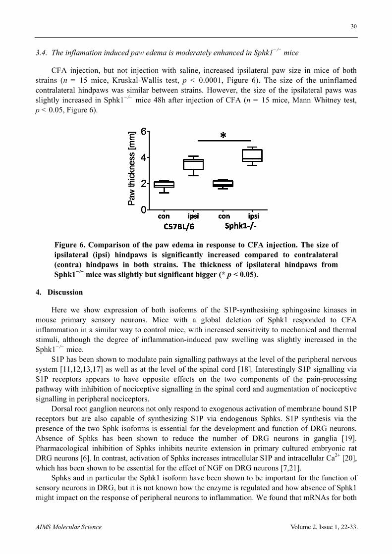
3.4. The inflamation induced paw edema is moderately enhanced in Sphk1−/− mice
CFA injection, but not injection with saline, increased ipsilateral paw size in mice of both strains (n = 15 mice, Kruskal-Wallis test, p < 0.0001, Figure 6). The size of the uninflamed contralateral hindpaws was similar between strains. However, the size of the ipsilateral paws was slightly increased in Sphk1−/− mice 48h after injection of CFA (n = 15 mice, Mann Whitney test, p < 0.05, Figure 6).
Figure 6. Comparison of the paw edema in response to CFA injection. The size of ipsilateral (ipsi) hindpaws is significantly increased compared to contralateral (contra) hindpaws in both strains. The thickness of ipsilateral hindpaws from Sphk1−/− mice was slightly but significant bigger (* p < 0.05).
4. Discussion
Here we show expression of both isoforms of the S1P-synthesising sphingosine kinases in mouse primary sensory neurons. Mice with a global deletion of Sphk1 responded to CFA inflammation in a similar way to control mice, with increased sensitivity to mechanical and thermal stimuli, although the degree of inflammation-induced paw swelling was slightly increased in the Sphk1−/− mice.
S1P has been shown to modulate pain signalling pathways at the level of the peripheral nervous system [11,12,13,17] as well as at the level of the spinal cord [18]. Interestingly S1P signalling via S1P receptors appears to have opposite effects on the two components of the pain-processing pathway with inhibition of nociceptive signalling in the spinal cord and augmentation of nociceptive signalling in peripheral nociceptors.
Dorsal root ganglion neurons not only respond to exogenous activation of membrane bound S1P receptors but are also capable of synthesizing S1P via endogenous Sphks. S1P synthesis via the presence of the two Sphk isoforms is essential for the development and function of DRG neurons. Absence of Sphks has been shown to reduce the number of DRG neurons in ganglia [19]. Pharmacological inhibition of Sphks inhibits neurite extension in primary cultured embryonic rat DRG neurons [6]. In contrast, activation of Sphks increases intracellular S1P and intracellular Ca2+ [20], which has been shown to be essential for the effect of NGF on DRG neurons [7,21].
Sphks and in particular the Sphk1 isoform have been shown to be important for the function of sensory neurons in DRG, but it is not known how the enzyme is regulated and how absence of Sphk1 might impact on the response of peripheral neurons to inflammation. We found that mRNAs for both
31
AIMS Molecular Science Volume 2, Issue 1, 22-33.
isoforms are expressed in DRGs with a higher relative expression of Sphk2 compared to the Sphk1. The higher expression of Sphk2 in peripheral sensory neurons is in agreement with studies that show a higher expression of Sphk2 in brain [22] and spinal cord [18]. Despite these differences in expression levels, Sphk1 seems to be involved in the response to persistent peripheral inflammation since the mRNA expression of Sphk1 but not Sphk2 was changed in DRG explants after CFA. The relative Sphk1 mRNA levels were lower in ipsilateral compared to contralateral DRG explants whereas mRNA expression levels of the pro-inflammatory cytokine TNF and the peptide NPY were increased as described previously [23,24]. This indicates that the CFA injection sufficiently induced changes in L3-5 DRG and that Sphk1 mRNA was down-regulated upon peripheral inflammation. It cannot be ruled out that the reduced expression of Sphk1 was a global response of sensory neurons. A global effect of inflammation could be related to the fact that DRGs do not possess the blood brain barrier of central neurons and therefore neurons are in direct contact to components of the blood [25]. Inflammatory mediators either from the blood or released from intra-ganglionic non-neuronal structures [23] can access all DRG neurons.
To further investigate if the reduction of Sphk1 is related to the response to inflammation, we used mice with a null mutation for Sphk1 [14] and compared the expression of TNF and NPY in response to peripheral inflammation between strains. Down-regulation of Sphk1 mRNA using siRNA decreases the expression of the pro-inflammatory TNF in LPS-activated microglia, [26]. In contrast, absence of Sphk1 (Sphk1−/−) increases the inflammatory response in the mouse brain [27]. In our model, absence of Sphk1 had a significant effect on the paw swelling in response to CFA application. However, transcription levels for TNF and NPY were not affected in DRGs in response to peripheral inflammation by the genetic deletion of Sphk1. The reason for this absence of an effect seems not mediated by transcriptional compensation of Sphk2 since Sphk2 mRNA levels were similar in C57BL/6 and Sphk1−/− DRG. However, this does not exclude a functional compensatory mechanism with increased activity of Sphk2 to compensate for the loss of Sphk1. The similarity in inflammation with respect to expression of TNF and NPY was reflected by the fact that the level of nocifensive behaviour and the degree of mechanical and heat hypersensitivity were similar in transgenic and control animals. Our results are in agreement with other studies showing that Sphk1−/− mice develop a normal inflammatory response [28].
In summary, Sphk1 mRNA is expressed in virtually all sensory DRG neurons and its expression changes in response to peripheral inflammation. However, deficiency of Sphk1 did not impact inflammation-dependent expression of pro-inflammatory markers in DRG, nor did the lack of Sphk1 result in any significant nocifensive behaviour changes.
5. Conclusion
The bioactive sphingolipid sphingosine 1-phosphate (S1P) has been shown to modulate pain signalling via activation of S1P receptors that are expressed in nociceptive dorsal root ganglion (DRG) neurons. One paracrine source might be S1P generated from Sphk1 in sensory neurons. The Sphk1 isoform is expressed in sensory DRG neurons and Sphk1 mRNA is reduced in DRG in response to peripheral inflammation. However, absence of Sphk1 has no effect on the expression of inflammatory marker proteins and nocifensive behaviour. Therefore, Sphk1 in sensory DRG neurons seems not to be directly involved in the pain signalling in response to chronic peripheral inflammation.
32
AIMS Molecular Science Volume 2, Issue 1, 22-33.
Acknowledgements
The research was supported by grants from the Australian National Health & Medical Research Council (project grant 535055, RVH and ILG) and the Flinders Medical Research Foundation.
Conflict of interest
All authors declare no conflicts of interest in this paper.
Reference
1. Maceyka M, Harikumar KB, Milstien S, et al. (2012) Sphingosine-1-phosphate signaling and its role in disease. Trends Cell Biol 22: 50-60.
2. Pitson SM (2011) Regulation of sphingosine kinase and sphingolipid signaling. Trends Biochem Sci 36: 97-107.
3. Mizugishi K, Yamashita T, Olivera A, et al. (2005) Essential role for sphingosine kinases in neural and vascular development. Mol Cell Biol 25: 11113-11121.
4. Chan H, Pitson SM (2013) Post-translational regulation of sphingosine kinases. Biochim Biophys Acta 1831: 147-156.
5. Baker DA, Barth J, Chang R, et al. (2010) Genetic sphingosine kinase 1 deficiency significantly decreases synovial inflammation and joint erosions in murine TNF-alpha-induced arthritis. J Immunol 185: 2570-2579.
6. Toman RE, Payne SG, Watterson KR, et al. (2004) Differential transactivation of sphingosine-1-phosphate receptors modulates NGF-induced neurite extension. J Cell Biol 166: 381-392.
7. Zhang YH, Vasko MR, Nicol GD (2006) Intracellular sphingosine 1-phosphate mediates the increased excitability produced by nerve growth factor in rat sensory neurons. J Physiol 575: 101-113.
8. Zhang YH, Fehrenbacher JC, Vasko MR, et al. (2006) Sphingosine-1-phosphate via activation of a G-protein-coupled receptor(s) enhances the excitability of rat sensory neurons. J Neurophysiol 96: 1042-1052.
9. He XH, Zang Y, Chen X, et al. (2010) TNF-alpha contributes to up-regulation of Nav1.3 and Nav1.8 in DRG neurons following motor fiber injury. Pain 151: 266-279.
10. Hokfelt T, Brumovsky P, Shi T, et al. (2007) NPY and pain as seen from the histochemical side. Peptides 28: 365-372.
11. Mair N, Benetti C, Andratsch M, et al. (2011) Genetic evidence for involvement of neuronally expressed S1P(1) receptor in nociceptor sensitization and inflammatory pain. PLoS One 6: e17268.
12. Camprubi-Robles M, Mair N, Andratsch M, et al. (2013) Sphingosine-1-phosphate-induced nociceptor excitation and ongoing pain behavior in mice and humans is largely mediated by S1P3 receptor. J Neurosci 33: 2582-2592.
13. Salvemini D, Doyle T, Kress M, et al. (2013) Therapeutic targeting of the ceramide-to-sphingosine 1-phosphate pathway in pain. Trends Pharmacol Sci 34: 110-118.
14. Allende ML, Sasaki T, Kawai H, et al. (2004) Mice deficient in sphingosine kinase 1 are rendered lymphopenic by FTY720. J Biol Chem 279: 52487-52492.
15. Pitman MR, Pham DH, Pitson SM (2012) Isoform-selective assays for sphingosine kinase activity. Methods Mol Biol 874: 21-31.
16. Hargreaves K, Dubner R, Brown F, et al. (1988) A new and sensitive method for measuring thermal nociception in cutaneous hyperalgesia. Pain 32: 77-88.
33
AIMS Molecular Science Volume 2, Issue 1, 22-33.
17. Xie W, Strong JA, Kays J, et al. (2012) Knockdown of the sphingosine-1-phosphate receptor S1PR1 reduces pain behaviors induced by local inflammation of the rat sensory ganglion. Neurosci Lett 515: 61-65.
18. Coste O, Pierre S, Marian C, et al. (2008) Antinociceptive activity of the S1P-receptor agonist FTY720. J Cell Mol Med 12: 995-1004.
19. Meng H, Yuan Y, Lee VM (2011) Loss of sphingosine kinase 1/S1P signaling impairs cell growth and survival of neurons and progenitor cells in the developing sensory ganglia. PLoS One 6: e27150.
20. Pollock J, McFarlane SM, Connell MC, et al. (2002) TNF-alpha receptors simultaneously activate Ca2+ mobilisation and stress kinases in cultured sensory neurones. Neuropharmacology 42: 93-106.
21. Nicol GD (2008) Nerve growth factor, sphingomyelins, and sensitization in sensory neurons. Sheng Li Xue Bao 60: 603-604.
22. Blondeau N, Lai Y, Tyndall S, et al. (2007) Distribution of sphingosine kinase activity and mRNA in rodent brain. J Neurochem 103: 509-517.
23. Li Y, Ji A, Weihe E, et al. (2004) Cell-specific expression and lipopolysaccharide-induced regulation of tumor necrosis factor alpha (TNFalpha) and TNF receptors in rat dorsal root ganglion. J Neurosci 24: 9623-9631.
24. Ji RR, Zhang X, Wiesenfeld-Hallin Z, et al. (1994) Expression of neuropeptide Y and neuropeptide Y (Y1) receptor mRNA in rat spinal cord and dorsal root ganglia following peripheral tissue inflammation. J Neurosci 14: 6423-6434.
25. Kubicek L, Kopacik R, Klusakova I, et al. (2010) Alterations in the vascular architecture of the dorsal root ganglia in a rat neuropathic pain model. Ann Anat 192: 101-106.
26. Nayak D, Huo Y, Kwang WX, et al. (2010) Sphingosine kinase 1 regulates the expression of proinflammatory cytokines and nitric oxide in activated microglia. Neuroscience 166: 132-144.
27. Grin'kina NM, Karnabi EE, Damania D, et al. (2012) Sphingosine kinase 1 deficiency exacerbates LPS-induced neuroinflammation. PLoS One 7: e36475.
28. Michaud J, Kohno M, Proia RL, et al. (2006) Normal acute and chronic inflammatory responses in sphingosine kinase 1 knockout mice. FEBS Lett 580: 4607-4612.
© 2015, Rainer V. Haberberger, et al., licensee AIMS. This is an open access article distributed under the terms of the Creative Commons Attribution License (http://creativecommons.org/licenses/by/4.0)