
Phosphorylation Regulates myo-Inositol-3-phosphate Synthase A ...
Sphingosine 1-phosphate regulates regeneration andfibrosis after liver injury via sphingosine 1-phosphatereceptor 2
Hitoshi Ikeda,1,*,† Naoko Watanabe,*,† Isao Ishii,§ Tatsuo Shimosawa,* Yukio Kume,*Tomoaki Tomiya,† Yukiko Inoue,† Takako Nishikawa,† Natsuko Ohtomo,† Yasushi Tanoue,†
Satoko Iitsuka,* Ryoto Fujita,* Masao Omata,† Jerold Chun,** and Yutaka Yatomi*
Department of Clinical Laboratory Medicine,* and Department of Gastroenterology,† Graduate School ofMedicine, The University of Tokyo, 7-3-1 Hongo, Bunkyo-ku, Tokyo, Japan; Department of Molecular andCellular Neurobiology,§ Gunma University Graduate School of Medicine, Gunma, Japan; and Department ofMolecular Biology,** Helen L. Dorris Child and Adolescent Neuropsychiatric Disorder Institute, The ScrippsResearch Institute, La Jolla, CA
Abstract Sphingosine 1-phosphate (S1P), a bioactive lipidmediator, stimulates proliferation and contractility in hepaticstellate cells, the principal matrix-producing cells in the liver,and inhibits proliferation via S1P receptor 2 (S1P2) in hepa-tocytes in rats in vitro. A potential role of S1P and S1P2 inliver regeneration and fibrosis was examined in S1P2-deficientmice. Nuclear 5-bromo-2′-deoxy-uridine labeling, proliferat-ing cell nuclear antigen (PCNA) staining in hepatocytes, andthe ratio of liver weight to body weight were enhanced at48 h in S1P2-deficient mice after a single carbon tetrachlo-ride (CCl4) injection. After dimethylnitrosamine (DMN) ad-ministration with a lethal dose, PCNA staining in hepatocyteswas enhanced at 48 h and survival rate was higher in S1P2-deficient mice. Serum aminotransferase level was unalteredin those mice compared with wild-type mice in both CCl4-and DMN-induced liver injury, suggesting that S1P2 inactiva-tion accelerated regeneration not as a response to enhancedliver damage. After chronic CCl4 administration, fibrosis wasless apparent, with reduced expression of smooth-musclea-actin-positive cells in the livers of S1P2-deficient mice,suggesting that S1P2 inactivation ameliorated CCl4-inducedfibrosis due to the decreased accumulation of hepatic stel-late cells. Thus, S1P plays a significant role in regenera-tion and fibrosis after liver injury via S1P2.—Ikeda, H., N.Watanabe, I. Ishii, T. Shimosawa, Y. Kume, T. Tomiya,Y. Inoue, T. Nishikawa, N. Ohtomo, Y. Tanoue, S. Iitsuka,R. Fujita, M. Omata, J. Chun, and Y. Yatomi. Sphingosine1-phosphate regulates regeneration and fibrosis after liverinjury via sphingosine 1-phosphate receptor 2. J. Lipid Res.2009. 50: 556–564.
Supplementary key words liver regeneration • liver fibrosis • hepato-cyte • hepatic stellate cell • hepatic myofibroblast
Sphingosine 1-phosphate (S1P), which elicits a widevariety of cell responses (1), has emerged as a novel lipidintracellular mediator. S1P was shown to act as an intracel-lular second messenger of platelet-derived growth factorand serum in their mitogenic actions in cultured fibroblasts(2, 3), and furthermore, intracellular levels of S1P and cer-amide were reported to determine cell survival or death (4,5). However, evidence indicating that S1P also acts as an ex-tracellular mediator has been reported; some of the diverseeffects of S1P, such as stimulation of cell proliferation orcontractility, are known to be sensitive to pertussis toxin(6) or ADP-ribosyltransferase C3 from Clostridium botulinum(7, 8), suggesting that S1P may activate a receptor coupledto G protein(s). Indeed, recent investigation has revealedthat S1P acts through at least five high-affinity G protein-coupled receptors referred to as S1P1–5 (9, 10). Regardingthe source of S1P in vivo, it is shown to be stored in platelets(11), and recent data using conditional knockouts of sphin-gosine kinases support release of S1P from erythrocytes(12, 13). These findings suggest that S1P has normal in vivoroles as well as potentially pathophysiological roles as a cir-culating paracrine mediator, a view further supported bythe phenotypes of S1P receptor mutants (10, 14, 15).
S1P receptors are also expressed in the liver (14). To in-vestigate the function of S1P in liver pathophysiology, wehave determined the effect of S1P on liver cells in culture.We first demonstrated that S1P stimulates proliferationand contractility in rat hepatic stellate cells in culture;the stimulation of proliferation is pertussis toxin-sensitive,
This work was supported by the Japanese Society of Laboratory Medicine Fund forthe Promotion of Scientific Research (H.I.) and by National Institutes of HealthGrants NS-048478 and R01 DA-019674 ( J.C.).
Manuscript received 23 September 2008 and in revised form 24 October 2008.
Published, JLR Papers in Press, October 27, 2008.DOI 10.1194/jlr.M800496-JLR200
Abbreviations: ALT, alanine aminotransferase; ALP, alkaline phos-phatase; BrdU, 5-bromo-2′-deoxy-uridine; CCl4, carbon tetrachloride;DMN, dimethylnitrosamine; PCNA, proliferating cell nuclear antigen;S1P, sphingosine 1-phosphate.
1 To whom correspondence should be addressed.e-mail: [email protected]
Copyright © 2009 by the American Society for Biochemistry and Molecular Biology, Inc.
556 Journal of Lipid Research Volume 50, 2009 This article is available online at http://www.jlr.org
by guest, on October 1, 2018
ww
w.jlr.org
Dow
nloaded from
and the stimulation of contractility is C3 exotoxin-sensitive(16). This stimulatory effect of S1P on contractility in thosecells was found to be via S1P2 with Rho activation (17). Onthe other hand, we revealed that S1P inhibits proliferationin cultured rat hepatocytes. With the use of a specific an-tagonist to S1P2 and C3 exotoxin, this inhibitory effect ofS1P on hepatocyte proliferation in culture involved Rhoactivation via S1P2 (18). Furthermore, the administrationof S1P in 70% hepatectomized rats indeed reduces a re-sponse of hepatocytes to synthesize DNA (18).
Irrespective of the insults, such as viruses, alcohol abuse,or drugs, the regenerative and wound-healing responsesgenerally occur in the liver after the injury, and the persis-tence of these responses may result in liver fibrosis (19,20). Hepatocytes play a major role in liver regeneration(19) as do hepatic stellate cells in the wound-healing re-sponse, and hence, liver fibrosis (20). The enhanced prolif-eration and contractility of hepatic stellate cells are amongthe main features of liver fibrosis (21, 22). Although ourprevious evidence was obtained mainly by in vitro studiesand indicated a pharmacologically inhibitory effect of S1P
on liver regeneration in vivo, it raised the possibility thatS1P, and potentially S1P2, might play a pathophysiologicalrole in the liver after the injury. Thus, we planned to ex-tend our study using S1P2-deficient mice to clarify this point.
In this context, Serrier-Lanneau et al. (23) recently re-ported that the wound-healing response to acute liver in-jury elicited by carbon tetrachloride (CCl4) was reduced inS1P2
2/2 mice with reduced accumulation of hepatic myo-fibroblasts and that hepatic myofibroblasts isolated fromS1P2
2/2 mice did not proliferate in response to S1P. Thereis controversy as to whether hepatic myofibroblasts are dis-tinct from hepatic stellate cells. Hepatic myofibroblasts havebeen studied as the cells developed from hepatic stellatecells by transdifferentiation (24–27). In contrast, hepaticmyofibroblasts have been reported to belong to a cell popu-lation different from hepatic stellate cells (28). Whateverthe case, hepatic myofibroblasts have been assumed tobe a principle matrix-producing cell of the diseased liver(29). On the other hand, the regenerative response aftera single injection of CCl4 in S1P2
2/2 mice was not differ-ent from that in wild-type mice, suggesting that S1P2 inac-
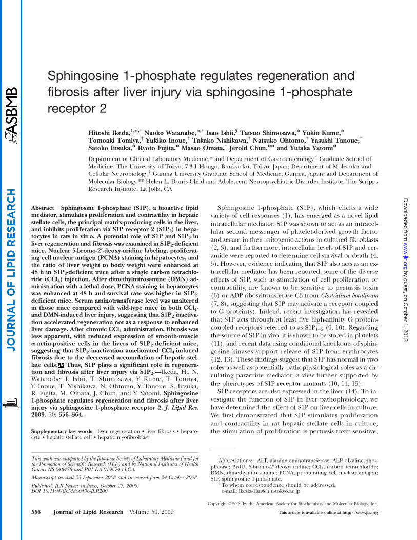
Fig. 1. Regenerative response in hepatocytes and nonparenchymal cells of wild-type and S1P22/2 livers in acute injury induced by carbon
tetrachloride (CCl4). A single injection of CCl4 was performed in wild-type (WT) and S1P22/2 mice. 5-Bromo-2′-deoxy-uridine (BrdU) was
injected intraperitoneally at 1 h before euthanization at 24, 48, 72, and 96 h after CCl4 injection. The nuclear labeling in hepatocytes wasdetermined using a BrdU labeling and detection kit II. Representative BrdU labeling in the livers of wild-type and S1P2
2/2 mice at 48 h afterCCl4 injection is shown (A). Bar 5 50 mm. The number of BrdU-positive cells was determined as the mean of five random areas at 400-foldmagnification in each section (B). Columns and bars represent means 6 SEM of four animals. Immunohistochemical analysis of proliferat-ing cell nuclear antigen (PCNA) was also done using a PCNA staining kit. Representative photomicrographs of the liver of wild-type andS1P2
2/2 mice at 48 h after CCl4 injection are shown (C). Bar 5 50 mm. Arrows indicate positive hepatocytes, and arrowheads, positivenonparenchymal cells. The percentages of PCNA-positive hepatocytes (D) and nonparenchymal cells (E) were determined with five ran-dom areas at 400-fold magnification in each section. Columns and bars represent means 6 SEM of four animals. The asterisk indicates asignificant difference from wild type in Studentʼs t-test (P , 0.05).
Sphingosine 1-phosphate and the liver 557
by guest, on October 1, 2018
ww
w.jlr.org
Dow
nloaded from
tivation did not affect hepatocyte regeneration (23). Withthese findings, we aimed to perform more-detailed exam-ination of hepatocyte regeneration in acute liver injury elic-ited by dimethylnitrosamine (DMN) in addition to CCl4and to determine whether reduced wound-healing re-sponse could lead to reduced liver fibrosis after chronicCCl4 administration in S1P2
2/2 mice.
MATERIALS AND METHODS
AnimalsHeterozygous S1P2
1/2 mice were originally generated by ourgroup on the (129/Sv 3 129/J)F1 (an embryonic stem cell ori-gin)/C57BL/6N (a blastocyst origin) mixed background (30).In this study, they were backcrossed onto the inbred C57BL/6Nstrain (Clea Japan, Tokyo, Japan) for 7–8 generations to achieve.99.2% genetic homogeneity, and the obtained S1P2
1/2 mice
were bred to produce S1P22/2 mice. Deletion of S1P2 in these
S1P22/2 mice has been repeatedly confirmed by the complete
absence of s1p2 gene expression in tissues (e.g., adult lung, spleen,brain, heart, cochlea, and embryonic fibroblasts) in which s1p2 isnormally expressed (15, 30, 31) as well as the specific appearanceor disappearance of some S1P-mediated cellular signaling/sys-temic effects (15, 30–32). Age-matched wild-type C57BL/6N micewere used as controls. They were fed a standard pelleted diet andwater ad libitum under normal laboratory conditions of 12 h light/dark cycles. All animals received humane care, and the researchwas conducted in conformity with Public Health Service policyon the humane care and use of laboratory animals. The experi-mental protocol was approved by the Animal Research Committeeof the University of Tokyo and followed National Institutes ofHealth guidelines for the care and use of laboratory animals.
CCl4 treatmentTo assess the responses to a single CCl4 administration, male
and female wild-type and S1P22/2 mice, 8–10 weeks of age, were
Fig. 2. The ratio of liver weight to body weight, body weight loss, and liver function tests in wild-type and S1P22/2 mice in acute liver injury
induced by CCl4. A single injection of CCl4 was performed in wild-type (WT) and S1P22/2 mice. The ratio of liver weight to body weight was
measured in untreated wild-type and S1P22/2 mice from 24 to 96 h at euthanization after CCl4 injection (A). The alteration of body weight
was determined by comparison of body weights before CCl4 injection with those at 24, 48, 72, and 96 h after CCl4 injection (B). Serumconcentrations of alanine aminotransferase (ALT) (C) and alkaline phosphatase (ALP) (D) were measured in wild-type and S1P2
2/2 miceat 24, 48, 72, and 96 h after CCl4 injection. Columns and bars represent means 6 SEM of four animals. The asterisk indicates a significantdifference from wild type in Studentʼs t-test (P , 0.05).
558 Journal of Lipid Research Volume 50, 2009
by guest, on October 1, 2018
ww
w.jlr.org
Dow
nloaded from

injected intraperitoneally with 0.5 ml/kg body weight of CCl4 dis-solved in the same amount of olive oil (1:1) (23), in which liverinjury, assessed by serum aminotransferase level, was nongenderspecific. To develop liver fibrosis, female wild-type and S1P2
2/2
mice, 8 weeks of age, were injected intraperitoneally with 1 ml/kgbody weight of CCl4 dissolved in the same amount of olive oil (1:1),twice a week for 4 weeks (33, 34). Control mice received injectionof the carrier (olive oil) alone. Each group consisted of four to fivemice. For the experiment on chronic CCl4 treatment, the liverswere harvested at 4 days after the end of the administration.
DMN treatmentMale and female wild-type and S1P2
2/2 mice, 8–10 weeks of age,were injected intraperitoneally with 15 mg/kg body weight DMNdissolved in HBSS (0.3%) (GIBCO) (35), in which liver injury, as-sessed by serum aminotransferase level, was nongender specific.
Measurement of nuclear labeling by5-bromo-2′-deoxy-uridine
One hour before euthanization, 50 mg/4 ml/kg body weightof 5-bromo-2′-deoxy-uridine (BrdU) (Roche Molecular Biochem-icals, Mannheim, Germany) was injected intraperitoneally, andthen the mice were euthanized at 24, 48, 72, and 96 h afterCCl4 administration. The liver was excised, immediately fixed in10% formalin, and embedded in paraffin. The nuclear labelingwas measured using BrdU labeling and detection kit II (RocheMolecular Biochemicals). The number of BrdU-positive hepato-cytes was determined as the mean of five random areas at 400-foldmagnification in each section.
Immunohistochemical analysis of proliferating cellnuclear antigen
Immunohistochemical staining for proliferating cell nuclearantigen (PCNA) was performed in liver tissue using a PCNA stain-ing kit (Zymed Laboratories) in accordance with the protocolspecified by the manufacturer. The ratio of PCNA-positive hepa-tocytes to all hepatocytes was determined with five random areasat 400-fold magnification in each section in CCl4-treated mice andwith ten random areas in DMN-treated mice, because submassivehemorrhagic necrosis was focally found in DMN-treated liversas previously described (36). The percentage of PCNA-positivenonparenchymal cells was determined with five random areasat 400-fold magnification in each section in CCl4-treated mice.
Measurement of liver functionThe serum levels of alanine aminotransferase (ALT) and alka-
line phosphatase (ALP) were determined using an automatedanalyzer (Hitachi 7170; Hitachi Instruments Service Co., Ltd.,Tokyo, Japan).
Histological analysis of fibrosisTissue sections (4 mm thick) of liver specimens fixed in for-
malin and embedded in paraffin were analyzed with Massonʼs tri-chrome staining (37). The microscopic fields (five per each slide)were blindly selected and captured with the aid of the Nikon Dig-ital Camera DXM1200 (NIKON, Japan). A fibrous portion stainedin blue was extracted, and the extent of liver fibrosis was quantifiedby the technique reported previously (38) by calculating the areaof fibrosis/area of section using image analysis software of the pub-lic domain Scion Image (Scion Corporation).
Immunohistochemical analysis of smooth-muscle a-actinImmunohistochemical analysis of smooth-muscle a-actin was
performed in liver tissue fixed in formalin and embedded in par-
affin using a Vector M.O.M. immunodetection kit (Vector Labora-tories, Burlingame, CA) in accordance with the protocol specifiedby the manufacturer, with a 1:5 dilution of mouse monoclonal anti-body to smooth-muscle a-actin (Sigma). The quantitation of thepositive staining area for smooth-muscle a-actin was performedblindly on four or five liver fragments per animal using the com-puter software of the public domain Scion Image developed bythe Scion Corporation.
Statistical analysisData are represented as mean 6 SEM. An ANOVA followed by
Tukeyʼs honest significance differences test (Tukeyʼs HSD) orStudentʼs t-test were used to determine whether significant differ-ences existed between groups. Fisherʼs exact test was performedwhen appropriate. Differences were considered to be significantat P values of ,0.05.
RESULTS
Regenerative response of hepatocytes to CCl4-inducedacute liver injury was enhanced in S1P2
2/2 miceAfter a single CCl4 administration, DNA synthesis of he-
patocytes determined by BrdU labeling peaked at 48 h in
Fig. 3. PCNA-positive hepatocytes of wild-type and S1P22/2 livers
in acute liver injury induced by dimethylnitrosamine (DMN). Asingle injection of DMN was performed in wild-type (WT) andS1P2
2/2 mice. Immunohistochemical analysis of PCNA was doneusing a PCNA staining kit. Representative photomicrographs of theliver of wild-type and S1P2
2/2 mice at 48 h after DMN injection areshown (A). Bar 5 50 mm. Arrows indicate positive hepatocytes. Thepercentage of PCNA-positive hepatocytes was determined with tenrandom areas at 400-fold magnification in each section (B). Col-umns and bars represent means6 SEM of four animals. The asteriskindicates a significant difference from wild type in Studentʼs t-test(P , 0.05).
Sphingosine 1-phosphate and the liver 559
by guest, on October 1, 2018
ww
w.jlr.org
Dow
nloaded from
both wild-type and S1P22/2 mice (Fig. 1). As depicted in
Fig. 1, BrdU-positive hepatocytes seemed more apparentin S1P2
2/2 mice than in wild-type mice (Fig. 1A) at 48 hafter CCl4 administration, and in fact, the number ofBrdU-positive hepatocytes at 48 h after CCl4 administra-tion in S1P2
2/2 mice was significantly increased by 1.8-foldcompared with wild-type mice (Fig. 1B). On the other hand,BrdU-positive hepatocytes were not found in both wild-type
and S1P22/2 mice treated with olive oil alone. We next per-
formed immunohistochemical analysis of PCNA in wild-typeand S1P2
2/2 livers after a single CCl4 administration. Asdemonstrated in Fig. 1, PCNA-positive hepatocytes weremore apparent in S1P2
2/2 mice than in wild-type mice(Fig. 1C) at 48 h after CCl4 administration. The number ofPCNA-positive hepatocytes significantly increased by 1.7-foldin S1P2
2/2 mice compared with wild-type mice (Fig. 1D).
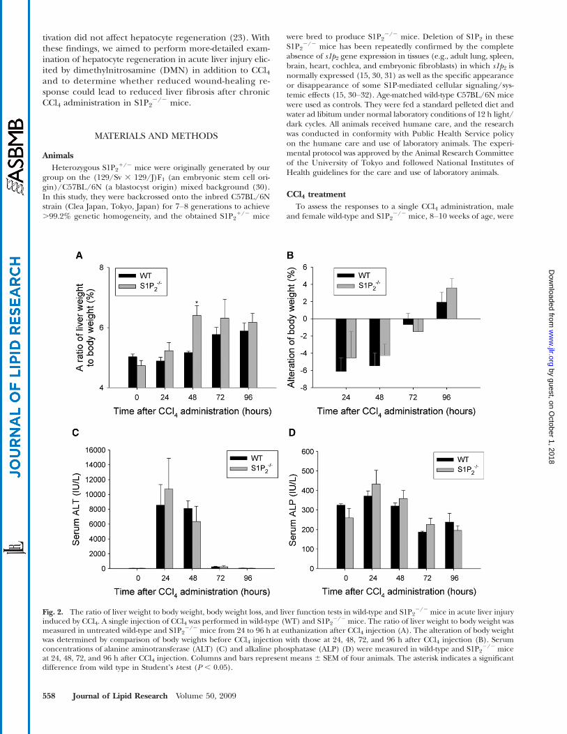
Fig. 4. The ratio of liver weight to body weight, body weight loss, liver function test, and survival rate in wild-type and S1P22/2 mice in acute
liver injury induced by DMN. A single injection of DMN was performed in wild-type (WT) and S1P22/2 mice. The ratio of liver weight to
body weight (A), body weight loss (B), and serum concentration of ALT (C) and ALP (D) were measured at 48 h after DMN injection.Columns and bars represent means 6 SEM of four animals. Survival rate was also determined in wild-type (n 5 17) and S1P2
2/2 (n 5 16)mice up to 120 h after DMN injection (E). Significantly higher survival rate in S1P2
2/2 mice was determined at 96 and 120 h after DMNinjection in Fisherʼs exact test (P , 0.05).
560 Journal of Lipid Research Volume 50, 2009
by guest, on October 1, 2018
ww
w.jlr.org
Dow
nloaded from
On the other hand, it is of note that PCNA-positive non-parenchymal cells were less apparent in S1P2
2/2 mice com-pared with wild-type mice (Fig. 1C). The percentage ofPCNA-positive nonparenchymal cells in S1P2
2/2 mice wasreduced to 62% of that in wild-type mice (Fig. 1E). In bothwild-type and S1P2
2/2 mice treated with olive oil alone,PCNA-positive cells were not determined. These resultssuggest that the regenerative response after CCl4 adminis-tration is enhanced in hepatocytes, but reduced in nonpa-renchymal cells in S1P2
2/2 mice.We then analyzed both liver weight and body weight at
euthanization after a single injection of CCl4. The ratio ofliver weight to body weight was significantly higher inS1P2
2/2 mice than in wild-type mice at 48 h after CCl4administration, but not different between wild-type andS1P2
2/2 mice at 24, 72, and 96 h (Fig. 2A). Both wild-typeand S1P2
2/2 mice lost their body weight maximally at 24 hafter CCl4 administration and then gradually gained weightup to 96 h, as shown in Fig. 2B. The alterations of bodyweight after CCl4 administration were not different be-tween wild-type and S1P2
2/2 mice. These results suggestthat the regenerative response in the liver after CCl4 ad-ministration is enhanced in S1P2
2/2 mice.To examine the extent of liver damage elicited by CCl4
administration, serum levels of ALT and ALP were mea-sured up to 96 h after treatment. Serum ALT level peakedat 24 h after CCl4 administration and was not significantlydifferent between wild-type and S1P2
2/2 mice (Fig. 2C),as previously reported (23). Furthermore, serum level ofALP was not different between wild-type and S1P2
2/2 mice(Fig. 2D). These results suggest that the extent of liverdamage caused by a single injection of CCl4 was not differ-ent in S1P2
2/2 mice compared with wild-type mice.
Enhanced regenerative response of hepatocytes toDMN-induced acute liver injury led to increased survivalrate in S1P2
2/2 miceTo determine whether the enhanced hepatocyte regen-
eration in S1P22/2 mice could be the specific phenomenon
in CCl4-induced acute liver injury, the regenerative responseof hepatocytes in S1P2
2/2 mice was also examined in DMN-induced acute liver injury (35). In this experiment, BrdUlabeling was not suitable for determining the proliferativeresponse, because significant amounts of ascites were foundin both wild-type and S1P2
2/2 mice in which BrdU was ad-ministered intraperitoneally. Thus, we determined the pro-liferative response by PCNA staining. At 48 h after DMNadministration, PCNA-positive hepatocytes were more ap-parent in S1P2
2/2 mice than in wild-type mice (Fig. 3A).The percentage of PCNA-positive hepatocytes increased by1.5-fold in S1P2
2/2 mice compared with wild-type mice(Fig. 3B). On the other hand, PCNA-positive hepatocyteswere not found in both wild-type and S1P2
2/2 mice treatedwith vehicle alone. This result suggests that the regenerativeresponse is also enhanced after DMN administration in he-patocytes in S1P2
2/2 mice. At the same time point, the ratioof liver weight to body weight was not different betweenwild-type and S1P2
2/2 mice (Fig. 4A), and the trend ofmore body weight loss in wild-type mice was noted (P 5
0.05116) (Fig. 4B). Serum levels of ALT (Fig. 4C) andALP (Fig. 4D) were not different between wild-type andS1P2
2/2 mice at the same time point, suggesting that theextent of DMN-induced liver damage was not different inS1P2
2/2 mice compared with wild-type mice.Although we next planned to determine the regenerative
response in hepatocytes in wild-type and S1P22/2 mice be-
yond 48 h after DMN administration, a substantial numberof wild-type mice were found to be dead at 72 h after DMNadministration. Thus, our experiment was changed to deter-mine survival rate of wild-type and S1P2
2/2 mice with DMNintoxication. As shown in Fig. 4E, survival rate was signif-icantly higher in S1P2
2/2 mice compared with wild-typemice at 96 to 120 h after DMN injection. Thus, increasedregenerative response in hepatocytes after DMN intoxica-tion may lead to increased survival rate in S1P2
2/2 mice.
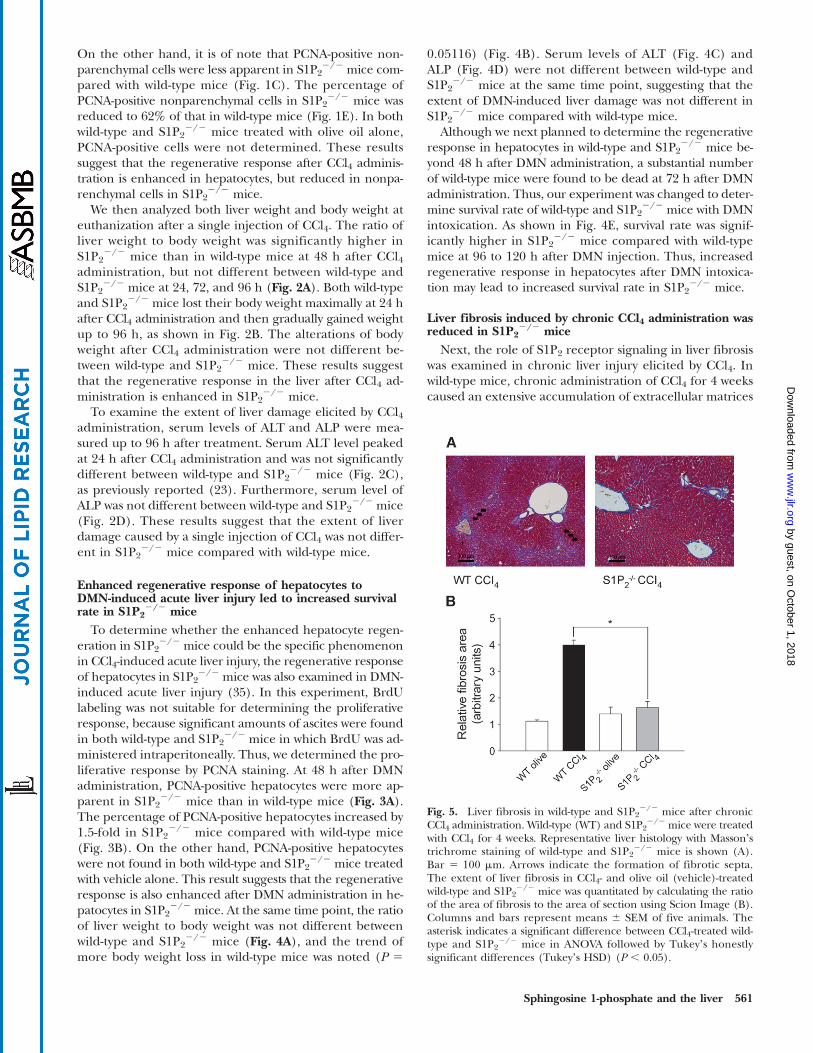
Liver fibrosis induced by chronic CCl4 administration wasreduced in S1P2
2/2 miceNext, the role of S1P2 receptor signaling in liver fibrosis
was examined in chronic liver injury elicited by CCl4. Inwild-type mice, chronic administration of CCl4 for 4 weekscaused an extensive accumulation of extracellular matrices
Fig. 5. Liver fibrosis in wild-type and S1P22/2 mice after chronic
CCl4 administration. Wild-type (WT) and S1P22/2 mice were treated
with CCl4 for 4 weeks. Representative liver histology with Massonʼstrichrome staining of wild-type and S1P2
2/2 mice is shown (A).Bar 5 100 mm. Arrows indicate the formation of fibrotic septa.The extent of liver fibrosis in CCl4- and olive oil (vehicle)-treatedwild-type and S1P2
2/2 mice was quantitated by calculating the ratioof the area of fibrosis to the area of section using Scion Image (B).Columns and bars represent means 6 SEM of five animals. Theasterisk indicates a significant difference between CCl4-treated wild-type and S1P2
2/2 mice in ANOVA followed by Tukeyʼs honestlysignificant differences (Tukeyʼs HSD) (P , 0.05).
Sphingosine 1-phosphate and the liver 561
by guest, on October 1, 2018
ww
w.jlr.org
Dow
nloaded from
with bridging fibrosis in the liver, whereas in S1P22/2
mice, such dense fibrotic septum development with nod-ule formation was not found (Fig. 5A). Quantitative analy-sis of fibrosis development revealed that the mean fibroticarea in the livers in wild-type mice was significantly largerthan that in S1P2
2/2 mice treated with CCl4. The extent ofliver fibrosis in S1P2
2/2 mice was approximately 40% thatof wild-type mice (Fig. 5B). On the other hand, liver histol-ogy in S1P2
2/2 mice administered olive oil alone, i.e., with-out CCl4 treatment, was essentially unaltered compared withthat of wild-type mice administered identically (Fig. 5B). Inaddition, serum ALT levels at the time of euthanization af-ter chronic CCl4 administration for 4 weeks were essentiallyunaltered in wild-type and S1P2
2/2 mice; 586 20 IU/l (n55) and 51 6 30 IU/l (n 5 5), respectively. These resultssuggest that S1P2 inactivation causes less liver fibrosis in re-sponse to chronic CCl4 administration.
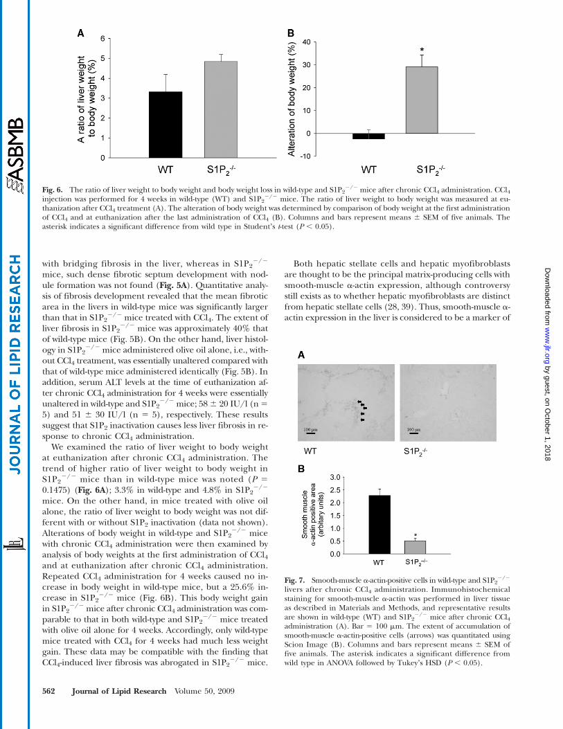
We examined the ratio of liver weight to body weightat euthanization after chronic CCl4 administration. Thetrend of higher ratio of liver weight to body weight inS1P2
2/2 mice than in wild-type mice was noted (P 50.1475) (Fig. 6A); 3.3% in wild-type and 4.8% in S1P2
2/2
mice. On the other hand, in mice treated with olive oilalone, the ratio of liver weight to body weight was not dif-ferent with or without S1P2 inactivation (data not shown).Alterations of body weight in wild-type and S1P2
2/2 micewith chronic CCl4 administration were then examined byanalysis of body weights at the first administration of CCl4and at euthanization after chronic CCl4 administration.Repeated CCl4 administration for 4 weeks caused no in-crease in body weight in wild-type mice, but a 25.6% in-crease in S1P2
2/2 mice (Fig. 6B). This body weight gainin S1P2
2/2 mice after chronic CCl4 administration was com-parable to that in both wild-type and S1P2
2/2 mice treatedwith olive oil alone for 4 weeks. Accordingly, only wild-typemice treated with CCl4 for 4 weeks had much less weightgain. These data may be compatible with the finding thatCCl4-induced liver fibrosis was abrogated in S1P2
2/2 mice.
Both hepatic stellate cells and hepatic myofibroblastsare thought to be the principal matrix-producing cells withsmooth-muscle a-actin expression, although controversystill exists as to whether hepatic myofibroblasts are distinctfrom hepatic stellate cells (28, 39). Thus, smooth-muscle a-actin expression in the liver is considered to be a marker of
Fig. 7. Smooth-muscle a-actin-positive cells in wild-type and S1P22/2
livers after chronic CCl4 administration. Immunohistochemicalstaining for smooth-muscle a-actin was performed in liver tissueas described in Materials and Methods, and representative resultsare shown in wild-type (WT) and S1P2
2/2 mice after chronic CCl4administration (A). Bar 5 100 mm. The extent of accumulation ofsmooth-muscle a-actin-positive cells (arrows) was quantitated usingScion Image (B). Columns and bars represent means 6 SEM offive animals. The asterisk indicates a significant difference fromwild type in ANOVA followed by Tukeyʼs HSD (P , 0.05).
Fig. 6. The ratio of liver weight to body weight and body weight loss in wild-type and S1P22/2 mice after chronic CCl4 administration. CCl4
injection was performed for 4 weeks in wild-type (WT) and S1P22/2 mice. The ratio of liver weight to body weight was measured at eu-
thanization after CCl4 treatment (A). The alteration of body weight was determined by comparison of body weight at the first administrationof CCl4 and at euthanization after the last administration of CCl4 (B). Columns and bars represent means 6 SEM of five animals. Theasterisk indicates a significant difference from wild type in Studentʼs t-test (P , 0.05).
562 Journal of Lipid Research Volume 50, 2009
by guest, on October 1, 2018
ww
w.jlr.org
Dow
nloaded from
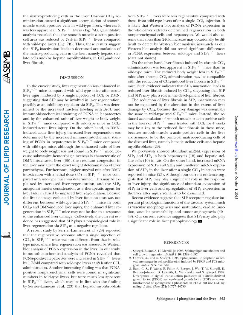
the matrix-producing cells in the liver. Chronic CCl4 ad-ministration caused a significant accumulation of smooth-muscle a-actin-positive cells in wild-type livers, whereas itwas less apparent in S1P2
2/2 livers (Fig. 7A). Quantitativeanalysis revealed that the smooth-muscle a-actin-positivecell area was reduced by 78% in S1P2
2/2 livers comparedwith wild-type livers (Fig. 7B). Thus, these results suggestthat S1P2 inactivation leads to decreased accumulation ofthe matrix-producing cells in the liver, namely hepatic stel-late cells and/or hepatic myofibroblasts, in CCl4-inducedliver fibrosis.
DISCUSSION
In the current study, liver regeneration was enhanced inS1P2
2/2 mice compared with wild-type mice after acuteliver injury induced by a single injection of CCl4 or DMN,suggesting that S1P may be involved in liver regeneration,possibly as an inhibitory regulator via S1P2. This was deter-mined by the increased nuclear labeling with BrdU andimmunohistochemical staining of PCNA in hepatocytesand by the enhanced ratio of liver weight to body weightin S1P2
2/2 mice compared with wild-type mice in CCl4-induced acute liver injury. On the other hand, in DMN-induced acute liver injury, increased liver regeneration wasdetermined by the increased immunohistochemical stain-ing of PCNA in hepatocytes in S1P2
2/2 mice comparedwith wild-type mice, although the enhanced ratio of liverweight to body weight was not found in S1P2
2/2 mice. Be-cause submassive hemorrhagic necrosis is characteristic ofDMN-intoxicated liver (36), the resultant congestion inthe liver may affect the exact weight determination of liverparenchyma. Furthermore, higher survival rate after DMNintoxication with a lethal dose (35) in S1P2
2/2 mice com-pared with wild-type mice was determined. This may be ex-plained by increased liver regeneration, and the S1P2
antagonist merits consideration as a therapeutic agent forliver failure caused by impaired liver regeneration. Becausethe liver damage evaluated by liver function tests was notdifferent between wild-type and S1P2
2/2 mice in bothCCl4- and DMN-induced liver injury, the enhanced liver re-generation in S1P2
2/2 mice may not be due to a responseto the enhanced liver damage. Collectively, the current evi-dence has suggested that S1P plays a physiological role inliver regeneration via S1P2 as a negative regulator.
A recent study by Serrier-Lanneau et al. (23) reportedthat the regenerative response after a single injection ofCCl4 in S1P2
2/2 mice was not different from that in wild-type mice, where liver regeneration was assessed by Westernblot analysis of PCNA expression in the liver. In our study,immunohistochemical analysis of PCNA revealed thatPCNA-positive hepatocytes were increased in S1P2
2/2 liversby 1.7-fold compared with wild-type livers at 48 h after CCl4administration. Another interesting finding was that PCNA-positive nonparenchymal cells were found in significantnumbers in wild-type livers, but were much less apparentin S1P2
2/2 livers, which may be in line with the findingby Serrier-Lanneau et al. (23) that hepatic myofibroblasts
from S1P22/2 livers were less regenerative compared with
those from wild-type livers after a single CCl4 injection. Itis likely that Western blot analysis of PCNA expression inthe whole-liver extracts determined regeneration in bothnonparenchymal cells and hepatocytes. We would also as-sume that a less than 2-fold increase may occasionally be dif-ficult to detect by Western blot analysis, inasmuch as ourWestern blot analysis did not reveal significant differencesin PCNA expression between wild-type and S1P2
2/2 livers(data not shown).
On the other hand, liver fibrosis induced by chronic CCl4administration was less apparent in S1P2
2/2 mice than inwild-type mice. The reduced body weight loss in S1P2
2/2
mice after chronic CCl4 administration may be compatiblewith the reduction of CCl4-induced liver fibrosis in S1P2
2/2
mice. Such evidence indicates that S1P2 inactivation leads toreduced liver fibrosis induced by CCl4, suggesting that S1Pand S1P2 may play a role in the development of liver fibrosis.
The reduction of liver fibrosis in S1P2 inactivation maynot be explained by the alteration in the extent of liverdamage by CCl4, because the liver damage was essentiallythe same in wild-type and S1P2
2/2 mice. Instead, the re-duced accumulation of smooth-muscle a-actin-positive cellsin the livers of S1P2
2/2 mice treated with CCl4 for 4 weeksmay be a key to the reduced liver fibrosis in those mice,because smooth-muscle a-actin-positive cells in the liverare assumed to be the principal matrix-producing cells ofthe diseased liver, namely hepatic stellate cells and hepaticmyofibroblasts (29).
We previously showed abundant mRNA expression ofS1P1 and S1P2 in both hepatocytes (18) and hepatic stel-late cells (16) in rats. On the other hand, increased mRNAexpression of S1P2 and S1P3 and unaltered mRNA expres-sion of S1P1 in the liver after a single CCl4 injection werereported in mice (23). Although our current evidence sug-gests that S1P2 may play a significant role in the responseto liver injury, the significance of abundant expression ofS1P1 in liver cells and upregulation of S1P3 expression inthe liver after injury remains to be examined.
Recent evidence suggests that S1P receptors regulate im-portant physiological functions of the vascular system, suchas vascular morphogenesis and maturation, cardiac func-tion, vascular permeability, and tumor angiogenesis (40–43). Our current evidence suggests that S1P2 may also playa significant role in liver pathophysiology.
REFERENCES
1. Spiegel, S., and A. H. Merrill, Jr. 1996. Sphingolipid metabolism andcell growth regulation. FASEB J. 10: 1388–1397.
2. Olivera, A., and S. Spiegel. 1993. Sphingosine-1-phosphate as sec-ond messenger in cell proliferation induced by PDGF and FCS mito-gens. Nature. 365: 557–560.
3. Rani, C. S., F. Wang, E. Fuior, A. Berger, J. Wu, T. W. Sturgill, D.Beitner-Johnson, D. LeRoith, L. Varticovski, and S. Spiegel. 1997.Divergence in signal transduction pathways of platelet-derivedgrowth factor (PDGF) and epidermal growth factor (EGF) receptors.Involvement of sphingosine 1-phosphate in PDGF but not EGF sig-naling. J. Biol. Chem. 272: 10777–10783.
Sphingosine 1-phosphate and the liver 563
by guest, on October 1, 2018
ww
w.jlr.org
Dow
nloaded from
4. Cuvillier, O., G. Pirianov, B. Kleuser, P. G. Vanek, O. A. Coso, S.Gutkind, and S. Spiegel. 1996. Suppression of ceramide-mediatedprogrammed cell death by sphingosine-1-phosphate. Nature. 381:800–803.
5. Edsall, L. C., G. G. Pirianov, and S. Spiegel. 1997. Involvement ofsphingosine 1-phosphate in nerve growth factor-mediated neuronalsurvival and differentiation. J. Neurosci. 17: 6952–6960.
6. Goodemote, K. A., M. E. Mattie, A. Berger, and S. Spiegel. 1995.Involvement of a pertussis toxin-sensitive G protein in the mito-genic signaling pathways of sphingosine 1-phosphate. J. Biol. Chem.270: 10272–10277.
7. Tosaka, M., F. Okajima, Y. Hashiba, N. Saito, T. Nagano, T. Watanabe,T. Kimura, and T. Sasaki. 2001. Sphingosine 1-phosphate contractscanine basilar arteries in vitro and in vivo: possible role in pathogen-esis of cerebral vasospasm. Stroke. 32: 2913–2919.
8. Ohmori, T., Y. Yatomi, M. Osada, F. Kazama, T. Takafuta, H. Ikeda,and Y. Ozaki. 2003. Sphingosine 1-phosphate induces contraction ofcoronary artery smooth muscle cells via S1P2. Cardiovasc. Res. 58:170–177.
9. Ishii, I., N. Fukushima, X. Ye, and J. Chun. 2004. Lysophospholipidreceptors: signaling and biology. Annu. Rev. Biochem. 73: 321–354.
10. Gardell, S. E., A. E. Dubin, and J. Chun. 2006. Emerging medicinalroles for lysophospholipid signaling. Trends Mol. Med. 12: 65–75.
11. Yatomi, Y., F. Ruan, J. Ohta, R. J. Welch, S. Hakomori, and Y. Igarashi.1995. Quantitative measurement of sphingosine 1-phosphate in bio-logical samples by acylation with radioactive acetic anhydride. Anal.Biochem. 230: 315–320.
12. Pappu, R., S. R. Schwab, I. Cornelissen, J. P. Pereira, J. B. Regard, Y.Xu, E. Camerer, Y. W. Zheng, Y. Huang, J. G. Cyster, et al. 2007.Promotion of lymphocyte egress into blood and lymph by distinctsources of sphingosine-1-phosphate. Science. 316: 295–298.
13. Chun, J. 2007. Immunology. The sources of a lipid conundrum.Science. 316: 208–210.
14. Yang, A. H., I. Ishii, and J. Chun. 2002. In vivo roles of lysophospho-lipid receptors revealed by gene targeting studies in mice. Biochim.Biophys. Acta. 1582: 197–203.
15. Herr, D. R., N. Grillet, M. Schwander, R. Rivera, U. Muller, and J.Chun. 2007. Sphingosine 1-phosphate (S1P) signaling is required formaintenance of hair cells mainly via activation of S1P2. J. Neurosci. 27:1474–1478.
16. Ikeda, H., Y. Yatomi, M. Yanase, H. Satoh, H. Maekawa, I. Ogata, Y.Ozaki, Y. Takuwa, S. Mochida, and K. Fujiwara. 2000. Biological activ-ities of novel lipid mediator sphingosine 1-phosphate in rat hepaticstellate cells. Am. J. Physiol. Gastrointest. Liver Physiol. 279: G304–G310.
17. Ikeda, H., K. Nagashima, M. Yanase, T. Tomiya, M. Arai, Y. Inoue, K.Tejima, T. Nishikawa, N. Watanabe, M. Omata, et al. 2004. Sphingo-sine 1-phosphate enhances portal pressure in isolated perfused livervia S1P2 with Rho activation. Biochem. Biophys. Res. Commun. 320:754–759.
18. Ikeda, H., H. Satoh, M. Yanase, Y. Inoue, T. Tomiya, M. Arai, K. Tejima,K. Nagashima, H. Maekawa, N. Yahagi, et al. 2003. Antiproliferativeproperty of sphingosine 1-phosphate in rat hepatocytes involves ac-tivation of Rho via Edg-5. Gastroenterology. 124: 459–469.
19. Michalopoulos, G. K., and M. C. DeFrances. 1997. Liver regenera-tion. Science. 276: 60–66.
20. Friedman, S. L. 2000. Molecular regulation of hepatic fibrosis, anintegrated cellular response to tissue injury. J. Biol. Chem. 275:2247–2250.
21. Minato, Y., Y. Hasumura, and J. Takeuchi. 1983. The role of fat-storingcells in Disse space fibrogenesis in alcoholic liver disease. Hepatology.3: 559–566.
22. Rockey, D. C., C. N. Housset, and S. L. Friedman. 1993. Activation-dependent contractility of rat hepatic lipocytes in culture and in vivo.J. Clin. Invest. 92: 1795–1804.
23. Serriere-Lanneau, V., F. Teixeira-Clerc, L. Li, M. Schippers, W. deWries, B. Julien, J. Tran-Van-Nhieu, S. Manin, K. Poelstra, J. Chun,et al. 2007. The sphingosine 1-phosphate receptor S1P2 triggers he-patic wound healing. FASEB J. 21: 2005–2013.
24. Adachi, M., Y. Osawa, H. Uchinami, T. Kitamura, D. Accili, and D. A.Brenner. 2007. The forkhead transcription factor FoxO1 regulatesproliferation and transdifferentiation of hepatic stellate cells. Gastro-enterology. 132: 1434–1446.
25. Kojima, N., M. Hori, T. Murata, Y. Morizane, and H. Ozaki. 2007.Different profiles of Ca21 responses to endothelin-1 and PDGF inliver myofibroblasts during the process of cell differentiation. Br. J.Pharmacol. 151: 816–827.
26. Park, E. J., Y. Z. Zhao, Y. C. Kim, and D. H. Sohn. 2007. Bakuchiol-induced caspase-3-dependent apoptosis occurs through c-Jun NH2-terminal kinase-mediated mitochondrial translocation of Bax in ratliver myofibroblasts. Eur. J. Pharmacol. 559: 115–123.
27. Watson, M. R., K. Wallace, R. G. Gieling, D. M. Manas, E. Jaffray,R. T. Hay, D. A. Mann, and F. Oakley. 2008. NF-kappaB is a criticalregulator of the survival of rodent and human hepatic myofibro-blasts. J. Hepatol. 48: 589–597.
28. Knittel, T., D. Kobold, B. Saile, A. Grundmann, K. Neubauer, F.Piscaglia, and G. Ramadori. 1999. Rat liver myofibroblasts and he-patic stellate cells: different cell populations of the fibroblast line-age with fibrogenic potential. Gastroenterology. 117: 1205–1221.
29. Saile, B., N. Matthes, K. Neubauer, C. Eisenbach, H. El-Armouche,J. Dudas, and G. Ramadori. 2002. Rat liver myofibroblasts and he-patic stellate cells differ in CD95-mediated apoptosis and response toTNF-alpha. Am. J. Physiol. Gastrointest. Liver Physiol. 283: G435–G444.
30. Ishii, I., X. Ye, B. Friedman, S. Kawamura, J. J. Contos, M. A.Kingsbury, A. H. Yang, G. Zhang, J. H. Brown, and J. Chun. 2002.Marked perinatal lethality and cellular signaling deficits in micenull for the two sphingosine 1-phosphate (S1P) receptors, S1P(2)/LP(B2)/EDG-5 and S1P(3)/LP(B3)/EDG-3. J. Biol. Chem.277: 25152–25159.
31. Goparaju, S. K., P. S. Jolly, K. R. Watterson, M. Bektas, S. Alvarez, S.Sarkar, L. Mel, I. Ishii, J. Chun, S. Milstien, et al. 2005. The S1P2receptor negatively regulates platelet-derived growth factor-inducedmotility and proliferation. Mol. Cell. Biol. 25: 4237–4249.
32. Means, C. K., C. Y. Xiao, Z. Li, T. Zhang, J. H. Omens, I. Ishii, J.Chun, and J. H. Brown. 2007. Sphingosine 1-phosphate S1P2 andS1P3 receptor-mediated Akt activation protects against in vivo myo-cardial ischemia-reperfusion injury. Am. J. Physiol. Heart Circ. Physiol.292: H2944–H2951.
33. Simeonova, P. P., R. M. Gallucci, T. Hulderman, R. Wilson, C.Kommineni, M. Rao, and M. I. Luster. 2001. The role of tumor necro-sis factor-alpha in liver toxicity, inflammation, and fibrosis inducedby carbon tetrachloride. Toxicol. Appl. Pharmacol. 177: 112–120.
34. Kanno, K., S. Tazuma, and K. Chayama. 2003. AT1A-deficient miceshow less severe progression of liver fibrosis induced by CCl(4). Bio-chem. Biophys. Res. Commun. 308: 177–183.
35. Morrison, V., and J. Ashby. 1994. Reconciliation of five negative andfour positive reports of the activity of dimethylnitrosamine in themouse bone marrow micronucleus assay. Mutagenesis. 9: 361–365.
36. Jin, Y. L., H. Enzan, N. Kuroda, Y. Hayashi, H. Nakayama, Y. H.Zhang, M. Toi, E. Miyazaki, M. Hiroi, L. M. Guo, et al. 2003. Tissueremodeling following submassive hemorrhagic necrosis in rat liversinduced by an intraperitoneal injection of dimethylnitrosamine.Virchows Arch. 442: 39–47.
37. Bedossa, P., and T. Poynard. 1996. An algorithm for the grading ofactivity in chronic hepatitis C. The METAVIR Cooperative StudyGroup. Hepatology. 24: 289–293.
38. Caballero, T., A. Perez-Milena, M. Masseroli, F. OʼValle, F. J. Salmeron,R. M. Del Moral, and G. Sanchez-Salgado. 2001. Liver fibrosis as-sessment with semiquantitative indexes and image analysis quantifi-cation in sustained-responder and non-responder interferon-treatedpatients with chronic hepatitis C. J. Hepatol. 34: 740–747.
39. Rockey, D. C., J. K. Boyles, G. Gabbiani, and S. L. Friedman. 1992.Rat hepatic lipocytes express smooth muscle actin upon activationin vivo and in culture. J. Submicrosc. Cytol. Pathol. 24: 193–203.
40. Sanchez, T., T. Estrada-Hernandez, J. H. Paik, M. T. Wu, K.Venkataraman, V. Brinkmann, K. Claffey, and T. Hla. 2003. Phos-phorylation and action of the immunomodulator FTY720 inhibitsvascular endothelial cell growth factor-induced vascular permeabil-ity. J. Biol. Chem. 278: 47281–47290.
41. Chae, S. S., J. H. Paik, H. Furneaux, and T. Hla. 2004. Require-ment for sphingosine 1-phosphate receptor-1 in tumor angiogen-esis demonstrated by in vivo RNA interference. J. Clin. Invest. 114:1082–1089.
42. LaMontagne, K., A. Littlewood-Evans, C. Schnell, T. OʼReilly, L.Wyder, T. Sanchez, B. Probst, J. Butler, A. Wood, G. Liau, et al.2006. Antagonism of sphingosine-1-phosphate receptors by FTY720inhibits angiogenesis and tumor vascularization. Cancer Res. 66:221–231.
43. Sanna, M. G., S. K. Wang, P. J. Gonzalez-Cabrera, A. Don, D. Marsolais,M. P. Matheu, S. H. Wei, I. Parker, E. Jo, W. C. Cheng, et al. 2006.Enhancement of capillary leakage and restoration of lympho-cyte egress by a chiral S1P1 antagonist in vivo. Nat. Chem. Biol. 2:434–441.
564 Journal of Lipid Research Volume 50, 2009
by guest, on October 1, 2018
ww
w.jlr.org
Dow
nloaded from


![DihydroceramideDesaturaseInhibitionbyaCyclopropanated ...downloads.hindawi.com/journals/jl/2011/724015.pdfbut also for those of sphingosine-1-phosphate (review: [12]). Dihydroceramide](https://static.fdocuments.in/doc/165x107/60291fd8cdc0c448707e1227/dihydroceramidedesaturaseinhibitionbyacyclopropanated-but-also-for-those-of.jpg)