Somites Branchial arches · 2020-02-06 · development of pharyngeal arches resembles formation of...
48
Primordia of the brain Primordia of the nose Primordia of the eye Primordia of the liver Primordia of the heart Upper limbs bud Lower limbs bud Somites WEEK 4 EMBRYO General features Branchial arches Dr.shatarat shatarat
Transcript of Somites Branchial arches · 2020-02-06 · development of pharyngeal arches resembles formation of...
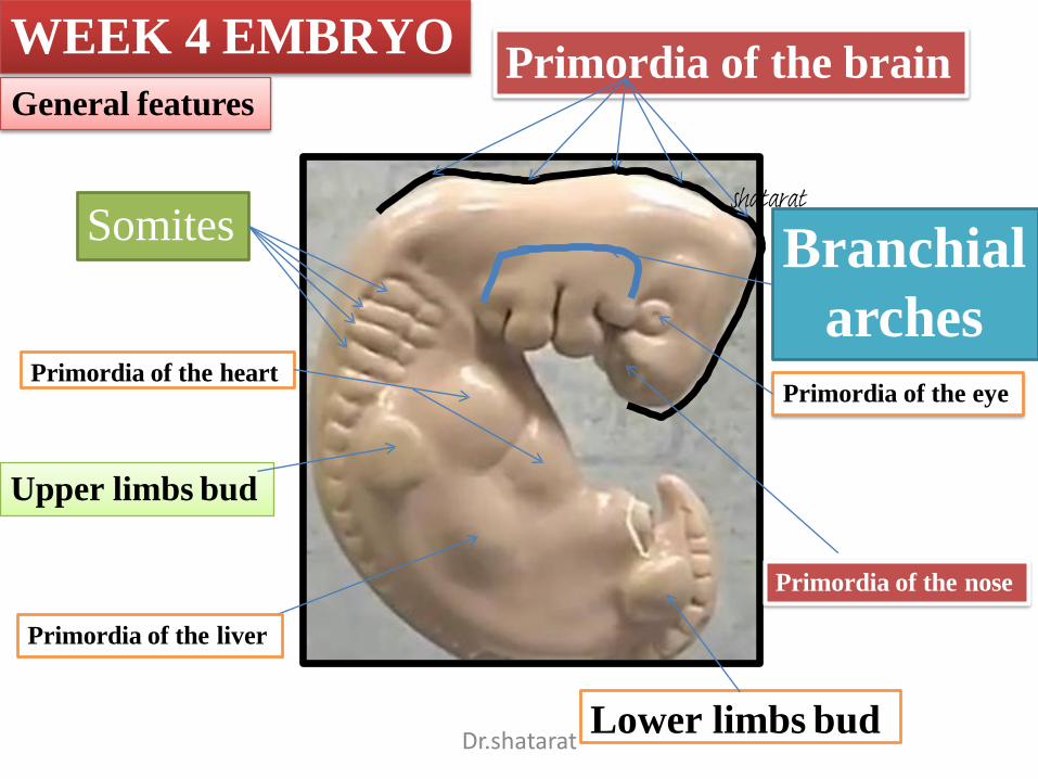
Primordia of the brain
Primordia of the nose
Primordia of the eye
Primordia of the liver
Primordia of the heart
Upper limbs bud
Lower limbs bud
Somites
WEEK 4 EMBRYO
General features
Branchial
arches
Dr.shatarat
shatarat
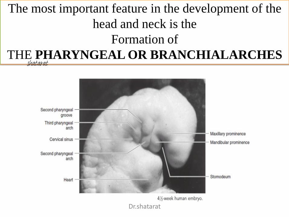
The most important feature in the development of the
head and neck is the
Formation of
THE PHARYNGEAL OR BRANCHIALARCHES
Dr.shatarat
shatarat
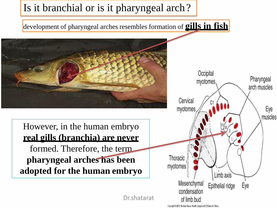
development of pharyngeal arches resembles formation of gills in fish
However, in the human embryo
real gills (branchia) are never
formed. Therefore, the term
pharyngeal arches has been
adopted for the human embryo
Is it branchial or is it pharyngeal arch?
Dr.shatarat
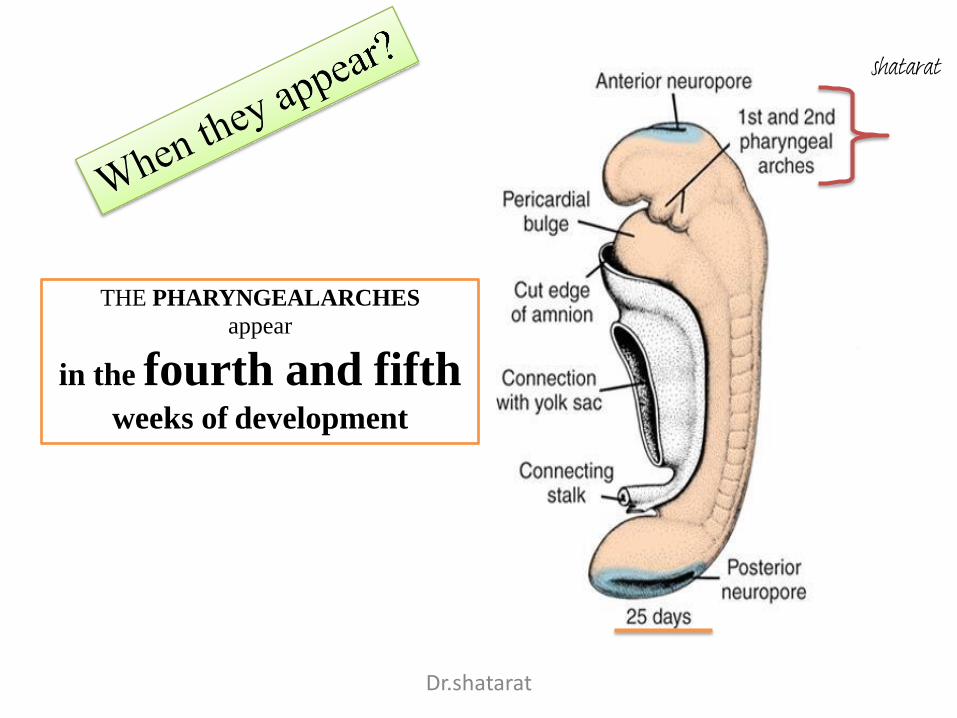
THE PHARYNGEALARCHES
appear
in the fourth and fifthweeks of development
Dr.shatarat
shatarat
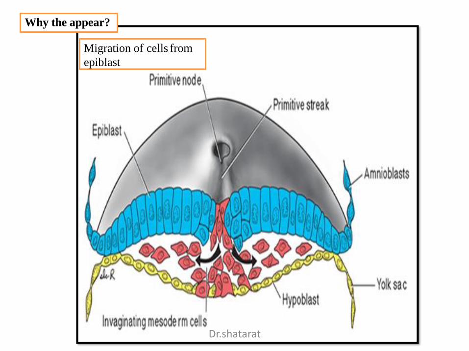
Why the appear?
Migration of cells from
epiblast
Dr.shatarat
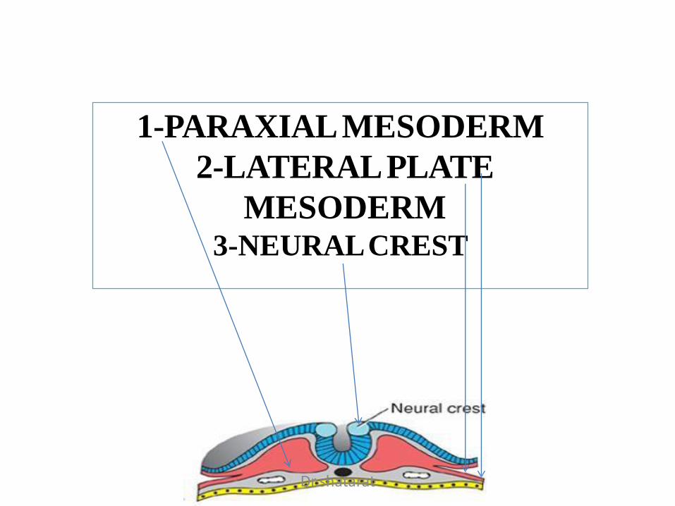
1-PARAXIALMESODERM
2-LATERALPLATE
MESODERM3-NEURALCREST
Dr.shatarat
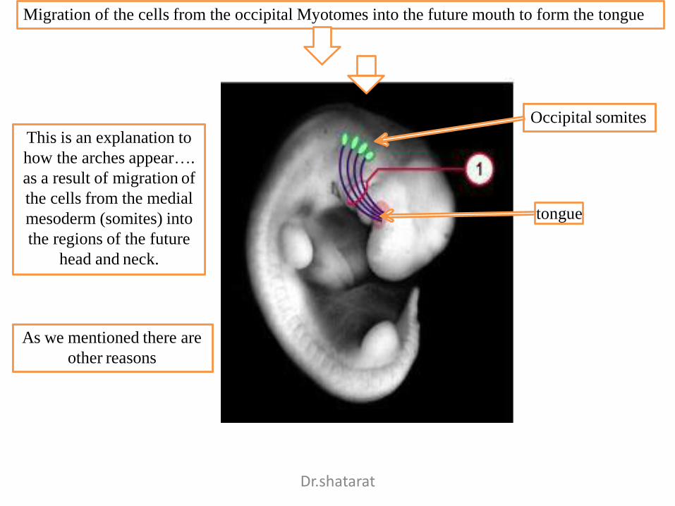
Migration of the cells from the occipital Myotomes into the future mouth to form the tongue
This is an explanation to
how the arches appear….
as a result of migration of
the cells from the medial
mesoderm (somites) into
the regions of the future
head and neck.
As we mentioned there are
other reasons
Occipital somites
tongue
Dr.shatarat
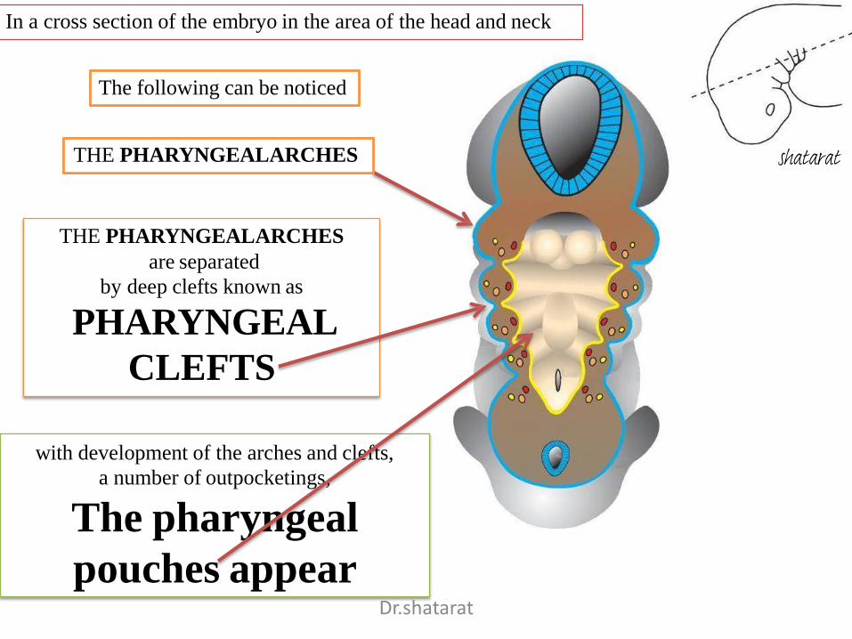
THE PHARYNGEALARCHES
are separated
by deep clefts known as
PHARYNGEAL
CLEFTS
with development of the arches and clefts,
a number of outpocketings,
The pharyngeal
pouches appear
In a cross section of the embryo in the area of the head and neck
The following can be noticed
THE PHARYNGEALARCHES
Dr.shatarat
shatarat
1-PHARYNGEAL ARCHs
Dr.shatarat
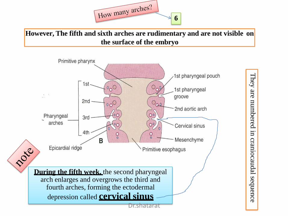
However, The fifth and sixth arches are rudimentary and are not visible on
the surface of the embryo
During the fifth week, the second pharyngeal arch enlarges and overgrows the third and
fourth arches, forming the ectodermal
depression called cervical sinus
6
They
aren
um
bered
incran
iocau
dal
sequen
ce
Dr.shatarat
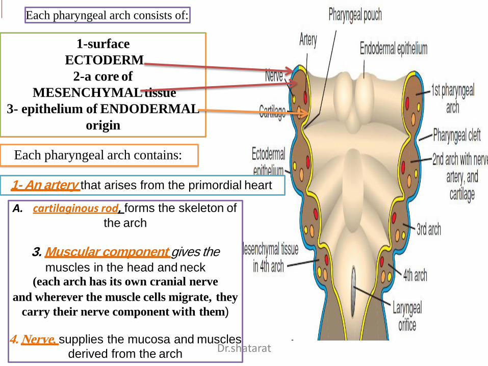
1-surface
ECTODERM
2-a core of
MESENCHYMALtissue
3- epithelium of ENDODERMAL
origin
Each pharyngeal arch consists of:
1- An artery that arises from the primordial heart
Each pharyngeal arch contains:
A. cartilaginous rod, forms the skeleton of
the arch
3. Muscular component gives themuscles in the head and neck
(each arch has its own cranial nerve
and wherever the muscle cells migrate, they
carry their nerve component with them)
4. Nerve, supplies the mucosa and muscles
derived from the archDr.shatarat
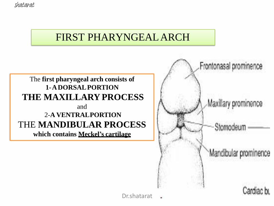
FIRST PHARYNGEALARCH
The first pharyngeal arch consists of
1-ADORSALPORTION
THE MAXILLARY PROCESSand
2-A VENTRALPORTION
THE MANDIBULAR PROCESSwhich contains Meckel’s cartilage
Dr.shatarat
shatarat
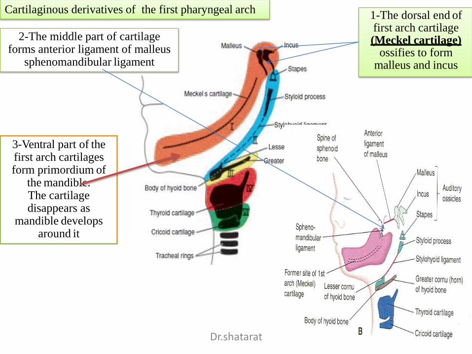
Cartilaginous derivatives of the first pharyngeal arch1-The dorsal end of first arch cartilage (Meckel cartilage)
ossifies to form malleus and incus
2-The middle part of cartilage forms anterior ligament of malleus
sphenomandibular ligament
3-Ventral part of the first arch cartilages form primordium of
the mandible.The cartilage disappears as
mandible develops around it
Dr.shatarat
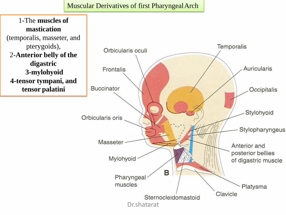
Muscular Derivatives of first PharyngealArch
1-The muscles of
mastication
(temporalis, masseter, and
pterygoids),
2-Anterior belly of the
digastric
3-mylohyoid
4-tensor tympani, and
tensor palatini
Dr.shatarat
The nerve supply to the muscles of
the first arch is provided by
the mandibular branch of the trigeminal nerve
Since mesenchyme from the first arch
also contributes
to the dermis of the face,
sensory supply to the skin of the face
is provided by
ophthalmic, maxillary, and mandibular
branches of the trigeminal nerve.
Dr.shatarat
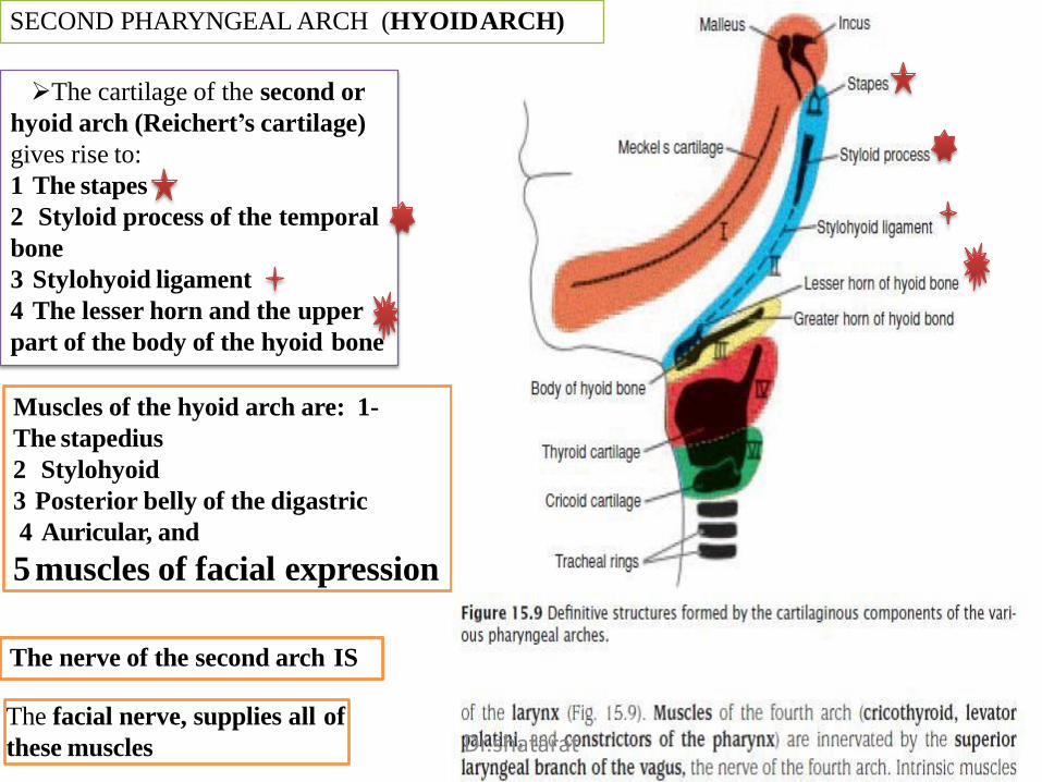
The cartilage of the second or
hyoid arch (Reichert’s cartilage)
gives rise to:
1 The stapes
2 Styloid process of the temporal
bone
3 Stylohyoid ligament
4 The lesser horn and the upper
part of the body of the hyoid bone
Muscles of the hyoid arch are: 1-
The stapedius
2 Stylohyoid
3 Posterior belly of the digastric
4 Auricular, and
5 muscles of facial expression
The facial nerve, supplies all of
these muscles
The nerve of the second arch IS
SECOND PHARYNGEAL ARCH (HYOIDARCH)
Dr.shatarat
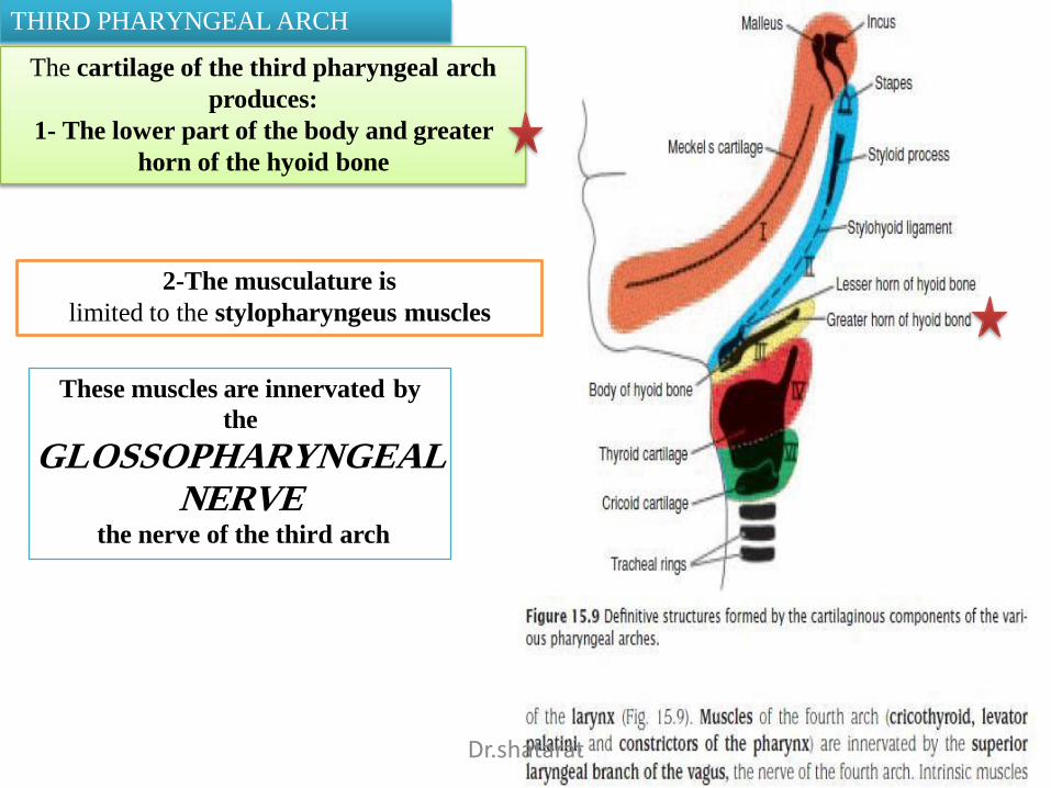
THIRD PHARYNGEAL ARCH
The cartilage of the third pharyngeal arch
produces:
1- The lower part of the body and greater
horn of the hyoid bone
These muscles are innervated by
the
GLOSSOPHARYNGEALNERVE
the nerve of the third arch
2-The musculature is
limited to the stylopharyngeus muscles
Dr.shatarat
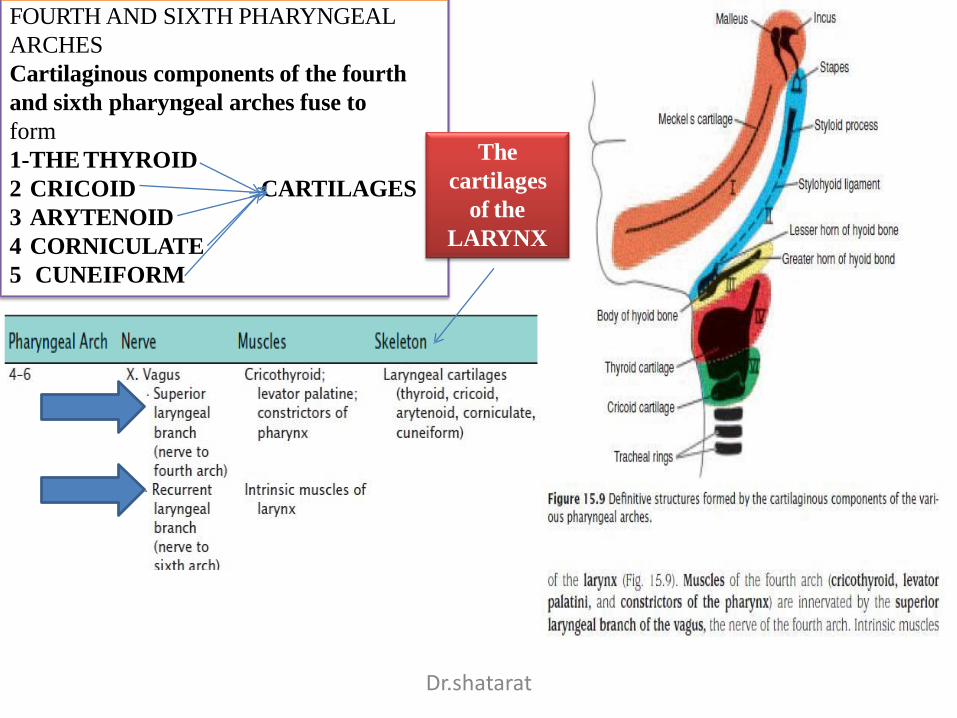
FOURTH AND SIXTH PHARYNGEAL
ARCHES
Cartilaginous components of the fourth
and sixth pharyngeal arches fuse to
form
1-THE THYROID
CARTILAGES2 CRICOID
3 ARYTENOID
4 CORNICULATE
5 CUNEIFORM
The
cartilages
of the
LARYNX
Dr.shatarat

2-PHARYNGEAL POUCHES
Dr.shatarat
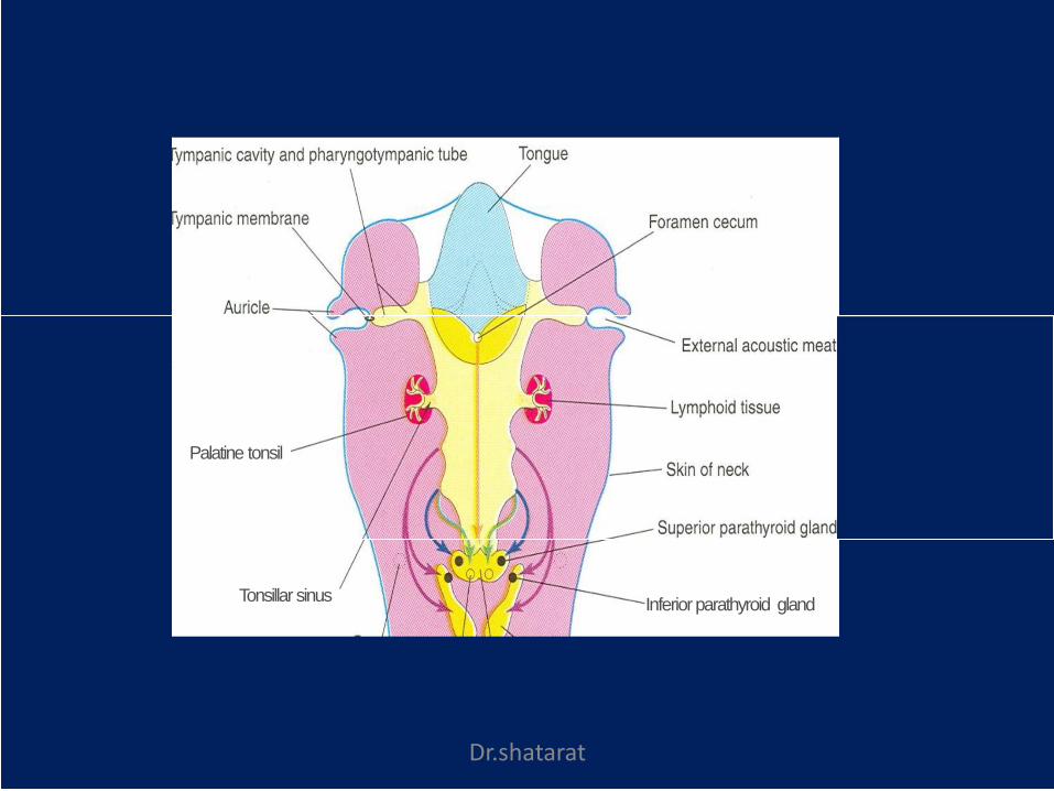
Palatine tonsil
Tonsillar sinusInferior parathyroid gland
Dr.shatarat
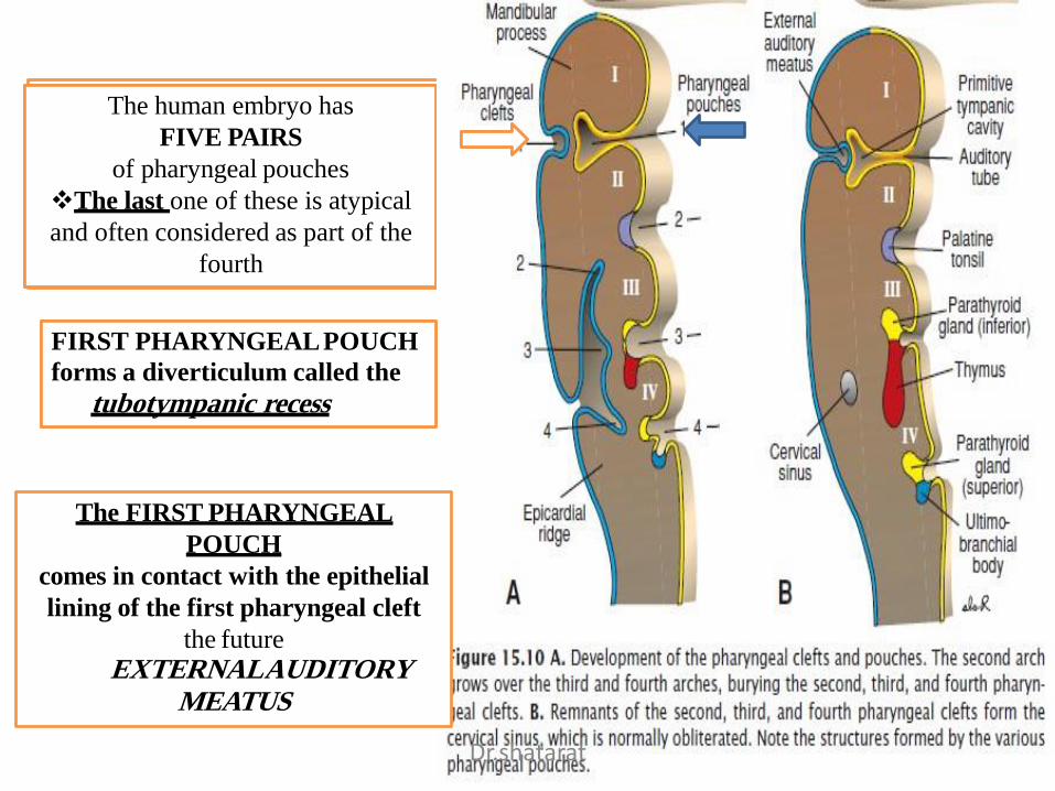
The human embryo has
FIVE PAIRS
of pharyngeal pouches
The last one of these is atypical
and often considered as part of the
fourth
FIRST PHARYNGEALPOUCH
forms a diverticulum called the
tubotympanic recess
The FIRST PHARYNGEAL
POUCH
comes in contact with the epithelial
lining of the first pharyngeal cleft
the future
EXTERNALAUDITORYMEATUS
Dr.shatarat
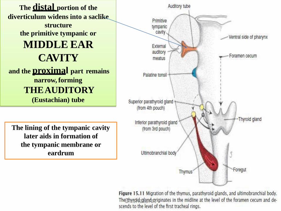
The distal portion of the
diverticulum widens into a saclike
structure
the primitive tympanic or
MIDDLE EAR
CAVITYand the proximal part remains
narrow, forming
THEAUDITORY(Eustachian) tube
The lining of the tympanic cavity
later aids in formation of
the tympanic membrane or
eardrum
Dr.shatarat
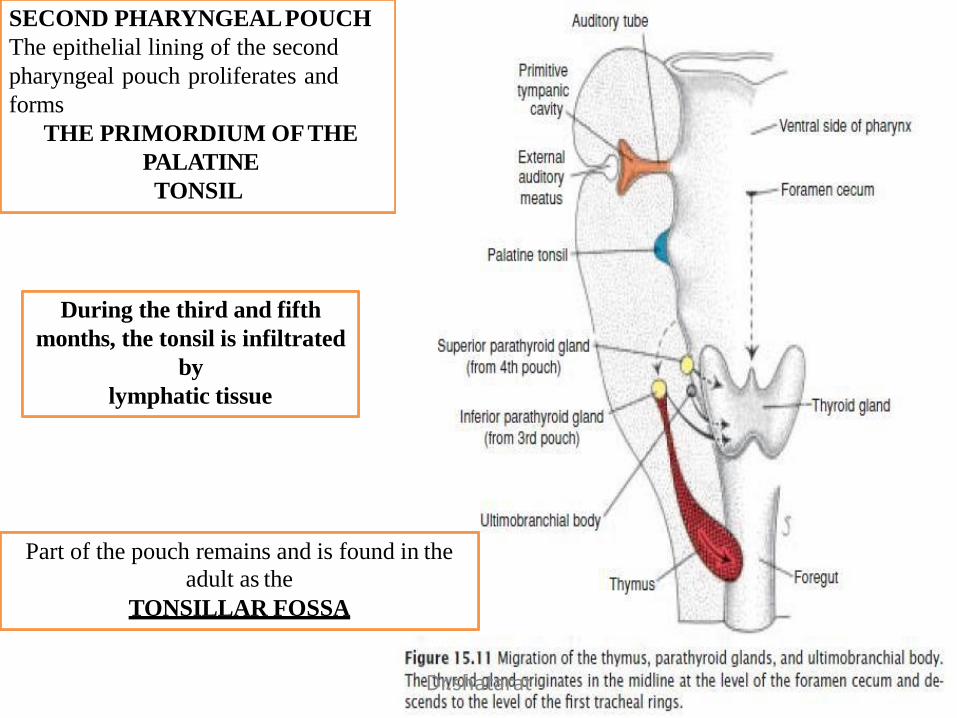
SECOND PHARYNGEALPOUCH
The epithelial lining of the second
pharyngeal pouch proliferates and
forms
THE PRIMORDIUM OFTHE
PALATINE
TONSIL
During the third and fifth
months, the tonsil is infiltrated
by
lymphatic tissue
Part of the pouch remains and is found in the
adult as the
TONSILLAR FOSSA
Dr.shatarat
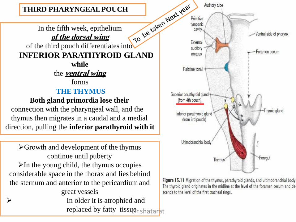
THIRD PHARYNGEALPOUCH
In the fifth week, epithelium
of the dorsal wingof the third pouch differentiates into
INFERIOR PARATHYROID GLANDwhile
the ventral wingforms
THE THYMUS
Both gland primordia lose their
connection with the pharyngeal wall, and the
thymus then migrates in a caudal and a medial
direction, pulling the inferior parathyroid with it
Growth and development of the thymus
continue until puberty
In the young child, the thymus occupies
considerable space in the thorax and lies behind
the sternum and anterior to the pericardium and
great vessels
In older it is atrophied and
replaced by fatty tissueDr.shatarat
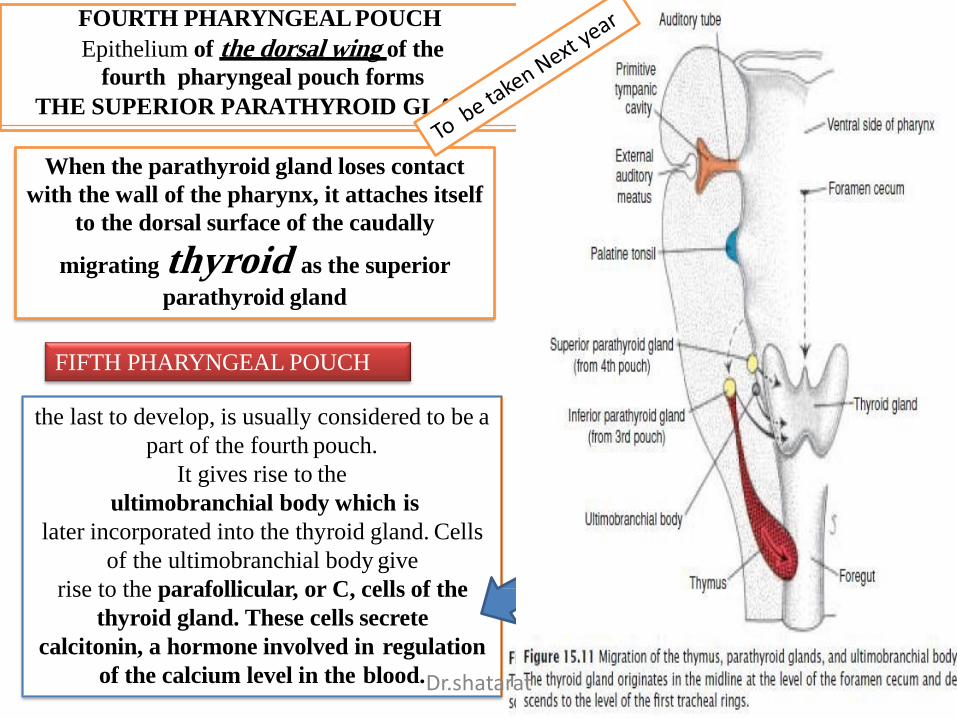
FOURTH PHARYNGEAL POUCH
Epithelium of the dorsal wing of the
fourth pharyngeal pouch forms
THE SUPERIOR PARATHYROID GLAND
FIFTH PHARYNGEAL POUCH
the last to develop, is usually considered to be a
part of the fourth pouch.
It gives rise to the
ultimobranchial body which is
later incorporated into the thyroid gland. Cells
of the ultimobranchial body give
rise to the parafollicular, or C, cells of the
thyroid gland. These cells secrete
calcitonin, a hormone involved in regulation
of the calcium level in the blood.
When the parathyroid gland loses contact
with the wall of the pharynx, it attaches itself
to the dorsal surface of the caudally
migrating thyroid as the superior
parathyroid gland
Dr.shatarat
3-Pharyngeal Clefts
Dr.shatarat
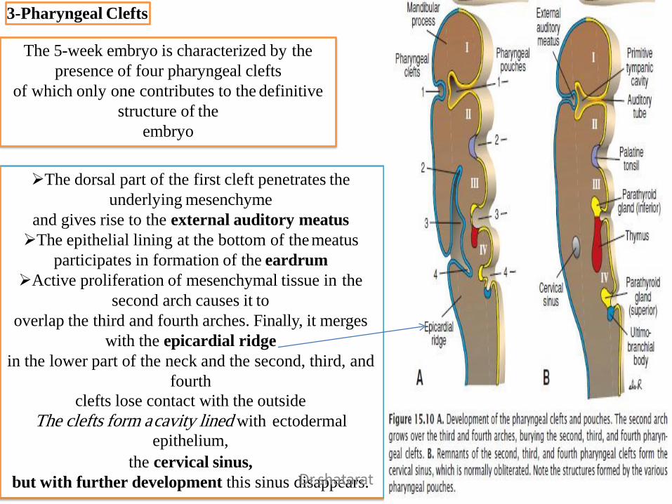
3-Pharyngeal Clefts
The 5-week embryo is characterized by the
presence of four pharyngeal clefts
of which only one contributes to the definitive
structure of the
embryo
The dorsal part of the first cleft penetrates the
underlying mesenchyme
and gives rise to the external auditory meatus
The epithelial lining at the bottom of the meatus
participates in formation of the eardrum
Active proliferation of mesenchymal tissue in the
second arch causes it to
overlap the third and fourth arches. Finally, it merges
with the epicardial ridge
in the lower part of the neck and the second, third, and
fourth
clefts lose contact with the outside
The clefts form a cavity lined with ectodermal
epithelium,
the cervical sinus,
but with further development this sinus disappears.Dr.shatarat

Development of the face
Dr.shatarat
Neural crest
Dr.shatarat
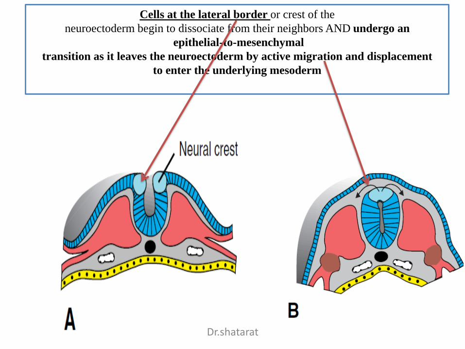
Cells at the lateral border or crest of the
neuroectoderm begin to dissociate from their neighbors AND undergo an
epithelial-to-mesenchymal
transition as it leaves the neuroectoderm by active migration and displacement
to enter the underlying mesoderm
Dr.shatarat
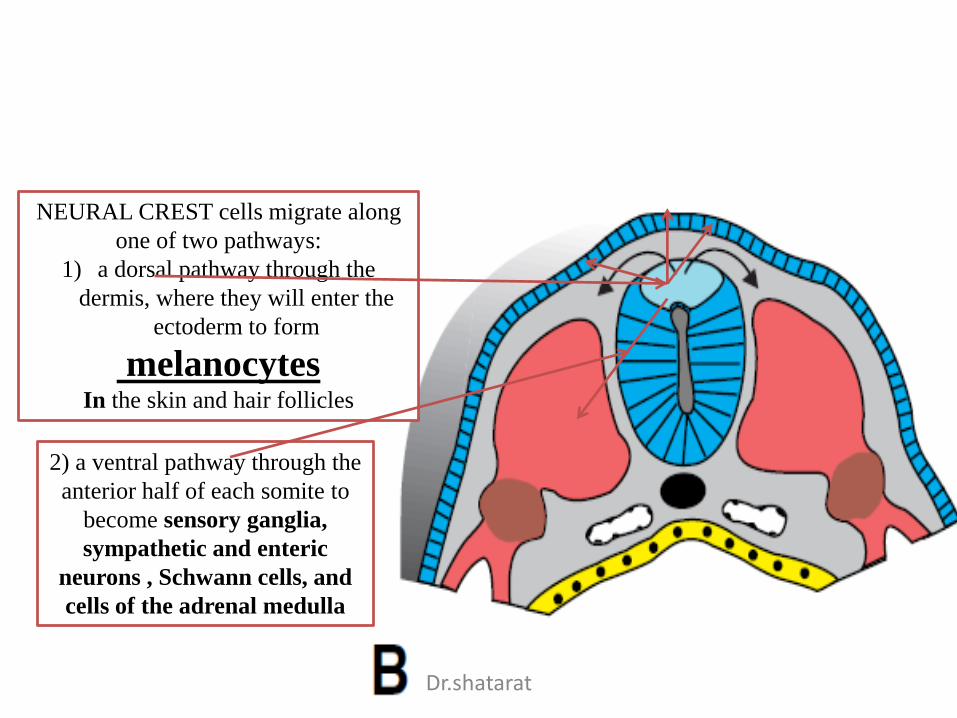
NEURAL CREST cells migrate along
one of two pathways:
1) a dorsal pathway through the
dermis, where they will enter the
ectoderm to form
melanocytes In the skin and hair follicles
2) a ventral pathway through the
anterior half of each somite to
become sensory ganglia,
sympathetic and enteric
neurons , Schwann cells, and
cells of the adrenal medulla
Dr.shatarat
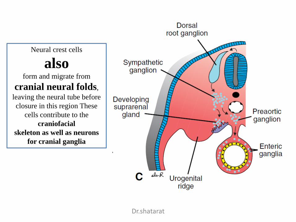
Neural crest cells
alsoform and migrate from
cranial neural folds,
leaving the neural tube before
closure in this region These
cells contribute to the
craniofacial
skeleton as well as neurons
for cranial ganglia
Dr.shatarat
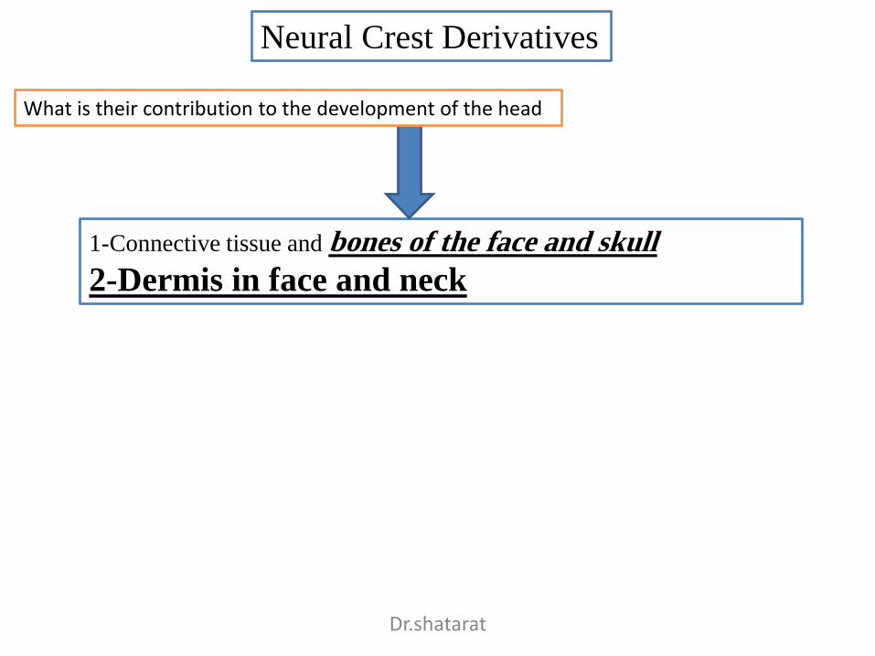
1-Connective tissue and bones of the face and skull
2-Dermis in face and neck
Neural Crest Derivatives
What is their contribution to the development of the head
Dr.shatarat
Dr.shatarat
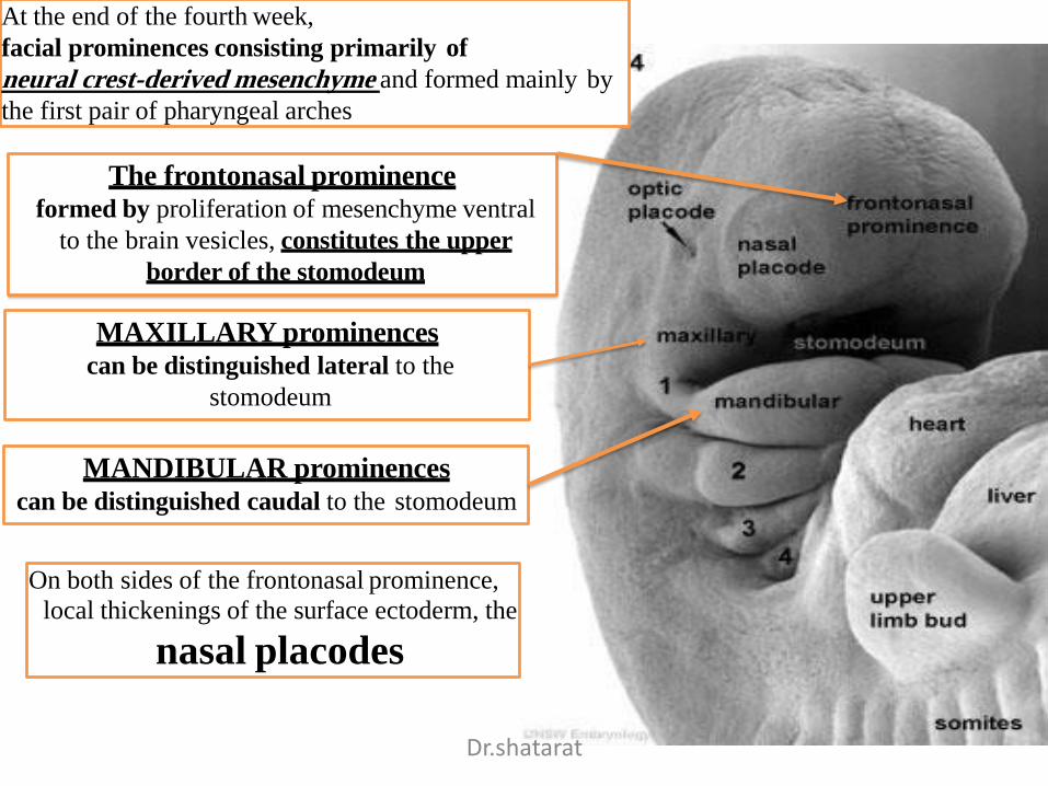
At the end of the fourth week,
facial prominences consisting primarily of
neural crest-derived mesenchyme and formed mainly by
the first pair of pharyngeal arches
The frontonasal prominenceformed by proliferation of mesenchyme ventral
to the brain vesicles, constitutes the upper
border of the stomodeum
On both sides of the frontonasal prominence,
local thickenings of the surface ectoderm, the
nasal placodes
MAXILLARY prominences can be distinguished lateral to the
stomodeum
MANDIBULAR prominencescan be distinguished caudal to the stomodeum
Dr.shatarat
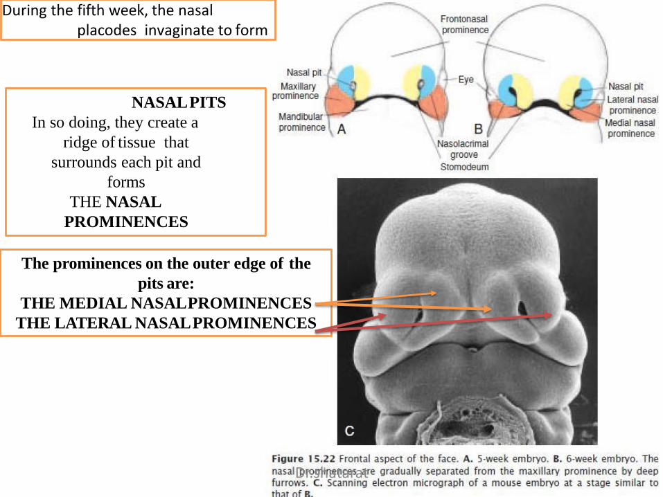
During the fifth week, the nasal placodes invaginate to form
NASALPITS
In so doing, they create a
ridge of tissue that
surrounds each pit and
forms
THE NASAL
PROMINENCES
The prominences on the outer edge of the
pits are:
THE MEDIAL NASALPROMINENCES
THE LATERAL NASALPROMINENCES
Dr.shatarat
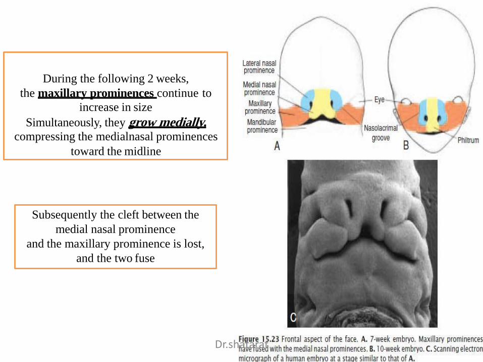
During the following 2 weeks,
the maxillary prominences continue to
increase in size
Simultaneously, they grow medially, compressing the medialnasal prominences
toward the midline
Subsequently the cleft between the
medial nasal prominence
and the maxillary prominence is lost,
and the two fuse
Dr.shatarat
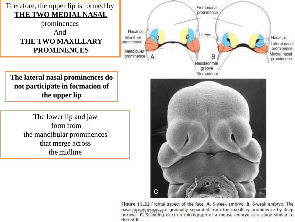
Therefore, the upper lip is formed by
THE TWO MEDIALNASAL
prominences
And
THE TWO MAXILLARY
PROMINENCES
The lateral nasal prominences do
not participate in formation of
the upper lip
The lower lip and jaw
form from
the mandibular prominences
that merge across
the midline
Dr.shatarat
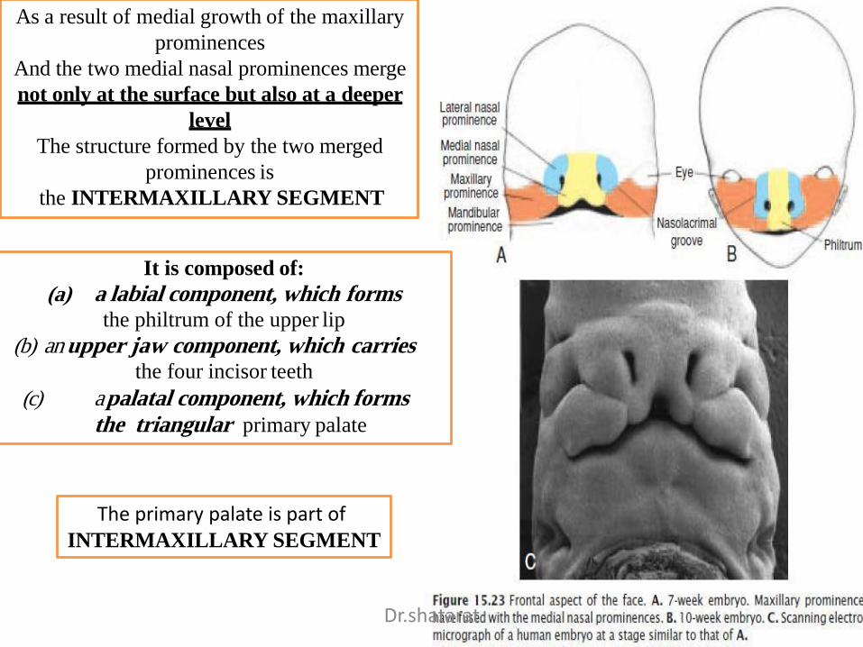
As a result of medial growth of the maxillary
prominences
And the two medial nasal prominences merge
not only at the surface but also at a deeper
level
The structure formed by the two merged
prominences is
the INTERMAXILLARY SEGMENT
It is composed of:
(a) a labial component, which formsthe philtrum of the upper lip
(b) an upper jaw component, which carriesthe four incisor teeth
(c) a palatal component, which forms the triangular primary palate
The primary palate is part of INTERMAXILLARY SEGMENT
Dr.shatarat
Development of the palate
The primary palate is part of INTERMAXILLARY SEGMENT
Dr.shatarat
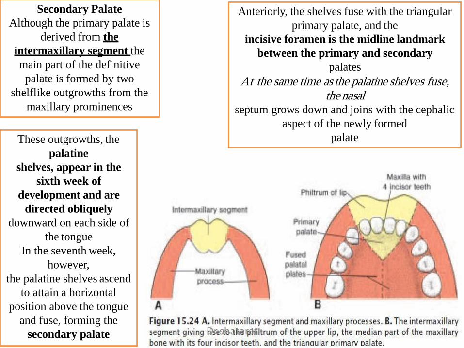
Secondary Palate
Although the primary palate is
derived from the
intermaxillary segment the
main part of the definitive
palate is formed by two
shelflike outgrowths from the
maxillary prominences
These outgrowths, the
palatine
shelves, appear in the
sixth week of
development and are
directed obliquely
downward on each side of
the tongue
In the seventh week,
however,
the palatine shelves ascend
to attain a horizontal
position above the tongue
and fuse, forming the
secondary palate
Anteriorly, the shelves fuse with the triangular
primary palate, and the
incisive foramen is the midline landmark
between the primary and secondary
palates
At the same time as the palatine shelves fuse, the nasal
septum grows down and joins with the cephalic
aspect of the newly formed
palate
Dr.shatarat
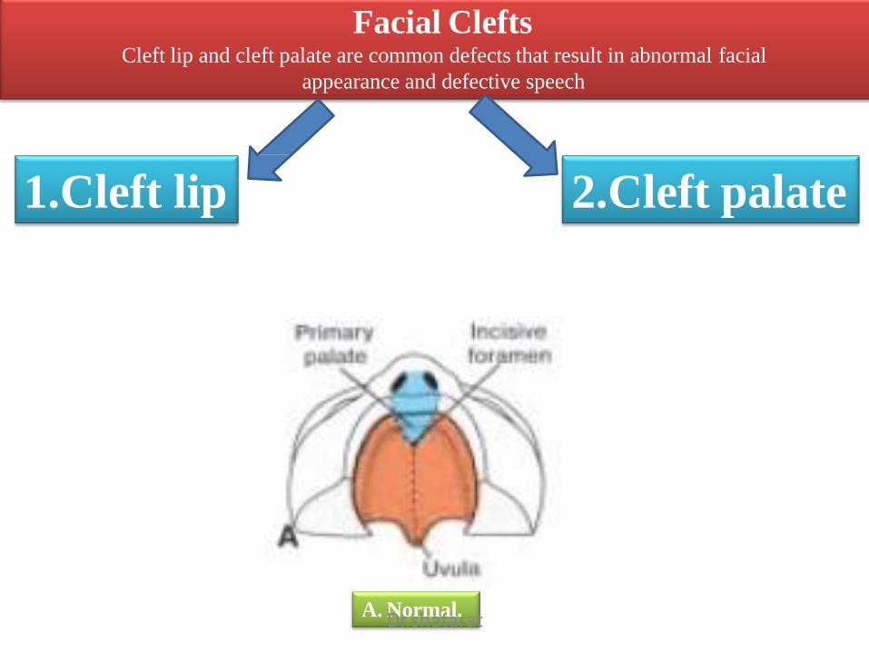
Facial Clefts
A. Normal.
Cleft lip and cleft palate are common defects that result in abnormal facial
appearance and defective speech
1.Cleft lip 2.Cleft palate
Dr.shatarat
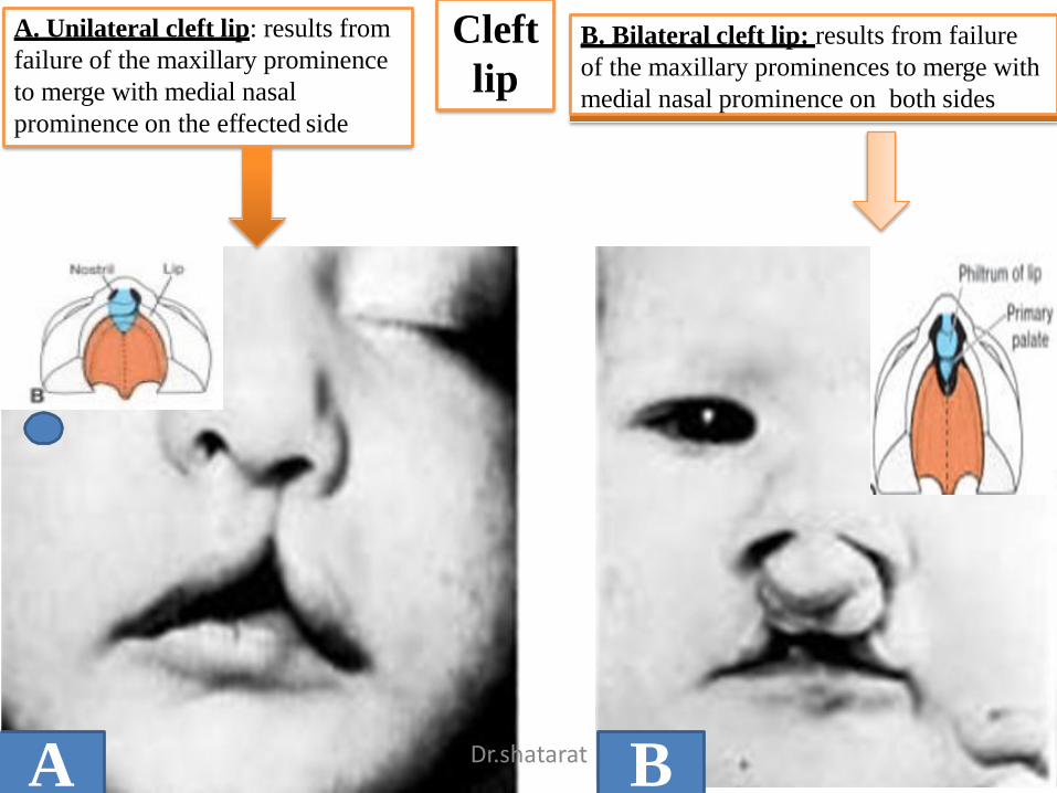
Cleft
lip
A. Unilateral cleft lip: results from
failure of the maxillary prominence
to merge with medial nasal
prominence on the effected side
B. Bilateral cleft lip: results from failure
of the maxillary prominences to merge with
medial nasal prominence on both sides
A BDr.shatarat
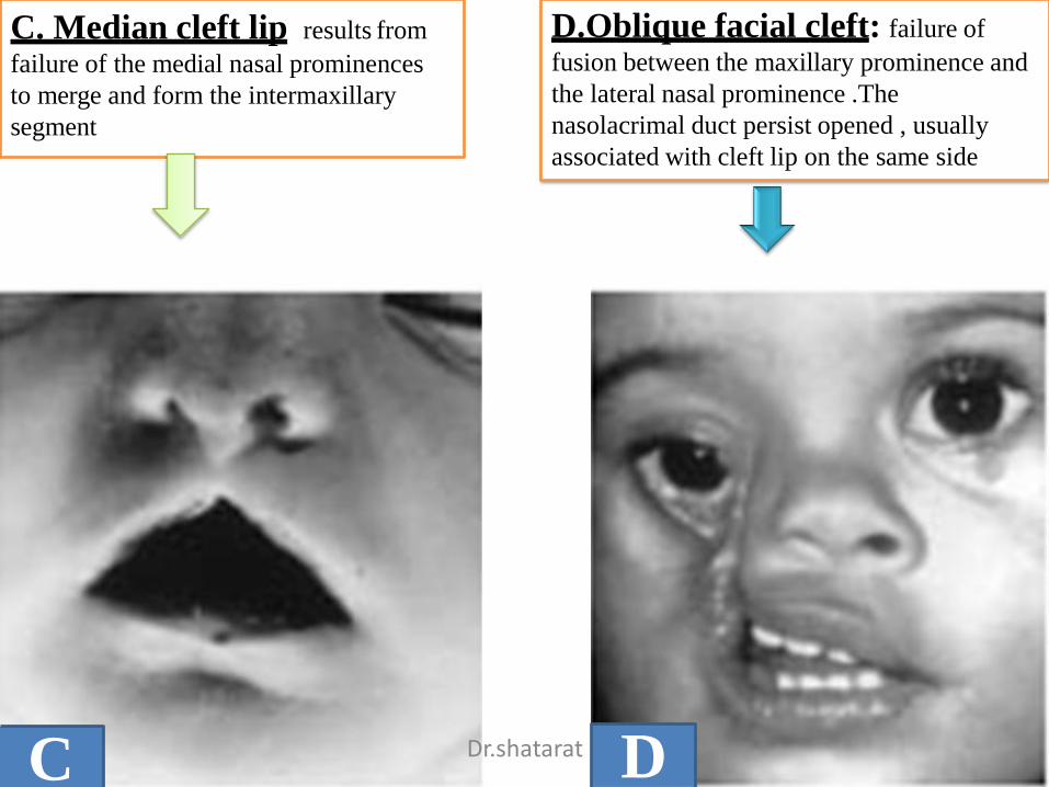
C. Median cleft lip: results from
failure of the medial nasal prominences
to merge and form the intermaxillary
segment
D.Oblique facial cleft: failure of
fusion between the maxillary prominence and
the lateral nasal prominence .The
nasolacrimal duct persist opened , usually
associated with cleft lip on the same side
DC Dr.shatarat
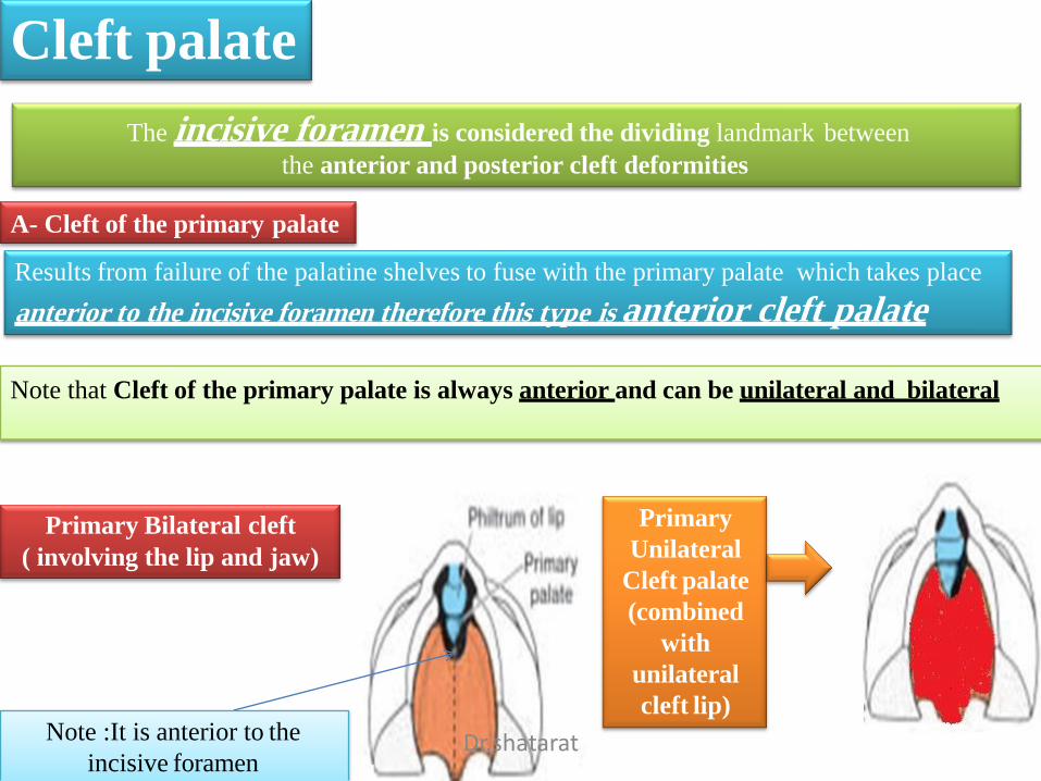
Cleft palate
The incisive foramen is considered the dividing landmark between
the anterior and posterior cleft deformities
A- Cleft of the primary palate
Results from failure of the palatine shelves to fuse with the primary palate which takes place
anterior to the incisive foramen therefore this type is anterior cleft palate
Note that Cleft of the primary palate is always anterior and can be unilateral and bilateral
Primary Bilateral cleft
( involving the lip and jaw)
Note :It is anterior to the
incisive foramen
Primary
Unilateral
Cleft palate
(combined
with
unilateral
cleft lip)
Dr.shatarat
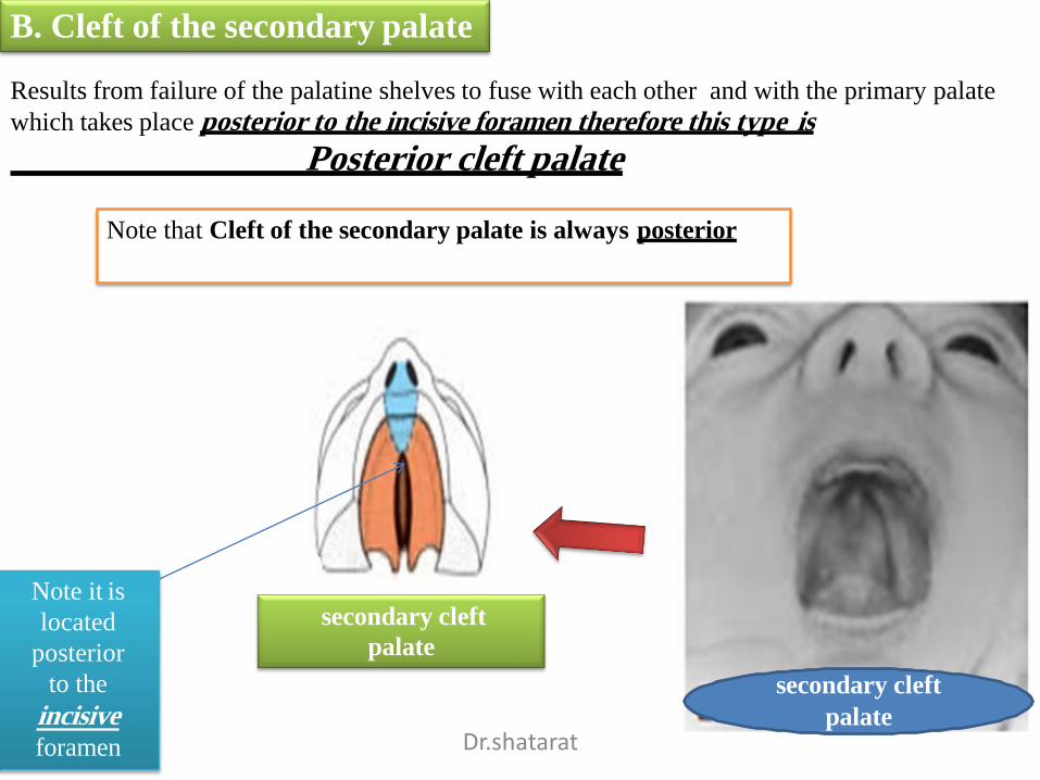
B. Cleft of the secondary palate
Results from failure of the palatine shelves to fuse with each other and with the primary palate
which takes place posterior to the incisive foramen therefore this type is
Posterior cleft palate
Note that Cleft of the secondary palate is always posterior
secondary cleft
palate
Note it is
located
posterior
to the
incisive foramen
secondary cleft
palateDr.shatarat
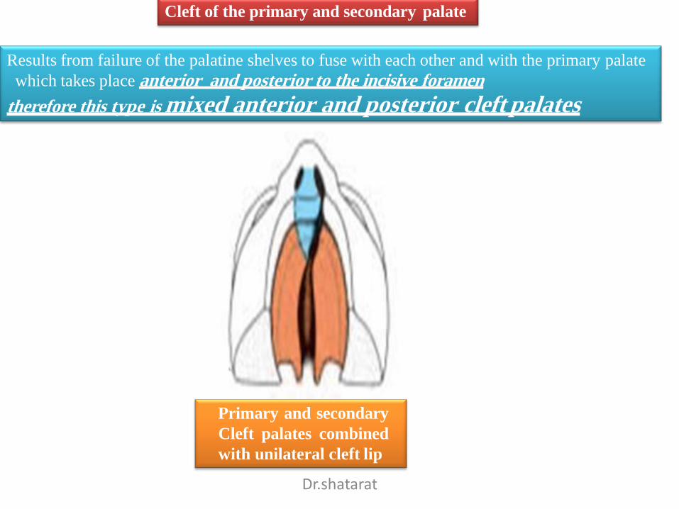
Cleft of the primary and secondary palate
Results from failure of the palatine shelves to fuse with each other and with the primary palate
which takes place anterior and posterior to the incisive foramen
therefore this type is mixed anterior and posterior cleft palates
Primary and secondary
Cleft palates combined
with unilateral cleft lip
Dr.shatarat
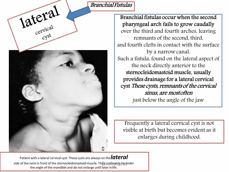
Branchial fistulas occur when the second pharyngeal arch fails to grow caudally
over the third and fourth arches, leaving remnants of the second, third,
and fourth clefts in contact with the surface by a narrow canal.
Such a fistula, found on the lateral aspect of the neck directly anterior to the
sternocleidomastoid muscle, usually provides drainage for a lateral cervical
cyst These cysts, remnants of the cervicalsinus, are mostoften
just below the angle of the jaw
BranchialFistulas
Frequently a lateral cervical cyst is not visible at birth but becomes evident as it
enlarges during childhood.
Patient with a lateral cervical cyst. These cysts are always on thelateralside of the neck in front of the sternocleidomastoid muscle. They commonly lie under
the angle of the mandible and do not enlarge until later inlife.Dr.shatarat