![Leonardo da Vinci (1452–1519) and his depictions of the ... · of the spine has been credited to Leonardo [14, 15], a topic to which we will return later in this article. Although](https://static.fdocuments.in/doc/165x107/5f61847eefea1e6a8f2b4cb5/leonardo-da-vinci-1452a1519-and-his-depictions-of-the-of-the-spine-has-been.jpg)
Some of the earliest depictions of the human spine: a ... · Some of the earliest depictions of the...
3
COVER EDITORIAL Some of the earliest depictions of the human spine: a glimpse into European history Peter C. Oakes 1 & Christina Kirkpatrick 1 & Jens R. Chapman 2 & Rod J. Oskouian 2 & R. Shane Tubbs 1,3 Received: 10 April 2017 /Accepted: 13 April 2017 /Published online: 25 April 2017 # Springer-Verlag Berlin Heidelberg 2017 Abstract Introduction Drawings of the human form have a history al- most as old as mankind itself. However, illustrations of the human spine as seen with the vertebral column were not seen until much later. This paper reviews some of the early European depictions of the human vertebral column from the twelfth (e.g., Fünfbilderserie BBone-Man^: 1152 A.D.) and thirteenth (e.g., Ashmole 1292) centuries. Man’ s under- standing of his body has evolved over hundreds of years. Conclusions This glimpse into our past and early drawings of the human spine illustrate how this particular anatomical structure was perceived almost a millennium ago and would not be structurally correct renditions until Leonardo da Vinci in the fifteenth century. Keywords European . History . Art . Drawings . Vertebral column . Anatomy Background It has been speculated that the origin of neurosurgery can be traced back nearly 5000 years to the ancient Egyptians. Some have suggested that one of the earliest recorded neurosurgical procedures took place at this point in history: a traction reduc- tion of the cervical spine of an early Egyptian ruler. The pro- cedure is said to have served as a template for the Egyptian myth detailing the resurrection of Osiris by his wife and son. Several other references have suggested the god recovered his strength after the efforts of his family [1]. While the facts are difficult to establish, this story raises several questions about the history of the spine in medicine. The present paper will briefly address differing views concerning the history of de- pictions of the human spine. Sudhoff ’ s findings During the early twentieth century, Karl Sudhoff (1853– 1938), a German medical historian, was a voracious explorer of pre-Vesalian anatomical drawings. Sudhoff was put in charge of a newly formed Institut für Geschichte der Medizin (Institute of Historical Medicine) at the University of Leipzig in 1905 and promoted to a newly established chair to help his work. Sudhoff scavenged literary works across Europe and discovered several groups of drawings [2]. One, dating back to 1292, came from the Ashmolean library in Oxford (Fig. 1), while another came from the BProvençal manuscript^, now housed in the University of Basel’ s library (see Fig. 2)[2]. However, the oldest depictions of the human spine were found in the cloister of Prüfening outside Ratisbon, Bavaria, Germany, and dated to 1158 A.D. (see Fig. 3)[2, 3]. Sudhoff ’ s investigation quickly expanded into the wider ques- tion of the earliest medical anatomical depictions of the human body. For Sudhoff, this BBone-Man^ was one in a series of five similar anatomical depictions [4]. The figures that Sudhoff discovered followed a some- what formulaic pattern. Generally, a series of five fig- ures would depict the skeletal, nervous, muscular, ve- nous, and arterial systems [2]. In view of this near- * R. Shane Tubbs [email protected] 1 Seattle Science Foundation, Seattle, WA, USA 2 Swedish Neuroscience Institute, Swedish Medical Center, Seattle, Washington, USA 3 Department of Anatomical Sciences, St. George’ s University, St. George’ s, Grenada Childs Nerv Syst (2018) 34:179–181 DOI 10.1007/s00381-017-3419-9
Transcript of Some of the earliest depictions of the human spine: a ... · Some of the earliest depictions of the...
COVER EDITORIAL
Some of the earliest depictions of the human spine: a glimpseinto European history
Peter C. Oakes1 & Christina Kirkpatrick1& Jens R. Chapman2
& Rod J. Oskouian2&
R. Shane Tubbs1,3
Received: 10 April 2017 /Accepted: 13 April 2017 /Published online: 25 April 2017# Springer-Verlag Berlin Heidelberg 2017
AbstractIntroduction Drawings of the human form have a history al-most as old as mankind itself. However, illustrations of thehuman spine as seen with the vertebral column were not seenuntil much later. This paper reviews some of the earlyEuropean depictions of the human vertebral column fromthe twelfth (e.g., Fünfbilderserie BBone-Man^: 1152 A.D.)and thirteenth (e.g., Ashmole 1292) centuries. Man’s under-standing of his body has evolved over hundreds of years.Conclusions This glimpse into our past and early drawings ofthe human spine illustrate how this particular anatomicalstructure was perceived almost a millennium ago and wouldnot be structurally correct renditions until Leonardo da Vinciin the fifteenth century.
Keywords European . History . Art . Drawings . Vertebralcolumn . Anatomy
Background
It has been speculated that the origin of neurosurgery can betraced back nearly 5000 years to the ancient Egyptians. Somehave suggested that one of the earliest recorded neurosurgical
procedures took place at this point in history: a traction reduc-tion of the cervical spine of an early Egyptian ruler. The pro-cedure is said to have served as a template for the Egyptianmyth detailing the resurrection of Osiris by his wife and son.Several other references have suggested the god recovered hisstrength after the efforts of his family [1]. While the facts aredifficult to establish, this story raises several questions aboutthe history of the spine in medicine. The present paper willbriefly address differing views concerning the history of de-pictions of the human spine.
Sudhoff’s findings
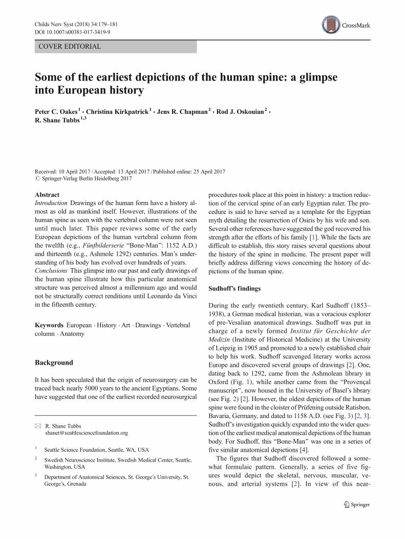
During the early twentieth century, Karl Sudhoff (1853–1938), a German medical historian, was a voracious explorerof pre-Vesalian anatomical drawings. Sudhoff was put incharge of a newly formed Institut für Geschichte derMedizin (Institute of Historical Medicine) at the Universityof Leipzig in 1905 and promoted to a newly established chairto help his work. Sudhoff scavenged literary works acrossEurope and discovered several groups of drawings [2]. One,dating back to 1292, came from the Ashmolean library inOxford (Fig. 1), while another came from the BProvençalmanuscript^, now housed in the University of Basel’s library(see Fig. 2) [2]. However, the oldest depictions of the humanspine were found in the cloister of Prüfening outside Ratisbon,Bavaria, Germany, and dated to 1158 A.D. (see Fig. 3) [2, 3].Sudhoff’s investigation quickly expanded into the wider ques-tion of the earliest medical anatomical depictions of the humanbody. For Sudhoff, this BBone-Man^ was one in a series offive similar anatomical depictions [4].
The figures that Sudhoff discovered followed a some-what formulaic pattern. Generally, a series of five fig-ures would depict the skeletal, nervous, muscular, ve-nous, and arterial systems [2]. In view of this near-
* R. Shane [email protected]
1 Seattle Science Foundation, Seattle, WA, USA2 Swedish Neuroscience Institute, Swedish Medical Center, Seattle,
Washington, USA3 Department of Anatomical Sciences, St. George’s University, St.
George’s, Grenada
Childs Nerv Syst (2018) 34:179–181DOI 10.1007/s00381-017-3419-9
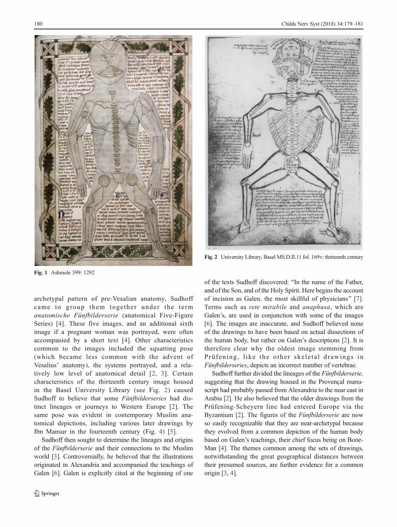
archetypal pattern of pre-Vesalian anatomy, Sudhoffcame to group them together under the te rmanatomische Fünfbilderserie (anatomical Five-FigureSeries) [4]. These five images, and an additional sixthimage if a pregnant woman was portrayed, were oftenaccompanied by a short text [4]. Other characteristicscommon to the images included the squatting pose(which became less common with the advent ofVesalius’ anatomy), the systems portrayed, and a rela-tively low level of anatomical detail [2, 3]. Certaincharacteristics of the thirteenth century image housedin the Basel University Library (see Fig. 2) causedSudhoff to believe that some Fünfbilderseries had dis-tinct lineages or journeys to Western Europe [2]. Thesame pose was evident in contemporary Muslim ana-tomical depictions, including various later drawings byIbn Mansur in the fourteenth century (Fig. 4) [5].
Sudhoff then sought to determine the lineages and originsof the Fünfbilderserie and their connections to the Muslimworld [3]. Controversially, he believed that the illustrationsoriginated in Alexandria and accompanied the teachings ofGalen [6]. Galen is explicitly cited at the beginning of one
of the texts Sudhoff discovered: BIn the name of the Father,and of the Son, and of the Holy Spirit. Here begins the accountof incision as Galen, the most skillful of physicians^ [7].Terms such as rete mirabile and anaphusa, which areGalen’s, are used in conjunction with some of the images[6]. The images are inaccurate, and Sudhoff believed noneof the drawings to have been based on actual dissections ofthe human body, but rather on Galen’s descriptions [2]. It istherefore clear why the oldest image stemming fromPrüfen ing , l i ke the o the r ske l e t a l d rawings inFünfbilderseries, depicts an incorrect number of vertebrae.
Sudhoff further divided the lineages of the Fünfbilderserie,suggesting that the drawing housed in the Provençal manu-script had probably passed from Alexandria to the near east inArabia [2]. He also believed that the older drawings from thePrüfening-Scheyern line had entered Europe via theByzantium [2]. The figures of the Fünfbilderserie are nowso easily recognizable that they are near-archetypal becausethey evolved from a common depiction of the human bodybased on Galen’s teachings, their chief focus being on Bone-Man [4]. The themes common among the sets of drawings,notwithstanding the great geographical distances betweentheir presumed sources, are further evidence for a commonorigin [3, 4].
Fig. 2 University Library, Basel MS D.II.11 fol. 169v: thirteenth century
Fig. 1 Ashmole 399: 1292
180 Childs Nerv Syst (2018) 34:179–181
However, not all researchers agree with Sudhoff’shypothesis of an Alexandrian progenitor, arguing thatancient Greek and Byzantine writings on medicine havenot yet yielded a Fünfbilderserie [4]. Some, on the ba-sis of translations by Hunain ibn Ishâq, suggest that thedrawings originated in the ancient world or Islamicmedicine. Others believe they began to appear inEurope around the time Galen was rediscovered in theWest [4].
It is unlikely that the true nature of the origin of theFünfbilderserie will ever be completely elucidated.However, perhaps a more realistic and exciting goal wouldbe to discover a depiction of the human spine from an evenearlier date. Ancient China is believed to have severalFünfbilderserie-like drawings, discovered after ourPrüfening-Scheyern, which were supposedly based on a mucholder text written during the Sung Dynasty (960–1127 A.D.)[3]. Some authors remain convinced that the Egyptiansportrayed the spine before these Galenic drawings but with alack of evidence, this claim is difficult to support [3].Whatever the case, the most anatomically correct renditionsof the human spine would not appear until the works ofLeonard da Vinci in the 1400s [8].
Compliance with ethical standards
Conflicts of interest The authors have no conflicts of interest to report.
References
1. Filler AG (2007) A historical hypothesis of the first recorded neuro-surgical operation: Isis, Osiris, Thoth, and the origin of the djedcross. Neurosurg Focus 23:E6
2. Choulant JL, Garrison FH, Streeter EC, Frank M (1920) History andbibliography of anatomic illustration in its relation to anatomic sci-ence and the graphic arts. University of Chicago Press, Chicago
3. Cowdry EV (1921) A comparison of ancient Chinese anatomicalcharts with the ‘Fünfbilderserie’ of Sudhoff. Anat Rec 22:1–25
4. Frampton M (2008) Embodiments of will: anatomical and physio-logical theories of voluntary animal motion from Greek antiquity tothe Latin Middle Ages, 400 B.C.-A.D. 1300. VDM Verlag Dr.Müller Aktiengellsellschaft & Co. KG, Saarbrücken
5. Thebirthofananatomical icon(1999)MedicalHistory(Suppl)19:74–1036. French RK (1984) An origin for the bone text of the ‘five-figure
series’. Franz Steiner Verlag, Stuttgart7. Prioreschi P (2003) Medieval Medicine. Hroatius Press, Omaha8. BowenG,Gonzales J, Iwanaga J, Fisahn C, LoukasM, Oskouian RJ,
Tubbs RS (2017) Leonardo da Vinci (1452-1519) and his depictionsof the human spine. Childs Nerv Syst Mar 10:1–4. doi:10.1007/s00381-017-3354-9
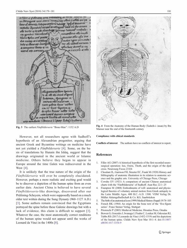
Fig. 4 From the Anatomy of the Human Body (Tashrih-i- insan) by IbnMansur near the end of the fourteenth century
Fig. 3 The earliest Fünfbilderserie BBone-Man^: 1152 A.D
Childs Nerv Syst (2018) 34:179–181 181