
Solutions For Movement Impairments Of The Lower Quadrant · Solutions For Movement Impairments Of...
141
Solutions For Movement Impairments Of The Lower Quadrant : a manual therapy based case study Koen Schoolmeesters, MSc Physiotherapy, Manual Therapist Kinetic Control Accredited tutor
Transcript of Solutions For Movement Impairments Of The Lower Quadrant · Solutions For Movement Impairments Of...

Solutions For Movement Impairments Of The Lower Quadrant :
a manual therapy based case study
Koen Schoolmeesters,
MSc Physiotherapy, Manual Therapist
Kinetic Control Accredited tutor

• Kinesitherapeut KUL FaBeR
• Manueel Therapeut SOMT ’98, …….
• Groepspraktijk voor kinesitherapie en manuele therapie Move To Heal
• Assistent Manuele Therapie FaBeR – KUL
• Kinetic Control Accredited Tutor 2006
Introductie
• Product van MPS
• Internationaal onderwijs-/bijscholingsteam
• 1995 – > 20 years !!
• Chichester, UK
• Comerford Mark & Mottram Sarah
• Evidence based concept

Movement Performance SolutionsMission Statement
Through innovative, evidence-based solutions, Movement Performance Solutions educates and trains professionals in sport, health, fitness and rehab to enhance movement efficiency and better understand how to prevent and manage musculoskeletal injury and pain that can affect movement and compromise performance in their patients, players and clients.
http://www.movementperformancesolutions.com/http://www.kineticcontrol.com/http://www.theperformancematrix.com/


• Instability (structural-functional)•Stability dysfunction•Give•Maladaptive movement pattern•Motor control dysfunction•Movement dysfunction•Control impairment•Movement control impairment•UnControlled Movement UCM

Patho-anatomic to patho-kinesiology to
kinesio-pathology(Sahrmann)

MovementImpairment
?
Pain
Function & Disability
Recurrence
Is pain or dysfunction a ‘downstream’ process of presence of movement impairments?
This module supplies a route to address movement impairments, influencing presence of pain, recurrence of pain and function
OPTIMISING MOVEMENT HEALTH www.kineticcontrol.com
Treating Pain

•Subjects employ strategies or patterns of synergistic recruitment that are normally reserved for high loadfunction (mobiliser dominant)… to perform low load postural control and normal non-fatiguing functional movements
(Sterling et al 2001 2005, Hodges 2003, Moseley & Hodges 2006, Hodges & Moseley 2003, Jull 2000, Falla et al 2004a&b, 2005, Lee 1999, Sahrmann 2002, Richardson et al 2004,
Dankaerts et al 2006, O’Sullivan 2005, O’Sullivan et al 2006)
In the presence of chronic or recurrent musculo-skeletal pain

These altered strategies are referred to in the research and clinical literature as:
• Substitution strategies• Compensatory movements• Muscle imbalance• Faulty movements • Co-contraction rigidity• Control impairments
Altered Strategies


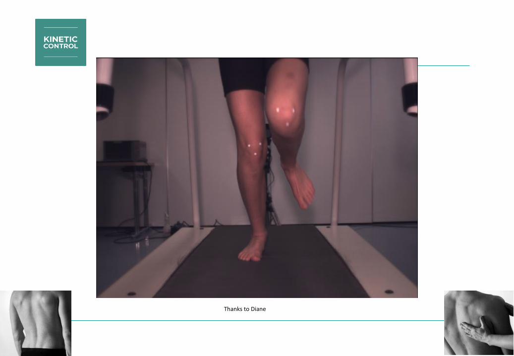
Hitching during unloaded hip flexion is a compensation in this patient with groin pain
Ideal
Dysfunctional
global
stabiliser ?

Stabiliser/Mobiliser recruitment patterns
Painfree (normal / ideal)
0
20
40
60
80
100
Low load
High load
Mobiliser
Stabiliser
Musculo-skeletal Pain
0
20
40
60
80
100
Low load
High load
Mobiliser
Stabiliser
Significant pain related change with low load ...
? need to emphasise low threshold re-training

• These high load strategies… to perform low load postural control
• are reversible with low threshold training
• High load training (if it is the only training) is likely to re-enforce
or maintain this state
• There is often no need to stop high load training
… just add low threshold motor control training
Cause – consequence ?
In the presence of chronic or recurrent musculo-skeletal pain

Study 1 (cross sectional)
Decreased variability in postural control strategies in young people with NS-LBP is associated with altered proprioceptive
reweigthing
Claeys K, Dankaerts W, Brumagne S e.a.,
European J of Applied Physiology 2011; 111 : 115-123
Decreased Variability in young people with NS-LBP

Postural control strategy - STANDINGVariability in proprioceptive postural control
• ankle strategy - the inverted pendulum control model : rigid segments movingaround 1 joint : esp by calf muscles
• multisegmental control model : multiple corrections at different joints andvertebrae by the deep segmental spinal muscles – most optimal

Sensory reweighting and LBP
•Non injured subjects can shift relatively quickly from one postural strategy to another
•LBP subjects use the same control strategy regardless the condition, compensating for the sensory deficit• ‘One strategy all fits’ (Hodges)

Sensory reweighting and LBP
Persons with recurrent LBP
•prefer significantly ankle muscle proprioceptive control for both standing without foam and on foam compared to healthy individuals
Brumagne e.a. 2008, Persons with current low back pain exhibit a rigid
postural control strategy, Eur Spine J 17 : 1177-1184

Fw sway
Bw sway
Postural control strategy –Variability in proprioceptive postural control
STANDING + VIBRATION
LBP : larger Bw sway in triceps vibration
More reliance on anklesignals during stablestanding conditions
Ankle-steered strategy
LBP : less (expected) Fwsway in LM vibration
Less reliance on LB signals during unstableconditions where the LB signals are expected to play a predominant rolein postural control

• decreased variabilty of movement strategies
•Lack of choices

•Habitual movements
•Sustained positions

Prospective study 4 : Young individuals with a more ankle-steerd proprioceptive
control strategy may develop mild non-specific low back pain
Claeys K, Dankaerts W, Brumagne S e.a.,
European J Electromyogr Kinesiol 2014

People with a dominant ankle proprioceptive use in unstable standing showed an almost four times higher risk to develop LBP in the future.
This indicates that, in this young population with mild LBP,
• LBP may be caused by a different proprioceptive weighting and
• that the observed differences in proprioceptive postural control werenot only the result of pain which was frequently suggested in earliercross-sectional studies.
High reliance on ankle proprioceptive inputs and a reduced ability to adaptthis proprioceptive use when necessary (on foam) in people who developLBP may result in a less-tuned spinal control during postural tasks withmechanical stress, spinal injury and pain as consequense.
risk for developping or sustaining mild LBP within 2 years – comparing NoLBP-LBP vs NoLBP-NoLBP

• increased postural sway (force plate, w/o vibration)
• postural differences in usual standing and sitting
• psychosocial variables (4DQ 4 Dimensional Questionnaire –
FABQ Fear-avoidance Beliefs Questionnaire <14 – TSK Tampa)
• physical activity level (FAI Physical Activity Index )
NO risk for developping or sustaining mild LBP within 2 years – comparing NoLBP-LBP vs NoLBP-NoLBP

Movement Performance Solutions
http://www.movementperformancesolutions.com/http://www.kineticcontrol.com/http://www.theperformancematrix.com/

Development Links
Kinetic Control: Systematic analysis of movement and function
Evidence based
Development of clinical assessment of stability
dysfunction (based on low trheshold
recruitment efficiency)
Diagnosis of movement dysfunction
Development of principles of stability training
Local & global motor control testing & Rx
Clinical reasoning led exercise prescription
Integrated 'Core Stability' retraining
Application of Ax & Rx to the whole body
Performance Stability: 'Performance Matrix' (evidence based)
Assessment of low & high force
performance function
Systematic analysis of performance
'weak links'
Development of risk management
(assessment and correction)
Integrated multi-dimensional core
stability training
High standard training and accreditation
Stretching & Strengthening
(Historical Developments)
Sahrmann:
global
- whole body
Janda:
global
- trunk & limbs
Muscle Balance:
Restrictions & Compensation
'Traditional' Strengthening:
overload training
- power & endurance
'Core' Strengthening:
(McGill)
overload training
- trunk & girdles
Task Specific Training
& Functional Integration
Analysis of
'Alternative' Therapies
& Approaches
Motor Control Research
& Training Model:
(Hodges,Jull, Richardson)
- local spinal control
- global trunk stability
Kinetic Control:
- research literature review
- analysis & development of
movement system model
- research
- clinical evaluation &
application
Model of Clinical Movement Analysis &
Movement Dysfunction Diagnosis
- Sahrmann (Direction Susceptible to Motion)
Kinetic Control (Site & Direction of Uncontrolled Motion)
- O'Sullivan & Dankaerts (Control Impairment)
Flexibility:
- contractile tissue
- connective tissue

‘Alternative’ Approaches:- common features
• multi-joint movements
• slow movements
• low force movements
• large range movements
• co-ordination and control of rotation
• smooth transition of concentric eccentric movement
• concept of a ‘core’
• awareness of gravity
• awareness of posture
• co-ordinated breathing
• intermittent static hold of position
• control of the centre of mass of one body segment with respect to adjacent ones
• proximal control for distal movement
• positive mental attitude

Development Links
Kinetic Control: Systematic analysis of movement and function
Evidence based
Development of clinical assessment of stability
dysfunction (based on low trheshold
recruitment efficiency)
Diagnosis of movement dysfunction
Development of principles of stability training
Local & global motor control testing & Rx
Clinical reasoning led exercise prescription
Integrated 'Core Stability' retraining
Application of Ax & Rx to the whole body
Performance Stability: 'Performance Matrix' (evidence based)
Assessment of low & high force
performance function
Systematic analysis of performance
'weak links'
Development of risk management
(assessment and correction)
Integrated multi-dimensional core
stability training
High standard training and accreditation
Stretching & Strengthening
(Historical Developments)
Sahrmann:
global
- whole body
Janda:
global
- trunk & limbs
Muscle Balance:
Restrictions & Compensation
'Traditional' Strengthening:
overload training
- power & endurance
'Core' Strengthening:
(McGill)
overload training
- trunk & girdles
Task Specific Training
& Functional Integration
Analysis of
'Alternative' Therapies
& Approaches
Motor Control Research
& Training Model:
(Hodges,Jull, Richardson)
- local spinal control
- global trunk stability
Kinetic Control:
- research literature review
- analysis & development of
movement system model
- research
- clinical evaluation &
application
Model of Clinical Movement Analysis &
Movement Dysfunction Diagnosis
- Sahrmann (Direction Susceptible to Motion)
Kinetic Control (Site & Direction of Uncontrolled Motion)
- O'Sullivan & Dankaerts (Control Impairment)
Flexibility:
- contractile tissue
- connective tissue

Local Stability Role/Strategy
•muscle stiffness to control segmental translation
• no or minimal length change in function movements
• +/- anticipatory recruitment in all directions prior to functional loading provides protective stiffness
• +/- activity is continuous and independent of the direction of movement
• proprioceptive input role
(review: Comerford & Mottram 2001)

Transversus Abdominis - LBP
-0.2 -0.1 0 0.1Time (s)
AD
TrA
IO
EO
RA
MF
-0.2 -0.1 0 0.1Time (s)
MD
TrA
IO
EO
RA
MF
-0.2 -0.1 0 0.1Time (s)
Flexion Abduction Extension
PD
TrA
IO
EO
RA
MF
(Hodges & Richardson 1996 Spine 21: 2640-2650)
LBP
• A motor control deficit is present in subjects with low back pain
• Anticipatory recruitment of transversus is significantly delayed
• This results in stability failure• The timing delay
• Is independent of the type or nature of pathology
• persist long after the resolution of back pain (Hodges & Richardson 1995,1996)

Translational UCM

Development Links
Kinetic Control: Systematic analysis of movement and function
Evidence based
Development of clinical assessment of stability
dysfunction (based on low trheshold
recruitment efficiency)
Diagnosis of movement dysfunction
Development of principles of stability training
Local & global motor control testing & Rx
Clinical reasoning led exercise prescription
Integrated 'Core Stability' retraining
Application of Ax & Rx to the whole body
Performance Stability: 'Performance Matrix' (evidence based)
Assessment of low & high force
performance function
Systematic analysis of performance
'weak links'
Development of risk management
(assessment and correction)
Integrated multi-dimensional core
stability training
High standard training and accreditation
Stretching & Strengthening
(Historical Developments)
Sahrmann:
global
- whole body
Janda:
global
- trunk & limbs
Muscle Balance:
Restrictions & Compensation
'Traditional' Strengthening:
overload training
- power & endurance
'Core' Strengthening:
(McGill)
overload training
- trunk & girdles
Task Specific Training
& Functional Integration
Analysis of
'Alternative' Therapies
& Approaches
Motor Control Research
& Training Model:
(Hodges,Jull, Richardson)
- local spinal control
- global trunk stability
Kinetic Control:
- research literature review
- analysis & development of
movement system model
- research
- clinical evaluation &
application
Model of Clinical Movement Analysis &
Movement Dysfunction Diagnosis
- Sahrmann (Direction Susceptible to Motion)
Kinetic Control (Site & Direction of Uncontrolled Motion)
- O'Sullivan & Dankaerts (Control Impairment)
Flexibility:
- contractile tissue
- connective tissue

Movement Dysfunction +/- related to pain
“Faulty movements can induce pathology”
…The path of least resistance
t just be a result of it”
Patho-anatomic to patho-kinesiology to
kinesio-pathology (Shirley Sahrmann)

Relative Stiffness - Relative Flexibility
•Relatively flexible structures compensate for relatively stiffer structures creating direction related stress and strain
• If 2 joint muscles lack extensibility or are overactive, - they limit normal motion which must be compensated for elsewhere in the movement system
• If 1 joint muscles lack ability to adequately shorten or are inefficient or “weak”, - they allow excessive motion

Flexion UCM hips
Lumbar Flexion UCM –
Restricted hipflexion
Lumbar FlexionUCM – restrictiedthoracic flexion
Thoracic Flexion UCM Normal flexion
pattern
• Path of least resistance
• Move in the direction of least resistance

UnControlledmovement

•Habitual movements
•Sustained positions
Range UCM

Development Links
Kinetic Control: Systematic analysis of movement and function
Evidence based
Development of clinical assessment of stability
dysfunction (based on low trheshold
recruitment efficiency)
Diagnosis of movement dysfunction
Development of principles of stability training
Local & global motor control testing & Rx
Clinical reasoning led exercise prescription
Integrated 'Core Stability' retraining
Application of Ax & Rx to the whole body
Performance Stability: 'Performance Matrix' (evidence based)
Assessment of low & high force
performance function
Systematic analysis of performance
'weak links'
Development of risk management
(assessment and correction)
Integrated multi-dimensional core
stability training
High standard training and accreditation
Stretching & Strengthening
(Historical Developments)
Sahrmann:
global
- whole body
Janda:
global
- trunk & limbs
Muscle Balance:
Restrictions & Compensation
'Traditional' Strengthening:
overload training
- power & endurance
'Core' Strengthening:
(McGill)
overload training
- trunk & girdles
Task Specific Training
& Functional Integration
Analysis of
'Alternative' Therapies
& Approaches
Motor Control Research
& Training Model:
(Hodges,Jull, Richardson)
- local spinal control
- global trunk stability
Kinetic Control:
- research literature review
- analysis & development of
movement system model
- research
- clinical evaluation &
application
Model of Clinical Movement Analysis &
Movement Dysfunction Diagnosis
- Sahrmann (Direction Susceptible to Motion)
Kinetic Control (Site & Direction of Uncontrolled Motion)
- O'Sullivan & Dankaerts (Control Impairment)
Flexibility:
- contractile tissue
- connective tissue

• Instability (structural-functional)•Stability dysfunction•Give•Maladaptive movement pattern•Motor control dysfunction•Movement dysfunction•Control impairment•Movement control impairment•UnControlled Movement UCM

Development Links
Kinetic Control: Systematic analysis of movement and function
Evidence based
Development of clinical assessment of stability
dysfunction (based on low trheshold
recruitment efficiency)
Diagnosis of movement dysfunction
Development of principles of stability training
Local & global motor control testing & Rx
Clinical reasoning led exercise prescription
Integrated 'Core Stability' retraining
Application of Ax & Rx to the whole body
Performance Stability: 'Performance Matrix' (evidence based)
Assessment of low & high force
performance function
Systematic analysis of performance
'weak links'
Development of risk management
(assessment and correction)
Integrated multi-dimensional core
stability training
High standard training and accreditation
Stretching & Strengthening
(Historical Developments)
Sahrmann:
global
- whole body
Janda:
global
- trunk & limbs
Muscle Balance:
Restrictions & Compensation
'Traditional' Strengthening:
overload training
- power & endurance
'Core' Strengthening:
(McGill)
overload training
- trunk & girdles
Task Specific Training
& Functional Integration
Analysis of
'Alternative' Therapies
& Approaches
Motor Control Research
& Training Model:
(Hodges,Jull, Richardson)
- local spinal control
- global trunk stability
Kinetic Control:
- research literature review
- analysis & development of
movement system model
- research
- clinical evaluation &
application
Model of Clinical Movement Analysis &
Movement Dysfunction Diagnosis
- Sahrmann (Direction Susceptible to Motion)
Kinetic Control (Site & Direction of Uncontrolled Motion)
- O'Sullivan & Dankaerts (Control Impairment)
Flexibility:
- contractile tissue
- connective tissue

Movement Performance Solutions
http://www.theperformancematrix.com/

Movement Performance Solutions
http://www.movementperformancesolutions.com/http://www.kineticcontrol.com/http://www.theperformancematrix.com/
1. ASSESSMENT
• The presence of movement control impairments identifies
kinesio-pathological subgroups of patients within the
musculoskeletal pain population.
• The identification of the site and direction of uncontrolled
movement allows targeted and focused retraining to
optimally manage pain and improve movement function

Diagnosis of UCM
The site of uncontrolled motion
• is the site of movement control impairment
&
• is the most likely site of pathology and symtoms
• because tissues around are excessively compressed,
impinged, strained
Diagnosis of UCM
The direction of uncontrolled motion
• is the direction of movement control impairment
&
• is the direction of pain producing movements • Because it relates to the direction of tissue stress or
strain

• Movement retraining interventions that are matched to
correcting specific impairments can improve the efficiency
and cost effectiveness of decreasing recurrence and
improving movement health
• Movement control retraining changes both central nervous
system neurophysiology and peripheral tissue structure and
function
2. REHABILITATION

It’s all about cognitive retraining, not about exercises !
Movement control retraining changes both • CNS NFS and • peripheral tissue structure and function
RETRAIN UCM – FIX it

Movement Performance Solutions
http://www.movementperformancesolutions.com/http://www.kineticcontrol.com/http://www.theperformancematrix.com/

© Koen Schoolmeesters – FaBeR -
KULeuven
Models for diagnosis and
classifcation of CLBP
1. Patho-anatomical model
2. Peripheral pain generator model
3. Neuro-physiological model
4. Psycho-social model
5. Mechanical loading model
6. Signs and symptoms model
7. Motor control model
8. Biopsychosocial modelO’Sullivan P, 2005, Diagnosis and classification of chronic low back pain disorders : maladaptive
movement and motor control impairments as underlying mechanism, Manual Therapy 10:242-255
(p243-245)

Various Diagnoses for 1 patient !
1. Medical –2. Anatomical – (‘complaining’ tissue/structure)
3. Manual Therapeutic – (restrictions, end feel)
4. Movement Control – (UnControlled Movement patterns, movement control impairment)
5. Neuro-Dynamic-6. Psycho-social – Presenting pain mechanisms
(sensitization,affective,…)

•Articular restriction
•Myofascial restriction
•Neurodynamic restriction
Pain and Restriction
The site of the pain refers to the site of the
restriction
Manual therapeutic diagnosis

• Manipulations –• HVLA technique• Passive
Mobilisations
• Sustained stretch• Active stretch
• Inhibitory• facilitatory
• Muscle energy techniques
• Neurodynamic mobilisation
Treatment Restrictions

• (give)
•movement impairment
•movement control impairment
• UnControlled Movement
Pain & Stability Dysfunction
The site of the pain refers to the site of the
UCM
Movement control diagnosis

• Manipulations –• HVLA technique• Passive
Mobilisations
• Sustained stretch• Active stretch
• Inhibitory• facilitatory
• Muscle energy techniques
• Neurodynamic mobilisation
Movement control retraining
Treatment UnControlled Movement

It’s all about cognitive retraining, not about exercises !
Movement control retraining changes both • CNS NFS and • peripheral tissue structure and function
RETRAIN UCM – FIX it

And/Or
Objective
The site of the pain refers to the site of the
restriction
The site of the pain refers to the site of the
UCM

Background Case Study Best Clinical Practice

CLINICAL EXAMINATION

Case Study - History
• 54-year old recreational sportsman (long distance (speed)
walking - hiking, strengthening exercises for back muscles,
sometimes jogging)
• symptoms :
– Low back-buttock pain left (LS-junction – SIJ region)
– Intermittent pain dorsal thigh, muscle cramping left leg
– intermittent groin pain and ventral thigh pain (after jogging)

Case Study - History
• Time frame : 4 months
• Reason : move out parents house ; also ‘missed a step’ by
using the ladder (regional overload, no history of LBP)
• Evolution : no change
• Provocation :
– LBP : sitting in train, sitting tall, turn around in bed, supine
– Groin pain : walking, jogging (need to stop after 3 km)
• Disability : hiking, jogging
• Medication : -

•Posture : • tall 1,87m, atlethic 54 yr man, • well trained leg- and back muscles, rather hypotone
abdominal wall
•Active lumbar movements : • All Lx ROM restricted• flexion hip + Lx restricted ROM (> extension), • Lx extension (+ 3D extension homonymous left) provocative for
LBP left• sidebend hinge Upper Lx• High ilia ?
Observation ROM & quality

Various Diagnoses for 1 patient !
•Medical – Piriformis syndrome •Anatomical – (‘complaining’ tissue/structure)
•Manual Therapeutic – (restrictions, end feel)
•Movement Control – (UnControlled Movement patterns, movement control impairment)
• Neuro-Dynamic-•Psycho-social – Presenting pain mechanisms
(sensitization,affective,…)

•Pain distribution – other Sx – NFS
•Differential diagnosis
•Selection of relevant clinical tests
1. Patho-anatomical diagnosis pain producing tissue ?

• hip ?•Upper Lx – TLj ? •SIG ?
•Lower Lx –LSj ?
Structural analysis

Symptoms Differentialdiagnosis
Tool - Test
Piriformis sydrome Length test : provocative test but piriformis not short
No neurological signs
What about Piriformis syndrome as pain producing tissue ?

www.kineticcontrol.comOPTIMISING MOVEMENT HEALTH

Quickly…..Exlcusion of hip pathology
Symptoms Differential diagnosis Tool - Test
Hip (pre)artrosis
Hip impingement
(psoas bursitis, - tendinitis, …)
History -
Active movement -
Passive movement
Capsular pattern –
Drehman sign –
Quadrant test -

Symptoms Differentialdiagnosis
Tool - Test
SIG
• dysfunction ?
• Instability ?
• UCM ?
History : missed a step of the ladder
Palpation : hyperalgesia sacral sulcus -fortin area
Active movement : restricted hip flexionand lumbar flexion…..
Provocation tests :
• thigh thrust positive
• sacral thrust ???
Is SIJ pain producing structure ?
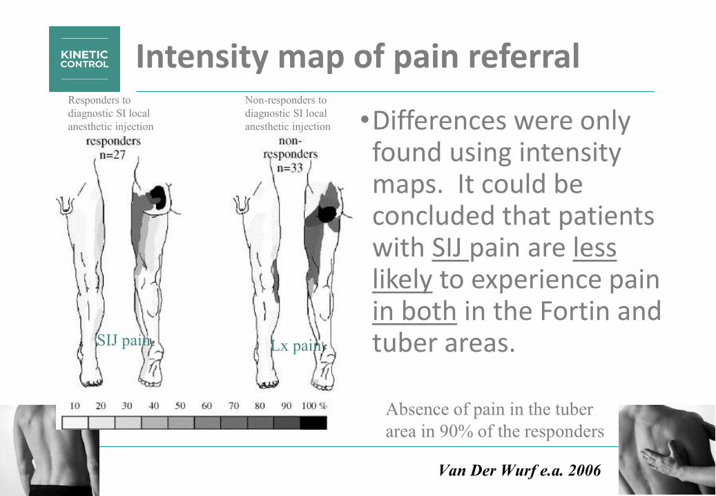
•Differences were only found using intensity maps. It could be concluded that patients with SIJ pain are less likely to experience pain in both in the Fortin and tuber areas.
Intensity map of pain referralResponders to
diagnostic SI local
anesthetic injection
Non-responders to
diagnostic SI local
anesthetic injection
SIJ pain Lx pain
Absence of pain in the tuber
area in 90% of the responders
Van Der Wurf e.a. 2006

Thigh thrust: PPPP test
Supine lying: hip 90° flexion, knee in flexion (slight adduction)
Fixation sacrum
Mobilising pressure through knee/femur
Look for provocation (and range and end feel)
Cave: load on medial hip Mens et al. 2001 hypothesize that the PPPP
test shows whether the pelvic system has been previously overloaded

Distraction
Supine lying or crook lying
Arms crossed, hands medial to SIAS
Push innominates to lateral and posterior
Test for pain/dysesthesia

Anterior compression, posterior gapping
Side lying, ± 90° hips and knees
Hands on anterolateral aspect of the uppermost iliac crest
Apply an anteromedial force
Test for pain/dysesthesia?

Sacral thrust
Apply a postero-anterior force on the sacrum
The presumed action is an anterior shearing force of the sacrum on both the ilia
Test for pain/dysesthesia?
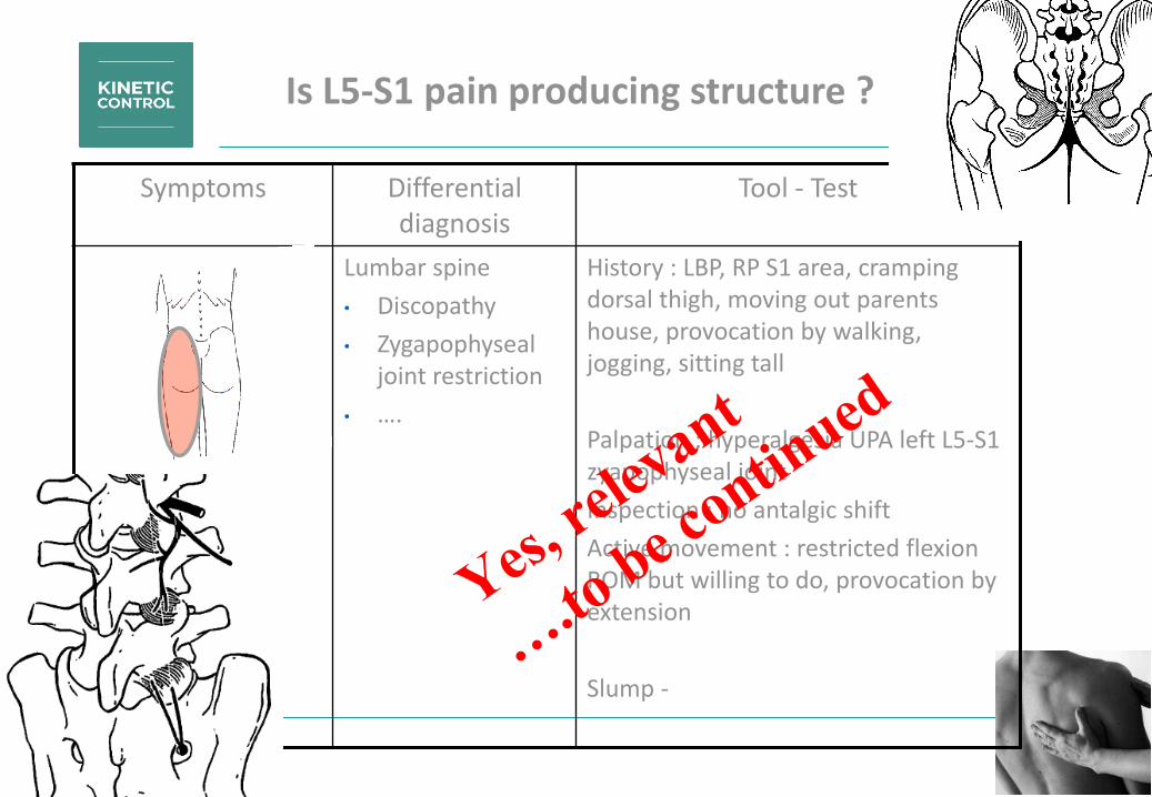
Symptoms Differentialdiagnosis
Tool - Test
Lumbar spine
• Discopathy
• Zygapophysealjoint restriction
• ….
History : LBP, RP S1 area, crampingdorsal thigh, moving out parentshouse, provocation by walking, jogging, sitting tall
Palpation : hyperalgesia UPA left L5-S1 zyapophyseal joint
Inspection : no antalgic shift
Active movement : restricted flexionROM but willing to do, provocation byextension
Slump -
Is L5-S1 pain producing structure ?

• palpation : SIPS left lower, SISA left higher (?), TI no difference
• kinematic tests : • standing trunk flexion : left SIPS more cranial• Gillet hip flexion : ipsilateral left +, contralateral left +
•Left ilium anterior rotation stiffened endfeel
•Short arm glide stiffened endfeel
2. Manual therapeutic diagnosis articular restriction SIJ
Left innominate anterior
rotation restriction

Standing trunk flexion:
• Feet ± 15 cm wide
• Palpate PSIS
• Patient flexes maximally
• Monitor movement of PSIS’s
• Normally both PSIS’s should move symmetrically
• Positive if one PSIS has moved more cephalad
•
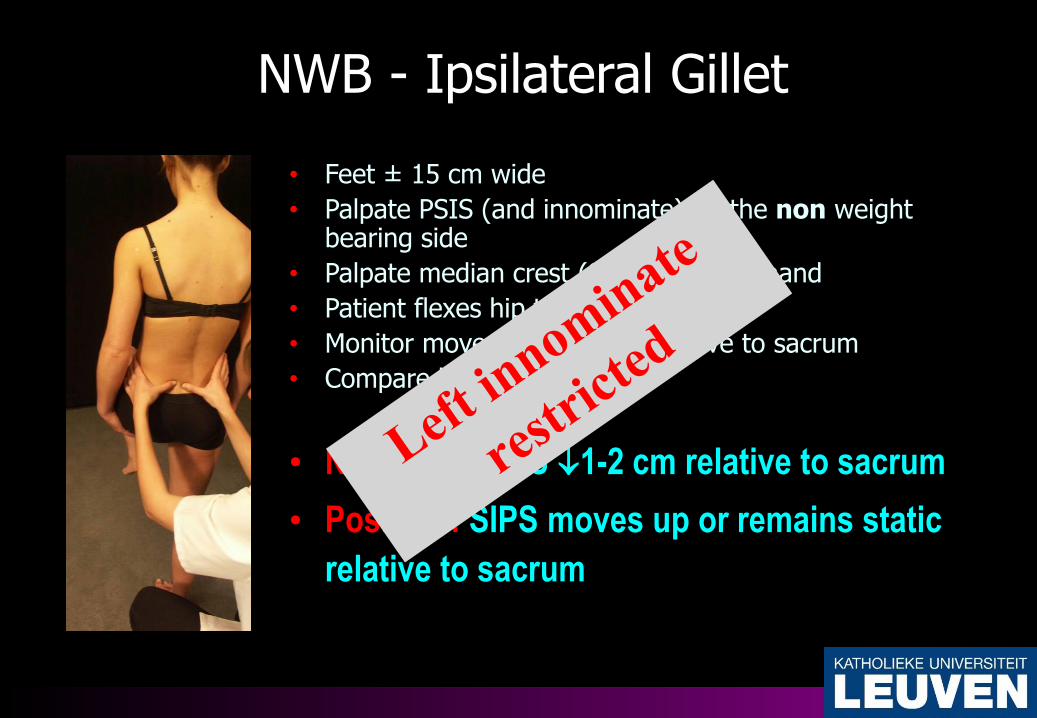
NWB - Ipsilateral Gillet
• Feet ± 15 cm wide
• Palpate PSIS (and innominate) on the non weight bearing side
• Palpate median crest (S2) with other hand
• Patient flexes hip to 90°-120°
• Monitor movement of PSIS relative to sacrum
• Compare left & right
• Negative : SIPS 1-2 cm relative to sacrum
• Positive : SIPS moves up or remains static
relative to sacrum

WB - Contralateral Gillet
• Feet ± 15 cm wide
• Palpate PSIS (and innominate) on weight bearingside
• Palpate median crest (S2) with other hand
• Patient flexes hip to 90°-120°
• Monitor movement of PSIS relative to sacrum
• Compare left & right
• Negative : SIPS 1-2 cm relative to sacrum
• Positive : SIPS moves up or remains static
relative to sacrum

Intra-articular glide: short arm
First determine treatment plane (cranial to cranial and slightly lateral)
Apply a gentle oscillatory force in:
Cranial direction
Caudal direction
Analyze neutral and elastic zone and compare to other side
(Diane Lee 2004)

Innominate anterior
Prone
Fixation sacrum contralateral below S2 (apex)
Mobilising hand with ulnar side (pisiforme) on SIPS
Direction: anterior, lateral and cranial

• palpation : SIPS left lower, SISA left higher (?), TI no difference
• kinematic tests : • standing trunk flexion : left SIPS more cranial
• Gillet hip flexion : ipsilateral left +, contralateral right +
• Left ilium anterior rotation stiffened endfeel
• Short arm glide stiffened endfeel
•ROM hip prone : lateral rotation ROM left↑, medial rotation ROM left ↓
2. Manual therapeutic diagnosis articular restriction SIJ
Left innominate anterior
rotation restriction

• UP/A L5-S1 left painful
• active 3D extension left painful at left side
•Sidelying gap zygapophyseal joints• Left not restricted – normal endfeel• Right restricted – stiffer endfeel
2. Manual therapeutic diagnosis articular restriction L5-S1 right - BST

© Kim Daniels – Koen Schoolmeesters K.U. Leuven
Lumbar Spine Rotation Sidelying
Localiseren op het juiste segment !

• P/A mid thoracic spine stiffened
• passive 3D-extension homonymous +
2. Manual therapeutic diagnosis articular restriction Thoracic Spine

© Koen Schoolmeesters & Pieter Derycke – KULeuven
– ‘08-’09
Passive non-physiological movements
PA
Central/bilateral
Unilateral
Transverse pressure

© Koen Schoolmeesters & Pieter Derycke – KULeuven
– ‘08-’09
Passive physiological movements
Test for
ROM
Endfeel
Symptom provocation

• L5-S1 right – BST• Left innominate
anterior rotation• Mid-Thoracic
extension

1. Site of UCM
2. Direction of UCM
3. Treshold of UCM
•Site of pain refers to site of UCM
•Provocative direction refers to direction of UCM
3. Movement control impairmentdiagnosis

Control of Direction TEST

• History : LBP left – during extension(-rotation) related activities
• Posture : well trained leg- and back muscles, rather hypotone abdominal wall
• Active Movements :• provocative extension (+ 3D homonymous)
• Restricted lumbar flexion (short ES ? High ilia ?)
3. Movement control impairment diagnosis
hypothesis : L5-S1 extension-
rotation UCM ?

Identifying movement control impairments with movement control tests : control of direction of UCM
3. Movement control impairmentdiagnosis
Motor Control Testing Process
• Observe (natural movement for relative stiffness & relative
flexibility
• Teach (Visual, Auditory, Kinaesthetic)
• Test (without feedback, support or cueing)
• Rate or or
• Relate (to symptomatic movements &
functional patterns
• Rehab (Clinical Priority)

• Standing forward lean : can he prevent Lx extension ?
• Knee lower abdominal test : can he prevent Lx extension-lumboplevic rotation ?
• standing hip extension : can he prevent Lx extension- lumboplevicrotation ?
Selection of test
• Extension and/or rotation
• Functional link : standing tests ~lifting, hiking
3. Movement control tests

Standing Forward Lean: Control lumbar extension while moving the hips
Tests for uncontrolled movement of the low back
Classify site: low backDirection: extensionThreshold: low
Manage Movement – Solutions for the low back
www.kineticcontrol.comOPTIMISING MOVEMENT HEALTH
Identifying movement control impairments with movement control tests
Standing forward lean VX SOE and FB of hands and wall to
prevent upper lumbar extension
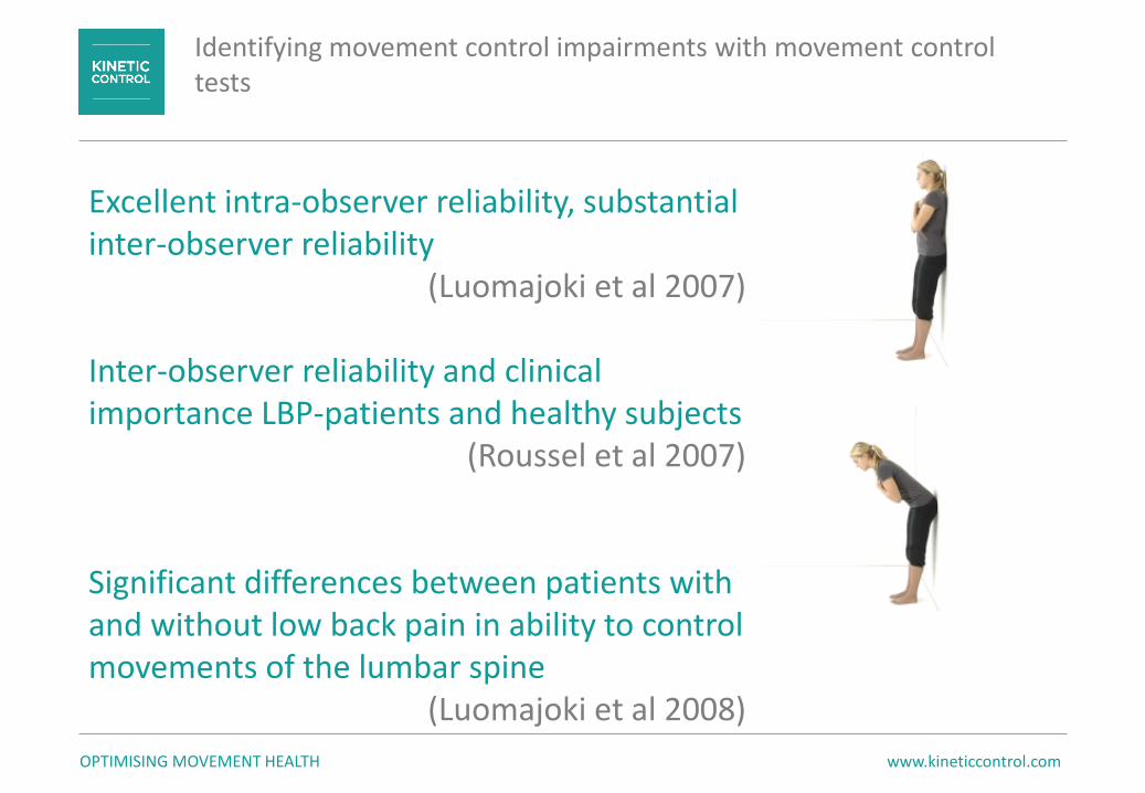
Excellent intra-observer reliability, substantial inter-observer reliability
(Luomajoki et al 2007)
Inter-observer reliability and clinical importance LBP-patients and healthy subjects
(Roussel et al 2007)
Significant differences between patients with and without low back pain in ability to control movements of the lumbar spine
(Luomajoki et al 2008)
www.kineticcontrol.comOPTIMISING MOVEMENT HEALTH
Identifying movement control impairments with movement control tests
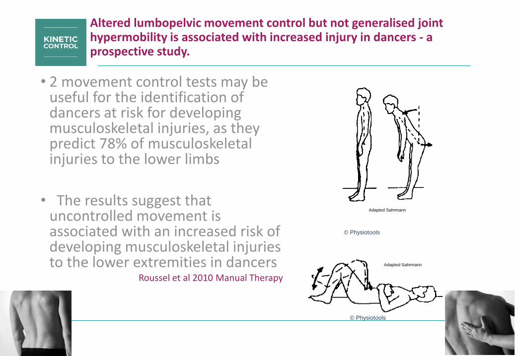
Altered lumbopelvic movement control but not generalised joint hypermobility is associated with increased injury in dancers - a prospective study.
• 2 movement control tests may be useful for the identification of dancers at risk for developing musculoskeletal injuries, as they predict 78% of musculoskeletal injuries to the lower limbs
• The results suggest that uncontrolled movement is associated with an increased risk of developing musculoskeletal injuriesto the lower extremities in dancers
Roussel et al 2010 Manual Therapy
Adapted Sahrmann
Adapted Sahrmann
© Physiotools
© Physiotools

www.kineticcontrol.comOPTIMISING MOVEMENT HEALTH
Uncontrolled MovementSite: Low back and pelvis; Direction: Rotation - extension; Threshold: Low
Knee Lift Abdominal Test (KLAT)
Inter-observer reliability and clinical importance of the KLAT (Roussel)
The PBU
Lower both feet w/o Lx extension-rotation
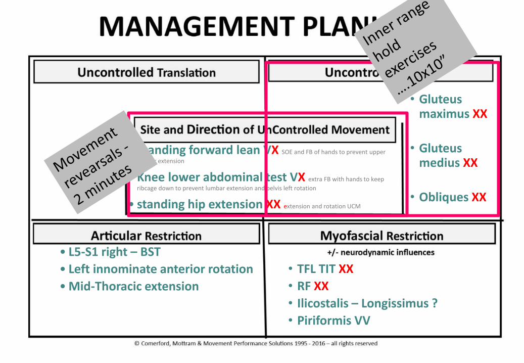
• Knee lower abdominal test VX extra FB with hands to keep ribcagedown to prevent lumbar extension and pelvis left rotation

Standing Hip Extension : Control lumbar extension and lumbopelvic rotation while extending the hip till -15°
Tests for uncontrolled movement of the low back
Classify site: low backDirection: extension - rotationThreshold: low
Manage Movement – Solutions for the low back
www.kineticcontrol.comOPTIMISING MOVEMENT HEALTH
Identifying movement control impairments with movement control tests
• standing hip extension XX extension and pelvis left rotation UCM

• Standing forward lean VX SOE and FB of hands to prevent upperlumbar extension
• Knee lower abdominal test VX extra FB with hands to keep ribcagedown to prevent lumbar extension and pelvis left rotation
• standing hip extension XX extension and pelvis left rotation UCM
3. Movement control tests
hypothesis confirmed
L5-S1 extension rotation
(pelvis faces left) UCM ?

• Standing forward lean VX SOE and FB of hands to prevent upper
lumbar extension
• Knee lower abdominal test VX extra FB with hands to keep
ribcage down to prevent lumbar extension and pelvis left rotation
• standing hip extension XX extension and rotation UCM
• L5-S1 right – BST
• Left innominate anterior rotation
• Mid-Thoracic extension
• Gluteus maximus
• Gluteus medius
• Obliques
• TFL TIT
• RF
• Ilicostalis – Longissimus
• piriformis

• Gluteus maximus XX cocontraction rigidity, lateral hip rotation, Lx extension before inner range
• Gluteus medius posterior XX hip flexion before inner range
•Obliques XX
Testing recruitment efficiency

• Standing forward lean VX SOE and FB of hands to prevent upper
lumbar extension
• Knee lower abdominal test VX extra FB with hands to keep
ribcage down to prevent lumbar extension and pelvis left rotation
• standing hip extension XX extension and rotation UCM
• L5-S1 right – BST
• Left innominate anterior rotation
• Mid-Thoracic extension
• Gluteus maximus XX
• Gluteus medius XX
• Obliques XX
• TFL TIT XX
• RF XX
• Ilicostalis – Longissimus ?
• Piriformis VV

Extensibility Tensor Fascia LataeModified Thomas Test

RESTRICTION
Mid thoracicextension restriction
L5-S1 zygapophysealrestriction right
BST
Ilium anteriorrotation restrictionleft
UCM
(Mid thoracic flexion UCM)
L5-S1 left zygapophyseal
extension rotation UCM
Inefficient Oblique
abdominals
Short/ dominant
TFL TIT and RF
Short/ dominant Longissimus Iliocostalis
Inefficient glutei

TREATMENT
REHABILITATION

• Standing forward lean VX SOE and FB of hands to prevent upper
lumbar extension
• Knee lower abdominal test VX extra FB with hands to keep
ribcage down to prevent lumbar extension and pelvis left rotation
• standing hip extension XX extension and rotation UCM
• L5-S1 right – BST
• Left innominate anterior rotation
• Mid-Thoracic extension
• Gluteus maximus XX
• Gluteus medius XX
• Obliques XX
• TFL TIT XX
• RF XX
• Ilicostalis – Longissimus ?
• Piriformis VV

© Kim Daniels – Koen Schoolmeesters K.U. Leuven
Lumbar Rotation Manipulation
Faculteit Bewegings- en
Revalidatiewetenschappen
Positioned in flexion

© Kim Daniels – Koen Schoolmeesters K.U. Leuven
Prone
Fixation sacrum contralateral below S2 (apex)
Mobilising hand with ulnar side (pisiforme) on SIPS
Direction: anterior, lateral and cranial
Left innominate anterior rotation manipulation

© Kim Daniels – Koen Schoolmeesters K.U. Leuven
Left innominate anterior rotation manipulation

© Kim Daniels – Koen Schoolmeesters K.U. Leuven
Innominate anterior: home exercise

© Kim Daniels – Koen Schoolmeesters K.U. Leuven
• Starting position of the patient : sitting,
arms crossed with hands on the
shoulders
• fixation : fixation of the cranial
vertebra by the therapists chest
(or towel) while he/she is holding
the patients elbows (“compression”)
• Mobilisation : the therapist is moving his bodyweight from the
front foot towards the rear foot (from kneeflexion towards knee extension)
Sitting Thoracic Gap Manipulation

© Kim Daniels – Koen Schoolmeesters K.U. Leuven
Supine Thoracic Gap Manipulation
© Kim Daniels – Koen Schoolmeesters K.U. Leuven
• Standing forward lean VX SOE and FB of hands to prevent upper
lumbar extension
• Knee lower abdominal test VX extra FB with hands to keep
ribcage down to prevent lumbar extension and pelvis left rotation
• standing hip extension XX extension and rotation UCM
• L5-S1 right – BST
• Left innominate anterior rotation
• Mid-Thoracic extension
• Gluteus maximus XX
• Gluteus medius XX
• Obliques XX
• TFL TIT XX
• RF XX
• Ilicostalis – Longissimus ?
• Piriformis VV

•
Recruiting Oblique Abdominals to control extension-rotation under low
load

..and lifting one leg alternating

• learn to control lumbar extension andlumboplevic rotation
• Visual : look, PBU
•Auditive : bring your lower ribs down to the ilia
•Kinaesthetic : thumb on lower ribs, fingers on ilia…close this….
Recruiting Obliques Abdominals

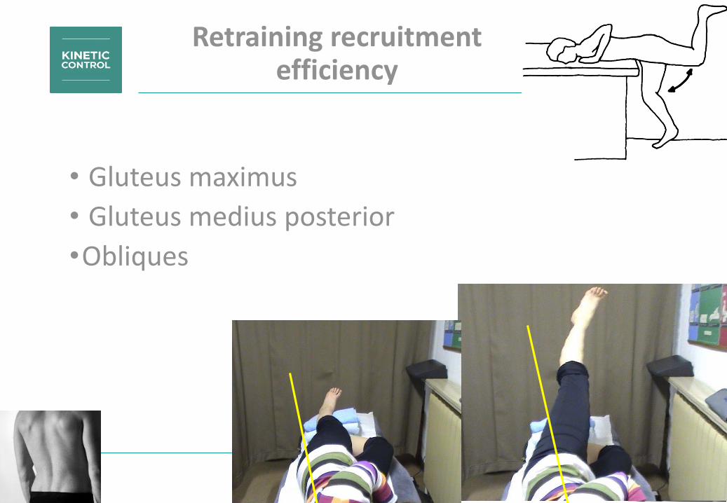
• Gluteus maximus
• Gluteus medius posterior
•Obliques
Retraining recruitment efficiency

Recruiting Glutei to control extension under low load
• against the wall
• lack of ROM
•poor recruitment
• hips andknee flexed
•…10 x 10 seconds

standing postural correction
• without wall
•During ADL
•Submaximal tonic recruitment of gluts
•No cocontraction rigidity
•Breathing ! Talking !

Movement Control Exercise : standing hip extension
• with wall
•Standing hip extension
•Submaximal tonic recruitment of gluts & obliques
•No cocontraction rigidity
•Breathing ! Talking !

Movement Control Exercise : standing hip extension
• without wall
•Standing hip extension
•Submaximal tonic recruitment of gluts & obliques
•No cocontraction rigidity
•Breathing ! Talking !

Movement Control Exercise : lunge

Movement Control Exercise : walking
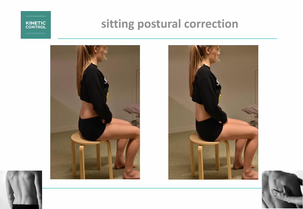
sitting postural correction
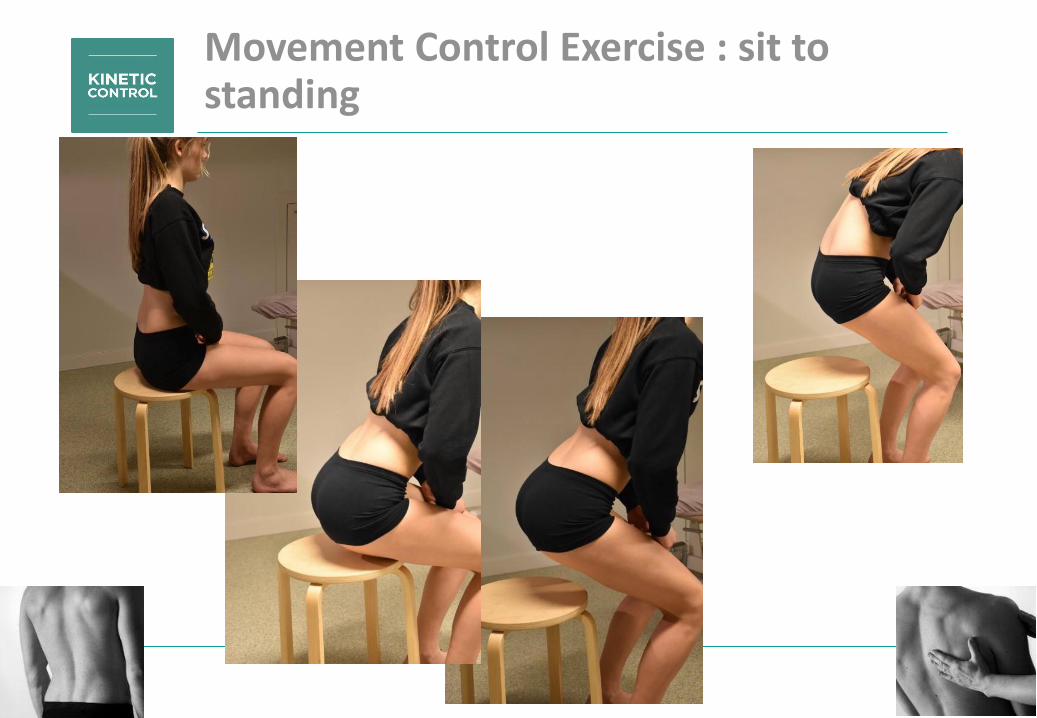
Movement Control Exercise : sit to standing

Movement Control Exercise : standing lean w/o extension UCM

Movement Control Exercise : learning to bend the lumbar spine

Movement Control Exercise : Bridge Single Leg Extension (gluts & obliques)

Movement Control Exercise : Small Knee Bend without hipflexion and knee valgus

• 2 movement control exercises – 2 minutes – 2/day
• ADL postural control : standing-sitting-walking
Home exercises

© Kim Daniels – Koen Schoolmeesters K.U. Leuven
• Standing forward lean VX SOE and FB of hands to prevent upper
lumbar extension
• Knee lower abdominal test VX extra FB with hands to keep
ribcage down to prevent lumbar extension and pelvis left rotation
• standing hip extension XX extension and rotation UCM
• L5-S1 right – BST
• Left innominate anterior rotation
• Mid-Thoracic extension
• Gluteus maximus XX
• Gluteus medius XX
• Obliques XX
• TFL TIT XX
• RF XX
• Ilicostalis – Longissimus ?
• Piriformis VV

Regaining extensibility
• Passive sustained stretch 2 minutes
• Active inhibitory stretch 3x30 seconds

• Passive sustained stretch 2 minutes
• Active inhibitory stretch 3x30 seconds
Home exercises

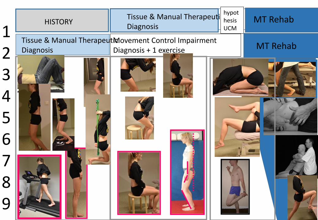
HISTORYTissue & Manual Therapeutic Diagnosis
hypothesis UCM
Movement Control Impairment Diagnosis + 1 exercise
Tissue & Manual Therapeutic Diagnosis
MT Rehab
Session
1 2 MT Rehab

HISTORYTissue & Manual Therapeutic Diagnosis
hypothesis UCM
Movement Control Impairment Diagnosis + 1 exercise
Tissue & Manual Therapeutic Diagnosis
MT Rehab
Session
1 23456789
MT Rehab
Movement Control exercises
Extensibility
stretches (active –passive)

HISTORYTissue & Manual Therapeutic Diagnosis
hypothesis UCM
Movement Control Impairment Diagnosis + 1 exercise
Tissue & Manual Therapeutic Diagnosis
MT Rehab
1 234
MT Rehab

HISTORYTissue & Manual Therapeutic Diagnosis
hypothesis UCM
Movement Control Impairment Diagnosis + 1 exercise
Tissue & Manual Therapeutic Diagnosis
MT Rehab
1 2345678
MT Rehab
Extensibility
stretches (active –passive)
HISTORYTissue & Manual Therapeutic Diagnosis
hypothesis UCM
Movement Control Impairment Diagnosis + 1 exercise
Tissue & Manual Therapeutic Diagnosis
MT Rehab
1 23456789
MT Rehab

•Always combination of MT and Movementcontrol Lumbar rotation-extension • Session 1-3 : espec MT SIJ• Session 4 : more L5-S1 techniques and obliques-
glutes ….
• Continued to walk & run - Distance increasedgradually
•fluctuating evolution, often ventral thigh painafter hiking
•Session 7 : better ☺
Evolution

24/12 31/12 : pain ventral thigh one day after running, no provocation during
running5/1 : run w/o Sx, left buttock less sensitive, no groin pain anymore12/1 : started to work again, some more pain during night (turn
around), buttock pain sitting in train20/1 : hiking 30 km, sensitive in low back left + RP left ventral thigh,
supine is provocative30/1 : in the morning pain left SIJ with RP dorsal leg, once walking Sx
improve, turning around provocative9/2 : better ! Walked 25 km, also 36 km yesterday w/o Sx23/2 : no Sx last week, also no after 50 km walking9/3 : no Sx anymore despite hiking
Evolution