Soluciones Horizontales - Single Consulting
11
JOURNAL OF VIROLOGY, Aug. 2009, p. 8051–8061 Vol. 83, No. 16 0022-538X/09/$08.000 doi:10.1128/JVI.00427-09 Copyright © 2009, American Society for Microbiology. All Rights Reserved. Loss of MicroRNA Targets in the 3 Untranslated Region as a Mechanism of Retroviral Insertional Activation of Growth Factor Independence 1 Magdalena Julia Dabrowska, 1,2 Karen Dybkaer, 1,2 Hans Erik Johnsen, 1 Bruce Wang, 3 Matthias Wabl, 4 and Finn Skou Pedersen 2 * Department of Haematology, Aalborg Hospital, Aarhus University Hospital, Aarhus, Denmark 1 ; Department of Molecular Biology, Aarhus University, Aarhus, Denmark 2 ; Picobella L.L.C., 863 Mitten Road, Suite 101, Burlingame, California 94010 3 ; and Department of Microbiology and Immunology, University of California, San Francisco, California 94143 4 Received 27 February 2009/Accepted 20 May 2009 The non-oncogene-bearing retrovirus SL3-3 murine leukemia virus induces strictly T-cell lymphomas with a mean latency of 2 to 4 months in mice of the NMRI-inbred (NMRI-i) strain. By high-throughput sequencing of retroviral tags, we have identified the genomic region carrying the transcriptional repressor and oncogene growth factor independence 1 (Gfi1) as a frequent target for SL3-3 in the NMRI-i mouse genome. Twenty-four SL3-3 insertions were identified within a 1-kb window of the 3 untranslated region (3UTR) of the Gfi1 gene, a clustering pattern unique for this lymphoma model. Expression analysis determined that the Gfi1 gene was transcriptionally activated by SL3-3 insertions, and an upregulation of Gfi1 protein expression was detected for tumors harboring insertions in the Gfi1 3UTR. Here we provide data in support of a mechanism by which retroviral insertions in the Gfi1 3UTR decouple microRNA-mediated posttranscriptional regulation. The non-oncogene-bearing murine leukemia viruses (MLVs) induce leukemias and lymphomas when injected into newborn susceptible mice (1, 21, 75). The major determinant of MLV latency and disease specificity is the retroviral enhancer in the U3 region of the MLV long terminal repeat (LTR) (3, 5, 9, 17, 19, 24, 37, 38, 50, 52, 69, 70, 71). It comprises conserved areas which hold densely packed binding sites for several host tran- scription factors, including Runx, NF-1, Ets, c-Myb, the glu- cocorticoid response element, and basic helix-loop-helix fac- tors. Small nucleotide alterations in the different binding sites influence latency, confer variations in cell-specific expression, and shift disease patterns from lymphoma to plasmacytoma, myeloid leukemia, megakaryoblastic leukemia, erythroleuke- mia, and mixed phenotype. The wild-type (wt) SL3-3 is a highly pathogenic ecotropic MLV that induces precursor T-cell lym- phomas with a mean latency of 2 to 4 months and primary manifestations in thymus, spleen, and mesenteric lymph nodes when injected into mice of the NMRI-inbred (NMRI-i) strain (19, 43, 51). Tumor induction by SL3-3 and other MLVs is a complex process, where the most well defined step involves integration of the viral genome into the host genome and deregulation of nearby proto-oncogenes or tumor suppressors (6, 8, 10, 28, 53, 65, 66, 67). The effect of the provirus depends on its integration position relative to the target gene, where the most frequent mechanisms of insertional mutagenesis are en- hancement and LTR promotion, both of which result in either upregulation of the wt gene and protein or generation of chi- meric transcripts. Another way by which gene expression can be affected by retroviral insertions is by loss of regulatory regions. Early studies of insertional mutagenesis have demon- strated that retroviral integrations in the 3 untranslated re- gions (3UTRs) of genes may result in generation of prema- turely terminated transcripts or transcripts with increased mRNA stability and elevated protein synthesis (6, 8, 10, 67). The 3UTR may also harbor other regulatory sequences, namely, binding sites for microRNAs (miRNAs), which are noncoding 22-nucleotide RNAs encoded from introns or in- tergenic regions in the genome (36). They act by targeting primarily the 3UTRs of mRNAs and mediate posttranscrip- tional downregulation of gene expression by complete comple- mentarity or partial binding of their 5-end nucleotides 2 to 7 (seed region) to mRNA targets (39). Theoretically, the short seed sequence permits a single miRNA to act on multiple target sites, and thereby each miRNA is able to recognize an average of 100 different mRNAs (2, 41). The genomic locus on murine chromosome 5 encoding the transcriptional repressor and oncogene growth factor indepen- dence 1 (Gfi1) (25) and neuroblastoma 4S oncogene ecotropic viral integration site 5 (Evi5) (40) (hereafter also referred to as the gfi1 locus) is a frequent integration locus in T-cell lympho- mas induced by Moloney MLV (MoMLV) (48, 62, 65) and in B-cell lymphomas induced by the Akv MLV (72, 73). Previous studies have demonstrated that retroviral insertions within the gfi1 locus lead to transcriptional activation of the Gfi1 gene (62, 65). Gfi1 is a key regulator of stem cell quiescence (29, 82) and plays a significant role in T-cell development (26, 54, 64, 81) and lineage commitment (80). It further influences matu- ration of myeloid precursors into granulocytes and monocytes and acts in limiting the inflammatory immune response (31). Gfi1 has a major oncogenic potential and has been associated with both murine and human cancers (15, 32, 59, 68). In this study we have identified 130 retroviral insertions in the gfi1 locus and addressed their effect on Gfi1 mRNA and * Corresponding author. Mailing address: Department of Molecular Biology, Aarhus University, C.F. Møllers Alle ´, Bldg. 1130, DK-8000 Aarhus C, Denmark. Phone: 4589422614. Fax: 4586196500. E-mail: [email protected]. Published ahead of print on 27 May 2009. 8051 on April 14, 2019 by guest http://jvi.asm.org/ Downloaded from
Transcript of Soluciones Horizontales - Single Consulting
JOURNAL OF VIROLOGY, Aug. 2009, p. 8051–8061 Vol. 83, No. 160022-538X/09/$08.00�0 doi:10.1128/JVI.00427-09Copyright © 2009, American Society for Microbiology. All Rights Reserved.
Loss of MicroRNA Targets in the 3� Untranslated Region as aMechanism of Retroviral Insertional Activation of Growth
Factor Independence 1�
Magdalena Julia Dabrowska,1,2 Karen Dybkaer,1,2 Hans Erik Johnsen,1 Bruce Wang,3Matthias Wabl,4 and Finn Skou Pedersen2*
Department of Haematology, Aalborg Hospital, Aarhus University Hospital, Aarhus, Denmark1; Department of Molecular Biology,Aarhus University, Aarhus, Denmark2; Picobella L.L.C., 863 Mitten Road, Suite 101, Burlingame, California 940103; and
Department of Microbiology and Immunology, University of California, San Francisco, California 941434
Received 27 February 2009/Accepted 20 May 2009
The non-oncogene-bearing retrovirus SL3-3 murine leukemia virus induces strictly T-cell lymphomas witha mean latency of 2 to 4 months in mice of the NMRI-inbred (NMRI-i) strain. By high-throughput sequencingof retroviral tags, we have identified the genomic region carrying the transcriptional repressor and oncogenegrowth factor independence 1 (Gfi1) as a frequent target for SL3-3 in the NMRI-i mouse genome. Twenty-fourSL3-3 insertions were identified within a 1-kb window of the 3� untranslated region (3�UTR) of the Gfi1 gene,a clustering pattern unique for this lymphoma model. Expression analysis determined that the Gfi1 gene wastranscriptionally activated by SL3-3 insertions, and an upregulation of Gfi1 protein expression was detected fortumors harboring insertions in the Gfi1 3�UTR. Here we provide data in support of a mechanism by whichretroviral insertions in the Gfi1 3�UTR decouple microRNA-mediated posttranscriptional regulation.
The non-oncogene-bearing murine leukemia viruses (MLVs)induce leukemias and lymphomas when injected into newbornsusceptible mice (1, 21, 75). The major determinant of MLVlatency and disease specificity is the retroviral enhancer in theU3 region of the MLV long terminal repeat (LTR) (3, 5, 9, 17,19, 24, 37, 38, 50, 52, 69, 70, 71). It comprises conserved areaswhich hold densely packed binding sites for several host tran-scription factors, including Runx, NF-1, Ets, c-Myb, the glu-cocorticoid response element, and basic helix-loop-helix fac-tors. Small nucleotide alterations in the different binding sitesinfluence latency, confer variations in cell-specific expression,and shift disease patterns from lymphoma to plasmacytoma,myeloid leukemia, megakaryoblastic leukemia, erythroleuke-mia, and mixed phenotype. The wild-type (wt) SL3-3 is a highlypathogenic ecotropic MLV that induces precursor T-cell lym-phomas with a mean latency of 2 to 4 months and primarymanifestations in thymus, spleen, and mesenteric lymph nodeswhen injected into mice of the NMRI-inbred (NMRI-i) strain(19, 43, 51). Tumor induction by SL3-3 and other MLVs is acomplex process, where the most well defined step involvesintegration of the viral genome into the host genome andderegulation of nearby proto-oncogenes or tumor suppressors(6, 8, 10, 28, 53, 65, 66, 67). The effect of the provirus dependson its integration position relative to the target gene, where themost frequent mechanisms of insertional mutagenesis are en-hancement and LTR promotion, both of which result in eitherupregulation of the wt gene and protein or generation of chi-meric transcripts. Another way by which gene expression canbe affected by retroviral insertions is by loss of regulatory
regions. Early studies of insertional mutagenesis have demon-strated that retroviral integrations in the 3� untranslated re-gions (3�UTRs) of genes may result in generation of prema-turely terminated transcripts or transcripts with increasedmRNA stability and elevated protein synthesis (6, 8, 10, 67).The 3�UTR may also harbor other regulatory sequences,namely, binding sites for microRNAs (miRNAs), which arenoncoding 22-nucleotide RNAs encoded from introns or in-tergenic regions in the genome (36). They act by targetingprimarily the 3�UTRs of mRNAs and mediate posttranscrip-tional downregulation of gene expression by complete comple-mentarity or partial binding of their 5�-end nucleotides 2 to 7(seed region) to mRNA targets (39). Theoretically, the shortseed sequence permits a single miRNA to act on multipletarget sites, and thereby each miRNA is able to recognize anaverage of 100 different mRNAs (2, 41).
The genomic locus on murine chromosome 5 encoding thetranscriptional repressor and oncogene growth factor indepen-dence 1 (Gfi1) (25) and neuroblastoma 4S oncogene ecotropicviral integration site 5 (Evi5) (40) (hereafter also referred to asthe gfi1 locus) is a frequent integration locus in T-cell lympho-mas induced by Moloney MLV (MoMLV) (48, 62, 65) and inB-cell lymphomas induced by the Akv MLV (72, 73). Previousstudies have demonstrated that retroviral insertions within thegfi1 locus lead to transcriptional activation of the Gfi1 gene(62, 65). Gfi1 is a key regulator of stem cell quiescence (29, 82)and plays a significant role in T-cell development (26, 54, 64,81) and lineage commitment (80). It further influences matu-ration of myeloid precursors into granulocytes and monocytesand acts in limiting the inflammatory immune response (31).Gfi1 has a major oncogenic potential and has been associatedwith both murine and human cancers (15, 32, 59, 68).
In this study we have identified 130 retroviral insertions inthe gfi1 locus and addressed their effect on Gfi1 mRNA and
* Corresponding author. Mailing address: Department of MolecularBiology, Aarhus University, C.F. Møllers Alle, Bldg. 1130, DK-8000Aarhus C, Denmark. Phone: 4589422614. Fax: 4586196500. E-mail:[email protected].
� Published ahead of print on 27 May 2009.
8051
on April 14, 2019 by guest
http://jvi.asm.org/
Dow
nloaded from

protein expression. Our results suggest that integrations in theGfi1 3�UTR contribute to increased protein synthesis througha mechanism including loss of potential miRNA binding sites.
MATERIALS AND METHODS
Tumors and isolation of retroviral tags. Tumors originated from previouslypublished (17, 18, 19, 20, 27, 43, 45, 51, 69, 70) and unpublished pathogenicitystudies of wt and enhancer mutated SL3-3, Akv, and Reilly-Finkel-Biskis (RFB)MLVs. Large-scale analysis of integrated retroviruses, performed by a splinkler-ette-based PCR method described previously (78), was able to identify 120 wtand enhancer-mutated SL3-3 integrations in the genomic region carrying Gfi1from a total of 790 SL3-3 tags. Seven Akv integrations and three RFB integra-tions from 2,800 Akv tags and 85 RFB tags, respectively, were identified in thegfi1 locus.
PCR and sequencing. Total RNA was extracted from snap-frozen tissue by useof TRIzol extraction reagent (Invitrogen). Full-genome cDNA was synthesizedusing the first-strand cDNA synthesis kit (GE Healthcare) according to themanufacturer’s recommendations. PCR for identifying alternative transcriptswas performed with a Gfi1 exon 2 forward primer (5�-CCGACTCTCAGCTTACCGAG-3�) and a Gfi1 exon 5 reverse primer (5�-CTGTGTGGATGAAGGTGTGTTT-3�) (DNA Technology). PCR for identifying retroviral insertions inthe Gfi1 3�UTR was performed with a Gfi1 exon 6 forward primer (5�-CTCAGGAGGCACCGAGAGA-3�) and SL3-3 reverse primer (5�-CCCCAGAAATAGCTAAAACAACAACAGTTTCAA-3�) (DNA Technology). PCR fragmentswere purified on GFX columns (GE Healthcare) and sequenced by use of aBigDye Terminator v3.1 cycle sequencing kit (Applied Biosystems).
Real-time PCR analysis. Real-time PCR amplifications for gene mRNA quan-tification were performed using TaqMan expression assays for Gfi1(Mm00515853_m1) and Ywhaz (Mm01158417_g1). For miRNA quantification,
cDNA was synthesized according to the TaqMan MicroRNA assay protocol byuse of a TaqMan MicroRNA reverse transcription kit and TaqMan MicroRNAassays for miR-155 (001806), miR-142-3p (001189), miR-330 (001062), miR-133a(002246), miR-34b-3p (002618), miR-879 (002473), miR-466l (002804), miR-10a(002288), and miR-467g (002811). Samples were set up in 20-�l reaction mix-tures with 10 �l TaqMan universal PCR master mix, no AmpErase UNG, 0.5 �lTaqMan primer-probe, and 9 �l cDNA. All TaqMan reagents were purchasedfrom Applied Biosystems. To obtain amplification efficiency, samples for genequantification were run at four-point dilutions (1:10, 1:50, 1:100, and 1:500) andsamples for miRNA quantification were run at three dilutions (1:10, 1:50, and1:100). Each measurement was performed in duplicates. Controls without tem-plate and controls without reverse transcriptase for each tumor sample wereincluded. Samples for Gfi1 quantification were normalized to Ywhaz (the house-keeping genes Ubc, Tfrc, B2m, and Gapdh were tested on 10 thymic, 10 splenic,and 10 mesenteric lymph node samples, where Ywhaz showed the most stableexpression). miRNA expression was normalized to snoRNA420 (001239) (Ap-plied Biosystems). Each tumor sample was further normalized to its own tissuecontrol counterpart.
Western blot analysis. Protein extraction was performed by homogenization of60 to 120 ng snap-frozen tissue in 75 mM NaCl, 100 mM Tris-HCl (pH 8), 5 mMEDTA (pH 8), and 1 mM phenylmethylsulfonyl fluoride. Protein concentrationwas determined by use of a bicinchoninic acid assay kit (Pierce Biotechnology)according to the manufacturer’s recommendations. Five micrograms of proteinfrom each sample was loaded onto Criterion XT 12% bis-Tris precast gels(Bio-Rad) and run in 0.5� Criterion XT MOPS (morpholinepropanesulfonicacid) running buffer (Bio-Rad). Proteins were transferred onto a polyvinylidenefluoride membrane (Millipore Corporation), and blocking was performed over-night at 4°C in TBS-T (20 mM Tris-HCl [pH 7.6], 200 mM NaCl) containing 5%(wt/vol) fat-free milk and 0.05% Tween 20 (Sigma). Gfi1 primary antibody(ab21061) (Abcam) or �-actin primary antibody (sc-1616) (Santa Cruz Biotech-
TABLE 1. Retroviral integrations in the Gfi1 3�UTR
Integrationa Tissueb Virusc Provirusorientationd
Proviruspositione Reference(s)
1 S SL3-3 UCR � 6641 45; unpublished data2* ML SL3-3 wt � 6646 513 S SL3-3 Ea/s � 6653 174* ML SL3-3 wt � 6654 515 S SL3-3 wt � 6654 516*** S SL3-3 UCR � 6656 45; unpublished data7 S SL3-3 wt � 6658 518 T SL3-3 (SL3-2Env) � 6694 Unpublished data9 S Akv1-99 Egre�Ea/s � 6715 6910 T SL3-3 wt � 6734 5111 ML SL3-3 wt � 6734 5112 S RFB wt � 6734 Unpublished data13** T SL3-3 Turbo � 6787 20, 5114 S SL3-3 GR�Ea/s � 6791 1715** T SL3-3 Turbo � 6819 20, 5116 T SL3-3 Turbo � 6819 20, 5117 T SL3-3 wt � 6826 5118 T SL3-3 wt � 6842 5119 T SL3-3 wt � 6919 5120*** S SL3-3 UCR � 6024 45; unpublished data21 S SL3-3 wt � 6932 5122 S Akv1-99 Runx � 7024 19, 7023 T SL3-3 Turbo � 7065 20, 5124**** S SL3-3 GR�Ea/s � 7068 1725 S SL3-3 wt � 7070 5126**** S SL3-3 GR�Ea/s � 7086 1727 S SL3-3 (AkvIN) � 7267 Unpublished data
a Integrations that have been identified at more than one position in the Gfi1 3�UTRs from different purification rounds are here considered independent integrationevents and indicated by asterisks, where the same number of asterisks indicates that the integrations are derived from the same tumor.
b T, thymus; S, spleen; ML, mesenteric lymph node.c NMRI-i mice were infected with wt SL3-3 (51), Akv (43, 69), and RFB (unpublished data) MLVs as well as several SL3-3 and Akv mutants with mutations in host
transcription factor binding sites: Runx (19, 70), UCR (reference 45 and unpublished data), Egre and Ea/s (17, 69), Turbo (2�18-3) (20, 51), glucocorticoid responseelement (17, 70), SL3-2Env (SL3-3 envelope replaced with SL3-2 envelope) (unpublished data), AkvIN (SL3-3 integrase replaced with Akv integrase) (unpublisheddata), an Akv1-99 (single enhancer repeat variant of Akv) (43).
d Integrated virus position in the same (�) transcriptional direction as Gfi1.e Retrovirus integration position from the Gfi1 transcriptional start site. The Gfi1 3�UTR is at positions 6606 to 7690 from the Gfi1 transcriptional start site.
8052 DABROWSKA ET AL. J. VIROL.
on April 14, 2019 by guest
http://jvi.asm.org/
Dow
nloaded from
nology) was diluted 1:1,000 in TBS-T–0.05% Tween 20 and incubated with themembranes for 1 h at room temperature. Secondary horseradish peroxidase-conjugated goat anti-rabbit antibody (sc-2004) or rabbit anti-goat antibody (sc-2768) (Santa Cruz Biotechnology) was diluted 1:5,000 in blocking solution andincubated with the membranes for 30 min at room temperature. Membraneswere washed in TBS-T. All samples were run simultaneously, and incubation ofthe membranes with antibodies was performed in the same solution to ensuresample comparability. The antigen-antibody complexes were visualized by use ofan ECL Western blotting detection kit (GE Healthcare). The Western blot wasrepeated for 25 of the tumors with protein from a new round of purification toensure reproducibility in observed expression patterns (data not shown).
Plasmid constructs and luciferase reporter assay. The SL3-3 LTR, Gfi13�UTR, and Gfi1 3�UTR–SL3-3 constructs from tumor 2ML, 16T, and 25S(integration position are indicated in Table 1) were amplified using NotI andXhoI site-containing primers: Gfi1 3�UTR�XhoI forward primer (5�-CACTCGAGGTACCCTGGCAGCCCGCAA), Gfi1 3�UTR�NotI reverse primer (5�-CAGCGGCCGCGTAATAATCTTAATACTTTATTAAG-3�), SL3-3�XhoI for-ward primer (5�-CACTCGAGAATGAAAGACCCCTTCATAAGG-3�), andSL3-3�NotI reverse primer (5�-CAGCGGCCGCAATGAAAGACCCCCAGGCTGG-3�). Constructs were ligated into the PsiCheck-2 vector (Promega). 293Tcells were cultured in 48-well plates with 2 � 104 cells/well in Dulbecco modifiedEagle medium containing 10% fetal bovine serum (Invitrogen) and maintainedat 37°C with 5% CO2 for 24 h prior to transfection. Cells were transfected by useof calcium phosphate in triplicates with 200 ng vector and 30 nM pre-miRNAprecursor (PM13058 and PM10398) and anti-miRNA inhibitor (AM13058 andAM10398) (Ambion). Transfections were run in miRNA series so that all con-structs were simultaneously cotransfected with a particular miRNA. Renilla/fireflyactivity was measured after 30 h by use of a dual-luciferase reporter assay (Promega)on a FLUOstar Optima luminometer. Renilla/firefly values for the construct with thewt Gfi1 3�UTR were on average 2.5-fold lower than those for the SL3-3 LTR, 2ML,16T, and 25S constructs. Renilla/firefly values for the different constructs were nor-malized to values for the control transfection with no added miRNAs. The resultspresented here are representative of at least two independent transfection experi-ments for each miRNA, meaning that approximately the same downregulationpatterns were observed in both experimental sets for each miRNA.
RESULTS
The tumors assayed in this study originated from previouslypublished and unpublished pathogenicity studies involving
mainly wt SL3-3 (51) and Akv (43, 69), as well as SL3-3 andAkv mutated in the host transcription factor binding sites nu-clear factor 1 (NF1) (18), Runx (19, 70), and glucocorticoidresponse element (17, 69) and the basic helix-loop-helix motifsEgre and Ea/s (17, 69). A panel of tumors originated fromexperimental studies of SL3-3 with replaced envelope and in-tegrase sequences from SL3-2 and Akv, respectively (unpub-lished data). Furthermore, tumors induced by SL3-3 mutatedin the upstream conserved region (UCR) (reference 45 andunpublished data) and the variant with two 18-bp deletions(SL3-3 Turbo) (20, 51) were included in this study. High-throughput sequencing of integrated retroviruses identified2,800 and 790 tags in tumors induced by Akv and SL3-3, re-spectively. Additionally, 85 tags were obtained from tumorsinduced by the RFB MLV, which causes lymphomas, osteope-trosis, and osteomas when injected into NMRI-i mice (23, 63).
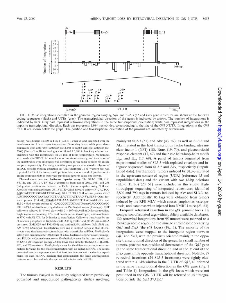
Frequent retroviral insertion in the gfi1 genomic locus. Bycomparison of isolated tags within publicly available databases,130 retroviral integrations from 95 tumors were mapped to a150-kb genomic region on the murine chromosome 5 carryingGfi1 and Evi5 (the gfi1 locus) (Fig. 1). The majority of theintegrations were mapped to the intergenic region betweenGfi1 and Evi5, with the provirus oriented mainly in the oppo-site transcriptional direction of the genes. In a small number oftumors, provirus was positioned downstream of the Gfi1 genein the same transcriptional direction and in the 3� end of theEvi5 gene in the opposite transcriptional direction. Notably, 27retroviral insertions (24 SL3-3 insertions) were tightly clus-tered within a 1-kb window in the 3�UTR of Gfi1, all orientedin the same transcriptional direction as the Gfi1 gene (Fig. 1and Table 1). Integrations in the gfi1 locus which were notpositioned in the Gfi1 3�UTR will be referred to as “integra-tions outside the Gfi1 3�UTR.”
FIG. 1. MLV integrations identified in the genomic region carrying Gfi1 and Evi5. Gfi1 and Evi5 gene structures are shown at the top withcoding sequences (black) and UTRs (gray). The transcriptional direction of the genes is indicated by arrows. The number of integrations isindicated by bars. Gray bars represent retroviral integrations in the same transcriptional orientation; white bars represent integrations in theopposite transcriptional direction. Each bar represents 1,084 nucleotides, corresponding to the size of the Gfi1 3�UTR. Integrations in the Gfi13�UTR are shown below the graph. The position and transcriptional orientation of the provirus are indicated by arrowheads.
VOL. 83, 2009 miRNA TARGET LOSS BY RETROVIRAL INSERTION IN Gfi1 3�UTR 8053
on April 14, 2019 by guest
http://jvi.asm.org/
Dow
nloaded from

The vast majority of the tumors with 3�UTR insertions wereinduced by the wt or enhancer-mutated SL3-3. Akv insertionwas identified in only two tumors, and one tumor was found topossess an integration of the RFB MLV. Each integration inthe Gfi1 3�UTR was validated by PCR using specific primerspositioned in the sixth exon of Gfi1 and in the SL3-3 LTR. Inall cases, sequencing revealed the presence of chimeric tran-scripts containing both Gfi1 and SL3-3 LTR sequences (datanot shown). The exact position of the provirus with respect tothe Gfi1 gene is indicated in Table 1.
This region was the most frequently targeted locus in theNMRI-i mouse genome and contained retroviral insertions in15% (120 of 790) of all SL3-3-induced tumors, where 90% (120of 130) were integrations of wt SL3-3 or SL3-3 enhancer mu-tants (data not shown). There was no significant correlationbetween integration patterns and virus mutants. Our observa-tions demonstrated that the genomic locus carrying Gfi1, andthe Gfi1 3�UTR in particular, are hot spots for retroviral in-sertions in the SL3-3/NMRI-i lymphoma model.
Retroviral insertions in the gfi1 genomic locus generatetruncated forms of Gfi1 mRNA. To study Gfi1 mRNA expres-sion, 40 tumors were selected for splicing analysis based onaccessibility and integration relative to the Gfi1 gene. Samplesincluded tumor material from thymus, spleen, and mesentericlymph node. PCR was performed using gene-specific primerscomplementary to sequences in the second and fifth exons inthe murine Gfi1 gene. Sequencing revealed the presence ofthree alternative Gfi1 transcripts, none of which have beenpreviously identified (Fig. 2). The transcripts were character-ized by exon 4 skipping (alternative transcript 1) and use of
alternative 5� and 3� splice sites in exons 3 and 4, respectively(alternative transcripts 2 and 3, respectively). Moreover, alter-native transcripts 1 and 2 had maintained their open readingframes. In the panel “integrations in the Gfi1 3�UTR,” tran-scripts 1 and 3 were detected in SL3-3 (7 of 14)- and Akv (1 of2)-induced tumors, while transcript 2 was detected in all tu-mors from this tumor group. Five of the tumors with insertionsin the Gfi1 3�UTR had all three alternative transcripts. Intumors with insertions outside the Gfi1 3�UTR, transcripts 1and 3 were detected only in SL3-3-induced tumors (in 1 of 19and 7 of 19 cases, respectively), while transcript 2 was identi-fied in both SL3-3 (16 of 19)- and Akv (1 of 4)-induced tumors.Due to a lack of tumor material, it was not possible to includemore Akv- or RFB-induced tumors. Table 2 summarizes thefrequency of alternative splicing within these two tumorgroups. The alternative transcripts showed relatively equal dis-tribution among the thymus, spleen, and mesenteric lymph
TABLE 2. Frequency of Gfi1 alternative splicing
Alternativetranscript
No. of tumors with alternative transcript/no. oftumors analyzeda
Integrations in Gfi1 3�UTR Integrations outsideGfi1 3�UTR
SL3-3 Akv RFB SL3-3 Akv RFB
1 7/14 1/2 0/1 1/19 0/4 0/02 14/14 2/2 1/1 16/19 1/4 0/03 7/14 1/2 0/1 7/19 0/4 0/0
a From a total of 40 tumors analyzed.
FIG. 2. Gfi1 alternative transcripts identified by sequencing. The wt Gfi1 is shown at the top. Coding exons are in black, UTRs are in gray. Threealternative transcripts were detected in SL3-3- and Akv-induced tumors, here referred to as alternative transcripts 1, 2, and 3. Sequencing revealed exon4 skipping (alternative transcript 1) and use of alternative 5� and 3� splice sites in exons 3 and 4, respectively (alternative transcripts 2 and 3).
8054 DABROWSKA ET AL. J. VIROL.
on April 14, 2019 by guest
http://jvi.asm.org/
Dow
nloaded from

node tumors, with no apparent correlation to either integra-tion position, virus variant, or provirus orientation. Alternativesplicing was also detected in MLV-induced tumors withoutknown integration on chromosome 5 but not in uninfectedtissue or in either of the control cell lines, L691, MPC11, orNIH 3T3, indicating that the aberrant splicing of the Gfi1 geneobserved in our study is a general phenomenon of MLV-induced lymphomas. We have not determined the relativeabundances of the transcripts, and the alternative splicing wasnot investigated further in this study.
Gfi1 is transcriptionally activated by the SL3-3 MLV. Pre-vious small-scale studies have demonstrated that all MoMLVinsertions in the genomic region carrying Gfi1 and Evi5 acti-vate the Gfi1 gene, leading to a three- to sixfold transcriptionalupregulation (62, 65). To evaluate the effect of retrovirus in-tegration in the gfi1 genomic locus on Gfi1 mRNA expression,43 tumors were screened by TaqMan real-time PCR (Fig. 3A).Our data confirmed a general upregulation of Gfi1 mRNAregardless of the position of the provirus in this 150-kb region.Notably, an up-to-200-fold upregulation in tumor 19T and a10- to 100-fold mRNA increase in 16 other tumors were ob-served. The transcription level of Gfi1 was found to be signif-icantly elevated in nearly all tumors analyzed, regardless oftissue type or provirus orientation. The upregulation was most
prominent in SL3-3-induced tumors but was not observed inAkv-induced tumors, possibly indicating that Gfi1 upregulationtakes place primarily in development of T-cell lymphomas. Innormal tissue, Gfi1 was most abundant in spleen, with a some-what lower expression in mesenteric lymph node and thymus.
Further expression analysis of Evi5 mRNA (data notshown) revealed significant Evi5 upregulation in Akv-in-duced tumors without known integrations on chromosome5, suggesting an oncogenic potential for Evi5 in B-cell lym-phomagenesis. Evi5 was also activated in the RFB-inducedtumor 12S harboring integration in the Gfi1 3�UTR but notin the Akv-induced tumor 22S. Expression for tumor 9S wasnot investigated, and no other Akv tumors with insertions inthe gfi1 locus were included in the study due to a lack oftumor material. In the SL3-3-induced tumors from bothtumor groups, Evi5 expression varied, with no unambiguousexpression pattern.
Decoupling of Gfi1 mRNA and protein expression in tumorsharboring retroviral insertions in the Gfi1 3�UTR. To investi-gate the correlation between Gfi1 mRNA and protein expres-sion, Western blot analysis was performed with a polyclonalantibody detecting Gfi1 at 50 to 55 kDa (Fig. 3B). Surprisingly,our results demonstrated major differences in Gfi1 proteinexpression, which appeared to be most abundant in tumors
FIG. 3. (A) Gfi1 expression in MLV-induced lymphomas. TaqMan real-time PCR was performed on 43 tumors harboring integrations in theGfi1 3�UTR and elsewhere in the gfi1 locus as well as on the three control tissues, i.e., thymus (T), spleen (S), and mesenteric lymph node (ML).Tumors are indicated by numbers. �, Akv integrations; ��, RFB integrations. Gfi1 mRNA expression was normalized to the expression of thetyrosine 3-monooxygenase housekeeping gene, Ywhaz. Thymic, splenic, and mesenteric lymph node tumors were further normalized to thymus,spleen, and mesenteric lymph node tissue controls, respectively. All control tissue was normalized to the thymus control. (B) Gfi1 proteinexpression in tumors with integrations in the Gfi1 3�UTR and outside of the Gfi1 gene. Tumors are indicated by numbers. Gfi1 was detected at50 to 55kDa. �-Actin and amido black protein stainings were used as a loading controls.
VOL. 83, 2009 miRNA TARGET LOSS BY RETROVIRAL INSERTION IN Gfi1 3�UTR 8055
on April 14, 2019 by guest
http://jvi.asm.org/
Dow
nloaded from

possessing insertions in the Gfi1 3�UTR. Gfi1 protein wasidentified at a relative high level in all tumors with Gfi1 3�UTRinsertions except 9S, 11ML, 19T, 25S, and 26S, which showedvague or no protein expression. In tumors with integrationselsewhere in the gfi1 locus, only 38S, 39T, 41T, 48S, and 51Sexpressed the Gfi1 protein at equally high levels. No (or vague)protein expression was observed in the remaining tumors fromthis panel, and no Gfi1 protein was detected in normal-tissuecontrols. Based on the decoupled mRNA and protein expres-sion patterns, we hypothesized that integrations in the Gfi13�UTR might disrupt a potential posttranscriptional miRNAregulation of Gfi1. Previous studies have indicated that Gfi1might be targeted by several miRNAs (7), and numerous pre-dicted miRNA target sites in the Gfi1 3�UTR (Fig. 4A) can
also be found in the miRNA registries (http://microrna.sanger.ac.uk and http://microrna.org). However, experimental valida-tion of whether Gfi1 is subjected to miRNA regulation has notto our knowledge been presented yet. From alignment of pre-dicted potential target sites for different miRNAs in the Gfi13�UTRs of various species, miR-142-3p and miR-155 showedthe most conservation in the seed binding region (Fig. 4B).Moreover, miR-142-3p displayed perfect base pairing of nu-cleotides 2 to 8, while a single wobble at position 6 was presentin the miR-155 seed sequence. Due to the conservation be-tween species and to the already established expression ofmiR-142-3p and miR-155 in T and B lymphocytes (47), thesewere selected as main candidates for further analysis, andreal-time quantitative PCR was performed on all 46 samples.
FIG. 4. (A) Predicted miRNA binding sites in the Gfi1 3�UTR from miRNA registries (http://microrna.sanger.ac.uk and http://microrna.org,April 2009). Positions of the different miRNA binding sites in the Gfi1 3�UTR are indicated by boxes, and miRNAs are indicated by numbers.miRNAs indicated by black boxes were selected for expression analysis. (B) Alignment of miR-142-3p and miR-155 binding sites in differentspecies. Conserved nucleotides are boxed. Presumed binding sites and nucleotide sequences for miR-142-3p and miR-155 are shown, andcomplementary nucleotides are connected by lines. G � U base pairing is indicated by dashed lines. (C) miR-142-3p and miR-155 expression in 43tumors with integrations in the Gfi1 3�UTR and outside the Gfi1 gene and three control tissue, i.e., thymus (T), spleen (S), and mesenteric lymphnode (ML), assayed by TaqMan real-time PCR. The data were calculated by the �CT method, and the values were normalized to snoRNA420.Thymic, splenic, and mesenteric lymph node tumors were further normalized to thymus, spleen, and mesenteric lymph node tissue controls,respectively. Expression for tissue controls is shown as normalized to snoRNA420. Lines indicate mean values for miR-142-3p and miR-155expression. (D) Real-time PCR expression analysis for miR-155, miR-142, miR-34b-3p, miR-10a, miR-466l, miR-133a, and miR-330 on 12 tumorswith integrations in the Gfi1 3�UTR and elsewhere in the gfi1 locus and three control tissues, i.e., thymus (T), spleen (S), and mesenteric lymphnode (ML). The data were processed as described above.
8056 DABROWSKA ET AL. J. VIROL.
on April 14, 2019 by guest
http://jvi.asm.org/
Dow
nloaded from
Of the remaining miRNAs, which showed no major conserva-tion between species in the region binding the miRNA seedsequence (alignment not shown), miR-330, miR-133a, miR-34b-3p, miR-10a, miR-879, miR-466l, and miR-467g were se-lected for expression analysis. Thymus, spleen, mesentericlymph node, and 12 tumors from the two tumor groups withand without Gfi1 protein expression were assayed for miRNAexpression. All data were calculated by the �CT method, andthe values were normalized to snoRNA420 and tissue controls(Fig. 4C). miR-879 and miR-467g were not detected in any ofthe samples. miR-142-3p and miR-155 expression patterns var-ied, while expression of miR-330, miR-133a, miR-34b-3p, miR-10a, and miR-466l was mainly downregulated in comparison tocontrol tissue. There was no significant difference in expressionbetween the two tumor groups for any of the miRNAs. Theexpression data indicate that the increase in Gfi1 protein intumors with integrations in the Gfi1 3�UTR was not due to adecrease in miRNA levels.
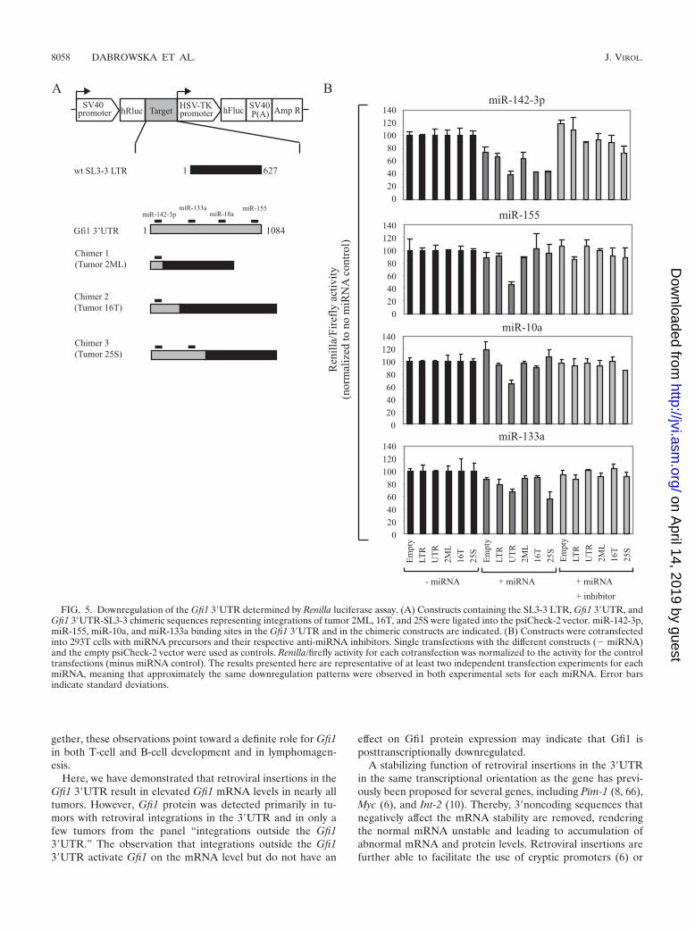
Downregulation of the Gfi1 3�UTR by miR-142-3p, miR-155,miR-10a, and miR-133a. To determine if any of the selectedmiRNAs were able to recognize the 3�UTR and mediate trans-lational regulation of the Gfi1 transcript, we made a Renilla/luciferase reporter system with different constructs containingthe Gfi1 3�UTR, SL3-3 LTR, and 3�UTR-SL3-3 LTR se-quences representing retrovirus integration in the tumors2ML, 16T, and 25S (Fig. 5A). The 2ML and 16T constructscontained only the miR-142-3p binding site, while 25S alsocontained the miR-330 and miR-133a binding sites. The Gfi13�UTR contained all miRNA binding sites.
Our data (Fig. 5B) demonstrated that miR-142-3p was ca-pable of downregulating all constructs, including the emptypsiCheck-2 vector, indicating that the effect was not specific forthe Gfi1 3�UTR only. In all cases, downregulation by miR-142-3p was rescued by cotransfection with miR-142-3p inhibi-tor, establishing a specific effect of miR-142-3p on all con-structs. Screening of the psiCheck-2 vector sequence detecteda perfect seed match between miR-142-3p and Renilla (posi-tions 982 to 987 and 1075 to 1080 [data not shown]). Likewise,screening of the SL3-3 LTR sequence identified weak mir-142-3p complementarity (nucleotides 2 to 6 with one G � Ubase pairing and nucleotides 3 to 9 with two G � U base pair-ings) (not shown in Fig. 5).
In contrast, both miR-155 and miR-10a were able to down-regulate the Gfi1 3�UTR significantly in comparison to theSL3-3 LTR, and a full rescue was observed in both cases.miR-133a downregulated the Gfi1 3�UTR and 25S constructs,and a small knockdown was also observed on the SL3-3 LTR.A region complementary to the miR-133a seed sequence wasfound in the SL3-3 LTR (nucleotides 2 to 7 with one G � Ubase-pairing), however, downregulation of the 2ML and 16Tconstructs was not observed. The miRNAs 34b-3b, 330, and466l did not have a specific effect on any of the constructs (datanot shown). Our data suggested that miR-155, miR-10a, andmiR-133a were able to recognize and bind to sequencespresent in the 3�UTR of Gfi1 and that the main silencingeffect of miR-142-3p was due to recognition of additionalbinding to complementarity sequences in Renilla and possi-bly also the SL3-3 LTR.
DISCUSSION
The mechanism of insertional mutagenesis in murine modelsand identification of retroviral insertion sites by high-through-put screening of the mouse genome have been widely used inidentification of genes contributing to murine lymphomagen-esis (34, 48, 73).
By large-scale analysis of integrated retroviruses in MLV-infected NMRI-i mice, we have identified the genomic regioncarrying Gfi1 as the most frequently targeted locus and haveaddressed the effect of these insertions on expression of theGfi1 gene. Gfi1 has previously been identified as a commonintegration site for several retroviruses, including MoMLV (22,30, 62, 65), Akv (72, 73), and MCF (40). Accumulating retro-viral insertions identified in various mouse strains have madeGfi1 a highly targeted gene in MLV-induced lymphomas, with82 integrations available from the Retrovirus Tagged CancerGene Database (http://rtcgd.abcc.ncifcrf.gov/) and many morewhich have been identified in recent studies (4, 76, 78). Wehere report on further identification of 130 MLV insertions inand adjacent to Gfi1. The majority of the integrations were ofwt or enhancer-mutated SL3-3. In most of the tumors theprovirus was found in the intergenic region between Gfi1 andEvi5 in the opposite transcriptional direction, displaying inte-gration patterns similar to those described previously (http://rtcgd.abcc.ncifcrf.gov/). Additionally, a tight cluster of 24SL3-3 insertions was mapped to a 1-kb region in the Gfi13�UTR. Such clustering in the Gfi1 gene has not been docu-mented in other virus/host models and appears to be uniquefor SL3-3 in the NMRI-i mouse strain.
The differences in integration patterns between studies areoften a result of different combinations of mouse genetic back-ground and virus strain. For instance, both Gfi1 and Myc arefrequently targeted in by MoMLV in p27kip (30)- and Cdkn2a(44)-deficient mice of the C57Bl6/129 strain but are rarelytargeted in BHX2 mice. Likewise, the SL3-3 Turbo enhancervariant has distinct integration hot spots in the c-Myc promotercompared to the wt SL3-3 (51), and the wt SL3-3 has differentintegration patterns in the Fos/Jdp2/Batf locus in comparisonto other SL3-3 enhancer variants (55). The variation of targetsin different model systems may reflect different but overlappingpathways to lymphoma development (55).
To determine the effect of the provirus on Gfi1 expression,Gfi1 mRNA and protein levels were determined in 43 tumors.In agreement with earlier studies (62, 65), we found that MLVintegration activated Gfi1 expression regardless of provirusposition. Gfi1 mRNA upregulation was most profound in SL3-3-induced tumors, supporting the involvement of Gfi1 in T-celllymphomas. In Akv-induced lymphomas Gfi1 was downregu-lated compared to in control tissue, strongly indicating thatGfi1 does not have an oncogenic effect in B-cell lymphomagen-esis. Thus, we failed to support previous reports of frequentAkv integration in this locus (72, 73). However, evidence fromseveral studies points toward a role for Gfi1 in development ofB-cell tumors, a notion supported by findings of plasmacytosisin Gfi1-deficient mice (56) and increased Gfi1 levels in a subsetof murine B-cell lymphomas in the marginal zone (68). Fur-thermore, Gfi1 expression has been detected in early B-cellprogenitors (81), and it has been suggested that Gfi1 controlscytokine-dependent B-cell differentiation (57). Taken to-
VOL. 83, 2009 miRNA TARGET LOSS BY RETROVIRAL INSERTION IN Gfi1 3�UTR 8057
on April 14, 2019 by guest
http://jvi.asm.org/
Dow
nloaded from
gether, these observations point toward a definite role for Gfi1in both T-cell and B-cell development and in lymphomagen-esis.
Here, we have demonstrated that retroviral insertions in theGfi1 3�UTR result in elevated Gfi1 mRNA levels in nearly alltumors. However, Gfi1 protein was detected primarily in tu-mors with retroviral integrations in the 3�UTR and in only afew tumors from the panel “integrations outside the Gfi13�UTR.” The observation that integrations outside the Gfi13�UTR activate Gfi1 on the mRNA level but do not have an
effect on Gfi1 protein expression may indicate that Gfi1 isposttranscriptionally downregulated.
A stabilizing function of retroviral insertions in the 3�UTRin the same transcriptional orientation as the gene has previ-ously been proposed for several genes, including Pim-1 (8, 66),Myc (6), and Int-2 (10). Thereby, 3�noncoding sequences thatnegatively affect the mRNA stability are removed, renderingthe normal mRNA unstable and leading to accumulation ofabnormal mRNA and protein levels. Retroviral insertions arefurther able to facilitate the use of cryptic promoters (6) or
FIG. 5. Downregulation of the Gfi1 3�UTR determined by Renilla luciferase assay. (A) Constructs containing the SL3-3 LTR, Gfi1 3�UTR, andGfi1 3�UTR-SL3-3 chimeric sequences representing integrations of tumor 2ML, 16T, and 25S were ligated into the psiCheck-2 vector. miR-142-3p,miR-155, miR-10a, and miR-133a binding sites in the Gfi1 3�UTR and in the chimeric constructs are indicated. (B) Constructs were cotransfectedinto 293T cells with miRNA precursors and their respective anti-miRNA inhibitors. Single transfections with the different constructs (� miRNA)and the empty psiCheck-2 vector were used as controls. Renilla/firefly activity for each cotransfection was normalized to the activity for the controltransfections (minus miRNA control). The results presented here are representative of at least two independent transfection experiments for eachmiRNA, meaning that approximately the same downregulation patterns were observed in both experimental sets for each miRNA. Error barsindicate standard deviations.
8058 DABROWSKA ET AL. J. VIROL.
on April 14, 2019 by guest
http://jvi.asm.org/
Dow
nloaded from
destroy important regulatory elements such as A/U-rich re-gions implicated in mRNA destabilization (79). Based on thepreferred integration clustering in the 5� end of the Gfi13�UTR and the Gfi1 protein expression patterns observedhere, we speculated on whether retroviral integrations de-coupled miRNA binding to the Gfi1 3�UTR, resulting in anincrease in protein synthesis. In this case, it would be possiblethat the high Gfi1 protein expression observed in some of thetumors from the panel “integrations outside of the Gfi13�UTR” (38S, 39T, 41T, 48S, and 51S) could reflect deregula-tion of other proteins important for miRNA processing, al-though we did not succeed in identifying such integrations.
The Gfi1 3�UTR holds predicted binding sites for severalmiRNAs, including miR-142-3p and miR-155, which are foundwith relatively high abundance in most hematopoietic cells (47)and show highly conserved binding sites in the Gfi1 3�UTRs ofvarious species. Real-time PCR expression analysis for themiRNAs 155 and 142-3p showed varying expression patterns,while miRNAs 34b-3p, 10a, 466l, 133a, and 330 demonstrateda general downregulation in most of the tumors in comparisonto control tissue. Our data indicated that the increase in Gfi1protein in tumors with integrations in the Gfi1 3�UTR was notdue to a decrease in miRNA levels for any of the miRNAsinvestigated here.
In order to determine if any of these miRNAs could bepotential downregulators of Gfi1, Renilla luciferase reporterassays with different constructs were performed. Our resultsdemonstrated that miR-142-3p was able to downregulate allconstructs, suggesting that this downregulation results from aninteraction with competing target sequences in the psiCheck-2vector and the SL3-3 LTR. Previous studies have demon-strated that multiple target sites can potentially increase thedegree of translational suppression (13), possibly explainingthe higher silencing observed here for miR-142-3p on the3�UTR 16T and 25S constructs. Only a minor downregulationby miR-142-3p was observed on the 2ML construct, althoughthis contained the full binding site for miR-142-3p. The inte-gration in tumor 2ML is positioned just three nucleotidesdownstream of the miRNA binding region, possibly influenc-ing the structure of the small Gfi1 3�UTR fragment. Severalstudies have addressed the role of mRNA structure in miRNAtarget recognition and suggest that the affinity of binding of amiRNA to its mRNA target is determined by both the se-quence and structure of the mRNA (11, 12, 14, 33, 42). Apossible explanation for the variability that we observed in ourexperiments may simply arise from differences in accessibilityimposed by the sequence surrounding the target.
In contrast, miR-155, miR-10a, and miR-133a all had adownregulating effect on the Gfi1 3�UTR construct, possiblysuggesting a role for these miRNAs in posttranscriptional reg-ulation of the Gfi1 gene. Furthermore, the 25S construct,which was the only chimeric construct containing the miR133abinding site, was also downregulated. Together, our resultssupport the hypothesis that Gfi1 may be downregulated by oneor more miRNAs. However, we have assessed the function ofonly a small number of potential miRNAs. Other miRNAsmay also have an effect on Gfi1 regulation. To further validatewhether any of the miRNAs investigated here targets the Gfi1gene, additional experiments, including miRNA knockdown in
different cell lines and subsequent analysis of Gfi1 expression,need to be performed.
Our results suggest that retroviral integrations in the Gfi13�UTR contribute to Gfi1 activation and possibly T-cell lym-phomagenesis through loss of miRNA binding sites. In themajority of the tumors with insertions elsewhere in the gfi1locus, no Gfi1 protein expression was observed. It is unclearhow these integrations contribute to the development of thesetumors. Ccnd3, Myc/Pvt1, Ras2, and RasGrp1, which were pre-viously identified as possible Gfi1 cooperative partners in lym-phoma development (76), were found as recurring integrationsin several of the tumors with integrations in the gfi1 locus.Development of T-cell lymphomas in tumors with integrationsoutside the Gfi1 3�UTR that do not express the Gfi1 proteincould be a result of activation of these or other oncogenes. Ofthe 130 insertions, Ccnd3 was a cotarget in 10 tumors and wasalso found as a target in tumor 42S, 45ML, 49ML, and 50ML;Myc/Pvt1was targeted four times, including in 8T and 48S;Ras2 was cotargeted in 9 tumors, including 42S; and RasGRP1was cotargeted in 5 tumors including 37 M and 43ML. We donot know how Ccnd3, Myc/Pvt1, Ras2, and RasGrp1 are ex-pressed in the tumors investigated in this study, and the tech-niques used to identify retroviral tags do not necessarily iden-tify all integrations. Overall, more extensive analyses need tobe performed in order to obtain a clearer impression of howthese tumors were initiated.
In comparison to tumor development through insertionalmutagenesis of proto-oncogenes or tumor suppressors, a re-cently discovered mode of tumor induction includes retroviraltargeting of miRNA loci and deregulation of single miRNAs ormiRNA cistrons. The SL3-3 retrovirus has been shown to ac-tivate the 17-92 miRNA cistron (78), while the avian leukosisvirus targets the BIC gene (the chromosomal region encodingmiR-155) (16, 74) and the Radiation MLV frequently inte-grates into a locus encoding a group of five differentiallyspliced noncoding RNAs known as Kis2 (35). Retroviral inte-gration in all these regions caused significant upregulation ofthe miRNA clusters, demonstrating a role for these miRNAsin oncogenesis.
In this study, we have introduced an SL3-3/NMRI-i modelwith high retroviral integration frequency in the gfi1 locus andderegulated Gfi1 mRNA and protein expression patterns. Ourdata indicate that retroviral insertions in the Gfi1 3�UTR con-tribute to activation of Gfi1 by loss of regulatory regions im-portant for miRNA posttranscriptional downregulation of thegene. It is possible that such loss of regulatory regions in the3�UTR of the human GFI1 gene might likewise contribute tohuman lymphomagenesis. The human GFI1 gene is carried inthe chromosomal region 1p22 (58), a locus commonly affectedin several cancers, including mantle cell lymphoma (60, 61),mucosa-associated lymphoid tissue lymphoma (77), and neu-roblastoma (46). Although there has been no direct correlationbetween translocations in this region and the effect on theGFI1 gene, our studies support the importance of this genomicregion in tumor development. In humans, precursor T-celllymphoma is a rare disease with a poor prognosis but with aclear diagnostic parallel to the same type of tumors observed inmurine models (49). In time, the results presented here maycontribute to understanding of the oncogenic mechanisms by
VOL. 83, 2009 miRNA TARGET LOSS BY RETROVIRAL INSERTION IN Gfi1 3�UTR 8059
on April 14, 2019 by guest
http://jvi.asm.org/
Dow
nloaded from
which Gfi1 is involved in development of both murine andhuman T-cell lymphomas.
ACKNOWLEDGMENTS
The technical assistance of Astrid van der Aa Kuhle is gratefullyacknowledged.
This project was supported by grants from The Karen Elise JensenFoundation (M.J.D. and K.D.), the Danish Cancer Society (H.E.J. andF.S.P.), and The Danish Agency for Science Technology and Innova-tion (F.S.P.) and by NIH grant R01AI41570 (M.W.).
REFERENCES
1. Ben-David, Y., E. B. Giddens, K. Letwin, and A. Bernstein. 1991. Erythro-leukemia induction by Friend murine leukemia virus: insertional activationof a new member of the ets gene family, Fli-1, closely linked to c-ets-1. GenesDev. 6:908–918.
2. Brennecke, J., A. Stark, R. B. Russell, and S. M. Cohen. 2005. Principles ofmicroRNA-target recognition. PLoS Biol. 3:e85–e99.
3. Celander, D., and W. A. Haseltine. 1987. Glucocorticoid regulation of mu-rine leukemia virus transcription elements is specified by determinantswithin the viral enhancer region. J. Virol. 61:269–275.
4. Chakraborty, J., H. Okonta, H. Bagalb, S. J. Lee, B. Fink, R. Changanam-kandat, and J. Duggan. 2008. Retroviral gene insertion in breast milk me-diated lymphomagenesis. Virology 377:100–109.
5. Chatis, P. A., C. A. Holland, J. W. Hartley, W. P. Rowe, and N. Hopkins.1983. Role for the 3� end of the genome in determining disease specificity ofFriend and Moloney murine leukemia viruses. Proc. Natl. Acad. Sci. USA80:4408–4411.
6. Corcoran, L. M., J. M. Adams, A. R. Dunn, and S. Cory. 1984. Murine Tlymphomas in which the cellular myc oncogene has been activated by retro-viral insertion. Cell 37:113–122.
7. Costa, I. G., S. Roepcke, and A. Schliep. 2007. Gene expression trees inlymphoid development. BMC Immunol. 8:25–44.
8. Cuypers, H. T., G. Selten, W. Quint, M. Zijlstra, E. R. Maandag, W. Boelens,P. van Wezenbeek, C. Melief, and A. Berns. 1984. Murine leukemia virus-induced T-cell lymphomagenesis: integration of proviruses in a distinct chro-mosomal region. Cell 37:141–150.
9. DesGroseillers, L., and P. Jolicoeur. 1984. The tandem direct repeats withinthe long terminal repeat of murine leukemia viruses are primary determinantof their leukemogenic potential. J. Virol. 52:945–952.
10. Dickson, C., R. Smith, S. Brookes, and G. Peters. 1990. Proviral insertionswithin the int-2 gene can generate multiple anomalous transcripts but leavethe protein-coding domain intact. J. Virol. 64:784–793.
11. Didiano, D., and O. Hobert. 2006. Perfect seed pairing is not a generallyreliable predictor for miRNA-target interactions. Nat. Struct. Mol. Biol.13:849–851.
12. Didiano, D., and O. Hobert. 2008. Molecular architecture of a miRNA-regulated 3�UTR. RNA 14:1297–1317.
13. Doench, J. G., C. P. Petersen, and P. A. Sharp. 2003. siRNAs can functionas miRNAs. Genes Dev. 17:438–442.
14. Doench, J. G., and P. A. Sharp. 2004. Specificity of microRNA target selec-tion in translational repression. Genes Dev. 18:504–511.
15. Dwivedi, P. P., P. H. Anderson, J. L. Omdahl, H. L. Grimes, H. A. Morris,and B. K. May. 2005. Identification of growth factor independent-1 (GFI1)as a repressor of 25-hydroxyvitamin D 1-alpha hydroxylase (CYP27B1) geneexpression in human prostate cancer cells. Endocrinol. Relat. Cancer 12:351–365.
16. Eis, P. S., W. Tam, L. Sun, A. Chadburn, Z. Li, M. F. Gomez, E. Lund, andJ. E. Dahlberg. 2005. Accumulation of miR-155 and BIC RNA in human Bcell lymphomas. Proc. Natl. Acad. Sci. USA 102:3627–3632.
17. Ejegod, D., K. D. Sørensen, I. Mossbrugger, L. Quintanilla-Martinez, J.Schmidt, and F. S. Pedersen. 2009. Control of pathogenicity and diseasespecificity of a T-cell lymphomagenic gammaretrovirus by E-box motifs butnot by an overlapping glucocorticoid response element. J. Virol. 83:336–346.
18. Ethelberg, S., B. Hallberg, J. Lovmand, J. Schmidt, A. Luz, T. Grundstrom,and F. S. Pedersen. 1997. Second-site proviral enhancer alterations in lym-phomas induced by enhancer mutants of SL3-3 murine leukemia virus:negative effects of nuclear factor 1 binding site. J. Virol. 71:1196–1206.
19. Ethelberg, S., J. Lovmand, J. Schmidt, A. Luz, and F. S. Pedersen. 1997.Increased lymphomagenicity and restored disease specificity of AML1 site(core) mutant SL3-3 murine leukemia virus by a second-site enhancer vari-ant evolved in vivo. J. Virol. 71:7273–7280.
20. Ethelberg, S., A. B. Sørensen, J. Schmidt, A. Luz, and F. S. Pedersen. 1997.An SL3-3 murine leukemia virus enhancer variant more pathogenic than thewild type obtained by assisted molecular evolution in vivo. J. Virol. 71:9796–9799.
21. Fan, H., B. K. Brightman, B. R. Davis, and Q. X. Li. 1991. Leukemogenesisby Moloney murine leukemia virus, p. 155–174. In H. Y. Fan et al. (ed.),Viruses that affect the immune system. American Society for Microbiology,Washington, DC.
22. Gilks, C. B., S. E. Bear, H. L. Grimes, and P. N. Tsichlis. 1993. Progressionof interleukin-2 (IL-2)-dependent rat T-cell lymphoma lines to IL-2-inde-pendent growth following activation of a gene (Gfi-1) encoding a novel zincfinger protein. Mol. Cell. Biol. 13:1759–1768.
23. Gimbel, W., J. Schmidt, J. Barack-Werner, A. Luz, P. G. Strauss, V. Erfle,and T. Werner. 1996. Molecular and pathogenic characterization of the RFBosteoma virus: lack of oncogene and induction of osteoma, osteopetrosis andlymphoma. Virology 224:533–538.
24. Golemis, E. A., N. A. Speck, and N. Hopkins. 1990. Alignment of U3 regionsequences of mammalian type C viruses: identification of highly conservedmotifs and implications for enhancer design. J. Virol. 64:534–542.
25. Grimes, H. L., T. O. Chan, P. A. Zweidler-McKay, B. Tong, and P. N.Tsichlis. 1996. The Gfi-1 proto-oncoprotein contains a novel transcriptionalrepressor domain, SNAG, and inhibits G1 arrest induced by interleukin-2withdrawal. Mol. Cell. Biol. 16:6263–6272.
26. Grimes, H. L., C. B. Gilks, T. O. Chan, S. Porter, and P. N. Tsichlis. 1996.The Gfi-1 protooncoprotein represses Bax expression and inhibits T-celldeath. Proc. Natl. Acad. Sci. USA 93:14569–14573.
27. Hallberg, B., J. Schmidt, A. Luz, F. S. Pedersen, and T. Grundstrom. 1991.SL3-3 enhancer factor I transcriptional activators are required for tumorformation by SL3-3 murine leukemia virus. J. Virol. 65:4177–4181.
28. Hayward, W. S., B. G. Neel, and S. M. Astrin. 1981. Activation of a cellularonc gene by promoter insertion in ALV-induced lymphoid leukosis. Nature290:475–480.
29. Hock, H., M. J. Hamblen, H. M. Rooke, J. W. Schindler, S. Saleque, Y.Fujiwara, and S. H. Orkin. 2004. Gfi-1 restricts proliferation and preservesfunctional integrity of haematopoietic stem cells. Nature 431:1002–1007.
30. Hwang, H. C., C. P. Martins, Y. Bronkhorst, E. Randel, A. Berns, M. Fero,and B. E. Clurman. 2002. Identification of oncogenes collaborating withp27kip1 loss by insertional mutagenesis and high-throughput insertion siteanalysis. Proc. Natl. Acad. Sci. USA 99:11293–11298.
31. Karsunky, H., H. Zeng, T. Schmidt, B. Zevnik, R. Kluge, K. W. Schmid, U.Duhrsen, and T. Moroy. 2002. Inflamatory reactions and severe neutropeniain mice lacking the transcriptional repressor Gfi1. Nat. Genet. 30:295–300.
32. Kazanjian, A., D. Wallis, N. Au, R. Nigam, K. J. T. Venken, P. T. Cagle, B. F.Dickey, H. J. Bellen, C. B. Gilks, and H. L. Grimes. 2004. Growth factorindependence-1 is expressed in primary human neuroendocrine lung carci-nomas and mediates the differentiation of murine pulmonary neuroendo-crine cells. Cancer Res. 64:6874–6882.
33. Kertesz, M., N. Iovino, U. Unnerstall, U. Gaul, and E. Segal. 2007. The roleof site accessibility in miRNA target recognition. Nat. Genet. 39:1278–1284.
34. Kim, R., A. Trubetskoy, T. Suzuki, N. A. Jenkins, N. G. Copeland, and J.Lenz. 2003. Genome-based identification of cancer genes by proviral taggingin mouse retrovirus-induced T-cell lymphomas. J. Virol. 77:2056–2062.
35. Landais, S., S. Landry, P. Legault, and E. Rassart. 2007. Oncogenic poten-tial of the miR-106-363 cluster and its implication in human T-cell leukemia.Cancer Res. 67:5699–5707.
36. Lee, R. C., R. L. Feinbaum, and V. Ambros. 1993. The C. elegans hetero-chronic gene lin-4 encodes small RNAs with antisense complementarity tolin-14. Cell 75:843–854.
37. Lenz, J., D. Celander, R. L. Crowther, R. Patarca, D. W. Perkins, and W. A.Haseltine. 1984. Determination of the leukaemogenicity of a murine retro-virus by sequences within the long terminal repeat. Nature 308:467–470.
38. Lewis, A. F., T. Stacy, W. R. Green, L. Taddesse-Heath, J. W. Hartley, andN. A. Speck. 1999. Core-binding factor influences the disease specificity ofMoloney murine leukemia virus. J. Virol. 73:5535–5547.
39. Lewis, B. P., C. B. Burge, and D. P. Bartel. 2005. Conserved seed pairing,often flanked by adenosines, indicates that thousands of human genes aremicroRNA targets. Cell 120:15–20.
40. Liao, X., A. M. Buchberg, N. A. Jenkins, and N. G. Copeland. 1995. Evi-5, acommon site of retroviral integration in AKXD T-cell lymphomas, mapsnear Gfi1 on mouse chromosome 5. J. Virol. 69:7132–7137.
41. Lim, L. P., N. C. Lau, P. Garrett-Engele, A. Grimson, J. M. Schelter, J.Castle, D. P. Bartel, P. S. Linsley, and J. M. Johnson. 2005. Microarrayanalysis shows that some microRNAs downregulate large numbers of targetmRNAs. Nature 433:769–773.
42. Long, D., R. Lee, P. Williams, C. Y. Chan, V. Ambros, and Y. Ding. 2007.Potent effect of target structure on microRNA function. Nat. Struct. Mol.Biol. 14:287–294.
43. Lovmand, J., A. B. Sørensen, J. Schmidt, M. Østergaard, A. Luz, and F. S.Pedersen. 1998. B-cell lymphoma induction by Akv murine leukemia virusesharboring one or both copies of the tandem repeat in the U3 enhancer.J. Virol. 72:5745–5756.
44. Lund, A. H., G. Turner, A. Trubetskoy, E. Verhoeven, E. Wientjens, D.Hulsman, R. Russell, R. A. DePinho, J. Lenz, and M. van Lohuizen. 2002.Genome-wide retroviral insertional tagging of genes involved in cancer inCdkn2a-deficient mice. Nat. Genet. 32:160–165.
45. Ma, S. L., J. Lovmand, A. B. Sørensen, A. Luz, J. Schmidt, and F. S.Pedersen. 2003. Triple basepair changes within and adjacent to the con-served YY1 motif upstream of the U3 enhancer repeats of SL3-3 murineleukemia virus cause a small but significant shortening of latency of T-lymphoma induction. Virology 313:638–644.
8060 DABROWSKA ET AL. J. VIROL.
on April 14, 2019 by guest
http://jvi.asm.org/
Dow
nloaded from
46. Mead, R. S., and J. K. Cowell. 1995. Molecular characterization of a (1;10)(p22;q21) constitutional translocation from a patient with neuroblastoma.Cancer Genet. Cytogenet. 81:151–157.
47. Merkerova, M., M. Belickova, and H. Bruchova. 2008. Differential expres-sion of microRNA in hematopoietic cell lineages. Eur. J. Haematol. 81:304–310.
48. Mikkers, H., J. Allen, P. Knipscheer, L. Romeijn, A. Hart, E. Vink, and A.Berns. 2002. High-throughput retroviral tagging to identify components ofspecific signaling pathways in cancer. Nat. Genet. 32:153–159.
49. Morse, H. C., III, M. R. Anver, T. N. Fredrickson, D. C. Haines, A. W.Harris, N. L. Harris, E. S. Jaffe, S. C. Kogan, I. C. MacLennan, P. K.Pattengale, and J. M. Ward. 2002. Hematopathology subcommittee of theMouse Models of Human Cancers Consortium. Bethesda proposals forclassification of lymphoid neoplasms in mice. Blood 100:246–258.
50. Nielsen, A. L., P. L. Nørby, F. S. Pedersen, and P. J.ørgensen. 1996. Variousmodels of basic helix-loop-helix protein-mediated regulation of murine leu-kemia virus transcription in lymphoid cells. J. Virol. 70:5893–5901.
51. Nielsen, A. A., A. B. Sørensen, J. Schmidt, and F. S. Pedersen. 2005. Analysisof wild-type and mutant SL3-3 murine leukemia virus insertions in the c-mycpromoter during lymphomagenesis reveals target site hot spots, virus-depen-dent patterns, and frequent error-prone gap repair. J. Virol. 79:67–78.
52. Nieves, A., L. S. Levy, and J. Lenz. 1997. Importance of a c-Myb binding sitefor lymphomagenesis by the retrovirus SL3-3. J. Virol. 71:1213–1219.
53. Nusse, R., and H. E. Varmus. 1982. Many tumors induced by the mousemammary tumor virus contain a provirus integrated in the same region of thehost genome. Cell 31:99–109.
54. Pargmann, D., R. Yucel, C. Kosan, I. Saba, L. Klein-Hitpass, S. Schimmer,F. Heyd, U. Dittmer, and T. Moroy. 2007. Differential impact of the tran-scriptional repressor Gfi1 on mature CD4� and CD8� T lymphocyte func-tion. Eur. J. Immunol. 37:3551–3563.
55. Rasmussen, M. H., A. B. Sørensen, D. W. Morris, J. C. Dutra, E. K. En-gelhard, C. L. Wang, J. Schmidt, and F. S. Pedersen. 2005. Tumor model-specific proviral insertional mutagenesis of the Fos/Jdp2/Batf locus. Virology337:353–364.
56. Rathinam, C., H. Lassmann, M. Mengel, and C. Klein. 2008. Transcriptionfactor Gfi1 restricts B-cell mediated autoimmunity. J. Immunol. 181:6222–6229.
57. Rathinam, C., and C. Klein. 2007. Transcriptional repressor Gfi1 integratescytokine-receptor signals controlling B-cell differentiation. PLoS ONE2:e306–e317.
58. Roberts, T., and J. K. Cowell. 1997. Cloning of the human Gfi-1 gene and itsmapping to chromosome region 1p22. Oncogene 14:1003–1005.
59. Sakai, I., H. Yamauchi, M. Yasukawa, H. Kohno, and S. Fujita. 2001.Expression of the Gfi-1 gene in HTLV-I-transformed T cells. Int. J. Hema-tol. 73:507–516.
60. Salaverria, I., B. Espinet, A. Carrio, D, Costa, L, Astier, J. Slotta-Huspenina,L, Quintanilla-Martinez, F. Fend, F. Sole, D. Colomer, S. Serrano, R. Miro,S. Bea, and E. Campo. 2008. Multiple recurrent chromosomal breakpoints inmantle cell lymphoma revealed by a combination of molecular cytogenetictechniques. Genes Chromosomes Cancer 47:1086–1097.
61. Sander, S., L. Bullinger, E. Leupolt, A. Benner, D. Kienle, T. Katzenberger,J. Kalla, G. Ott, H. K. Muller-Hermelink, T. F. Barth, P. Moller, P. Lichter,H. Dohner, and S. Stilgenbauer. 2008. Genomic aberrations in mantle celllymphoma detected by interphase fluorescence in situ hybridization. Inci-dence and clinicopathological correlations. Haematologica 93:680–687.
62. Scheijen, B., J. Jonkers, D. Acton, and A. Berns. 1997. Characterization ofpal-1, a common proviral insertion site in murine leukemia virus-inducedlymphomas of c-myc and pim-1 transgenic mice. J. Virol. 71:9–16.
63. Schmidt, J., K. Lumniczky, B. D. Tzschaschel, H. L. Guenther, A. Luz, S.Riemann, W. Gimbel, V. Erfle, and R. G. Erben. 1999. Onset and dynamicsof osteosclerosis in mice induced by Reilly-Finkel-Biskis (RFB) murine leu-kemia virus. Am. J. Pathol. 155:557–570.
64. Schmidt, T., H. Karsunky, B. Rodel, B. Zevnik, H-P. Elsasser, and T. Moroy.1998. Evidence implicating Gfi-1 and Pim-1 in pre-T-cell differentiation stepsassociated with �-selection. EMBO J. 17:5349–5359.
65. Schmidt, T., M. Zornig, R. Benke, and T. Moroy. 1996. MoMuLV proviralintegrations identified by Sup-F selection in tumors from infected myc/pimbitransgenic mice correlate with activation of the gfi-1 gene. Nucleic AcidsRes. 24:2528–2534.
66. Selten, G. H., H. T. Cuypers, and A. Berns. 1985. Proviral activation of theputative oncogene Pim-1 in MuLV induced T-cell lymphomas. EMBO J.4:1793–1798.
67. Selten, G. H., H. T. Cuypers, M. Zijlstra, C. Melief, and A. Berns. 1984.Involvement of c-Myc in T cell lymphomas in mice: frequency and mecha-nism of activation. EMBO J. 3:3215–3222.
68. Shin, M. S., T. N. Fredrickson, J. W. Hartley, T. Suzuki, K. Akagi, and H. C.Morse III. 2004. High-throughput retroviral tagging for identification ofgenes involved in initiation and progression of mouse splenic marginal zonelymphomas. Cancer Res. 64:4419–4427.
69. Sørensen, K. D., S. Kunder, L. Quintanilla-Martinez, J. Sørensen, J.Schmidt, and F. S. Pedersen. 2007. Enhancer mutations of Akv murineleukemia virus inhibit the induction of mature B-cell lymphomas and shiftdisease specificity towards the more differentiated plasma cell stage. Virol-ogy 362:179–191.
70. Sørensen, K. D., L. Quintanilla-Martinez, S. Kunder, J. Schmidt, and F. S.Pedersen. 2004. Mutation of all Runx (AML1/core) sites in the enhancer ofT-lymphomagenic SL3-3 murine leukemia virus unmasks a significant po-tential for myeloid leukemia induction and favors enhancer evolution towardinduction of other disease patterns. J. Virol. 78:13216–13231.
71. Speck, N. A., B. Renjifo, E. Golemis, T. N. Fredrickson, J. W. Hartley, andN. Hopkins. 1990. Mutation of the core or adjacent LVb elements of theMoloney murine leukemia virus enhancer alters disease specificity. GenesDev. 4:233–242.
72. Suzuki, T., K. Minehata, K. Agaki, N. A. Jenkins, and N. G. Copeland. 2006.Tumor suppressor gene identification using retroviral insertional mutagen-esis in Blm-deficient mice. EMBO J. 25:3422–3431.
73. Suzuki, T., H. Shen, K. Akagi, H. C. Morse, J. D. Malley, D. Q. Naiman, N. A.Jenkins, and N. G. Copeland. 2002. New genes involved in cancer identifiedby retroviral tagging. Nat. Genet. 32:166–174.
74. Tam, W., D. Ben-Yehuda, and W. S. Hayward. 1997. bic, a novel geneactivated by proviral insertions in avian leukosis virus-induced lymphomas, islikely to function through its noncoding RNA. Mol. Cell. Biol. 17:1490–1502.
75. Tsichlis, P. N., and P. A. Lazo. 1991. Virus-host interactions and the patho-genesis of murine and human oncogenic retroviruses. Curr. Top. Microbiol.Immunol. 171:95–173.
76. Uren, A. G., J. Kool, K. Matentzoglu, J. de Ridder, J. Mattison, M. vanUitert, W. Lagcher, D. Sie, E. Tanger, T. Cox, M. Reinders, T. J. Hubbard,J. Rogers, J. Jonkers, L. Wessels, D. J. Adams, M. van Lohuizen, and A.Berns. 1998. Large-scale mutagenesis in p19ARF- and p53-deficient miceidentifies cancer genes and their collaborative networks. Cell 133:727–741.
77. Vega, F., and L. J. Medeiros. 2001. Marginal-zone B-cell lymphoma ofextranodal mucosa-associated lymphoid tissue type: molecular genetics pro-vides new insights into pathogenesis. Adv. Anat. Pathol. 8:313–326.
78. Wang, C. L., B. B. Wang, G. Bartha, L. Li, N. Channa, M. Klinger, N.Killeen, and M. Wabl. 2006. Activation of an oncogenic microRNA cistronby provirus integration. Proc. Natl. Acad. Sci. USA 103:18680–18684.
79. Wingett, D., R. Reeves, and N. S. Magnuson. 1992. Characterization of thetestes-specific pim-1 transcript in rat. Nucleic Acids Res. 20:3183–3189.
80. Yucel, R., H. Karsunky, L. Klein-Hitpass, and T. Moroy. 2003. The tran-scriptional repressor Gfi1 affects development of early uncommitted c-kit�T cell progenitors and CD4/CD8 lineage decision in the thymus. J. Exp. Med.197:831–844.
81. Yucel, R., C. Kosan, F. Heyd, and T. Moroy. 2004. Gfi1:Green fluorescentprotein knock-in mutant reveals different expression and autoregulation ofthe growth factor independence 1 (Gfi1) gene during lymphocyte develop-ment. J. Biol. Chem. 279:40906–40917.
82. Zeng, H., R. Yucel, C. Kosan, L. Klein-Hitpass, and T. Moroy. 2004. Tran-scription factor Gfi1 regulates self-renewal and engraftment of hematopoi-etic stem cells. EMBO J. 23:4116–4125.
VOL. 83, 2009 miRNA TARGET LOSS BY RETROVIRAL INSERTION IN Gfi1 3�UTR 8061
on April 14, 2019 by guest
http://jvi.asm.org/
Dow
nloaded from