Society of Vascular and Interventional Neurology 8th ... Meeting/2015/SVIN Abstract...
97
Society of Vascular and Interventional Neurology 8 th Annual Meeting Poster Session Details & Presenter List October 17, 2015 | 6:00-9:00pm | Calusa Terrace Hyatt Regency Coconut Point | Bonita Springs, FL The dinner and reception will take place from 6:00-6:45pm. Stand-by Times: Authors are mandated to attend their posters during the following stand by times: Odd-Numbered Posters: 6:45 – 7:30pm Even-Numbered Posters: 7:30 – 8:15pm We encourage presenters to stay for the entire session! Logistics: • Put up your poster early! We encourage you to put up your poster early so that attendees can view it throughout the meeting. The Calusa Terrace will be open starting Friday, October 16 th at 6:30 am for set-up. • Poster boards are 4 ft. high by 8 ft. wide. Please plan your poster size accordingly. Pins will be provided. • All posters must be torn down by 10:30am on Sunday, October 18 th . All posters that are not taken down will be discarded. ABSTRACT DISCLAIMER: All Abstract information in this supplement is published as submitted.
Transcript of Society of Vascular and Interventional Neurology 8th ... Meeting/2015/SVIN Abstract...

Society of Vascular and Interventional Neurology
8th Annual Meeting
Poster Session Details & Presenter List October 17, 2015 | 6:00-9:00pm | Calusa Terrace Hyatt Regency Coconut Point | Bonita Springs, FL
The dinner and reception will take place from 6:00-6:45pm.
Stand-by Times: Authors are mandated to attend their posters during the following stand by times: Odd-Numbered Posters: 6:45 – 7:30pm Even-Numbered Posters: 7:30 – 8:15pm
We encourage presenters to stay for the entire session!
Logistics:
• Put up your poster early! We encourage you to put up your poster early so that attendees can view it throughout the meeting. The Calusa Terrace will be open starting Friday, October 16th at 6:30 am for set-up.
• Poster boards are 4 ft. high by 8 ft. wide. Please plan your poster size accordingly. Pins will be provided.
• All posters must be torn down by 10:30am on Sunday, October 18th. All posters that are not taken down will be discarded.
ABSTRACT DISCLAIMER: All Abstract information in this supplement is published as submitted.

Walk Around with the Professor
Join experts in the field and “Walk Around with the Professor” during this year’s poster session. Below is a list of the Professors and the schedule. Poster numbers, titles and authors are included in the program. View the complete abstracts electronically on the attendee portal and via the mobile app!
Odd-Numbered Poster Groups: 6:45 – 7:30pm Even-Numbered Poster Groups: 7:30 – 8:15pm
Professor Name Poster Group Poster Group
Sam Zaidat, MD, MS, FSVIN Odd Posters 1-19
Randall C. Edgell, MD, FSVIN Even Posters 2-20
Andrei V. Alexandrov, MD Odd Posters 21-39
Mohamed Teleb, MD Even Posters 22-40
Alex Abou-Chebl, MD, FSVIN Odd Posters 41-59
Italo Linfante, MD, FSVIN Even Posters 42-60
Edgar Samaniego, MD Odd Posters 61-79
Muhammad Asif Taqi, MD Even Posters 62-80
Jawad Kirmani, MD, FSVIN Odd Posters 81-91
Raul Nogueira, MD, FSVIN Even Posters 82-90
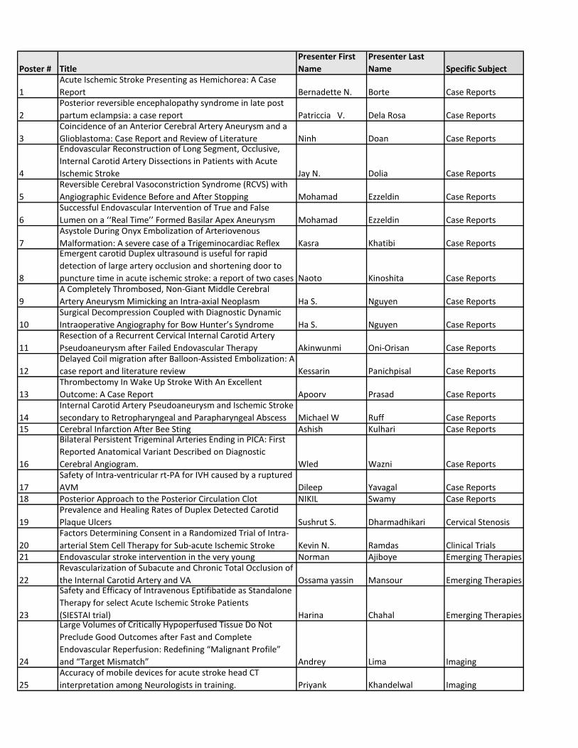
Poster # Title
Presenter First
Name
Presenter Last
Name Specific Subject
1
Acute Ischemic Stroke Presenting as Hemichorea: A Case
Report Bernadette N. Borte Case Reports
2
Posterior reversible encephalopathy syndrome in late post
partum eclampsia: a case report Patriccia V. Dela Rosa Case Reports
3
Coincidence of an Anterior Cerebral Artery Aneurysm and a
Glioblastoma: Case Report and Review of Literature Ninh Doan Case Reports
4
Endovascular Reconstruction of Long Segment, Occlusive,
Internal Carotid Artery Dissections in Patients with Acute
Ischemic Stroke Jay N. Dolia Case Reports
5
Reversible Cerebral Vasoconstriction Syndrome (RCVS) with
Angiographic Evidence Before and After Stopping Mohamad Ezzeldin Case Reports
6
Successful Endovascular Intervention of True and False
Lumen on a ‘‘Real Time’’ Formed Basilar Apex Aneurysm Mohamad Ezzeldin Case Reports
7
Asystole During Onyx Embolization of Arteriovenous
Malformation: A severe case of a Trigeminocardiac Reflex Kasra Khatibi Case Reports
8
Emergent carotid Duplex ultrasound is useful for rapid
detection of large artery occlusion and shortening door to
puncture time in acute ischemic stroke: a report of two cases Naoto Kinoshita Case Reports
9
A Completely Thrombosed, Non-Giant Middle Cerebral
Artery Aneurysm Mimicking an Intra-axial Neoplasm Ha S. Nguyen Case Reports
10
Surgical Decompression Coupled with Diagnostic Dynamic
Intraoperative Angiography for Bow Hunter’s Syndrome Ha S. Nguyen Case Reports
11
Resection of a Recurrent Cervical Internal Carotid Artery
Pseudoaneurysm after Failed Endovascular Therapy Akinwunmi Oni-Orisan Case Reports
12
Delayed Coil migration after Balloon-Assisted Embolization: A
case report and literature review Kessarin Panichpisal Case Reports
13
Thrombectomy In Wake Up Stroke With An Excellent
Outcome: A Case Report Apoorv Prasad Case Reports
14
Internal Carotid Artery Pseudoaneurysm and Ischemic Stroke
secondary to Retropharyngeal and Parapharyngeal Abscess Michael W Ruff Case Reports15 Cerebral Infarction After Bee Sting Ashish Kulhari Case Reports
16
Bilateral Persistent Trigeminal Arteries Ending in PICA: First
Reported Anatomical Variant Described on Diagnostic
Cerebral Angiogram. Wled Wazni Case Reports
17
Safety of Intra-ventricular rt-PA for IVH caused by a ruptured
AVM Dileep Yavagal Case Reports18 Posterior Approach to the Posterior Circulation Clot NIKIL Swamy Case Reports
19
Prevalence and Healing Rates of Duplex Detected Carotid
Plaque Ulcers Sushrut S. Dharmadhikari Cervical Stenosis
20
Factors Determining Consent in a Randomized Trial of Intra-
arterial Stem Cell Therapy for Sub-acute Ischemic Stroke Kevin N. Ramdas Clinical Trials21 Endovascular stroke intervention in the very young Norman Ajiboye Emerging Therapies
22
Revascularization of Subacute and Chronic Total Occlusion of
the Internal Carotid Artery and VA Ossama yassin Mansour Emerging Therapies
23
Safety and Efficacy of Intravenous Eptifibatide as Standalone
Therapy for select Acute Ischemic Stroke Patients
(SIESTAI trial) Harina Chahal Emerging Therapies
24
Large Volumes of Critically Hypoperfused Tissue Do Not
Preclude Good Outcomes after Fast and Complete
Endovascular Reperfusion: Redefining “Malignant Profile”
and “Target Mismatch” Andrey Lima Imaging
25
Accuracy of mobile devices for acute stroke head CT
interpretation among Neurologists in training. Priyank Khandelwal Imaging
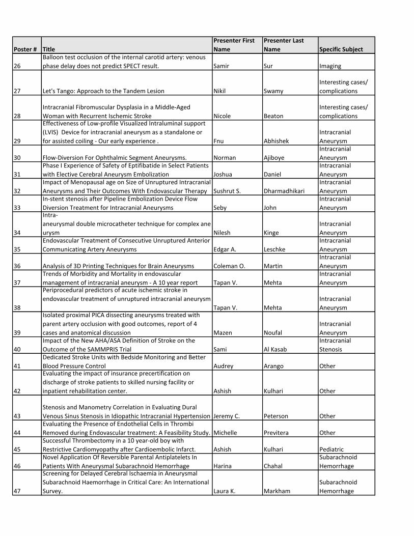
Poster # Title
Presenter First
Name
Presenter Last
Name Specific Subject
26
Balloon test occlusion of the internal carotid artery: venous
phase delay does not predict SPECT result. Samir Sur Imaging
27 Let's Tango: Approach to the Tandem Lesion Nikil Swamy
Interesting cases/
complications
28
Intracranial Fibromuscular Dysplasia in a Middle-Aged
Woman with Recurrent Ischemic Stroke Nicole Beaton
Interesting cases/
complications
29
Effectiveness of Low-profile Visualized Intraluminal support
(LVIS) Device for intracranial aneurysm as a standalone or
for assisted coiling - Our early experience . Fnu Abhishek
Intracranial
Aneurysm
30 Flow-Diversion For Ophthalmic Segment Aneurysms. Norman Ajiboye
Intracranial
Aneurysm
31
Phase I Experience of Safety of Eptifibatide in Select Patients
with Elective Cerebral Aneurysm Embolization Joshua Daniel
Intracranial
Aneurysm
32
Impact of Menopausal age on Size of Unruptured Intracranial
Aneurysms and Their Outcomes With Endovascular Therapy Sushrut S. Dharmadhikari
Intracranial
Aneurysm
33
In-stent stenosis after Pipeline Embolization Device Flow
Diversion Treatment for Intracranial Aneurysms Seby John
Intracranial
Aneurysm
34
Intra-
aneurysmal double microcatheter technique for complex ane
urysm Nilesh Kinge
Intracranial
Aneurysm
35
Endovascular Treatment of Consecutive Unruptured Anterior
Communicating Artery Aneurysms Edgar A. Leschke
Intracranial
Aneurysm
36 Analysis of 3D Printing Techniques for Brain Aneurysms Coleman O. Martin
Intracranial
Aneurysm
37
Trends of Morbidity and Mortality in endovascular
management of intracranial aneurysm - A 10 year report Tapan V. Mehta
Intracranial
Aneurysm
38
Periprocedural predictors of acute ischemic stroke in
endovascular treatment of unruptured intracranial aneurysm
Tapan V. Mehta
Intracranial
Aneurysm
39
Isolated proximal PICA dissecting aneurysms treated with
parent artery occlusion with good outcomes, report of 4
cases and anatomical discussion Mazen Noufal
Intracranial
Aneurysm
40
Impact of the New AHA/ASA Definition of Stroke on the
Outcome of the SAMMPRIS Trial Sami Al Kasab
Intracranial
Stenosis
41
Dedicated Stroke Units with Bedside Monitoring and Better
Blood Pressure Control Audrey Arango Other
42
Evaluating the impact of insurance precertification on
discharge of stroke patients to skilled nursing facility or
inpatient rehabilitation center. Ashish Kulhari Other
43
Stenosis and Manometry Correlation in Evaluating Dural
Venous Sinus Stenosis in Idiopathic Intracranial Hypertension Jeremy C. Peterson Other
44
Evaluating the Presence of Endothelial Cells in Thrombi
Removed during Endovascular treatment: A Feasibility Study. Michelle Previtera Other
45
Successful Thrombectomy in a 10 year-old boy with
Restrictive Cardiomyopathy after Cardioembolic Infarct. Ashish Kulhari Pediatric
46
Novel Application Of Reversible Parental Antiplatelets In
Patients With Aneurysmal Subarachnoid Hemorrhage Harina Chahal
Subarachnoid
Hemorrhage
47
Screening for Delayed Cerebral Ischaemia in Aneurysmal
Subarachnoid Haemorrhage in Critical Care: An International
Survey. Laura K. Markham
Subarachnoid
Hemorrhage

Poster # Title
Presenter First
Name
Presenter Last
Name Specific Subject
48
Institution of Code NeuroIntervention and Its Impact on
Reaction and Treatment Times Joshua Daniel
Systems Of Stroke
Care
49
Optimizing Financial Perfomance at a Large Comprehensive
Stroke Center Thomas Devlin
Systems Of Stroke
Care
50
Improving Door to Puncture Times (Need of Hour): Pilot
Quality Improvement Project. Priyank Khandelwal
Systems Of Stroke
Care
51
Overcoming Barriers to Reduce Door to Needle times in
Acute Ischemic Stroke Patients: Field to CT Spozhmy Panezai
Systems Of Stroke
Care
52
A Brain Attack protocol achieving better door to needle time
in stroke Dennys Reyes
Systems Of Stroke
Care
53
Acute stroke and stroke mimics in two different brain attack
protocols at Cleveland Clinic Florida. Dennys Reyes
Systems Of Stroke
Care
54 Response to Recent Trials in a Major Tertiary Stroke Center Hazem Shoirah
Systems Of Stroke
Care
55
Stroke VAN: A Large Artery Stroke Screening Tool for the ED
and Field Use Mohamed S. Teleb
Systems Of Stroke
Care
56
Preliminary experience with Precipitating Hydrophobic
Injectable Liquid (PHIL) in treating cranial AVMs and fistulas Edgar A. Samaniego
Vascular
Malformations
57
Outcome After Treatment of Spinal Dural Arteriovenous
Fistula: a Single-Institution Case Series Anita Tipirneni
Vascular
Malformations
58
Spinal Dural Ateriovenous Fistulas Mimicking Demyelinating
Disease Anita Tipirneni
Vascular
Malformations
59
Post-Acute Ischemic Stroke Thrombectomy and Obesity
Paradox Michael G. Abraham Ischemic Stroke
60
High Safety Outcomes Using Proximal Protection Device with
Carotid Stenting of Long Carotid Stenoses Kunakorn Atchaneeyasakul Ischemic Stroke
61
Elevated INR does not predict failure of mechanical
thrombectomy in acute ischemic strokes secondary to large
vessel occlusion. Garrett Bennett Ischemic Stroke
62
Eptifibatide is Safe and may Improve Outcomes in Stroke
Patients Undergoing Thrombectomy after Receiving IVtPA Jaskiran Brar Ischemic Stroke
63
Efficacy of IV tPA in Treatment of Large Vessel Ischemic
Strokes Not Amenable to Endovascular Therapy Jaskiran Brar Ischemic Stroke
64
Emergent Distal Mechanical Thrombectomy for Acute Stroke
Using the Mindframe Capture LP System: Initial Single Center
Experience Russell Cerejo Ischemic Stroke
65
Utility of thromboelastogram in optimizing antithrombotic
strategy in secondary stroke prevention Vikas Gupta Ischemic Stroke
66
Unchanged Utilization of Endovascular Treatment in Acute
Ischemic Stroke Patients in the post IMS-III era Ameer E. Hassan Ischemic Stroke
67
Sensitivity and Specificity of the Prehospital NIHSS for
Screening Intra-arterial Thrombectomy Candidates:
Experience on a Mobile Stroke Unit Amanda L. Jagolino Ischemic Stroke
68
Outcomes of Acute Stroke Interventions in Posterior
Circulation Gurmeen Kaur Ischemic Stroke
69
Outcomes of TREVO - The Latest Stentriever in Acute Stroke
Interventions - A Single Centre Experience Gurmeen Kaur Ischemic Stroke
70
Acute Stroke Thrombectomy Outcomes for Patients
Transferred Directly to the Angiosuite in an Effort to Reduce
Delay to Reperfusion Cynthia L. Kenmuir Ischemic Stroke
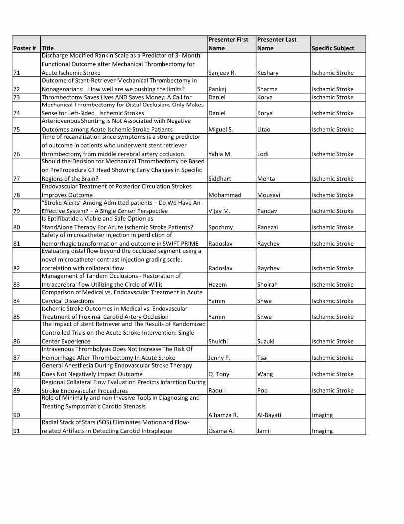
Poster # Title
Presenter First
Name
Presenter Last
Name Specific Subject
71
Discharge Modified Rankin Scale as a Predictor of 3- Month
Functional Outcome after Mechanical Thrombectomy for
Acute Ischemic Stroke Sanjeev R. Keshary Ischemic Stroke
72
Outcome of Stent-Retriever Mechanical Thrombectomy in
Nonagenarians: How well are we pushing the limits? Pankaj Sharma Ischemic Stroke73 Thrombectomy Saves Lives AND Saves Money: A Call for Daniel Korya Ischemic Stroke
74
Mechanical Thrombectomy for Distal Occlusions Only Makes
Sense for Left-Sided Ischemic Strokes Daniel Korya Ischemic Stroke
75
Arteriovenous Shunting is Not Associated with Negative
Outcomes among Acute Ischemic Stroke Patients Miguel S. Litao Ischemic Stroke
76
Time of recanalization since symptoms is a strong predictor
of outcome in patients who underwent stent retriever
thrombectomy from middle cerebral artery occlusion. Yahia M. Lodi Ischemic Stroke
77
Should the Decision for Mechanical Thrombectomy be Based
on PreProcedure CT Head Showing Early Changes in Specific
Regions of the Brain? Siddhart Mehta Ischemic Stroke
78
Endovascular Treatment of Posterior Circulation Strokes
Improves Outcome Mohammad Mousavi Ischemic Stroke
79
“Stroke Alerts” Among Admitted patients – Do We Have An
Effective System? – A Single Center Perspective VIjay M. Pandav Ischemic Stroke
80
Is Eptifibatide a Viable and Safe Option as
StandAlone Therapy For Acute Ischemic Stroke Patients? Spozhmy Panezai Ischemic Stroke
81
Safety of microcatheter injection in perdiction of
hemorrhagic transformation and outcome in SWIFT PRIME Radoslav Raychev Ischemic Stroke
82
Evaluating distal flow beyond the occluded segment using a
novel microcatheter contrast injection grading scale:
correlation with collateral flow Radoslav Raychev Ischemic Stroke
83
Management of Tandem Occlusions - Restoration of
Intracerebral flow Utilizing the Circle of Willis Hazem Shoirah Ischemic Stroke
84
Comparison of Medical vs. Endoavscular Treatment in Acute
Cervical Dissections Yamin Shwe Ischemic Stroke
85
Ischemic Stroke Outcomes in Medical vs. Endovascular
Treatment of Proximal Carotid Artery Occlusion Yamin Shwe Ischemic Stroke
86
The Impact of Stent Retriever and The Results of Randomized
Controlled Trials on the Acute Stroke Intervention: Single
Center Experience Shuichi Suzuki Ischemic Stroke
87
Intravenous Thrombolysis Does Not Increase The Risk Of
Hemorrhage After Thrombectomy In Acute Stroke Jenny P. Tsai Ischemic Stroke
88
General Anesthesia During Endovascular Stroke Therapy
Does Not Negatively Impact Outcome Q. Tony Wang Ischemic Stroke
89
Regional Collateral Flow Evaluation Predicts Infarction During
Stroke Endovascular Procedures Raoul Pop Ischemic Stroke
90
Role of Minimally and non Invasive Tools in Diagnosing and
Treating Symptomatic Carotid Stenosis
Alhamza R. Al-Bayati Imaging
91
Radial Stack of Stars (SOS) Eliminates Motion and Flow-
related Artifacts in Detecting Carotid Intraplaque Osama A. Jamil Imaging

Poster 1 Acute Ischemic Stroke Presenting as Hemichorea: A Case Report Bernadette N Borte MD1, Jose A Ramos PhD1 1USD Medical Center/ Neuroscience, Sioux Falls, SD, USA, 2USD Medical Center/ Neuroscience, Sioux Falls, SD, USA Introduction: Background: Hyperkinetic movement disorders following stroke are rare as they are reported to occur in 1 percent of strokes. Hemichorea is the most common movement disorder reported to occur after stroke. Hemichorea is arrhythmic high-amplitude unilateral movements often worsened by voluntary movement and not bothersome to the individual. It is important to recognize sudden onset hemichorea as a presenting feature of acute ischemic stroke as it can impact appropriate acute stroke care. Methods: Clinical Vignette: A 61-year-old Caucasian male presented to the emergency room with sudden onset high-amplitude arrhythmic movements of the right upper and lower extremity. The patient had sudden onset of these movements at his place of work. He was complaining of weakness of the right side, but did not find the hyperkinetic movements bothersome. He had normal blood sugar and an elevated blood pressure. Neurology was consulted for odd movements. The Patient underwent MRI brain imaging for mild right side weakness and decreased sensation with hemichorea. Imaging revealed a left head of the caudate acute ischemic stroke. Conclusions: Discussion: We conclude that it is important to recognize sudden onset hemichorea as a presenting feature of acute ischemic stroke as it can impact appropriate acute stroke care. Furthermore, early recognition can aide in localization as this typically correlates to basal ganglia infarction in association with small vessel disease. Keywords: Acute stroke, Ischemic stroke Financial Disclosures: The authors had no disclosures. Grant Support: None.
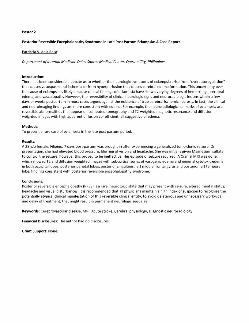
Poster 2 Posterior Reversible Encephalopathy Syndrome in Late Post Partum Eclampsia: A Case Report Patriccia V. dela Rosa1 Department of Internal Medicine Delos Santos Medical Center, Quezon City, Philippines Introduction: There has been considerable debate as to whether the neurologic symptoms of eclampsia arise from “overautoregulation” that causes vasospasm and ischemia or from hyperperfusion that causes cerebral edema formation. This uncertainty over the cause of eclampsia is likely because clinical findings of eclampsia have shown varying degrees of hemorrhage, cerebral edema, and vasculopathy.However, the reversibility of clinical neurologic signs and neuroradiologic lesions within a few days or weeks postpartum in most cases argues against the existence of true cerebral ischemic necrosis. In fact, the clinical and neuroimaging findings are more consistent with edema. For example, the neuroradiologic hallmarks of eclampsia are reversible abnormalities that appear on computed tomography and T2-weighted magnetic resonance and diffusion-weighted images with high apparent diffusion co- efficient, all suggestive of edema. Methods: To present a rare case of eclampsia in the late post partum period. Results: A 28 y/o female, Filipino, 7 days post-partum was brought in after experiencing a generalized tonic-clonic seizure. On presentation, she had elevated blood pressure, blurring of vision and headache. She was initially given Magnesium sulfate to control the seizure, however this proved to be ineffective. Her episode of seizure recurred. A Cranial MRI was done, which showed T2 and diffusion weighted images with subcortical zones of vasogenic edema and minimal cytotoxic edema in both occipital lobes, posterior parietal lobes, posterior cingulums, left middle frontal gyrus and posterior left temporal lobe, findings consistent with posterior reversible encephalopathy syndrome. Conclusions: Posterior reversible encephalopathy (PRES) is a rare, neurotoxic state that may present with seizure, altered mental status, headache and visual disturbances. It is recommended that all physicians maintain a high index of suspicion to recognize the potentially atypical clinical manifestation of this reversible clinical entity, to avoid deleterious and unnecessary work-ups and delay of treatment, that might result in permanent neurologic sequelae Keywords: Cerebrovascular disease, MRI, Acute stroke, Cerebral physiology, Diagnostic neuroradiology Financial Disclosures: The author had no disclosures. Grant Support: None.
Poster 3 Coincidence of an Anterior Cerebral Artery Aneurysm and a Glioblastoma: Case Report and Review of Literature Ninh Doan1, Ha S Nguyen1, Michael Gelsomino1, Saman Shabani1, Wade Mueller1, Osama Zaidat2 1Medical College of Wisconsin / Neurosurgery, Milwaukee, WI, USA, 2Medical College of Wisconsin / Neurology, Milwaukee, WI, USA Introduction: The association between glioblastoma and intracranial aneurysm is rare. Treatment guidelines do not exist, while operative mortality and morbidity are significantly high. To our knowledge, no prior cases have employed endovascular therapy for the treatment of these intra-tumor intracranial aneurysms followed by tumor resection. Methods: Case report and review of literature Results: A 74 year-old male, history of a left A2 aneurysm, presented after a motor vehicle accident at low speeds. Imaging was concerning for a possible traumatic brain contusion, an aneurysmal hemorrhage given history of left A2 aneurysm, or a hemorrhage from an underlying tumor given profound edema. The patient was discussed at brain tumor board, where the plan was to address the aneurysm followed by resection of the mass versus close monitoring with subsequent imaging. The high risk of re-hemorrhage, given the real possibility of an aneurysmal hemorrhage, motivated prompt treatment of the aneurysm. Patient was taken to the angiography suite; an antero-superiorly projecting azygous A2 aneurysm, measuring 4.5 mm x 5.5 mm with a neck width at 3.5 mm and a small daughter sac, was completely obliterated with primary coiling. The following day, he underwent a left craniotomy along a forehead skin crease for mass excision. Final pathology revealed glioblastoma. Patient recovered well from both procedures, with a baseline neurological exam. Patient subsequently underwent hypofractionated radiation and temodar. Conclusions: To our knowledge, no prior cases have employed endovascular therapy for the treatment of these intracranial aneurysms. We emphasize that efforts to introduce less invasive elements may improve the overall outcomes in this rare patient population. Keywords: Aneurysm, Tumors Financial Disclosures: The authors had no disclosures. Grant Support: None.
Poster 4 Endovascular Reconstruction of Long Segment, Occlusive, Internal Carotid Artery Dissections in Patients with Acute Ischemic Stroke Jay N Dolia1, Rebecca M Sugg University of Mississippi Medical Center/Department of Neurology, Jackson, MS, USA Introduction: Current evidence supports endovascular treatment for acute ischemic stroke patients (AIS) with distal carotid occlusion. However, there is limited evidence (IIb, C) to support cervical artery revascularization/stenting and even less describing revascularization of long segment occlusive internal carotid artery dissections. We present a series of five cases that support revascularization through stent reconstruction of long segment cervical ICA occlusive dissections in the acute setting. Methods: We performed a retrospective review of patients presenting with acute ischemic stroke who underwent neurointerventional revascularization at our institution. We identified five patients with stroke due to extensive ICA dissection extending from the proximal cervical segment of the ICA to the skull base and report demographic, procedural, and outcome measures. Results: We identified two males and three females (age range 43-60 years, mean age, 51.4) all with long segment occlusive dissections of the Left Internal Carotid Artery stemming from the carotid bifurcation to the carotid terminus. Average pre-procedural NIHSS was 16.8(range 8-22). Median onset of symtpoms to revascularization time was 375 minutes (range 295-703). Median door to revascularization was 173 minutes (range 121-221). Revascularization with carotid reconstruction was performed with resultant TICI of 3 patients with IIB, 1 with IIC, and 1 with III. One patient, who had received IV alteplase, had hemorrhagic transformation on 24 hour CT head (PH1 hemorrhage classification), but with no increase in NIHSS. Mean NIHSS upon discharge was 8.6 (range 3-15). Conclusions: Endovascular management of AIS secondary to reconstruction of long segment ICA dissections/occlusions is feasible and can result in improved immediate outcome. Keywords: Acute Ischemic Stroke Intervention, Carotid, Carotid stenting and angioplasty, Endovascular therapy, TICI Financial Disclosures: The authors had no disclosures. Grant Support: None.
Poster 5 Reversible Cerebral Vasoconstriction Syndrome (RCVS) with Angiographic Evidence Before and After Stopping Venlafaxine Mohamad Ezzeldin1, Narges Moghimi, Venkata Dandamudi University of Texas Medical Branch, Galveston, TX, USA Introduction: Reversible cerebral vasoconstriction syndrome (RCVS) is a well-defined but under-diagnosed clinical entity described as transient and reversible intracranial vasospasm in patients with thunderclap headache (TCH) and near normal CSF analysis. It has been reported in association with Subarachnoid hemorrhage, hypertensive encephalopathy and in exposure to various drugs including serotonin-norepinephrine reuptake inhibitors (SNRIs). Here we report a unique case of Venlafaxine associated RCVS as demonstrated on the digital subtraction imaging. Methods: Retrospective review of a clinical case including clinical course and neuroimages. Results: A 43 year old female who presented with seven days history of TCH that started after sexual intercourse. Neurological exam was unremarkable. On the day of her presentation, Head CT scan without contrast, CT Venogram and CT Angiography (CTA) of the head and neck were all reported to be normal. CSF analysis showed no evidence of xanthochromia. Digital substraction angiography (DSA) showed “beading pattern” in the left; Anterior Cerebral Artery A 2 segment, Middle Cerebral Artery M2 branches, mid segment posterior intracerebellar artery and posterior cerebral artery. She was weaned off venlafaxine slowly, and discharged home on verapamil. Repeat DSA six weeks later showed complete resolution of vasospasm. These findings along with the clinical history were highly suggestive of Venlafaxine associated RCVS. Conclusions: RCVS is well-defined but under-diagnosed clinical entity. Diagnostic challenges persist as the mostly commonly used non invasive cerebral angiography might miss vasospasm in medium to small vessels. We report for the first time a case of RCVS attributed to venlafaxine with normal initial CTA and DSA evidence of vasospasm and its complete resolution after stopping Venlafaxine. Keywords: Angiogram, Vasospasm Financial Disclosures: The authors had no disclosures. Grant Support: None.

Poster 6 Successful Endovascular Intervention of True and False Lumen on a ‘‘Real Time’’ Formed Basilar Apex Aneurysm Mohamad Ezzeldin1, Narges Moghimi, Venkata Dandamudi University of Texas Medical branch, Galveston, TX, USA Introduction: Intracranial pseudoaneurysm formation due to non traumatic rupture of a cerebral saccular aneurysm is very rare. One case of real time angiographic evidence of pseudoaneurysm formation has been reported. Endovascular coil embolization in the acute phase carries a high risk of repeated aneurysm rupture for which clipping is recommended. We report a unique case of successful endovascular intervention on a basilar apex pseudoaneurysm involving the false lumen. Methods: Retrospective review of a clinical case including clinical course and neuroimages. Results: A 57-year-old male with past medical history of hypertension presented with one day history of sudden onset severe headache. CT scan of the head demonstrated subarachnoid hemorrhage, fisher grade 4 with evidence of early hydrocephalus. Clinical grade was Hunt and Hess grade 2. CT angiogram of the head demonstrated basilar apex and anterior communicating artery (ACom) aneurysms. The pattern of the bleeding on CT head suggested basilar apex aneurysm as a ruptured aneurysm. Cerebral angiogram demonstrated superiorly and posterior pointing basilar apex aneurysm measuring 4.5 mm deep x 3.5 mm wide with a wide neck of 2.5 mm in addition to the ACom aneurysm. After placing the guide catheter in the left vertebral artery, rebleeding from the basilar tip aneurysm was noted. Biplane run demonstrated remodeling of the aneurysm dome harboring a bilobed appearance without any contrast extravasation. Successful endovascular coil embolization of the ruptured basilar apex aneurysm was performed with no evidence for residual filling. Follow up angiogram showed unchanged appearance of the recently treated aneurysm. He was discharged in stable condition. Conclusions: Real time angiographic evidence of intracranial pseudoaneurysm formation is extremely rare. We are reporting for the first time a case of real time angiographic evidence of basilar apex pseudoaneurysm formation with successful endovascular intervention involving the false lumen. Keywords: Aneurysm, Angiogram, Coiling, SAH Financial Disclosures: The authors had no disclosures. Grant Support: None.

Poster 7 Asystole During Onyx Embolization of Arteriovenous Malformation: A Severe Case of a Trigeminocardiac Reflex Kasra Khatibi1, Omar Choudhri2, Huy Do3 1Department of Neurology, Stanford University, Stanford, CA, USA, 2Department of Neurosurgery, Stanford University, Stanford, CA, USA, 3Departments of Radiology and Neurosurgery, Stanford University, Stanford, CA, USA Introduction: Trigeminal-cardiac reflex (TCR) during stimulation of sensory branches of trigeminal nerve can lead to hemodynamic instability. This phenomenon has been described during ophthalmologic, craniofacial, and skull base surgeries. TCR has been rarely described in endovascular Onyx embolization of dural arteriovenous fistula (dAVF). We report a case of TCR in the setting of endovascular embolization of an arteriovenous malformation (AVM) for the first time, and review the available evidence in Onyx embolization and patholphysiology. Methods: A 16-year-old boy with a previously treated cerebellar AVM was found to have recurrence of the AVM with arterial feeders from braches of the external carotid and bilateral superior cerebellar, posterior inferior cerebellar and vertebral arteries. For treatment the middle meningeal artery was catheterized through which DMSO and Onyx was injected with satisfactory penetration into the nidus and the feeders. Near completion of embolization, the patient became bradycardic and proceeded to asystole. He was resuscitated with chest compressions, and boluses of atropine and vasopressors with to return of spontaneous circulation. His hemodynamics normalized with normal echocardiography and patent coronaries. We used PubMed to identify reported cases of Onyx and other endovascular embolization complicated by hemodynamic instability. Results: We found 15 cases of endovascular Onyx embolization complicated by clinically significant hemodynamic changes in the treatment of dAVFs, cavernous carotid fistulas and juvenile nasopharygeal angiofibromas but not AVMs. In all cases arterial supply to the nidus involved the sensory receptive field of the trigeminal nerve. Hemodynamic changes have been reported in the setting of injection of DMSO prior to introduction of Onyx, as well as during Onyx injection and cast formation. Conclusions: TCR can lead to significant hemodynamic changes during endovascular onyx embolization of vascular malformations (both AVM and dAVF) involving the receptive field of the trigeminal nerve. Therefore, the anesthesiologist should be made aware of treatment approach prior to intervention. Keywords: Angiogram, AVM embolization, Onyx Financial Disclosures: The authors had no disclosures. Grant Support: None.

Poster 8 Emergent Carotid Duplex Ultrasound is useful for Rapid Detection of Large Artery Occlusion and Shortening Door to Puncture Time in Acute Ischemic Stroke: A Report of Two Cases Naoto Kinoshita1, Hiroshi Yamagami1, Takuro Arimizu2, Mikito Hayakawa2, Yuichi Miyazaki2, Kazunori Toyoda2, Kazuyuki Nagatsuka1 1Department of Neurology, National Cerebral and Cardiovascular Center, Suita, Osaka, Japan, 2Department of Cerebrovascular Medicine, National Cerebral and Cardiovascular Center, Suita, Osaka, Japan Introduction: Carotid Duplex ultrasound is a well-established technique for the evaluation of the site of carotid axis occlusion. In emergency setting, it can detect candidates for endovascular therapy (EVT) easier and faster than CTA/MRA. Here we report two cases in which the carotid ultrasound in emergency room contributed to shorten the door to puncture time in acute ischemic stroke patients. Results: Case 1: A 59-year-old man was presented to our hospital with impaired consciousness and left hemiplegia (NIHSS score 21), 5 hours after the last known well time. Carotid Duplex ultrasound on arrival showed the side-to-side ratio of the end-diastolic flow velocity in the CCAs greater than 1.4. We diagnosed distal right ICA occlusion and activated the endvascular team. After the confirmation of ischemic lesion volume (DWI-ASPECTS 8) and right ICA occlusion by MRI/MRA, EVT was initiated with door to groin-puncture time of 89 minutes. Successful reperfusion (TICI 2b) was achieved using Penumbra 5MAX ACE, and he was discharged on day 18 with modified Rankin scale (mRS) score of 1. Case 2: A 44-year-old man with history of ventricular tachycardia was transferred to our hospital with left hemiparesis (NIHSS score 15), 10 hours after the last known well time. The diffusion weighted MRI before the transfer revealed no ischemic lesions, but MRA had not been examined. Emergent carotid Duplex ultrasound on arrival showed loss of end-diastolic flow in the right ICA, suspected distal ICA occlusion. Without reexamination of CT/MRI, EVT was initiated with door to groin-puncture time of 39 minutes. Complete reperfusion (TICI 3) was achieved using Penumbra 5MAX ACE, and his neurological deficits were fully improved on day 2. Conclusions: Emergency carotid Duplex ultrasound is useful for rapid detection of large artery occlusion and it can contribute to shorten the door to puncture time. Keywords: Acute stroke, Endovascular therapy, Ultrasound Financial Disclosures: The authors had no disclosures. Grant Support: None.

Poster 9 A Completely Thrombosed, Non-Giant Middle Cerebral Artery Aneurysm Mimicking an Intra-axial Neoplasm Ha S Nguyen1, Ninh B Doan1, Gerald Eckardt1, Michael Gelsomino1, Saman Shabani1, W. Douglas Brown2, Wade Mueller1, Glen Pollock1 1Medical College of Wisconsin / Neurosurgery, Milwaukee, WI, USA, 2Medical College of Wisconsin / Radiology, Milwaukee, WI, USA Introduction: Few reports exist regarding thrombosed aneurysms where initial work up was concerning for neoplasm. To date, no published reports exist regarding a thrombosed middle cerebral artery aneurysm, where primary workup and treatment plan was directed toward a preliminary diagnosis of intra-axial neoplasm. Methods: Case Report and Review of Literature Results: We report a 43-year-old female who presented with a generalized tonic-clonic seizure attributed to a lesion along the right superior temporal gyrus; the lesion enhanced on initial MR as well as on follow up MR. Subsequent vascular studies and metastatic work up were negative. A craniotomy with image guidance was performed and an intraoperative diagnosis of a thrombosed aneurysm along a branch off the middle cerebral artery was made. The aneurysm was clipped. The patient had an uneventful post-operative course. Conclusions: Completely thrombosed, non-giant aneurysms may mimic an intra-axial neoplasm. Typical imaging features for thrombosed aneurysms may be missed, especially if the aneurysms are small, where resolution of intraluminal contents is more difficult to appreciate. Although imaging may be consistent for a neoplasm, there should be suspicion for a potential underlying aneurysm, which would alter surgical planning. Keywords: Aneurysm, Angiogram, Clipping, Diagnostic neuroradiology Financial Disclosures: The authors had no disclosures. Grant Support: None.

Poster 10 Surgical Decompression Coupled with Diagnostic Dynamic Intraoperative Angiography for Bow Hunter’s Syndrome Ha S Nguyen1, Ninh B Doan, Gerald Eckardt, Glen Pollock Medical College of Wisconsin / Neurosurgery, Milwaukee, WI, USA Introduction: Bow hunter’s syndrome, also known as rotational vertebrobasilar insufficiency, arises from mechanical compression of the vertebral artery during neck rotation. Surgical options have been the mainstay treatment of choice. Postoperative imaging is typically used to assess adequate decompression. On the other hand, intraoperative assessment of decompression has been rarely reported. Methods: Case Report and Literature Review Results: A 52-year-old male began to see “black spots”, and experienced pre-syncope whenever he rotated his head towards the right. The patient ultimately underwent a dynamic diagnostic cerebral angiogram, which revealed a dominant right vertebral artery and complete proximal occlusion of the right vertebral artery with the head rotated towards the right. Subsequently, the patient underwent an anterior transcervical approach to the right C6/7 transverse process. Bone removal occurred along the anterior wall of the C6 foramen transversarium, followed by the upper portion of the anterior C6 body medially, and the transverse process of C6 laterally. An oblique osseofibrous band was noted to extend across the vertebral artery; it was dissected and severed. An intraoperative cerebral angiogram confirmed no existing compression of the vertebral artery with the head rotated towards the right. The patient recovered from surgery without issues; he denied recurrence of preoperative symptoms at follow up. Conclusions: The authors report the third instance where intraoperative dynamic angiography was employed with good outcomes. Although intraoperative cerebral angiography is an invasive procedure, which prompts additional risks, the authors believe the modality affords better, real-time visualization of the vertebral artery, allowing for assessment of the adequacy of the decompression. This advantage may reduce the probability for a second procedure, which has its own set of risks, and may counteract the risks involved with intraoperative dynamic angiography. Keywords: Angiogram, Diagnostic neuroradiology, TIA, Vertebral Financial Disclosures: The authors had no disclosures. Grant Support: None.
Poster 11 Resection of a Recurrent Cervical Internal Carotid Artery Pseudoaneurysm after Failed Endovascular Therapy Akinwunmi Oni-Orisan1, Ha S Nguyen, Joseph Cochran, Glen Pollock Medical College of Wisconsin / Neurosurgery, Milwaukee, WI, USA Introduction: Recurrence of a cervical internal carotid artery (ICA) pseudoaneurysm initially treated by endovascular means is rare. We report an instance where a patient returned with a recurrent, enlarging cervical ICA pseudoaneursym, 15 years after initial complete, endovascular occlusion of the ICA. Methods: Case Report and Review of Literature Results: Patient is a 64-year-old male with a history of a right cervical ICA pseudoaneurysm diagnosed 15 years ago after a car accident. At the time, he received endovascular occlusion of his right ICA. Recent serial imaging demonstrated progressive enlargement of his pseudoaneurysm, up to 6 cm x 5 cm x 5.5 cm, without evidence of internal flow or extravasation. Due to dysphagia and hoarseness, resection of the pseudoaneurysm was recommended. Dissection occurred down to the lesion, where its borders were skeletonized. Its stump at the proximal ICA was mobilized and clamped; the lesion was incised and the existing thrombus, as well as the coil mass, was removed. The distal ICA appeared completely scarred with no retrograde filling. There were branches from the external carotid artery that appeared to supply the pseudoaneurysm. The scarred remnant of the distal ICA was sutured and the stump at the proximal ICA was ligated. Once hemostasis was obtained, closure occurred via anatomical layers. Postoperatively, the patient woke up well; at discharge, he exhibited no respiratory distress or dysphagia. At 5 months follow-up, a CT angiography of the neck revealed no evidence for a residual pseudoaneurysm. He continues on lifelong aspirin. Conclusions: Recurrence of a cervical ICA pseudoaneursym is rare. We caution that such a clinical scenario is possible, even 15 years after endovascular occlusion of the ICA. Branches from the external carotid artery may feed the pseudoaneursym and cause recurrence. This mechanism has not been reported. Perhaps longer clinical follow-up is necessary, especially if endovascular therapy is the initial treatment option. Keywords: Aneurysm, Endovascular therapy Financial Disclosures: The authors had no disclosures. Grant Support: None.

Poster 12 Delayed Coil migration after Balloon-Assisted Embolization: A Case Report and Literature Review Kessarin Panichpisal1, Nazli Janjua1, Wassana Plengsombat2 1Asia Pacific Comprehensive Stroke Institute, Pomona, CA, USA, 2Bumrungrad International Hospital, Bangkok, Thailand Introduction: Delayed coil migration is very rare or might be underreported. Methods: We report our case of delayed coil migration and performed a literature review using search terms delayed coil migration in Pubmed. From these cases, we collected clinical and radiographic data and analyzed them using descriptive statistics. Results: A 56-yr-old woman with subarachnoid hemorrhage (SAH), Hunt and Hess grade 3, from a small anterior communicating artery aneurysm underwent coil embolization with balloon remodeling with small residual neck. A follow up CT head at 1 week (performed for hydrocephalus evaluation) was stable whereas a 2 week follow up CT revealed protrusion of a tail of the coil, confirmed on subsequent angiography. After a period of 1 month following the SAH event and shunt placement, she underwent stent assisted parent vessel reconstruction. She had no neurological deficits. We identified total 14 patients including our case with delayed coil migration after coil embolization. Mean age was 56.4 +/-12.93 years old. Nine patients (64%) had SAH. Average aneurysm size was 5.55 +/- 1.96 mm. The aneurysm locations were middle cerebral artery bifurcation (3), anterior communicating artery (3), posterior communicating artery(3), anterior choroidal artery (2), superior hypophyseal artery (2), and basilar artery (1). Delayed coil migration occurred in 3 cases after balloon-assisted embolization and 2 after stent assisted embolization. Nine patients were symptomatic from coil migration. Six patients required surgical management. Nine patients had good recovery. Conclusions: Delayed coil migration is an infrequent phenomenon after aneurysm embolization but may be more common with smaller aneurysms. Adjunctive techniques such as balloon remodeling or parent vessel reconstruction with stents do not eliminate its occurrence. Keywords: Aneurysm Embolization, Angiogram, Balloon assisted, Stent assisted, SAH Financial Disclosures: The authors had no disclosures. Grant Support: None.

Poster 13 Thrombectomy In Wake Up Stroke With An Excellent Outcome: A Case Report Apoorv Prasad 1, Anuradha Duleep , Sameer Sharma Upstate Medical University, Syracuse, NY, USA Results: A 50 year old woman was brought to our emergency department by air ambulance after she woke up that morning with right facial droop and right sided weakness. She was an active smoker, with a previous left middle cerebral artery stroke but no residual deficits. Patient was last seen normal at 9.30 pm previous night and the stroke code was activated at 7.24 am. The patient was emergently seen by neurology service and her NIHSS score was 23. On examination she had left gaze preference, right sided facial droop, expressive aphasia and severe dysarthria with no anti-gravity strength in right arm and leg. Since the patient was out of window for intravenous thrombolysis, she received CT angiogram of head and neck. An abrupt cutoff of the left MCA with a hyperdense MCA sign on the non-contrast CT head was noted. Emergent CT perfusion scan was performed and revealed a large ischemic penumbra spanning majority of left MCA territory. She was taken for therapeutic cerebral arteriogram immediately in spite of being 13 hours from last known normal. A complete occlusive thrombus in the mid M1 segment of the left middle cerebral artery was found. Mechanical thrombectomy was performed after 2 passes with TREVO clot retrieval device under manual aspiration and anterograde flow arrest, resulting in complete recanalization of the left middle cerebral artery, with TICI 3 flow in the left MCA territory. The patient's NIHSS score reduced to 9 following the procedure. She was admitted to neurological intensive care unit after the procedure for closer monitoring and systolic blood pressure target of 120-160. Clinically, the patient continued to improve and her NIHSS score was zero four days after the procedure when she was discharged home. Conclusions: The excellent clinical outcome in our case highlights the fact that patient’s with wake-up stroke should not to be excluded from acute intervention on the basis of unknown time of onset of symptoms. Keywords: Acute stroke, Recanalization, Endovascular therapy, Ischemic stroke, CT perfusion Financial Disclosures: The authors had no disclosures. Grant Support: None.
Poster 14 Internal Carotid Artery Pseudoaneurysm and Ischemic Stroke Secondary to Retropharyngeal and Parapharyngeal Abscess Michael W Ruff1, Deena M Nasr1, James P Klaas1, Deborah L Renaud 2 1Department of Neurology, Rochester, MN, USA, 2Department of Neurology Division of Child and Adolescent Neurology, Rochester, MN, USA Introduction: The clinical course of a 2 year old boy with retropharyngeal abscess complicated by internal carotid artery pseudoaneurysm, demonstrated by CTA, is described. Methods: A two year old boy developed reduced range of neck motion, pain, and swelling at the base of his left jaw and anterior neck. Computed tomography with contrast demonstrated a large retropharyngeal abscess and left internal carotid artery pseudoaneurysm. A conventional angiogram confirmed the presence of a large pseudoaneurysm measuring 1.3 cm x 3.6 with no antegrade flow distal to the pseudoaneurysm and substantial collateral flow across the circle of Willis, with filling of the left anterior and middle cerebral arteries via the anterior and posterior communicating arteries. Results: Endovascular occlusion resulted in non-filling of the left internal carotid artery, pseudoaneurysm, and left internal jugular vein at the base of the skull. Following the procedure, the patient was noted to have mild right hemiparesis. Magnetic resonance imaging demonstrated restricted diffusion in the left frontal lobe consistent with an acute/ subacute ischemic infarct. There was also a small area of T2/FLAIR hyper-intensity in the white matter of the left parietal lobe, which was also consistent with ischemic stroke. Conclusions: Mycotic carotid artery pseudoaneurysm is a complication of deep neck infections. In children this may occur secondary to pharyngeal and retropharyngeal infections. Missed or delayed diagnosis can result in stroke, carotid rupture, and death. Treatment involves both antibiotic therapy for 4-6 weeks and either surgical ligation, resection, and grafting, or endovascular occlusion of the affected vessel. Although there is very limited data regarding endovascular occlusion, it is currently the favored method as it is felt to have less operative risk. The main risk of these procedures is ischemic stroke. Keywords: Pediatric intervention, Aneurysm Embolization Financial Disclosures: The authors had no disclosures. Grant Support: None.
Poster 15 Cerebral Infarction After Bee Sting Ashish Kulhari1, Ashley Rogers, Wei Xiong, Michael Degeorgia Case Western Reserve University/Neurology, Cleveland, OH, USA Introduction: To report a case of right MCA-territory infarction after a bee sting in a 44 year-old healthy man. Methods: Extensive literature review was done to describe the clinical presentation, pathophysiology and management of cerebral infarction after bee sting. Results: CASE: A 44 year-old man with hypertension was working outside when he was stung by a bee. He acutely developed a diffuse rash and urticaria but had no cardiorespiratory symptoms. About an hour later, he suddenly developed left hemiplegia, left facial droop and severe dysarthria and was taken to the ED by ambulance. There, he had left hemiparesis, mild left facial weakness and mild dysarthria (NIHSS score 4). Initial vital signs and labs were normal. ECG showed normal sinus rhythm. Head CT was normal, however, brain MRI showed multiple areas of diffusion restriction in the right MCA territory with no signal on MRA in the distal right ICA and MCA. He was treated with intravenous t-PA. Because of his low NIHSS score, endovascular intervention as not done. CTA head performed 24 hours later showed multiple regions of mild-moderate vasoconstriction in bilateral large intracranial vessels. Evaluation for causes of stroke, including echocardiogram, telemetry, and lipid panel, was not revealing. Aspirin and atorvastatin were started for secondary stroke prevention. At discharge, a mild left facial droop remained but he had normal strength in the left arm and leg. DISCUSSION: Cerebral infarction after a bee sting is rare but can occur from 2h to 24h after the sting. The mechanism is believed to stem from both allergic and toxic reactions to the bee venom including the release of phospholipase A2, resulting in an increase in thromboxane and leukotrienes, vasoconstriction, platelet activation, and thrombus formation. Conclusions: Cerebral infarction after a bee sting is rare but may occur secondary to both allergic and toxic reactions to bee venom. Keywords: Ischemic stroke, Pathophysiology, Lytics, Vasospasm Financial Disclosures: The authors had no disclosures. Grant Support: None.

Poster 16 Bilateral Persistent Trigeminal Arteries Ending in PICA: First Reported Anatomical Variant Described on Diagnostic Cerebral Angiogram. Wled Wazni1, Sam Zaidat1, Alicia Castonguay1, Ali Abdallah2, Mazen Noufal1, Ahsan Sattar1 1Medical College of Wisconsin/Neurology, Milwaukee, WI, USA, 2Detroit Mercy, Detroit, MI, USA Introduction: A persistent trigeminal artery (PTA) is a rare embryonic anastomosis between the anterior and posterior circulation. This anastomosis usually occurs between the internal carotid and basilar artery. PTA variants (PTAV) have also been reported and described as an anastomosis between the internal carotid artery (ICA) and the branches of the basilar artery. We report a case of a 43-year-old female presenting with a intra parenchymal hemorrhage secondary to a cavernous malformation. She was found to have bilateral PTAVs ending in the posterior inferior cerebellar Artery (PICA) on a diagnostic cerebral angiogram (DSA). The prevalence of bilateral PTAVs is about 0.0012%, to our knowledge, this is the first reported case of bilateral PTAV ending in PICA described on DSA. Knowledge of this potential anatomical variants is of great importance in avoiding neurologic complications during vascular and endovascular neurosurgical procedures. We will discuss the first reported case of bilateral PTAVs ending in the posterior inferior cerebellar Artery (PICA) with discuss of its embryological origin and associated vascular malformations. Keywords: Angiogram, Neurointerventional education, Diagnostic neuroradiology, Cerebral arteriovenous malformations, Cerebral blood flow Financial Disclosures: The authors had no disclosures. Grant Support: None.
Poster 17 Safety of Intra-Ventricular rt-PA for IVH caused by a Ruptured AVM Dileep R Yavagal2, Sushrut S Dharmadhikari1, Amer M Malik2 1Wayne State University, Detroit, MI, USA, 2University of Miami, Miami, FL, USA Introduction: Intra-ventricular recombinant tissue plasminogen activator (rt-PA) has improved outcomes of intra-ventricular hemorrhage (IVH), a disease otherwise associated with a poor prognosis. However, patients with suspected or untreated arteriovenous malformations (AVMs) have been excluded from clinical trials. We present a patient with IVH secondary to a ruptured AVM safely treated with intra-ventricular rt-PA. Methods: A 48-year-old Hispanic male with history of dermatomyositis presented to the emergency room with sudden left sided weakness. En-route to CT, he became lethargic. CT Brain revealed extensive IVH with acute hydrocephalus, which was treated with placement of an external ventricular drain (EVD) with improvement. CT-angiogram performed did not reveal presence of a vascular malformation. A catheter cerebral angiogram was planned due to ongoing suspicion of a vascular malformation. Prior to this, the patient became acutely altered and lethargic. CT imaging revealed worsening hydrocephalus. EVD was noted to be draining and was dropped to the ground. Overnight, EVD drained well with no improvement in neurological exam. Repeat CT revealed improved hydrocephalus, but with left lateral ventricle dilatation likely secondary to obstruction of Foramen of Monro. Risks and benefits of intra-ventricular rt-PA were discussed with family and a decision was made to treat. Results: 3 doses of intra-ventricular rt-PA (1mg each) were administered with resolution of midline blood and lateral ventricular dilatation with clinical improvement. Catheter cerebral angiogram revealed a right posterior cerebral artery branch draining into an early draining vein further draining into the inferior sagittal sinus. This was thought to be a ruptured AVM without a clear nidus. Repeat catheter angiogram with possible embolization was planned after discharge. In-spite of additional in-hospital complications, the patient gradually improved, had his EVD removed and was ultimately discharge home. Conclusions: Intra-ventricular rt-PA can safely be administered in certain patients with IVH with ruptured AVM’s. Keywords: Cerebral arteriovenous malformations, Intracerebral Hemorrhage, TPA, Fibrinolytics, Ruptured Financial Disclosures: The authors had no disclosures. Grant Support: None.

Poster 18 Posterior Approach to the Posterior Circulation Clot NIKIL SWAMY1, SONAL MEHTA PALMETTO HEALTH RICHLAND, COLUMBIA, SC, USA Introduction: Symptomatic acute basilar artery thrombosis with associated bilateral vertebral occlusion is a unique entity, occurring in ~3% of posterior circulation ischemic events. Common etiologies include atherosclerosis, giant cell arteritis, trauma, spontaneous dissection. It is associated with a poor prognosis and high rate of recurrent ischemia. We report one such interesting case and a review of the literature. Methods: A 63 year-old-male presented with right arm/leg weakness, visual-field cut, dysarthria. He had a seizure earlier with return to baseline. A crisis MRI showed bilateral PCA territory infarcts with petechial hemorrhage and was excluded from tissue plasminogen candidacy. CT angiogram showed a basilar thrombus with thread-like right and proximally occluded left vertebral artery. Intervention was pursued at 3 hours. NIHSS was 8, with post-intervention improvement to 4. Results: Four-vessel angiogram revealed a basilar-tip clot with minimal left and absent right P1 filling, and occluded left vertebral origin with ascending cervical collaterals. After additional views of the right P2, a Marksman microcatheter was used to make the first pass with the Solitare device, and following intercalation for 5 minutes, the Penumbra aspiration pump was engaged for suction with simultaneous clot retraction. Repeat run demonstrated patency of the basilar-tip, right P1 and proximal P2, with filling defects in the left P1 and P2 segments. A second pass with the same arrangement revealed a patent left PCA and basilar-tip with a persistent small right P2 filling defect. Injection of the left P1 demonstrated a narrow and irregular left vertebral artery suggestive of V1 dissection. A TICI grade of 2 was observed. Conclusions: The approach taken here is in contrast to other reports of re-canalization through the anterior circulation. Given the grave prognosis and lack of randomized control trials, case reports such as this are needed to add to the empiric knowledge base for approaches to vascular intervention. Keywords: Acute Ischemic Stroke Intervention, Basilar, Vertebral, Endovascular, Invention Financial Disclosures: The authors had no disclosures. Grant Support: None.
Poster 19 Prevalence and Healing Rates of Duplex Detected Carotid Plaque Ulcers Sushrut S Dharmadhikari1, Bennett A2, Campo Nelly2, Romano Jose2, Koch Sebastian2 1Wayne State University, Detroit, MI, USA, 2University of Miami, Miami, FL, USA Introduction: Carotid plaque ulcers confer an increased risk for stroke and TIA in both symptomatic and asymptomatic carotid artery stenosis. Little is known about healing rates of ulcers or development of new ulcers. Carotid Duplex studies are easily obtained and repeated to non-invasively monitor progression of carotid stenosis. We aimed to determine prevalence and healing rates of ultrasound detected carotid plaque ulcers in an outpatient neurosonology laboratory. Methods: We retrospectively reviewed our database of 5837 carotid Duplex studies performed in an outpatient ultrasound laboratory affiliated with the neurological department of an academic center. A total of 3215 patients underwent a first Duplex study, and 2522 studies were obtained in follow-up. A carotid ulcer was defined as a deep indentation in a carotid plaque which had a minimum width of 2mm and a well-defined back wall, as determined by B-mode, color, and B-flow imaging. Results: A total of 127(4%) patients had a carotid ulcer. Demographics were available on 111 patients. Ulceration was associated with increased age (Mean - Ulcer 69.1 ± 8.4 yrs vs No ulcer 64.3 ± 13.5; p< 0.001), hypertension (90.1% vs 50.6%; p< 0.001), dyslipidemia (79.3% vs 64.7%; p< 0.001), coronary artery disease (CAD) (32.4% vs 16.3%; p< 0.001) and current smoking (19.8% vs 7.9%; p< 0.001). Ulcer was found on initial study in 82 (3%) patients. Ipsilateral degree of stenosis for patients with a carotid ulcer exceeded 50% in 36% (46/127). 65 ulcers were followed serially. Median number of scans was 6 over a mean follow-up of 42±30 months. 31/65 (48%) healed. No factors including statin use could be associated with ulcer healing. Conclusions: We report a low prevalence of duplex detected carotid plaque ulcers. Ulceration was associated with an increased age, hypertension, dyslipidemia, CAD and current smoking. Carotid ulcers healed in approximately 50% patients with follow-up, and, in a minority of patients, a new ulcer developed during serial studies. Factors associated with ulcer healing remain poorly understood. Keywords: Atherosclerosis, Carotid, Extracranial stenosis, Pathophysiology, Ultrasound Financial Disclosures: The authors had no disclosures. Grant Support: None.

Poster 20 Factors Determining Consent in a Randomized Trial of Intra-Arterial Stem Cell Therapy for Sub-Acute Ischemic Stroke Kevin N. Ramdas1, Ryan N. Pafford1, Diogo C. Haussen3, Delmas McBee2, J. Neal Rutledge2, David Y. Huang4, Sean I. Savitz5, James Hinson6, Dileep R. Yavagal1 1Department of Neurology- University of Miami Leonard M. Miller School of Medicine / Jackson Memorial Hospital, Miami, FL, USA, 2Department of Neurology - University Medical Center at Brackenridge, Austin, TX, USA, 3Departments of Neurology, Neurosurgery and Radiology - Emory University School of Medicine / Grady Memorial Hospital Marcus Stroke and Neuroscience Center, Atlanta, GA, USA, 4Department of Neurology, The University of North Carolina School of Medicine Chapel Hill, NC, USA, 5Department of Neurology, The University of Texas Medical School Houston, TX, USA, 6Cytomedix, Inc Durham, NC, USA Introduction: Obtaining individual consent for randomized clinical trials can be challenging. Stroke poses an additional difficulty because patients are often functionally disabled at the time of consent. We investigated the rate of consent among patients and legally authorized representatives (LAR), as well as factors determining consent in the first US intra-arterial autologous stem cell trial - RECOVER Stroke. Methods: In this trial, eligible patients were identified systematically at one week from the stroke ictus in participating centers. We performed a retrospective analysis of data from two out of eight actively recruiting centers in the trial. Recorded data included age, gender, race, National Institutes of Health stroke scale score (NIHSSS), lesion location, prior recanalization therapy and mode of consent. Results: 44 stroke cases were approached for enrollment. The mean age was 65±17 yr, and 59% were male. 15 (34%) patients self-consented and 33 (66%) were approached via LAR, leading to 22 (50%) being enrolled in the trial. A univariate analysis for factors associated with enrollment revealed that consent refusal was more common in cases involving older (68 ± 12 vs. 61 ± 12; p = 0.04) and male (81 vs. 40%; p< 0.01) patients. Ethnicity was also found to be associated with the probability of enrollment. The mode of consenting had no impact on consenting/refusal rates (p=0.55). The multivariate logistic regression analysis revealed males as the only variable independently associated with better rates of overall consent (OR 7.2; 95% CI 1.6-31.5; p=0.007). Sensitivity analyses revealed that the only factors independently associated with overall LAR-consenting after multivariate regression was lower NIHSSS (OR 0.9; 95% CI 0.8-1.08; p=0.6) not significant and IV-tPA (OR 3.3; 95% CI 0.52-21.92; p=0.2). Conclusions: There was a relatively high rate of consent among eligible patients in the first US intra-arterial trial of stem cell therapy for stroke. Approaching LAR for consent was found not to influence consenting rates. Keywords: Stem cell therapy, Clinical trial, Intra-arterial therapy, Interventional neuroradiology, Stroke Financial Disclosures: The authors had no disclosures. Grant Support: None.
Poster 21 Endovascular Stroke Intervention in the Very Young Norman Ajiboye1 Thomas Jefferson University Hospital, Philadelphia, PA, USA Introduction: This study aims to evaluate the use of endovascular therapy to treat very young (≤35 years)patients with acute ischemic stroke from large vessel occlusion. Methods: We identified from a prospectively maintained database young patients (≤35 years) undergo-ing endovascular intervention for AIS at two cerebrovascular referral centers. The study only includedpatients with a confirmed large vessel occlusion. Modified Rankin scale (mRS) scores were determinedat 90 days during a follow-up visit. Results: A total of 15 patients met the inclusion criteria. Mean age was 27.93 years ± 6.75 years (range:9–35 years). On admission, the mean NIHSS score was 14.07 ± 9.16. Mechanical thrombectomy was per-formed using the Solitaire FR device in 4 of 15 (26.67%) patients and the Merci/Penumbra systems in 11(73.33%) patients. Successful recanalization (TICI 2–3) was achieved in all but one patient (14/15; 93.33%).Only one patient (6.67%) had a hemorrhagic conversion following intervention; he later expired. The rateof 90-day favorable outcome (mRS 0–2) was 86.67% (13/15). Conclusions: Endovascular treatment in the very young population may be carried out with limited com-plications and attain remarkably high rate of recanalization and favorable outcome. This study supportsthe role of aggressive management strategies for very young patients with large vessel occlusion. Keywords: Aneurysm, Endovascular therapy Financial Disclosures: The authors had no disclosures. Grant Support: None.
Poster 22 Revascularization of Subacute and Chronic Total Occlusion of the Internal Carotid Artery and VA Ossama yassin Mansour1 Alexandria University Hospital, Alexandria, Egypt Introduction: The natural course of symptomatic carotid or vertebral artery occlusion with hemo-dynamic impairment is poor. Surgical revascularization may improve the outcome; however, its efficacy has not been established yet. The goal of this study was to characterize the technical and clinical outcomes protection. following endovascular recanalization of the ICA and Vaunder cerebral circulatory Methods: Endovascular recanalization was attempted in 14 patients with symptomatic 11 ICA occlusions and 3 VA occlusions . The duration of the occlusion ranged from 10 days to 6 months (mean, 2.5 months),and the mean length of the occlusion was 95 mm. Cerebral hemodynamics ipsilateral to the side of the occlusion were severely impaired in all patients. The endovascular procedure was performed under total cerebral circulatory protection, beginning with proximal protection with a subsequent switch to distal protection (in 11 cases ) after successful guide wire passage. Results: The occlusion was recanalized successfully in 12 out 14 of patients (85.7%), resulting in improve-ment of ipsilateral cerebral hemodynamics without symptomatic stroke. Small asymptomatic ischemic lesions were detected in 3 of 14 patients (21%) on DWI, and 1 patient developed a mild groin hematoma. Ischemic episodes did not recur during the mean follow-up period of 19 months. However, 2 patients experienced asymptomatic reocclusion, which was retreated successfully without complications due to failure maintenance of antiplatelet and anticoagulant therapy. Conclusions: Endovascular revascularization of an ICA occlusion is feasible and well-tolerated inpatients with subacute or chronic total occlusion of the ICA. Keywords: Carotid, Basilar, Collateral, Endovascular therapy, Ischemic stroke Financial Disclosures: The authors had no disclosures. Grant Support: None.

Poster 23 Safety and Efficacy of Intravenous Eptifibatide as Standalone Therapy for select Acute Ischemic Stroke Patients (SIESTAI trial) Harina Chahal1, Siddhart Mehta, Mohammad Moussavi, Jaskiran Brar, Daniel Korya, Josh Daniels, Mena Samaan, Rushil Kalola, Azka Shaikh, Mohammed Hussain, Alex Yang Ge, Jawad Kirmani JFK Medical Center, Edison, NJ, USA Introduction: There is existence of data on the successful application of Eptifibatide in coronary interventions. Our objective was to report the results of an open labeled retrospective registry to evaluate the safety and efficacy of administering IV Eptifibatide as a standalone therapy for anterior circulation stroke in patients ineligible for IVtPA or neurointervention. Methods: All patients with acute ischemic events between 20102015 that presented to our university affiliated comprehensive stroke center were reviewed. Patients that presented with anterior circulation ischemic strokes who received Eptifibatide as standalone therapy were identified. Eptifibatide was administered intravenously as a 135μg/kg bolus, then a 0.5μg/kg/min infusion. Charts were reviewed for all patients to assess for complications, initial NIHSS (INIHSS) 24 hour NIHSS, discharge mRS. Mean change in NIHSS was abstracted using GraphPad Quick Calcs Website. Results: Of a total patient population of 2,831, a total of 45 patients received IV Eptifibatide for anterior circulation ischemic strokes for a mean duration of 32.5 hours (range 2-171). The mean age was 71. The mean INIHSS was 7.14 and discharge NIHSS was 3.8. The difference between INIHSS and discharge NIHSS was 3.3 (p< 0.001). Good outcome discharge mRS (0-2) was seen in 77% of patients (n=34). No complications were observed except for a patient who exhibited knee hemarthroses. Conclusions: Application of IV Eptifibatide in achieving recanalization and preventing extension may be a safe and possibly efficacious standalone therapy in acute anterior circulation ischemic stroke patients ineligible for other neurointerventions. Larger randomized trials are required to corroborate our findings. Keywords: Ischemic stroke, Medical management, Thrombolytics Financial Disclosures: The authors had no disclosures. Grant Support: None.
Poster 24 Large Volumes of Critically Hypoperfused Tissue Do Not Preclude Good Outcomes after Fast and Complete Endovascular Reperfusion: Redefining “Malignant Profile” and “Target Mismatch” Andrey Lima1, Diogo Haussen, Seena Dehkharghani, Rebello Leticia, Belagaje Samir, Anderson Aaron, Nogueira Raul Emory University, Atlanta, GA, USA Introduction: Acute ischemic stroke (AIS) patients with large volumes of severe hypoperfusion (Tmax>10s>100cc) have a higher likelihood of intracranial hemorrhage (ICH) and/or poor outcomes following reperfusion. We aim to evaluate the impact of the presence and extent of Tmax>10s CTP-lesions in patients undergoing successful treatment with the contemporary endovascular technology. Methods: Retrospective database review of endovascular AIS treatment between September2010–March2015 for patients with anterior circulation occlusions with baseline RAPID CTP and full reperfusion (mTICI3). The primary outcome was the impact of the Tmax>10s lesion spectrum on infarct growth. Secondary safety and efficacy outcomes included parenchymal hematomas and good clinical outcomes (90-day modified Rankin Scale 0-2). Results: Out of 684 treated patients, 113 patients fit the inclusion criteria. Patients with and without Tmax>10s>100cc had similar ages, times from CTP to reperfusion, anesthesia modalities, and rates of IV rt-PA and stent-retriever usage. On univariate analysis, patients with Tmax>10s>100cc(n=37) had significantly higher baseline NIHSS(20.7±3.8vs.17.0±5.9,p < 0.01), more ICA-T occlusions(29%vs.9%,p=0.02), and larger baseline(38.6±29.6vs.11.7±15.8cc,p< 0.01) and final(60.7±60.0vs.29.4±33.9cc,p< 0.01) infarct volumes as compared to patients without Tmax>10s>100cc(n=76). However, there were no significant differences in infarct growth (22.1±51.6vs.17.8±32.4cc,p=0.78), severe intracranial hemorrhage (PH2:2%vs.4%,p=0.73), good outcomes (90-day mRS 0-2:56%vs.59%,p=0.83), or 90-day mortality (16%vs.7%,p=0.28). On multivariate analysis only baseline NIHSS (OR: 1.19, 95% CI [1.06-1.34], p< 0.01) and baseline infarct core volume (OR: 1.05, 95% CI [1.02-1.08], p< 0.01) were independently associated with Tmax>10s>100cc. Conclusions: Large Tmax>10s lesion volumes are not associated with any clinically relevant ICH and do not preclude good outcomes in patients undergoing endovascular reperfusion with the contemporary technology. Keywords: Acute Ischemic Stroke Intervention, Angiographic CT perfusion, Endovascular therapy, Imaging Financial Disclosures: The authors had no disclosures. Grant Support: None.

Poster 25 Accuracy of Mobile Devices for Acute Stroke Head CT Interpretation among Neurologists in Training. Priyank Khandelwal1, Clotilde Balucani 2, Qing Hao2, Leah Steinberg 2, Jeremy Weedon2, Sebina Bulic2, Ellie Dancour2, Carlos Escasena2, Kester Phillip2, Jihan Grany2, Steven R.Levine2 1Birgham and Womens Hopsital/ Interventional Neuro, Boston, MA, USA, 2Suny Downstate Hopsital, New York, NY, USA Introduction: Advances in technology has allowed physicians access to radiological images remotely facilitating their rapid interpretation. The diagnostic performance of this approach for acute stroke head CT scan interpretation among neurologists in training has not been evaluated. We tested accuracy of mobile devices (iPad and iPhone) in the interpretation of Head CT compared to standard Picture Archiving and Communication System (PACs) Radiology station among Neurologists in training. Methods: 9 readers (3 vascular neurology fellows, 3 PGY-4 and 3 PGY-3 neurology residents) independently interpreted 20 preselected acute stroke CTs. Images were viewed on iPad 2(1024x768 pixels) and iPhone 4 (960x640 pixels) using ResMD® software (Calgary Scientific, Calgary, Canada), and Radiology PACS station (gold standard). Readers recorded CT findings including Acute Ischemic Signs (AIS), Non-Acute Ischemic Signs (NAIS) and Hyperdense MCA (HMCA). A generalized, mixed linear model was constructed. Model-generated estimates of sensitivity & specificity (with 95% confidence intervals) were calculated. Results: For AIS, iPad had sensitivity of 83% (95%CI 69-92) and specificity of 60% (95%CI 41-76) compared to iPhone [sensitivity 79% (95%CI 60-90); specificity 58% (95%CI 36-77)]. For NAIS iPad sensitivity was 63% (95%CI 24-90) and specificity 77% (95%CI 37-95). iPhone sensitivity was 75% (95%CI 53-89) and specificity was 73% (95%CI 48-89). HMCA identification sensitivity on the iPad was 58% (95%CI 36-77) and specificity was 93% (95%CI 86-96) and iPhone sensitivity was 52% (95%CI 29-74) and specificity 90% (95%CI 81-95). Conclusions: Among neurologists in training (residents and stroke fellows) mobile devices had good to excellent sensitivity for identification of AIS. Mobile devices also has good to excellent specificity for the identification of the HMCA. Larger studies testing newer generation mobile devices and a greater breadth of findings are needed for comparison with traditional PACs interpretation prior to routine clinical practice. Keywords: Imaging, Decision analysis, Acute Ischemic Stroke Intervention, Diagnostic neuroradiology, New technique Financial Disclosures: The authors had no disclosures. Grant Support: None.

Poster 26 Balloon Test Occlusion of the Internal Carotid Artery: Venous Phase Delay does not predict SPECT Result. Samir Sur1, Brian M. Snelling, Sudheer Ambekar, Eric C. Peterson, Dileep R. Yavagal, Mohamed S. Elhammady University of Miami Miller School of Medicine/Department of Neurosurgery, Miami, FL, USA Introduction: Balloon test occlusion (BTO) of the internal carotid artery is commonly used to predict feasibility of carotid sacrifice in the management of head and neck and skull base tumors and certain cerebral aneurysms. Technetium-99m Single Photon Emission Computed Tomography (SPECT), is routinely performed to increase the sensitivity of BTO. The purpose of this study is to evaluate whether venous phase delay predicts SPECT results during balloon test occlusion of the internal carotid artery (ICA). Methods: We obtained institutional review board approval to review the medical records of consecutive patients who underwent balloon test occlusion of an internal carotid artery between April 2008 to April 2014. Patients who underwent successful clinical and angiographic BTO followed by SPECT imaging were included. The difference in onset between the venous phase of each hemisphere was determined independently by two physicians and compared with stratified, qualitative assessment of the SPECT imaging which was performed by our radiologists. Results: A total of 57 patients met criteria for the study. Twenty-seven patients showed no evidence of ischemia on SPECT imaging while 30 showed evidence of hypoperfusion. The average venous delay was 0.68s (range: 0-3s) and 0.96s (range: 0-3s) respectively. Two-sample t-test demonstrated no significant difference between the two groups (p=0.24). Further stratification of patients demonstrating hypoperfusion on SPECT (mild vs moderate-to-severe) did not yield any correlation with venous phase delay by ANOVA (p=0.22). Conclusions: Our study demonstrates the lack of correlation between observed venous phase delay and SPECT results in our series of patient undergoing BTO of the internal carotid artery. These results underscore the importance of a patient-specific approach to management and utilization of multiple diagnostic modalities in determining optimum management in patients being considered for carotid sacrifice. Further data and analyses are needed to better determine correlation of angiographic findings and SPECT results with clinical outcomes. Keywords: Angiogram, Carotid, Interventional neuroradiology, Intravascular imaging, Angiographic technology Financial Disclosures: The authors had no disclosures. Grant Support: None.

Poster 27 Let's Tango: Approach to the Tandem Lesion NIKIL SWAMY1, SONAL MEHTA PALMETTO HEALTH RICHLAND, COLUMBIA, SC, USA Introduction: Symptomatic acute basilar artery thrombosis with associated bilateral vertebral occlusion is a unique and rare entity, occurring in ~3% of posterior circulation ischemic events; common etiologies being atherosclerosis, giant cell arteritis, trauma and spontaneous dissection. These challenging interventional cases are associated with a poor prognosis and high rate of recurrent ischemia. We report one such interesting case and a review of the literature. Methods: Case Report and review of the literature. Results: A 63 year-old-male with hemiparesis and hemianopsia was excluded from IV thrombolysis due to MRI showing bilateral PCA territory infarcts with petechial hemorrhage. Initial diagnostic angiogram revealed patent anterior circulation, basilar tip clot with minimal left P1 and absent right P1 filling, and occluded left vertebral origin with collateral flow through ascending cervical branches. A glide wire was navigated through the expected location of the left vertebral artery ostium. The diagnostic catheter was exchanged out for a coaxial system consisting of a Neuron 088 Max and an 058 Navien catheter. The Navien was navigated to the distal vertebral artery and a Marksman microcatheter was navigated through it into the right PCA. Clot location was confirmed and a solitaire thrombectomy performed with Penumbra aspiration. A control angiogram then revealed recanalization of the basilar artery, but with distal occlusion of the right PCA as well as occlusion of the left PCA. In a similar fashion a thrombectomy was performed in the left PCA. Control angiogram revealed TICI grade 2B recanalization. NIHSS improved from 8 to 4 after intervention. Conclusions: The presence of tandem lesions in an acute stroke setting present a challenge for endovascular treatment. Various methods of treatment have been described including angioplasty with or without stenting of the proximal lesion. Our report describes the potential for stand-alone thrombectomy in the acute setting, which may be considered in such cases. Keywords: Acute Ischemic Stroke Intervention, Basilar, Endovascular, Revascularization, Thrombosis Financial Disclosures: The authors had no disclosures. Grant Support: None.

Poster 28 Intracranial Fibromuscular Dysplasia in a Middle-Aged Woman with Recurrent Ischemic Stroke Nicole Beaton1, Sebastian Koch1, Yvan Maque-Acosta1, Amer M Malik2 1Department of Neurology, University of Miami Miller School of Medicine, Miami, FL, USA, 2Department of Pathology, University of Miami Miller School of Medicine, Miami, FL, USA Introduction: Fibromuscular dysplasia (FMD) is a rare, non-atheromatous, non-inflammatory disease of unknown etiology affecting primarily the renal, extracranial carotid and vertebral arteries. FMD has rarely been reported in the intracranial arteries and here we report a case of recurrent ischemic stroke in a middle-aged woman with autopsy-proven intracranial FMD. Methods: 55-year-old woman with hyperthyroidism was followed for episodes of expressive aphasia and right hemiparesis secondary to recurrent ischemic stroke. Initially, the left middle cerebral artery (MCA) territory was involved; however, over the ensuing twenty years the disease process progressed to involve the entire Circle of Willis despite secondary prevention with aspirin, clopidogrel and risk factor management. Results: Workup showed elevated C-reactive protein and low thyroid stimulating hormone in the setting of multinodular goiter. Infectious, autoimmune and embolic etiologies were excluded. Digital subtraction angiogram (DSA) revealed “string of beads” appearance in the mid to distal segment of bilateral cervical internal carotid arteries and concentric, segmental narrowing in distal portions of the left MCA and its branches. Eventually, the bilateral MCAs, anterior cerebral arteries, and posterior cerebral arteries showed concentric, segmental narrowing. Autopsy of the brain showed focal, concentric arterial wall thickening throughout the entire Circle of Willis suggestive of intimal intracranial FMD. Conclusions: FMD is a rare disease most commonly seen in the renal, extracranial carotid and vertebral arteries and has very rarely been described in the intracranial arteries. This case illustrates the natural history and progression of FMD in this patient and elucidates the limited treatment options for preventing disease progression and secondary complications. FMD most predominantly affects younger to middle-aged females and if the disease process goes unrecognized it can have devastating consequences. It is important to continue research endeavors into the genetic and biologic determinants of FMD in order to develop better prevention and therapeutic strategies for this condition. Keywords: Acute stroke, Angiogram, intra caranial stenosis, Ischemic stroke, Diagnostic neuroradiology Financial Disclosures: The authors had no disclosures. Grant Support: None.

Poster 29 Effectiveness of Low-profile Visualized Intraluminal Support (LVIS) Device for Intracranial Aneurysm as a Standalone or for Assisted Coiling - Our Early Experience. Fnu Abhishek1, Sushrut Dharmadhikari, Vikram Jadhav, Aniel Majjhoo, Andrew Xavier Wayne State University, Detroit, MI, USA Introduction: Wide neck and small aneurysms have been challenge for endovascular coiling. With New class of devices as LVIS which are used mainly for small wide neck aneurysms have a potential as standalone devices. We wanted to acknowledge the use as LVIS only and LVIS assisted coil embolization, its technical challenges and early followup. Methods: Retrospective case series with review of current published literature. We are reporting our experience of with 26 devices between March 2015 to July 2015. Results: Low-profile Intraluminal Support Device is a technically feasible option for stent alone for smaller aneurysms and for assisted coiling of wide neck intracranial aneurysms. Out of 19 Patients and 26 devices there were 2 acute stent occlusions (0.07 %). After initial deployment of the stent immediate resolution of the aneurysm or >50% decrease in size of the aneurysm happened in 60% of devices based on post operative angiogram. Conclusions: Low Profile Visualized Intraluminal Support Device is a feasible and safe option as a stand alone or with assisted coiling wide neck small aneurysms. Keywords: Aneurysm, Endovascular therapy Financial Disclosures: The authors had no disclosures. Grant Support: None.
Poster 30 Flow-Diversion for Ophthalmic Segment Aneurysms. Norman Ajiboye1 Thomas Jefferson University Hospital, Philadelphia, PA, USA Introduction: Ophthalmic segment aneurysms (OSA) are treated to prevent future rupture and to improve visual field function and acuity. Recently, high rates of aneurysm occlusion and low rates of recurrence have been reported for various intracranial aneurysms treated with pipeline embolization device (PED) Methods: Patients undergoing PED therapy for unruptured OSA received 75 mg/d of clopidogrel and 81 mg/d of aspirin for 10 days prior to the intervention. Platelet function tests were routinely performed using aspirin assay and P2Y12 assay (VerifyNow; Accumetrics, San Diego, California) to obtain a level of platelet inhibition of 30% to 90%. Patients with ruptured aneurysms were loaded 8 hours prior to surgery with 600 mg of clopidogrel and 650 mg of aspirin. Results: Adequate angiographic (DSA) and clinical follow-up was available for all patients. At final angiographic follow-up (mean: 9.86 months, range: 3-39 months), 77.27% (34/44) had complete occlusion, 6.81% (3/44) had near-complete occlusion, and 15.90% (7/44) had incomplete occlusion. Therefore, combined complete and near-complete occlusion (>90%) was observed in 84% of the treated OSA (37/44). Of the 22 symptomatic OSA, complete resolution or significant improvement was noted in 72.72% (16/22), while worsening of symptoms occurred in 4.54% (1/22). Five patients out of 22 (22.72%; 5/22) had no significant changes in their symptoms. Further subgroup evaluation showed that visual symptoms completely resolved or significantly improved in 75.00% (9/12) of the cases, while headache and retrorobital pain completely resolved or improved in 73.68% (14/19). Patient outcomes are displayed in table 1. The rate of retreatment was (3/44; 6.8%); all three retreated aneurysms had PED placement. We registered one major complication only, which was a clinically significant stroke producing a complication rate of 2.27% (1/44). We had no hemorrhagic complication and no vessel perforation. The mortality rate was 0%. Conclusions: The use of flow-diversion may be considered for first-line treatment, mainly when surgery is better avoided. Keywords: Aneurysm Embolization, Endovascular therapy Financial Disclosures: The authors had no disclosures. Grant Support: None.
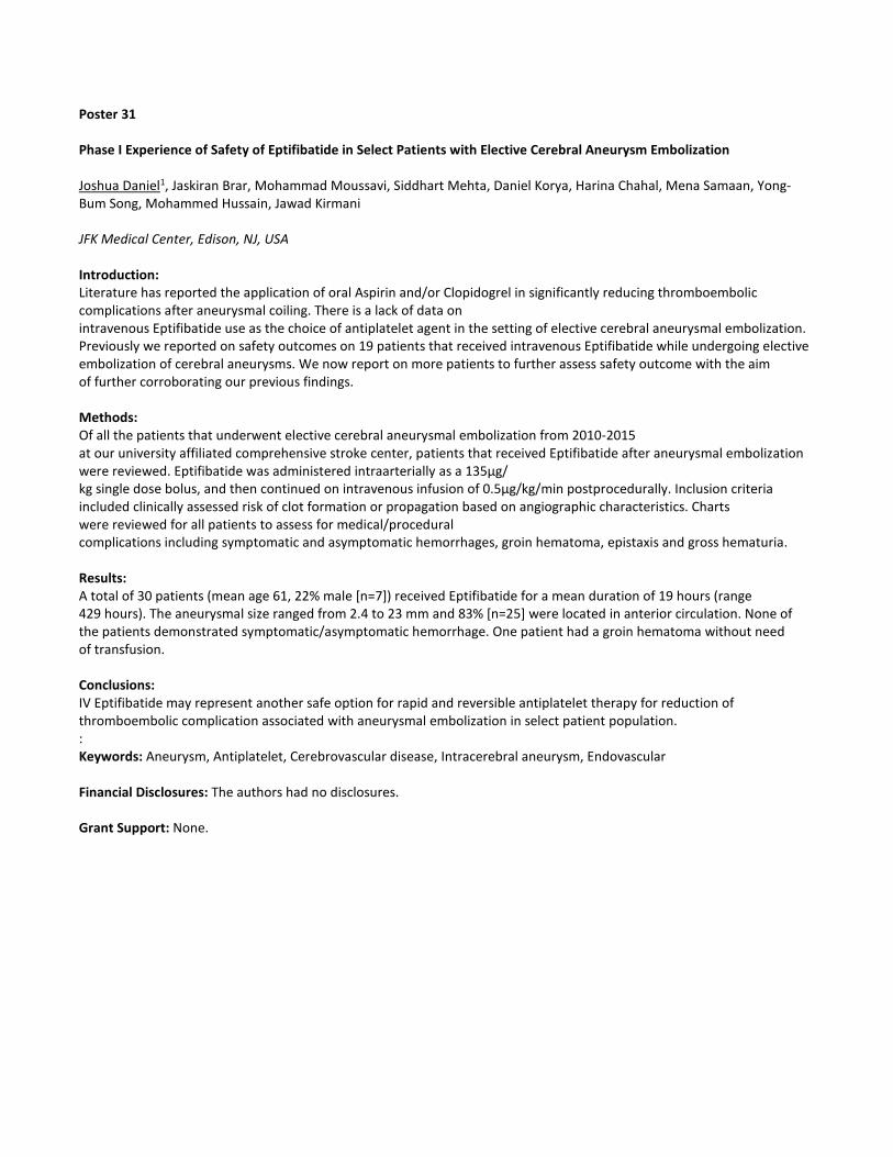
Poster 31 Phase I Experience of Safety of Eptifibatide in Select Patients with Elective Cerebral Aneurysm Embolization Joshua Daniel1, Jaskiran Brar, Mohammad Moussavi, Siddhart Mehta, Daniel Korya, Harina Chahal, Mena Samaan, Yong-Bum Song, Mohammed Hussain, Jawad Kirmani JFK Medical Center, Edison, NJ, USA Introduction: Literature has reported the application of oral Aspirin and/or Clopidogrel in significantly reducing thromboembolic complications after aneurysmal coiling. There is a lack of data on intravenous Eptifibatide use as the choice of antiplatelet agent in the setting of elective cerebral aneurysmal embolization. Previously we reported on safety outcomes on 19 patients that received intravenous Eptifibatide while undergoing elective embolization of cerebral aneurysms. We now report on more patients to further assess safety outcome with the aim of further corroborating our previous findings. Methods: Of all the patients that underwent elective cerebral aneurysmal embolization from 2010-2015 at our university affiliated comprehensive stroke center, patients that received Eptifibatide after aneurysmal embolization were reviewed. Eptifibatide was administered intraarterially as a 135μg/ kg single dose bolus, and then continued on intravenous infusion of 0.5μg/kg/min postprocedurally. Inclusion criteria included clinically assessed risk of clot formation or propagation based on angiographic characteristics. Charts were reviewed for all patients to assess for medical/procedural complications including symptomatic and asymptomatic hemorrhages, groin hematoma, epistaxis and gross hematuria. Results: A total of 30 patients (mean age 61, 22% male [n=7]) received Eptifibatide for a mean duration of 19 hours (range 429 hours). The aneurysmal size ranged from 2.4 to 23 mm and 83% [n=25] were located in anterior circulation. None of the patients demonstrated symptomatic/asymptomatic hemorrhage. One patient had a groin hematoma without need of transfusion. Conclusions: IV Eptifibatide may represent another safe option for rapid and reversible antiplatelet therapy for reduction of thromboembolic complication associated with aneurysmal embolization in select patient population. : Keywords: Aneurysm, Antiplatelet, Cerebrovascular disease, Intracerebral aneurysm, Endovascular Financial Disclosures: The authors had no disclosures. Grant Support: None.

Poster 32 Impact of Menopausal Age on Size of Unruptured Intracranial Aneurysms and their Outcomes with Endovascular Therapy Sushrut S Dharmadhikari1, Atchaneeyasakul Kunakorn2, Sudheer Ambekar2, Dileep R Yavagal2 1Wayne State University, Detroit, MI, USA, 2University of Miami, Miami, FL, USA Introduction: Unruptured Intracranial Aneurysms (UIAs) occur about twice more commonly in women compared to men. Prevalence increases rapidly in women with age. Impact of menopausal age, if any, on UIAs and treatment outcomes with Endovascular Therapy (ET) has not been well studied. We hypothesized that higher estrogen in pre-menopausal women may have a protective effect on presentation size and treatment outcomes. Methods: We performed a retrospective analysis of consecutive female patients with UIAs treated with ET at our academic tertiary care center. UIA characteristics, complications and outcomes were recorded and compared. Results: 117 patients were included. 23 patients in premenopausal group (PRM) and 94 in postmenopausal group (POM). 93.6% of aneurysms in PRM were in ICA segments (p< 0.05). Hence only ICA segment aneurysms further studied. Baseline characteristics were similar in 2 groups. Mean size of aneurysms in PRM 8.6 ± 3.9mm vs 10.8 ± 5.6mm in POM (p=0.055). Trend to higher aneurysm neck size seen in POM (4.7 ± 2.5mm vs 3.7 ± 1.7mm; p=0.07). Number of aneurysm lobes higher in PRM (1.23 ± 0.54 vs 1.07 ± 0.31; p=0.18). In multivariate analysis, number of lobes showed significance (p=0.026). Complications and outcomes similar. Conclusions: Loss of protective effect of estrogen may be responsible for higher UIA size with increased rupture risk in POM. Peri and Post menopausal women with UIAs may need to be monitored frequently and treated aggressively. Larger prospective trial is needed to confirm these findings. Keywords: Endovascular therapy, Intracerebral aneurysm, Pathophysiology, Embolization, Unruptured Financial Disclosures: The authors had no disclosures. Grant Support: None.

Poster 33 In-stent stenosis after Pipeline Embolization Device Flow Diversion Treatment for Intracranial Aneurysms Seby John1, Mark Bain, Ferdinand Hui, Muhammad S. Hussain, Thomas Masaryk, Peter Rasmussen, Gabor Toth Cerebrovascular Center, Cleveland Clinic, Cleveland, USA Introduction: There is scant information on in-stent stenosis following flow diversion treatment of intracranial aneurysms with Pipeline Embolization Device (PED). We aimed to assess the incidence, severity, nature, and clinical consequences of in-stent stenosis on routine angiographic follow-up following treatment with PED. Methods: Retrospective study of patients who underwent treatment of intracranial aneurysm(s) with PED. In-stent stenosis was assessed on follow-up catheter angiography performed at 6, 12 and 24 months after treatment. In-stent stenosis was defined as a growth process beyond the limits of the metallic mesh. Intimal hyperplasia was labeled when the above measurement was < 20%, and present uniformly throughout the stent margins. In-stent stenosis represented an area of parent vessel narrowing, often times focal, graded as mild (< 25%), moderate (25-50%), or severe (>50%). Results: Between 6/2011-4/2015, 80 patients were treated with PED. Angiographic follow-up was available in 44 (55%) patients (mean 12 months). Thirty-seven (84%) patients had aneurysms in the anterior circulation, and 7 (16%) in the posterior circulation. Average measurement of the largest aneurysm diameter was 12.6 mm (range 2-37 mm). A mean of 1.5 (range 1-5) stents were used, with 17 (39%) requiring >1 stent. Concomitant coil embolization was performed in 7 (16%) patients. Two (5%) patients required balloon angioplasty post stent deployment for optimal wall apposition. In-stent stenosis was detected in 6 (13.6%) patients, at a median of 6 months. Stenosis was mild in 2/6 (33%) and moderate in 4/6 (67%) patients, with no cases of severe stenosis. None of the in-stent stenoses caused flow limitation, clinical symptoms, or required re-treatment. Thirty-six (82%) patients had either minimal intimal hyperplasia (12 [27%]), or no stenosis (24 [55%]). Conclusions: Treatment with PED was associated with a 13.6% rate of in-stent stenosis, detected on first angiographic follow-up, at a median of 6 months. None were symptomatic, or needed re-treatment. Keywords: Pipeline, Aneurysm Embolization Financial Disclosures: The authors had no disclosures. Grant Support: None.

Poster 34 Intra-Aneurysmal Double Microcatheter Technique for Complex Aneurysm NILESH KINGE1, ATULABH VAJPEYEE2, RAMA KANT3, NARENDRA MAL4 1GBH AMERICAN HOSPITAL, UDAIPUR, India, 2GBH AMERICAN HOSPITAL, UDIAPUR, India, 3GBH AMERICAN HOSPITAL, UDAIPUR, India, 4GBH AMERICAN HOSPITAL UDAIPUR, India Introduction: Objective: This study deals with one of the neurointerventional technique for treatment of complex aneurysm which imparts several challenges for complete occlusion without compromising parent vessel. Methods: Material and methods: Ten patients with wide neck intracranial aneurysm were treated using endovascular method of double microcatheter between December 2012 to September 2013. All patients had presented with SAH. All were followed up for at least 1 year. Detail demographic information including patient age, sex, presentation, clinical manifestations (Hunt and Hess scale), aneurysm morphology (neck, height, width, and length), aneurysm location were noted and proper follow up was kept. All follow up were done with MRA and at least once a cerebral DSA was done after 1 months. Angiographic results were classified using Raymond-Roy classification system. Modified ranking score was (mRS) recorded at the time of admission ,at the time of discharge and thereafter at 1month,3months,6months and 1 year. Aneurysm were coiled using double microcathetor technique. Results: Result: All aneurysms were successfully embolized using double microcatheter technique. DSA done immediately post embolization showed class I filling in 9 patients and class II filling in 1 patient. The mean neck is 2.91 mm (range 2 to 4 mm) and mean dome of neck ratio was 1.34 (range 0.89 to 2.14). Modified Rankin scale (mRS) was less than 2 immediately after the procedure and subsequently till one year of follow-up. Conclusions: Conclusion: Compared to other methods for treating these subsets of aneurysms, this modified intra-aneurysmal double catheter technique is safe, effective, and not technically demanding. Keywords: Aneurysm, Aneurysm Embolization, Access catheters, Coiling, SAH Financial Disclosures: The authors had no disclosures. Grant Support: None.

Poster 35 Endovascular Treatment of Consecutive Unruptured Anterior Communicating Artery Aneurysms John M. Leschke1, Ahsan Sattar , Kaiz Asif, Wled Wazni, Mazen Noufal, Sam O. Zaidat Medical College of Wisconsin, MILWAUKEE, WI, USA Introduction: The anterior communicating (ACom) artery is the most common location of cerebral aneurysms harboring up to 30%-37% of all treated aneurysms. Untreated, ACom aneurysms are responsible for up to 40% of subarachnoid hemorrhage. Endovascular coiling is a well-established alternative to neurosurgical clipping but the choice of treatment remains debated and heavily relies on the risks associated with repair. We report the immediate and long-term clinical as well as radiographic outcomes of non-selected consecutive patients with unruptured anterior communicating artery (ACOM) aneurysms treated with endovascular technique. Methods: A prospectively maintained single institution neuro-endovascular database was accessed to identify consecutive cases of un-ruptured ACom aneurysms treated endovascularly between 2006 and 2013. Clinical, demographic, radiographic, and procedural data were retrospectively obtained through chart review. Results: A cohort of 60 consecutive patients with unruptured ACom artery aneurysms were treated using endovascular technique including 28 women and 32 men with a mean age of 62.5 yrs. The average maximum aneurysm diameter was 7.4 mm. Dome to neck ratio was less than 2 for 46 (76.6%) of the aneurysms. Aneurysm projection was anterior-superior in 29 (48.3%), anterior-inferior in 23 (40%) and posterior-superior in 8 (13.3%). Primary coiling was performed for 30 (50%) aneurysms, balloon assistance was used for 2 (3%), stent support was used for 30 (50%). Complete aneurysm occlusion was achieved for 44 (79.3%) aneurysms and near complete for 11 (18.3%) aneurysms. Intra-operative perforation (IOP) occurred in 2 (3.3%) patients and a thromboembolic event (TEE) occurred in 2 (3.3%) patient without clinical worsening. Median follow up with imaging was 6.1 months. There were no instances of re-bleeding during follow up. A small neck residual occurred in 10 (16.6%) aneurysms. 2 (3%) were re-canalized and retreated with additional primary coiling and stent assistance. Conclusions: Endovascular treatment of unselected unruptured ACom aneurysms can be performed with acceptable rates of complications and recanalization. Keywords: Aneurysm, Coiling, Balloon assisted, Endovascular, Endovascular therapy Financial Disclosures: The authors had no disclosures. Grant Support: None.
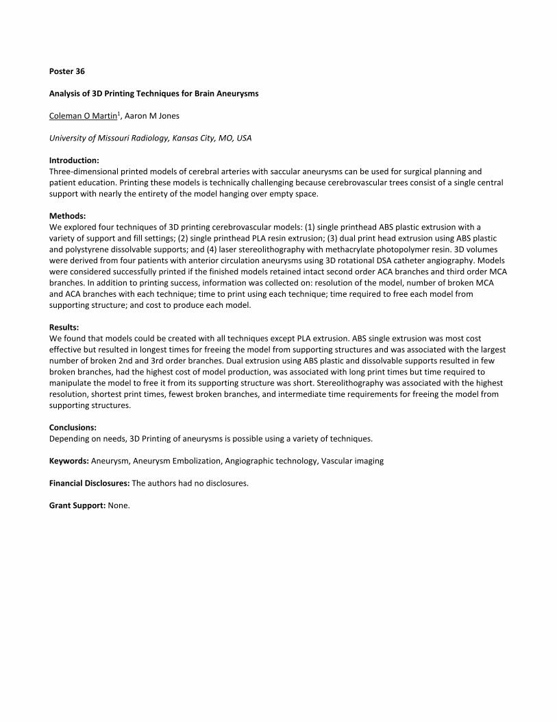
Poster 36 Analysis of 3D Printing Techniques for Brain Aneurysms Coleman O Martin1, Aaron M Jones University of Missouri Radiology, Kansas City, MO, USA Introduction: Three-dimensional printed models of cerebral arteries with saccular aneurysms can be used for surgical planning and patient education. Printing these models is technically challenging because cerebrovascular trees consist of a single central support with nearly the entirety of the model hanging over empty space. Methods: We explored four techniques of 3D printing cerebrovascular models: (1) single printhead ABS plastic extrusion with a variety of support and fill settings; (2) single printhead PLA resin extrusion; (3) dual print head extrusion using ABS plastic and polystyrene dissolvable supports; and (4) laser stereolithography with methacrylate photopolymer resin. 3D volumes were derived from four patients with anterior circulation aneurysms using 3D rotational DSA catheter angiography. Models were considered successfully printed if the finished models retained intact second order ACA branches and third order MCA branches. In addition to printing success, information was collected on: resolution of the model, number of broken MCA and ACA branches with each technique; time to print using each technique; time required to free each model from supporting structure; and cost to produce each model. Results: We found that models could be created with all techniques except PLA extrusion. ABS single extrusion was most cost effective but resulted in longest times for freeing the model from supporting structures and was associated with the largest number of broken 2nd and 3rd order branches. Dual extrusion using ABS plastic and dissolvable supports resulted in few broken branches, had the highest cost of model production, was associated with long print times but time required to manipulate the model to free it from its supporting structure was short. Stereolithography was associated with the highest resolution, shortest print times, fewest broken branches, and intermediate time requirements for freeing the model from supporting structures. Conclusions: Depending on needs, 3D Printing of aneurysms is possible using a variety of techniques. Keywords: Aneurysm, Aneurysm Embolization, Angiographic technology, Vascular imaging Financial Disclosures: The authors had no disclosures. Grant Support: None.
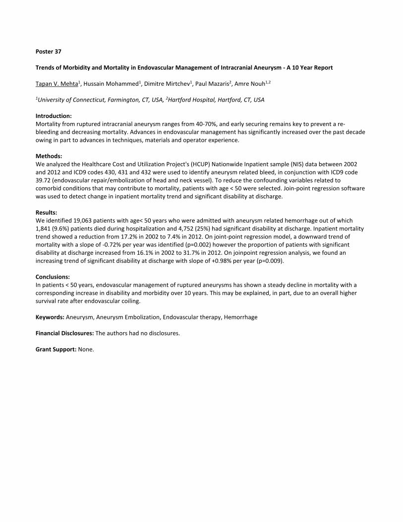
Poster 37 Trends of Morbidity and Mortality in Endovascular Management of Intracranial Aneurysm - A 10 Year Report Tapan V. Mehta1, Hussain Mohammed1, Dimitre Mirtchev1, Paul Mazaris2, Amre Nouh1,2 1University of Connecticut, Farmington, CT, USA, 2Hartford Hospital, Hartford, CT, USA Introduction: Mortality from ruptured intracranial aneurysm ranges from 40-70%, and early securing remains key to prevent a re-bleeding and decreasing mortality. Advances in endovascular management has significantly increased over the past decade owing in part to advances in techniques, materials and operator experience. Methods: We analyzed the Healthcare Cost and Utilization Project's (HCUP) Nationwide Inpatient sample (NIS) data between 2002 and 2012 and ICD9 codes 430, 431 and 432 were used to identify aneurysm related bleed, in conjunction with ICD9 code 39.72 (endovascular repair/embolization of head and neck vessel). To reduce the confounding variables related to comorbid conditions that may contribute to mortality, patients with age < 50 were selected. Join-point regression software was used to detect change in inpatient mortality trend and significant disability at discharge. Results: We identified 19,063 patients with age< 50 years who were admitted with aneurysm related hemorrhage out of which 1,841 (9.6%) patients died during hospitalization and 4,752 (25%) had significant disability at discharge. Inpatient mortality trend showed a reduction from 17.2% in 2002 to 7.4% in 2012. On joint-point regression model, a downward trend of mortality with a slope of -0.72% per year was identified (p=0.002) however the proportion of patients with significant disability at discharge increased from 16.1% in 2002 to 31.7% in 2012. On joinpoint regression analysis, we found an increasing trend of significant disability at discharge with slope of +0.98% per year (p=0.009). Conclusions: In patients < 50 years, endovascular management of ruptured aneurysms has shown a steady decline in mortality with a corresponding increase in disability and morbidity over 10 years. This may be explained, in part, due to an overall higher survival rate after endovascular coiling. Keywords: Aneurysm, Aneurysm Embolization, Endovascular therapy, Hemorrhage Financial Disclosures: The authors had no disclosures. Grant Support: None.
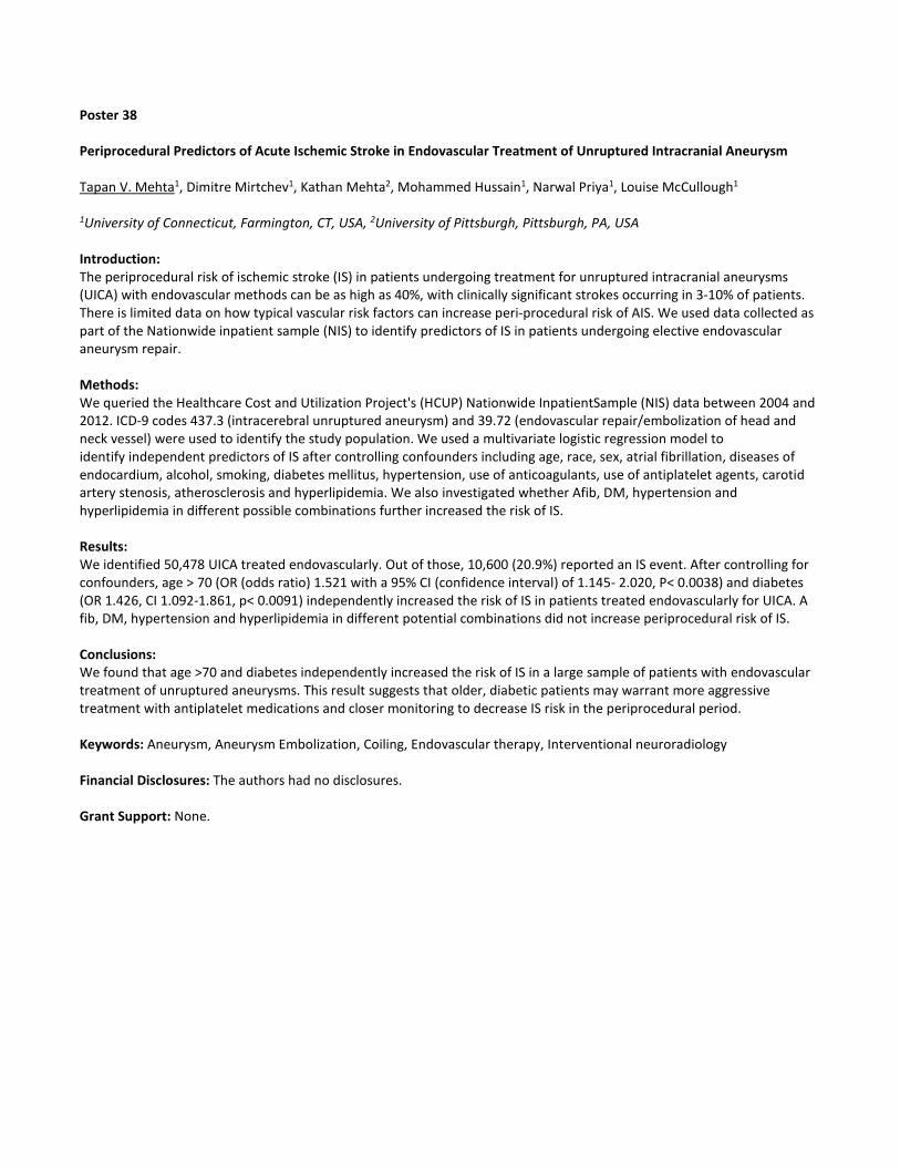
Poster 38 Periprocedural Predictors of Acute Ischemic Stroke in Endovascular Treatment of Unruptured Intracranial Aneurysm Tapan V. Mehta1, Dimitre Mirtchev1, Kathan Mehta2, Mohammed Hussain1, Narwal Priya1, Louise McCullough1 1University of Connecticut, Farmington, CT, USA, 2University of Pittsburgh, Pittsburgh, PA, USA Introduction: The periprocedural risk of ischemic stroke (IS) in patients undergoing treatment for unruptured intracranial aneurysms (UICA) with endovascular methods can be as high as 40%, with clinically significant strokes occurring in 3-10% of patients. There is limited data on how typical vascular risk factors can increase peri-procedural risk of AIS. We used data collected as part of the Nationwide inpatient sample (NIS) to identify predictors of IS in patients undergoing elective endovascular aneurysm repair. Methods: We queried the Healthcare Cost and Utilization Project's (HCUP) Nationwide InpatientSample (NIS) data between 2004 and 2012. ICD-9 codes 437.3 (intracerebral unruptured aneurysm) and 39.72 (endovascular repair/embolization of head and neck vessel) were used to identify the study population. We used a multivariate logistic regression model to identify independent predictors of IS after controlling confounders including age, race, sex, atrial fibrillation, diseases of endocardium, alcohol, smoking, diabetes mellitus, hypertension, use of anticoagulants, use of antiplatelet agents, carotid artery stenosis, atherosclerosis and hyperlipidemia. We also investigated whether Afib, DM, hypertension and hyperlipidemia in different possible combinations further increased the risk of IS. Results: We identified 50,478 UICA treated endovascularly. Out of those, 10,600 (20.9%) reported an IS event. After controlling for confounders, age > 70 (OR (odds ratio) 1.521 with a 95% CI (confidence interval) of 1.145- 2.020, P< 0.0038) and diabetes (OR 1.426, CI 1.092-1.861, p< 0.0091) independently increased the risk of IS in patients treated endovascularly for UICA. A fib, DM, hypertension and hyperlipidemia in different potential combinations did not increase periprocedural risk of IS. Conclusions: We found that age >70 and diabetes independently increased the risk of IS in a large sample of patients with endovascular treatment of unruptured aneurysms. This result suggests that older, diabetic patients may warrant more aggressive treatment with antiplatelet medications and closer monitoring to decrease IS risk in the periprocedural period. Keywords: Aneurysm, Aneurysm Embolization, Coiling, Endovascular therapy, Interventional neuroradiology Financial Disclosures: The authors had no disclosures. Grant Support: None.

Poster 39 Isolated Proximal PICA Dissecting Aneurysms Treated with Parent Artery Occlusion with Good Outcomes, Report of 4 Cases and Anatomical Discussion Mazen Noufal1, Sam Zaidat, Brian-Fred Fitzsimmons, John Lynch, Marc Lazzaro Department of Neurology, Medical College of Wisconsin, Milwaukee, WI, USA Introduction: Dissecting PICA aneurysms represent a rare cause of subarachnoid hemorrhage. Those aneurysms are often fusiform, have no neck, and arise from a pathologic artery segment, which often make primary coiling or clipping of the aneurysm sac not feasible. Treatment is especially challenging when the anterior- and lateral medullary segments are involved due to concern for brainstem ischemica. Preservatory surgical treatment options include PICA-AICA bypass, PICA-PICA bypass, and surgical wrapping. Methods: Chart review Results: Four patients (3 adults and a 4-year-old child) presented with subarachnoid hemorrhage, hydrocephalus, and decreased consciousness had dissecting aneurysms of proximal PICA. One patient had an embolic infarction in the cerebellar tonsil, and another had rare anatomical variant of duplicate PICA origin. Cerebral angiogram revealed good size of ipsilateral AICA (>1/2 of PICA size) in all patients, with evident PICA-AICA anastomoses in three. All patients were treated with PICA sacrifice at the origin (2 with coil embolization and 2 with n-BCA glue embolization). One patient had asymptomatic embolic cerebellar infarcts, another had cerebellar infarction caused ataxia, and two had no ischemic lesions. None had brainstem infarction. Two patients had mRS of 0, one had mRS of 2, and another had mRS of 3. Conclusions: Perforator branches of PICA intermingle and overlap with those arising from vertebral artery. It is reporteded that 75% of lateral medullary infarctions were associated with VA occlusion, and 12% with PICA occlusion. PICA anastomoses with AICA and SCA are documented. While utmost consideration to preserve PICA should be the principle when treating its lesions, the above collateral and overlapping circulation is probably underestimated in daily practice. Angiographic evaluation of AICA size and its anastomosis with PICA might help judging the safety of PICA sacrifice when other treatment options are less desirable. Our results are consistent with previous reports that dissecting aneurysms of proximal PICA can be treated with arterial sacrifice with good outcome in most patients. Keywords: Aneurysm, SAH Financial Disclosures: The authors had no disclosures. Grant Support: None.
Poster 40 Impact of the New AHA/ASA Definition of Stroke on the Outcome of the SAMMPRIS Trial Sami Al Kasab1, Michael Lynn2, Tanya Turan1, Colin P Derdyn3, David Fiorella4, Bethany F Lane1, Jean Montgemry2, L Scott Janis5, Marc I Chimowitz1 1MUSC, Charleston, SC, USA, 2Emory University, Atlanta, GA, USA, 3Washington University, St.Louis, MT, USA, 4SUNY at Bufalo Sc of med Stony Brook, NY, USA, 5NIH/NINDS Bethesda, MD, USA Introduction: Atherosclerotic intracranial arterial stenosis is one of the most common causes of stroke worldwide and is associated with a high risk of recurrent stroke. •SAMMPRIS study previously showed that aggressive medical management is superior to stenting in patients with intracranial arterial atherosclerosis. •The AHA/ASA have recently recommended that cerebral infarction associated with temporary symptoms or signs lasting < 24 hours (that we refer to as CITS) should be considered a stroke. • CITS in the territory was not included in the primary end point in SAMMPRIS for the following reasons: 1. Unclear clinical significance 2.Concern for ascertainment bias 1.Patients who had stenting: •hospitalized for the procedure à imaging when experiencing a change in neurologic exam. •may be more likely to have imaging for TIAs during follow-up because of the presence of a stent. 2.Patients in the medical arm typically not hospitalized at enrollment à less likely to have imaging if experienced transient symptoms while at home. Methods: Comparison: Medical vs. Stenting groups •Primary endpoint: •Any stroke, CITS or death within 30 days after enrollment, or •Stroke or CITS in the territory of the qualifying artery beyond 30 days. •For evaluation of ascertainment bias: Compared the use of brain MRI after TIAs in both treatment groups. Results: The percentage of patients with reported TIAs who underwent brain MRI : •69% (34/49) in the Medical group vs. •61% (28/46) in the Stenting group (p=0.40). Primary Endpoint Primary Endpoint or CITS Medical PTAS Medical PTAS 1 6 12 Conclusions: CITS in the territory occurred more frequently during follow-up in the PTAS group than in the medical group This cannot be attributed to imaging ascertainment bias. These results imply an even higher benefit from medical therapy over stenting and a higher risk of symptomatic cerebral infarction in both groups than originally described in SAMMPRIS. Keywords: Atherosclerosis Financial Disclosures: The authors had no disclosures. Grant Support: None.
Poster 41 Dedicated Stroke Units with Bedside Monitoring and Better Blood Pressure Control Audrey Arango1, Florence Chukwuneke, Briana DeCarvalho, Yong- Bum Song, Jaskiran Brar, Siddhart Mehta, Mohammad Moussavi, Spozhmy Panezai, Jawad Kirmani JFK Medical Center, Edison, NJ, USA Introduction: Patients with ischemic or hemorrhagic stroke require strict BP control to prevent hemorrhagic transformation or hematoma expansion. Acute elevations in BP are often treated with IV labetalol. Standard automated BP monitoring alerts practitioners to trends that lead to more steady control of BP rather than frequent acute interventions. We evaluated patients who were treated in a stroke unit after the institution of automated bedside monitoring to determine if there was a significant difference in care. Methods: Patients were evaluated over a 12month period (2014) after the incorporation of bedside automated BP monitoring in a dedicated stroke unit at a university affiliated, comprehensive stroke center. The number of times each patient during this time frame received IV labetalol for acute elevations in BP was compared with a time period spanning 12months prior (2013); there was no automated BP monitoring performed. The average interventions were compared with a t-test by using SPSS V22. Comparisons of patient population and type of pathology were matched appropriately. Results: Of the 1,326 patients who presented for ischemic or hemorrhagic strokes during the 24 month period evaluated, 25 required multiple injections of IV labetalol for acute BP control. Of these, 12 patients were on automated vital signs and BP monitoring, and 13 were not. The mean number of IV labetalol interventions implemented in the group being monitored was 2.8, while the mean number of treatments given to patients not being monitored was 5.9 (p=.016). Conclusions: In our study there is a trend towards better blood pressure control with adequate adjustment of oral medications for monitored patients in our dedicated stroke unit. Prevention of sudden elevations in BP may translate into lower rates of hemorrhagic transformation or hematoma expansion and confer better outcomes in stroke patients. Larger prospective studies are required to corroborate our findings. Keywords: Blood pressure management in acute stroke, Stroke Financial Disclosures: The authors had no disclosures. Grant Support: None.

Poster 42 Evaluating the Impact of Insurance Precertification on Discharge of Stroke Patients to Skilled Nursing Facility or Inpatient Rehabilitation Center. Ashish Kulhari1, Andrew L Smith1, Julie Wolfram2, Anthony Furlan1 1Case Western Reserve University/Neurology, Cleveland, OH, USA, 2Case Western Reserve University/ General Medics, Cleveland, OH, USA Introduction: The purpose of this study is to determine if the common insurance practice of requiring precertification before a medically ready stroke patient can be discharged to a skilled nursing facility or inpatient rehabilitation center causes a delay in discharge. Eliminating delays in discharge of stroke patients is important given the increasing demands for health care efficiency after the passage of the Affordable Health Care Act. The objective of this study was to determine whether insurance precertification prior to discharge to a skilled nursing facility or inpatient rehabilitation center for medically ready stroke patients causes a significant delay in discharge. Methods: A retrospective chart review of 1007 patients who were admitted to our comprehensive stroke center with the primary diagnosis of stroke over a 12 month period was performed. 315 patients met the inclusion criterion of a primary diagnosis of stroke who required discharge to a skilled nursing facility or inpatient acute rehabilitation. All 315 patients were medically cleared for discharge to a skilled nursing facility or inpatient acute rehabilitation by a board certified vascular neurologist. Results: Of these 315 patients, 131 required insurance precertification and 184 did not. All 131 patients who required precertification had private health insurance. The patients who required insurance precertification had an average delay of discharge of 1.5 days and those patients who did not require precertification had an average delay of discharge of 0.8 days (P –value < 10-4). Conclusions: The results of this study demonstrate that insurance precertification leads to delay in discharge and increased hospital costs for stroke patients. Keywords: Stroke, Health economic Financial Disclosures: The authors had no disclosures. Grant Support: None.
Poster 43 Stenosis and Manometry Correlation in Evaluating Dural Venous Sinus Stenosis in Idiopathic Intracranial Hypertension Jeremy C Peterson1, Michael Abraham2 1Neurosurgery Kansas University Medical Center, Kansas City, KS, USA, 2Neurology/Interventional Neurology Kansas University Medical Center, Kansas City, KS, USA Introduction: A certain percentage of patients with idiopathic intracranial hypertension (IIH) have venous sinus stenosis. A number of patients are undergoing interventional evaluation of stenosis with venography and manometry in consideration for stenting. To our knowledge there have been no studies to evaluate the correlation or lack thereof between degree of sinus stenosis and manometry pressure gradients accross the stenosis. Methods: We retrospectively reviewed the imaging and manometry findings for 39 consecutive patients undergoing angiographic evaluation for stenosis in patients diagnosed with idiopathic intracranial hypertension. This yielded 67 sinus imaging and manometry readings for evaluation. Twelve data points or readings were excluded because they showed no evidence for any stenosis and were essentially normal. A linear regression model was created looking for the correlation of percentage stenosis to pressure gradient recordings. Results: The correlation coefficient was r=0.4204 with an r squared of 0.1767 at a p-value of 0.0014. The mean degree of stenosis was 56% with a standard deviation of 17% stenosis. The minimum degree of stenosis was 15% and the maximum 90%. The mean pressure gradient was 17.56 mmHg with a standard deviation of 7.66 mmHg. The lowest pressure reading was 6 mmHg and the highest was 42 mmHg. Conclusions: Our finding highlight that there is a weak level of correlation between the degree of sinus stenosis and manometry pressure gradient in this subset of patients. This likely reflects the variability and distensibility of the dural sinus system as compared to the arterial circulation. This finding highly suggests that if an idiopathic intracranial hypertension patient is being evaluated for dural sinus stenosis and there is any degree of stenosis seen on diagnostic imaging, the patient needs to undergo manometry readings accross the level of stenosis before deciding if the stenosis should be treated by stenting. Keywords: Angiogram, Cerebral sinus and venous thrombosis, Endovascular, Intracranial Stenosis stenting and angioplasty, Stenting Financial Disclosures: The authors had no disclosures. Grant Support: None.

Poster 44 Evaluating the Presence of Endothelial Cells in Thrombi Removed during Endovascular Treatment: A Feasibility Study. Michelle Previtera1, Amitabha Sengupta, Daniel Korya, Mohammad Moussavi, Siddhart Mehta, Jawad Kirmani JFK Medical Center, Edison, NJ, USA Introduction: Acute ischemic stroke patients that do not respond to intravenous thrombolysis undergo mechanical thrombectomy upon documentation/clinical suspicion of large vessel occlusion (LVO). Sometimes damage to blood vessel layers may result from mechanical embolectomy/thrombectomy. No studies have evaluated the trauma to blood vessel walls sustained from this procedure. We hypothesize that we can evaluate blood vessel wall damage by determining whether endothelial cells adhere to the thrombectomy device and/or are on the edges of the removed thrombus. Methods: This study was approved by the IRB and all samples were collected with the consent of the donor. Thrombi were collected from stroke patients that underwent thrombectomy. Patient demographics, complication logs, outcomes and device characteristics were noted. Procedure times, number of passes and recannalization status was also documented. Thrombi were immediately placed in paraformaldehyde and transferred for analysis. Results: The effort was led by the stroke basic science lab in collaboration with the clinical team in an effort to find timely answers in view of recent Multi Center Prospective Randomized Control trials using Stentrievers. A total of 15 clots from 10 patients (Males, n=6) have been collected. Initial pilot data for feasibility of protocol and preliminary histopathological data is to be presented at the ISC annual meeting Conclusions: The study will infer whether damage is sustained by the endothelium during thrombectomy. It may allow for comparisons of techniques and devices, which in turn may facilitate future thrombectomy methods and device engineering. Keywords: Endovascular therapy Financial Disclosures: The authors had no disclosures. Grant Support: None.

Poster 45 Successful Thrombectomy in a 10 year-old boy with Restrictive Cardiomyopathy after Cardioembolic Infarct. Ashish Kulhari1, Yasir Saleem, Krishna Kandregula, Ciro Ramos-Estebanez Case Western Reserve University/Neurology, Cleveland, OH, USA Introduction: To showcase the opportunity for mechanical thrombectomy in pediatric populations with large cerebral vessel occlusion Methods: Literature pertinent to cerebral thrombectomy in pediatric populations with acute stroke was reviewed. Results: CASE: A 10-year-old boy with unknown medical history was brought to our hospital with left hemiparesis, hypoesthesia and dysarthria (NIHSS 7) after 5.5 hours of last seen normal. CT head showed evolving hypodensity in right MCA territory and hyperdense MCA sign. MRI/A brain showed small core infarct and large penumbra in right MCA territory with right M1 cut off. MRA neck was normal. Although he was out of intravenous t-PA window, he underwent mechanical thrombectomy. Successful right MCA recanalization (TICI 2b) was achieved. Emergent transesophageal echocardiogram showed bilateral severe atrial enlargement with normal ventricular size and function, highly suggestive of restrictive cardiomyopathy. Cardiac MRI confirmed the findings. No intracardiac thrombus was seen. 24 hours later he was started on anticoagulation for secondary stroke prevention. Right ventricular endomyocardial biopsy showed cardiomyocyte hypertrophy with endocardial and superficial interstitial fibrosis. Genetic tests for restrictive cardiomyopathy were sent. Hypercoagulable work up was negative. Cardiac defibrillator was placed given high chances of arrhythmias. On discharge, NIHSS was 1 (left pronator drift). Currently awaiting heart transplantation and genetic tests results. DISCUSSION: Pediatric stroke is very rare. There are no evidence based guidelines about the management of acute pediatric stroke. Our case is interesting because he presented out of the intravenous t-PA window but within the therapeutic period for mechanical thrombectomy, according to the adult stroke guidelines. Despite of presenting with NIHSS below 10, we pursued mechanical thrombectomy because of the small core infarct and large penumbra. Conclusions: Mechanical thrombectomy might allow the opportunity for improving the outcomes in pediatric populations given their typically excellent collateral circulation and potential lower infarct volume. Keywords: Ischemic stroke, Mechanical thrombectomy, Pediatric intervention Financial Disclosures: The authors had no disclosures. Grant Support: None.

Poster 46 Novel Application Of Reversible Parental Antiplatelets in Patients with Aneurysmal Subarachnoid Hemorrhage Harina Chahal1, Siddhart Mehta, Mohammad Moussavi, Daniel Korya, Jaskiran Brar, Mena Samaan, Joshua Daniel, Yong-Bum Song, Jawad Kirmani JFK Medical Center, Edison, NJ, USA Introduction: The International Subarachnoid Aneurysm Trial (ISAT) showed a greater likelihood of survival free 1 year disability in patients undergoing endovascular coiling who were started on antiplatelet agents after SAH compared with neurosurgical clipping. However, data on safety of acute parental antiplatelet agents after aneurysmal coiling is lacking. Previously we reported on 5 patients that received acute intraprocedural eptifibatide followed by IV infusions. We now report 7 more patients to further asses safety of IV eptifibatide in patients presenting with acute subarachnoid hemorrhage undergoing endovascular coiling for aneurysmal embolization. Methods: All the patients from 20092015 who presented to our university affiliated comprehensive stroke center with aneurysmal subarachnoid hemorrhage and underwent endovascular coiling were evaluated. Eptifibatide was administered intraarterially as a 135μg/kg single bolus, and then continued on intravenous infusion of 0.5μg/ kg/min postprocedurally. Charts were reviewed to assess for complications including symptomatic and asymptomatic intra and extracranial hemorrhages, groin hematomas, epistaxis and gross hematuria. Results: Of the total of 101 patients treated with coil embolization during this period, 12 patients (mean age 55 years, received acute intraprocedural Eptifibatide followed by IV infusion for a mean duration of 71 hours (range 15.5316 hours). Various reasons for use of Eptifibatide included: stent assist coiling [n=3], multiple stents for flow diversion [n=2], partial coil prolapse [n=2], angioplasty for vasospasm (n=1) and vascular lumen flow compromise [n=4]. None of the patients demonstrated symptomatic/asymptomatic hemorrhage, groin hematoma, epistaxis or hematuria.. Conclusions: Our results highlight safety of administering IV Eptifibatide to prevent thrombotic complications after endovascular coil embolization in select patients with aneurysmal subarachnoid hemorrhage. Multicenter prospective trials are warranted to corroborate our finding. Keywords: Aneurysm, Antiplatelet, Cerebrovascular disease, Subarachnoid, Aneurysm Embolization Financial Disclosures: The authors had no disclosures. Grant Support: None.

Poster 47 Screening for Delayed Cerebral Ischaemia in Aneurysmal Subarachnoid Haemorrhage in Critical Care: An International Survey. Laura K Markham1, Milo Hollingworth2, Aimum Jamjoon3, PR Chen4, A Goddard5, A Coulthard6, Michael Soderman7, Ketan R Bulsara8 1University of Bristol Medical School, Bristol, United Kingdom, 2North Bristol Trust, Bristol, United Kingdom, 3British Neurosurgical Trainee Research Collaborative, London, United Kingdom, 4University of Texas Medical School Houston, TX, USA, 5Leeds General Hospital Leeds, United Kingdom, 6University of Queensland Brisbane, Australia, 7Karolinska Institute Stockholm, Sweden, 8Yale University Medical School New Haven, CT, USA Introduction: Delayed cerebral ischaemia (DCI) is a significant cause of morbidity and mortality in aneurysmal subarachnoid haemorrhage (aSAH). Radiological techniques including Transcranial Doppler (TCD), Computer Tomography Angiography (CTA) and Digital Subtraction Angiography (DSA) are used in screening for DCI; however, there is no consensus about how best to use them. Methods: We conducted an international survey of DCI screening strategies used in aSAH amongst members of the Congress of Neurological Surgeons, British Society of Neuroradiologists, European Society of Neuroradiologists and the Australian and New Zealand Interventional Neuroradiology Collaborative. Data from 371 respondents from 32 countries were analyzed. Results: The most common screening technique was daily TCD, which was reported by 59% (220/371). Less than daily TCD was reported by 3% (11/371). Use of CTA or DSA at day 5 was reported by 16% (61/371) and 16% (59/371) respectively. Other screening options included Computer Tomography Perfusion (13), Electroencephalography (1), Near Infrared Spectroscopy (1) and Single-Photon Emission Computer Tomography (1). 20% (73/371) did not use any screening strategy. Amongst British respondents, 59% (24/41) did not use a screening strategy and use of daily TCD, less than daily TCD, CTA and DSA were lower at 20% (8/41), 7% (3/41), 7% (3/41) and 15% (6/41) respectively. Conclusions: Screening for DCI is subject to variability in practice internationally involving costly and risky investigations. Moreover, the relationship between DCI screening and aSAH outcome is unclear. We propose a trainee-led multicentre UK-based cohort study to explore the association between DCI screening and aSAH outcome to pave the way to a clinical trial. Keywords: Imaging, Transcranial doppler, Subarachnoid hemorrhage, Cerebral, Neuromonitoring Financial Disclosures: The authors had no disclosures. Grant Support: None.
Poster 48 Institution of Code NeuroIntervention and its Impact on Reaction and Treatment Times Joshua Daniel1, Jaskiran Brar, Spozhmy Panezai, Ravdeep Oberoi, Florence Chukwuneke, Siddhart Mehta, Daniel Korya, Harina Chahal, Mohammad Moussavi, Jawad Kirmani JFK Medical Center, Edison, NJ, USA Introduction: Various strategies have been implemented to reduce acute stroke treatment times. Our Comprehensive Stroke Center instituted Code Neurointervention (NI) on May 1, 2014 for the purpose of rapidly assembling the NI team and rapidly providing acute endovascular therapy. Methods: We performed a retrospective analysis of all patients who had Code NI called from May 1, 2014 to April 30, 2015 and compared them to patients who underwent acute endovascular treatment the prior year (Non Code NI). The following parameters were compared: decision to recanalization and door to recanalization times. Further analysis was done to compare patients presenting during business hours (Monday-Friday 8am-5pm). Results: There were 28 Code N and the previous year 25 patients underwent acute endovascular. Mean decision to recanalization times were 106 (Code NI) vs 115 minutes (Non Code NI) (p< 0.0.6) during work hours and 154 (Code NI) vs 139 minutes (Non Code NI) (p< 0.37) during off hours. Door to recanalization times during work hours were 169 (Code NI) vs 173 minutes (Non Code NI) (p< 0.85) and while off hours they were 252 (Code NI) vs 243minutes (Non Code NI) (p< 0.75). Subset analysis of time parameters for patients in Code NI group showed mean decision to recanalization times of 106 minutes during work hours vs 154 minutes off work hours (p< 0.004). Mean door torecanalization times were 169 minutes vs 251 minutes (p< 0.0003), respectively. Conclusions: Institution of Code Neurointervention significantly improved time to treatment during work hours as compared to off hours. Rapid assembly of the neurointervention team, rapid availability of imaging and angiography suite, and greater number of resources during work hours likely contribute to these differences. Further initiatives, such as improving neurointervention staff availability on site during off hours or cross training other staff already on site during these off hour times can further improve these acute endovascular intervention time parameters. Keywords: Door to groin puncture, Acute stroke, Health economic Financial Disclosures: The authors had no disclosures. Grant Support: None.

Poster 49 Optimizing Financial Perfomance at a Large Comprehensive Stroke Center Thomas Devlin1, Traci Jennings1, Ken Berry2, Blaise Baxter3, Peter Catalano4, Abdelazim Sirelkhatim1 1Erlanger Health System/Department of Neurology, Chattanooga, TN, USA, 2Erlanger Health System/Payer Relations and Performance, Chattanooga, TN, USA, 3Erlanger Health System/Department of Radiology, Chattanooga, TN, USA, 4The Pleiades Foundation Chattanooga, TN, USA Introduction: Most comprehensive stroke centers represent complex multimillion dollar investments on the part of hospital corporations. Such investments mandate ongoing detailed financial optimization and oversight. Furthermore, many strategic clinical program decisions should only be made on the basis of a detailed understanding of financial metrics and ramifications. Financial analysis of perfomance measures were carried out at our large comprehensive stroke center over a five year period. Key drivers of financial optimization were identified for the overall stroke program and endovascular program in specific. Methods: Stroke patients were identified in yearly cohorts as those coded out as ICD 430 - 436 inclusive at the time of discharge. Endovascular patients were identified most reliably from a list of patients gathered in the endovascualr suite as thrombectomy DRG identification did not prove reliable. A financial oversight committee led my phsicians and composed of hospital staff analyists from coding, payer relations and reimbursement, and insurance oversight departments met monthly to analyze data over a five year period. Results: During the five year period, yearly patient volume increased from 1379 to 2195. Despite an initial drop in the per patient revenue due to decreasing reimbursements, total Contribution Margin increased steadily with a net increase of approximately 3.1 million due to a combination of increased volume, minimiztion of coding errors, and optimization of appropriate DRG assignments. Endovascular therapy contributed to only about 6% of the overall Contribution Margin of the program and to only about 3% of the net. Conclusions: Comprehensive stroke centers must develop a detailed financial tool to analyze all aspects of financial performance of their program. While the overall contribution of the endovascular program represents only a small portion of program revenues, the ability of a stroke center to provide endovascular services is vital for increasing patient volume which drives overall robust finaical operations. Keywords: Acute stroke Financial Disclosures: The authors had no disclosures. Grant Support: None.

Poster 50 Improving Door to Puncture Times (Need of Hour): Pilot Quality Improvement Project. Priyank Khandelwal1, Kevin Ramdas2, Oliver Otite2, Nicole Beaton2, Anita Tipirneni2, Luis Deldago2, Eric Peterson2, Dileep Yavagal.2 1Birgham and Womens Hopsital/ Interventional Neuro, Boston, MA, USA, 2University of Miami/ Department of Neurology, Miami, FL, USA Introduction: Delays in mechanical thrombectomy (MT) lead to worse outcomes in large artery occlusions. Little is known regarding the magnitude & reasons for these delays and the impact quality improvement (QI) initiatives can have. In this pilot quality improvement project at our university center we sought to analyze delays and impact of QI. Methods: We collected demographics and time variables for EVT on patients coming directly to ER with AIS from 1/ 2008 to 2/2015 (retrospective phase (RP)) and 3/2015 to7/2015 (prospective phase (PP). The baseline demographics, time of last known well to groin puncture (GP), door to groin puncture (DTP), and CT to suite were collected . In RP our usual workflow was in order of, ER- stroke alert activation- CT scan- IV tPA- MRI- activation of endovascular team- GP. After extensive analysis of retrospective data and multidisciplinary team meeting we formulated plan of prestroke alert activation- direct arrival of patient in CT scan (bypassing ER)- stroke team arrival – activation of endovascular team if suspicion of large vessel occlusion/ high NIH - CT/CTA- TPA in CT scanner- Rapid GP. Results: 165 ( N132 RP and 32 PP) patients were included in study that underwent MT. Baseline demographics were similar in RP and PP group. Generalized liner model after adjusting for baseline variables using fisher exact test showed significant reduction in time across all variables. Comparison RP vs PP,Last known well to groin puncture (275 (211-395) vs 187 (130-330) P=.0023 ), Imaging-Angio-suite time (136(98-190) vs 71(52-93) P=0001, Imaging to Recanalization time (213 (170-245) vs 130 (110-170) P=.0001) &Door to puncture 195(162-255) vs 100 (90-105) P=.0001 ). Conclusions: In hospital factors, like EVT based on MRI and late activation of endovascular team leads to overall increase in time delays. We found that Prehospital notification, bypassing ER, use of CT & CTA (Instead of MRI/MRA), early activation of endovascular team and parallel workflow during decision-making can lead to substantial reduction in DTP. Keywords: Acute stroke, Endovascular therapy, Door to groin puncture Financial Disclosures: The authors had no disclosures. Grant Support: None.
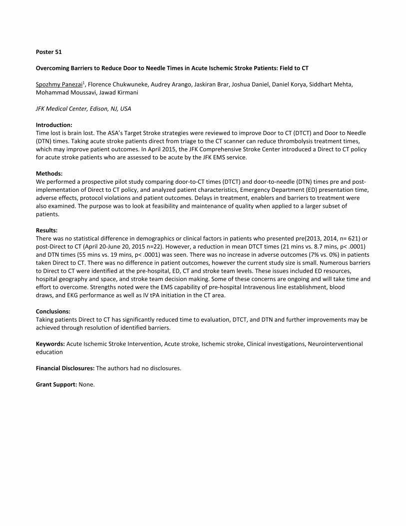
Poster 51 Overcoming Barriers to Reduce Door to Needle Times in Acute Ischemic Stroke Patients: Field to CT Spozhmy Panezai1, Florence Chukwuneke, Audrey Arango, Jaskiran Brar, Joshua Daniel, Daniel Korya, Siddhart Mehta, Mohammad Moussavi, Jawad Kirmani JFK Medical Center, Edison, NJ, USA Introduction: Time lost is brain lost. The ASA’s Target Stroke strategies were reviewed to improve Door to CT (DTCT) and Door to Needle (DTN) times. Taking acute stroke patients direct from triage to the CT scanner can reduce thrombolysis treatment times, which may improve patient outcomes. In April 2015, the JFK Comprehensive Stroke Center introduced a Direct to CT policy for acute stroke patients who are assessed to be acute by the JFK EMS service. Methods: We performed a prospective pilot study comparing door-to-CT times (DTCT) and door-to-needle (DTN) times pre and post-implementation of Direct to CT policy, and analyzed patient characteristics, Emergency Department (ED) presentation time, adverse effects, protocol violations and patient outcomes. Delays in treatment, enablers and barriers to treatment were also examined. The purpose was to look at feasibility and maintenance of quality when applied to a larger subset of patients. Results: There was no statistical difference in demographics or clinical factors in patients who presented pre(2013, 2014, n= 621) or post-Direct to CT (April 20-June 20, 2015 n=22). However, a reduction in mean DTCT times (21 mins vs. 8.7 mins, p< .0001) and DTN times (55 mins vs. 19 mins, p< .0001) was seen. There was no increase in adverse outcomes (7% vs. 0%) in patients taken Direct to CT. There was no difference in patient outcomes, however the current study size is small. Numerous barriers to Direct to CT were identified at the pre-hospital, ED, CT and stroke team levels. These issues included ED resources, hospital geography and space, and stroke team decision making. Some of these concerns are ongoing and will take time and effort to overcome. Strengths noted were the EMS capability of pre-hospital Intravenous line establishment, blood draws, and EKG performance as well as IV tPA initiation in the CT area. Conclusions: Taking patients Direct to CT has significantly reduced time to evaluation, DTCT, and DTN and further improvements may be achieved through resolution of identified barriers. Keywords: Acute Ischemic Stroke Intervention, Acute stroke, Ischemic stroke, Clinical investigations, Neurointerventional education Financial Disclosures: The authors had no disclosures. Grant Support: None.
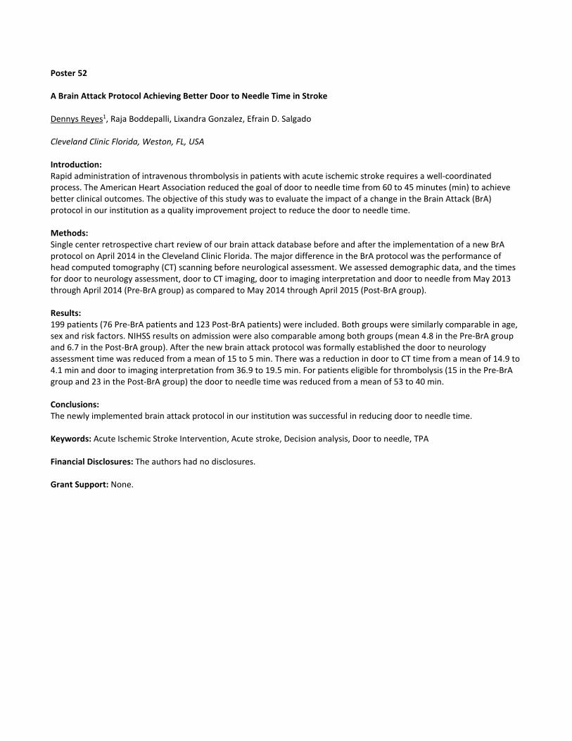
Poster 52 A Brain Attack Protocol Achieving Better Door to Needle Time in Stroke Dennys Reyes1, Raja Boddepalli, Lixandra Gonzalez, Efrain D. Salgado Cleveland Clinic Florida, Weston, FL, USA Introduction: Rapid administration of intravenous thrombolysis in patients with acute ischemic stroke requires a well-coordinated process. The American Heart Association reduced the goal of door to needle time from 60 to 45 minutes (min) to achieve better clinical outcomes. The objective of this study was to evaluate the impact of a change in the Brain Attack (BrA) protocol in our institution as a quality improvement project to reduce the door to needle time. Methods: Single center retrospective chart review of our brain attack database before and after the implementation of a new BrA protocol on April 2014 in the Cleveland Clinic Florida. The major difference in the BrA protocol was the performance of head computed tomography (CT) scanning before neurological assessment. We assessed demographic data, and the times for door to neurology assessment, door to CT imaging, door to imaging interpretation and door to needle from May 2013 through April 2014 (Pre-BrA group) as compared to May 2014 through April 2015 (Post-BrA group). Results: 199 patients (76 Pre-BrA patients and 123 Post-BrA patients) were included. Both groups were similarly comparable in age, sex and risk factors. NIHSS results on admission were also comparable among both groups (mean 4.8 in the Pre-BrA group and 6.7 in the Post-BrA group). After the new brain attack protocol was formally established the door to neurology assessment time was reduced from a mean of 15 to 5 min. There was a reduction in door to CT time from a mean of 14.9 to 4.1 min and door to imaging interpretation from 36.9 to 19.5 min. For patients eligible for thrombolysis (15 in the Pre-BrA group and 23 in the Post-BrA group) the door to needle time was reduced from a mean of 53 to 40 min. Conclusions: The newly implemented brain attack protocol in our institution was successful in reducing door to needle time. Keywords: Acute Ischemic Stroke Intervention, Acute stroke, Decision analysis, Door to needle, TPA Financial Disclosures: The authors had no disclosures. Grant Support: None.
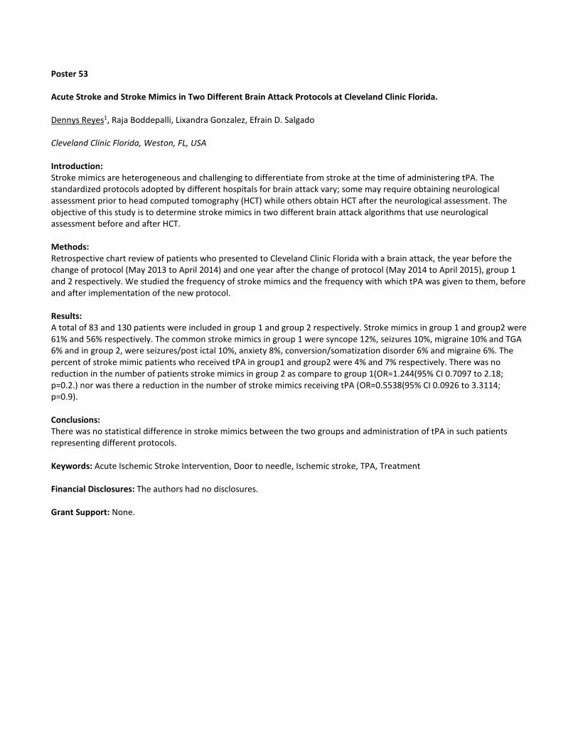
Poster 53 Acute Stroke and Stroke Mimics in Two Different Brain Attack Protocols at Cleveland Clinic Florida. Dennys Reyes1, Raja Boddepalli, Lixandra Gonzalez, Efrain D. Salgado Cleveland Clinic Florida, Weston, FL, USA Introduction: Stroke mimics are heterogeneous and challenging to differentiate from stroke at the time of administering tPA. The standardized protocols adopted by different hospitals for brain attack vary; some may require obtaining neurological assessment prior to head computed tomography (HCT) while others obtain HCT after the neurological assessment. The objective of this study is to determine stroke mimics in two different brain attack algorithms that use neurological assessment before and after HCT. Methods: Retrospective chart review of patients who presented to Cleveland Clinic Florida with a brain attack, the year before the change of protocol (May 2013 to April 2014) and one year after the change of protocol (May 2014 to April 2015), group 1 and 2 respectively. We studied the frequency of stroke mimics and the frequency with which tPA was given to them, before and after implementation of the new protocol. Results: A total of 83 and 130 patients were included in group 1 and group 2 respectively. Stroke mimics in group 1 and group2 were 61% and 56% respectively. The common stroke mimics in group 1 were syncope 12%, seizures 10%, migraine 10% and TGA 6% and in group 2, were seizures/post ictal 10%, anxiety 8%, conversion/somatization disorder 6% and migraine 6%. The percent of stroke mimic patients who received tPA in group1 and group2 were 4% and 7% respectively. There was no reduction in the number of patients stroke mimics in group 2 as compare to group 1(OR=1.244(95% CI 0.7097 to 2.18; p=0.2.) nor was there a reduction in the number of stroke mimics receiving tPA (OR=0.5538(95% CI 0.0926 to 3.3114; p=0.9). Conclusions: There was no statistical difference in stroke mimics between the two groups and administration of tPA in such patients representing different protocols. Keywords: Acute Ischemic Stroke Intervention, Door to needle, Ischemic stroke, TPA, Treatment Financial Disclosures: The authors had no disclosures. Grant Support: None.

Poster 54 Response to Recent Trials in a Major Tertiary Stroke Center Hazem Shoirah1,2, Justin R Mascitelli1, Natalie Wilson1, Peter Shearer1, Johanna Fifi1, Srinivasan Paramasivam1, Stephan Mayer1, Stanley Tuhrim1, J Mocco1 1Mount Sinai Medical Center, New York City, NY, USA, 2University of Pittsburgh Medical Center, Pittsburgh, PA, USA Introduction: The recent pivotal intra-arterial treatment (IAT) trials for acute ischemic stroke (AIS) necessitate immediate system changes in tertiary stroke centers. Our center formed a multi-disciplinary stroke response coalition. We reduced emergency department (ED) time by creating a streamlined door-to-CT pathway. We created a resident-led instant messaging platform that connected all AIS responders within our center and its satellite hospitals. We hypothesize that this will improve AIS response time and patient outcomes. Methods: We retrospectively reviewed patients who underwent IAT for AIS between January 2014 and April 2015. We divided patients in two cohorts based on presentation prior to or after implementation of the changes. Using t-test we compared both cohorts for monthly rates of IAT, their demographics, imaging modality, door to puncture time and discharge modified Rankin scale (MRS). Results: There were 32 and 37 patients in the pre- and post-implementation cohorts respectively. The monthly rate of IAT was higher post-implementation (7.2 vs 2.8, p = 0.04) with a trend for more IAT through transfers (34.1% vs 15.6%, p= 0.07). CTA was used more often post-implementation though that did not reach significance (62.2% vs 43.8%, p = 0.13). Door to IAT time was reduced post-implementation (103.5 vs 180.8, p = 0.002) and groin puncture to first pass was achieved more rapidly (34.3 vs 53, p = 0.01). There was no difference in age, gender, NIHSS at presentation, IV tPA use, complication rate or MRS at discharge. More patients in the post-implementation group had anterior circulation strokes (100% vs 87.5%, p = 0.03) with a trend for early neurological improvement (59.5% vs 43.8%, p = 0.2). Conclusions: Our study shows that multi-disciplinary coalitions and implementation of telecommunication can result in system-wide changes with immediate and significant impact on patient care. This model can be applied broadly. Keywords: Acute stroke, Interventional neuroradiology, Mechanical thrombectomy Financial Disclosures: The authors had no disclosures. Grant Support: None.

Poster 55 Stroke VAN: A Large Artery Stroke Screening Tool for the ED and Field Use Mohamed S Teleb1 Banner Desert Medical Center, Mesa, AZ, USA Introduction: Introduction: Identification of large artery stroke (LAS) has become increasingly important with the recent publications of five favorable acute stroke thrombectomy trials. Unfortunately, there are limited number of LAS screening scales being utilized in the field or emergency department (ED). None were identified in Pubmed or Google Scholar searches. Popularly used prehospital stroke screening tools such as the CPSS or LAPSS were not designed for LAS screening. A quick, simple screening tool was designed and tested in the ED using nurse responders. Methods: VAN, a screening tool designed based on neurovascular anatomy was devised. Nurses were trained to use this screening tool. VAN was used in the trauma bay prior to patients being evaluated by a doctor and before CT scan of head. Accuracy of VAN screened patients with LAS was analyzed for positive predictive value (PPV), sensitivity, negative predictive value (NPV), and specificity. Results: 62 acute stroke codes were activated during the screening period. 19 were VAN positive and 24 had an NIHSS of 6 or greater. 14 patients had LAS and were in the VAN positive and NIHSS of 6 or greater group. Both had sensitivity of 100% but VAN had a 74% PPV while NIHSS only had 58%. No patients that were VAN negative or with a NIHSS < 6 had a LAS (both had 100% NPV). However, VAN had 90% specificity vs 74% for NIHSS. Conclusion: In this small cohort, the VAN screening tool accurately identified LAS and allowed for early activation of the Neurointerventional team prior to CT scan. A larger study to validate this screening tools use in the ED and in the field for triage is warranted. A larger study is also needed to validate whether its use can decrease door to puncture time for LAS. Keywords: Stroke, Scale, Neurointerventional education Financial Disclosures: The author had no disclosures. Grant Support: None.

Poster 56 Preliminary Experience with Precipitating Hydrophobic Injectable Liquid (PHIL) in Treating Cranial AVMs and Fistulas Edgar A Samaniego1, Santiago Ortega2 1Hospital Eugenio Espejo, Quito, Ecuador, 2University of Iowa Hospitals and Clinics, Iowa City, IA, USA Introduction: PHIL is a new embolic agent that can be used in the treatment of AVMs and fistulas. We describe our preliminary experience. Methods: Between June and August 2015 five patients: 3 with AVMs and 2 with fistulas were treated with PHIL 25%. Two patients were treated exclusively with PHIL and three with Onyx + PHIL. The three patients had ruptured AVMs (Spetzler-Martin grade II, III and III). One patient was treated acutely due to neurological deterioration and the other four patients were treated in a programmed fashion. Results: The patient with pial fistula had an asymptomatic procedural complication with a piece of PHIL migrating to a frontal artery. The decreased radiopacity of PHIL may have contributed to this complication. There were no other procedural complications. Two of three patients with AVMs were cured in one session. The third AVM was embolized in a 50%, as it was decided to perform staged embolization of the nidus due to its size and multiple afferents. The two patients with cranial fistulas were cured. Both patients were treated with Onyx and PHIL. In 4/5 patients with used a detachable tip microcatheter due to prolonged injection times. 4/5 patients were discharged with the same neurological condition. The patient with the AVM Spetzler-Martin III that was cured with one session of PHIL died during hospitalization due to complications of the initial hemorrhage. This patient was treated acutely due to neurological deterioration. A patient with a cortical AVM Spetzler-Martin grade II seized after the procedure. We did not encounter any other peri-procedural complications. Conclusions: PHIL appears to be a great embolic agent that can be used for treatment of AVMs and fistulas. It is less radiopaque than Onyx and extreme caution should be used when injecting the embolic agent. Keywords: AVM embolization, Embolization, Hemorrhage Financial Disclosures: The authors had no disclosures. Grant Support: None.

Poster 57 Outcome After Treatment of Spinal Dural Arteriovenous Fistula: a Single-Institution Case Series Anita Tipirneni1, Kunakorn Atchaneeyasakul1, Pankaj Sharma1, Nirav H. Shah1, Dileep Yavagal1,2 1University of Miami Miller School of Medicine, Department of Neurology, Miami, FL, USA, 2University of Miami Miller School of Medicine, Department of Neurosurgery, Miami, FL, USA Introduction: Spinal dural arteriovenous fistulas (SDAVF) are an uncommon but treatable cause of myelopathy. Many patients improve after treatment, however it is not known what, if any, factors may increase the odds of clinical improvement. Methods: We performed a retrospective chart review of PACS imaging system for the term “spinal dural arteriovenous fistula”. Inclusion criteria included any confirmed case of SDAVF. Exclusion criteria included no intervention and no follow-up. Our primary outcome was defined as any improvement in myelopathy symptoms and was assessed by blinded evaluators comparing initial assessments to final follow-up. No improvement and any improvement groups were compared across demographics, vascular risk factors, smoking history, fistula level, type of symptoms, time to diagnosis and type of treatment. Bivariate analysis was used to further evaluate any significant differences on T-test or Chi-square analysis. Results: Symptom progression was halted after treatment in all patients. Mean follow-up time was 9 months. Raters were able to come to consensus on all patients for outcome of any improvement. There were no differences in demographics, vascular risk factors, fistula level, treatment type, smoking history or time to diagnosis between no improvement and any improvement groups. There was a significant difference in bowel/bladder/impotence symptoms between these two groups with the any improvement group being more likely to have had these symptoms (82% vs. 33%, p=0.03). Bivariate analysis revealed this association to be significant as well, with an odds ratio of 9 (95% CI 1.14-71, p=0.04). Conclusions: In patients with SDAVF, those who experienced bowel/bladder/impotence symptoms were 9 times more likely to have symptom improvement than those who did not. These represent an unexpected result given these types of symptoms are known to be the least likely to resolve following fistula treatment. Further investigations into clinical differences are warranted. Keywords: Spinal malformation therapy, Treatment Financial Disclosures: The authors had no disclosures. Grant Support: None.

Poster 58 Spinal Dural Ateriovenous Fistulas Mimicking Demyelinating Disease Anita Tipirneni1, Melissa R Ortega1, Amer M Malik1, Dileep R Yavagal1,2 1University of Miami Miller School of Medicine, Department of Neurology, Miami, FL, USA, 2University of Miami Miller School of Medicine, Department of Neurological Surgery, Miami, FL, USA Introduction: Spinal dural arteriovenous fistulas (SDAVF) are an uncommon but treatable cause of myelopathy. Their diagnosis is often delayed as they can mimic other diseases, especially demyelinating disease, on imaging. Methods: We recruited five consecutive patients referred to an outpatient neurologist with expertise in neuroimmunology at our university medical center or referred to a tertiary academic hospital for a definitive diagnosis. We analyzed demographics, imaging, time to diagnosis, and disability at follow-up. Results: All patients presented with a clinical picture of subacute to chronic myelopathy and were later diagnosed as having SDAVF. Mean time from symptom onset to diagnosis was 1.4 years. All patients were male and 80% were elderly (i.e. >70 years old). Ethnic background was diverse and included three Caucasian patients, one African American patient, and one Hispanic patient. All patients had abnormal T2 signal on spine MRIs in addition to T2 flow voids or an increase in number, size and tortuosity of spinal vessels. Time to diagnosis and treatment was shorter in those who received a full spinal and intracranial angiography early in their presentation than in those who did not. All patients underwent successful embolization or surgical intervention with obliteration of the fistula, after which progression of symptoms were halted with improvement of symptoms in 80% of patients. Conclusions: The diagnosis of SDAVF is commonly delayed due to symptoms mimicking other spinal diseases such as demyelinating disease. Once diagnosed, the rate of successful endovascular and surgical treatment is very high. Keywords: Endovascular, Interventional neuroradiology, Imaging Financial Disclosures: Dr. Yavagal has worked as a consultant and received consulting fees from Aldegen/Cytomedix Inc., EV3 Neurovascular/Covidien, and Stryker Neurovascular. Grant Support: None.

Poster 59 Post-Acute Ischemic Stroke Thrombectomy and Obesity Paradox Michael G. Abraham1, Matthew Sharrock, Rawan Albadareen, Sanjeev Keshary, Manoj Mittal University of Kansas Medical Center, Kansas City, KS, USA Introduction: A recurring theme for physicians is to promote weight loss in their patients. Growing evidence, though, demonstrates that there is an inverse relationship between obesity and outcome in acute ischemic stroke patients as well as in rates of hemorrhagic transformation, termed the ‘obesity paradox’. To date, this is the first case series evaluating the obesity paradox in acute ischemic stroke (AIS) patients treated with endovascular thrombectomy. Methods: Single-centered, retrospective chart review was performed of all patients who underwent ischemic stroke endovascular interventions between 2008 and 2014. Demographics, comorbidities, prior medication use, stroke characteristics, procedure related factors, and 3 month outcome (good outcome=MRS≤2) were compared between patients with or without obesity (BMI>30). Chi square/Fisher exact test and t-test/Wilcoxon Rank Sums were used for categorical and continuous variables. Significant predictors (p< 0.10) from univariate analysis were entered into a multivariate logistic regression model to study the independent effect of obesity on patient’s outcome. Results: Out of 97 patients who underwent endovascular stroke interventions, 35 (36%) patients were obese. Obese were significantly younger than non-obese (63 vs 68 years, p=0.07), had prior history of diabetes (31% vs 8%, p=0.003), less likely on anticoagulation therapy (6% vs 19%, p=0.07), had less anterior (69% vs 84%, p=0.08) strokes, less post procedure ICH (0% vs 8%, p=0.08), and more patients with good outcome (50% vs 28%, p=0.03). In a multivariate logistic regression model controlling for age, diabetes, anterior circulation, and prior anticoagulation usage, obesity was associated with good outcome (OR 2.83, 95% CI 1.01-8.28). Conclusions: Our single center endovascular AIS registry demonstrates a trend towards better outcomes in patients with a body mass index (BMI) greater than 30. The exact explanation of this finding is not clearly known. This finding should be confirmed in a multicenter registry and a population based study. Keywords: Stroke, Endovascular therapy, Cerebrovascular disease, Functional recovery in stroke, mRS Financial Disclosures: The authors had no disclosures. Grant Support: None.
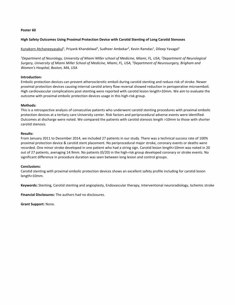
Poster 60 High Safety Outcomes Using Proximal Protection Device with Carotid Stenting of Long Carotid Stenoses Kunakorn Atchaneeyasakul1, Priyank Khandelwal3, Sudheer Ambekar2, Kevin Ramdas1, Dileep Yavagal1 1Department of Neurology, University of Miami Miller school of Medicine, Miami, FL, USA, 2Department of Neurological Surgery, University of Miami Miller School of Medicine, Miami, FL, USA, 3Department of Neurosurgery, Brigham and Women's Hospital, Boston, MA, USA Introduction: Embolic protection devices can prevent atherosclerotic emboli during carotid stenting and reduce risk of stroke. Newer proximal protection devices causing internal carotid artery flow reversal showed reduction in perioperative microemboli. High cardiovascular complications post-stenting were reported with carotid lesion length>10mm. We aim to evaluate the outcome with proximal embolic protection devices usage in this high-risk group. Methods: This is a retrospective analysis of consecutive patients who underwent carotid stenting procedures with proximal embolic protection devices at a tertiary care University center. Risk factors and periprocedural adverse events were identified. Outcomes at discharge were noted. We compared the patients with carotid stenosis length >10mm to those with shorter carotid stenosis. Results: From January 2011 to December 2014, we included 27 patients in our study. There was a technical success rate of 100% proximal protection device & carotid stent placement. No periprocedural major stroke, coronary events or deaths were recorded. One minor stroke developed in one patient who had a string sign. Carotid lesion length>10mm was noted in 20 out of 27 patients, averaging 14.9mm. No patients (0/20) in the high-risk group developed coronary or stroke events. No significant difference in procedure duration was seen between long lesion and control groups. Conclusions: Carotid stenting with proximal embolic protection devices shows an excellent safety profile including for carotid lesion length>10mm. Keywords: Stenting, Carotid stenting and angioplasty, Endovascular therapy, Interventional neuroradiology, Ischemic stroke Financial Disclosures: The authors had no disclosures. Grant Support: None.
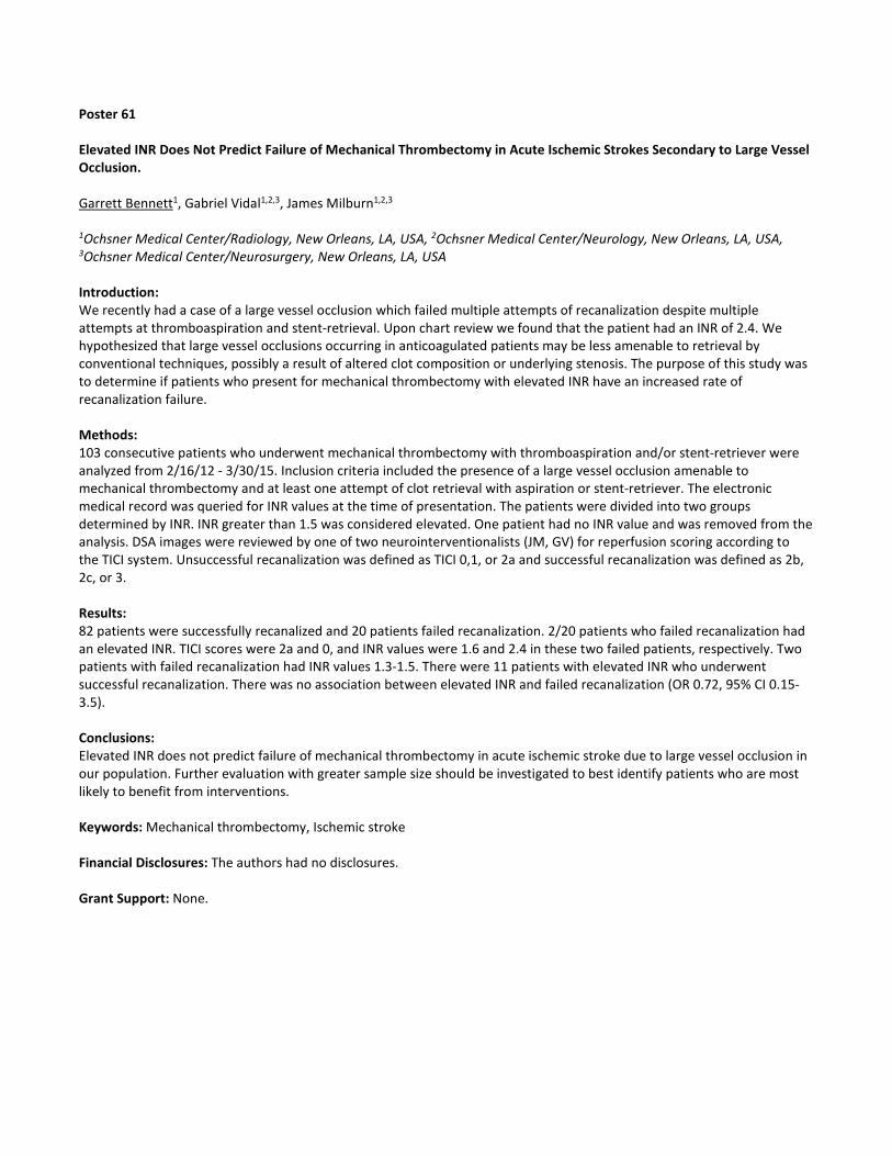
Poster 61 Elevated INR Does Not Predict Failure of Mechanical Thrombectomy in Acute Ischemic Strokes Secondary to Large Vessel Occlusion. Garrett Bennett1, Gabriel Vidal1,2,3, James Milburn1,2,3 1Ochsner Medical Center/Radiology, New Orleans, LA, USA, 2Ochsner Medical Center/Neurology, New Orleans, LA, USA, 3Ochsner Medical Center/Neurosurgery, New Orleans, LA, USA Introduction: We recently had a case of a large vessel occlusion which failed multiple attempts of recanalization despite multiple attempts at thromboaspiration and stent-retrieval. Upon chart review we found that the patient had an INR of 2.4. We hypothesized that large vessel occlusions occurring in anticoagulated patients may be less amenable to retrieval by conventional techniques, possibly a result of altered clot composition or underlying stenosis. The purpose of this study was to determine if patients who present for mechanical thrombectomy with elevated INR have an increased rate of recanalization failure. Methods: 103 consecutive patients who underwent mechanical thrombectomy with thromboaspiration and/or stent-retriever were analyzed from 2/16/12 - 3/30/15. Inclusion criteria included the presence of a large vessel occlusion amenable to mechanical thrombectomy and at least one attempt of clot retrieval with aspiration or stent-retriever. The electronic medical record was queried for INR values at the time of presentation. The patients were divided into two groups determined by INR. INR greater than 1.5 was considered elevated. One patient had no INR value and was removed from the analysis. DSA images were reviewed by one of two neurointerventionalists (JM, GV) for reperfusion scoring according to the TICI system. Unsuccessful recanalization was defined as TICI 0,1, or 2a and successful recanalization was defined as 2b, 2c, or 3. Results: 82 patients were successfully recanalized and 20 patients failed recanalization. 2/20 patients who failed recanalization had an elevated INR. TICI scores were 2a and 0, and INR values were 1.6 and 2.4 in these two failed patients, respectively. Two patients with failed recanalization had INR values 1.3-1.5. There were 11 patients with elevated INR who underwent successful recanalization. There was no association between elevated INR and failed recanalization (OR 0.72, 95% CI 0.15-3.5). Conclusions: Elevated INR does not predict failure of mechanical thrombectomy in acute ischemic stroke due to large vessel occlusion in our population. Further evaluation with greater sample size should be investigated to best identify patients who are most likely to benefit from interventions. Keywords: Mechanical thrombectomy, Ischemic stroke Financial Disclosures: The authors had no disclosures. Grant Support: None.

Poster 62 Eptifibatide is Safe and May Improve Outcomes in Stroke Patients Undergoing Thrombectomy after Receiving IVtPA Jaskiran Brar1, Siddhart Mehta, Mohammad Moussavi, Daniel Korya, Harina Brar, Swathi Kondapalli, Mena Saman, Aska Shaikh, Rushil Kalola, Yong- Bum Song, Jawad Kirmani JFK Medical Center, Edison, NJ, USA Introduction: Recent trials have shown significant improvement in outcome for patients with acute ischemic stroke (AIS) undergoing mechanical thrombectomy and IV tPA. In addition to its acute antiplatelet properties eptifibatide may also reduce acute inflammatory response following neurovascular intervention. Our goal was to evaluate the potential benefit of adding IV eptifibatide to mechanical thrombectomy and IV tPA. Methods: Patients who presented to a community based university affiliated comprehensive stroke center from 2012-2015 with AIS over a 2 year period were included in the study. Only patients who received thrombectomy after IV tPA were included. A subgroup of those patients also received IV eptifibatide as a continuous drip during and after the procedure. The initial NIH Stroke Score (NIHSS) and 24hour NIHSS were compared between the two groups with paired samples t-test using SPSS Version 22. Results: A total of 866 patients were evaluated, and 139 met the study criteria. All patients received mechanical thrombectomy after IV tPA; 70 also received a bolus dose of 135 mcg/kg of eptifibatide followed by 0.5 mcg/kg/min continuous drip. The mean duration of the drip was 23.8 minutes (SD 14.13). There were no significant differences in complication or hemorrhage rates between groups. The mean initial minus 24 hour NIHSS (Initial-24) for the patients receiving only IV tPA/thrombectomy was 1.6. Patients who also received eptifibatide had a mean Initial- 24 of 3.6. The paired mean difference was 2 (95% CI .193.8; p=.03), favoring the addition of eptifibatide. Conclusions: The addition of eptifibatide bolus followed by a continuous drip for a mean of 24 hours to IV tPA/thrombectomy was associated with a significantly better 24 hour postprocedure outcome. The mechanism of action may be related to the suppression of inflammation and potential prevention of re-thrombosis after treatment. No additional complications were noted with eptifibatide. A larger prospective trial is warranted to corroborate our findings. Keywords: New technique, Acute Ischemic Stroke Intervention, Endovascular therapy Financial Disclosures: The authors had no disclosures. Grant Support: None.

Poster 63 Efficacy of IV tPA in Treatment of Large Vessel Ischemic Strokes Not Amenable to Endovascular Therapy Jaskiran Brar1, Spozhmy Panezai, Joshua Daniel, Jawad Kirmani, Siddhart Mehta, Daniel Korya, Florence Chukwuneke, Mohammad Moussavi JFK Medical Center, Edison, NJ, USA Introduction: Intravenous Tpa remains the standard of care for distal large vessels occlusions (M2, M3, P1, P2, A1, A2) not amenable to endovascular treatment. Recent trials (SWIFT PRIME, REVASCAT, EXTENDIA and THERAPY) showed that 28-39% of acute stroke patients with proximal large vessel occlusions treated with IV tpa alone achieved mRS 0-2 at 90 days while 26-38% achieved mRS 3-4, and 26-36%achieved mRS 5-6. We analyzed patients admitted to our institution with acute ischemic stroke secondary to distal large vessel occlusions to assess if IV tPA alone was effective in improving discharge NIHSS from admission NIHSS and discharge modified Rankin Score (mRS). Methods: We conducted a retrospective review of acute ischemic stroke patients who received IV tPA only from Jan 1, 2012December 30, 2014 at our community based university affiliated comprehensive stroke center. CTA and/or MRA images were reviewed to confirm the presence of a distal large vessel occlusion (M2, M3, P1, P2, A1 and A2). Admission NIHSS, discharge NIHSS, and discharge mRS were abstracted. GraphPad Quick Calcs Web site was used to obtain descriptive statistics. Results: Of the 162 patients reviewed, 14 had distal large vessel occlusions on imaging. One patient was excluded due to incomplete data. Patients were mostly female (71%), with mean age of 73.5 years (SD = 11.8). Mean admission NIHSS was 6.2 (SD = 2.8) and discharge NIHSS was 1.8 (SD =1.6). Mean discharge mRS was 1.4 (SD=0.9). 92% of patients were discharged with mRS of 0-2 and 8% with discharge mRS 3-4 (Fig 1). Conclusions: Distal large vessel occlusions are less amenable to endovascular therapy due to more distal location, tortuous vessel anatomy, and smaller vessel caliber. In such cases, IV tPA alone may be effective in improving discharge NIHSS and mRS. Limitations of this study included small patient population and inconsistent admission mRS. Keywords: mRS, Endovascular therapy, Medical management Financial Disclosures: The authors had no disclosures. Grant Support: None.
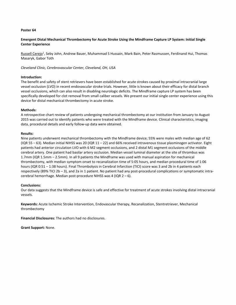
Poster 64 Emergent Distal Mechanical Thrombectomy for Acute Stroke Using the Mindframe Capture LP System: Initial Single Center Experience Russell Cerejo1, Seby John, Andrew Bauer, Muhammad S Hussain, Mark Bain, Peter Rasmussen, Ferdinand Hui, Thomas Masaryk, Gabor Toth Cleveland Clinic, Cerebrovascular Center, Cleveland, OH, USA Introduction: The benefit and safety of stent retrievers have been established for acute strokes caused by proximal intracranial large vessel occlusion (LVO) in recent endovascular stroke trials. However, little is known about their efficacy for distal branch vessel occlusions, which can also result in disabling neurologic deficits. The Mindframe capture LP system has been specifically developed for clot removal from small caliber vessels. We present our initial single center experience using this device for distal mechanical thrombectomy in acute stroke. Methods: A retrospective chart review of patients undergoing mechanical thrombectomy at our institution from January to August 2015 was carried out to identify patients who were treated with the Mindframe device. Clinical characteristics, imaging data, procedural details and early follow-up data were obtained. Results: Nine patients underwent mechanical thrombectomy with the Mindframe device; 55% were males with median age of 62 (IQR 55 – 63). Median initial NIHSS was 20 (IQR 11 – 22) and 66% received intravenous tissue plasminogen activator. Eight patients had anterior circulation LVO with 6 M2 segment occlusions, and 2 distal M1 segment occlusions of the middle cerebral artery. One patient had basilar artery occlusion. Median vessel luminal diameter at the site of thrombus was 1.7mm (IQR 1.5mm – 2.5mm). In all 9 patients the Mindframe was used with manual aspiration for mechanical thrombectomy, with median symptom onset to recanalization time of 5:05 hours, and median procedural time of 1:06 hours (IQR 0:51 – 1:38 hours). Final Thrombolysis in Cerebral Infarction (TICI) score was 3 and 2b in 4 patients each respectively (89% TICI 2b – 3), and 2a in 1 patient. No patient had any post-procedural complications or symptomatic intra-cerebral hemorrhage. Median post-procedure NIHSS was 4 (IQR 2 – 6). Conclusions: Our data suggests that the Mindframe device is safe and effective for treatment of acute strokes involving distal intracranial vessels. Keywords: Acute Ischemic Stroke Intervention, Endovascular therapy, Recanalization, Stentretriever, Mechanical thrombectomy Financial Disclosures: The authors had no disclosures. Grant Support: None.

Poster 65 Utility of Thromboelastogram in Optimizing Antithrombotic Strategy in Secondary Stroke Prevention Vikas Gupta1, Rahat Suddle, Waleed Rahiem University of Missouri, Neurology, Columbia, MO, USA Introduction: Aspirin and clopidogrel resistance increases relative risk of recurrent ischemic stroke. Thromboelastogram is a readily available objective measure of platelet inhibition induced by aspirin and clopidogrel which can be used to evaluate efficacy and optimize antithrombotic regimen in secondary stroke prevention. Methods: Retrospective review of a prospectively maintained database of all patients admitted to the inpatient stroke service. Selected patients demonstrated evidence of acute ischemic stroke on noncontrast head CT or MRI and were taking either aspirin or Plavix or both prior to admission. Patients with transient ischemic attack, atrial fibrillation, hemorrhagic stroke, intracranial neoplasm were excluded. Patients were divided into two groups based on presence or absence of traditional risk factors for ischemic stroke. The adequacy of antiplatelet activity was defined by percentage of arachidonic acid and ADP inhibition on thromboelastogram. Patients were classified as responders or nonresponder based upon>= 50% inhibition of either arachidonic acid or ADP induced platelet aggregation. Results: A total of 40 patients between were analyzed. 15/40 (37.5%) were aspirin nonresponder while 25/40 (62.5%) were clopidogrel non-responsive. Aspirin non responsiveness was seen in 5 out of 8 (62.5%) patients with no identifiable risk factors whereas 10/31 (32.2%) patients with one or more risk factors. 4/9 (44%) patients with absent risk factors demonstrated no responsiveness to clopidogrel while a higher level of lack of response to clopidogrel was seen in 17/29 (58.6%) patients with one or more risk factors. Conclusions: We found a high percentage of patients with acute ischemic stroke with inadequate response to aspirin and/or clopidogrel. Thromboelastography offers an objective assessment of platelet function and may be used to optimize secondary stroke prevention. A small sample size limits interpretation of our study. Keywords: Acute stroke, Coagulation, Ischemic stroke, Stroke, Thrombosis Financial Disclosures: The authors had no disclosures. Grant Support: None.

Poster 66 Unchanged Utilization of Endovascular Treatment in Acute Ischemic Stroke Patients in the Post IMS-III Era Ameer E. Hassan1, Ahmed A. Malik2, Wondwossen Tekle1, Adnan I Qureshi2 1UTSCHSA - Valley Baptist, Harlingen, TX, USA, 2Zeenat Qureshi Stroke Institute, St Cloud, MN, USA Introduction: Background: The results of Interventional Management of Stroke (IMS) III, MR RESCUE, and SYNTHESIS EXPANSION trials are expected to affect the utilization of endovascular treatment for acute ischemic stroke. Objective: To evaluate trends in utilization of endovascular treatment among acute ischemic stroke patients after the publication of IMS III, MR RESCUE, and SYNTHESIS EXPANSION trials. Methods: Methods: We obtained data for patients admitted to hospitals with a primary diagnosis of ischemic stroke using data from Minnesota Hospital Association. We determined the rate of utilization of endovascular treatment for acute ischemic stroke from March 2012 to December 2012 and March 2013 to December 2013. Outcomes were classified based on discharge disposition. Results: Results: Of the 13,408 patients with ischemic stroke, 6765 (50.5%) were admitted with ischemic stroke prior to, and 6643 (49.5%) after the publication of trials’ results. The proportion of patients who underwent endovascular treatment was unchanged: 2.3% of ischemic stroke patients in the pre-IMS III group vs 2.4% in the post-IMS III group (p=0.6). The estimated number of patients undergoing any thrombolytic treatment increased (7.1% versus 7.8%,p=0.1). There was a significant reduction in length of stay in the post IMS III group, 3.8 ± 4.1 vs 3.6 ± 3.5 (p=0.02). The rates of discharge to home remained unchanged in the pre- and post- IMS III groups (3646 (53.9%) vs 3585 (54.0%), p=0.9). Conclusions: Conclusion: Contrary to expectations, there has been no reduction in the utilization of endovascular treatment among acute ischemic stroke patients after IMS-III, MR RESCUE and SYNTHESIS EXPANSION results were published. Keywords: Acute Ischemic Stroke Intervention, Acute stroke, Neurointerventional program Financial Disclosures: AE Hassan - Consultant for Medtronic, microvention, Stryker and GE Healthcare. Grant Support: None.
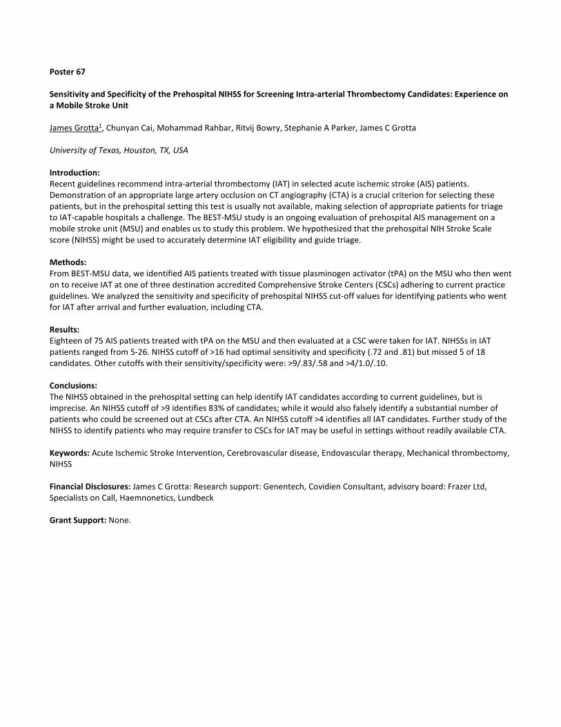
Poster 67 Sensitivity and Specificity of the Prehospital NIHSS for Screening Intra-arterial Thrombectomy Candidates: Experience on a Mobile Stroke Unit James Grotta1, Chunyan Cai, Mohammad Rahbar, Ritvij Bowry, Stephanie A Parker, James C Grotta University of Texas, Houston, TX, USA Introduction: Recent guidelines recommend intra-arterial thrombectomy (IAT) in selected acute ischemic stroke (AIS) patients. Demonstration of an appropriate large artery occlusion on CT angiography (CTA) is a crucial criterion for selecting these patients, but in the prehospital setting this test is usually not available, making selection of appropriate patients for triage to IAT-capable hospitals a challenge. The BEST-MSU study is an ongoing evaluation of prehospital AIS management on a mobile stroke unit (MSU) and enables us to study this problem. We hypothesized that the prehospital NIH Stroke Scale score (NIHSS) might be used to accurately determine IAT eligibility and guide triage. Methods: From BEST-MSU data, we identified AIS patients treated with tissue plasminogen activator (tPA) on the MSU who then went on to receive IAT at one of three destination accredited Comprehensive Stroke Centers (CSCs) adhering to current practice guidelines. We analyzed the sensitivity and specificity of prehospital NIHSS cut-off values for identifying patients who went for IAT after arrival and further evaluation, including CTA. Results: Eighteen of 75 AIS patients treated with tPA on the MSU and then evaluated at a CSC were taken for IAT. NIHSSs in IAT patients ranged from 5-26. NIHSS cutoff of >16 had optimal sensitivity and specificity (.72 and .81) but missed 5 of 18 candidates. Other cutoffs with their sensitivity/specificity were: >9/.83/.58 and >4/1.0/.10. Conclusions: The NIHSS obtained in the prehospital setting can help identify IAT candidates according to current guidelines, but is imprecise. An NIHSS cutoff of >9 identifies 83% of candidates; while it would also falsely identify a substantial number of patients who could be screened out at CSCs after CTA. An NIHSS cutoff >4 identifies all IAT candidates. Further study of the NIHSS to identify patients who may require transfer to CSCs for IAT may be useful in settings without readily available CTA. Keywords: Acute Ischemic Stroke Intervention, Cerebrovascular disease, Endovascular therapy, Mechanical thrombectomy, NIHSS Financial Disclosures: James C Grotta: Research support: Genentech, Covidien Consultant, advisory board: Frazer Ltd, Specialists on Call, Haemnonetics, Lundbeck Grant Support: None.
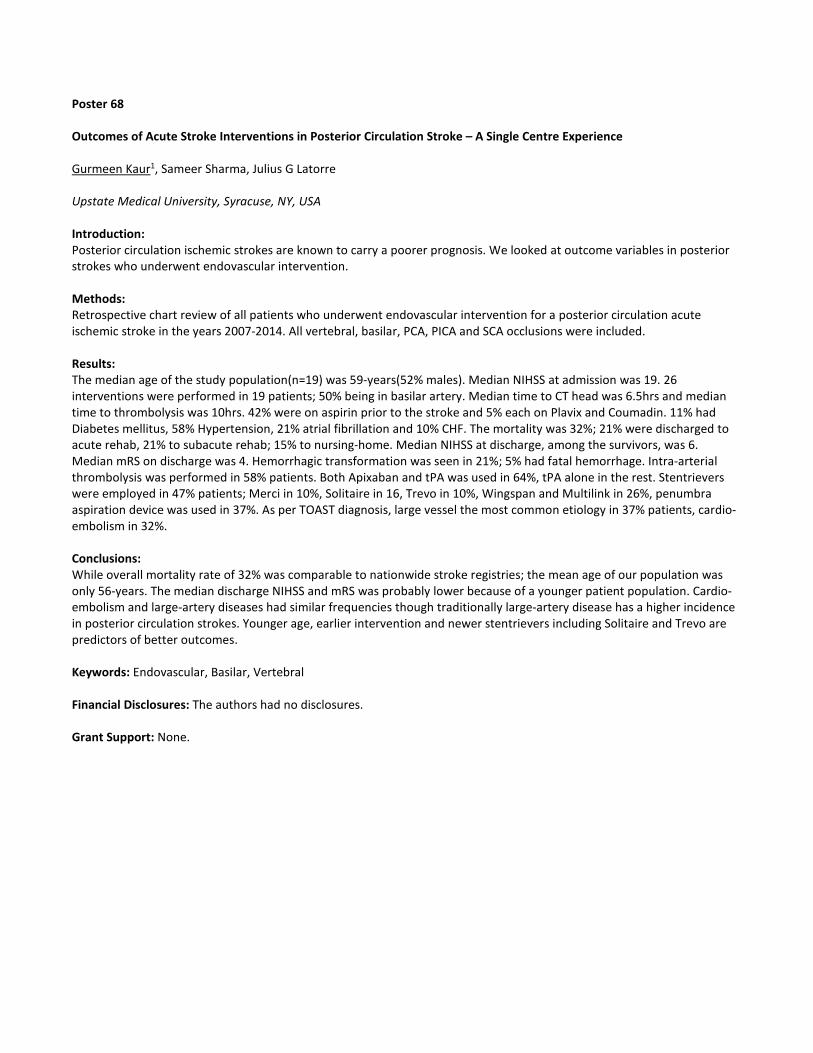
Poster 68 Outcomes of Acute Stroke Interventions in Posterior Circulation Stroke – A Single Centre Experience Gurmeen Kaur1, Sameer Sharma, Julius G Latorre Upstate Medical University, Syracuse, NY, USA Introduction: Posterior circulation ischemic strokes are known to carry a poorer prognosis. We looked at outcome variables in posterior strokes who underwent endovascular intervention. Methods: Retrospective chart review of all patients who underwent endovascular intervention for a posterior circulation acute ischemic stroke in the years 2007-2014. All vertebral, basilar, PCA, PICA and SCA occlusions were included. Results: The median age of the study population(n=19) was 59-years(52% males). Median NIHSS at admission was 19. 26 interventions were performed in 19 patients; 50% being in basilar artery. Median time to CT head was 6.5hrs and median time to thrombolysis was 10hrs. 42% were on aspirin prior to the stroke and 5% each on Plavix and Coumadin. 11% had Diabetes mellitus, 58% Hypertension, 21% atrial fibrillation and 10% CHF. The mortality was 32%; 21% were discharged to acute rehab, 21% to subacute rehab; 15% to nursing-home. Median NIHSS at discharge, among the survivors, was 6. Median mRS on discharge was 4. Hemorrhagic transformation was seen in 21%; 5% had fatal hemorrhage. Intra-arterial thrombolysis was performed in 58% patients. Both Apixaban and tPA was used in 64%, tPA alone in the rest. Stentrievers were employed in 47% patients; Merci in 10%, Solitaire in 16, Trevo in 10%, Wingspan and Multilink in 26%, penumbra aspiration device was used in 37%. As per TOAST diagnosis, large vessel the most common etiology in 37% patients, cardio-embolism in 32%. Conclusions: While overall mortality rate of 32% was comparable to nationwide stroke registries; the mean age of our population was only 56-years. The median discharge NIHSS and mRS was probably lower because of a younger patient population. Cardio-embolism and large-artery diseases had similar frequencies though traditionally large-artery disease has a higher incidence in posterior circulation strokes. Younger age, earlier intervention and newer stentrievers including Solitaire and Trevo are predictors of better outcomes. Keywords: Endovascular, Basilar, Vertebral Financial Disclosures: The authors had no disclosures. Grant Support: None.
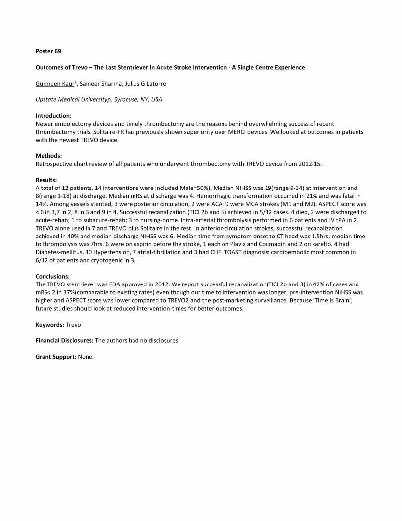
Poster 69 Outcomes of Trevo – The Last Stentriever in Acute Stroke Intervention - A Single Centre Experience Gurmeen Kaur1, Sameer Sharma, Julius G Latorre Upstate Medical Universityp, Syracuse, NY, USA Introduction: Newer embolectomy devices and timely thrombectomy are the reasons behind overwhelming success of recent thrombectomy trials. Solitaire-FR has previously shown superiority over MERCI devices. We looked at outcomes in patients with the newest TREVO device. Methods: Retrospective chart review of all patients who underwent thrombectomy with TREVO device from 2012-15. Results: A total of 12 patients, 14 interventions were included(Male=50%). Median NIHSS was 19(range 9-34) at intervention and 8(range 1-18) at discharge. Median mRS at discharge was 4. Hemorrhagic transformation occurred in 21% and was fatal in 14%. Among vessels stented, 3 were posterior circulation, 2 were ACA, 9 were MCA strokes (M1 and M2). ASPECT score was < 6 in 3,7 in 2, 8 in 3 and 9 in 4. Successful recanalization (TICI 2b and 3) achieved in 5/12 cases. 4 died, 2 were discharged to acute-rehab; 1 to subacute-rehab; 3 to nursing-home. Intra-arterial thrombolysis performed in 6 patients and IV tPA in 2. TREVO alone used in 7 and TREVO plus Solitaire in the rest. In anterior-circulation strokes, successful recanalization achieved in 40% and median discharge NIHSS was 6. Median time from symptom onset to CT head was 1.5hrs; median time to thrombolysis was 7hrs. 6 were on aspirin before the stroke, 1 each on Plavix and Coumadin and 2 on xarelto. 4 had Diabetes-mellitus, 10 Hypertension, 7 atrial-fibrillation and 3 had CHF. TOAST diagnosis: cardioembolic most common in 6/12 of patients and cryptogenic in 3. Conclusions: The TREVO stentriever was FDA approved in 2012. We report successful recanalization(TICI 2b and 3) in 42% of cases and mRS< 2 in 37%(comparable to existing rates) even though our time to intervention was longer, pre-intervention NIHSS was higher and ASPECT score was lower compared to TREVO2 and the post-marketing surveillance. Because ‘Time is Brain’; future studies should look at reduced intervention-times for better outcomes. Keywords: Trevo Financial Disclosures: The authors had no disclosures. Grant Support: None.
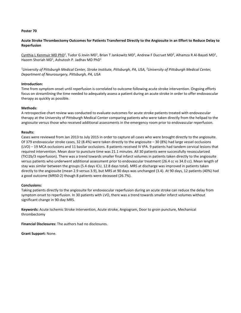
Poster 70 Acute Stroke Thrombectomy Outcomes for Patients Transferred Directly to the Angiosuite in an Effort to Reduce Delay to Reperfusion Cynthia L Kenmuir MD PhD1, Tudor G Jovin MD1, Brian T Jankowitz MD2, Andrew F Ducruet MD2, Alhamza R Al-Bayati MD1, Hazem Shoriah MD1, Ashutosh P. Jadhav MD PhD1 1University of Pittsburgh Medical Center, Stroke Institute, Pittsburgh, PA, USA, 2University of Pittsburgh Medical Center, Department of Neurosurgery, Pittsburgh, PA, USA Introduction: Time from symptom onset until reperfusion is correlated to outcome following acute stroke intervention. Ongoing efforts focus on streamlining the time needed to adequately assess a patient during an acute stroke in order to offer endovascular therapy as quickly as possible. Methods: A retrospective chart review was conducted to evaluate outcomes for acute stroke patients treated with endovascular therapy at the University of Pittsburgh Medical Center comparing patients who were taken directly from the helipad to the angiosuite versus those who received additional assessments in the emergency room prior to endovascular reperfusion. Results: Cases were reviewed from Jan 2013 to July 2015 in order to capture all cases who were brought directly to the angiosuite. Of 379 endovascular stroke cases, 32 (8.4%) were taken directly to the angiosuite – 30 (8%) had large vessel occlusions (LVO) – 19 MCA occlusions and 11 basilar occlusions. 8 patients received IV tPA. 9 patients had tandem cervical lesions that required intervention. Mean door to puncture time was 21.1 minutes. All 30 patients were successfully revascularized (TICI2b/3 reperfusion). There was a trend towards smaller final infarct volumes in patients taken directly to the angiosuite versus patients who underwent additional assessment prior to endovascular treatment (26.4 cc vs 34.0 cc). Mean length of stay was similar between the groups (5.4 days ICU, 12.8 days total). MRS at discharge was improved in patients taken directly to the angiosuite (mean 2.9 versus 3.9), but MRS at 90 days was unchanged (3.4). At 90 days, 12 patients (40%) had a good outcome (MRS0-2) though 8 patients were deceased (26.7%). Conclusions: Taking patients directly to the angiosuite for endovascular reperfusion during an acute stroke can reduce the delay from symptom onset to reperfusion. In 30 patients with LVO, there was a trend towards smaller infarct volumes without significant change in 90-day MRS. Keywords: Acute Ischemic Stroke Intervention, Acute stroke, Angiogram, Door to groin puncture, Mechanical thrombectomy Financial Disclosures: The authors had no disclosures. Grant Support: None.
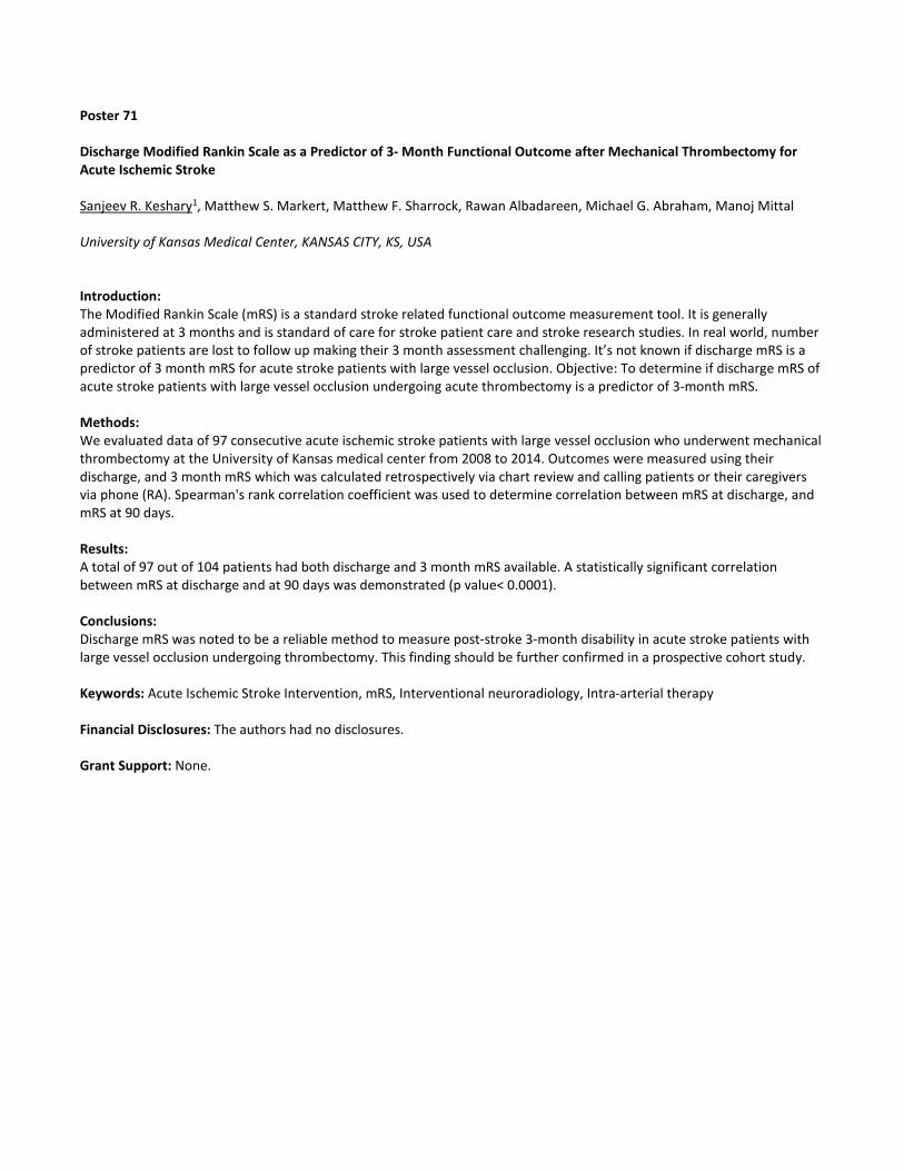
Poster 71 Discharge Modified Rankin Scale as a Predictor of 3- Month Functional Outcome after Mechanical Thrombectomy for Acute Ischemic Stroke Sanjeev R. Keshary1, Matthew S. Markert, Matthew F. Sharrock, Rawan Albadareen, Michael G. Abraham, Manoj Mittal University of Kansas Medical Center, KANSAS CITY, KS, USA Introduction: The Modified Rankin Scale (mRS) is a standard stroke related functional outcome measurement tool. It is generally administered at 3 months and is standard of care for stroke patient care and stroke research studies. In real world, number of stroke patients are lost to follow up making their 3 month assessment challenging. It’s not known if discharge mRS is a predictor of 3 month mRS for acute stroke patients with large vessel occlusion. Objective: To determine if discharge mRS of acute stroke patients with large vessel occlusion undergoing acute thrombectomy is a predictor of 3-month mRS. Methods: We evaluated data of 97 consecutive acute ischemic stroke patients with large vessel occlusion who underwent mechanical thrombectomy at the University of Kansas medical center from 2008 to 2014. Outcomes were measured using their discharge, and 3 month mRS which was calculated retrospectively via chart review and calling patients or their caregivers via phone (RA). Spearman's rank correlation coefficient was used to determine correlation between mRS at discharge, and mRS at 90 days. Results: A total of 97 out of 104 patients had both discharge and 3 month mRS available. A statistically significant correlation between mRS at discharge and at 90 days was demonstrated (p value< 0.0001). Conclusions: Discharge mRS was noted to be a reliable method to measure post-stroke 3-month disability in acute stroke patients with large vessel occlusion undergoing thrombectomy. This finding should be further confirmed in a prospective cohort study. Keywords: Acute Ischemic Stroke Intervention, mRS, Interventional neuroradiology, Intra-arterial therapy Financial Disclosures: The authors had no disclosures. Grant Support: None.

Poster 72 Outcome of Stent-Retriever Mechanical Thrombectomy in Nonagenarians: How well are we pushing the limits? Pankaj Shamra2, Priyank Khandelwal1, Kevin Ramdas2, Amer Malik2, Dilleep Yavagl2 1Birgham and Womens Hopsital/ Interventional Neuro, Boston, MA, USA, 2University of Miami/ Department of Neurology, Miami, FL, USA Introduction: Improvement in medical care and socio-economic growth over the past few decades illustrate improvement in life span, allowing individuals to enjoy good quality of life above 90 years of age. However, nonagenarians are at the highest risk of acute ischemic stroke (AIS). Recently published clinical trials consistently showed benefit of endovascular therapy in reducing the mortality and disability from ischemic stroke. However only two of these new studies included patients more than 80 years of age & very few nonagenarians. Therefore, we plan to study the outcome of Endovascular therapy in the rapidly growing nonagenarian population. Methods: We conducted an extensive single centric retrospective chart review of all eligible thrombectomy cases of nonagenarian patients presented with AIS and large vessel occlusion in the anterior circulation that could be treated within 8 hours with stent retriever. We plan to study the functional independence measured by MRS and NIHSS at 90 days. Results: There were 10 consecutive nonagenarian patients from 2012 to 2015 who received endovascular treatment at our institution with stent retriever. The median age was 92 (90-95) years. The median baseline National Institute of Health Stroke Scale (NIHSS) was 21 (14-22). All patients received standard of care along with endovascular therapy, 5/10 (50 %) patients received IV TPA. At discharge, the median modified Rankin Score (mRS), 0-2 was 10% (1/10), 0-3 was. 30% (3/10). At 90-day follow up, the median mRS for favorable outcome 0-2 was 1/8 (12.5%) & (0-3) was 37 % (3/8, 2 patient lost to follow up). The discharge mortality rate was 30 % and the rate of symptomatic intracranial hemorrhage related to procedure was 10% (1/10). Conclusions: Mechanical thrombectomy with stent retrievers in nonagenarians may achieve a good outcome in few of them. While this may be better than medical therapy, this needs to be studied further to increase rate of good outcomes in this important stroke population. Keywords: Acute Ischemic Stroke Intervention, Stentretriever, Decision analysis, Endovascular therapy Financial Disclosures: The authors had no disclosures. Grant Support: None.
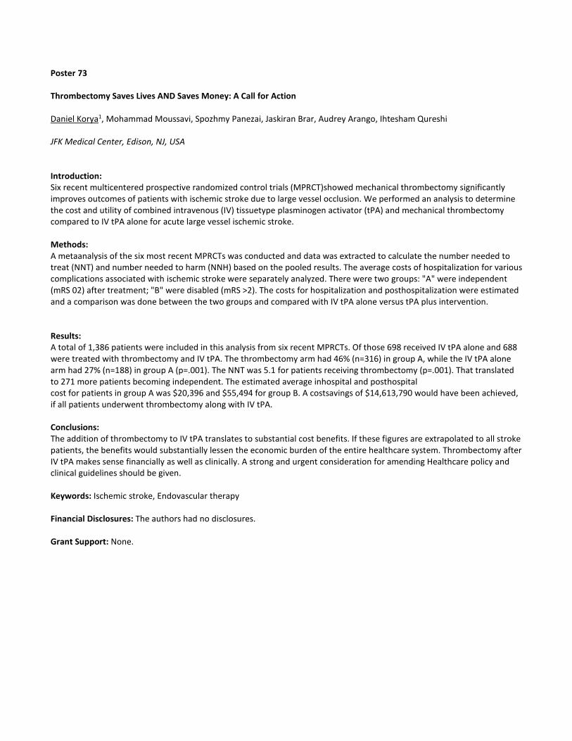
Poster 73 Thrombectomy Saves Lives AND Saves Money: A Call for Action Daniel Korya1, Mohammad Moussavi, Spozhmy Panezai, Jaskiran Brar, Audrey Arango, Ihtesham Qureshi JFK Medical Center, Edison, NJ, USA Introduction: Six recent multicentered prospective randomized control trials (MPRCT)showed mechanical thrombectomy significantly improves outcomes of patients with ischemic stroke due to large vessel occlusion. We performed an analysis to determine the cost and utility of combined intravenous (IV) tissuetype plasminogen activator (tPA) and mechanical thrombectomy compared to IV tPA alone for acute large vessel ischemic stroke. Methods: A metaanalysis of the six most recent MPRCTs was conducted and data was extracted to calculate the number needed to treat (NNT) and number needed to harm (NNH) based on the pooled results. The average costs of hospitalization for various complications associated with ischemic stroke were separately analyzed. There were two groups: "A" were independent (mRS 02) after treatment; "B" were disabled (mRS >2). The costs for hospitalization and posthospitalization were estimated and a comparison was done between the two groups and compared with IV tPA alone versus tPA plus intervention. Results: A total of 1,386 patients were included in this analysis from six recent MPRCTs. Of those 698 received IV tPA alone and 688 were treated with thrombectomy and IV tPA. The thrombectomy arm had 46% (n=316) in group A, while the IV tPA alone arm had 27% (n=188) in group A (p=.001). The NNT was 5.1 for patients receiving thrombectomy (p=.001). That translated to 271 more patients becoming independent. The estimated average inhospital and posthospital cost for patients in group A was $20,396 and $55,494 for group B. A costsavings of $14,613,790 would have been achieved, if all patients underwent thrombectomy along with IV tPA. Conclusions: The addition of thrombectomy to IV tPA translates to substantial cost benefits. If these figures are extrapolated to all stroke patients, the benefits would substantially lessen the economic burden of the entire healthcare system. Thrombectomy after IV tPA makes sense financially as well as clinically. A strong and urgent consideration for amending Healthcare policy and clinical guidelines should be given. Keywords: Ischemic stroke, Endovascular therapy Financial Disclosures: The authors had no disclosures. Grant Support: None.
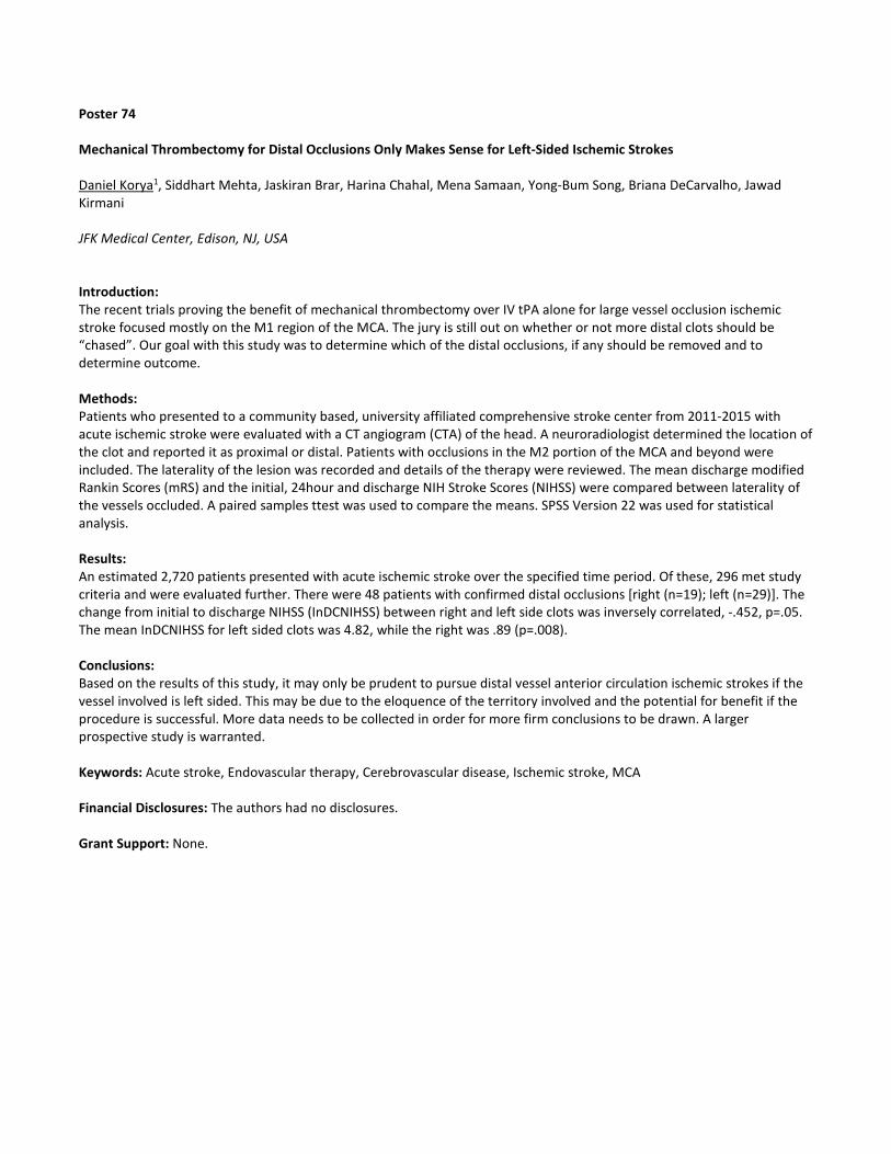
Poster 74 Mechanical Thrombectomy for Distal Occlusions Only Makes Sense for Left-Sided Ischemic Strokes Daniel Korya1, Siddhart Mehta, Jaskiran Brar, Harina Chahal, Mena Samaan, Yong-Bum Song, Briana DeCarvalho, Jawad Kirmani JFK Medical Center, Edison, NJ, USA Introduction: The recent trials proving the benefit of mechanical thrombectomy over IV tPA alone for large vessel occlusion ischemic stroke focused mostly on the M1 region of the MCA. The jury is still out on whether or not more distal clots should be “chased”. Our goal with this study was to determine which of the distal occlusions, if any should be removed and to determine outcome. Methods: Patients who presented to a community based, university affiliated comprehensive stroke center from 2011-2015 with acute ischemic stroke were evaluated with a CT angiogram (CTA) of the head. A neuroradiologist determined the location of the clot and reported it as proximal or distal. Patients with occlusions in the M2 portion of the MCA and beyond were included. The laterality of the lesion was recorded and details of the therapy were reviewed. The mean discharge modified Rankin Scores (mRS) and the initial, 24hour and discharge NIH Stroke Scores (NIHSS) were compared between laterality of the vessels occluded. A paired samples ttest was used to compare the means. SPSS Version 22 was used for statistical analysis. Results: An estimated 2,720 patients presented with acute ischemic stroke over the specified time period. Of these, 296 met study criteria and were evaluated further. There were 48 patients with confirmed distal occlusions [right (n=19); left (n=29)]. The change from initial to discharge NIHSS (InDCNIHSS) between right and left side clots was inversely correlated, -.452, p=.05. The mean InDCNIHSS for left sided clots was 4.82, while the right was .89 (p=.008). Conclusions: Based on the results of this study, it may only be prudent to pursue distal vessel anterior circulation ischemic strokes if the vessel involved is left sided. This may be due to the eloquence of the territory involved and the potential for benefit if the procedure is successful. More data needs to be collected in order for more firm conclusions to be drawn. A larger prospective study is warranted. Keywords: Acute stroke, Endovascular therapy, Cerebrovascular disease, Ischemic stroke, MCA Financial Disclosures: The authors had no disclosures. Grant Support: None.
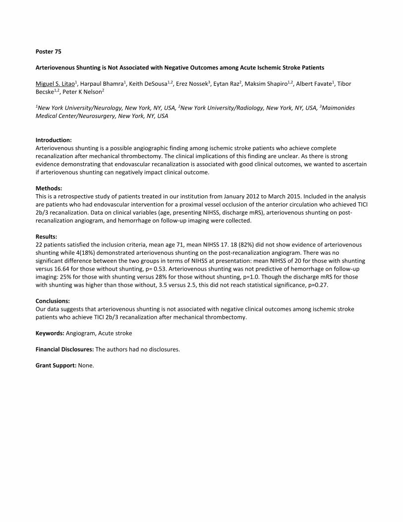
Poster 75 Arteriovenous Shunting is Not Associated with Negative Outcomes among Acute Ischemic Stroke Patients Miguel S. Litao1, Harpaul Bhamra1, Keith DeSousa1,2, Erez Nossek3, Eytan Raz2, Maksim Shapiro1,2, Albert Favate1, Tibor Becske1,2, Peter K Nelson2 1New York University/Neurology, New York, NY, USA, 2New York University/Radiology, New York, NY, USA, 3Maimonides Medical Center/Neurosurgery, New York, NY, USA Introduction: Arteriovenous shunting is a possible angiographic finding among ischemic stroke patients who achieve complete recanalization after mechanical thrombectomy. The clinical implications of this finding are unclear. As there is strong evidence demonstrating that endovascular recanalization is associated with good clinical outcomes, we wanted to ascertain if arteriovenous shunting can negatively impact clinical outcome. Methods: This is a retrospective study of patients treated in our institution from January 2012 to March 2015. Included in the analysis are patients who had endovascular intervention for a proximal vessel occlusion of the anterior circulation who achieved TICI 2b/3 recanalization. Data on clinical variables (age, presenting NIHSS, discharge mRS), arteriovenous shunting on post-recanalization angiogram, and hemorrhage on follow-up imaging were collected. Results: 22 patients satisfied the inclusion criteria, mean age 71, mean NIHSS 17. 18 (82%) did not show evidence of arteriovenous shunting while 4(18%) demonstrated arteriovenous shunting on the post-recanalization angiogram. There was no significant difference between the two groups in terms of NIHSS at presentation: mean NIHSS of 20 for those with shunting versus 16.64 for those without shunting, p= 0.53. Arteriovenous shunting was not predictive of hemorrhage on follow-up imaging: 25% for those with shunting versus 28% for those without shunting, p=1.0. Though the discharge mRS for those with shunting was higher than those without, 3.5 versus 2.5, this did not reach statistical significance, p=0.27. Conclusions: Our data suggests that arteriovenous shunting is not associated with negative clinical outcomes among ischemic stroke patients who achieve TICI 2b/3 recanalization after mechanical thrombectomy. Keywords: Angiogram, Acute stroke Financial Disclosures: The authors had no disclosures. Grant Support: None.

Poster 76 Time of Recanalization since Symptoms is a Strong Predictor of Outcome in Patients who Underwent Stent Retriever Thrombectomy from Middle Cerebral Artery Occlusion. Yahia M Lodi1,2,3, Varun V Reddy1,2, Ashok Devasenapathy1,2, George Petro2, Anas Hourani3, Chun-An Chou3 1Upstate Medical University, Binghamton Campus, Binghamton, NY, USA, 2UHS-Wilson Regional Medical Center, Johnson City, NY, USA, 3SSIE Department at Binghamton University, Binghamton, NY, USA Introduction: The outcome of acute ischemic stroke (AIS) patients who recanalize < 3 versus >3 hours since symptoms have not been clearly investigated especially, those with large artery occlusion (LAO) and underwent stent retriever thrombectomy (SRT) . Objective: To identify the predictors of outcome in AIS patients from middle cerebral artery (MCA) occlusion with large clot burden (LCB >8 mm) and underwent SRT, who recanalize < 3 versus >3 hours since symptoms. Methods: Consecutive AIS patients with MCA occlusion with LCB who underwent SRT were enrolled from 2012 to 2014. Outcome was measured using modified Rankin Scale (mRS) at 30 days. The SAS software was used to analyze the data. Results: 21 AIS patients who underwent SRT in MCA; 52% right MCA, 48% left, mean age 70.62±13.94 years and mean admission NIHSS 17±5. Complete (TICI3) and near-complete (TICI2b) recanalization was observed in 90.50% and 9.50% respectively with mean passes of 1.71±1. Time to recanalization from symptoms was 230±160 minutes. Presenting NIHSS of 17 which dropped to 7, 5 and 2 at immediate, 24 hours and 30 days post SRT respectively. Good outcome (mRS ≤2) was observed in 66.13% and poor outcome in 28.57% including mortality in 9.5%. In univariate analysis, recanalization time, immediate and 24 hours post SRT NIHSS were predictors of outcome (p= 0.0039, 0.003 and 0.043 respectively). Patient's age, gender and presenting NIHSS were not predictors of outcome. In multivariate analysis, time of recanalization since symptoms (p=0.01) and baseline mRS (p=0.01) continued to be the predictors of good outcome. Conclusions: Our study demonstrates that AIS patients from MCA who recanalize less than 3 hours of symptoms onset have very good chance of good outcome compared to those who recanilize more than 3 hours. Therefore, all AIS patients with LAO should offer early SRT to achieve a good functional outcome. Further studies are required. Keywords: Acute Ischemic Stroke Intervention, Mechanical thrombectomy, MCA, mRS, Endovascular therapy Financial Disclosures: The authors had no disclosures. Grant Support: None.

Poster 77 Should the Decision for Mechanical Thrombectomy be Based on PreProcedure CT Head Showing Early Changes in Specific Regions of the Brain? Siddhart Mehta1, Daniel Korya, Mohammad Moussavi, Jaskiran Brar, Mena Samaan, Swathi Kondapalli, Briana DeCarvalho, Jawad Kirmani JFK Medical Center, Edison, NJ, USA Introduction: The treatment of acute ischemic stroke has evolved over the past several years to utilize neuroimaging in guiding therapy. With regard to IV tPA and thrombectomy, recent endovascular therapy trials have utilized the ASPECT score in determining if intervention should be attempted. We sought to evaluate different regions of interest on the ASPECT score to determine if specific areas of injury should be weighed more heavily during decision making. Methods: We evaluated the preintervention CT scans of the head on all patients who received IV tPA and mechanical thrombectomy during the last two years at a community based, university affiliated comprehensive stroke center. All 20 regions of interest (ROIs) of the ASPECT score were compared with each other with regard to initial NIH stroke score, discharge NIHSS, delta NIHSS and modified Rankin Score to determine if one or more regions were associated with worse outcome. SPSS version 22 was used to determine Spearman rho values and paired samples t-test. Results: A total of 864 patients presented with acute ischemic stroke, of which 70 patients received IV tPA followed by mechanical thrombectomy and were included in the study. The 4 ROIs with the greatest correlation with worse outcome as rated by discharge mRS were the right and left M5-M6 [4.2 (p=.001, 95%CI 3.54.8) ; 4.3 (p=.001, 95%CI 3.45.1); 4.3 (p=.001, 95%CI 3.45.2); 4.2 (p=.001, 95%CI 3.64.8), respectively]. Conclusions: Early changes defined as hypodensity in the M5 and M6 ROIs on either side of the preintervention head CT were associated with significantly worse outcomes. A modified ASPECT score should be considered to better prognosticate patients and guide the appropriateness of endovascular therapy in select patients. These findings should be validated in a larger population and a longer followup period. Keywords: Acute Ischemic Stroke Intervention, Imaging, ASPECTS Financial Disclosures: The authors had no disclosures. Grant Support: None.

Poster 78 Endovascular Treatment of Posterior Circulation Strokes Improves Outcome Mohammad Mousavi1, Siddhart Mehta, Daniel Korya, Jaskiran Brar, Azka Shaikh, Rushil Kalola, Shayan Nizam, Spozhmy Panezai, Jawad Kirmani, Alex JFK Medical Center, Edison, NJ, USA Introduction: Patients presenting with acute ischemic stroke due to a large vessel occlusion of the anterior circulation (usually M1), are now offered thrombectomy after 6 major trials demonstrated superior results over IV tPA alone. However, there are few trials evaluating the benefit of clot retrieval in posterior circulation strokes. Our goal was to evaluate the benefit of thrombectomy for posterior circulation strokes. Methods: We conducted a comprehensive record review of patients who underwent acute endovascular intervention for posterior circulation ischemia at a community based, university affiliated comprehensive stroke center during a 5 year period (20102015).The initial NIHSS, discharge NIHSS and discharge mRS were abstracted. We compared discharge mRS to composite 90 DAYS mRS of six recent multicentered prospective randomized control trials (MPRCT) for acute endovascular treatment of acute ischemic stroke involving large vessel occlusions (SWIFT PRIME, ESCAPE, MR CLEAN, EXTENDIA, REVASCAT, THERAPY). Results: There were 2,980 patients who presented with acute ischemic stroke during the prespecified time period. Of those 310 were posterior circulation strokes; of which, 34 underwent endovascular treatment. These were compared with 1,386 patients with proximal anterior circulation strokes included in the recent MPRCTs. The mean age was 62 (SD=12). Mean initial NIHSS was 12 (SD=11). Mean change in NIHSS was 4 (SD =6). Mean discharge NIHSS was 9 (SD=11). Mean discharge mRS was 2.6 (SD=2.1). Conclusions: There were 2,980 patients who presented with acute ischemic stroke during the prespecified time period. Of those 310 were posterior circulation strokes; of which, 34 underwent endovascular treatment. These were compared with 1,386 patients with proximal anterior circulation strokes included in the recent MPRCTs. The mean age was 62 (SD=12). Mean initial NIHSS was 12 (SD=11). Mean change in NIHSS was 4 (SD =6). Mean discharge NIHSS was 9 (SD=11). Mean discharge mRS was 2.6 (SD=2.1). Keywords: Endovascular therapy, Acute stroke Financial Disclosures: The authors had no disclosures. Grant Support: None.

Poster 79 “Stroke Alerts” Among Admitted Patients – Do We Have An Effective System? – A Single Center Perspective VIjay M Pandav1, Trevor Gregath, Ankur Garg The University of Oklahoma, Department of Neurology, Oklahoma City, OK, USA Introduction: Rapid revascularization in acute ischemic stroke (AIS) with intravenous thrombolysis (tPA) and/or intra-arterial treatment decreases morbidity and mortality. Despite this very few stroke patients receive this treatment in timely fashion due to lack of community awareness. In contrast, in-hospital “Stroke Alerts”(SA) are expected to be called quickly due to immediate availability of trained healthcare professionals. The aim of the present study was to evaluate efficacy of in-hospital SA system - in identifying and treating AIS at our comprehensive stroke center. Methods: In-hospital, non-emergency room “Stroke Alerts” were retrospectively reviewed for the period January 2013 to December 2014. Results: A total of 673 SA were called during the study period of which 65 (10.35%) were in-hospital calls. Mean age was 64 (SD±15) with 52% men. Median NIHSS was 7.5. Fifty patients were on regular floor, 11 in the Intensive Care Unit (ICU) and 4 in the step-down unit. The mean time for initiating SA since last know normal was 200 (SD ±256) min. The mean time since last know normal to initiating SA was 196 min (SD±259) for floor, 279 min (SD±269) for ICU and 35 min (SD±26) for step down unit (one-way ANOVA p=0.26). Final diagnosis after SA was AIS in 22(33%), intracerebral hemorrhage in 4(16%) and non-stroke/stroke mimic in 36(55%). Among the SA, 15/50 (30%) on the floor, 5/11 (45.45%) in ICU and 2/4 (50%) in step-down units diagnosed with AIS (X2 1.125. p=0.56). Only 2/22(3%) received IV tPA and 1/22 received intra-arterial treatment. The common reasons for not receiving IV tPA for AIS diagnosis was being “outside treatment window” (27.27%). Conclusions: Conclusion: In our cohort we report a significant delay in identifying AIS among in-hospital patients. Recognition of such deficiency among healthcare professionals calls for hospital-wide staff training as level of admission also had no effect in regards to prompt or correct identification of AIS patients. Keywords: Acute stroke Financial Disclosures: The authors had no disclosures. Grant Support: None.

Poster 80 Is Eptifibatide a Viable and Safe Option as StandAlone Therapy For Acute Ischemic Stroke Patients? Spozhmy Panezai1, Daniel Korya, Mohammad Moussavi, Siddhart Mehta, Jaskiran Brar, Harina Chahal, Josh Daneil, Alex Yang Ge, Mena Samaan, Jawad Kirmani JFK Medical Center, Edison, NJ, USA Introduction: The list of contraindications for IV tPA in acute ischemic stroke (AIS) is often too long and may lead to physicians opting to offer no treatment for certain strokes. An alternative treatment is proposed in cases where IV tPA is not an option. We compared the stroke severity, outcomes and safety of IV eptifibatide when compared with IV tPA. Methods: Patients who presented to a community based university affiliated comprehensive stroke center from 201215 with AIS over a twoyear period were included in the study. Those who received IV tPA were compared with patients who only received IV eptifibatide. The initial NIH Stroke Score (NIHSS), 24hour NIHSS, discharge NIHSS (DCNIHSS), discharge mRS (DCmRS) and symptomatic ICH rates were compared with a paired samples t-test to determine significance of difference between the means. Results: A total of 864 patients presented with AIS in the evaluated time period and of those 166 met study criteria. There were 119 patients who received IV tPA alone (group A) and 47 patients received eptifibatide (group B). The mean initial NIHSS, 24NIHSS, DCNIHSS, DCmRS and percent bleeding complications for group A were: 11.2, 10.8, 8.6, 3.1 and 6%. For group B the figures were: 6.7, 4.8, 4.3, 1.7 and 0%, respectively. Group A was compared with group B in a paired samples t-test and yielded 4.3, 6.2, 6, 1.5 (p=.0001 to .04) for initial, 24hour, discharge NIHSS and discharge mRS, respectively. The difference between initial and discharge NIHSS between the two groups was 2.7 (p=.009), favoring IV tPA. Conclusions: In patients who are either outside the time window or with contraindications to IV tPA, eptifibatide may be a safe alternative and appears to be efficacious. None of the patients who were started on eptifibatide had bleeding complications and they had a statistically significant improvement in their level of disability and stroke severity at discharge. A limitation of this study is that patients in group A had significantly worse initial NIHSS compared with group B. Keywords: Medical management, Lytics, Acute stroke Financial Disclosures: The authors had no disclosures. Grant Support: None.

Poster 81 Safety of Microcatheter Injection in Perdiction of Hemorrhagic Transformation and Outcome in SWIFT PRIME Radoslav Raychev1, 2, Reza Jahan3, Jeffrey Saver1, Raul G. Nogueira4, Mayank Goyal5, Vitor M Pereira6, Elad I Levy7, Christophe Cognard8, David Liebeskind9 1Department of Neurology and Comprehensive Stroke Center, David Geffen School of Medicine at the UCLA, Los Angeles, CA, USA, 2Saddleback Memorial Hospital, Laguna Hills, CA, USA, 3Division of Interventional Neuroradiology, UCLA, Los Angeles, CA, USA, 4Marcus Stroke and Neuroscience Center, Grady Memorial Hospital, Department of Neurology, Emory University School of Medicine Atlanta, GA, USA, 5Departments of Radiology and Clinical Neurosciences, University of Calgary Calgary, Canada, 6Division of Neuroradiology and Division of Neurosurgery, Department of Medical Imaging and Department of Surgery, Toronto Western Hospital, University Health Network, University of Toronto Toronto, Canada, 7Department of Neurosurgery, SUNY at Buffalo Buffalo, NY, USA, 8Department of Diagnostic and Therapeutic Neuroradiology, University Hospital of Toulouse Toulouse, France, 9Neurovascular Imaging Research Core, Department of Neurology, UCLA Los Angeles, CA, Canada Introduction: Prior data from older revascularization methods suggested that microcatheter contrast injection (MCI) is associated with higher risk of hemorrhagic transformation (HT). We analyzed the SWIFT PRIME trial to evaluate the impact of pre-intervention MCI on hemorrhagic transformation and outcome after Solitaire thrombectomy. Methods: Angiography of the endovascular arm in SWIFT PRIME (n=98) was reviewed by 3 independent readers, followed by consensus adjudications. A 3-point grading scale was use to define MCI flow. Multivariate analysis was conducted to evaluate the impact of MCI on HT and outcome, including the following clinical and procedural parameters: Age, MCI flow grade (poor, good, moderate), baseline NIHSS, number of MCI injections, baseline core volume, presence of MCI (y/n), time to TICI 2a, modified TICI, baseline SBP, original TICI, baseline hyperglycemia (serum glucose > 140 mg/dl, baseline ASPECTS Results: MCI was observed in 59% (N=51) of patients with prevalence of M2 location 71% (N=36/51). Pre-deployment distal emboli were observed in 12% (N=6/51). Median number of MCI was 1 (1-7). None of the MCI variables (presence, number, and grades) correlated with HT, SAH, PH, and clinical outcome. Among all included variables, only lower ASPECTS at screening correlated with both HT (p=0.0078; OR 0.501) and non-disabled outcome (mRS 0-2) at 90 days (p=0.0361; OR 1.6). Other factors influencing clinical outcome in the analyzed data were age for non-disabled outcome (p=0.0109), time to TICI 2a for NIHSS at 7-10 days (p=0.009), and baseline core volume for NIHSS at 90 days (p=0.0226). Conclusions: Evaluation of distal flow, anatomic position and distal emboli with MCI prior to Solitaire deployment is safe with no statistical evidence of impact on HT and clinical outcome. Lower likelihood of non-disabled outcome after thrombectomy may be expected in patient with lower ASPECTS and older age. HT may also be observed more frequently in patients with lower baseline ASPECTS. Keywords: Acute stroke, Hemorrhagic transformation, ASPECTS, Angiogram, Mechanical thrombectomy Financial Disclosures: The authors had no disclosures. Grant Support: None.

Poster 82 Evaluating Distal Flow Beyond the Occluded Segment Using a Novel Microcatheter Contrast Injection Grading Scale: Correlation with Collateral Flow Radoslav Raychev1, 2, Reza Jahan3, Jeffrey Saver1, Raul G. Nogueira4, Mayank Goyal5, Vitor M Pereira6, Elad I Levy7, Dileep R Yavagal8, Christophe Cognard9, David Liebeskind10 1Department of Neurology and Comprehensive Stroke Center, David Geffen School of Medicine at the UCLA, Los Angeles, CA, USA, 2Saddleback Memorial Hospital, Laguna Hills, CA, USA, 3Division of Interventional Neuroradiology, UCLA, Los Angeles, CA, USA, 4Marcus Stroke and Neuroscience Center, Grady Memorial Hospital, Department of Neurology, Emory University School of Medicine Atlanta, CA, USA, 5Departments of Radiology and Clinical Neurosciences, University of Calgary Calgary, Canada, 6Division of Neuroradiology and Division of Neurosurgery, Department of Medical Imaging and Department of Surgery, Toronto Western Hospital, University Health Network, University of Toronto Toronto, Canada, 7Department of Neurosurgery, SUNY at Buffalo Buffalo, NY, USA, 8University of Miami &Jackson Memorial Hospitals Miami, FL, USA, 9Department of Diagnostic and Therapeutic Neuroradiology, University Hospital of Toulouse Toulouse, France, 10Neurovascular Imaging Research Core, Department of Neurology, UCLA Los Angeles, CA, USA Introduction: Given the strong emphasis on rapid revascularization, complete pre-intervention angiographic collateral assessment may not be routinely performed. In presence of complete proximal occlusion, the direction of flow within the occluded vessel and its distal branches is retrograde. The evaluation of distal flow beyond the occlusion via microcatheter contrast injection (MCI) prior to stentriever deployment may provide useful information of anatomic location, presence of emboli and collaterals. We analyzed the SWIFT PRIME trial to evaluate the role of MCI flow with respect to collateral flow. Methods: Angiography of the endovascular arm in SWIFT PRIME (n=98) was reviewed by 3 independent readers, followed by consensus adjudications. The MCI flow was graded as 1. Poor – confined to the M2 segments; 2. Moderate – confined to the M3 segments; 3. Good – extending into the M4 segments. MCI and ASITN/SIR collateral grades were scored only when available, prior to use of the Solitaire device. Qhi Square test was used to investigate the relationship of collateral grades with MCI grades. Results: Both MCI and collaterals were concurrently available in 40 subjects. Poor MCI flow was seen in 23% (n=49), moderate in 43% (n=17), and good in 35% (N=14) of analyzed subjects. The most common MCI location was M2 (65 %) and pre-deployment distal emboli were observed in 7.5 % (N = 3). Dichotomized correlation between ASITN/SIR collateral grading defined as poor (grade 1 and 2) and good (grade3 and 4) demonstrated strong inverse relationship of poor collaterals with good (3) MCI flow (p=0.004; OR 8.25). Conclusions: Poor collateral flow strongly correlates with good microcatheter injection flow in the endovascular arm of SPRIME. This finding warrants further validation in larger cohort as MCI flow may be influenced by individual operator's technique and choice of syringe size. Keywords: Acute Ischemic Stroke Intervention, Angiogram, Cerebral blood flow, Collateral, Mechanical thrombectomy Financial Disclosures: The authors had no disclosures. Grant Support: None.
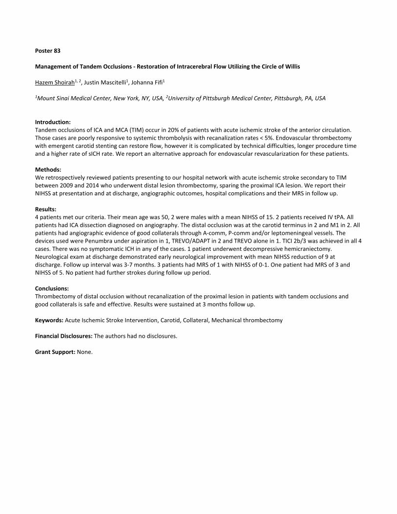
Poster 83 Management of Tandem Occlusions - Restoration of Intracerebral Flow Utilizing the Circle of Willis Hazem Shoirah1, 2, Justin Mascitelli1, Johanna Fifi1 1Mount Sinai Medical Center, New York, NY, USA, 2University of Pittsburgh Medical Center, Pittsburgh, PA, USA Introduction: Tandem occlusions of ICA and MCA (TIM) occur in 20% of patients with acute ischemic stroke of the anterior circulation. Those cases are poorly responsive to systemic thrombolysis with recanalization rates < 5%. Endovascular thrombectomy with emergent carotid stenting can restore flow, however it is complicated by technical difficulties, longer procedure time and a higher rate of sICH rate. We report an alternative approach for endovascular revascularization for these patients. Methods: We retrospectively reviewed patients presenting to our hospital network with acute ischemic stroke secondary to TIM between 2009 and 2014 who underwent distal lesion thrombectomy, sparing the proximal ICA lesion. We report their NIHSS at presentation and at discharge, angiographic outcomes, hospital complications and their MRS in follow up. Results: 4 patients met our criteria. Their mean age was 50, 2 were males with a mean NIHSS of 15. 2 patients received IV tPA. All patients had ICA dissection diagnosed on angiography. The distal occlusion was at the carotid terminus in 2 and M1 in 2. All patients had angiographic evidence of good collaterals through A-comm, P-comm and/or leptomeningeal vessels. The devices used were Penumbra under aspiration in 1, TREVO/ADAPT in 2 and TREVO alone in 1. TICI 2b/3 was achieved in all 4 cases. There was no symptomatic ICH in any of the cases. 1 patient underwent decompressive hemicraniectomy. Neurological exam at discharge demonstrated early neurological improvement with mean NIHSS reduction of 9 at discharge. Follow up interval was 3-7 months. 3 patients had MRS of 1 with NIHSS of 0-1. One patient had MRS of 3 and NIHSS of 5. No patient had further strokes during follow up period. Conclusions: Thrombectomy of distal occlusion without recanalization of the proximal lesion in patients with tandem occlusions and good collaterals is safe and effective. Results were sustained at 3 months follow up. Keywords: Acute Ischemic Stroke Intervention, Carotid, Collateral, Mechanical thrombectomy Financial Disclosures: The authors had no disclosures. Grant Support: None.

Poster 84 Comparison of Medical vs. Endoavscular Treatment in Acute Cervical Dissections Yamin Shwe1, Chunyan Cai, Anjail Sharrief, Amrou Sarraj Univertisy of Texas Health Sciences Center at Houston, Houston, TX, USA Introduction: Cervical artery dissection (CAD) is an important cause of acute ischemic stroke (AIS) in young patients and can be associated with recurrent thromboembolism. There are currently a limited number of studies to guide clinical management. Our aim is to compare outcomes in acute CAD (cervical or vertebral artery) treated with antiplatelet (AP) vs. anticoagulation (AC) vs. endovascular stenting (ES). Methods: We conducted a retrospective analysis of consecutive AIS due to CAD admitted to our comprehensive stroke center from January 2011 to June 2015. Our primary outcome was stroke recurrence at discharge or day 7; secondary outcomes were safety (hemorrhage or neurological worsening) and functional outcome (mRS) at day 7 or discharge. Fisher’s exact test was used to conduct pairwise comparisons on clinical outcomes between groups. Results: A total of 74 patients were included in the study. The majority of patients had carotid dissections in ES (89%) compared to AC (47%) (p=0.03). While stroke recurrence was lower in ES and AP (11%) compared to AC (13%), there was no statistical difference between the 3 treatment groups (ES vs. AP p=1.0, ES vs. AC p=0.77). Worsening neurological symptoms during hospitalization were similar among all treatment groups (22%) (p=1.0). Bleeding rates were higher in AP (14.8%) vs. ES (11.1%) and AC (5.3%) but without statistical difference (ES vs. AP p=1.0, ES vs. AC p=0.5). AC group had the lowest mortality rate (0%) compared to AP (19%) and ES (11%). Conclusions: Our study did not find any significant difference among stroke recurrence at discharge or on day 7 among patients treated with ET compared with AC and AP therapy. This study is limited by a small sample size, particularly in the ET group. Large, prospective studies are needed to determine the difference in outcomes among patients with CAO treated with ET vs AC and AP therapy. Keywords: Endovascular therapy, Interventional neuroradiology, Stroke, Carotid stenting and angioplasty, Acute stroke Financial Disclosures: The authors had no disclosures. Grant Support: None.

Poster 85 Ischemic Stroke Outcomes in Medical vs. Endovascular Treatment of Proximal Carotid Artery Occlusion Yamin Shwe1, Chunyan Cai, Anjail Z. Sharrief, Amrou Sarraj University of Texas Health Sciences Center at Houston, Houston, TX, USA Introduction: Acute ischemic stroke (AIS) due to proximal carotid artery occlusion (pCAO) can be associated with significant neurological deficits and poor outcome without timely intervention and successful reperfusion. Intravenous thrombolytics (IT) have low recanalization rates in pCAO and these patients were excluded from recent randomized controlled trials which showed superiority of endovascular therapy (EVT) over IT.The purpose of this study is to investigate clinical outcomes in AIS due to pCAO treated with medical vs. endovascular treatment. Methods: We conducted a retrospective chart review of patients who underwent IT or EVT±IT for all types of pCAO from January 2008 to June 2015. Our primary outcome was the functional outcome at discharge measured by modified Rankin score (mRS) 0-3. The secondary outcomes were hemorrhagic transformation (HT), neurological worsening (NW), symptomatic hemorrhage (sICH) and death. Logistic regression analysis was used to compare outcomes between the two groups. Results: A total of 133 patients were included in the study. There were no significant differences in functional outcome mRS (0-3) between ET and IT groups, 22% vs.16% (p=0.35, OR 1.71, 95% CI 0.55, 5.34). IV tPA was given in 56% IT vs. 72% EVT (p=0.14). While there was a shift towards better outcomes in the mRS distribution in EVT group (22% vs. 16%), the treatment effect did not reach significance (OR 1.71, 95% CI (0.55, 5.34), p=0.35). At discharge the rate of hemorrhagic transformation was 25.9% (ET) and 14.2% (IT) (p=0.14), worsening neurologic symptoms were 25.9% (ET) and 20.8% (IT), (p=0.56) and symptomatic hemorrhage were 3.7% (ET) and 0% (IT) (p=0.2). Conclusions: Our study did not show difference in discharge functional outcomes between EVT and IT in AIS with pCAO. Our results are limited by small sample size and retrospective nature. Future prospective studies randomizing patients to medical vs. endovascular treatments are warranted to guide management. Keywords: Carotid stenting and angioplasty, Cerebrovascular disease, Endovascular therapy, Ischemic stroke, Interventional neuroradiology Financial Disclosures: The authors had no disclosures. Grant Support: None.
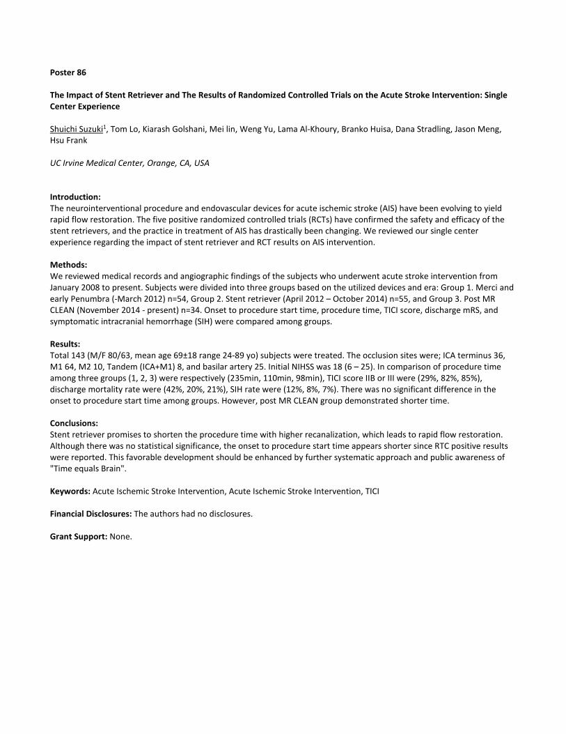
Poster 86 The Impact of Stent Retriever and The Results of Randomized Controlled Trials on the Acute Stroke Intervention: Single Center Experience Shuichi Suzuki1, Tom Lo, Kiarash Golshani, Mei lin, Weng Yu, Lama Al-Khoury, Branko Huisa, Dana Stradling, Jason Meng, Hsu Frank UC Irvine Medical Center, Orange, CA, USA Introduction: The neurointerventional procedure and endovascular devices for acute ischemic stroke (AIS) have been evolving to yield rapid flow restoration. The five positive randomized controlled trials (RCTs) have confirmed the safety and efficacy of the stent retrievers, and the practice in treatment of AIS has drastically been changing. We reviewed our single center experience regarding the impact of stent retriever and RCT results on AIS intervention. Methods: We reviewed medical records and angiographic findings of the subjects who underwent acute stroke intervention from January 2008 to present. Subjects were divided into three groups based on the utilized devices and era: Group 1. Merci and early Penumbra (-March 2012) n=54, Group 2. Stent retriever (April 2012 – October 2014) n=55, and Group 3. Post MR CLEAN (November 2014 - present) n=34. Onset to procedure start time, procedure time, TICI score, discharge mRS, and symptomatic intracranial hemorrhage (SIH) were compared among groups. Results: Total 143 (M/F 80/63, mean age 69±18 range 24-89 yo) subjects were treated. The occlusion sites were; ICA terminus 36, M1 64, M2 10, Tandem (ICA+M1) 8, and basilar artery 25. Initial NIHSS was 18 (6 – 25). In comparison of procedure time among three groups (1, 2, 3) were respectively (235min, 110min, 98min), TICI score IIB or III were (29%, 82%, 85%), discharge mortality rate were (42%, 20%, 21%), SIH rate were (12%, 8%, 7%). There was no significant difference in the onset to procedure start time among groups. However, post MR CLEAN group demonstrated shorter time. Conclusions: Stent retriever promises to shorten the procedure time with higher recanalization, which leads to rapid flow restoration. Although there was no statistical significance, the onset to procedure start time appears shorter since RTC positive results were reported. This favorable development should be enhanced by further systematic approach and public awareness of "Time equals Brain". Keywords: Acute Ischemic Stroke Intervention, Acute Ischemic Stroke Intervention, TICI Financial Disclosures: The authors had no disclosures. Grant Support: None.

Poster 87 Intravenous Thrombolysis Does Not Increase The Risk of Hemorrhage After Thrombectomy in Acute Stroke Jenny P. Tsai1, Michael Mlynash1, Christian Federau2, Soren Christensen1, Sun Kim1, Maarten G. Lansberg1 1Stanford Stroke Center, Department of Neurology, Stanford University Medical Center, Stanford, CA, USA, 2Division of Neuroradiology, Department of Radiology, Stanford University, Stanford, CA, USA Introduction: Intra-arterial thrombectomy (IAT) is effective in rapidly establishing reperfusion in emergent large-vessel occlusions. Although current guidelines recommend intravenous tissue plasminogen activator (IV tPA) for all eligible patients, it is unclear if tPA increases the risk of clinically significant intracranial hemorrhage (ICH) after IAT. Using data from the CT Perfusion to predict Response to recanalization in Ischemic Stroke Project (CRISP), a multi-center, NIH-sponsored prospective cohort study, we assessed whether IV tPA is a predictor of ICH after IAT. We hypothesized that IV tPA increases the risk of symptomatic ICH after IAT. Methods: We leveraged data for patients that underwent IAT for anterior circulation large vessel occlusions. We evaluated the association between IV tPA and symptomatic ICH using Fisher’s exact test. We defined ICH as parenchymal hemorrhage (PH) grades 1 and 2 (ECASS II criteria) on follow-up MRI or CT within 36 hours. We considered ICH as symptomatic if NIHSS increased by ≥4 points. In the logistic regression analysis, we prespecified variables for adjustment if associated with symptomatic ICH at a level of p< 0.1. These variables included age, baseline NIHSS and ASPECTS, pre-admission anti-thrombotic use, glucose, systolic blood pressure, infarct core volume, Tmax >10s volume, location of arterial occlusion, time to reperfusion, and reperfusion status. Results: The analysis included 191 patients: 87 (46%) received IV tPA before IAT. Symptomatic ICH affected 4.6% of patients post tPA, and 2.9% of patients with IAT alone (p=0.70). Only baseline NIHSS was associated with symptomatic ICH in the univariate analysis (p=0.058). Adjusted for NIHSS, IV tPA shows no statistically significant association with symptomatic ICH (p=0.54). Conclusions: Use of intravenous tPA is not associated with an increased risk of symptomatic ICH in patients undergoing IAT. This is in keeping with the 2015 AHA/ASA guidelines that support treatment of all eligible patients with IV tPA, even if they are being considered for intra-arterial thrombectomy. Keywords: Acute Ischemic Stroke Intervention, Thrombolytics, Hemorrhagic transformation Financial Disclosures: The CRISP study is sponsored by an NIH grant. Grant Support: None.

Poster 88 General Anesthesia During Endovascular Stroke Therapy Does Not Negatively Impact Outcome Q. Tony Wang1,2,3, Rachel Lehner1,2, Rivkah Epstein1, Isaac Rosenblum1, Nicole Eiden1, Madison Stellfox1, Amy Aquilina1, Noorie Pednekar3, Michael F. Stiefel1,2 1Westchester NeuroVascular Institute, Westchester Medical Center Health Network, Valhalla, NY, USA, 2Department of Neurosurgery, Division of NeuroVascular Surgery, Westchester Medical Center, New York Medical College, Valhalla, NY, USA, 3Departement of Neurology, Westchester Medical Center, New York Medical College, Valhalla, NY, USA Introduction: Recent randomized trials, including MR CLEAN, have demonstrated that endovascular therapy alone or in conjunction with intravenous tPA, compared to IV tPA alone, improves outcomes in patients with large vessel acute ischemic stroke. Sub-group analysis of the MR CLEAN found that patients undergoing general anesthesia (GA) during the procedure did worse than those with non-general anesthesia (non-GA). Current guidelines favor non-GA over GA with limited supporting evidence. We sought to review our experience and outcomes routinely using GA in patients undergoing stroke endovascular therapy with similar techniques. Methods: Medical records for 53 consecutive patients with anterior circulation strokes, who underwent endovascular therapy under GA, were reviewed. National Institutes of Health Stroke Scale (NIHSS) on admission and discharge, and modified Rankin scales (mRS) at discharge were recorded. Results: Our cohort consisted of 33 males and 20 females with a mean age of 60.5 (range of 40 to 83). Fifty-two patients were transferred from outside primary stroke centers, while 1 patient presented directly to our institution. Median NIHSS on admission was 15. The median time of symptoms onset to endovascular therapy was 338 minutes, with interquartile range of 300 to 403 minutes. TICI 2b-3 was achieved in 64.5% (20/31) of recorded patients. At discharge, mortality was 15.1% (8/53), median NIHSS was 4 (from 43 live and recorded patients) and 43.4% (23/53) of patients had a mRS of 0-2. Conclusions: MR CLEAN’s sub group analysis reported better outcomes of 25.9% with a mRS 0-2 and 16.5% mortality in non-GA patients, compared to 14.3% with a mRS 0-2 and a mortality of 21.4% in those receiving GA. Despite a longer time from symptom onset to treatment, our outcomes for those undergoing endovascular stroke treatment under GA compare favorably to that reported in the literature with non-GA. Randomized trials examining the types of anesthesia during endovascular stroke therapy are warranted. Keywords: General Anesthesia, Ischemic stroke, mRS, NIHSS, Endovascular therapy Financial Disclosures: The authors had no disclosures. Grant Support: None.
Poster 89 Regional Collateral Flow Evaluation Predicts Infarction during Stroke Endovascular Procedures POP Raoul1,3, MANISOR Monica1, WOLFF Valérie2, MARESCAUX Christian2, BEAUJEUX Rémy1, SIMU Mihaela3
Interventional Neuroradiology Department , Strasbourg University Hospitals, France1Vascular Neurology Department, Strasbourg University Hospitals, France2 Neurology Department, Timisoara County Emergency Hospital, Romania3
Introduction: We developed a new system for grading of leptomeningeal collateral flow in angiography. We investigated its diagnostic value for the prediction of infarction during stroke endovascular procedures. Methods: We evaluated all consecutive patients treated for an anterior circulation occlusion between 2009 and 2013. Two readers performed a zonal collateral circulation evaluation in 5 cortical regions based on the vascular anatomy. Regional scores were correlated with the presence of infarction in the same cortical region on pretreatment and follow-up imaging. Global collateral scores were correlated with initial and final infarct volumes and clinical scores. Results: In 89 patients with 408 cortical zones we found good correlation between the degree of collateral flow and the absence of infarction in the same region on pretreatment imaging (AUC of ROC curve 0.82, p<0.0001). In a subgroup of 37 recanalized patients (TICI 3) with 173 cortical regions, retrograde collateral flow to the proximal M4 segment predicted the absence of infarction in the same region on follow-up imaging (PPV 88,7% , NPV 83,8%). We found good inter-rater agreement for the presence of collateral flow to the M4 proximal segment or further – kappa 0.77 (p=0.05, 95%CI 0.66-0.88). The number of cortical regions with good collateral flow (0-5) was correlated with the intial infarct volume (p=0.003). All patients with 4-5 cortical regions had infarct volumes ≤70ml. Conclusions: Anatomic collateral flow evaluation can provide a real-time estimation of the size and location of irreversible ischemia during stroke endovascular procedures. Financial Disclosures: The authors had no disclosures. Grant Support: None.
Poster 90 Role of Minimally and Non Invasive Tools in Diagnosing and Treating Symptomatic Carotid Stenosis Alhamza R. Al-Bayati1, Amin Aghaebrahim, David M. Panczykowski, Gregory M. Weiner, Cynthia L. Kenmuir, Hazem Shoirah, Ruba R. Al-Ramadhani, Ashutosh P. Jadhav, Tudor G. Jovin, Brian T. Jankowitz University of Pittsburgh Medical Center/Neurology, Pittsburgh, PA, USA Introduction: Prior trials, such as NASCET, assessed the degree of carotid stenosis based on digital subtraction angiography (DSA), which consequently has become the gold standard reference in selecting patients for carotid revascularization. DSA is still underutilized in evaluation of symptomatic carotid stenosis. Methods: This population-based cohort study included all patients undergoing evaluation of carotid stenosis between July 2014 and June 2015. Clinical data was prospectively collected and patients were excluded if information was missing or incomplete regarding imaging and or treatment. Primary outcome was need for carotid revascularization (either endarterectomy or angioplasty/stenting). Significant stenosis was defined according to AHA Guidelines as carotid stenosis >70% on non-invasive imaging (ultrasound, MRA, CTA) and >50% stenosis on catheter angiography. Agreement was assessed by Cohen's kappa coefficient; discriminatory power and calibration of these imaging modalities were assessed using multiple regression analyses and indicated by the area under the receiver operating characteristic curve (AUC). Results: A sample of 169 out of 265 patients was available for analysis, of which 45% underwent revascularization for symptomatic carotid artery stenosis. Non-invasive imaging included CTA (58%), MRA (34%), and ultrasound (8%). Agreement between diagnostic modalities was 75% (expected 55%) with a Kappa of 0.46, representing fair agreement (p=0.001). Catheter angiography demonstrated superior discriminatory ability for prediction of carotid stenosis requiring revascularization (AUC 0.81 vs non-invasive AUC 0.64, p< 0.001). All modalities displayed adequate calibration for predicting need for revascularization. Conclusions: Despite the agreement of both non-invasive and minimally invasive measures in evaluating carotid stenosis, catheter angiography showed a superior trend in predicting high-grade carotid stenosis that warranted revascularization. Keywords: Carotid stenting and angioplasty, Diagnostic neuroradiology, Stenting, CEA, Angiographic technology Financial Disclosures: The authors had no disclosures. Grant Support: None.

Poster 91 Radial Stack of Stars (SOS) Eliminates Motion and Flow-related Artifacts in Detecting Carotid Intraplaque Hemorrhage Osama A Jamil 1, Laura Eisenmenger 2, Booth Aldred 2, Seong-Eun Kim3, Scott McNally2 1University of Utah-Department of Neurology, Salt Lake City, UT, USA, 2University of Utah-Department of Radiology, Salt Lake City, UT, USA, 3 University of Utah-Utah Center for Advanced Imaging Research, Salt Lake City, UT, USA Introduction: Carotid MRI detected intraplaque hemorrhage (IPH) identifies plaques at increased risk of future and recurrent stroke.Carotid IPH is detected with heavily T1 weighted sequences including the magnetization prepared rapid acquisition gradient echo (MPRAGE) sequence.However, our prior research has shown that the MPRAGE sequence can be limited by motion and flow artifact at 3T.The objective was to develop/evaluate a 3D carotid wall imaging technique to improve carotid IPH detection.We hypothesized that the radial stack of stars(SOS) sequence incorporated with inversion recovery (IR) preparation would offer reduced motion artifact and more robust flow suppression due to its inherent oversampling of central k-space. Methods: IRB approval and informed consent was obtained from 31 patients with carotid disease.Subjects underwent 3T carotid MRI with a custom carotid coil.Using a 3D MPRAGE sequence capable of both Cartesian and SOS trajectories,inversion IR preparation was implemented and optimized to suppress blood flow before each TFL readout train.Following the IR,the same radial or ky line for all partitions was acquired with a linear k-space ordering in the slice direction.The parameters were:coronal,FOV=160x160 mm2,isotropic voxel dimension=0.73 mm3,TE/TR=2.5/8.0 ms,64 slices per slab,TI=350 ms.Scan time was 5 minutes for each trajectory with two averages. Measurement parameters were matched for SOS and Cartesian acquisitions. Results: 25/62 carotid arteries imaged had carotid IPH by 3 reviewer consensus.3D IR-prep-SOS demonstrated excellent blood suppression and clear visualization of IPH relative to wall and lumen.Image quality (on a scale from 1-5) was significantly higher with SOS compared to Cartesian (mean 3.74 vs. 3.11, p< .001).SOS acquisition yielded sharper image features with less motion and flow artifact.There was excellent interobserver agreement between 3 reviewers with SOS (Kappa .89) higher than that of Cartesian (Kappa=.84). Conclusions: 3D IR-prep-SOS improved carotid MPRAGE images by decreasing artifact due to motion and incomplete blood flow suppression.This technique may better provide a more robust method to study the clinical impact of carotid IPH. Keywords: Acute stroke, Atherosclerosis, Cerebral blood flow, Clinical investigations, MRA Financial Disclosures: The authors had no disclosures. Grant Support: None.