Sleep Study Interpretation - APSR homepage Study Interpretation Gina S. de los Reyes, MD, MHPED,...
79
Sleep Study Interpretation Gina S. de los Reyes, MD, MHPED, FPCCP, FPSSM
Transcript of Sleep Study Interpretation - APSR homepage Study Interpretation Gina S. de los Reyes, MD, MHPED,...
Sleep Study
InterpretationGina S. de los Reyes, MD, MHPED,
FPCCP, FPSSM
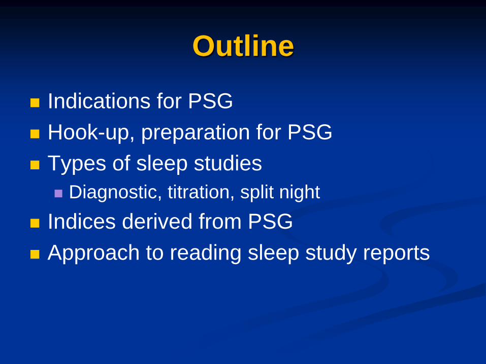
Outline
Indications for PSG
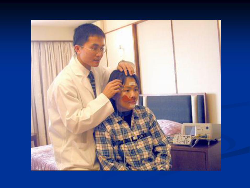
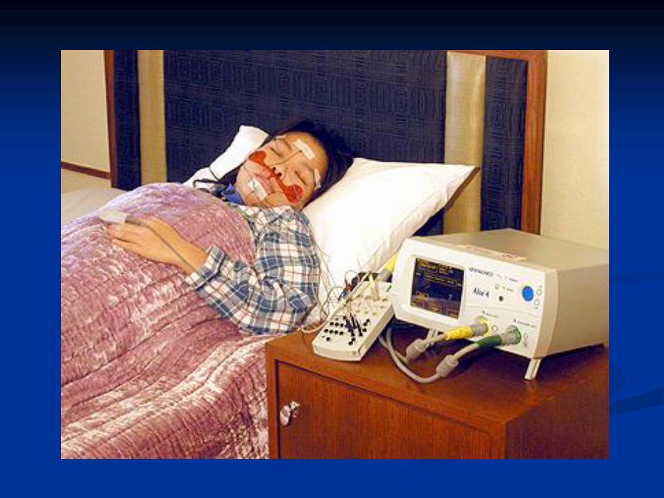
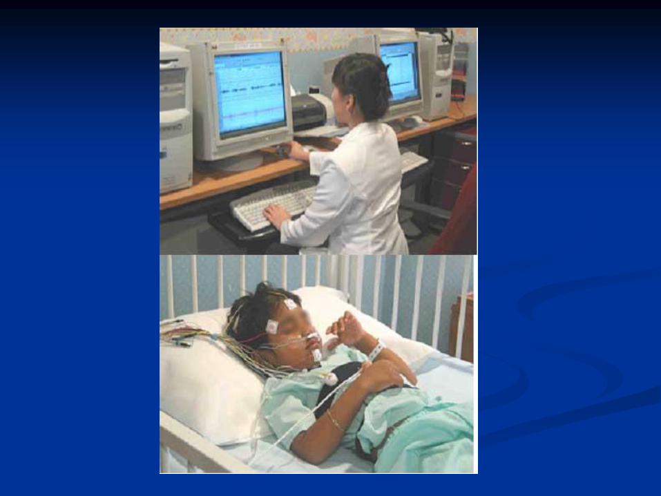
Hook-up, preparation for PSG
Types of sleep studies
Diagnostic, titration, split night
Indices derived from PSG
Approach to reading sleep study reports
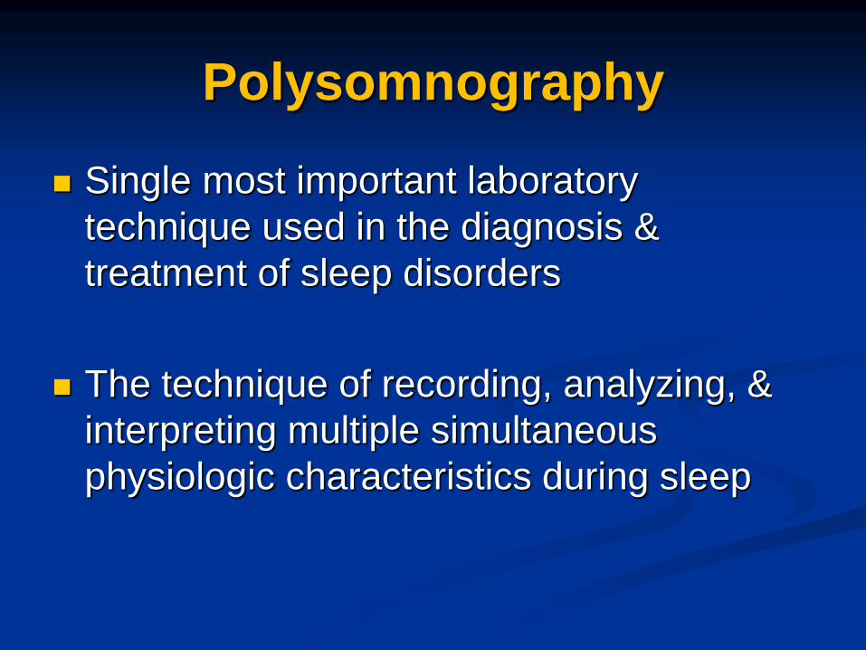
Polysomnography
Single most important laboratory
technique used in the diagnosis &
treatment of sleep disorders
The technique of recording, analyzing, &
interpreting multiple simultaneous
physiologic characteristics during sleep
Indications
Excessive daytime sleepiness
Obstructive sleep apnea
Breathing difficulties during sleep
Behavior disturbances during sleep
Poor sleep quality or Insomnia to exclude
other sleep disorders
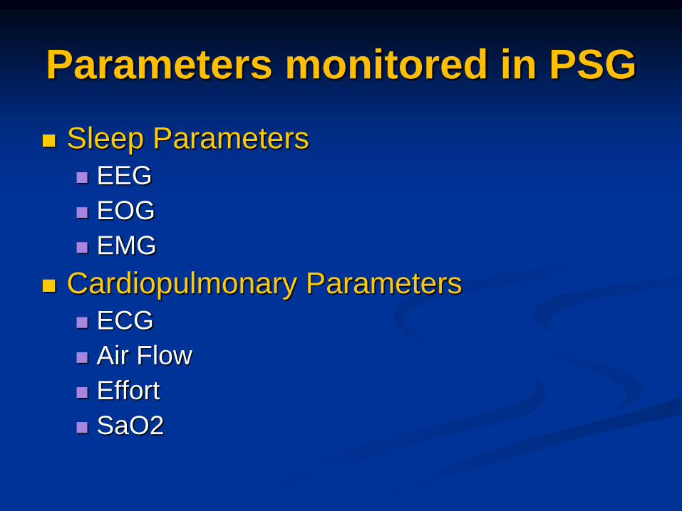
Parameters monitored in PSG
Sleep Parameters
EEG
EOG
EMG
Cardiopulmonary Parameters
ECG
Air Flow
Effort
SaO2
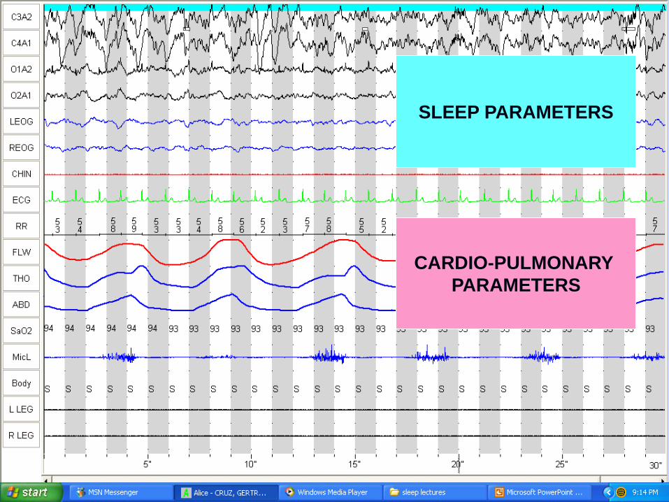
SLEEP PARAMETERS
CARDIO-PULMONARY
PARAMETERS

Amplitude
Time
1 sec
Moving Paper
Input 1(Exploring electrode)
Input 2 (Reference electrode)
+
-
Differential Amplifier
Cycles per second or Hz
uV
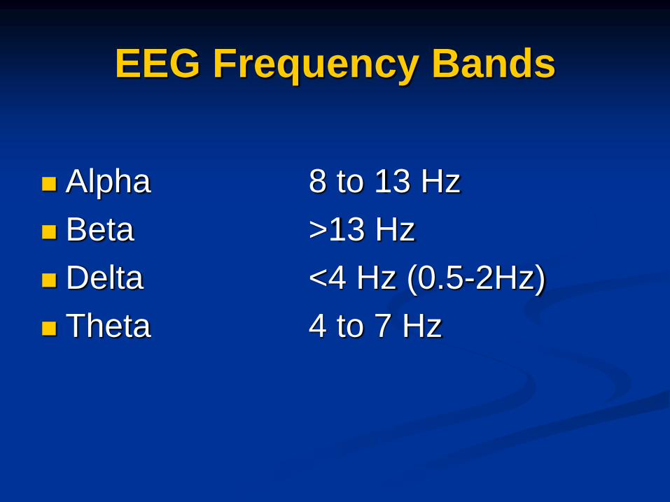
EEG Frequency Bands
Alpha 8 to 13 Hz
Beta >13 Hz
Delta <4 Hz (0.5-2Hz)
Theta 4 to 7 Hz
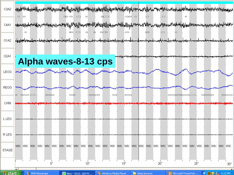
Alpha waves-8-13 cps

Beta waves->13 cps
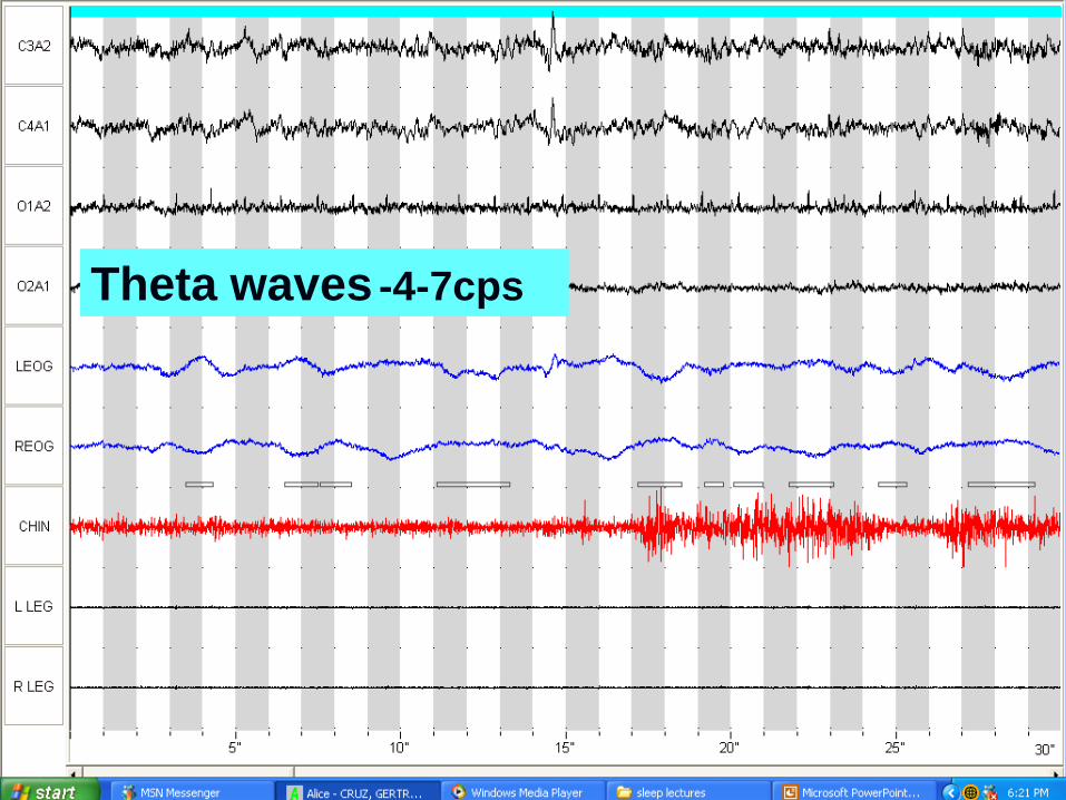
Theta waves -4-7cps
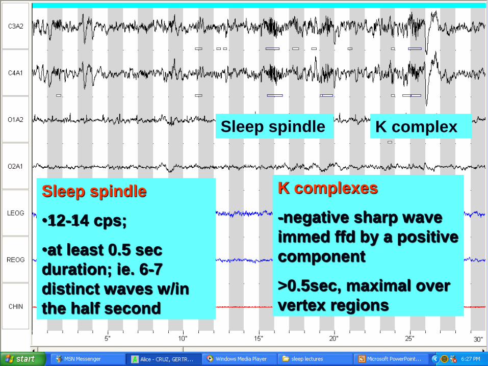
Sleep spindle K complex
Sleep spindle
•12-14 cps;
•at least 0.5 sec
duration; ie. 6-7
distinct waves w/in
the half second
K complexes
-negative sharp wave
immed ffd by a positive
component
>0.5sec, maximal over
vertex regions
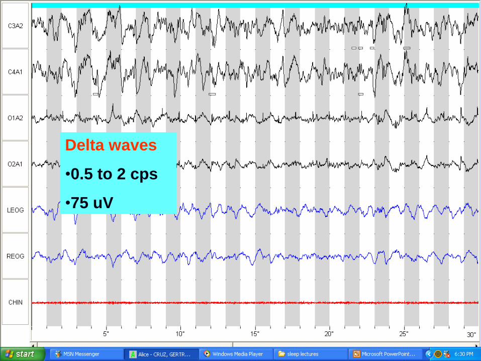
Delta waves
•0.5 to 2 cps
•75 uV

Sleep Stages
Stage W
Non- REM
Stage N1
Stage N2
Stage N3
Stage R
*AASM Manual, 2007
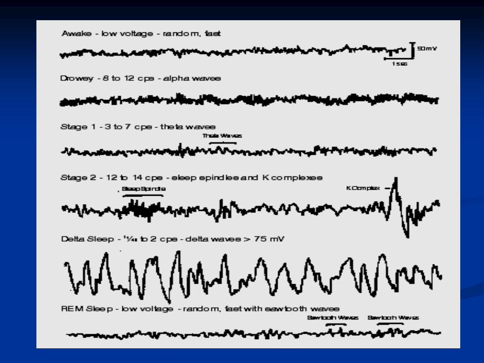
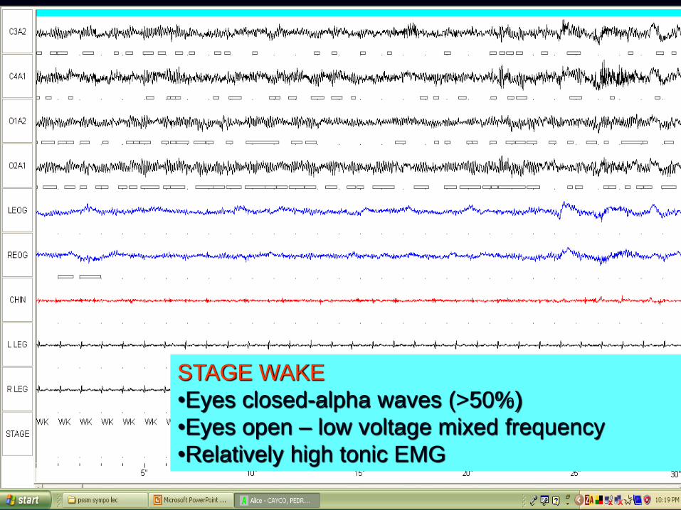
STAGE WAKE
•Eyes closed-alpha waves (>50%)
•Eyes open – low voltage mixed frequency
•Relatively high tonic EMG
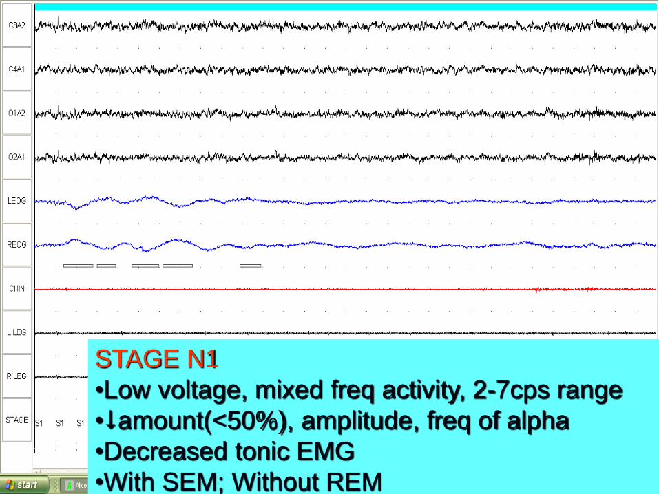
STAGE N1
•Low voltage, mixed freq activity, 2-7cps range
•amount(<50%), amplitude, freq of alpha
•Decreased tonic EMG
•With SEM; Without REM
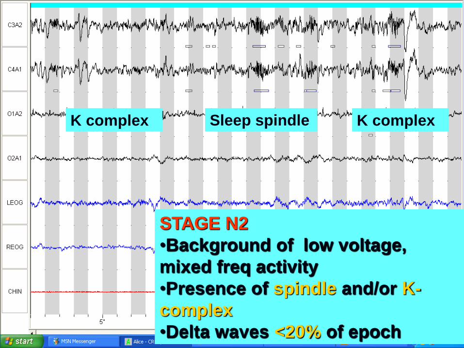
Sleep spindle K complex
STAGE N2
•Background of low voltage,
mixed freq activity
•Presence of spindle and/or K-
complex
•Delta waves <20% of epoch
K complex
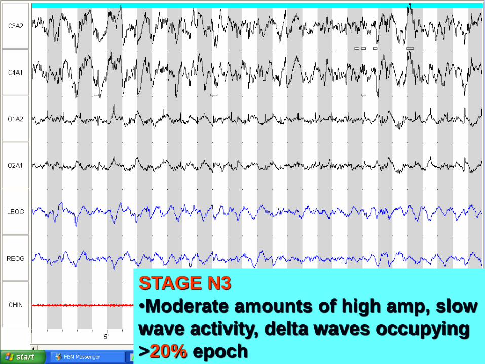
STAGE N3
•Moderate amounts of high amp, slow
wave activity, delta waves occupying
>20% epoch
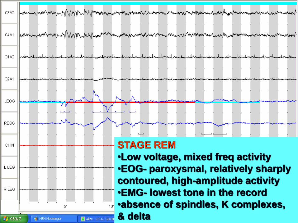
STAGE REM
•Low voltage, mixed freq activity
•EOG- paroxysmal, relatively sharply
contoured, high-amplitude activity
•EMG- lowest tone in the record
•absence of spindles, K complexes,
& delta
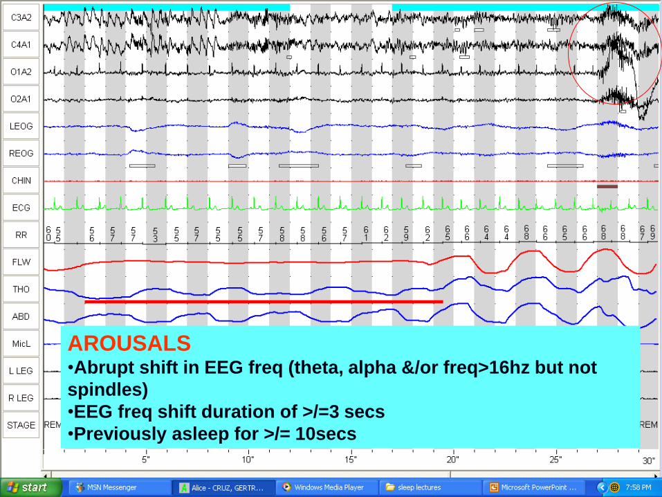
AROUSALS •Abrupt shift in EEG freq (theta, alpha &/or freq>16hz but not
spindles)
•EEG freq shift duration of >/=3 secs
•Previously asleep for >/= 10secs
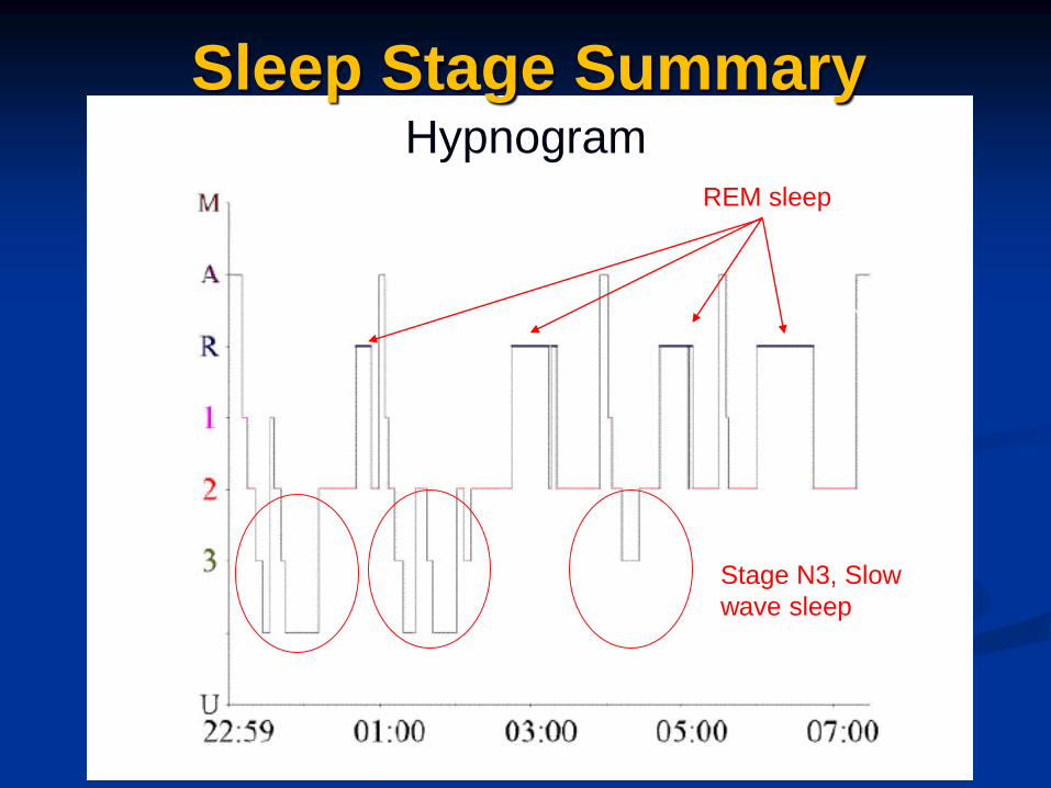
Hypnogram
Sleep Stage Summary
REM sleep
Stage N3, Slow
wave sleep

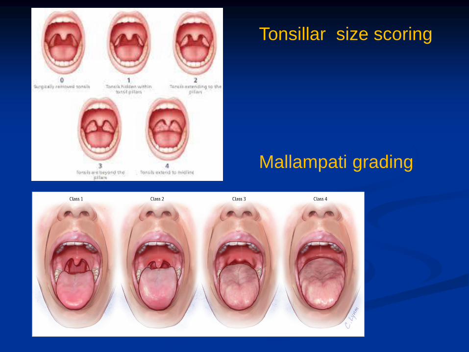
Tonsillar size scoring
Mallampati grading

Situation Chance of Dosing (0-3)
Sitting and reading 0 1 2 3
Watching television 0 1 2 3
Sitting inactive in a public place – ex theater
or meeting
0 1 2 3
As a passenger in a car for an hour without a
break
0 1 2 3
Lying down to rest in the afternoon 0 1 2 3
Sitting and talking to someone 0 1 2 3
Sitting quietly after lunch (when you’ve had no
alcohol)
0 1 2 3
In a car, while stopped in traffic 0 1 2 3
TOTAL
SCORE
0 = would never dose 2=moderate chance of dozing
1= slight chance of dozing 3=high chance of dozing ESS>10
EPWORTH SLEEPINESS SCALE

SLEEP PARAMETERS
CARDIO-PULMONARY
PARAMETERS
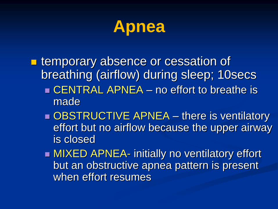
Apnea
temporary absence or cessation of breathing (airflow) during sleep; 10secs
CENTRAL APNEA – no effort to breathe is made
OBSTRUCTIVE APNEA – there is ventilatory effort but no airflow because the upper airway is closed
MIXED APNEA- initially no ventilatory effort but an obstructive apnea pattern is present when effort resumes
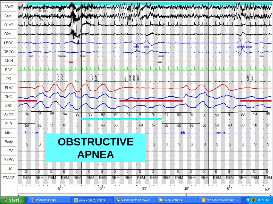
OBSTRUCTIVE
APNEA
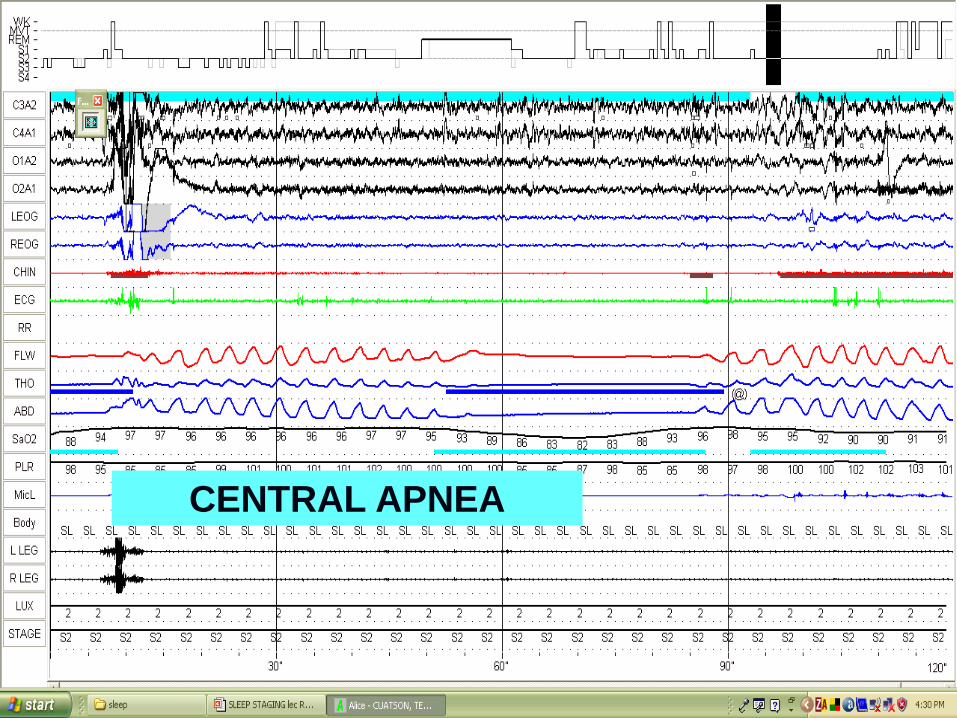
CENTRAL APNEA
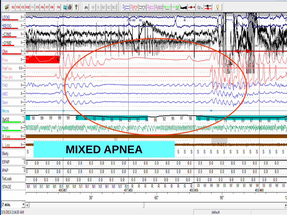
MIXED APNEA
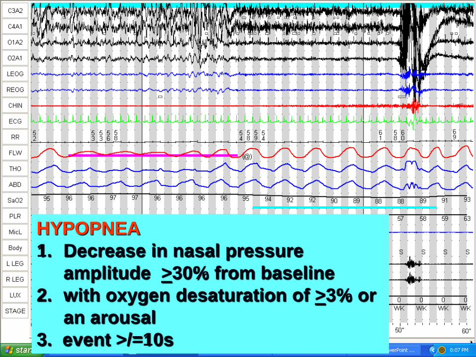
HYPOPNEA
1. Decrease in nasal pressure
amplitude >30% from baseline
2. with oxygen desaturation of >3% or
an arousal
3. event >/=10s
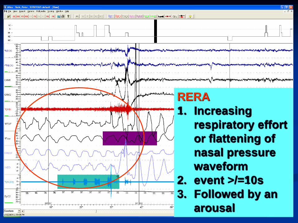
RERA
1. Increasing
respiratory effort
or flattening of
nasal pressure
waveform
2. event >/=10s
3. Followed by an
arousal
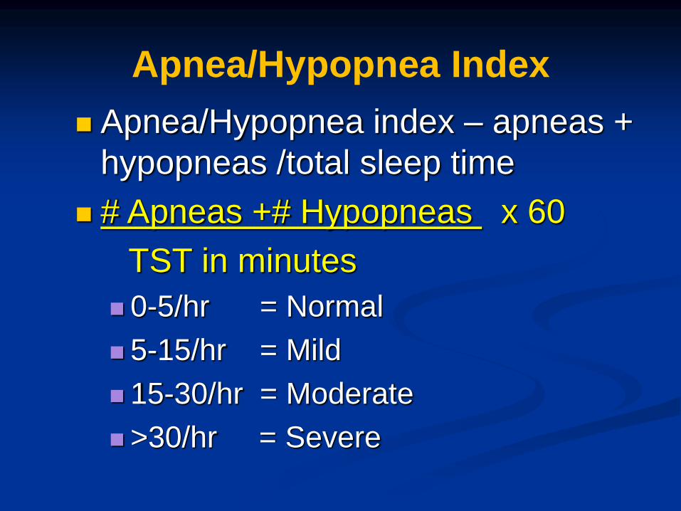
Apnea/Hypopnea Index
Apnea/Hypopnea index – apneas +
hypopneas /total sleep time
# Apneas +# Hypopneas x 60
TST in minutes
0-5/hr = Normal
5-15/hr = Mild
15-30/hr = Moderate
>30/hr = Severe
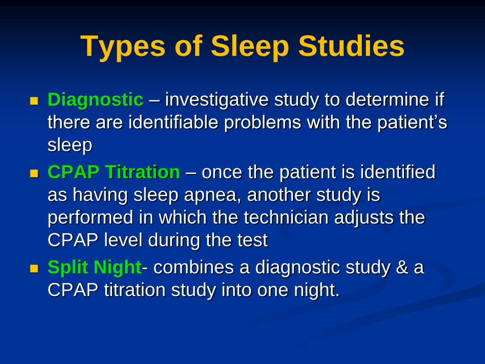
Types of Sleep Studies
Diagnostic – investigative study to determine if
there are identifiable problems with the patient’s
sleep
CPAP Titration – once the patient is identified
as having sleep apnea, another study is
performed in which the technician adjusts the
CPAP level during the test
Split Night- combines a diagnostic study & a
CPAP titration study into one night.
IV.41
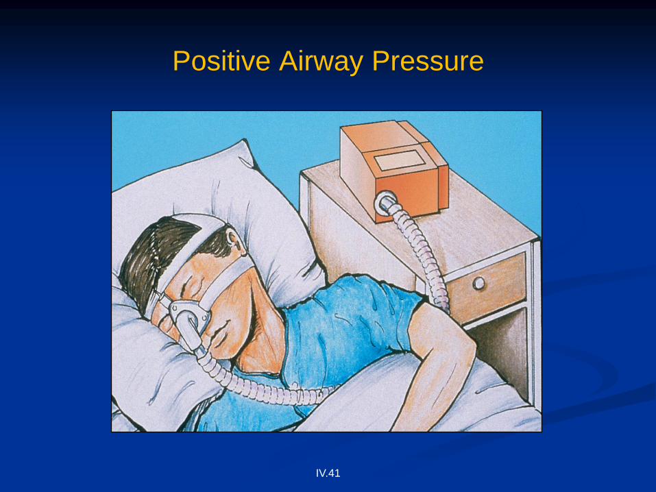
Positive Airway Pressure
IV.42
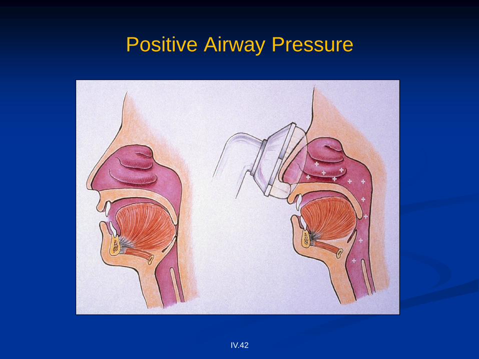
Positive Airway Pressure


Indices derived from PSGSleep Related Indices
Time in Bed (TIB)
Total recording time (TRB)
Total sleep time (TST)
Sleep Efficiency >90%
Sleep Latency <20 mins
REM latency 90-120 mins
Wake after Sleep Onset (WASO) <20 mins
Sleep Period Time (SPT)
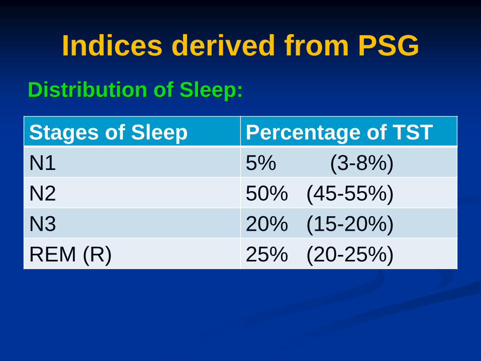
Indices derived from PSG
Stages of Sleep Percentage of TST
N1 5% (3-8%)
N2 50% (45-55%)
N3 20% (15-20%)
REM (R) 25% (20-25%)
Distribution of Sleep:
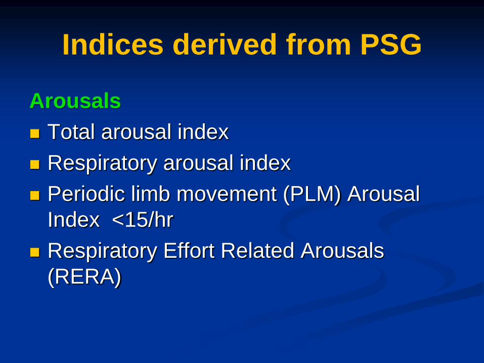
Indices derived from PSG
Arousals
Total arousal index
Respiratory arousal index
Periodic limb movement (PLM) Arousal
Index <15/hr
Respiratory Effort Related Arousals
(RERA)

Indices derived from PSG
Abnormal activity during the study
Periodic Limb movements index (PLMI)
Bruxism
ECG

Indices derived from PSG
Respiratory Indices
Apnea Hypopnea index (AHI)
RERA index
Respiratory Disturbance Index (RDI)
Oxygen saturation indices

Apnea/Hypopnea Index
Apnea/Hypopnea index – apneas +
hypopneas /total sleep time
# Apneas +# Hypopneas x 60
TST in minutes
0-5/hr = Normal
5-15/hr = Mild
15-30/hr = Moderate
>30/hr = Severe

Factors affecting interpretation
Sleep Quantity & Quality – decreased
sleep quantity & poor sleep efficiency will
overestimate AHI
Absent REM sleep – underestimate AHI
since apneas & hypopneas tend to be
worse in REM sleep when respiratory
muscles are more hypotonic
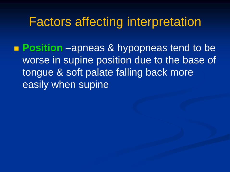
Factors affecting interpretation
Position –apneas & hypopneas tend to be
worse in supine position due to the base of
tongue & soft palate falling back more
easily when supine
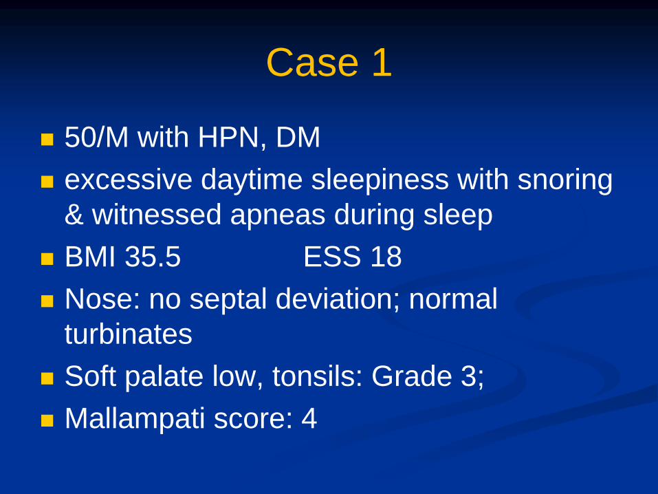
Case 1
50/M with HPN, DM
excessive daytime sleepiness with snoring
& witnessed apneas during sleep
BMI 35.5 ESS 18
Nose: no septal deviation; normal
turbinates
Soft palate low, tonsils: Grade 3;
Mallampati score: 4

Total sleep
time
463.5 min %Stage N3 13.5%
Time in bed 508.0 min %REM 22.2%
Sleep
Efficiency
91.2% Arousal
Index
45.4/hTST
Lowest satn 79% PLMI 2.8/h
NREM AHI 70.4/hr REM AHI 81.6/hr
AHI 72.9/hr

HR,BPM
30507090110130150170190
SpO2,%
50
60
70
80
90
100
Stage
N3
N2
N1
R
W
CA,sec
0
10
20
OA,sec
0
10
20
MA,sec
0
10
20
HYPO,sec
0
10
20
RERA,sec
0
10
20
10:02:48 PM 12 AM 1 AM 2 AM 3 AM 4 AM 5 AM 6 AM
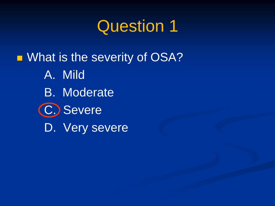
Question 1
What is the severity of OSA?
A. Mild
B. Moderate
C. Severe
D. Very severe
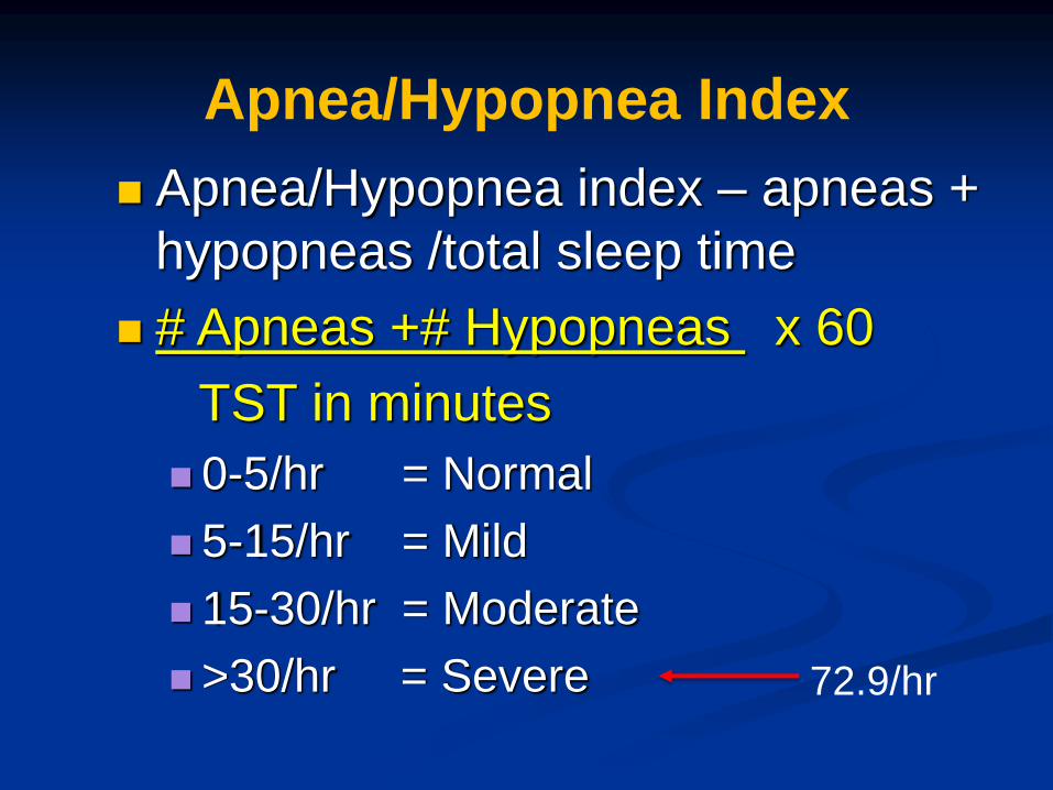
Apnea/Hypopnea Index
Apnea/Hypopnea index – apneas +
hypopneas /total sleep time
# Apneas +# Hypopneas x 60
TST in minutes
0-5/hr = Normal
5-15/hr = Mild
15-30/hr = Moderate
>30/hr = Severe 72.9/hr
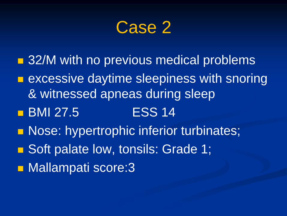
Case 2
32/M with no previous medical problems
excessive daytime sleepiness with snoring
& witnessed apneas during sleep
BMI 27.5 ESS 14
Nose: hypertrophic inferior turbinates;
Soft palate low, tonsils: Grade 1;
Mallampati score:3
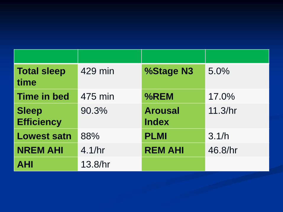
Total sleep
time
429 min %Stage N3 5.0%
Time in bed 475 min %REM 17.0%
Sleep
Efficiency
90.3% Arousal
Index
11.3/hr
Lowest satn 88% PLMI 3.1/h
NREM AHI 4.1/hr REM AHI 46.8/hr
AHI 13.8/hr

S S
L L
S
L
S
Pos
.PRSRRSLSPLPL
HR,BPM
30507090110130150170190
SpO2,%
5060708090100
Stage
N3
N2
N1
R
W
CA,sec
01020
OA,sec
01020
MA,sec
01020
HYPO,sec
01020
IPAP,cmH2O
0
5
10
15
20
EPAP,cmH2O
0
5
10
15
20
Tot. Leak.,L/min
0255075100
11:06:00 PM 1 AM 2 AM 3 AM 4 AM 5 AM 6 AM 7 AM
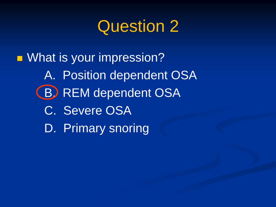
Question 2
What is your impression?
A. Position dependent OSA
B. REM dependent OSA
C. Severe OSA
D. Primary snoring

Case 3
55/F with HPN, CAD, dyslipidemic, s/p Coronary
bypass surgery.
Loud snoring, choking episodes during sleep,
falls asleep while driving
BMI 38.5, neck circumference = 42.5 cm, ESS of
16/24.
Nose: normal turbinates
Soft palate low, tonsils: Grade 2;
Mallampati score: 4
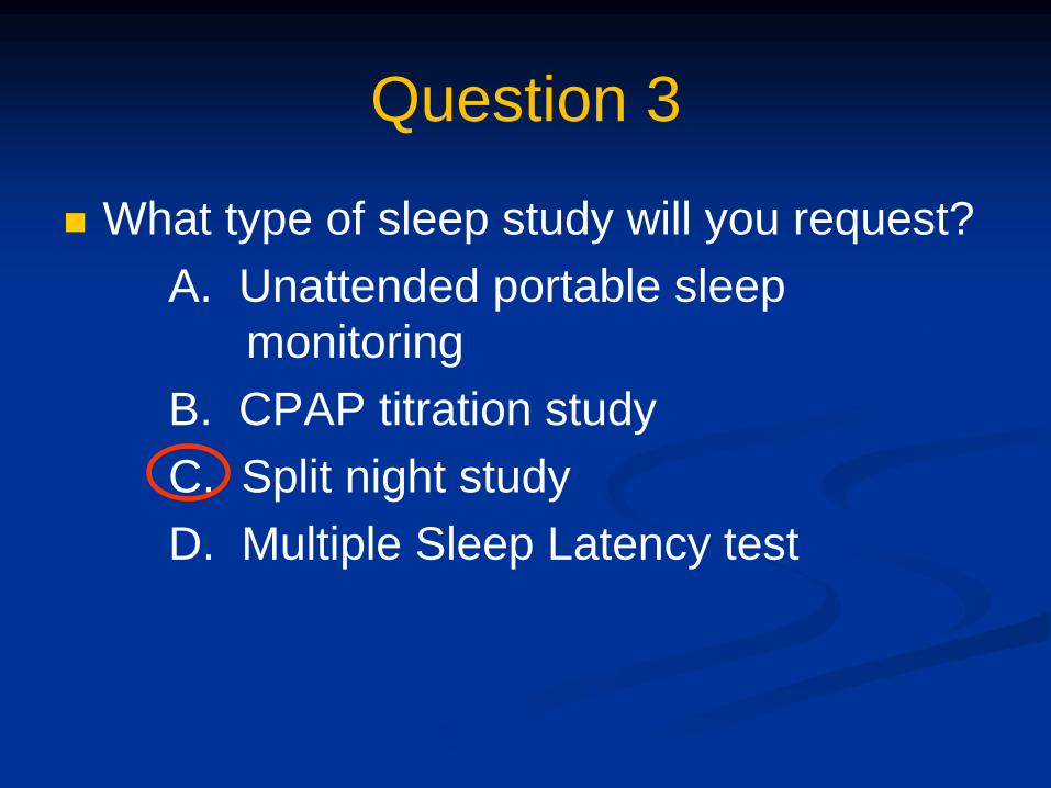
Question 3
What type of sleep study will you request?
A. Unattended portable sleep
monitoring
B. CPAP titration study
C. Split night study
D. Multiple Sleep Latency test
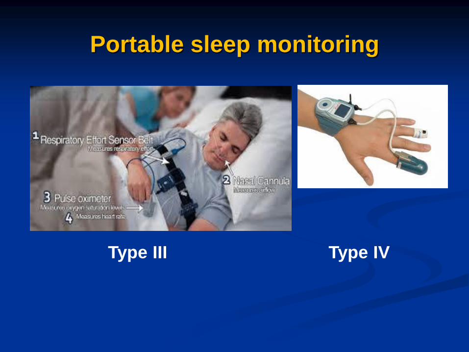
Type III Type IV
Portable sleep monitoring
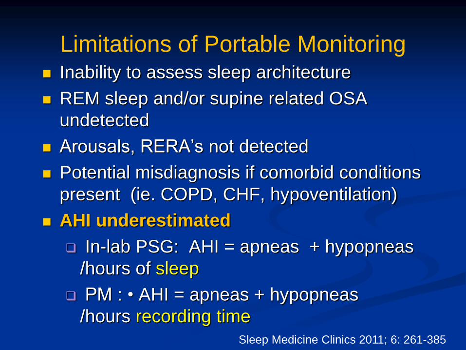
Limitations of Portable Monitoring Inability to assess sleep architecture
REM sleep and/or supine related OSA
undetected
Arousals, RERA’s not detected
Potential misdiagnosis if comorbid conditions
present (ie. COPD, CHF, hypoventilation)
AHI underestimated
In-lab PSG: AHI = apneas + hypopneas
/hours of sleep
PM : • AHI = apneas + hypopneas
/hours recording timeSleep Medicine Clinics 2011; 6: 261-385
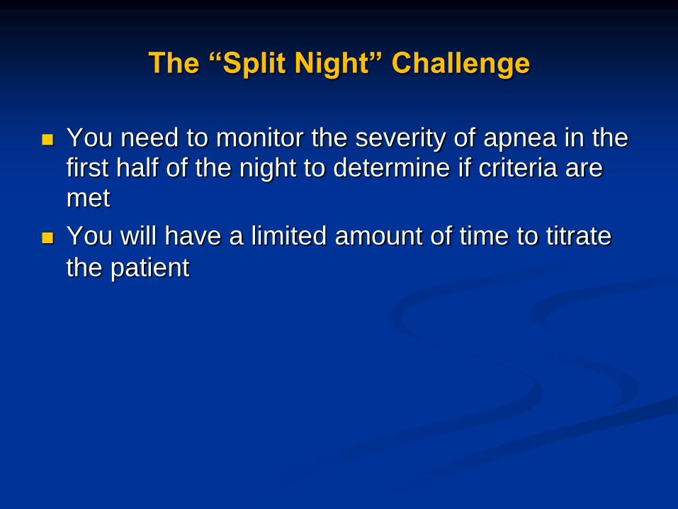
The “Split Night” Challenge
You need to monitor the severity of apnea in the first half of the night to determine if criteria are met
You will have a limited amount of time to titrate
the patient
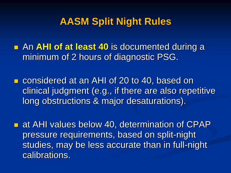
AASM Split Night Rules
An AHI of at least 40 is documented during a minimum of 2 hours of diagnostic PSG.
considered at an AHI of 20 to 40, based on clinical judgment (e.g., if there are also repetitive long obstructions & major desaturations).
at AHI values below 40, determination of CPAP pressure requirements, based on split-night studies, may be less accurate than in full-night calibrations.
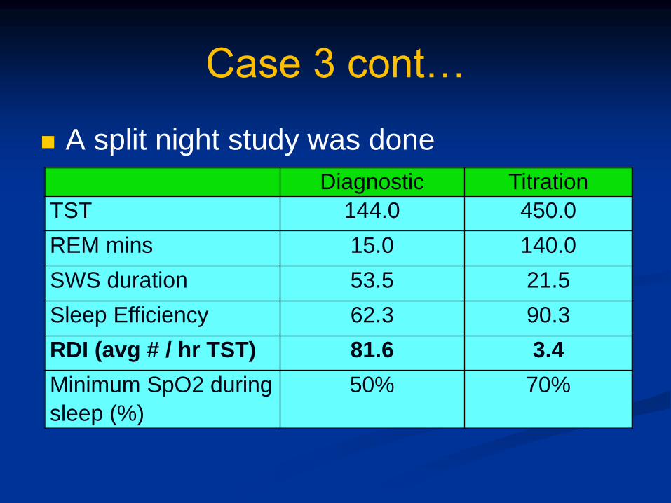
Case 3 cont…
A split night study was done
Diagnostic Titration
TST 144.0 450.0
REM mins 15.0 140.0
SWS duration 53.5 21.5
Sleep Efficiency 62.3 90.3
RDI (avg # / hr TST) 81.6 3.4
Minimum SpO2 during
sleep (%)
50% 70%
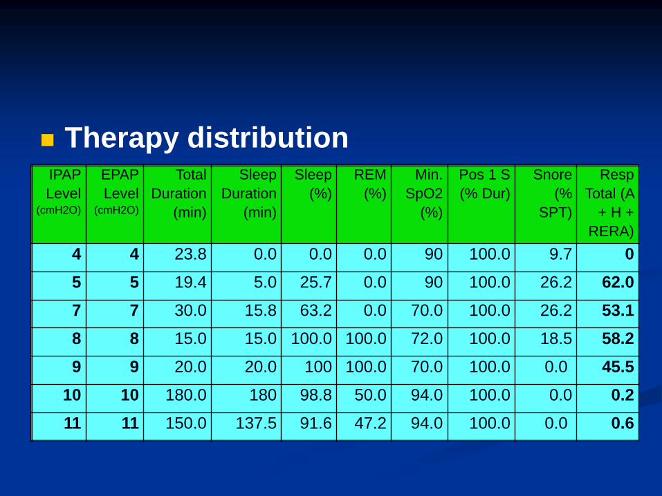
Therapy distributionIPAP
Level(cmH2O)
EPAP
Level (cmH2O)
Total
Duration
(min)
Sleep
Duration
(min)
Sleep
(%)
REM
(%)
Min.
SpO2
(%)
Pos 1 S
(% Dur)
Snore
(%
SPT)
Resp
Total (A
+ H +
RERA)
4 4 23.8 0.0 0.0 0.0 90 100.0 9.7 0
5 5 19.4 5.0 25.7 0.0 90 100.0 26.2 62.0
7 7 30.0 15.8 63.2 0.0 70.0 100.0 26.2 53.1
8 8 15.0 15.0 100.0 100.0 72.0 100.0 18.5 58.2
9 9 20.0 20.0 100 100.0 70.0 100.0 0.0 45.5
10 10 180.0 180 98.8 50.0 94.0 100.0 0.0 0.2
11 11 150.0 137.5 91.6 47.2 94.0 100.0 0.0 0.6

Night Hypnogram
S
Pos
.PRSRRSLSPLPL
HR,BPM
30507090110130150170190
SpO2,%
5060708090100
Stage
N3
N2
N1
R
W
CA,sec
01020
OA,sec
01020
MA,sec
01020
HYPO,sec
01020
RERA,sec
01020
4
8
57 8 9 10 11
9
IPAP,cmH2O
0
5
10
15
20
4 57 8 9 10 11
9
EPAP,cmH2O
0
5
10
15
20
Tot. Leak.,L/min
0255075100
9:54:07 PM 11 PM 12 AM 1 AM 2 AM 3 AM 4 AM 5 AM 6 AM
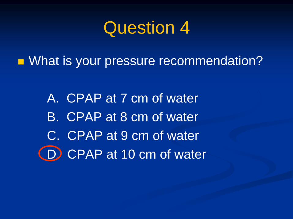
Question 4
What is your pressure recommendation?
A. CPAP at 7 cm of water
B. CPAP at 8 cm of water
C. CPAP at 9 cm of water
D. CPAP at 10 cm of water
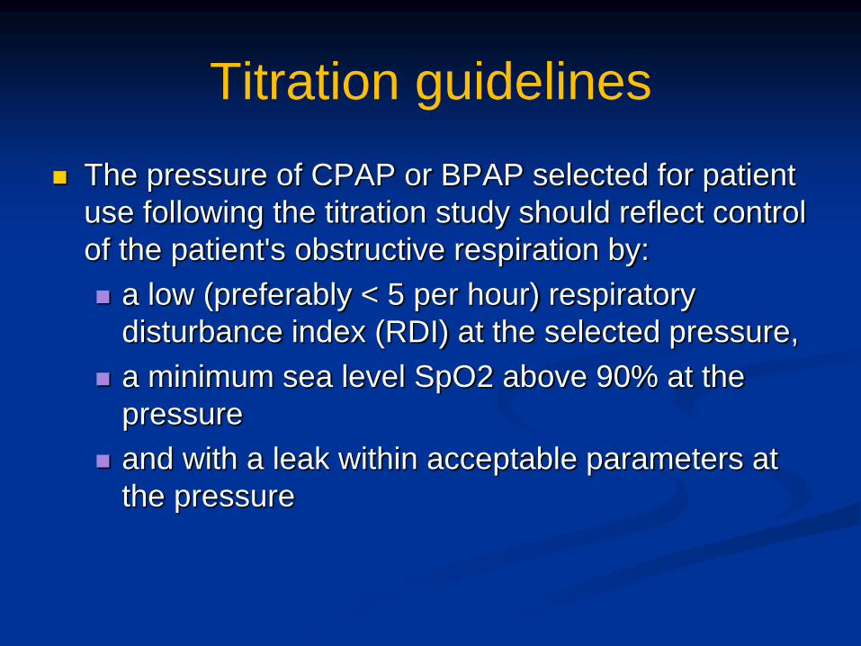
Titration guidelines
The pressure of CPAP or BPAP selected for patient
use following the titration study should reflect control
of the patient's obstructive respiration by:
a low (preferably < 5 per hour) respiratory
disturbance index (RDI) at the selected pressure,
a minimum sea level SpO2 above 90% at the
pressure
and with a leak within acceptable parameters at
the pressure
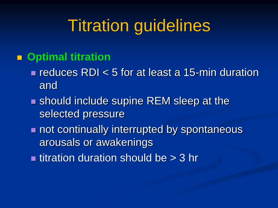
Titration guidelines
Optimal titration
reduces RDI < 5 for at least a 15-min duration
and
should include supine REM sleep at the
selected pressure
not continually interrupted by spontaneous
arousals or awakenings
titration duration should be > 3 hr
Question 5
When will the patient need a follow-up
PSG?
A. change in weight by 10%
B. recurrence of symptoms
C. intolerance of CPAP therapy
D. All of the above
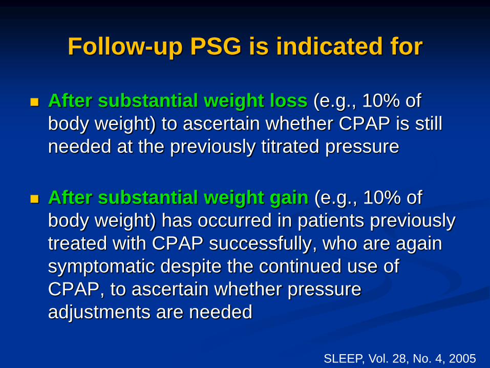
Follow-up PSG is indicated for
After substantial weight loss (e.g., 10% of
body weight) to ascertain whether CPAP is still
needed at the previously titrated pressure
After substantial weight gain (e.g., 10% of
body weight) has occurred in patients previously
treated with CPAP successfully, who are again
symptomatic despite the continued use of
CPAP, to ascertain whether pressure
adjustments are needed
SLEEP, Vol. 28, No. 4, 2005
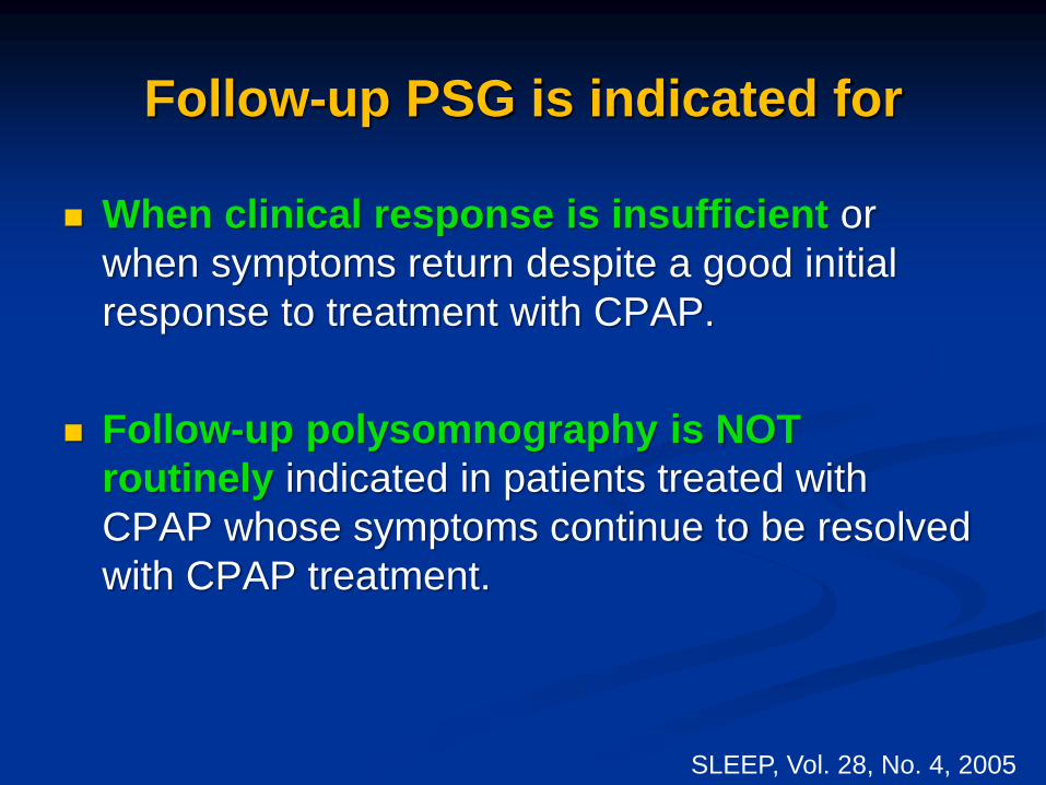
Follow-up PSG is indicated for
When clinical response is insufficient or
when symptoms return despite a good initial
response to treatment with CPAP.
Follow-up polysomnography is NOT
routinely indicated in patients treated with
CPAP whose symptoms continue to be resolved
with CPAP treatment.
SLEEP, Vol. 28, No. 4, 2005
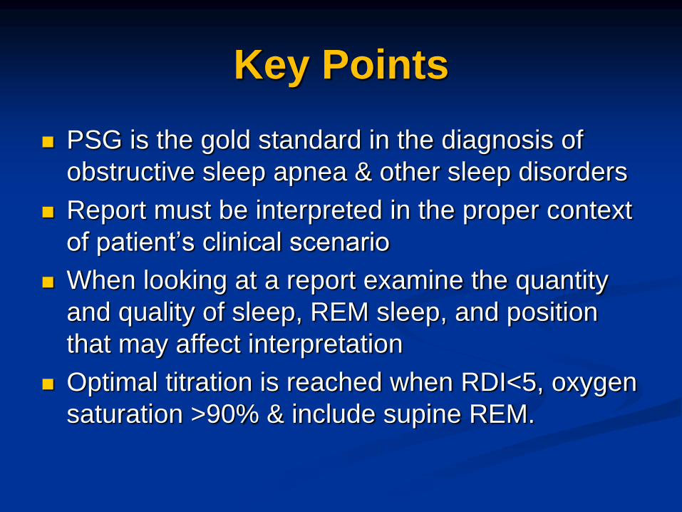
Key Points
PSG is the gold standard in the diagnosis of
obstructive sleep apnea & other sleep disorders
Report must be interpreted in the proper context
of patient’s clinical scenario
When looking at a report examine the quantity
and quality of sleep, REM sleep, and position
that may affect interpretation
Optimal titration is reached when RDI<5, oxygen
saturation >90% & include supine REM.


THANK YOU!!!


![UIHC Home Sleep Apnea Testing (HSAT) · 2018-10-12 · portable sleep-testing device (Peripheral Arterial Tonometry [PAT®]) and those measured by PSG. • Methods: 14 studies qualified](https://static.fdocuments.in/doc/165x107/5f792a255f98132bd25e45a4/uihc-home-sleep-apnea-testing-hsat-2018-10-12-portable-sleep-testing-device.jpg)