SLEEP DISTURBANCES AND COMORBIDITIES IN …hera.ugr.es/tesisugr/21468266.pdf · Fisioterapia y que...
95
Tesis doctoral internacional International PhD thesis ALTERACIONES DEL SUEÑO Y COMORBILIDADES EN POBLACIÓN MEDITERRÁNEA: EL CASO DE LA APNEA DEL SUEÑO SLEEP DISTURBANCES AND COMORBIDITIES IN MEDITERRANEAN POPULATION: THE SLEEP APNEA CASE Universidad de Granada Departamento de medicina legal, TOXICOLOGIA, antropología física y forense Marie Carmen Valenza 2012
Transcript of SLEEP DISTURBANCES AND COMORBIDITIES IN …hera.ugr.es/tesisugr/21468266.pdf · Fisioterapia y que...

Tesis doctoral internacional
International PhD thesis
ALTERACIONES DEL SUEÑO Y COMORBILIDADES EN POBLACIÓN
MEDITERRÁNEA: EL CASO DE LA APNEA DEL SUEÑO
SLEEP DISTURBANCES AND COMORBIDITIES IN MEDITERRANEAN
POPULATION: THE SLEEP APNEA CASE �
�
�
�
Universidad de Granada
Departamento de medicina legal, TOXICOLOGIA, antropología física y forense
Marie Carmen Valenza
2012

Editor: Editorial de la Universidad de GranadaAutor: Marie Carmen ValenzaD.L.: GR 1692-2013ISBN: 978-84-9028-572-5

Valenza MC, 2012
�
2
SSiigguuee ssiieemmpprree aaddeellaannttee,, aabbrriieennddoo nnuueevvaass ppuueerrttaass yy hhaacciieennddoo ccoossaass nnuueevvaass.. SSéé ccuurriioossoo…… ppoorrqquuee llaa ccuurriioossiiddaadd nnooss hhaaccee sseegguuiirr
nnuueevvooss ccaammiinnooss..
Walt Disney (los Robisons)
Dedicada a todas las personas de mi vida que se han sacrificado para hacer mis sueños realidad
This thesis is dedicated to those in my life who have sacrificed to make my dreams a reality

Valenza MC, 2012
�
3
ÍNDICE
Resumen……………………………………………………………….………………...…4
Abstract……………………………………………………………….……….……...……5
Introducción………………………………………………………………………...……..6
Introduction…………………………………………………………………….….....…..16
Objetivos………………………………………………………………………..…………26
Aims………………………………………………………………………………..……….27
Material y métodos (matherial and methods)…………………………..……......28
Resultados y discusión (Results and discussion)…………………………...………33
� Consideration of sleep dysfunction in rehabilitation………………………………34
� Alteration in Sleep Quality in Patients with Mechanical Insidious Neck Pain
and Whiplash-Associated Neck Pain………………………………..………….……42
� Epidemiology of sleep-related complaints associated with obstructive sleep
apnea, insomnia and non-restorative sleep in an at risk population in
Granada-Spain. …………………………………………………………………………53
� Factores de riesgo para el Síndrome Metabólico en una población con
Apnea del Sueño. Evaluación en un grupo de pacientes de Granada y
provincia: ESTUDIO GRANADA. ……………………………………………….….…..64
� Apnea del Sueño y Arritmias Ventriculares. …………………………………..……72
Conclusiones……………………………………………………………………………..85
Conclussions……………………………………………………………………………...86
Agradecimientos (Acknowledgments)…………………………………….………87
Bibliografía (References)……………………………………………………………....88

Valenza MC, 2012
�
4�
RESUMEN
El sueño ha sido hasta hace poco una entidad relegada a un lugar secundario
en la medicina, y de igual manera por el resto de las profesiones de la salud.
Sin embargo, las ciencias médicas han girado ahora su mirada hacia un
aspecto que ocupa un tercio de nuestra vida como el sueño y sus patologías.
El conocimiento de los efectos y utilidad del sueño han desencadenado este
cambio de situación, centrándose en la monitorización y tratamiento del sueño
profesionales de todas ciencias médicas.
La fisioterapia es una de las disciplinas que aun no se ha interesado en este
campo.
El sueño y su calidad, duración y arquitectura se han mostrado como datos
clínicos de gran relevancia en cualquier historia clínica.
En esta memoria de tesis se ha profundizado en las alteraciones del sueño
como síntoma clínico de gran importancia para la Fisioterapia, a través del
Síndrome de Apnea de Sueño como una de las patologías del sueño más
frecuentes.
Los resultados obtenidos en esta tesis demuestran que las alteraciones del
sueño son muy prevalentes en los pacientes que acuden a consulta de
Fisioterapia y que presentan una asociación significativa con el perfil clínico y
sintomático de los pacientes.
Adicionalmente, los resultados obtenidos han mostrado una asociación entre
diferentes factores de riesgo cardiovascular y la severidad de las alteraciones
del sueño en una población con apnea de sueño.

Valenza MC, 2012
�
5�
ABSTRACT
The sleep has been until recently an entity relegated to a secondary place in
medicine, and in a consecutive way elsewhere in the health professions.
However, medical sciences have turned his gaze to one aspect that occupies
one-third of our lives like the dream and its pathologies.
Knowledge of the effects and usefulness of sleep, have triggered this change in
status, becoming the monitoring and treatment of sleep an aspect in which
professionals of all health sciences are grouped.
Physiotherapy is one of the disciplines that have not been yet interested in this
field. Sleep and its quality, duration and architecture have proved to have
clinical relevance in any medical history, because it has recognized
repercussions in the patient symptoms and evolution.
With this memory of thesis we have pursued to show the importance of the sleep
disturbances as a common pathologies and main clinical symptom for the
physiotherapy, focussing our studies in Sleep apnoea.
The results obtained in this thesis suggests no doubt in that sleep disturbances
are a target of first level for physical therapy, that involve a little researched
aspect so far in patients who attend physical therapy consultation. At the same
time the results have allowed the development of a clinical profile of patients
with Mediterranean population in sleep apnea.
�
�

Valenza MC, 2012
�
6�
INTRODUCCIÓN
Esta tesis no pretende explorar una lista exhaustiva de los desordenes del
sueño; sin embargo, pretende esclarecer algunos de los desordenes más
prevalentes y sus asociaciones con otras patologías y síntomas. En esta
disertación se presenta una síntesis de la literatura sobre los factores asociados
a las alteraciones del sueño y su importancia terapéutica, al mismo tiempo
que analiza el perfil sintomático y patológico en un grupo poblacional con
alteraciones del sueño.
El sueño que se define en el plano de la conducta por la suspensión normal de
la conciencia y desde un punto de vista electrofisiológico por criterios de ondas
encefálicas específicas, consume un tercio de nuestra vida1. Por ello, en los
últimos años las patologías del sueño y sus repercusiones clínicas se han
destacado como un objetivo prioritario por gran parte de la comunidad
científica.
Ciclo de sueño vigilia: El sueño surge como un conjunto o constelación de
cambios fisiológicos rítmicos y reversibles en el que participan distintos sistemas
del organismo, regulados por el sistema nervioso central (SNC). Se caracteriza
por la abolición de la conciencia vigil y la reducción de la respuesta a los
estímulos ambientales, que se acompaña de cambios en múltiples funciones,
y que se puede dividir en al menos dos estados: sueño lento, en el cual se
reconocen 4 etapas, y sueño paradójico2.
Durante el sueño se producen múltiples cambios en el organismo que afectan
prácticamente a todos los sistemas; disminuye la frecuencia respiratoria y el
ritmo cardíaco, se relaja la musculatura y la temperatura corporal disminuye. A
su vez se segregan diversas hormonas que afectan a la regulación de la
energía, el peso, el crecimiento y el estrés. Simultáneamente se producen
procesos muy activos que implican cambios en el funcionamiento global del
cerebro.
Valenza MC, 2012
�
7�
La vigilia posee diversas características que le son propias, y entre las que se
encuentran el control homeostático de diversas funciones vitales
(cardiovasculares, respiratorias, endocrinas, etc). La organización y la ejecución
de movimientos que actuarán sobre el ambiente y la actividad psíquica que
continuamente nos acompaña, son también características indicativas de que
todo el cerebro, de una forma u otra, es utilizado para proporcionar la
condición de vigilia. Existe además, una forma de recepción y procesamiento
de la información sensorial proveniente del ambiente y del propio cuerpo
diferentes de aquellas efectuadas en el estado hípnico3.
Neurofisiología de la respiración durante el sueño
En la respiración se distinguen al menos dos procesos diferenciados: el
intercambio gaseoso alvéolo-capilar, y la renovación del aire o ventilación. De
estos dos procesos, el último es extraordinariamente dependiente del SNC, ya
que no tienen autonomía local y depende de una bomba neuromuscular
(fuelle torácico) y de la permeabilidad de la vía aérea superior que también
posee un importante componente neuromuscular.
La actividad ventilatoria es controlada por dos sistemas relativamente
interdependientes pero con diferentes propiedades:
1) sistema involuntario, automático, regido por las necesidades
metabólicas del organismo, que se puede alterar voluntariamente pero
sólo por un tiempo limitado (por ej., contener la respiración), activo en
condiciones de anestesia, coma y sueño lento. Esta información se
integra a nivel bulbo-protuberancial.
Este sistema está a cargo del mantenimiento del ritmo respiratorio. El
mecanismo por el cual ocurre esto todavía no está totalmente
aclarado; por ejemplo, las neuronas respiratorias no poseen
marcapasos como en el corazón. El control involuntario de la inspiración
y la espiración no depende de centros nerviosos específicos, sino de
una red difusa de neuronas interconectadas. El modelo que se acepta
actualmente consiste en grupos de neuronas o redes neuronales: grupo
A, grupo respiratorio dorsal; grupo B, grupo respiratorio ventral que forma
parte del núcleo retroambiguo; grupo C, localizado en la zona ventral

Valenza MC, 2012
�
8�
del núcleo retroambiguo. Un cuarto grupo de neuronas, el grupo P o
centro neumotáxico, cuya interacción origina el ritmo respiratorio
básico, que recibiría señales de los centros superiores, puede estimular
directamente al grupo C y llevar en último término a la inhibición de la
inspiración. Mientras la interacción entre los grupos A-C sería
responsable del ciclo respiratorio básico, los estímulos externos
originados en el centro neumotáxico y en los diversos receptores
modularían la actividad de los grupos neuronales, modificando la
velocidad del ciclo y la intensidad de la respuesta. De esta manera es
posible variar la frecuencia respiratoria y el volumen de aire corriente.
Este sistema neuronal está influido por el sistema reticular activador que
consiste en una red de neuronas que modula la actividad del sistema de
control por medio de cambios en el estado de excitabilidad del sistema
nervioso durante el sueño y la vigilia. El sistema reticular activador disminuye su
actividad durante el sueño. Estas estructuras, que filogenéticamente son más
antiguas, están reguladas, a su vez, por los centros superiores de la corteza
cerebral y el tálamo, que están involucrados en el control de actividades
voluntarias que conciernen al tórax y a los pulmones.
2) El otro mecanismo es voluntario, conductual, relacionado con las
actividades no respiratorias de la ventilación, como por ejemplo la fonación, la
deglución, la tos, etc; es activo en la vigilia y se cree que también en el sueño
paradójico (como algunos autores sugieren por el hecho de la irregularidad del
patrón respiratorio, que no responde a las modificaciones de los impulsos del
sistema automático vagales o químicos).
En líneas generales el sistema de control de la ventilación consta de receptores
localizados en diferentes áreas del organismo, que son activados por una
variada serie de estímulos químicos y físicos. Estos receptores envían mensajes
a un sistema de control que procesa la información y envía señales a los
efectores, los cuales modifican su actividad de acuerdo con el tipo de
mensaje recibido. Este sistema mantiene un nivel de ventilación estrechamente
vinculado a las necesidades metabólicas.

Valenza MC, 2012
�
9�
CONSECUENCIAS DE LAS ALTERACIONES DEL SUEÑO
El sueño es parte universal de nuestra vida. Su naturaleza recuperadora permite
sentirse descansado y con energías durante el día, como resultado de una
noche de buen sueño. Aun así, la mayoría de las personas tienen algún tipo de
problema con su sueño a lo largo de su vida debido al estrés, preocupaciones,
enfermedad, o similar. Desafortunadamente, muchas personas experimentan
problemas de sueño durante un periodo importante de tiempo antes de ser
diagnosticadas de un trastorno del sueño.
El sueño alterado tiene efectos que han sido menospreciados sobre la salud,
tal y como ha sido evidenciado por diversos informes de los sistemas e institutos
de Salud de multiples paises 4.
Figura 1: Interrelación entre alteraciones del sueño y salud general

Valenza MC, 2012
�
10�
Aunque el sueño alterado no recibe mucha atención respecto a otros
problemas sanitarios, existe gran cantidad de literatura sobre el sueño.
Muchos investigadores han estudiado el impacto sobre la salud pública de las
alteraciones del sueño. Se estima que millones de personas en los EEUU tienen
desórdenes del sueño crónicos, lo que supone un coste social de 90 millones
de dólares5 .
Muchos investigadores han examinado el efecto del sueño sobre diferentes
causas de mobimortalidad, patología cardiovascular y diabetes6.
Se sabe poco sobre todos los factores que pueden estar asociados al sueño poco
reparador. Sin embargo, existen gran cantidad de escalas para identificar aspectos
del sueño y muchas contienen información útil y completa7 .
El Plan Nacional de EEUU para la Investigación sobre los Desordenes del Sueño8 ha
planteado numerosas recomendaciones para investigaciones futuras que incluyen la
necesidad de mejores métodos de medición, al igual que recomienda la
focalización en las desigualdades debidas a género, grupo humano y perfil
socioeconómico en las terapéuticas del sueño8.
Las diferentes organizaciones para la salud insisten en estos últimos años sobre la
necesidad de examinar los efectos del sueño sobre la salud, ya que se están
convirtiendo en un problema de salud pública de primer nivel. Todos los
profesionales de la salud deben conocerlos y comprender su repercusión, mucho
mayor de lo que se había pensado hasta hace poco tiempo.
El síndrome de Apnea del Sueño (SAS)
También llamado síndrome de Apneas-Hipoapneas del Sueño es uno de los
trastornos del sueño de mayor prevalencia, oscilando en la población general
entre un 1 a un 2%9, y se caracteriza por un cuadro de episodios repetidos de
obstrucción de la vía aérea superior, acompañada por esfuerzo respiratorio
inefectivo y apneas seguidas habitualmente por respiraciones amplias de
recuperación (llamadas hiperpneas) durante el sueño, que provocan
constantes desaturaciones de la oxihemoglobina y despertares transitorios.

Valenza MC, 2012
�
11�
La apnea, definida como la interrupción completa del flujo inspiratorio superior
a 10 segundos, puede diferenciarse en función de su origen.
Si se mantiene el esfuerzo ventilatorio durante este tiempo, la apnea se
considerará de origen obstructivo, y si no existe este esfuerzo se considerará
como de origen central. Una hipoapnea es un episodio de definición más
compleja consistente en la obstrucción parcial de las vías aéreas superiores y
que produce una clara disminución del flujo aéreo en boca-nariz de duración
igual o superior a 10 segundos. Se acompaña de un despertar transitorio y/o
desaturación cíclica de la oxihemoglobina. Un índice de apnea e hipoapnea
(IAH) (número de apneas + número de hipoapneas por hora de sueño) superior
a 5 por hora se considera patológico.
El Síndrome de apnea de sueño suele afectar la vida diaria de los sujetos que la
padecen. Son numerosas las complicaciones que la gente experimenta debido a la
AS, siendo las complicaciones pulmonares y la somnolencia las más comunes.
Algunos síntomas frecuentemente asociados son las apneas observadas, los
procesos asfícticos, la fatiga diurna, ronquidos nocturnos importantes, despertares
frecuentes que pueden estar acompañados de nicturia, dolores de cabeza
frecuentes e hipertensión, que pueden ser observados por el propio sujeto o por su
entorno 10.
Accidentes de tráfico y pérdida de la productividad son dos de los problemas
más comunes causados por la somnolencia11.
Algunas de las repercusiones diurnas exhibidos por pacientes con síndrome de
apnea de sueño son somnolencia diurna, depresión, disminución de la libido,
fatiga, dolor de cabeza y ronquidos12 . Algunas de la complicaciones que
puede causar el SAS pueden ser metabolismo alterado de la glucosa,
patología cardiovascular aguda y crónica, accidente cerebro vascular y
deterioro cognitivo13.
Existen tres tipos de Apnea de Sueño, central obstructiva y mixta.
� La apnea central es aquella apnea en la que no se produce esfuerzo respiratorio
debido a un fallo de la conexión entre el sistema nervioso y el sistema respiratorio,
resultando una falta de señal hacia los músculos respiratorios.

Valenza MC, 2012
�
12�
�La apnea obstructiva, en la que se produce esfuerzo respiratorio. Los músculos
respiratorios se contraen, pero esta contracción es ineficaz debido a la obstrucción
total o parcial de la vía aérea superior.
�La apnea de tipo mixto, es una combinación de la apnea central y de la apnea
obstructiva. La apnea mixta comienza habitualmente como una apnea central y
continua como una apnea obstructiva tras la recuperación de la actividad muscular
respiratoria13.
Los pacientes pueden mostrar todas las formas de Apnea durante el sueño.
Habitualmente los sujetos presentan una combinación de todos los tipos y son
diagnosticados de aquel tipo que prevalece. La apnea obstructiva en la forma más
común de apnea y la apnea central es la menos frecuente, siendo diagnosticada
tan solo en el 10% de los casos12 . Todas las formas de AS muestran alguna forma de
respiración intermitente durante el sueño13.
El conocimiento de la apnea ha progresado gracias al aumento del conocimiento
sobre los mecanismos del sueño. El hecho de que la apnea del sueño se
caracterice por un colapso recurrente de la vía aérea superior durante el sueño ha
sido aceptado, sin embargo el mecanismo de producción exacto y su por qué es
algo que numerosos investigadores están intentando comprender14.
Más del 5% de los adultos se encuentran afectados por algún tipo de alteración
respiratoria durante el sueño10. La incidencia de la AS ha aumentado de manera
significativa durante los últimos diez años, debido a la mayor atención y
reconocimiento por parte de los diferentes profesionales sanitarios. Los estudios de
sueño son solicitados cada vez con más frecuencias solicitadas para el diagnóstico
de la apnea, permitiendo a los profesionales sanitarios determinar el tipo y severidad
de las alteraciones del sueño.
El Síndrome de Apnea Obstructiva del Sueño y alteraciones cardiovasculares
La apnea obstructiva del sueño (SAOS) es una patología de tipo crónico
caracterizada por obstrucciones repetidas de la vía aérea superior durante el sueño.
Recientes investigaciones demuestran una conexión entre estas oclusiones repetitivas
de la vía aérea superior y anormalidades cardiovasculares. Por ejemplo,

Valenza MC, 2012
�
13�
correlaciones significativas demostradas entre SAOS e hipertensión pulmonar
sistémica15,16,17, isquemia cardiaca y disritmias16,18 , y accidentes
cerebrovasculares16,19.
La hipertensión sistémica (HTA) está frecuentemente asociada al SAOS y puede estar
directamente relacionado con los episodios parciales y repetidos de apnea que
interrumpen los reflejos cardiovasculares durante el sueño20,21,22,23. La prevalencia de
la HTA en los pacientes con SAOS puede ser de un 50%24.
La reducción de flujo de aire durante el sueño se traduce en una reducción en los
niveles de oxigeno en sangre y un incremento en la retención de dióxido de
carbono; ambos causan un incremento en la actividad nerviosa simpática y
conducen aumentos de la tensión arterial25.
Los pacientes con SAOS también presentan un aumento de la actividad simpática
durante el día26,27. Estas fluctuaciones en el sistema nervioso simpático pueden estar
relacionadas con la patogénesis de la HTA.
Sin embargo, estudios que relacionan la SAOS con la HTA, han mostrado una
disminución de la actividad nerviosa simpática28 y la tensión arterial29,30 después del
tratamiento con presión espiratoria positiva (nCPAP)19.
Adicionalmente, la SAOS se asocia a los efectos agudos y crónicos de la simpático-
excitación, que dando lugar a una adaptación vascular reducida32. De cualquier
manera, se ha aceptado que el SAOS afecta de manera adversa la homeostasis de
diferentes mecanismos cardiovasculares, aunque los mecanismos reguladores
vasculares no han sido investigados en la SAOS, tales como la hipoxia asociada o las
sustancia vasoactivas derivadas (adenosina, prostanoides, endotelina y oxido nítrico
(NO)31).
El Sleep Heart Health Study conducido por Mehra encontró que los pacientes con
SAOS moderado se encuentran en serio riesgo de padecer patología vascular
oclusiva32.
Estos investigadores, al igual que Lavie y cols33 encontraron que el riesgo no se
incrementaba proporcionalmente en los sujetos con SAOS severo.
Estos estudios se confirman por las observaciones clínicas en las que aun en
presencia de desaturaciones nocturnas severas, no todos los SAOS sufren co-
morbilidades cardiovasculares34.
Valenza MC, 2012
�
14�
Como consecuencia, se sabe con certeza que aun existen mecanismos
desconocidos aún que relacionan el SAOS y algunas complicaciones
cardiovasculares.
Mecanismos de asociación entre apnea del sueño y arritmia cardíaca
Los mecanismos que vinculan la AS y las arritmias cardíacas continúan siendo
objeto de especulación y debate continuo.
Uno de los mecanismos potenciales de asociación es la interrelación entre el
efecto proarrítmico de los episodios intermitentes de hipoxia nocturna,
isquemia miocárdica y el aumento de la actividad simpática, lo que conlleva
un incremento de las concentraciones plasmáticas de catecolaminas35,36. Sin
embargo, las especulaciones sobre los diferentes mecanismos de asociación
tan solo han sido confirmadas parcialmente.
Adicionalmente, como resultado de la actividad simpático secundaria a los
episodios de apnea, se producen oscilaciones en la frecuencia cardiaca y en la
tensión arterial, al igual que un incremento de las arritmias ventriculares y
auriculares37. En un estudio sobre 400 pacientes, se registraron arritmias en más de la
mitad de los sujetos38. La mayoría de estas alteraciones cardiacas fueron
bradiarritmias con 43 de los 400 pacientes que registraron paradas sinusales de 2.5 a
13 segundos.
En estudios posteriores, las arritmias ventriculares fueron relativamente poco
comunes39. Algunos investigadores discuten la alta prevalencia de las arritmias
asociadas a la SAOS, justificándose en que los pacientes incluidos en los estudios
hasta ahora tienen apnea severa y no son representativos de un espectro más
amplio del síndrome37.
En resumen, el SAOS predispone a los individuos a un aumento de la morbi-
mortalidad cardiovascular24. Este mayor riesgo patológico se agrava por factores
como la edad, índice de masa corporal (IMC), hipertensión arterial y polisomnografía
con índices elevados40.
Por lo tanto, no puede ser subestimada la importancia de la detección temprana y
tratamiento precoz. Las intervenciones adecuadas han demostrado mejorar el
rendimiento del neuropsiquiátrico, la lucidez mental y la calidad de vida, los valores

Valenza MC, 2012
�
15�
de hipertensión diurna, y la reducción de desaturaciones nocturnas y las hipopneas
respiratorias16,41.
Diversos estudios epidemiológicos se han centrado en describir patologías
cardiovasculares concretas en grupos poblacionales; sin embargo, apenas existen
estudios en poblaciones mediterráneas y aun menos en poblaciones españolas.
Así, el presente estudio evaluó sistemáticamente la intrincada relación entre los
factores de riesgo cardiovascular y las diferentes severidades de apnea de sueño en
una población de Granada.

Valenza MC, 2012
�
16�
INTRODUCTION
This thesis doesn´t want to explore all the sleep disturbances, but it wants to
review some of the more prevalent disorders and its associations with other
pathologies and symptoms. This thesis presents a review of the literature about
the associate factors to the sleep disturbances and its therapeutic importance.
Sleep is defined, in a behavioral and electrophysiological way, as the
suspension of the consciousness and as specific encephalic waves. This
represents the third part of our lives1. In the last years the sleep pathologies and
its clinical repercussions have become a first objective by the scientific
community.
Sleep-wake cycle: the sleep is a group of rhythmic physiological changes who
are made by all the organic systems, always controlled by the central nervous
system. It’s characterized by the loose of the consciousness and the reduction
of the environment stimuli response. Its accompanied by a change in different
functions and can be divided in two stages: slow sleep (with four different
stages) and paradoxal sleep2.
During the sleep numerous changes are made, affecting practically all the
systems. It reduce the respiratory frequency and cardiac rhythm, it relax the
muscles and reduces body temperature. At the same time many hormones are
segregated regulating the energy, growing and stress process. Changes in the
global brain function occur at the same time becoming a very active process.
The consciousness time have its own characteristics that included the
homeostatic control of different vital functions. The organization and execution
of movements are a signal of the environment and the physic activity that
usually are together. There is a way of reception and information processing of
the sensorial information that becomes from the environment and from the own
body, different from the sleep processes3.
Valenza MC, 2012
�
17�
Breathing Neurophysiology during the sleep
In the respiration we can recognize two different processes: the renewal of air or
ventilation and the diffusion alveoli-capillary. Between this processes, the
diffusion is controlled by the central nervous system and is dependent of the
thoracic pump and the upper airway permeability.
Ventilatory activity is controlled by two relatively independent systems but with
different properties:
1) Involuntary system, automatic and regulated by the metabolic
necessities of the body. Is active in conditions of anesthesia, slow sleep
and all this information’s are processed in the brain protuberance level.
This system is maintaining the breathing rhythm. The relationship does not
fully understand.
2) The involuntary control of the inspiration and expiration is not controlled of
the specific nervous centers, is regulated by a diffuse mesh of neurons.
The actually accepted model consist in neurons groups: group A who is
the respiratory dorsal group, group B who is the ventral respiratory group,
and group C. a fourth group, called P or neumotaxic centre, can directly
origin the basic breathing rhythm. The interaction between A-C groups
will be responsible of the modulation of the breathing rhythm, changing
the velocity and intensity of the respiratory answer.
This neural system in influenced by the reticular system, the reticular system can
modulate de activity of the control system with changes in the excitability of the
nervous system during the sleep and wake cycle. The reticular system reduces its
activity during the sleep.
The other mechanism is voluntary, conductual and related to other activities that
are not only the respiration like phonation, deglution, cough…
In general, the control system of ventilation is made by receptors distributed in
different areas of the body that are activated by series is chemical and physical
stimuli. These receptors send messages to a control system that process the
information and send signals to the effectors who modify their activities in line
with the orders.

Valenza MC, 2012
�
18�
This system maintains the level of ventilation required to the metabolic
demands.
Consequences of sleep disturbances on health
Sleep is a universal part of life. Its naturally restorative properties allow many to
feel refreshed and energized during the day, as a result of a good night’s sleep.
However, most people experience a problem with sleep at some point, due to
stress, worry, physical illness, or the like. Unfortunately, many people experience
sleep problems over a longer period of time and are then diagnosed with a
sleep disorder. Such people fail to benefit from the recuperative effects of
sleep. Poor sleep has under-appreciated impacts on health, as evidenced by
numerous reports4. Although poor sleep may not receive as much attention as
other health problems, a wealth of literature exists on sleep.
Figure 1: Interrelationship between sleep disturbances and general health

Valenza MC, 2012
�
19�
Researchers have investigated the public health impact of poor sleep. It is
estimated that approximately 40 million United States citizens have chronic
sleep disorders, costing society roughly $90 billion dollars5 .
Many researchers examine sleep’s contribution to all-cause mortality,
cardiovascular disease, and diabetes6.
The medical objectives range from improving the medical management of
sleep problems to decreasing vehicular crashes that result from driver
sleepiness. Unfortunately, poor sleep does not always receive as much
consideration as other, perhaps more prominent, physical conditions. It is often
difficult, if not impossible, to identify if poor sleep is a side effect of another
condition or the cause of a particular health condition.
Little is understood about the many factors associated with poor sleep. While many
scales exist to assess sleep, few contain complete and adequate information7 .
The National Sleep Disorders Research Plan put forth several recommendations for
future research, including a need for more methods to measure sleep, as well as
focuses on gender, racial, and ethnic disparities in sleep disorder outcomes8.
While there are many complications that people experience due to SAS, pulmonary
complications and sleepiness are the most common complications. Some common
symptoms that may be indicative of sleep apnea are apnea; snorting, or choking that
their significant other may observe during the day; loud snoring every night; frequently
waking during the night which can be accompanied by the need to urinate; recurrent
headaches in the morning; and hypertension8.
The different health organizations are promoting in this last year’s about the clinical
importance to examine the sleep effects on health. The sleep disturbances are
becoming a public health burden and all the health professionals has to know them
and understand its repercussions.

Valenza MC, 2012
�
20�
Sleep Apnea Syndrome
The sleep apnea syndrome, also called apneas-hipopneas syndrome is one of
the major sleep disturbances in prevalence rates, rating from 1 to 2%9 .Is
characterized by repetitive obstruction of the upper airway, accompanying by
respiratory ineffective effort and consecutive apneas followed by deep
breathing cycles (called hiperpneas) during the sleep, with constantly
desaturation of oxyhemoglobine and transient awakes.
The apnea, defined as the complete interruption of inspiratory flow more than
10 seconds, can be differentiated by its origin. If the breathing work is
maintained during this time, the apnea can be considered obstructive, if it
doesn´t exists can be considered as central.
A hypopnea is an episode more complex, because consist in a partial
obstruction of the upper airways and a clear reduction of air flow in mouth-nose
of a 10 seconds or more duration. Is accompanied by a transient awake and/or
cyclic desaturation of the oxihemoglobin. And index of apnea and hipopnea
more than 5 per hour is considered pathological.
Sleep apnea syndrome can affect patients’ daily lives. There are many
complications and symptoms that are associated with sleep apnea. Motor
vehicle accidents and loss of productivity at work are two problems caused by
sleepiness10. Some of the symptoms exhibited by patients with sleep apnea
syndrome are daytime sleepiness, depression, decreased libido, fatigue,
headaches, and snoring11 . Some of the complications that sleep apnea
syndrome may cause are, glucose metabolism disorders, acute and chronic
cardiovascular disease, stroke, and cognitive decline12.
There are three different forms of sleep apnea syndrome (SAS), central, obstructive,
and mixed.
�Central SAS refers to sleep apnea in which there are no respiratory attempts because
there is a break in the connection between the nervous system and the respiratory
system resulting in no signals to the respiratory muscles.
�Obstructive SAS there are breathing movements, the respiratory muscles work, but
there is no airflow due to partial or incomplete obstruction of upper airways.

Valenza MC, 2012
�
21�
�Mixed SAS is a combination of central and obstructive SAS. Mixed SAS may start as
central SAS and then continue as obstructive SAS after recovery of respiratory
musculature activity15 .
Patients may exhibit all forms of SAS during a night of sleep. Usually patients exhibit a
combination of the types so they are diagnosed as the type of SAS that occurs the
most. Obstructive sleep apnea (OSA) is the most common type of sleep apnea, and
central sleep apnea is the least common type of sleep apnea, being diagnosed in
only 10% of cases16 . All of the forms of SAS exhibit some form of intermittent breathing
interruption during sleep17.
Knowledge about sleep apneas has progressed along with an increased knowledge
about mechanisms of sleep. The fact that sleep apnea is characterized by recurrent
collapse of the upper airway during sleep has been common knowledge for many
years.
Exactly how this occurs and why it only occurs in certain individuals is something that
researchers are still trying to fully understand18.
Up to 5% of adults are affected by some type of breathing problems during sleep19.
Incidences of OSA have increased greatly over the past ten years because medical
professionals are more aware of the signs and symptoms of this condition and are
recognizing it more. Sleep studies are now done to diagnose a person with sleep
apnea. This allows medical professionals to determine the type and severity of the
patients’ condition.

Valenza MC, 2012
�
22�
Sleep Apnea and cardiovascular alterations
Recent studies demonstrate a link between the repetitive occlusions in the upper
airway and cardiovascular abnormalities. For example, significant correlations have
been demonstrated between OSA and systemic and pulmonary hypertension20,21,22,
cardiac ischemia and dysrhythmias23,24, and cerebrovascular accidents25,26.
Systemic hypertension (HTN) is frequently associated with OSA and can be directly
ascribed to the repeated and partial apneic events that disrupt autonomic
cardiovascular reflexes during sleep27,28,29,30. The prevalence of HTN in OSA patients
may be as high as 50%31. Diminished airflow during sleep results in decreased blood
oxygen levels and increased carbon dioxide retention, both of which cause increased
sympathetic nerve activity resulting in elevations in blood pressure32.
Patients with OSA also exhibit increased sympathetic activity during the day33,34. These
fluctuations in sympathetic nervous system activity may be involved in the
pathogenesis of HTN.
Furthermore, studies supporting this link between OSA and HTN have reported a
decrease in resting sympathetic nervous system activity35 and blood pressure36,37
following nasal continuous positive airway pressure (nCPAP) treatment38.
In addition, OSA is associated with both acute and chronic effects of sympatho-
excitation, resulting in decreased vascular compliance38 [Chittenden et al, 2002]. It
has long been postulated that the increased vascular tone in OSA leads to abnormal
diurnal blood pressure control and increases in the incidence of myocardial infarction
and stroke. Therefore, it is likely that OSA adversely affects several different
cardiovascular homeostatic mechanisms. Although these vascular regulatory
mechanisms have not been thoroughly investigated in OSA patients, disease-related
hypoxia might abate vascular function by depravation of tissue-derived or blood
vessel-derived vasoactive substances such as adenosine, prostanoids, endothelin,
and nitric oxide (NO)39.
Attributed to these recent findings, researchers are now suggesting the processes of
neovascularization and the resultant decrease in vascular compliance may explain
the decline in morbidity and mortality associated with long-standing sleep apnea
pathology. The Sleep Heart Health Study recently indicated that patients with mild OSA
are at enhanced risk for occlusive vascular disease36.
Valenza MC, 2012
�
23�
However, these investigators also found that the risk did not rise further in patients with
more severe OSA. This appears to support the findings of Lavie et al42. These
researchers witnessed decreases in cardiovascular morbidity and mortality in ~1500
men Israeli men after the eighth decade of life.
These studies are supported by the clinical observations that, even in the presence of
severe nocturnal oxygen desaturations, not all OSA patients suffer from cardiovascular
co-morbidity43.
Consequently, it may be considered that certain as yet unrevealed mechanism(s)
shield some OSA patients from developing cardiovascular complications.
Valenza MC, 2012
�
24�
Mechanism relating SA and cardiac rhythm disturbances
The mechanism between AS and cardiac arrhythmias are a first research objective in
the last years.
One of the first links is the relationship between the proarrythmic effect of the hypoxic
episodes, the myocardial ischemia and the increase of sympathic activity. These
implies an increase of plasmatic concentration of cathecolamines44,45.
Additionally, As a result of increased sympathetic activity secondary to apneic
episodes, oscillations in both heart rate and blood pressure occur as well as an
increase in the frequency of atrial and ventricular arrhythmias46. In one large study of
400 patients, cardiac arrhythmias were noted in almost half the subjects46. The
majority of these cardiac disturbances noted were bradyarrhythmias with 43 of the
400 patients experiencing sinus arrest of 2.5 to 13 seconds. In subsequent studies,
ventricular arrhythmias were relatively uncommon47. Some investigators dispute the
high prevalence of arrhythmias associated with OSA, claiming that the patients in the
aforementioned studies had severe sleep apnea and were not representative of a
greater spectrum of disease severity49.
In summary, OSA predisposes individuals to increased cardiovascular morbidity and
mortality 50. This increased pathological risk is exacerbated by factors of age, body
mass index (BMI), elevated blood pressure, and aberrant polysomnography indices51.
Thus, the importance of early detection and treatment cannot be understated.
Adequate intervention has been shown to improve daytime alertness and quality of
life, daytime hypertension, neuropsychiatric performance, and reduce nocturnal
oxyhemoglobin desaturation and respiratory arousals52,53.
Different epidemiological studies have been focused in the description of
cardiovascular pathologies and risk factors in different populations groups, but there is
no frequent in mediterranean subjects and less frequent in Spanish populations.
The present study has the purpose to evaluate systematically the relationship between
cardiovascular risk factors and the different severities of sleep apnea in a Granada
population (Spain).

Valenza MC, 2012
�
25�

Valenza MC, 2012
�
26�
OBJETIVOS
General�
El objetivo general de esta tesis doctoral internacional es demostrar las
repercusiones sobre la salud de las alteraciones del sueño, profundizando en
el Síndrome de la Apnea del Sueño como patología central, en una
población de Granada.
Específicos
1. Determinar la importancia y repercusión que pueden tener las
alteraciones del sueño para la fisioterapia
2. Evidenciar y examinar las relaciones entre alteraciones del Sueño y dolor
3. Presentar la asociación entre alteraciones del sueño y perfil clínico en
población granadina
4. Profundizar en la interrelación entre factores de riesgo cardiovascular y
apnea de sueño
5. Estudiar los mecanismos de asociación entre apnea del sueño y Arritmia
cardiaca

Valenza MC, 2012
�
27�
AIMS
General�
The main purpose in this Phd international dissertation was to explore the
sleep disturbances effects on health, with a focus on Sleep Apnea as a
prevalent pathology in a Granada Population.
Specifics
1. To determine the importance and repercussions of sleep disturbances in
physiotherapy practice.
2. To evidence the relationship between sleep and pain
3. To present the association between sleep disturbances and clinical profile
in a population from Granada
4. To search in the relationship between cardiovascular risk factors and sleep
apnea syndrome
5. To study the association mechanism between sleep apnea and cardiac
arrythmias

Valenza MC, 2012
�
28�
MATERIAL Y MÉTODOS
Objetivo I
Determinar la importancia y repercusión que pueden tener las alteraciones del sueño para la fisioterapia
Articulo I:
Valenza MC, Rodenstein DO, Fernandez-de-las-Peñas C. Consideration of sleep dysfunction in rehabilitation. Journal of Bodywork and Movement Therapies, Volume 15, Issue 3, July 2011, 262-267.
Metodología:
Revisión bibliográfica exhaustiva de las diferentes bases de datos, Pubmed, Scopus, Ovid, Science Direct y Web of Science.
Palabras clave: Sleep disturbances, sleep alterations, sleep disorders, health consequences, clinical profile, pain, fatigue.

Valenza MC, 2012
�
29�
MATERIAL Y MÉTODOS
Objetivo II
Evidenciar y examinar las relaciones entre alteraciones del Sueño y dolor
Articulo II:
Valenza MC, Valenza G, Gonzalez-Jimenez E, Isabel-de-la-Llave-Rincon A, Arroyo-Morales M, Fernandez-de-las-Peñas C. Alteration in Sleep Quality in patients with mechanical insidious neck pain and whiplash-associated neck pain. American Journal of Physical Medicine & Rehabilitation. 2012 91,1:584-591
Metodologia:
Diseño: Estudio descriptivo con casos y controles.
Muestra: 19 sujetos con dolor mecánico cervical, 22 sujetos diagnosticadas de latigazo cervical y 18 controles sanos.
Variables estudiadas: dolor, discapacidad y calidad y perfil de sueño.
MATERIAL Y MÉTODOS

Valenza MC, 2012
�
30�
Objetivo III
Presentar la asociación entre alteraciones del sueño y perfil clínico en población granadina
Articulo III:
Valenza MC, Valenza G, Muñoz-Casaubon T, Botella-Lopez M, Puentedura EJ, Arroyo-Morales M, Fernandez-de-las-Peñas C. Epidemiology of sleep-related complaints associated with obstructive sleep apnea, insomnia and non-restorative sleep in an at-risk population in Granada, Spain. Sleep and Biological Rhythms 2012; 10 (3)222–230.
Metodologia:
Diseño: Estudio descriptivo.
Muestra: 1009 sujetos con derivados al laboratorio del sueño por sospecha de Apnea de Sueño.
Variables estudiadas: prevalencia de Apnea de sueño, Insomnio y sueño no reparador junto con el perfil clínico y sintomático y los resultados polisomnograficos
MATERIAL Y MÉTODOS

Valenza MC, 2012
�
31�
Objetivo IV
Profundizar en la interrelación entre factores de riesgo cardiovascular y
apnea de sueño
Articulo IV:
Valenza MC, Martín Martín L, González Jiménez E, Aguilar Cordero MJ, Botella
López M, Muñoz Casaubon T, Valenza Demet G. Factores de riesgo para el
síndrome metabólico en una población con apnea del sueño; evaluación en
un grupo de pacientes de Granada y provincia; estudio GRANADA. Nutrición
Hospitalaria 2012;27(4):1255-160.
Metodologia:
Diseño: Estudio descriptivo.
Muestra: 1019 sujetos con derivados al laboratorio del sueño por sospecha de Apnea de Sueño.
Variables estudiadas: prevalencia de factores de riesgo cardiovascular, perfil sintomático y resultados polisomnograficos
MATERIAL Y MÉTODOS

Valenza MC, 2012
�
32�
Objetivo V
Estudiar los mecanismos de asociación entre apnea del sueño y Arritmia
cardiaca
Articulo V:
Valenza MC, Valenza G, Muñoz-Casaubon T, Botella-Lopez M, Puentedura EJ,
Arroyo-Morales M, Fernandez-de-las-Peñas C. Apnea del Sueño y Arritmias
Ventriculares. Revista Iberoamericana de Arritmologia 2012 ;12 (2)208-318
�
Metodologia:
Revisión bibliográfica exhaustiva de las diferentes bases de datos, Pubmed, Scopus, Ovid, Science Direct y Web of Science.
Palabras clave: Sleep disturbances, sleep alterations, sleep disorders, cardiac arrhythmias, ventricular arrhythmias and atrial arrhythmias.
RESULTADOS

Valenza MC, 2012
�
33�
RESULTADOS
Los resultados de esta tesis se agrupan en torno a cinco artículos que han
sido publicados o aceptados para su publicación en revista de difusión
internacional.
Tres de las revistas en las que se han expuesto resultados relacionados con
esta tesis se encuentran incluidas en el Journal Citation Reports en diferentes
categorías como: rehabilitación, neurociencias y nutrición.
El índice de impacto de cada revista en las que se ha difundido artículos
relacionados con esta tesis y la posición en las categorías correspondientes
se exponen a continuación

Valenza MC, 2012
�
34�
The Journal of Bodywork and Movement Therapies brings you the latest
therapeutic techniques and current professional debate. Publishing highly
illustrated articles on a wide range of subjects this journal is immediately
relevant to everyday clinical practice in private, community and primary
health care settings.
Abstracting and Indexing
� CHID(AM � CINAHL � Calcium and Calcified Tissue � Cambridge Scientific Abstracts � Cochrane Center
� EMBASE � MANTIS � Medline/Index Medicus � Neuroscience Abstracts � Scopus

Valenza MC, 2012
34
RESULTADOS y DISCUSION (Results and Discussion)
Los resultados de esta tesis se agrupan en torno a cinco artículos que
han sido publicados o aceptados para su publicación en revista de
difusión internacional.
Tres de las revistas en las que se han expuesto resultados relacionados
con esta tesis se encuentran incluidas en el Journal Citation Reports en
diferentes categorías como: rehabilitación, neurociencias y nutrición.
El índice de impacto de cada revista en las que se ha difundido
artículos relacionados con esta tesis y la posición en las categorías
correspondientes se exponen a continuación

Valenza MC, 2012
�
34�
The Journal of Bodywork and Movement Therapies brings you the latest
therapeutic techniques and current professional debate. Publishing highly
illustrated articles on a wide range of subjects this journal is immediately
relevant to everyday clinical practice in private, community and primary
health care settings.
Abstracting and Indexing
� CHID(AM � CINAHL � Calcium and Calcified Tissue � Cambridge Scientific Abstracts � Cochrane Center
� EMBASE � MANTIS � Medline/Index Medicus � Neuroscience Abstracts � Scopus

Valenza MC, 2012
�
35�
Articulo I:
Valenza MC, Rodenstein DO, Fernandez-de-las-Peñas C. Consideration of sleep dysfunction in rehabilitation. Journal of Bodywork and Movement Therapies, Volume 15, Issue 3, July 2011, 262-267.
Abstract
The physiology of sleep is not completely understood but it is widely accepted that sleep is important to the human body in the recovery of metabolic and neurological processes. This paper summarizes the effects of sleep dysfunction on different systems and considers implications in the context of rehabilitation. When sleep is experimentally completely or partially curtailed important brain functions are impacted leading to psychological and neurological disturbances. Increased cortisol levels, reduction of glucose tolerance, and increased sympathetic nervous system activity have also been identified in healthy subjects under such conditions. Several studies show that 50–80% of patients with chronic pain suffer from sleep dysfunction. It has been suggested that on the one hand pain can cause sleep dysfunction and on the other hand that sleep dysfunction can aggravate pain. The physiologic mechanism behind this interaction is not completely clear; although most authors describe the relationship between pain and sleep dysfunction as aberrant processing of tactile-cutaneous sensory inputs at the meso-encephalic level and in the trigeminal nucleus both when asleep and awake. Decreased duration of sleep also increases heart rate, blood pressure and sympathetic activity magnifying the individual’s response to stressful stimuli. Possible causal mechanisms for the established connection between short sleep cycles and coronary pathology include sympathetic nervous system hyperactivity, increased blood pressure increase or reduced glucose tolerance. Finally, sleep and fatigue have traditionally been linked. Fatigue can have a physical etiology but is also associated with depression. Sleep alterations are also considered an important risk factor for psychological dysfunction and also mental illness. However, despite the noted repercussions of sleep dysfunction, studies investigating interventions to improve sleep have been limited in number. Benefits of exercise programs on sleep habits have been controversial with some have finding positive effects, whereas others did not find any significant effect. It is possible that the dose or intensity of exercise programs may have an important influence in the outcomes. It is our opinion that based on the multi-system repercussions of different sleep dysfunctions, evaluation of sleep habits should be considered fundamental in the context of rehabilitation and should be included as part of the clinical history of each patient attending physical therapy.

REVIEW
Consideration of sleep dysfunction in rehabilitation
Marie Carmen Valenza, PT, Msc a, Daniel O. Rodenstein, PhD b,Cesar Fernandez-de-las-Penas, PT, PhD c,*
aDepartment of Physical Therapy, Faculty of Health Sciences, Universidad de Granada, Spainb Pneumology Department, Cliniques Universitaires St. Luc University of Louvain, Brussels, BelgiumcDepartment of Physical Therapy, Occupational Therapy, Physical Medicine and Rehabilitation, Universidad Rey JuanCarlos, Alcorcon, Spain
Received 26 May 2010; received in revised form 4 July 2010; accepted 24 July 2010
KEYWORDSSleep dysfunction;Physical therapy;Rehabilitation;Pain;Sleep apnea
Summary The physiology of sleep is not completely understood but it is widely accepted thatsleep is important to the human body in the recovery of metabolic and neurological processes.This paper summarizes the effects of sleep dysfunction on different systems and considersimplications in the context of rehabilitation. When sleep is experimentally completely orpartially curtailed important brain functions are impacted leading to psychological and neuro-logical disturbances. Increased cortisol levels, reduction of glucose tolerance, and increasedsympathetic nervous system activity have also been identified in healthy subjects under suchconditions. Several studies show that 50e80% of patients with chronic pain suffer from sleepdysfunction. It has been suggested that on the one hand pain can cause sleep dysfunctionand on the other hand that sleep dysfunction can aggravate pain. The physiologic mechanismbehind this interaction is not completely clear; although most authors describe the relation-ship between pain and sleep dysfunction as aberrant processing of tactile-cutaneous sensoryinputs at the meso-encephalic level and in the trigeminal nucleus both when asleep andawake. Decreased duration of sleep also increases heart rate, blood pressure and sympatheticactivity magnifying the individual’s response to stressful stimuli. Possible causal mechanismsfor the established connection between short sleep cycles and coronary pathology includesympathetic nervous system hyperactivity, increased blood pressure increase or reducedglucose tolerance. Finally, sleep and fatigue have traditionally been linked. Fatigue can havea physical etiology but is also associated with depression. Sleep alterations are also consideredan important risk factor for psychological dysfunction and also mental illness. However,despite the noted repercussions of sleep dysfunction, studies investigating interventions toimprove sleep have been limited in number. Benefits of exercise programs on sleep habits havebeen controversial with some have finding positive effects, whereas others did not find any
* Corresponding author. Cesar Fernandez de las Penas, Facultad de Ciencias de la Salud, Universidad Rey Juan Carlos, Avenida de Atenass/n, 28922 Alcorcon, Madrid, Spain. Tel.: þ34 91 488 88 84; fax: þ34 91 488 89 57.
E-mail address: [email protected] (C. Fernandez-de-las-Penas).
ava i lab le at www.sc iencedi rect .com
journal homepage : www.e lsev ie r . com/ jbmt
Journal of Bodywork & Movement Therapies (2011) 15, 262e267
1360-8592/$ - see front matter ª 2010 Elsevier Ltd. All rights reserved.doi:10.1016/j.jbmt.2010.07.009

significant effect. It is possible that the dose or intensity of exercise programs may have animportant influence in the outcomes. It is our opinion that based on the multi-system repercus-sions of different sleep dysfunctions, evaluation of sleep habits should be considered funda-mental in the context of rehabilitation and should be included as part of the clinical historyof each patient attending physical therapy.ª 2010 Elsevier Ltd. All rights reserved.
Introduction
The physiology of sleep is not completely understood but itis widely accepted that sleep is important for recovery ofnormal metabolic and neurological activity (Shapiro andFlannigan, 1993). For instance, the REM phase of sleepsignificantly impacts memory and learning. Hence, anypathology or behaviour altering normal sleeping habits mayhave an important impact on health status. The structure ofsleep has been studied for many years and the conclusion isthat sleep is not a homogeneous phenomenon. It isdistributed in two phases: REM and non-REM phases, whichare electro-physiologically differentiated:
Non-REM phase
This phase is characterized by a high voltage-low frequencyelectroencephalogram and a cortical synchronization withboth sleep time and visible K complexes. Muscle tone isprogressively reduced in the deepest phases of the sleepwith the exception of the diaphragm tone, which is main-tained independently of the phase of sleep. Authors definethe non-REM as a phase of brain inactivity and relativeregulation of an active body (Carscadon and Dement, 1994).It is divided into four steps, numbered 1 to 4 depending ondepth, going from a shallow sleep in the phase 1 to thedeepest sleep in the phase 4. The non-REM phase iscontrolled by the ascending reticular activating system andby the supraquiasmatic nucleus. This phase representsa decrease of the basal metabolism and is associated withdecreased O2 consumption, CO2 production, heart rate,blood pressure, body temperature, and respiratory rate(Rosenberg-Adamsen et al., 1996).
REM phase
This phase is characterized by a low voltage-high frequencyelectroencephalogram, accompanied by rapid and coordi-nated ocular movements and muscle atony (Shapiro andFlannigan, 1993; Dement and Kleitman, 1957). Carscadonand Dement (1994) identified an inhibition of the spinalmotoneurons that may explain the observed reduction inmuscle tone.
It should be noted that no consensus exists for a defini-tion of normal sleep in terms of duration or length of REMand non-REM phases. What has been shown is that sleep isa cyclic process that repeats each 90 min, where the non-REM phase takes around 80% of the duration and the REMphase usually accounts for the remaining 20% (Carscadonand Dement, 1994). In addition, although duration ofnormal sleep has been established as around 7.5 h/day(Carscadon and Dement, 1994); some authors have defined
sleep dysfunction as less than 6 h per day (Buysse et al.,1989). The current paper summarizes the effects of sleepdysfunction on different physiological systems and presentsconsiderations in the context of rehabilitation.
Effects of sleep dysfunction on different bodysystems
Pathological modifications of the architecture, quality andquantity of sleep are called “sleep dysfunctions” and theseoccur in all age-groups. Most of the studies conducted onhealthy subjects have focused on curtailed sleep witheffects on the duration and composition of sleep architec-ture. Studies into complete or selective restriction of sleephave found important effects on brain function leading topsychological and neurological dysfunctions (Horne, 1985),changes in behavioural and psychological performance(Horne, 1985; Bonnet, 1986; Gillberg and Akersted, 1994),somnolence (Bonnet, 1986), and concentration difficulties(Horne, 1985). In addition, mood and life attitude areaffected with irritability, lack of vitality, anxiety andconfusion reported (Agnew et al., 1967; Horne, 1985).Physiological effects including increased cortisol levels,reduced glucose tolerance, and increased sympatheticnervous system activity (Spiegel et al., 1999) have beenfound in healthy subjects as a result of complete or partialsleep restriction. Tochibuko et al. (1996) reportedincreased blood pressure and sympathetic activity in indi-viduals sleeping only 3.6 h.
Sleep dysfunction and pain
The relationship between sleep and pain is complex (Smithand Haythornthwaite, 2004). Traditionally pain has beenconsidered a state characterized by hyper-vigilance state,whereas sleep is considered a state with reduced vigilance.The interest in the relationship between pain and sleep hasbeen identified in the literature as early as 1934 whenCopperman et al. (1934) demonstrated the effect of sleepdysfunction on the nociceptive process. Later studies(Walter et al., 1960; Smith and Haythornthwaite, 2004)have demonstrated an apparent bidirectionality in thisrelationship with pain causing sleep dysfunction and sleepdysfunction aggravating pain. More recent studies havedemonstrated that changes in the nociceptive processresult from sleep disturbances appear particularly insubjects with selective REM phase restriction (Older et al.,1998; Lentz et al., 1999; Arima et al., 2001; Onen et al.,2001). The physiologic mechanism related to theseprocesses is not completely clear although most authorsdescribe it as an aberrant processing of tactile-cutaneoussensory inputs at the meso-encephalic level and in the
Consideration of sleep dysfunction in rehabilitation 263
trigeminal nucleus both when asleep and awake (Lavigneet al., 2005, pp.1246e1255). Several studies have shownthat 50e80% of patients with chronic pain also experiencesleep dysfunctions (Pilowsky et al., 1985; Atkinson et al.,1988; Morin et al., 1998; Smith et al., 2000). The moststudied pathologies in the context of sleep cycles havebeen fibromyalgia syndrome, low back pain and whiplash(Menefee et al., 2000; Schlesinger et al., 2001). Menefeeet al. (2000) identified a positive correlation betweenpain intensity and severity and sleep dysfunction.
A number of studies have also investigated serotoninproduction and its role in sleep and the pain. Kundermannet al. (2004) demonstrated that serotonergic dysfunctionwas related to increased thermal pain sensitivity, while Weiet al. (2008) found that serotoninergic receptors in thespinal cord have a complex role in the control of sleep-restriction induced cutaneous hypersensitivity. However, atthis time understanding of the mechanisms underlying theassociation between serotonergic dysfunction, pain andsleep remains limited (Ohayon, 2009).
Headaches have also been associated with sleepdysfunction (Alberti, 2006) and numerous studies haveexamined this relationship (Ødegard et al., 2010). A rela-tionship has been reported between headaches, low sleepefficiency, frequent waking and reduction of the low wavesof sleep in the tracing during or at the end of the REM phase(Chervin and Zallek, 2001; Dexter and Weitzman, 1970;Manzoni et al., 1981) Up to 55% of patients with head-aches present with primary sleep disorders such as apnea orrestless legs syndrome (Spierings et al., 1996). Patientsdiagnosed with sleep apnea also usually suffer morningmigraines (Idiman et al., 2004).
However, it is not clear whether pain causes orcontributes to these sleep abberations. Nevertheless, itseems clear that when pain and sleep interact, the bio-logical capacity and behaviour of the individual iscompromised causing a decrease in quality of life.
Sleep disturbances and hemodynamic alterations
Sleep and arterial rigidityThe associations between sleep disorders and cardiovas-cular and neurological control has important clinicalimplications. Various studies have shown that sleeprestriction increases heart rate, blood pressure andsympathetic activity predisposing the individual to aninadequate response to stressful stimuli (Tofler et al., 1990;Krachman et al., 1995; Masahiko et al., 2000). It is postu-lated that the renin-angiotensin system is activated causingendothelial vasoconstriction (Willich et al., 1987).
Coronary pathologyThe possible connection between sleep duration and itseffect on coronary artery disease has been widely investi-gated and it has been found that sleeping less than 7e8 hper night increases the incidence of cardiac related death(Schwartz et al., 1999; Ayas et al., 2003; Gangwisch et al.,2006; Wingard and Berkman, 1983; Kripke et al., 1979;Sehdev and Hutchins, 2001). Mechanisms for the correla-tion between short sleep and coronary pathology mayinclude sympathetic hyperactivity, increased blood
pressure or reduced glucose tolerance. Indicating thecomplexity of the associations, Spiegel et al. (1999) andTochibuko et al. (1996) found an increase in blood pressurewith brief sleep restriction but a decrease in blood pressurewith long sleep restriction.
Sleep dysfunctions and fatigue
In the context of sleep dysfunction, fatigue deservesspecial attention due to its possible multi-factorialetiology. Fatigue can be defined as a distressing, persistent,subjective sense of tiredness or exhaustion that is notproportional to physical or emotional activity and inter-feres with daily activities and functioning. Indicating thelikely complicated interaction of sleep dysfunction andfatigue, several studies have reported relationshipsbetween sleep alterations and depression (Bianchi et al.,2005), sleep alterations and fatigue (Girgrah et al., 2003)and fatigue and depression (Huang and Lin, 2009). Whendealing with physical fatigue several studies have founda greater psychological rather than musculoskeletalcomponent (Sehdev and Hutchins, 2001; Bianchi et al.,2005). Schaefer (1995) suggested a possible interactionbetween sleep, fatigue and fibromyalgia. Fatigue is one ofthe 5 criteria indicating clinical remission in patients withrheumatoid arthritis (Pincus et al., 2007).
Sleep disturbances and musculoskeletal function
The psychological and physiological effects resulting fromsleep alteration on musculoskeletal function have beendescribed yet remain controversial. Martin (1981) suggesteda cause-and-effect relationship between sleep dysfunction,psychological effects andmusculoskeletal function changes.It has been suggested that the greatest effect of sleepdisturbances would be a reduction in high-intensity physicalexercise tolerance (Martin, 1981). Evidence suggests thatathletes are concerned about the effects of inadequaterestful sleep on their physical performance (Leger et al.,2005), although the effects of sleep restriction on physicalperformance (e.g. anaerobic power, muscle strength,stamina, heart rate, ventilation and oxygen consumption)are not clearly understood (Souissi et al., 2003).
Rodgers et al. (1995) found that a 48-h sleep restrictionperiod caused a considerable reduction in performance inphysical activities that required from 30% to 45% of VO2maxwithout affecting the anaerobic power. Souissi et al. (2003)proved that the length of restriction of sleep period couldbe important as the peak-power was not affected after 24 hof sleep restriction; however, 26 h of sleep restriction didsignificantly affect significantly the peak-power.
Sleep disturbances, cognitive and psychologicalalterations
It seems that prolonged sleep restrictions have a relevanteffect on cognitive and emotional function (Pilcher andHuffcutt, 1996). Different studies conducted on healthysubjects found progressive cognitive function deterioration(e.g., lack of concentration or memory) related to sleepdysfunction (Dinges et al., 1997; Van Dongen et al., 2003).
264 M.C. Valenza et al.

Among the different cognitive functions, memory has mostoften been the subject of study (Bell-McGinty et al., 2004;Muet al., 2005; Lim et al., 2007). Other cognitive alterations,e.g., verbal learning (Thomas et al., 2000; Drummond et al.,2005), divided attention (Drummond et al., 2001), decisionmaking processes (Venkatraman et al., 2007) or emotionalresponse to images (Michael and Lisa, 2008) have also beenstudied but to a lesser extent.
Sleep alterations are also considered an important riskfactor for psychological dysfunction andmental illness (Changet al., 1997; Koren et al., 2002; Argoff, 2007). For instance,people with schizophrenia usually have long periods ofweakness due to decreased duration of sleep whereas peoplewith obsessive symptoms require less sleep (Chang et al.,1997). People suffering from depression have precociouswaking with a subsequent difficulty falling sleep again, whilesubjects with anxiety have problems in getting to sleep.
It has been shown that serotoninergic system is involvedin the regulation of sleep and wakefulness (Cifariello et al.,2008). In particular, REM-phase sleep depends on thedecrease of serotoninergic tone within brain stem struc-tures. Sleep restriction also induces an activation of sero-toninergic neurons due to prolonged wakefulness. Further,the common neurobiological mechanisms resulting fromsleep restriction suggest that sleep loss in insomniac ordepressed patients might be an endogenous compensatoryprocess (Adrien, 2002).
In addition, poor sleep has also been associated withemotional stress. This has been shown in the form ofdepression, hostility, fatigue and confusion (Atkinson et al.,1988). Sleep alterations in patients with chronic pain havealso been connected with a modification of sensitivity topain, which may become a perpetuating factor in the cycleconnecting chronic pain with sleep alterations anddepression (Moldofsky and Scarisbrick, 1976).
Nevertheless, the way in which sleep restriction affectscognitive function is treated differently by differentauthors, probably due to discrepancies between studies.
Sleep apnea
Sleep apnea obstructive syndrome (SAOS) is produced by anintermittent and repetitive occlusion of the superior airwayduring sleep, causing a complete (apnea) or partial (hypo-pnea) interruption of the airflow. It is one of the morestudied sleep dysfunctions due to its multiple repercussionsin different body systems. In fact, sleep apnea is anexample of sleep alteration with a great quantity of asso-ciated pathologies. The prevalence of SAOS ranges from 4to 6% in males and is reported as 2% in females (Younget al., 1993; Marın et al., 1997).
Apneas andhypopneas havea variable duration andaffectcardiorespiratory homeostasis in different ways. Its repeti-tion during sleep, sometimes several hundred times in onenight, and day after day for years, gives rise to importantdisturbances in the central nervous system, myocardial andbrain perfusion and the systemic and pulmonary bloodcirculation. Diurnal hyper-somnia, snoring and a spousalreport of apnea pauses are the 3 main symptoms. Definitivediagnosis is based on polysomnographic monitorization or onnocturnal cardiorespiratory polygraphy. The lack of sleephas
also been associated with behaviour and personality disor-ders (depressive syndrome, irritability or paranoia), lack ofmemory, intellectual deterioration and diminished motorability and perceptive skills. Further, obstructive apneas alsocause important alterations in intrapulmonary gas exchangeincreasing the risk of cardiovascular (Krieger et al., 1989;Hung et al., 1990) and systemic hypertension (Palomaki,1991). Similarly, these patients often also present witha greater incidenceof cardiac arrhythmia (sinusbradycardia,sinus blockade, auricle-ventricular jamming) and nocturnalsudden death (Shepard, 1994).
Considerations in the context of rehabilitation
Despite the repercussion of sleep dysfunctions, studies thathave investigated interventions for improving sleep are fewand far between (Page et al., 2006). Although no definitiveevidence is currently available, non-pharmacologicalinterventions have shown positive findings in promotinghigh-quality sleep and daytime functioning (Page et al.,2006). For instance, it has been identified that exerciseprograms improve sleep habits in obese patients with mildto moderate sleep apnea (Barnes et al., 2009) and that theyimprove central sleep apnea in patients with chronic heartfailure (Yamamoto et al., 2007). Further, Tai Chi exercisesenhanced sleep stability in patients with chronic heartfailure (Yeh et al., 2008). On the contrary, moderate-intensity walking or low-intensity yoga were not effectivein improving sleep quality (Elavsky and McAuley, 2007). It ispossible that dose or intensity of exercise programs have animportant influence in the outcomes.
Finally, different hands-on techniques may be also usedfor improving clinical implications of sleep disturbances. Infact, oropharyngeal exercises significantly reduced severityand symptoms in individuals with sleep apnea obstructivesyndrome (Guimaraes et al., 2009). No further scientificevidence related to hands-on techniques and sleep changesis available.
Based on the systemic repercussions of different sleepdysfunctions reviewed in the current paper, evaluation ofsleep habits should be considered a fundamental clinicalcompetency in contemporary physical therapy (Coren, 2009).In fact, questions on sleep dysfunction should be included inthe clinical history of each patient attending a physicaltherapy clinic allowing the therapist to tailor management toalso this possible aspect of a patient’s presentation.
Buysse et al. (1989) developed “The Pittsburgh SleepQuality Index” to assess the quality of sleep in psychiatricconditions. Cliniciansmay also use this Index in their patientsin relation to sleep. Some key questions can be recom-mended: 1) For how long do you sleep at night? 2) Do youusuallywakeupduring thenight? 3)Doyouneedmuch time toget to sleep? 4) Do you feel rested when you wake up in themorning? In our clinical practice these 4 questions help us, asclinicians, to provide an orientation with regard to thequality of sleep in our patients with chronic pain.
References
Adrien, J., 2002. Neurobiological bases for the relation betweensleep and depression. Sleep Med. Rev. 6, 341e351.
Consideration of sleep dysfunction in rehabilitation 265

Agnew, H.W.,Webb,W.B.,Williams, R.L., 1967. Comparisons of stagetour and 1-REM sleep deprivation. PerceptMot. Skills 24, 851e858.
Alberti, A., 2006. Headache and sleep. SleepMed. Rev. 10, 431e437.Argoff, C.E., 2007. The coexistence of neuropathic pain, sleep, and
psychiatric disorders: a novel treatment approach. Clin. J. Pain23, 15e22.
Arima, T., Svensson, P., Rasmussen, C., Nielsen, K.D.,Drewes, A.M., Arendt-Nielsen, L., 2001. The relationshipbetween selective sleep deprivation, nocturnal jaw-muscleactivity and pain in healthy men. J. Oral Rehabil. 28, 140e148.
Atkinson, J.H., Ancoli-Israel, S., Slater, M.A., Garfin, S.R.,Gillin, J.C.,1988. Subjective sleep disturbance in chronic back pain. Clin. J.Pain 65, 225e232.
Ayas, N.T., White, D.P., Manson, J.E., Stampfer, M.J., Speizer, F.E.,Malhotra, A., Hu, F.B., 2003. A prospective study of sleepduration and coronary heart disease in women. Arch. Intern.Med. 163, 205e209.
Barnes, M., Goldsworthy, U.R., Cary, B.A., Hill, C.J., 2009. A dietand exercise program to improve clinical outcomes in patientswith obstructive sleep apnea: a feasibility study. J. Clin. SleepMed. 5, 409e415.
Bell-McGinty, S., Habeck, C., Hilton, H.J., Rakitin, B.,Scarmeas, N., Zarahn, E., Flynn, J., et al., 2004. Identificationand differential vulnerability of a neural network in sleepdeprivation. Cereb. Cortex 14, 496e502.
Bianchi, G., Marchesini, G., Nicolino, F., Grazi ni, R., Sgarbi, D.,Loguercio, C., Abbiati, R., Zoli, M., 2005. Psychological statusand depression in patients with liver cirrhosis. Dig. Liver Dis. 37,593e600.
Bonnet, M.H., 1986. Performance and sleepiness followingmoderate sleep disruption and slow wave sleep deprivation.Physiol. Behav. 37, 915e918.
Buysse, D.J., Reynolds, C.F., Monk, T.H., Berman, S.R., Kupfer, D.J.,1989. The Pittsburgh sleep quality index: a new instrument forpsychiatric practice and research. Psychiatry Res. 28, 193e213.
Carscadon, M.A., Dement, W.C., 1994. Normal human sleep: anoverview. Principles Pract. Sleep Med., 13e23.
Chang, P.P., Ford, D.E., Mead, L.A., Cooper-Patrick, L., Klag, M.J.,1997. Insomnia in young men and subsequent depression. TheJohns Hopkins precursors study. Am. J. Epidemiol. 146, 105e114.
Chervin, R.D., Zallek, S.N., 2001. Sleep disordered breathing inpatients with cluster headache. Neurology 56, 984e988.
Cifariello, A., Pompili, A., Gasbarri, A., 2008. 5-HT (7) receptors inthe modulation of cognitive processes. Behav. Brain Res. 195,171e179.
Copperman, N.R., Mullin, F.J., Kleitman, N., 1934. Further obser-vations on the effects of prolonged sleeplessness. Curr. Opin.Neurol. 107, 589e594.
Coren, S., 2009. Sleep health and its assessment and managementin physical therapy practice: the evidence. Physiother. TheoryPract. 25, 442e452.
Dement, W., Kleitman, N., 1957. The relation of eye movementsduring sleep to dream activity: an objective method for thestudy of dreaming. J. Exp. Psicol. 53, 339e346.
Dexter, J.D., Weitzman, E.D., 1970. The relationship of nocturnalheadaches to sleep stage patterns. Neurology 20, 513e518.
Dinges, D.F., Pack, F., Williams, K., Gillen, K.A., Powell, J.W.,Ott, G.E., Aptowicz, C., Pack, A.I., 1997. Cumulative sleepi-ness, mood disturbances and psychomotor vigilance perfor-mance decrements during a week of sleep restricted to 4e5hours per night. Sleep 20, 267e277.
Drummond, S.P., Gillin, J.C., Brown, G.G., 2001. Increased cere-bral response during a divided attention task following sleepdeprivation. J. Sleep Res. 10, 85e92.
Drummond, S.P., Meloy, M.J., Yanagi, M.A., Orff, H.J., Brown, G.G.,2005. Compensatory recruitment after sleep deprivation and therelationship with performance. Psychiatry Res. Neuroimaging140, 211e223.
Elavsky, S., McAuley, E., 2007. Lack of perceived sleep improve-ment after 4-month structured exercise programs. Menopause14, 535e540.
Gangwisch, J.E., Heymsfield, S.B., Boden-Albala, B., Buijs, R.M.,Kreier, F., Pickering, T.G., Rundle, A.G., Zammit, G.K.,Malaspina, D., 2006. Short sleep duration as a risk factor forhypertension: analysis of the first national health and nutritionexamination survey. Hypertension, 833e839.
Gillberg, M., Akersted, T., 1994. Sleep restriction and SWS-suppression: effects on daytime alertness and night-timerecovery. J. Sleep Res. 3, 144e151.
Girgrah, N., Reid, G., Mackenzie, S., Wong, F., 2003. Cirrhoticcardiomyopathy: does it contribute to chronic fatigue anddecreased health-related quality of life in cirrhosis? Can.J. Gastroenterol. 17, 545e551.
Guimaraes, K.C., Drager, L.F., Genta, P.R., Marcondes, B.F.,Lorenzi-Filho, G., 2009. Effects of oropharyngeal exercises onpatients with moderate obstructive sleep apnea syndrome. Am.J. Respir. Crit. Care Med. 179, 962e966.
Horne, J.A., 1985. Sleep function, with particular reference tosleep deprivation. Ann. Clin. Res. 17, 199e208.
Huang, T., Lin, C., 2009. The mediating effects of depression onsleep disturbance and fatigue: symptom clusters in patientswith hepatocellular carcinoma. Cancer Nurs. 32, 398e403.
Hung, J., Whitford, E.G., Parsons, R.W., Hillman, D.R., 1990.Association of sleep apnea with myocardial infarction in men.Lancet 336, 261e264.
Idiman, F., Oztura, I., Baklan, B., Ozturk, V., Kursad, F., Pakoz, B.,2004. Headache in sleep apnea syndrome. Headache 44, 603e606.
Koren, D., Arnon, I., Lavie, P., Klein, E., 2002. Sleep complaints asearly predictors of posttraumatic stress disorder: a 1-yearprospective study of injured survivors of motor vehicle acci-dents. Am. J. Psychiatry 159, 855e857.
Krachman, S.L., D’Alonzo, G.E., Criner, G.J., 1995. Sleep in theintensive care unit. Chest 107, 1713e1720.
Krieger, J., Sforza, E., Apprill, M., Lampert, E., Weitzenblum, E.,1989. Pulmonary hypertension, hypoxernia and hypercapnia inobstructive sleep apnea patients. Chest 96, 729e737.
Kripke, D.F., Simons, R.N., Garfinkel, L., Hammond, E.C., 1979.Short and long sleep and sleeping pills. is increased mortalityassociated? Arch. Gen. Psychiatry 36, 103e116.
Kundermann, B., Spernal, J., Huber, M.T., Krieg, J.C.,Lautenbacher, S., 2004. Sleep deprivation affects thermal painthresholds but not somatosensory thresholds in healthy volun-teers. Psychosom. Med. 66, 932e937.
Lavigne, G.J., McMillan, D., Zucconi, M., 2005. Principles andPractice of Sleep Medicine, fourth ed, pp. 1246e1255.
Leger, D., Metlaine, A., Choudat, D., 2005. Insomnia and sleepdisruption: relevance for athletic performance. Clin. SportsMed. 224, 269e285.
Lentz, M.J., Landis, C.A., Rothermel, J., Shaver, J.L., 1999. Effectsof selective slow wave sleep disruption on musculoskeletal painand fatigue inmiddleagedwomen.J.Rheumatol. 26, 1586e1592.
Lim, J., Choo, W.C., Chee, M.W., 2007. Reproducibility of changesin behaviour and fMRI activation associated with sleep depri-vation in a working memory task. Sleep 30, 61e70.
Manzoni, G.C., Terzano, M.G., Moretti, G., Cocchi, M., 1981.Clinical observations on 76 cluster headache cases. Eur. Neurol.20, 88e94.
Marın, J.M., Gascon, J.M., Carrizo, S., Gispert, J., 1997. Prevalenceof sleep apnea syndrome in the Spanish adult population. Int.J. Epidemiol. 26, 381e386.
Martin, B.J., 1981. Effect of sleep deprivation on tolerance ofprolonged exercise. Eur. J. Appl. Physiol. Occup. Physiol. 47,345e354.
Masahiko, K., Bradley, G., Sigurdsson, G., Krzysztof, N.,Catherine, P.A., Virend, S.K., 2000. Effects of sleep deprivationon neural circulatory control. Hypertension 35, 1173e1175.
266 M.C. Valenza et al.

Menefee, L.A., Frank, E.D., Doghramji, K., Picarello, K., Park, J.J.,Jalali, S., Perez-Schwartz, L., 2000. Self-reported sleep qualityfor individuals with chronic pain. Clin. J. Pain 16, 290e297.
Michael, W.L., Lisa, Y.M., 2008. Functional neuro-imaging insightsinto how sleep and sleep deprivation affect memory andcognition. Curr. Opin. Neurol. 21, 417e423.
Moldofsky, H., Scarisbrick, P., 1976. Induction of neurasthenicmusculoskeletal pain syndrome by selective sleep stage depri-vation. Psychosom. Med. 38, 35e44.
Morin, C.M., Gibson, D., Wade, J., 1998. Self-reported sleep andmood disturbance in chronic pain patients. Clin. J. Pain 14,311e314.
Mu, Q., Nahas, Z., Johnson, K.A., et al., 2005. Decreased corticalresponse to verbal working memory following sleep deprivation.Sleep 28, 55e67.
Ødegard, S.S., Engstrøm, M., Sand, T., Stovner, L.J., Zwart, J.A.,Hagen, K., 2010. Associations between sleep disturbance andprimary headaches: the third Nord-Trøndelag Health Study. J.Headache Pain 11, 197e206.
Ohayon, M.M., 2009. Pain sensitivity, depression, and sleep depri-vation: links with serotoninergic dysfunction. J. Psychiatr. Res.43, 1243e1245.
Older, S.A., Battafarano, D.F., Danning, C.L., Ward, J.A., Grady, E.P.,Derman, S., Russell, I.J., 1998. The effects of delta wave sleepinterruption on pain thresholds and fibromyalgia-like symptoms inhealthy subjects; correlations with insulin-like growth factor. I. J.Rheumatol. 25, 1180e1186.
Onen, S.H., Alloui, A., Gross, A., Eschallier, A., Dubray, C., 2001.The effects of total sleep deprivation, selective sleep inter-ruption and sleep recovery on pain tolerance thresholds inhealthy subjects. J. Sleep Res. 10, 35e42.
Page, M.S., Berger, A.M., Johnson, L.B., 2006. Putting evidenceinto practice: evidence-based interventions for sleep-wakedisturbances. Clin. J. Oncol. Nurs. 10, 753e767.
Palomaki, H., 1991. Snoring and the risk of ischemic brain infarc-tion. Stroke 22, 1021e1025.
Pilcher, J.J., Huffcutt, A.I., 1996. Effects of sleep deprivation onperformance: a meta analysis. Sleep 19, 318e326.
Pilowsky, I., Crettenden, I., Townley, M., 1985. Sleep disturbancein pain clinic patients. Pain 23, 27e33.
Pincus, T., Maclean, R., Yazici, Y., Harrington, J.T., 2007. Quanti-tative measurement of patient status in the regular care ofpatients with rheumatic diseases over 25 years as a continuousquality improvement activity, rather than traditional research.Clin. Exp. Rheumatol. 25, 69e81.
Rodgers, C.D., Paterson, D.H., Cunningham, D.A., Noble, E.G.,Pettigrew, F.P., Myles, W.S., Taylor, A.W., 1995. Sleep depri-vation: effects on work capacity, self-paced walking, contrac-tile properties and perceived exertion. Sleep 18, 30e38.
Rosenberg-Adamsen, S., Kehlet, H., Dodds, C., Rosenberg, J.,1996. Postoperative sleep disturbances: mechanisms and clin-ical implications. Br. J. Anaesth. 76, 552e559.
Schaefer, K.M., 1995. Sleep disturbances and fatigue in womenwith fibromyalgia and chronic fatigue syndrome. J. Obstet.Gynecol. Neonatal. Nurs. 24, 229e233.
Schlesinger, I., Hering-Hanit, R., Dagan, Y., 2001. Sleep distur-bances after whiplash injury: objective and subjective findings.Headache 41, 586e589.
Schwartz, S., McDowell Anderson, W., Cole, S.R., Cornoni-Huntley, J., Hays, J.C., Blazer, D., 1999. Insomnia and heartdisease: a review of epidemiologic studies. J. Psychosom. Res.47, 313e333.
Sehdev, A.E.S., Hutchins, G.M., 2001. Problems with propercompletion and accuracy of the cause-of-death statement.Arch. Intern. Med. 161, 277e284.
Shapiro, C.M., Flannigan, M.J., 1993. Function of sleep. Br. Med. J.306, 383e385.
Shepard, J.W., 1994. Cardiorespiratory changes in obstructivesleep apnea. In: Kryger, M.H., Roth, T., Dement, W.C. (Eds.),Principles and Practice of Sleep Medicine, second ed. WBSaunders Co., Philadelphia, pp. 657e666.
Smith, M.T., Perlis, M.L., Smith, M.S., Giles, D.E., Carmody, T.P.,2000. Sleep quality and presleep arousal in chronic pain. J.Behav. Med. 23, 1e13.
Smith, M.T., Haythornthwaite, J.A., 2004. How do sleep distur-bance and chronic pain inter-relate? Insights from the longitu-dinal and cognitive-behavioral clinical trials literature. SleepMed. Rev. 8, 119e132.
Souissi, N., Sesboue, B., Gauthier, A., Larue, J., Davenne, D., 2003.Effects of one night’s sleep deprivation on anaerobic perfor-mance the following day. Eur. J. Appl. Physiol. 89, 359e366.
Spiegel, K., Leproult, R., Van Cauter, E., 1999. Impact of sleep debton metabolic and endocrine function. Lancet 354, 1435e1439.
Spierings, E., Sorbi, M., Haimowitz, B., Tellegen, B., 1996. Changesin daily hassles, mood, and sleep in the 2 days before a migraineheadache. Clin. J. Pain 12, 38e42.
Thomas, M., Sing, H., Belenky, G., et al., 2000. Neural basis ofalertness and cognitive performance impairments duringsleepiness. I. Effects of 24 h of sleep deprivation on wakinghuman regional brain activity. J. Sleep Res. 9, 335e352.
Tochibuko, O., Ikeda, A., Miyajima, E., Ishii, M., 1996. Effects ofinsufficient sleep on blood pressure monitored by a new mul-tibiomedical recorder. Hypertension 27, 1318e1324.
Tofler, G.H., Stone, P.H., Maclure, M., Edelman, E., Davis, V.G.,Robetson, T., Antman, E.M., Muller, J.E., 1990. Analysis ofpossible triggers of acute myocardial infarction (The MILISstudy). Am. J. Cardiol. 66, 22e27.
Van Dongen, H.P., Maislin, G., Mullington, J.M., Dinges, D.F., 2003.The cumulative cost of additional wakefulness: dose-responseeffects on neurobehavioral functions and sleep physiology fromchronic sleep restriction and total sleep deprivation. Sleep 26,117e126.
Venkatraman, V., Chuah, Y.M., Huettel, S.A., Chee, M.W., 2007.Sleep deprivation elevates expectation of gains andattenuates response to losses following risky decisions. Sleep30, 603e609.
Walter, R.D., Colbert, E.G., Koegler, R.R., Palmer, J.O., Bond, P.M.,1960. A controlled study of 14 and 6/sec EEG pattern. Arch. Gen.Psychiatry 2, 559e562.
Wei, H., Ma, A., Wang, Y.X., Pertovaara, A., 2008. Role of spinal 5-HT receptors in cutaneous hypersensitivity induced by REMsleep deprivation. Pharm. Res. 57, 469e475.
Willich, S.N., Levy, D., Rocco, M.B., Tofler, G.H., Stone, P.H.,Muller, J.E., 1987. Circadian variation in the incidence ofsudden cardiac death in the framingham heart study pop-ulation. Am. J. Cardiol. 60, 801e806.
Wingard, D.B., Berkman, L.F., 1983. Mortality risk associated withsleeping patterns among adults. Sleep 6, 102e107.
Yamamoto, U., Mohri, M., Shimada, K., Origuchi, H., Miyata, K.,Ito, K., Abe, K., Yamamoto, H., 2007. Six-month aerobic exer-cise training ameliorates central sleep apnea in patients withchronic heart failure. J. Card. Fail. 13, 825e829.
Yeh, G.Y., Mietus, J.E., Peng, C.K., Phillips, R.S., Davis, R.B.,Wayne, P.M., Goldberger, A.L., Thomas, R.J., 2008. Enhance-ment of sleep stability with Tai Chi exercise in chronic heartfailure: preliminary findings using an ECG-based spectrogrammethod. Sleep Med. 9, 527e536.
Young, T., Palta, M., Dempsey, J., Skatrud, J., Weber, S., Badr, S.,1993. The occurrence of sleep disordered breathing amongmiddle aged adults. J. Psychiatr. Res. 328, 1230e1235.
Consideration of sleep dysfunction in rehabilitation 267

Valenza MC, 2012
�
42�
American Journal of Physical Medicine &
Rehabilitation focuses on the practice,
research and educational aspects of
physical medicine and rehabilitation.
Monthly issues keep physiatrists up-to-date
on the optimal functional restoration of
patients with disabilities, physical treatment
of neuromuscular impairments, the
development of new rehabilitative
technologies, and the use of
electrodiagnostic studies. The Journal publishes cutting-edge basic and
clinical research, clinical case reports and in-depth topical reviews of interest
to rehabilitation professionals.
Topics include prevention, diagnosis, treatment, and rehabilitation of
musculoskeletal conditions, brain injury, spinal cord injury, cardiopulmonary
disease, trauma, acute and chronic pain, amputation, prosthetics and
orthotics, mobility, gait, and pediatrics as well as areas related to education
and administration. Other important areas of interest include cancer
rehabilitation, aging, and exercise. The Journal has recently published a series
of articles on the topic of outcomes research. This well-established journal is
the official scholarly publication of the Association of Academic Physiatrists
(AAP).
Journals Categories:
Sports Sciences 36/79
Rehabilitation 15/43
Impact Fator: 1.762

Valenza MC, 2012
�
43�
Articulo II:
Valenza MC, Valenza G, Gonzalez-Jimenez E, Isabel-de-la-Llave-Rincon A, Arroyo-Morales M, Fernandez-de-las-Peñas C. Alteration in Sleep Quality in patients with mechanical insidious neck pain and whiplash-associated neck pain. American Journal of Physical Medicine & Rehabilitation. 2012 91,1:
Abstract
OBJECTIVE: This study aimed to determine differences in sleep quality between patients with mechanical neck pain, patients with whiplash (WAD) pain, and healthy controls and to determine the relationship between the intensity of ongoing pain, disability, and sleep quality.
DESIGN: Nineteen patients with mechanical neck pain (4 men, 15 women; age, 40 ± 16 yrs), 22 with WAD (4 men, 18 women; age, 38 ± 15 yrs), and 18 comparable controls (4 men, 14 women; age, 41 ± 13 yrs) completed the Pittsburgh Sleep Quality Index to assess sleep quality. A numerical pain rate scale (0-10) and the Neck Disability Index (0-50) were collected for assessing neck pain and disability.
RESULTS: Significant differences in sleep quality (P < 0.001), sleep latency (P = 0.005), sleep efficiency (P = 0.002), sleep disturbances (P < 0.001), use of sleeping medication (P < 0.001), daytime dysfunction (P < 0.001), and total Pittsburgh Sleep Quality Index score (P < 0.001) but not for sleep duration (P = 0.096) were found; patients with mechanical neck pain and WAD pain exhibited higher scores in all components compared with healthy controls. Seventeen (77%) patients with WAD and 13 (68%) with mechanical neck pain reported poor sleep quality (Pittsburgh Sleep Quality Index score, >8). Significant positive correlations between mean intensity of ongoing pain with sleep quality (rs = 0.693; P < 0.001); sleep duration (rs = 0.433; P = 0.044); sleep efficiency (rs = 0.644; P = 0.001) and total Pittsburgh Sleep Quality Index score (rs = 0.643; P = 0.001) were found in patients with WAD pain; the higher the intensity of ongoing pain, the worse the sleep quality.
CONCLUSIONS: Sleep disturbances are a common finding in individuals with neck pain and are associated with the intensity of ongoing pain in WAD. It seems essential to address the ongoing cycle of pain and sleep disturbances as an integral part of the treatment of patients with neck pain.

Pain
ORIGINAL RESEARCH ARTICLE
Alteration in Sleep Quality in Patientswith Mechanical Insidious Neck Painand Whiplash-Associated Neck Pain
ABSTRACT
Valenza MC, Valenza G, Gonzalez-Jimenez E, De-la-Llave-Rincon AI,
Arroyo-Morales M, Fernandez-de-las-Penas C: Alteration in sleep quality in
patients with mechanical insidious neck pain and whiplash-associated neck
pain. Am J Phys Med Rehabil 2012;91:584Y591.
Objective: This study aimed to determine differences in sleep quality between
patients with mechanical neck pain, patients with whiplash (WAD) pain, and healthy
controls and to determine the relationship between the intensity of ongoing pain,
disability, and sleep quality.
Design: Nineteen patients with mechanical neck pain (4 men, 15 women; age,
40 T 16 yrs), 22 with WAD (4 men, 18 women; age, 38 T 15 yrs), and 18
comparable controls (4 men, 14 women; age, 41 T 13 yrs) completed the
Pittsburgh Sleep Quality Index to assess sleep quality. A numerical pain rate scale
(0Y10) and the Neck Disability Index (0Y50) were collected for assessing neck
pain and disability.
Results: Significant differences in sleep quality (P G 0.001), sleep latency (P =
0.005), sleep efficiency (P = 0.002), sleep disturbances (P G 0.001), use of
sleeping medication (P G 0.001), daytime dysfunction (P G 0.001), and total
Pittsburgh Sleep Quality Index score (P G 0.001) but not for sleep duration (P =
0.096) were found; patients with mechanical neck pain and WAD pain exhibited
higher scores in all components compared with healthy controls. Seventeen
(77%) patients with WAD and 13 (68%) with mechanical neck pain reported
poor sleep quality (Pittsburgh Sleep Quality Index score, 98). Significant positive
correlations between mean intensity of ongoing pain with sleep quality (rs = 0.693;
P G 0.001); sleep duration (rs = 0.433; P = 0.044); sleep efficiency (rs =
0.644; P = 0.001) and total Pittsburgh Sleep Quality Index score (rs = 0.643;
P = 0.001) were found in patients with WAD pain; the higher the intensity of
ongoing pain, the worse the sleep quality.
Conclusions: Sleep disturbances are a common finding in individuals with
neck pain and are associated with the intensity of ongoing pain in WAD. It seems
essential to address the ongoing cycle of pain and sleep disturbances as an integral
part of the treatment of patients with neck pain.
Key Words: Sleep, Pain, Cervical Spine, Whiplash
Authors:Marie Carmen Valenza, PT, MScGerad Valenza, PT, PhDEmilio Gonzalez-Jimenez, RNAna Isabel De-la-Llave-Rincon, PT, PhDManuel Arroyo-Morales, PT, MD, PhDCesar Fernandez-de-las-Penas, PT, PhD
Affiliations:From the Department of PhysicalTherapy, Universidad de Granada,Granada (MCV, GV, MA-M);Department of Nursing, Universidad deGranada, Granada (EG-J); andDepartment of Physical Therapy,Occupational Therapy, PhysicalMedicine and Rehabilitation ofUniversidad Rey Juan Carlos, Alcorcon,Madrid, Spain (AI-D-l-L-R, CF-d-l-P).
Correspondence:All correspondence and requests forreprints should be addressed to:Cesar Fernandez-de-las-Penas, PT,PhD, Facultad de Ciencias de la Salud,Universidad Rey Juan Carlos, Avenidade Atenas s/n 28922 Alcorcon,Madrid, Spain.
Disclosures:Financial disclosure statements havebeen obtained, and no conflicts ofinterest have been reported by theauthors or by any individuals in controlof the content of this article.
0894-9115/12/9107-0584/0American Journal of PhysicalMedicine & RehabilitationCopyright * 2012 by LippincottWilliams & Wilkins
DOI: 10.1097/PHM.0b013e31823c757c
584 Am. J. Phys. Med. Rehabil. & Vol. 91, No. 7, July 2012
Copyright © 2012 Lippincott Williams & Wilkins. Unauthorized reproduction of this article is prohibited.

Neck pain constitutes a significant health careproblem and can have an insidious (mechanical) ortraumatic (whiplash-associated [WAD] neck pain)onset. A systematic review reported 1-yr prevalencefor neck pain ranging from 16.7% to 75.1% with amean of 37.2%.1 The incidence rate of WAD variesacross different studies and countries, but it may beas high as 677 per 100,000 habitants.2 In addition,the economic burden associated with the manage-ment of neck pain is second only to low back painin annual workers’ compensation costs in UnitedStates.3
Sleep disturbances are considered an essen-tial element of a conceptual model for sleep-relatedproblems in patients with pain. In fact, several stud-ies have demonstrated that individuals with chronicpain present sleep disturbances.4,5 Bigatti et al.6 foundthat almost 95% of women with fibromyalgia syn-drome have poor sleep quality,6 whereas Theadomet al.7 reported that poor sleep quality had signifi-cant implications for health-related quality-of-life inthis pain syndrome.7 Other studies have shown thatsleep disorders were also present in patients withchronic low back pain and that worse sleep qualitywas associated with a higher impact on daily life.8 Itseems that poor sleep quality is related to pain in-tensity and psychologic distress, at least, in tempo-romandibular disorders.9 Therefore, it is likely thatsleep quality can be altered in patients with neckpain of insidious or traumatic onset affecting theirquality-of-life.
It has been suggested that mechanical insid-ious neck pain and WAD-associated neck pain exhibitdifference in nociceptive processing.10 Scott et al.11
concluded that mechanical insidious neck pain re-flects segmental local sensitization because mechan-ical pain hypersensitivity is restricted to the neckregion, whereas WAD can reflect an augmented cen-tral pain processing mechanism because pressurehypersensitivity is present in pain-free distant areas(widespread).11 It is possible that patients with WADneck pain also show different pattern of sleep qual-ity than do patients with mechanical insidious neckpain. In fact, Marty et al.8 suggested that sleep dis-turbances differ according to the pain syndromewith which they are associated.8 To the best of theauthors’ knowledge, no study has previously inves-tigated sleep quality in individuals with an insidious(mechanical) or traumatic (WAD) onset of neck pain.
Therefore, the aims of the current study were(1) to determine differences in sleep quality betweenpatients with mechanical neck pain and those withWAD neck pain compared with healthy controls
and (2) to determine the relationship between theintensity of ongoing pain, disability, and sleep qual-ity in patients with neck pain.
METHODS
ParticipantsIn this study, patients with insidious or trau-
matic onset of neck pain recruited from a regionalhospital were included. To be included in the insidi-ous mechanical neck pain group, patients should havegeneralized neck and shoulder pain, with symptomsprovoked by neck postures, neck movement, or pal-pation of the cervical musculature. Exclusion criteriaof these patients included (1) history of whiplashinjury, (2) history of cervical surgery, (3) diagnosisof cervical radiculopathy or myelopathy, (4) diagnosisof fibromyalgia syndrome, or (5) age younger than18 or older than 65 yrs. The medical history of eachpatient was solicited from their primary care phy-sician to assess the presence of any exclusion cri-teria or Bred flags[ (infection, osteoporosis).
To be included in the traumatic neck paingroup, patients should report neck pain as a result ofa motor vehicle accident. They were eligible if theymet the Quebec Task Force Classification of WAD II,that is, neck complaints and musculoskeletal signswithout evidence of conduction loss on neurologicexamination.12 Patients were excluded if they ex-perienced (1) concussion or loss of consciousnessduring the accident, (2) head or upper quadrant in-jury during the accident, (3) history of repetitivewhiplash, (4) previous diagnosis of primary head-ache, (5) psychiatric condition, (6) diagnosis of fi-bromyalgia, or (7) had a current claim for litigate orcompensation.
Finally, healthy subjects without history of neck/shoulder pain in the previous year, neck surgery orfracture, or neurologic disorders who responded toa local advertisement were also included.
The control group was searched for comparablesubjects in terms of age and sex. The protocol wasapproved by the local human research committeeand conducted after the declaration of Helsinki. Allsubjects signed an informed consent before theirinclusion in the study.
Demographic and Clinical DataDemographic data including age, sex, body
mass index, past medical history, and location andnature of the symptoms were collected. An 11-pointnumerical pain rating scale (0, no pain; 10, maxi-mum pain) was used to assess the current level ofneck pain. The numerical pain rating scale has been
www.ajpmr.com Sleep Disturbances and Neck Pain 585
Copyright © 2012 Lippincott Williams & Wilkins. Unauthorized reproduction of this article is prohibited.

demonstrated to be a reliable instrument to assesspain intensity.13
Patients also completed the Neck Disability In-dex (NDI) to measure self-reported disability. TheNDI consists of ten questions measured on a 6-pointscale (0, no disability; 5, full disability).14 The nu-meric score for each item is summed for a scoreranging from 0 to 50, where a higher score reflectsgreater disability. The NDI has been demonstratedto be a reliable and valid tool for the assessment ofneck disability.15,16 A systematic review found thatmost studies investigating the reliability of the NDIreported intraclass correlation coefficients rangingfrom 0.50 to 0.98, suggesting that the NDI has suf-ficient support and usefulness to be the most com-monly used self-report measure for neck pain.17
Assessment of Sleep QualityIn the current study, the Pittsburgh Sleep Qual-
ity Index (PSQI) was used to assess sleep quality.18
The PSQI is the most commonly used standardizedquestionnaire for the comprehensive assessment ofsleep quality. The PSQI appraises sleep quality overa 1-mo period through a standardized questionnairedifferentiating between Bgood[ and Bpoor[ sleepers.The PSQI consists of 19 self-rated questions and 5questions answered by bedmates or roommates. The19 questions are categorized into seven components(sleep quality, sleep latency, sleep duration, habitualsleep efficiency, sleep disturbances, sleeping medi-cation use, and daytime dysfunction) graded on ascore ranging from 0 to 3. PSQI items use varyingresponse categories that include recording usual bedtime, usual wake time, number of actual hours slept,and number of minutes to fall asleep, as well asforced-choice Likert-type responses. The sum of thescores for the components yields one global score,which ranges from 0 to 21, where a higher scoreindicates worse sleep quality. Buysse et al.19 havereported that the PSQI has good internal consistency(> = 0.83) and test-retest reliability (r = 0.85).19 Atotal score greater than 8.0 has been found to beindicative of poor sleep quality.20 In the current study,we considered total global score, the scores on eachcomponent and also the distribution of the re-sponses on each component of the PSQI.21
Statistical AnalysisData were analyzed using the SPSS version
17.0. The Kolmogorov-Smirnov test was used toanalyze the normal distribution of the variables (P 9
0.05). Differences in demographic and clinical fea-tures between both groups of patients and healthycontrols were compared using an analysis of vari-
ance for continuous data and W2 tests of indepen-dence for categorical data. For the first objective, aone-way analysis of variance test was used to ana-lyze the differences in the total global score and thescore on each component among the three groups(the Bonferroni test was applied as post hoc analy-sis). In addition, W2 tests were used to analyze thedifferences in the distribution of the responses oneach component of the PSQI among the three groups.Pain scores, duration of symptoms, and NDI did notfollow normal distribution (P = 0.007); therefore, forthe second objective of the study, the nonparametricSpearman rho (rs) test was used to analyze the as-sociation between intensity of ongoing pain, dura-tion of symptoms, and NDI with the scores on eachcomponent of the PSQI and the global PSQI score ineither neck pain group. The statistical analysis wasconducted at a 95% confidence level. A P value lessthan 0.05 was considered statistically significant.
RESULTSNineteen patients with mechanical insidious
neck pain (4 men, 15 women) aged 19Y56 yrs (mean,40 T 16 yrs), 22 patients with WAD (4 men, 18women) aged 18Y55 yrs (mean, 38 T 15 yrs), and 18comparable controls (4 men, 14 women) aged 18 to52 yrs (mean, 41 T 13 yrs) were included. No sig-nificant differences in sex (P = 0.879) and age (P =0.332) were found among the groups. Patients withmechanical insidious neck pain showed a longerduration of the symptoms compared with those withWAD neck pain (P = 0.001), but nonsignificant dif-ferences in the intensity of ongoing pain (P = 0.451)and NDI scores (P = 0.336) between groups werefound. A significant positive association betweenmean intensity of ongoing pain and NDI score wasfound in both patients with mechanical neck pain(rs = 0.533; P = 0.033) and with WAD (rs = 0.830;P = 0.003); the higher the pain intensity, the higherthe self-reported disability. Table 1 summarizes thedemographic and clinical data of patients with in-sidious neck pain, WAD, and healthy controls.
The analysis of variance revealed significant dif-ferences in the total PSQI score among the threegroups (F = 27.670; P G 0.001). Post hoc analysisfound that patients with insidious neck and WADneck pain exhibited higher scores (P G 0.001) thandid the healthy controls. No significant differences(P = 0.778) between mechanical and WAD neck painindividuals were found. Seventeen (77%) patientswith WAD and 13 (68%) with mechanical neck painreported poor sleep quality (PSQI score, 98). In ad-dition, significant differences for sleep quality (F =23.763, P G 0.001), sleep latency (F = 5.866, P =
586 Valenza et al. Am. J. Phys. Med. Rehabil. & Vol. 91, No. 7, July 2012
Copyright © 2012 Lippincott Williams & Wilkins. Unauthorized reproduction of this article is prohibited.

0.005), sleep efficiency (F = 6.952, P = 0.002), sleepdisturbances (F = 13.121, P G 0.001), use of sleepingmedication (F = 10.387, P G 0.001), and daytimedysfunction (F = 22.930, P G 0.001) but not for sleepduration (F = 2.448, P = 0.096) were also found(Table 2). Post hoc analysis revealed that both pa-tient groups exhibited higher scores in all compo-nents of the PSQI compared with controls (P G 0.01)and that patients with WAD neck pain showed worsesleep quality compared with those with mechanicalneck pain (P G 0.001).
Differences were also evident in the percent-age of patients reporting worse sleep quality (W2 =34.707, P G 0.001), worse sleep latency (W2 = 11.051,P = 0.047), less sleep efficiency (W2 = 19.668, P =0.003), worse sleep disturbances (W2 = 22.710,P = 0.001), higher use of sleeping medication (W2 =30.679, P G 0.001), and daytime dysfunction (W2 =37.832, P G 0.001). Tables 3 and 4 summarize thenumber of patients with either insidious or trau-matic onset of neck pain and healthy subjects re-porting each answer.
Finally, significant positive correlations be-tween the mean intensity of ongoing pain with sleepquality (rs = 0.693; P G 0.001), sleep duration (rs =0.433; P = 0.044), sleep efficiency (rs = 0.644; P =0.001), and total PSQI score (rs = 0.643; P = 0.001,Fig. 1A) were found in patients with traumatic on-set neck pain (WAD); the higher the intensity ofongoing pain, the worse the sleep quality; the lessthe sleep duration, the worse the sleep efficiencyand the higher the sleep disturbances (global PSQIscore). On the contrary, no significant associationsbetween intensity of ongoing pain and sleep quality(rs = 0.282; P = 0.243); sleep duration (rs = 0.294;P = 0.221); sleep efficiency (rs = 0.292; P = 0.226),and total PSQI score (rs = 0.141; P = 0.566; Fig. 1B)were found in individuals with mechanical insidiousneck pain. In addition, no significant associationsbetween the duration of pain symptoms and any ofthe subscales of the PSQI were found in either neckpain group (P 9 0.120). No significant associationbetween PSQI score and NDI score in individualswith either mechanical (rs = 0.228; P = 0.189) or
TABLE 2 PSQI scores on each component and global total score in patients with insidious or traumaticonset of neck pain and healthy controls compared with published normative and insomniac data
Mechanical NeckPain (n = 19)
WAD Neck Pain(n = 22)
Healthy Controls(n = 18)
Normative Data(n = 52)18
Insomniacs(n = 45)18
Sleep qualitya,b 1.5 T 1.0 2.4 T 0.8 0.7 T 0.6 0.4 T 0.5 2.0 T 0.9Sleep latencya 1.5 T 0.9 1.5 T 1.0 0.6 T 0.8 0.5 T 0.7 1.4 T 1.0Sleep duration 1.3 T 0.9 1.2 T 1.0 0.7 T 0.5 0.3 T 0.5 1.5 T 1.2Sleep efficiencya 0.9 T 0.8 1.1 T 1.1 0.1 T 0.3 0.1 T 0.3 1.5 T 1.3Sleep disturbancesa 1.9 T 0.5 1.6 T 0.5 0.9 T 0.7 1.0 T 0.4 1.4 T 0.6Use sleeping medicationa 1.5 T 1.3 1.6 T 1.3 0.1 T 0.4 0.1 T 0.3 1.2 T 1.3Daytime dysfunctiona 2.0 T 0.9 2.1 T 0.7 0.6 T 0.7 0.4 T 0.5 1.4 T 1.0Global PSQI scorea 10.2 T 3.8 11.5 T 3.9 3.7 T 2.0 2.8 T 1.7 10.4 T 4.6
Data are expressed as mean T SD.aBoth patient groups exhibit higher scores than healthy controls (analysis of variance, P G 0.001).bSignificant differences between patients with mechanical neck pain and WAD neck pain (P G 0.001).PSQI, Pittsburgh Sleep Quality Index; WAD, whiplash-associated.
TABLE 1 Demographic and clinical data of patients with insidious (mechanical) or traumatic (WAD) onsetof neck pain and healthy subjects
Healthy Controls (n = 18)Mechanical Neck Pain
(n = 19)WAD Neck Pain
(n = 22) Significance
Sex (male/female) 4/14 4/15 4/18 W2 = 0.25; P = 0.88Age, yrs 41 T 13 40 T 16 38 T 15 F = 4.61; P = 0.33BMI, kg/cm2 26.3 T 3.3 27.8 T 3.9 27.3 T 3.2 F = 1.96; P = 0.34Pain duration V 10.8 T 8.3 mos 22 T 8 days F = 12.26; P = 0.001a
NPRS (0Y10) V 6.2 T 1.5 6.9 T 1.6 F = 1.46; P = 0.45NDI (0Y50) V 23.0 T 8.6 26.8 T 9.6 F = 0.98; P = 0.34
Data are expressed as mean T SD.aStatistically significant difference between groups.BMI, body mass index; NDI, Neck Disability Index; NPRS, numerical pain rating scale; WAD, whiplash-associated.
www.ajpmr.com Sleep Disturbances and Neck Pain 587
Copyright © 2012 Lippincott Williams & Wilkins. Unauthorized reproduction of this article is prohibited.

WAD (rs = 0.229; P = 0.470) neck pain was alsofound.
DISCUSSIONIn the current study, we found that sleep dis-
turbances is a common finding in patients with
mechanical (insidious onset) or WAD (traumaticonset) neck pain. In fact, 77% patients with WADand 68% with mechanical neck pain reported poorsleep quality (PSQI score, 98). In addition, the in-tensity of ongoing pain was associated with sleepquality in patients with WAD but not in those with
TABLE 4 Number of patients with insidious (mechanical) or traumatic (WAD) onset of neck pain andhealthy subjects reporting sleep duration, habitual sleep efficiency, and use of sleepingmedication
Sleep Duration
97 hrs 6Y7 hrs 5Y6 hrs G5 hrs
Mechanical neck pain (n = 19) 3 9 5 2WAD neck pain (n = 22) 7 6 6 3Healthy controls (n = 18) 6 11 1 0
Sleep Efficiency
985% 84%Y75% 74%Y65% G65%
Mechanical neck pain (n = 19)a 7 7 5 0WAD neck pain (n = 22)a 9 6 3 4Healthy controls (n = 18) 16 2 0 0
Use of Sleep Medication
None G1 per wk 1Y2 per wk 92 per wk
Mechanical neck pain (n = 19)a 6 5 1 7WAD neck pain (n = 22)a,b 8 0 6 8Healthy controls (n = 18) 17 0 1 0
aSignificant different distribution compared with healthy control group (P G 0.001).bSignificant different distribution compared with mechanical neck pain (P G 0.01).WAD, whiplash-associated.
TABLE 3 Number of patients with insidious (mechanical) or traumatic (WAD) onset of neck pain andhealthy subjects reporting sleep quality, sleep latency, sleep efficiency, and daytimedysfunction perception
Very Good Good Bad Very Bad
Sleep qualityMechanical neck pain (n = 19)a 3 6 8 2WAD neck pain (n = 22)a,b 1 1 8 12Healthy controls (n = 18) 6 11 1 0
Sleep latencyMechanical neck pain (n = 19)a 3 6 7 3WAD neck pain (n = 22)a 3 6 7 7Healthy controls (n = 18) 10 5 3 0
Sleep disturbancesMechanical neck pain (n = 19)a 9 3 15 1WAD neck pain (n = 22)a,b 0 10 12 0Healthy controls (n = 18) 5 9 4 0
Daytime dysfunctionMechanical neck pain (n = 19)a 0 8 4 7WAD neck pain (n = 22)a,b 0 4 12 6Healthy controls (n = 18) 10 6 2 0
aSignificant different distribution compared with healthy control group (P G 0.001).bSignificant different distribution compared with mechanical neck pain (P G 0.01).WAD, whiplash-associated.
588 Valenza et al. Am. J. Phys. Med. Rehabil. & Vol. 91, No. 7, July 2012
Copyright © 2012 Lippincott Williams & Wilkins. Unauthorized reproduction of this article is prohibited.

mechanical neck pain; the higher the intensity ofongoing pain, the worse the sleep quality. No as-sociation between sleep quality and neck disabilitywas found.
Current results are similar to those previouslyfound in other chronic pain conditions. In our study,the mean PSQI score was 10 T 3.2 for patients withmechanical neck pain and 11.5 T 3.9 for those withWAD neck pain. This score is very similar to the PSQIscore showed by patients with chronic low back pain(mean, 10.9 T7.9),8 with fibromyalgia syndrome(mean, 11.2 T 3.9),6 or with heterogeneous benign,chronic pain (mean, 11.5 T 4.3).5 In addition, PSQIscores reported by patients with neck pain was alsovery similar to those published for individuals withinsomnia,18 supporting the presence of sleep dis-turbances in these patients. Finally, we should notethat the PSQI score of our control group was simi-lar to normative published data18; therefore, we canconclude that our control group showed normalPSQI values and that most of our patient group ex-hibited sleep disturbances. The current results sug-gest that sleep disorders are frequently associatedwith chronic pain syndromes; nevertheless, thesedisturbances differ according to the syndrome withwhich they are associated.8 In fact, the currentstudy demonstrated that patients with WAD showedworse sleep quality than did those with mechanicalneck pain.
We also found that higher intensity of ongo-ing pain, but not self-reported neck disability, wasassociated with worse sleep quality and more sleepdisturbances in patients with a traumatic onset ofneck pain but not in those with an insidious (me-chanical) onset pain. In agreement with current re-
sults, a relationship between pain intensity and sleepquality has been also reported in chronic low backpain.22 In addition, Menefee et al.23 found that painintensity was related to sleep quality and sleep la-tency but not to the remaining components of PSQIin individuals with degenerative spinal disease orpostlaminectomy syndrome who presented to a ter-tiary care outpatient patient pain center. On thecontrary, Chapman et al.24 did not find a relation-ship between mean pain intensity and sleep qualityin a sample of veterans presenting to a pain clinic.One possible reason may be the fact that patientswith insidious neck pain presented with chronicpain, whereas those with WAD presented with acutepain; however, this hypothesis is not supported bythe results of the current study because no signifi-cant association between the duration of pain symp-toms and sleep quality was found. Future studiesshould investigate the relationship between the on-sets of pain and sleep disturbances.
Whether cause or consequence, sleep disordersassociated with neck pain must be taken into ac-count in the overall management of these patientsin the same way as pain. It seems essential to ad-dress sleep disturbances as an integral part of theevaluation and treatment of patients with neck pain.In fact, a recent study has demonstrated that in-sufficient sleep at 16 yrs predicted the presence ofinsidious neck pain in both girls (odds ratio, 4.4;95% CI, 2.2Y9.0) and boys (odds ratio, 2.2; 95% CI,1.2Y4.1).25
Nevertheless, the mechanisms underlying therelation between pain and sleep disorders are com-plex and little understood.26 For instance, pain maybe considered as activator of the central nervous
FIGURE 1 Scatter plots of relationships between mean intensity of ongoing pain and PSQI score in patients withtraumatic onset of neck pain (A) or insidious onset of neck pain (B). Some points are overlapping. Apositive linear regression line is fitted to the data within the WAD group (A). WAD, whiplash-associated;PSQI, Pittsburgh Sleep Quality Index.
www.ajpmr.com Sleep Disturbances and Neck Pain 589
Copyright © 2012 Lippincott Williams & Wilkins. Unauthorized reproduction of this article is prohibited.

system areas responsible for wakefulness, at the sametime diminishing the effectiveness of areas respon-sible for sleep initiation (latency) and maintenance(lack of fragmentation).27 Furthermore, decreasedsensitivity of mu and delta opioid receptors or re-duced endorphin secretion may occur in patientswith worse sleep quality because prolonged depri-vation of rapid eye movement sleep may render theserotoninergic system unable to mediate the anal-gesic effects of opioid systems.28 It seems that sleepmodulation and pain regulation share a commonneurobiologic process with pain. The fact that painwas associated with self-reported disability andsleep quality but that sleep quality was not associ-ated with disability reflects the complexity of thisprocess. Another possible mechanism involved inthis process is the presence of posttraumatic stress,particularly in patients with WAD. Sterling andChadwick29 found that posttraumatic stress was as-sociated with greater same-hour pain and associatedwith fear of pain. It is possible that posttraumaticstress seen in patients with WAD contribute to im-paired sleep and nightmares in these subjects.
There exist a few limitations to the current study.First, the cross-sectional nature of the study does notpermit establishing a cause-and-effect relationshipbetween neck pain and sleep disturbances. Second,the sample size was small, which may explain thelack of significance in some outcomes. It is possiblethat some correlational analyses, particularly thoserelated to the NDI, were likely underpowered be-cause of the small sample size. In addition, we donot know whether the reported sleep disturbancescan be different between men and women becausethe current study included a greater number ofwomen. Third, the findings of our study cannot begeneralized to the general neck pain population be-cause patients evaluated in a regional hospital serviceprobably represent only a fraction of the overall neckpain population. Fourth, we did not collect data oncatastrophizing posttraumatic stress, anxiety, post-traumatic stress disorder, fear, uncomfortable sleepposition caused by restricted neck mobility, or po-tential medication adverse effects that could also berelated to sleep disturbances. Future longitudinalstudies with larger sample sizes and including all ofthese related outcomes are required to further con-firm a relationship between neck pain symptoms andsleep disturbances.
CONCLUSIONSSleep disturbances is a common finding in pa-
tients with mechanical (insidious onset) or WAD
(traumatic onset) neck pain. Seventy-seven percentof patients with WAD and 68% with mechanicalneck pain reported poor sleep quality (PSQI score,98). The intensity of pain but not self-reporteddisability was associated with sleep quality in WADbut not mechanical neck pain; the higher the in-tensity of pain, the worse the sleep quality. It seemsessential to address the vicious cycle of pain andsleep disturbances as an integral part of the evalu-ation and treatment of patients with neck pain.
REFERENCES
1. Fejer R, Ohm-Kyvik K, Hartvigsen J: The prevalence fneck pain in the world population: A systematic crit-ical review of the literature. Eur Spine J 2006;15:834Y48
2. Cassidy JD, Carroll LJ, Cote P, et al: Does multidis-ciplinary rehabilitation benefit whiplash recovery?Results of a population based incidence cohort study.Spine 2007;32:126Y31
3. Wright A, Mayer TG, Gatchel RJ: Outcomes ofdisabling cervical spine disorders in compensationinjuries: A prospective comparison to tertiary reha-bilitation response to chronic lumbar spine disorders.Spine 1999;24:178Y83
4. Sayar K, Arikan M, Yontem T: Sleep quality in chronicpain patients. Can J Psychiatry 2002;47:844Y8
5. Smith MT, Perlis ML, Smith MS et al: Sleep qualityand pre-sleep arousal in chronic pain. J Behav Med2000;23:1Y13
6. Bigatti SM, Hernandez AM, Cronan TA, et al: Sleepdisturbances in fibromyalgia syndrome: Relationshipto pain and depression. Arthritis Rheum 2008;59:961Y7
7. Theadom A, Cropley M, Humphrey KL: Exploringthe role of sleep and coping in quality of life in fi-bromyalgia. J Psychosom Res 2007;62:145Y51
8. Marty M, Rozenberg S, Duplan B et al: Quality ofsleep in patients with chronic low back pain: A case-control study. Eur Spine J 2008;17:839Y44
9. Yatani H, Studts J, Cordova M, et al: Comparison ofsleep quality and clinical and psychologic character-istics in patients with temporomandibular disorders.J Orofac Pain 2002;16:221Y8
10. Sterling M: Testing for sensory hypersensitivity orcentral hyper-excitability associated with cervical spine.J Manipulative Physiol Ther 2008;31:534Y9
11. Scott D, Jull G, Sterling M: Widespread sensoryhypersensitivity is a feature of chronic whiplash-associated disorder but not chronic idiopathic neckpain. Clin J Pain 2005;21:175Y81
12. Spitzer WO, Skovron ML, Salmi LR, et al: Scientificmonograph of the Quebec Task Force on WhiplashAssociated Disorders: Redefining Fwhiplash_ and itsmanagement. Spine 1995;20:8SY58S
590 Valenza et al. Am. J. Phys. Med. Rehabil. & Vol. 91, No. 7, July 2012
Copyright © 2012 Lippincott Williams & Wilkins. Unauthorized reproduction of this article is prohibited.

13. Jensen MP, Turbner JA, Romano JM, et al: Compar-ative reliability and validity of chronic pain intensitymeasures. Pain 1999;83:157Y62
14. Vernon H, Mior S: The neck disability index: A studyof reliability and validity. J Manipul Physiol Ther1991;14:409Y15
15. Hains F, Waalen J, Mior S: Psychometric propertiesof the neck disability index. J Manipul Physiol Ther1998;21:75Y80
16. Stratford PW, Riddle DL, Binkley JM, et al: Using theNeck Disability Index to make decisions concerningindividual patients. Physiother Can 1999;51:107Y12
17. Macdemid JC, Walton DM, Avery S, et al: Measurementproperties of the neck disability index: A systematicreview. J Orthop Sports Phys Ther 2009;39:400Y17
18. Cole JC, Dubois D, Kosinski M: Use of patient-reported sleep measures in clinical trials of paintreatment: A literature review and synthesis of cur-rent sleep measures and a conceptual model of sleepdisturbance in pain. Clin Ther 2007;29:2580Y8
19. Buysse DJ, Reynolds CF, Monk TH, et al: ThePittsburgh Sleep Quality Index: A new instrument forpsychiatric practice and research. Psychiatry Res1989;28:193Y213
20. Carpenter JS, Andrykowski MA: Psychometric eval-uation of the Pittsburgh Sleep Quality Index. J Psy-chosom Res 1998;45:5Y13
21. Cole JC, Motivala SJ, Buysse DJ, et al: Validation of a3-factor scoring model for the Pittsburgh sleep qual-ity index in older adults. Sleep 2006;29:112Y6
22. Marin R, Cyhan T, Miklos W: Sleep disturbance inpatients with chronic low back pain. Am J Phys MedRehabil 2006;85:430Y5
23. Menefee LA, Frank ED, Doghramji K, et al: Self-reported sleep quality and quality of life for individ-uals with chronic pain conditions. Clin J Pain2000;16:290Y7
24. Chapman JB, Lehman CL, Elliott J, et al: Sleepquality and the role of sleep medications for veteranswith chronic pain. Pain Med 2006;7:105Y14
25. Auvinen JP, Tammelin TH, Taimela SP, et al: Is in-sufficient quantity and quality of sleep a risk factorfor neck, shoulder and low back pain? A longitu-dinal study among adolescents. Eur Spine J 2010;19:641Y9
26. Smith MT, Haythornthwaite JA: How do sleep dis-turbance and chronic pain inter-relate? Insights fromthe longitudinal and cognitive-behavioral clinicaltrials literature. Sleep Med Rev 2004;8:119Y32
27. Perlis ML, Giles DE, Bootzin RR, et al: Alpha sleepand information processing, perception of sleep,pain, and arousability in fibromyalgia. Int J Neurosci1997;89:265Y80
28. Kundermann B, Krieg J, Schreiber W, et al: The ef-fect of sleep deprivation on pain. Pain Res Manag2004;9:25Y32
29. Sterling M, Chadwick BJ: Psychologic processes indaily life with chronic whiplash: Relations of post-traumatic stress symptoms and fear-of-pain to hourlypain and uptime. Clin J Pain 2010;26:573Y82
www.ajpmr.com Sleep Disturbances and Neck Pain 591
Copyright © 2012 Lippincott Williams & Wilkins. Unauthorized reproduction of this article is prohibited.
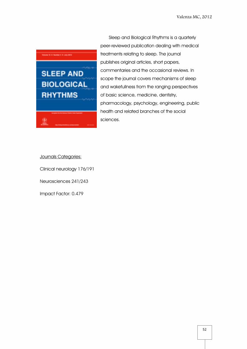
Valenza MC, 2012
�
52�
Sleep and Biological Rhythms is a quarterly
peer-reviewed publication dealing with medical
treatments relating to sleep. The journal
publishes original articles, short papers,
commentaries and the occasional reviews. In
scope the journal covers mechanisms of sleep
and wakefullness from the ranging perspectives
of basic science, medicine, dentistry,
pharmacology, psychology, engineering, public
health and related branches of the social
sciences.
Journals Categories:
Clinical neurology 176/191
Neurosciences 241/243
Impact Factor: 0.479
Valenza MC, 2012
�
53�
Articulo III:��
Valenza MC, Valenza G, Muñoz-Casaubon T, Botella-Lopez M, Puentedura EJ, Arroyo-Morales M, Fernandez-de-las-Peñas C. Epidemiology of sleep-related complaints associated with obstructive sleep apnea, insomnia and non-restorative sleep in an at-risk population in Granada, Spain. Sleep and Biological Rhythms 2012; 10 (3)222–230.
Abstract
Our aim was to estimate the frequency of sleep apnea, non-restorative sleep, and insomnia and their association with demographic factors, lifestyle behaviors and co-morbid conditions in an at-risk population in Spain. A total of 1009 individuals with suspected Obstructive Sleep Apnea Syndrome (OSAS) completed a questionnaire and personal interview including demographic data, lifestyle behaviors, sleep-wake schedule, diurnal and nocturnal symptoms and also co-morbid conditions. Laboratory tests included lung function test, echocardiography, arterial blood analysis, total blood counts, and standard polysomnography for diagnosing sleep apnea. The frequency of OSAS, insomnia and non-restorative sleep was 88.5%, 16.76%, 59.72%, respectively. The frequency of OSAS was lower in females than in males (OR 0.33, 95% CI 0.20–0.56) whereas the frequency of insomnia was higher (1.95, 1.29–2.95). People in the 61–70 year group exhibited higher frequency of OSAS (3.81, 1.51–9.58) and also insomnia (1.85, 1.08–4.32), whereas the obese exhibited higher probability of OSAS (1.80, 1.15–2.81). OSAS was associated with snoring (7.95, 1.57–24.58), nocturia (1.80, 1.12–2.89), daily fatigue (1.89, 1.12–3.17), apathy (0.53, 0.30–0.89), memory loss (0.44, 0.26–0.73), and hypertension (2.60, 1.41–4.78). Insomnia was associated with esophageal reflux (1.78, 1.21–2.61), awakening (3.39, 2.19–5.22), apathy (1.68, 1.11–2.55), and memory loss (1.58, 1.06–2.35). Non-restorative sleep was associated with nocturia (1.43, 1.01–2.05), gastro-esophageal reflux (2.08, 1.31–3.31), diaphoresis (3.31, 1.88–5.83), awakening (1.41, 1.01–2.02), daily fatigue (2.09, 1.47–2.97), memory loss (2.23, 1.40–3.54), irritability (2.26, 1.59–4.03) and headache (2.57, 1.65–4.00). The frequency of sleep-related breathing disorders was high in our sample. Different factors seem to be associated with OSAS, insomnia or non-restorative sleep.

ORIGINAL ARTICLEsbr_565 222..230
Epidemiology of sleep-related complaints associated with
obstructive sleep apnea, insomnia and non-restorative sleep
in an at-risk population in Granada, Spain
Marie Carmen VALENZA,1 Gerad VALENZA,1 Tomás MUÑOZ-CASAUBON,3
Miguel BOTELLA-LÓPEZ,2 Emilio J PUENTEDURA,5 Manuel ARROYO-MORALES1 andCésar FERNÁNDEZ-DE-LAS-PEÑAS4
1Department of Physical Therapy, 2Department of Morphological Sciences, Universidad de Granada, 3Laboratoryof Sleep Medicine, Hospital Clínico San Cecilio, Granada, 4Department of Physical Therapy, Occupational Therapy,Physical Medicine and Rehabilitation of Universidad Rey Juan Carlos, Alcorcón, Madrid, Spain, and 5Departmentof Physical Therapy, School of Allied Health Sciences, University of Nevada Las Vegas, Las Vegas, NV, USA
Abstract
Our aim was to estimate the frequency of sleep apnea, non-restorative sleep, and insomnia and theirassociation with demographic factors, lifestyle behaviors and co-morbid conditions in an at-riskpopulation in Spain. A total of 1009 individuals with suspected Obstructive Sleep Apnea Syndrome(OSAS) completed a questionnaire and personal interview including demographic data, lifestylebehaviors, sleep-wake schedule, diurnal and nocturnal symptoms and also co-morbid conditions.Laboratory tests included lung function test, echocardiography, arterial blood analysis, total bloodcounts, and standard polysomnography for diagnosing sleep apnea. The frequency of OSAS, insom-nia and non-restorative sleep was 88.5%, 16.76%, 59.72%, respectively. The frequency of OSAS waslower in females than in males (OR 0.33, 95% CI 0.20–0.56) whereas the frequency of insomnia washigher (1.95, 1.29–2.95). People in the 61–70 year group exhibited higher frequency of OSAS (3.81,1.51–9.58) and also insomnia (1.85, 1.08–4.32), whereas the obese exhibited higher probability ofOSAS (1.80, 1.15–2.81). OSAS was associated with snoring (7.95, 1.57–24.58), nocturia (1.80,1.12–2.89), daily fatigue (1.89, 1.12–3.17), apathy (0.53, 0.30–0.89), memory loss (0.44, 0.26–0.73), and hypertension (2.60, 1.41–4.78). Insomnia was associated with esophageal reflux (1.78,1.21–2.61), awakening (3.39, 2.19–5.22), apathy (1.68, 1.11–2.55), and memory loss (1.58, 1.06–2.35). Non-restorative sleep was associated with nocturia (1.43, 1.01–2.05), gastro-esophagealreflux (2.08, 1.31–3.31), diaphoresis (3.31, 1.88–5.83), awakening (1.41, 1.01–2.02), daily fatigue(2.09, 1.47–2.97), memory loss (2.23, 1.40–3.54), irritability (2.26, 1.59–4.03) and headache (2.57,1.65–4.00). The frequency of sleep-related breathing disorders was high in our sample. Differentfactors seem to be associated with OSAS, insomnia or non-restorative sleep.
Key words: insomnia, non-restorative sleep, Obstructive Sleep Apnea Syndrome.
Correspondence: Professor César Fernández-de-las-Peñas, Facultad de Ciencias de la Salud, Universidad Rey Juan Carlos,Avenida de Atenas s/n, 28922 Alcorcón, Madrid, Spain. Email: [email protected]
Financial support: No fund were received for this study.
Conflict of interest: None declared by any author.
Accepted 5 April 2012.
bs_bs_banner
Sleep and Biological Rhythms 2012; 10: 222–230 doi:10.1111/j.1479-8425.2012.00565.x
bs_bs_banner
222 © 2012 The AuthorsSleep and Biological Rhythms © 2012 Japanese Society of Sleep Research

INTRODUCTION
It seems that sleep disturbances are associated withgreater daytime sleepiness, deterioration of physical-psychological performance, impaired quality of life,1
cognitive deficits,2,3 psychological changes,4,5 and risk ofmotor vehicle accidents.6 Obstructive Sleep Apnea Syn-drome (OSAS) is the most common form of sleep dis-turbance (called sleep-related breathing disorders) andcharacterized by repetitive cessation of breathing duringsleep which is accompanied with sleep fragmentation,nocturnal hypoxemia, an excessive daytime sleepiness(Epworth Sleepiness Scale >10) and an Apnea-Hypopnea Index (AHI) of 5 or higher.7
Obstructive Sleep Apnea Syndrome is also associatedwith snoring, nocturnal respiratory arrest, repeated noc-turnal awakening, non-restorative sleep, diurnal fatigue,and altered concentration. Other sleep disturbancescommonly seen are non-restorative sleep, that is, areport of persistently feeling un-refreshed upon awak-ening in the presence of normal sleep duration occur-ring in the absence of a sleep disorder;8 and insomniadefined as a subjective perception of dissatisfaction withthe amount and/or quality of sleep including difficultyin initiating or maintaining sleep or early awakeningwith inability to fall asleep again.9
The prevalence of sleep disturbances has been con-ducted in several countries: United Kingdom,10 Den-mark,11 Greece,12 Australia,13 United States,14 China,15
India,16 Thailand,17 and Korea.18 All these studies havereported high prevalence of snoring and non-restorativesleep in the general population. Netzer et al. comparedthe prevalence of symptoms and risk of OSAS betweenUSA and Europe,19 and found that OSAS was moreprevalent (P < 0.001) in USA (35.8%) than in Europe(26.3%), and in men than in women (37.9% vs. 27.8%,respectively; P < 0.005).19 Nevertheless, the prevalenceof sleep disturbances differs by region20 and by coun-try.21,22 Therefore, it is important to conduct epidemio-logical studies in different regions in one country.
In Spain, few studies investigating the prevalence ofsleep disturbances have been conducted. Marin et al.estimated that 0.8% of women and 2.2% of men exhib-ited the minimal criteria for diagnosis of OSAS.23
Ohayon investigated the prevalence of non-restorativesleep in seven European countries (France, UK,Germany, Italy, Portugal, Spain, and Finland) and foundthat UK and Germany showed the highest prevalence ofnon-restorative sleep (16.1%, and 15.5%, respectively),whereas Spain exhibited the lowest (2.4%).24 No epide-miological studies have recently estimated the frequency
of sleep disturbances and their association with socio-demographic factors, lifestyle habits and co-morbidityin a Spanish population. Therefore, the objective of thecurrent study was to estimate the frequency of OSAS,non-restorative sleep and insomnia and their associationwith socio-demographic factors, lifestyle behavior andco-morbid conditions in a specific at-risk population inGranada, a southern region of Spain.
METHODS
A cross-sectional observational study was conducted.One thousand and nine (n = 1009) participants, 804menand 205 women, referred to the Sleep Laboratory ofHospital San Cecilio, Granada, Spain between January2008 and December 2010 with suspected OSAS wereincluded. Each participant completed a questionnaireand personal interview. The study protocol was approvedby the human research committee and conducted follow-ing the declaration of Helsinki. All subjects signed aninformed consent prior to their inclusion in the study.
In the questionnaire, participants answered generalquestions related to personal data and health informa-tion, as well as lifestyle behaviors, for example, smokingand alcohol consumption. The questionnaire includedthe Epworth Sleepiness Scale, which is used for assess-ment of subjective daytime sleepiness during face-to-face interviews.25,26 It consists of an eight-itemquestionnaire that requires the patient to rate their like-lihood of dozing-off in various situations on a 4-pointLikert scale (0: not at all likely to doze; 3: very likely todoze). Total score ranges from 0 to 24 with scores > 10indicating significant daytime sleepiness.25,26
During the interview, they were questioned in thepresence of their most closely-related person, preferablytheir spouse/partner. Participants and their partnersanswered questions about clinical characteristics ofOSAS and other sleep disorders, such as snoring, cessa-tion of breathing during sleep, and daytime somnolence,and also informed about co-morbid conditions diag-nosed by theirmedical doctor: cardio-pulmonary pathol-ogy, diabetes, hypertension, depressive symptoms,menopause, metabolic syndrome, chronic obstructivepulmonary disease. Any co-morbid condition wasrecorded when patients had a medical diagnosis.
Anthropometric data were measured by a trainednurse and consisted of: weight, height, age, sex, andabdominal and neck circumference. Weight wasmeasured in light clothes on a portable scale. Neckcircumference was measured just below the laryngealprominence using standardized methods.27
Sleep disturbances in Granada, Spain
223© 2012 The AuthorsSleep and Biological Rhythms © 2012 Japanese Society of Sleep Research

Medical laboratory tests included lung function test,arterial blood gas analysis, echocardiography and totalblood counts. In addition, participants underwent twonights of standard polysomnography for diagnosingOSAS and Continuous Positive Airway Pressure (CPAP)titration separately, including nasal and oral flow with athermistor, thoracic and abdominal respiratory move-ments with a strain gauge, and arterial oxygen saturationwith a finger oximeter. They went to bed at their chosentime and all woke up naturally without an alarm on bothnights. Data were stored in real time on Personal Com-puter Memory Card International Association (PCMCIA)cards. The equipment was retrieved the next morning.
Apnea was defined as complete cessation of airflowfor at least 10 s. Hypopnea was defined as a reduction ofrespiratory signals for at least 10 s associated withoxygen desaturation of 4%. Detailed protocol for centralscoring of sleep stages, arousals, and respiratory eventshas been described.28
In the current study, self-report insomnia and non-restorative sleep, and presence of OSAS were the depen-dent variables.
Statistical analyses
We first estimated demographic data, frequency of sleepdisturbance symptoms, and presence of co-morbidityconditions according to the presence of each dependentvariable (OSAS, insomnia, and non-restorative sleep).Bivariate logistic regression models were conducted toestimate the measure of association. A multivariateanalysis was performed using the variables statisticallysignificant within the bivariate analysis. Variables wereeliminated, one at each step, according to their signifi-cance in the model used (Wald statistic) and consideringthe model’s goodness-of-fit with regard to the previousstep. The effects of interaction among the variablesincluded in the final model were also examined. Theadjusted odds ratio (OR) for each factor is reported.Estimates were made by incorporating the samplingweights, using the “svy” (survey command) functions ofthe STATA program, which enabled us to incorporatethe sampling design into all statistical calculations(adjusted OR, logistic regression). The statistical signifi-cance was set at two-tailed a < 0.05.
RESULTS
Demographics and clinical features
Of the 1008 participants, 893 suffered from OSAS, 169from insomnia and 602 non-restorative sleep. There-
fore, the frequency of OSAS, insomnia and non-restorative sleep was 88.5%, 16.76% and 59.72%,respectively. A significant association between the pres-ence of OSAS and insomnia (OR 1.65, 95% CI 1.04–2.07) was found: subjects with OSAS exhibited a higherfrequency of insomnia than those without OSAS.
The frequency of OSAS was significantly higher (P <0.01) in males (91.10%) than in females (82.18%),whereas the frequency of insomnia and non-restorativesleep was significantly higher (P < 0.01) in females(29.41% and 67.16%, respectively) than in males(13.56%, 57.91% respectively). The frequency of OSASand insomnia showed the highest value in participantsin the 61–70 years group (93.99% and 25.68%, respec-tively). We also found that participants with a bodymass index (BMI) � 30 also showed a greater frequencyof OSAS (92.11%) than those with a BMI < 30(83.90%). Further, non-smokers and participants con-suming alcoholic drinks occasionally were more likelyto suffer insomnia (20.86%, 22.22% respectively) thansmokers (11.90%) or those consuming alcoholic drinksdaily (14.89%). Table 1 summarizes the frequency ofOSAS, insomnia and non-restorative sleep according tothe socio-demographic variables.
Nocturnal and diurnal symptoms
Participants with OSAS were more likely to snore(90.33%) and to experience nocturia (92.42%) thanthose without OSAS (53.33% and 85.81%, respec-tively). We found that individuals suffering frominsomnia were more likely to present with several noc-turnal symptoms (diaphoresis: 26.38%; self-reportedasphyxia: 24.30%; nocturia: 21.80%; gastro-esophagealreflux: 29.82%; awakening: 26.20%; nightmares:30.39%) than those individuals without insomnia (dia-phoresis: 13.84%; self-reported asphyxia: 13.81%;nocturia: 11.13%; gastro-esophageal reflux: 11.87%;awakening: 6.60%; or nightmares: 13.78%). Similarly,participants reporting non-restorative sleep sufferedhigher frequency of nocturnal symptoms (diaphoresis:91.49%; self-reported asphyxia: 81.98%; nocturia:69.55%; gastro-esophageal reflux: 86.50%; awakening:72.80%; nightmares: 91.16%) than those with a restor-ative sleep (diaphoresis: 50.13%; self-reported asphyxia:51.10%; nocturia: 48.84%; gastro-esophageal reflux:49.80%; awakening: 45.77%; nightmares: 52.91%).The frequency of these nocturnal symptoms duringsleeping time according to the presence of OSAS, insom-nia or non-restorative sleep is shown in Table 2.
MC Valenza et al.
224 © 2012 The AuthorsSleep and Biological Rhythms © 2012 Japanese Society of Sleep Research

Individuals with OSAS were less likely to experiencediurnal symptoms (apathy: 85.04%; irritability:85.52%; memory loss: 85.22%) than those withoutsleep apnea (apathy: 90.91%; irritability: 90.85%;memory loss: 91.20%). On the contrary, the presence ofdiurnal symptoms was more prevalent in participants
with insomnia (daily fatigue: 21.44%; headache:23.64%; apathy: 31.64%; irritability: 27.30%; concen-tration difficulty: 30.77%; memory loss: 29.38%; libidoloss: 37.14%) or non-restorative sleep (fatigue: 74.55%;headache: 82.48%; apathy: 90.88%; irritability: 90.1%;concentration difficulty: 90.13%; memory loss: 87.85%;
Table 1 Demographic and clinical data of the sample to the presence of obstructive sleep apnea syndrome (OSAS), insomniaor non-restorative sleep
Variable Categories n OSAS InsomniaNon-restorative
sleep
Sex†‡§ Male 804 91.10% 13.56% 57.91%Female 205 82.18% 29.41% 67.16%
Age†‡ 18–30 years 62 80.39% 15.69% 62.75%31–40 years 159 87.42% 19.38% 57.86%41–50 years 260 83.46% 16.67% 57.20%51–60 years 283 93.64% 15.14% 63.38%61–70 years 183 93.99% 25.68% 59.56%71–80 years 62 91.94% 18.75% 57.81%
Smoking habit‡ Smoker 353 89.17% 11.90% 58.36%Ex-smoker 321 90.88% 18.07% 61.88%Non-smoker 326 88.58% 20.86% 60.43%
Alcohol consumption‡ No drink 445 89.16% 19.06% 54.26%Occasionally 27 96.15% 22.22% 65.39%Daily 524 89.44% 14.89% 55.56%
Obesity† BMI < 30 356 83.90% 18.82% 57.58%BMI � 30 626 92.11% 15.81% 61.92%
†Significant differences (P < 0.01) for sleep apnea. ‡Significant differences (P < 0.01) for insomnia. §Significant differences (P < 0.01) fornon-restorative sleep. BMI, body mass index.
Table 2 Prevalence of nocturnal symptoms during sleeping time according to the presence of obstructive sleep apneasyndrome (OSAS), insomnia or non-restorative sleep
Variable Categories n OSAS InsomniaNon-restorative
sleep
Snoring† No 15 53.33% 13.33% 46.67%Yes 979 90.33% 17.06% 60.53%
DiaphoresisठNo 773 89.57% 13.84% 50.13%Yes 235 88.41% 26.38% 91.49%
Self-reported AsphyxiaठNo 724 89.83% 13.81% 51.10%Yes 284 87.94% 24.30% 81.98%
Nocturia†‡§ No 476 85.81% 11.13% 48.84%Yes 532 92.42% 21.80% 69.55%
Gastro-esophageal refluxठNo 733 89.81% 11.87% 49.80%Yes 275 87.96% 29.82% 86.50%
AwakeningठNo 485 90.02% 6.60% 45.77%Yes 523 88.63% 26.20% 72.80%
NightmaresठNo 827 89.28% 13.78% 52.91%Yes 181 89.39% 30.39% 91.16%
†Significant differences (P < 0.01) for sleep apnea. ‡Significant differences (P < 0.01) for insomnia. §Significant differences (P < 0.01) fornon-restorative sleep.
Sleep disturbances in Granada, Spain
225© 2012 The AuthorsSleep and Biological Rhythms © 2012 Japanese Society of Sleep Research

loss of libido: 85.71%) than in those without insomnia(fatigue: 11.04%; headaches: 14.21%; apathy: 11.91%;irritability: 12.45%; concentration difficulty: 12.53%;memory loss: 10.92%; loss of libido: 16.03%) or withrestorative sleep (daily fatigue: 41.72%; headache:51.23%; apathy: 48.16%; irritability: 47.34%; concen-tration difficulty: 50.65%; memory loss: 40.65%; libidoloss: 58.85%). The frequency of diurnal symptomsaccording to the presence of OSAS, insomnia or non-restorative sleep is shown in Table 3.
Co-morbid conditions
Hypertension, diabetes and cardiac pathology wereassociated with the presence of OSAS: participants withapnea exhibited higher frequency of these co-morbiddisorders than those participants without OSAS (P <0.01). Additionally, participants with insomnia or non-restorative sleep were more depressed (31.62% and88.46%, respectively) than those without insomnia ornon-restorative sleep (12.27%, 51.10% respectively).Finally, a lower proportion of participants with insomnia(7.58%) suffered from hyper-uremia as compared withthose without insomnia (17.41%). Frequency of OSAS,
insomnia, or non-restorative sleep related to co-morbidchronic diseases is summarized in Table 4.
Variables associated with thepresence of OSAS, insomnia ornon-restorative sleep
Table 5 summarizes the results of the multivariate analy-sis with the adjusted OR of those variables that wereassociated with the presence of OSAS, insomnia or non-restorative sleep. The analysis showed that femalegender decreased the probability of reporting OSAS (OR0.33, 95% CI 0.20–0.56), but increased the probabilityof reporting insomnia (OR 1.95, 95% CI 1.29–2.95). Inaddition, using the 18–30 year group as the referencecategory, participants within the 51–60 year (OR 3.59,95% CI 1.55–8.31) and 61–70 year (OR 3.81, 95% CI1.51–9.58) group reported significantly higher fre-quency of OSAS, and those within the 61–70 year groupreported significantly higher frequency of insomnia (OR1.85, 95% CI 1.08–4.32). We also found that obeseparticipants (BMI � 30) exhibited higher probability ofOSAS (OR 1.80, 95% CI 1.15–2.81).
Table 3 Prevalence of diurnal symptoms during sleeping time according to the presence of obstructive sleep apnea syndrome(OSAS), insomnia or non-restorative sleep
Variable Categories n OSAS InsomniaNon-restorative
sleep
Daily fatigueठNo 453 88.64% 11.04% 41.72%Yes 555 89.84% 21.44% 74.55%
HeadacheठNo 732 89.67% 14.21% 51.23%Yes 275 88.28% 23.64% 82.48%
Musculoskeletal pain No 956 89.35% 16.42% 59.37%Yes 52 88.46% 23.08% 67.31%
Psychiatric symptoms
Apathy†‡§ No 733 90.91% 11.91% 48.16%Yes 235 85.04% 31.64% 90.88%
Irritability†‡§ No 715 90.85% 12.45% 47.34%Yes 293 85.52% 27.30% 90.10%
Cognitive alterations
Concentration difficultyठNo 774 90.21% 12.53% 50.65%Yes 234 86.32% 30.77% 90.13%
Memory loss†‡§ No 687 91.20% 10.92% 40.65%Yes 321 85.22% 29.38% 87.85%
Loss of libidoठNo 973 89.12% 16.03% 58.85%Yes 35 94.29% 37.14% 85.71%
†Significant differences (P < 0.01) for sleep apnea. ‡Significant differences (P < 0.01) for insomnia. §Significant differences (P < 0.01) fornon-restorative sleep.
MC Valenza et al.
226 © 2012 The AuthorsSleep and Biological Rhythms © 2012 Japanese Society of Sleep Research

Table 4 Prevalence of co-morbid conditions according to the presence of obstructive sleep apnea syndrome (OSAS), insomniaor non-restorative sleep
Variable Categories n OSAS Insomnia Non-restorative sleep
DepressionठNo 774 90.23% 12.27% 51.10%Yes 234 86.21% 31.62% 88.46%
Chronic obstructive pulmonary disease No 984 89.24% 16.87% 60.22%Yes 24 91.67% 12.50% 41.67%
Metabolic syndrome† No 972 88.91% 16.77% 60.14%Yes 36 100% 16.67% 60.14%
Hypertension† No 670 86.43% 15.22% 60.09%Yes 338 94.96% 19.82% 59.17%
Diabetes† No 919 88.49% 16.10% 59.69%Yes 89 97.73% 23.60% 60.67%
Cardiac pathology† No 924 88.66% 16.23% 59.80%Yes 84 96.39% 22.62% 59.52%
Hyper-uraemia‡ No 942 88.87% 17.41% 60.15%Yes 66 95.45% 7.58% 54.55%
Hypercholesterolemia No 882 88.79% 16.55% 60.27%Yes 126 83.90% 18.82% 57.58%
†Significant differences (P < 0.01) for sleep apnea. ‡Significant differences (P < 0.01) for insomnia. §Significant differences (P < 0.01) fornon-restorative sleep.
Table 5 Variables significantly associated with a higher likelihood of presenting obstructive sleep apnea syndrome (OSAS),insomnia and non-restorative sleep
Variable Categories OSAS Insomnia Non-restorative sleep
Sex† Female 0.33 (0.20–0.56) 1.95 (1.29–2.95) NSAge‡ 31–40 years NS NS NS
41–50 years NS NS NS51–60 years 3.59 (1.55–8.31) NS NS61–70 years 3.81 (1.51–9.58) 1.85 (1.08–4.32) NS71–80 years NS NS NS
Obesity§ BMI � 30 1.80 (1.15–2.81) NS NS
Nocturnal symptomsSnoring¶ Yes 7.95 (2.57–24.58) NS NSNocturia¶ Yes 1.80 (1.12–2.89) NS 1.43 (1.01–2.05)Diaphoresis¶ Yes NS NS 3.31 (1.88–5.83)Gastro-esophageal reflux¶ Yes NS 1.78 (1.21–2.61) 2.08 (1.31–3.31)Awakening¶ Yes NS 3.39 (2.19–5.22) 1.41 (1.01–2.02)
Diurnal symptomsDaily fatigue¶ Yes 1.89 (1.12–3.17) NS 2.09 (1.47–2.97)Apathy¶ Yes 0.52 (0.30–0.89) 1.68 (1.11–2.55) NSMemory loss¶ Yes 0.44 (0.26–0.73) 1.58 (1.06–2.35) 2.23 (1.40–3.54)Irritability¶ Yes NS NS 2.62 (1.59–4.03)
Co-morbid conditionsHypertension¶ Yes 2.60 (1.41–4.78) NS NSHeadache¶ Yes NS NS 2.57 (1.65–4.00)
Reference categories: †Male; ‡18–30 years; §BMI (body mass index) < 30; ¶No. NS, not stated.
Sleep disturbances in Granada, Spain
227© 2012 The AuthorsSleep and Biological Rhythms © 2012 Japanese Society of Sleep Research

The analysis revealed a significant association ofOSAS with snoring (OR 7.95; 95% CI 1.57–24.58) andnocturia (OR 1.80, 1.12–2.89). Insomnia was signifi-cantly associated with the presence of gastro-esophagealreflux (OR 1.78, 95% CI 1.21–2.61) and awakening(OR 3.39, 95% CI 2.19–5.22). Further, non-restorativesleep was related to higher frequency of nocturia (OR1.43, 1.01–2.05), diaphoresis (OR 3.31, 1.88–5.83),gastro-esophageal reflux (OR 2.08, 1.31–3.31), andawakening (OR 1.41, 1.01–2.02).
In relation to diurnal symptoms, daily fatigue wasassociated with OSAS (OR 1.89, 1.12–3.17) and non-restorative sleep (2.09, 1.47–2.97); apathy was associ-ated with OSAS (0.53, 0.30–0.89) and insomnia (1.68,1.11–2.55); memory loss was associated with OSAS(0.44, 0.26–0.73), insomnia (1.58, 1.06–2.35), andnon-restorative sleep (2.23, 1.40–3.54); whereas irrita-bility was only associated with non-restorative sleep(2.26, 1.59–4.03). Finally, hypertension was co-morbidwith OSAS (OR 2.60, 1.41–4.78), and headache withnon-restorative sleep (OR 2.57, 1.65–4.00).
DISCUSSION
This is the first epidemiological study to investigatesleep disorders in a specific Spanish population referredto a hospital. Our study demonstrated a high frequencyof OSAS (88.5%), insomnia (16.7%), and non-restorative sleep (59.7%), data slightly superior thanprevious studies conducted in different countries.10–18 Aprevious study conducted in Zaragoza, a northeastregion of Spain, estimated a prevalence rate rangingfrom 1% to 2% of apnea.23 Ohayon reported that theprevalence of non-restorative sleep in a general popula-tion representative of seven European countries was10.8%.24 Higher frequency of sleep disturbances in thecurrent study may be related to the fact that our samplesize was derived from a specialized unit in a hospital;whereas previous studies included data from the generalpopulation. In contrast, others have reported higherprevalence rate (27.6%) of insomnia symptoms in theItalian population.29 It seems that the prevalence of sleepdisturbances varies by region and country depending oncultural differences on sleeping habits, differences inbedtime and wake-up hours, or differences in climatol-ogy and hours of light.20–22 Further, differences in preva-lence may be also explained by sample biases anddifferent diagnostic approaches. Nevertheless, our studyis the first one including an objective diagnosis of OSASwith polysomnography.
The frequency of OSAS was positively associated withmen, older age, obesity and snoring, which is consistentwith previous studies.10–22 Marin et al. also found thatindividuals with OSAS in Zaragoza were men, older andobese.23 A theoretical interaction between these threefactors may be that obesity tends to increase the size ofthe neck (neck obesity), which has been implicated inupper airway obstruction during sleep.30 In such cir-cumstances, obese people will snore more frequently.31
In fact, differences in obesity rates between USA andEurope has been proposed as one the main factorsrelated to differences in OSAS apnea prevalencebetween both countries.19 This is consistent with the factthat obesity is the most common predisposing factor forsuffering from OSAS.32 Nevertheless, the associationbetween OSAS and obesity (OR 1.8) in our study waslower than expected, probably because two thirds of oursample was obese.
The frequency of OSAS was more prevalent in men,which is in accordance with previous studies.19,23,33
Netzer et al. reported that men exhibited higher scoresof OSAS as caused by snoring and sleep apnea, support-ing the relationship between male gender, apnea, andsnoring.19 Redline et al. specifically investigated genderdifferences in sleep disturbances and have reported amale : female ratio for OSAS of 2:1.34 Importantly, theoccurrence of snoring is a predictor of subsequent diag-nosis of hypertension.35 In fact, individuals with sleepdisordered breathing are at increased risk for cardiovas-cular diseases, such as hypertension, angina, myocardialinfarction, and stroke.36 In our study, we found thathypertension was a highly co-morbid condition in sub-jects with OSAS supporting this relationship. Finally,some diurnal symptoms, for example, daily fatigue,apathy or memory loss, and nocturia (nocturnalsymptom) were also more prevalent in participants withOSAS, which is consistent with previous studies.37,38
Insomnia was associated with females and the elderly,gastro-esophageal reflux and awakening as nocturnalsymptoms, and apathy and memory loss as diurnalsymptoms in agreementwith previous data.24,39,40 A studysuggested that concentration difficulty and memory losswas highly present in older people with excessivedaytime sleepiness, which may be related to insomnia.41
Additionally, non-restorative sleep was associated withnocturia, diaphoresis, esophageal reflux, and awakeningas nocturnal symptoms, and daily fatigue, memory lossand irritability as diurnal symptoms. Current results arevery similar to those previously reported byOhayonwhoalso found that participants with non-restorative sleepreported more frequent irritability, physical, and mental
MC Valenza et al.
228 © 2012 The AuthorsSleep and Biological Rhythms © 2012 Japanese Society of Sleep Research

fatigue as compared to those with normal sleep, and alsoconsulted a physician twice as frequently for their sleep-ing difficulties than did participants with insomnia.24
Finally, headache was more frequent in individuals withnon-restorative sleep, confirming that sleep can be atrigger factor for headache.42
Although strengths of our study include a largesample size, the employment of personalized surveys,and an objective assessment of OSAS by polysomnog-raphy, there are also a number of significant limitations.First, we included a population derived from a specificregional hospital; therefore, current results should notbe extrapolated to the general population. Second, sincethis was a cross-sectional study, we cannot determinecause and effect relationships between OSAS, insomniaor non-restorative sleep with their associated symptoms.Finally, information obtained from personal interviewsmay be subject to recall errors or a tendency of indi-viduals to give socially desirable responses in the survey,particularly regarding lifestyle habits. Despite these limi-tations, our findings provide additional insight intosome clinical aspects of sleep-related breathing disor-ders in a specific Spanish population for which there islittle information at a population level. Therefore, datafrom our study are a valuable tool for evaluating theeffectiveness of the campaigns promoting breathinghealth care among people.
The current study has shown that OSAS, insomnia,and non-restorative sleep were highly prevalent sleep-related breathing disorders in a specific at-risk popula-tion in Granada, a southeast region of Spain. Currentresults show some similarities in risk factors for thesesleep-related breathing disorders with previous studies,but also exhibit different factors associated with OSAS,insomnia or non-restorative sleep. In fact, these differ-ences may be crucial for a proper distinction betweenthese sleep disorders.
REFERENCES
1 Yang EH, Hla KM, McHorney CA, Havighurst T, BadrMS, Weber S. Sleep apnea and quality of life. Sleep 2000;23: 535–41.
2 O’Hara R, Schröder CM, Kraemer HC et al. Nocturnalsleep apnea/hypopnea is associated with lower memoryperformance in APOE epsilon4 carriers. Neurology 2005;65: 642–4.
3 Kim HC, Young T, Matthews CG, Weber SM, WoodwardAR, Palta M. Sleep disordered breathing and neuropsy-chological deficits. A population-based study. Am. J.Respir. Crit. Care Med. 1997; 156: 1813–9.
4 Schröder CM, O’Hara R. Depression and ObstructiveSleep Apnea (OSA). Ann. Gen. Psychiatry 2005;4: 13. Available from URL: http://www.annals-general-psychiatry.com/content/4/1/13.
5 O’Hara R, Schröder CM. Unraveling the relationship ofobstructive sleep-disordered breathing to major depres-sive disorder. Sleep Med. 2006; 7: 101–3.
6 Sassani A, Findley LJ, Kryger M, Goldlust E, George C,Davidson TM. Reducing motor-vehicle collisions, costs,and fatalities by treating obstructive sleep apnea syn-drome. Sleep 2004; 27: 453–8.
7 Guilleminault C. Clinical features and evaluation ofobstructive sleep apnoea. In: Kryger MH, Roth T,Demment WC, eds. Principles and Practice of Sleep Medi-cine, 2nd edn. W B Saunders: Philadelphia, PA, 1994;667–77.
8 Stone KC, Taylor DJ, McCrae CS, Kalsekar A, LichsteinKL. Non-restorative sleep. Sleep Med. Rev. 2008; 12:275–88.
9 Estivill E, Bové A, García-Borreguero D et al. Consensuson drug treatment, definition and diagnosis for insom-nia. Clin. Drug Investig. 2003; 23: 351–85.
10 Ohayon M, Guillemenault C, Priest RG et al. Snoring andbreathing pauses during sleep: telephone interviewsurvey of a United Kingdom population sample. BMJ1997; 314: 860–3.
11 Jennum P, Sjøl A. Epidemiology of snoring and obstruc-tive sleep apnea in a Danish population, age 30–60. J.Sleep Res. 1992; 1: 240–44.
12 Vagiakis E, Kapsimalis F, Lagogianni I et al. Gender dif-ferences on polysomnographic findings in Greek sub-jects with obstructive sleep apnea syndrome. Sleep Med.2006; 7: 424–30.
13 Bearpark H, Elliot L, Grinstein R et al. Snoring and sleepapnea. A population study in Australian men. Am. J.Respir. Crit. Care Med. 1995; 151: 1459–65.
14 Hiestand M, Britz P, Goldman M et al. Prevalence ofsymptoms and risk of sleep apnea in the US population:results from the national sleep foundation sleep inAmerica 2005 poll. Chest 2006; 130: 780–6.
15 Ip MS, Lam B, Lauder I et al. A community study ofsleep-disordered breathing in middle-aged Chinese menin Hong Kong. Chest 2001; 119: 62–9.
16 Sharma S, Kumpawat S, Banga A, Goel A. Prevalenceand risk factors of obstructive sleep apnea syndromein population of Delhi, India. Chest 2006; 130: 149–56.
17 Suwanprathes P, Won C, Komoltri C, Nana A, Kotchab-hakdi N, Guilleminault C. Epidemiology of sleep-relatedcomplaints associated with sleep-disordered breath-ing in Bangkok, Thailand. Sleep Med. 2010; 11: 1025–30.
18 Kim J, In K, Kim J et al. Prevalence of sleep-disorderedbreathing in middle-aged Korean men and women. Am.J. Respir. Crit. Care Med. 2004; 170: 1108–13.
Sleep disturbances in Granada, Spain
229© 2012 The AuthorsSleep and Biological Rhythms © 2012 Japanese Society of Sleep Research

19 Netzer NC, Hoegel JJ, Loube D et al. Prevalence of symp-toms and risk of sleep apnea in primary care. Chest 2003;124: 1406–14.
20 Kapur V, Strohl KP, Redline S et al. Under-diagnosis ofsleep apnea syndrome in United States communities.Sleep Breath. 2002; 6: 49–54.
21 Vgontzas AN, Kales A. Sleep and its disorders. Annu. Rev.Med. 1999; 50: 387–400.
22 Young T, Peppard PE, Gottlieb DJ. Epidemiology ofobstructive sleep apnea: a population health perspective.Am. J. Respir. Crit. Care Med. 2002; 165: 1217–39.
23 Marin JM, Gascon JM, Carrizo S, Gispert J. Prevalence ofsleep apnoea syndrome in the Spanish adult population.Int. J. Epidemiol. 1997; 26: 381–6.
24 Ohayon MM. Prevalence and correlates of non-restorative sleep complaints. Arch. Intern. Med. 2005;165: 35–41.
25 Johns MW. A new method for measuring daytime sleepi-ness: the Epworth Sleepiness Scale. Sleep 1991; 14:540–5.
26 JohnsMW. Sleepiness in different situations measured bythe Epworth Sleepiness Scale. Sleep 1994; 17: 703–10.
27 Callaway CW, Chumlea WC, Bouchard C et al. Circum-ferences. In: Lohman TG, Roche AF, Martorell R, eds.Anthropometric Standardization Reference Manual. HumanKinetics: Champaign, IL, 1988; 41–2.
28 Redline S, Sanders MH, Lind BK et al. Methods forobtaining and analyzing polysomnography data for amulticenter study. Sleep 1998; 21: 759–67.
29 Ohayon MM, Smirne S. Prevalence and consequences ofinsomnia disorders in the general population of Italy.Sleep Med. 2002; 3: 115–20.
30 Davies RJO, Stradling JR. The relationship between neckcircumference, radiographic pharyngeal anatomy, andthe constructive sleep apnea syndrome. Eur. Respir. J.1990; 3: 509–14.
31 Stradling JR, Crosby JH. Predictors and prevalence ofobstructive sleep apnoea and snoring in 1001 middleaged men. Thorax 1991; 46: 85–90.
32 Young JB, Mc Donald JP. An investigation into the rela-tionship between the severity of obstructive sleep apnea/hypo-apnea syndrome and the vertical position of thehyoid bone. Surgeon 2004; 2: 145–51.
33 Shepertycky MR, Banno K, Kryger MH. Differencesbetween men and women in the clinical presentation ofpatients diagnosed with obstructive sleep apnea syn-drome. Sleep 2005; 28 (3): 309–14.
34 Redline S, Kump K, Tshler PV, Browner I, Ferrette V.Gender differences in sleep disordered breathing in acommunity-based sample. Am. J. Respir. Crit. Care Med.1994; 149: 722–26.
35 Peppard PE, Young T, Palta M et al. Prospective study ofthe association between sleep-disordered breathing andhypertension. N. Engl. J. Med. 2000; 342: 1378–84.
36 Phillips BG, Somers V. Sleep disordered breathing andrisk factors for cardiovascular disease. Curr. Opin. Pulm.Med. 2002; 8 (6): 516–20.
37 Umlauf MG, Chasens ER. Sleep disordered breathingand nocturnal polyuria: nocturia and enuresis. SleepMed. Rev. 2003; 7: 403–11.42,43.
38 Kjelsberg FN, Ruud EA, Stavem K. Predictors of symp-toms of anxiety and depression in obstructive sleepapnea. Sleep Med. 2005; 6: 341–6.
39 Benetó AM, Gómez-Siuriana E, Rubio-Sánchez P.Co-morbidity between sleep apnea and insomnia. SleepMed. Rev. 2009; 13: 287–93.
40 Ohayon MM. Epidemiology of insomnia: what we knowand what we still need to learn. Sleep Med. Rev. 2002; 6:97–111.
41 Ohayon MM, Vecchierini MF. Daytime sleepiness andcognitive impairment in the elderly population. Arch.Intern. Med. 2002; 162: 201–8.
42 Aguggia M, Cavallini M, Divito N et al. Sleep andprimary headaches. Neurol. Sci. 2011; 32: S51–4.
MC Valenza et al.
230 © 2012 The AuthorsSleep and Biological Rhythms © 2012 Japanese Society of Sleep Research

Valenza MC, 2012
�
63�
Included in EMBASE (Excerpta Medica),
MEDLINE (Index Medicus), Chemical
Abstracts, Cinahl, Cochrane plus, Ebsco,
Indice Médico Español, preIBECS, IBECS,
MEDES, SENIOR, ScIELO, Science Citation
Index Expanded (SciSearch), SCOPUS,
Cancerlit, Toxline, Aidsline y Health Planning
Administration
Journals Categories:
Nutrition and dietetics 37/59
Impact Factor: 1.120
Valenza MC, 2012
�
64�
Articulo IV:
Valenza MC, Martín Martín L, González Jiménez E, Aguilar Cordero MJ, Botella
López M, Muñoz Casaubon T, Valenza Demet G. Factores de riesgo para el
síndrome metabólico en una población con apnea del sueño; evaluación en
un grupo de pacientes de Granada y provincia; estudio GRANADA. Nutrición
Hospitalaria 2012;27(4):1255-160.
Resumen
Introducción: El síndrome metabólico se está convirtiendo en uno de los principales problemas de salud pública del siglo XXI. Se considera que la aparición del syndrome metabólico está determinada por la interacción de factores genéticos, ambientales y nerviosos centrales (disfunción de los centros hipotalámicos de hambre y saciedad) que generan dos alteraciones metabólicas importantes: la resistencia a la acción de la insulina y la obesidad visceral. La relación de este síndrome, que concentra en la actualidad al mundo científico, con las alteraciones del sueño sigue siendo un punto sin esclarecer. Aunque se ha teorizado sobre la relación causa efecto, se desconoce aún su interrelación convirtiéndose su estudio en un objetivo primario de la investigación epidemiológica. Muestra y métodos: Se reclutaron 1016 sujetos que acudieron al servicio de Fisiología Respiratoria del Hospital Universitario “San Cecilio” de Granada (España) por sospecha de Apnea de Sueño. Resultados: Se encontró una correlación significativa (p < 0,001) entre los valores de apneas hipopneas y los valores de saturación de Oxigeno nocturna con las diferentes alteraciones metabólicas asociadas al síndrome metabólico (Hipertensión, Diabetes y Obesidad). Por el contrario, no se encontraron diferencias estadísticamente significativas test (t-Student) en la mayoría de las variables entre el grupo NO-Apnea y el grupo Apnea moderada. Conclusiones: Los sujetos con apnea de sueño poseen significativamente más riesgo de desarrollar síndrome metabólico, y por lo tanto de presentar patología cardiovascular. Estos sujetos deben ser evaluados en este sentido para reducer la morbimortalidad asociada a estas patologías.
1255
Nutr Hosp. 2012;27(4):1255-1260ISSN 0212-1611 • CODEN NUHOEQ
S.V.R. 318
Original
Factores de riesgo para el síndrome metabólico en una población con apneadel sueño; evaluación en un grupo de pacientes de Granada y provincia;estudio GRANADAM.ª C. Valenza1, L. Martín Martín1, E. González Jiménez2, M.ª J. Aguilar Cordero2, M. Botella López3, T. Muñoz Casaubon4 y G. Valenza Demet1
1Departamento de Fisioterapia. Facultad de Ciencias de la Salud. Universidad de Granada. Granada. 2Departamento deEnfermería. Facultad de Ciencias de la Salud. Universidad de Granada. Granada. 3Laboratorio de Antropología Física yForense. Facultad de Medicina. Universidad de Granada. 4Laboratorio de Fisiología Respiratoria. Hospital Universitario SanCecilio. Granada. España.
RISK FACTORS FOR METABOLIC SYNDROME INA POPULATION WITH SLEEP APNEA;
EVALUATION IN A POPULATION OF GRANADA;THE GRANADA STUDY
Abstract
Introduction: The Metabolic Syndrome is one of thefirst health problems in the public health of the century.It’s consider that the beginning of the syndrome is deter-mined by numerous factors that developed two mainmetabolic disturbances: the insulin resistance and thecentral obesity. This relationship is concentrating thescientific world. As the cause-effect relationship has to beanswered, the epidemiologic research has focused onwithout results.
Material and methods: 1,016 subjects were recruited inthe sleep disorders laboratory in San Cecilio Hospitalwith sleep apnea suspicion.
Results: Significant correlation (p < 0,001) was foundbetween sleep apnea severity and nocturnal saturationvalues and the different metabolic disturbances related tothe metabolic syndrome (Hypertension, Diabetes andobesity). By the contrary, we doesn’t found significantdifferences between No-Sleep apnea group and moderatesleep apnea group in the majority of the variables.
Conclusions: Subjects with sleep apnea have signifi-cantly more possibilities to develop metabolic syndrome,and cardiovascular pathology. These subjects had to beevaluated in this sense to reduce the impact associated tothis pathology.
(Nutr Hosp. 2012;27:1255-1260)
DOI:10.3305/nh.2012.27.4.5825Key words: Sleep apnea. Metabolic syndrome. Risk fac-
tors.
Resumen
Introducción: El síndrome metabólico se está convir-tiendo en uno de los principales problemas de salud públicadel siglo XXI. Se considera que la aparición del síndromemetabólico está determinada por la interacción de factoresgenéticos, ambientales y nerviosos centrales (disfunción delos centros hipotalámicos de hambre y saciedad) que gene-ran dos alteraciones metabólicas importantes: la resisten-cia a la acción de la insulina y la obesidad visceral. La rela-ción de este síndrome, que concentra en la actualidad almundo científico, con las alteraciones del sueño siguesiendo un punto sin esclarecer. Aunque se ha teorizadosobre la relación causa efecto, se desconoce aún su interre-lación convirtiéndose su estudio en un objetivo primario dela investigación epidemiológica.
Muestra y métodos: Se reclutaron 1016 sujetos que acu-dieron al servicio de Fisiología Respiratoria del HospitalUniversitario “San Cecilio” de Granada (España) porsospecha de Apnea de Sueño.
Resultados: Se encontró una correlación significativa (p< 0,001) entre los valores de apneas hipopneas y los valoresde saturación de Oxigeno nocturna con las diferentes alte-raciones metabólicas asociadas al síndrome metabólico(Hipertensión, Diabetes y Obesidad). Por el contrario, nose encontraron diferencias estadísticamente significativastest (t-Student) en la mayoría de las variables entre elgrupo NO-Apnea y el grupo Apnea moderada.
Conclusiones: Los sujetos con apnea de sueño poseen sig-nificativamente más riesgo de desarrollar síndrome meta-bólico, y por lo tanto de presentar patología cardiovascular.Estos sujetos deben ser evaluados en este sentido para redu-cir la morbimortalidad asociada a estas patologías.
(Nutr Hosp. 2012;27:1255-1260)
DOI:10.3305/nh.2012.27.4.5825Palabras clave: Apnea del sueño. Síndrome metabólico. Fac-
tores de riesgo.
Correspondencia: Marie Carmen Valenza.Profesora del Departamento de Fisioterapia.Facultad de Ciencias de la Salud. Universidad de Granada.Av. de Madrid, s/n.18071 Granada. España.E-mail: [email protected]
Recibido: 25-II-2012.Aceptado: 11-IV-2012.

Introducción
El sueño, definido en el plano de la conducta por lasuspensión normal de la conciencia y desde un puntode vista electrofisiológico por criterios de ondas ence-fálicas específicas, consume un tercio de nuestra vida1.Por ello, en los últimos años las patologías del sueño ysus repercusiones clínicas comprenden un objetivoprioritario para la comunidad científica.
El Síndrome de Apnea del Sueño (SAS), tambiénllamado Síndrome de Apneas-Hipoapneas del sueño(SAHS)2, es tras el insomnio el trastorno del sueño máscomún. Su prevalencia se ha estimado en torno a un 5%en la población adulta3. Sin embargo, muchos autoresrefieren que estas cifras se incrementaran debido alaumento de los valores de obesidad a nivel mundial4,5.
Se caracteriza por un cuadro de episodios repetidosde obstrucción de la vía aérea superior acompañado poresfuerzo respiratorio inefectivo y apneas. Ello se acom-paña habitualmente por respiraciones amplias de recu-peración llamadas hiperapneas durante el sueño (AHI),que provocan constantes desaturaciones de la oxihe-moglobina y despertares transitorios.
Las alteraciones del sueño han sido en los últimosaños un punto de gran interés para la comunidad cientí-fica. Su enorme prevalencia en los países desarrolladosy su interrelación con gran cantidad de patologías sisté-micas6,7, es objetivo de primer orden para la investiga-ción en salud.
Recientemente, la interacción entre la Apnea desueño (AS) y la disfunción metabólica han sido objetode numerosas investigaciones. En particular la AS, hasido asociada de manera independiente a la Resistenciainsulínica, sugiriendo que la AS8 puede jugar un roldeterminante en el desarrollo de la diabetes tipo II y elllamado síndrome metabólico, que, como síndrome, esuna constelación de obesidad, resistencia a la insulina,hipertensión y dislipemia.
Las relaciones entre el sueño y el desarrollo del sín-drome metabólico se han demostrado tanto en sujetossanos como enfermos. Por una parte, la privación par-cial de sueño dá lugar a cambios similares a los propiosdel síndrome metabólico. Por otra, se ha sugerido queel síndrome de apnea del sueño es una manifestacióndel síndrome metabólico.
En 1998 Wilcox y cols.9 ya teorizaron sobre estarelación patológica entre apnea del sueño y SM, eincluso le dieron el nombre de síndrome Z.
Y aunque es objeto de gran cantidad de investigacio-nes al respecto, los investigadores aun no han desentra-ñado el origen de su interrelación.
Mientras los criterios para diagnosticar diabetesmellitus tipo 2, obesidad, e hipertensión arterial sonclaros, no ocurre lo mismo con el SM. Diferentes gru-pos de investigadores han desarrollado las definicionesexistentes y los criterios clínicos para el SM. Actual-mente se reconocen las del Grupo Europeo para el estu-dio de la Resistencia a la Insulina (EGIR), la OMS10, ygrupos del NCEP ATP III11; todos coinciden en que los
componentes básicos del SM son: obesidad, resistenciaa la insulina, dislipidemia e hipertensión arterial.
Sin embargo, emplean diferentes parámetros paramedir estas alteraciones. Según estos criterios, se ha esti-mado la prevalencia del SM en diferentes poblaciones yse han hecho comparaciones entre estos estudios12-17.
La hipoxia intermitente característica de la SA se aso-cia con cambios anatómicos en el sistema nervioso cen-tral (con consecuencias cognitivas)18, con reestructura-ción cardiovascular y respuesta anormal a estimulaciónsimpática19, y con resistencia a la insulina, aun en ausen-cia de obesidad y de hiperactividad simpática20.
Las últimas décadas se han desarrollado diferentesestudios sobre diferentes tipos de disfunción metabólicaen sujetos con SA21. Sin embargo, muy poca de esta lite-ratura ha caracterizado a los sujetos en términos de laalteración del sueño. Existe gran cantidad de artículosque se han centrado en la evidencia clínica de la contri-bución independiente de la AS para el desarrollo o agra-vamiento del síndrome metabólico. Sin embargo, el sín-drome metabólico y sus componentes (en concreto laobesidad y la resistencia insulínica) pueden tener unefecto causal en el desarrollo de la AS, proponiéndoseasí que la AS pudiese ser un síndrome metabólico en sí yun componente del SM22.
El objetivo de este estudio es caracterizar a una pobla-ción con desordenes del sueño en función de sus altera-ciónes endocrionometabólicas, en concreto, aquellasrelacionadas con el síndrome metabólico. Se trata de unode los primeros estudios desarrollados en poblaciónespañola con este tipo de características en las que con-fluyen dos grandes pandemias, las alteraciones delsueño y las alteraciones metabólicas.
Muestra
Se incluyeron 1.016 sujetos en el estudio, 811 hom-bres y 205 mujeres, derivados al servicio de Fisiologíarespiratoria y Laboratorio del Sueño del Hospital Uni-versitario San Cecilio de Granada. Se solicito la partici-pación a todos los sujetos derivados entre enero 2008 ydiciembre 2010 por sospecha de Síndrome de Apnea deSueno.
Metodología
Cada sujeto completó un cuestionario y una entre-vista personal. El protocolo del estudio fue aprobadopor el Comité Ético del Hospital San Cecilio (Granada)y se dirigió respetando la declaración de Helsinki en eldesarrollo de estudios realizados en humanos. Todoslos sujetos firmaron un consentimiento informado pre-vio a la participación en el estudio.
En el cuestionario los participantes respondían cues-tiones de tipo general relacionadas con datos persona-les e información de estilos de vida, como de su con-sumo de tabaco y alcohol. El formulario incluía el
1256 M.ª C. Valenza y cols.Nutr Hosp. 2012;27(4):1255-1260
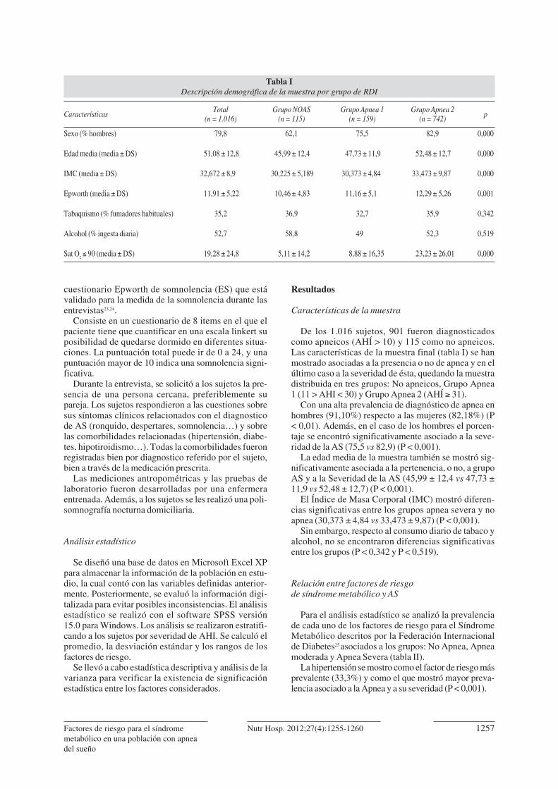
cuestionario Epworth de somnolencia (ES) que estávalidado para la medida de la somnolencia durante lasentrevistas23,24.
Consiste en un cuestionario de 8 items en el que elpaciente tiene que cuantificar en una escala linkert suposibilidad de quedarse dormido en diferentes situa-ciones. La puntuación total puede ir de 0 a 24, y unapuntuación mayor de 10 indica una somnolencia signi-ficativa.
Durante la entrevista, se solicitó a los sujetos la pre-sencia de una persona cercana, preferiblemente supareja. Los sujetos respondieron a las cuestiones sobresus síntomas clínicos relacionados con el diagnosticode AS (ronquido, despertares, somnolencia…) y sobrelas comorbilidades relacionadas (hipertensión, diabe-tes, hipotiroidismo…). Todas la comorbilidades fueronregistradas bien por diagnostico referido por el sujeto,bien a través de la medicación prescrita.
Las mediciones antropométricas y las pruebas delaboratorio fueron desarrolladas por una enfermeraentrenada. Además, a los sujetos se les realizó una poli-somnografía nocturna domiciliaria.
Análisis estadístico
Se diseñó una base de datos en Microsoft Excel XPpara almacenar la información de la población en estu-dio, la cual contó con las variables definidas anterior-mente. Posteriormente, se evaluó la información digi-talizada para evitar posibles inconsistencias. El análisisestadístico se realizó con el software SPSS versión15.0 para Windows. Los análisis se realizaron estratifi-cando a los sujetos por severidad de AHI. Se calculó elpromedio, la desviación estándar y los rangos de losfactores de riesgo.
Se llevó a cabo estadística descriptiva y análisis de lavarianza para verificar la existencia de significaciónestadística entre los factores considerados.
Resultados
Características de la muestra
De los 1.016 sujetos, 901 fueron diagnosticadoscomo apneicos (AHÍ > 10) y 115 como no apneicos.Las características de la muestra final (tabla I) se hanmostrado asociadas a la presencia o no de apnea y en elúltimo caso a la severidad de ésta, quedando la muestradistribuida en tres grupos: No apneicos, Grupo Apnea1 (11 > AHI < 30) y Grupo Apnea 2 (AHÍ � 31).
Con una alta prevalencia de diagnóstico de apnea enhombres (91,10%) respecto a las mujeres (82,18%) (P< 0,01). Además, en el caso de los hombres el porcen-taje se encontró significativamente asociado a la seve-ridad de la AS (75,5 vs 82,9) (P < 0,001).
La edad media de la muestra también se mostró sig-nificativamente asociada a la pertenencia, o no, a grupoAS y a la Severidad de la AS (45,99 ± 12,4 vs 47,73 ±11,9 vs 52,48 ± 12,7) (P < 0,001).
El Índice de Masa Corporal (IMC) mostró diferen-cias significativas entre los grupos apnea severa y noapnea (30,373 ± 4,84 vs 33,473 ± 9,87) (P < 0,001).
Sin embargo, respecto al consumo diario de tabaco yalcohol, no se encontraron diferencias significativasentre los grupos (P < 0,342 y P < 0,519).
Relación entre factores de riesgo de síndrome metabólico y AS
Para el análisis estadístico se analizó la prevalenciade cada uno de los factores de riesgo para el SíndromeMetabólico descritos por la Federación Internacionalde Diabetes25 asociados a los grupos: No Apnea, Apneamoderada y Apnea Severa (tabla II).
La hipertensión se mostro como el factor de riesgo másprevalente (33,3%) y como el que mostró mayor preva-lencia asociado a la Apnea y a su severidad (P < 0,001).
Factores de riesgo para el síndromemetabólico en una población con apneadel sueño
1257Nutr Hosp. 2012;27(4):1255-1260
Tabla IDescripción demográfica de la muestra por grupo de RDI
CaracterísticasTotal Grupo NOAS Grupo Apnea 1 Grupo Apnea 2
p(n = 1.016) (n = 115) (n = 159) (n = 742)
Sexo (% hombres) 79,8 62,1 75,5 82,9 0,000
Edad media (media ± DS) 51,08 ± 12,8 45,99 ± 12,4 47,73 ± 11,9 52,48 ± 12,7 0,000
IMC (media ± DS) 32,672 ± 8,9 30,225 ± 5,189 30,373 ± 4,84 33,473 ± 9,87 0,000
Epworth (media ± DS) 11,91 ± 5,22 10,46 ± 4,83 11,16 ± 5,1 12,29 ± 5,26 0,001
Tabaquismo (% fumadores habituales) 35,2 36,9 32,7 35,9 0,342
Alcohol (% ingesta diaria) 52,7 58,8 49,7 52,3 0,519
Sat O2� 90 (media ± DS) 19,28 ± 24,8 5,11 ± 14,2 8,88 ± 16,35 23,23 ± 26,01 0,000

Sin embargo el resto de los factores de riesgo, aun-que se presentaron como más prevalentes en el grupoApnea severa, no llegaron a la significación estadística.Tan solo el porcentaje de sujetos con IMC > 30 se mos-tró asociado de manera significativa a la Severidad dela Apnea (47,6 % vs 67% ) (P < 0,001).
Una de las consecuencias asociadas a los episodiosde apneas intermitentes y asociado de gran manera alos factores de riesgo cardiovascular, son los episodiosde hipoxia intermitente.
Se analizó la asociación entre los diferentes factoresde riesgo para el desarrollo de Síndrome Metabólicocon el porcentaje de tiempo de sueño en que el sujetosaturó con valores por debajo de 90 O2 (fig. 1).
La hipertensión y la diabetes se mostraron significa-tivamente asociadas a valores de saturación más bajos(P < 0,001 y P < 0,05) respectivamente.
Discusión/conclusión
Estudios previos ya han demostrado que la apnea delSueño se encuentra asociado de manera independientecon gran numero de factores de riesgo cardiovascularcomo la hipertensión26,27, la resistencia insulínica, laalteración de la tolerancia a la glucosa28,29 y la dislipe-mia30,31,32, en diferentes poblaciones.
Sin embargo, los estudios que se han desarrolladohasta el momento no han sido capaces de determinar elmecanismo de asociación entre la AS y el SíndromeMetabólico, teniendo en cuenta que este síndrome
agrupa a diversas alteraciones33 que también se encuen-tran asociadas a la AS.
Los datos existentes hasta el momento, han sidoobtenidos en estudios de corte prospectivo o retrospec-tivo, pero sin seguimiento ni con perfiles de sujetoscontrolados. Por lo tanto no han sido capaces de refle-jar la relación entre la AS y el Síndrome Metabólico. Elnúmero poblacional y las herramientas de medición,han permitido aumentar la calidad de estudios previosen una población de riesgo, como los sujetos con ASsevera.
Permitiendo además, cotejar una población extensacon cada una de las alteraciones endocrinometabolicasasociadas con el síndrome metabólico.
En este estudio, uno de los primeros en incluir pobla-ción española con AS, se demuestra que la AS está aso-ciada a mayor prevalencia de hipertensión, hipocoleste-rolemia, diabetes, dislipemia y obesidad. Encontrandouna significación estadística entre grupos entre laapnea de sueño severa y obesidad e hipertensión. Sinembargo, se han encontrado valores de prevalenciamuy similares entre grupos en los valores de hipocoles-terolemia. Estudios similares34 han encontrado diferen-cias significativas (p < 0,001) sin embargo sus gruposmuestrales, aun siendo menos numerosos, poseíangrupo AS y Grupo control emparejados por IMC, loque en nuestro estudio no ha sido posible replicar.
Los grandes estudios epidemiológicos que handemostrado que el síndrome de apnea obstructiva delsueño precede y predice la hipertensión arterial son elWisconsin Sleep Cohort Study35 y el Sleep Heart Health
1258 M.ª C. Valenza y cols.Nutr Hosp. 2012;27(4):1255-1260
Tabla IIPrevalencia de componentes del síndrome metabólico por índice RDI
CaracterísticasTotal Grupo NOAS Grupo Apnea 1 Grupo Apnea 2
p(n = 1.016) (n = 115) (n = 159) (n = 742)
Hipertensión (%) 33,3 18,4 23,1 37,7 0,000
Diabetes (%) 8,8 2,9 6,1 10,2 0,071
Hipercolesterolemia (%) 12,4 11,7 12,9 12,8 0,581
Dislipidemia (%) 5,8 1,9 5,4 6,6 0,276
Obesidad (%) IMC � 30 61,7 44,7 47,6 67 0,000
Síndrome metabólico (%) 3,5 0 2 4,1 0,090
** *
Fig. 1.—Tiempo nocturno desaturación O
2� 90 asociados
a los distintos factores deriesgo del síndrome metabó-lico. Se muestra el tiempo desaturación O
2nocturno < 90.
Valores expresados en %. *(p< 0,05); **(p < 0,001).
30
25
201510
5
0
Hiperten
sión
Hipercoles
terolem
ia
Diabete
s
Tiene No tiene
Dislipem
ia SM

Study36. En nuestro estudio, la hipertensión se ha mos-trado como el factor de riesgo más prevalente en nuestramuestra (33%), estos valores correlacionan con otrosestudios de corte similar. De hecho, al menos 30% de lospacientes hipertensos sufre síndrome de apnea obstruc-tiva del sueño37,38, este porcentaje puede aumentar hastael 70% o más si la hipertensión arterial es resistente.
Investigaciones recientes han mostrado una alta pre-valencia de diabetes tipo II en sujetos con AS sinembargo la relación de causalidad queda sin aclarar enla mayoría de estos. En nuestro estudio la diabetes hamostrado una prevalencia del 8,8% sobre la muestraglobal, mostrando una tendencia a su incremento juntoal grupo de mayor severidad pero sin alcanzar signifi-cación estadística (p = 0,071) y una asociación estadís-ticamente significativa asociada a bajos niveles desaturación (p > 0,005). Nuestros resultados han mos-trado una alta relación de la diabetes asociada a losvalores de saturación nocturna, estos resultados seencuentran en la línea de Iiyory y cols.39, en un modelocon ratones sin obesidad, describen que la hipoxemiaintermitente es causa de resistencia a la insulina40, loque explica por qué la severidad de la hipoxemia es elparámetro polisomnográfico que mejor se asocia conintolerancia a la glucosa en pacientes con síndrome deapnea obstructiva del sueño41.
Este estudio buscaba responder a la asociación entreprevalencia de factores de riesgo de síndrome metabó-lico y severidad de Apnea de Sueño. En nuestro estudiohemos encontrado la asociación entre algunos de ellosy el grupo de mayor apnea, sin embargo, otros estudioshan encontrado asociación incluso en el grupo de AHÍleve42. Una diferencia con nuestro estudio es la selec-ción de nuestros sujetos, ya que todos ellos acudían alservicio por sospecha de apnea de sueño, dificultandoesta asociación. Otros autores ya han referido que lasalteraciones del sueño, no únicamente la apnea, impli-can gran cantidad de factores de riesgo asociados tradi-cionalmente al síndrome metabólico43.
El sueño tiene un papel fundamental en la regulacióndel equilibrio energético. Las alteraciones metabólicasy la Apnea de Sueño presentaron en nuestra muestrauna importante asociación que debe, sin duda, serinvestigada en mayor profundidad. Sin duda, un mejorconocimiento de la relación entre AS y las alteracionesmetabólicas pueden tener importantes repercusionesen la salud pública.
Referencias
1. Purves D, Augustine GJ, Fitzpatrick D et al. Neurociencia. 3ªed. Madrid: Panamericana; 2007
2. American Academy of Sleep Medicine. International classifi-cation of sleep disorders, revised: diagnostic and codingmanual. Chicago (IL): American Academy of Sleep Medicine,2001 [online]. Available from URL: http://www.esst. org/adds/ICSD.pdf
3. Hossain JL, Shapiro CM. The prevalence, cost implications,and management of sleep disorders: an overview. Sleep Breath2002; 6 (2): 85-102.
4. Young T, Peppard PE, Gottlieb DJ. Epidemiology of obstruc-tive sleep apnea: a population health perspective. Am J RespirCrit Care Med 2002; 165: 1217-39.
5. Ruano Gil M, Silvestre Teruel V, Aguirregoicoa GarciaE,Criado Gomez L, Duque Lopez Y, Garcia-Blanch G. Nutri-ción, síndrome metabólico y obesidad mórbida. Nutr Hosp2011; 26: 759-764.
6. Álvarez Hernández J. Síndrome metabólico. Nutr Hosp 2010; 3(Suppl. 1): S3-S13.
7. Burgos Peláez R, Joaquim C, Puiggrós Llop C, ChicharroSerrano LL. Diabetes mellitus tipo 2 crónica. Nutr Hosp 2010;3 (Suppl. 1): S35-S45.
8. Bonsignore MR, Esquinas C, Barceló A, Sanchez-de-la-Torre M,Paternó A, Duran-Cantolla J, Marín JM, Barbé F. Metabolic syn-drome, insulin resistance and sleepiness in real-life obstructivesleep apnoea. Eur Respir J 2011 Nov 10. [Epub ahead of print].
9. Troxel WM, Buysse DJ, Matthews KA, Kip KE, Strollo PJ,Hall M, Drumheller O, Reis SE. Sleep symptoms predict thedevelopment of the metabolic syndrome. Sleep 2010; 33 (12):1633-40.
10. Trombetta IC, Somers VK, Maki-Nunes C, Drager LF, Toschi-Dias E, Alves MJ, Fraga RF, Rondon MU, Bechara MG,Lorenzi-Filho G, Negrão CE. Consequences of comorbid sleepapnea in the metabolic syndrome—implications for cardiovas-cular risk. Sleep 2010; 33 (9): 1193-9.
11. Papanas N, Steiropoulos P, Nena E, Tzouvelekis A, SkarlatosA, Konsta M, Vasdekis V, Maltezos E, Bouros D. Predictors ofobstructive sleep apnea in males with metabolic syndrome.Vasc Health Risk Manag 2010; 6: 281-6.
12. Aguilar-Salinas CA, Rojas R, Gomez-Perez FJ, Valles V, Rios-Torres JM, Franco A et al. Analysis of the agreement betweenthe World Health Organization criteria and the National Cho-lesterol Education Program-III definition of the metabolic syn-drome: results from a population-based survey. Diabetes Care2003; 26: 1635.
13. Ford ES, Giles WH, Dietz WH. Prevalence of the metabolicsyndrome among US adults: fi ndings from the third NationalHealth and Nutrition Examination Survey. JAMA 2002; 287:356-9.
14. Ford ES, Giles WH. A comparison of the prevalence of themetabolic syndrome using two proposed defi nitions. DiabetesCare 2003; 26: 575-8.
15. Jaber LA, Brown MB, Hammad A, Zhu Q, Herman WH. Theprevalence of the metabolic syndrome among arab americans.Diabetes Care 2004; 27: 234-8.
16. Al-Lawati JA, Mohammed AJ, Al-Hinai HQ, Jousilahti P. Pre-valence of the metabolic syndrome among Omani adults. Dia-betes Care 2003; 26: 1781-5.
17. Lorenzo C, Williams K, Gonzalez-Villalpando C, Haffner SM.The prevalence of the metabolic syndrome did not increase inMexico City between 1990-1992 and 1997-1999 despite morecentral obesity. Diabetes Care 2005; 28: 2480-5.
18. Yaouhi K, Bertran F, Clochon P, Mézenge F, Denise P, Foret J,Eustache F, Desgranges B. A combined neuropsychologicaland brain imaging study of obstructive sleep apnea. J Sleep Res2009; 18 (1): 36-48.
19. Dematteis M, Julien C, Guillermet C, Sturn N, et al. Intermit-tent hypoxia induces early functional cardiovascular remode-ling in mice. Am J Respir Crit Care Med 2008; 177: 227-235.
20. Iiyori N, Alonso LC, Li J, Sanders MH et al. Intermittent hypo-xia causes insulin resistance in lean mice independent of auto-nomic activity. Am J Respir Crit Care Med 2007; 175: 851-857.
21. Roest AM, Carney RM. Sleep apnea and the association withmetabolic syndrome and coronary heart disease. J PsychosomRes 2010; 68 (5): 503.
22. Vgontzas AN, Bixler EO, Chrousos GP. Sleep apnea is a mani-festation of the metabolic syndrome. Sleep Med Rev 2005; 9:211-224.
23. Johns MW. A new method for measuring daytime sleepiness:the Epworth Sleepiness Scale. Sleep 1991; 14: 540-5.
24. Sil A, Barr G. Assessment of predictive ability of Epworth sco-ring in screening of patients with sleep apnoea. J Laryngol Otol2011; 13: 1-8.
Factores de riesgo para el síndromemetabólico en una población con apneadel sueño
1259Nutr Hosp. 2012;27(4):1255-1260

25. Zimmet PM, MM Alberti KG, Serrano Ríos M. Una nueva defi-nición mundial del síndrome metabólico propuesta por la Fede-ración Internacional de Diabetes: fundamento y resultados. RevEsp Cardiol 2005; 58: 1371-6.
26. Peppard P, Young T, Palta M et al. Prospective study of theassociation between sleep-disordered breathing and hyperten-sion. N Engl J Med 2000; 342 (19): 1378-84.
27. Nieto F, Young T, Lind B et al. Association of sleep-disordered bre-athing, sleep apnea, and hypertension in a large community-basedstudy. Sleep Heart Health Study. JAMA 2000; 283 (14): 1829-36.
28. Vgontzas A, Papanicolaou D, Bixler E et al. Sleep apnea anddaytime sleepiness and fatigue: relation to visceral obesity,insulin resistance, and hypercytokinemia. J Clin EndocrinolMetab 2000; 85 (3): 1151-8.
29. Ip M, Lam B, Ng M et al. Obstructive sleep apnoea is indepen-dently associated with insulin resistance. Am J Respir Crit CareMed 2002; 165: 670-6.
30. Punjabi N, Sorkin J, Katzel L et al. Sleep-disordered breathingand insulin resistance in middle-aged and overweight men. AmJ Respir Crit Care Med 2002; 165 (5): 677-82.
31. Kiely J, McNicholas W. Cardiovascular risk factors in patientswith obstructive sleep apnoea syndrome. Eur Respir J 2000; 16(1): 128-33.
32. Lozano L, Tovar JL, Sampol G, Romero O, Jurado MJ, SegarraA, Espinel E, Ríos J, Untoria MD, Lloberes P. Continuous posi-tive airway pressure treatment in sleep apnea patients withresistant hypertension: a randomized, controlled trial. J Hyper-tens 2010; 28 (10): 2161-8.
33. Ozol D, Turkay C, Kasapoglu B, Karamanlı H, Yıldırım Z.Relationship between components of metabolic syndrome andpolysomnographic findings in obstructive sleep apnea. MetabSyndr Relat Disord 2011; 9 (1): 13-8.
34. Shahar E, Whitney C, Redline S et al. Sleep-disordered breat-hing and cardiovascular disease: cross-sectional results of the
Sleep Heart Health Study. Am J Respir Crit Care Med 2001;163 (1): 19-25.
35. Drager LF, Genta PR, Pedrosa RP, Nerbass FB, Gonzaga CC,Krieger EM, Lorenzi-Filho G. Characteristics and predictors ofobstructive sleep apnea in patients with systemic hypertension.Am J Cardiol 2010; 105 (8): 1135-9.
36. Kono M, Tatsumi K, Saibara T, Nakamura A, Tanabe N, Taki-guchi Y, Kuriyama T. Obstructive sleep apnea syndrome isassociated with some components of metabolic syndrome.Chest 2007; 131 (5): 1387-92.
37. Laaban JP, Mounier L, Roque d’Orbcastel O, Veale D, Bla-cher J, Melloni B, Cornette A, Muir JF, Chailleux E; ANTA-DIR Working group “CV risk in OSAS”; CMTS; ANTA-DIR.Cardiovascular risk factors in men and women withobstructive sleep apnoea syndrome. Respir Med 2010; 104(7): 1063-8.
38. Sjöström C, Lindberg E, Elmasry A, Hägg A, Svärdsudd K,Janson C. Prevalence of sleep apnoea and snoring in hyper-tensive men: a population based study. Thorax 2002; 57: 602-607.
39. Iiyori N, Alonso LC, Li J, Sanders MH, et al. Intermittent hypo-xia causes insulin resistance in lean mice independent of auto-nomic activity. Am J Respir Crit Care Med 2007; 175: 851-7.
40. Estellar PA Biología de la pared vascular y síndrome metabó-lico. Nutrición Hospitalaria 2005; (1): 5-17.
41. Sulit L, Storfer-Isser A, Kirchner HL, Redline S. Differences inpolysomnography predictors for hypertension and impairedglucose tolerance. Sleep 2006; 29: 777-83.
42. Marshall NS, Wong KK, Phillips CL, Liu PY, Knuiman MW,Grunstein RR. Is sleep apnea an independent risk factor for pre-valent and incident diabetes in the Busselton Health Study? J Clin Sleep Med 2009; 5 (1): 15-20.
43. Van Cauter E. Sleep disturbances and insulin resistance. DiabetMed 2010; 28 (12): 1455-62.
1260 M.ª C. Valenza y cols.Nutr Hosp. 2012;27(4):1255-1260

Valenza MC, 2012
�
71�
�
�
�
La Revista Iberoamericana de Arritmología (RIA) fue creada en España para
publicar y promover de manera expedita ideas y resultados científicos de
profesionales iberoamericanos en las área de electrofisiología y arritmias ó
estimulación cardiaca y desfibrilación.
Indexada en : Google Scholar, Cross Reference, DOAJ (Directory of Open
Access Journals) y Latindex.
�
�
�
�
�
�
�
�
�
�
�
�
�

Valenza MC, 2012
�
72�
Articulo V:
Valenza MC, Valenza G, Muñoz-Casaubon T, Botella-Lopez M, Puentedura EJ,
Arroyo-Morales M, Fernandez-de-las-Peñas C. Apnea del Sueño y Arritmias
Ventriculares. Revista Iberoamericana de Arritmologia 2012 ;12 (2)208-318
Resumen
Introducción: La asociación entre las alteraciones cardiovasculares y la apnea del sueño (AS) ha sido extensamente descrita. Sin embargo, la relación entre la AS y las arritmias ventriculares (AV) han sido poco exploradas. El objetivo de este artículo es revisar la bibliografía disponible y discutir el impacto pronóstico de esta asociación. Método: Revisión bibliográfica sistemática de los últimos 15 años en Medline, incluyendo ensayos clínicos y revisiones que relacionaran AS y AV en humanos, en idioma ingles y español. Resultados: La bibliografía ha mostrado una clara asociación entre las AV y la AS. En estudios sobre sujetos con patologías del ritmo cardíaco se encontró una prevalencia de hasta el 60% de AS, mientras que en estudios sobre sujetos con AS se ha encontrado un incremento de tres veces el riesgo de padecer extrasístoles ventriculares respecto a sujetos sin AS (OR 3.40; 95% CI 1.03-11.20, p=0.004). Conclusiones: La AS y las AV se encuentran interrelacionadas, sin embargo, diferentes hipótesis deben aún ser esclarecidas en esta relación. La importancia de esta relación merece ser estudiada de manera más profunda y sus consecuencias clínicas deben implicar de manera especial a los agentes sanitarios que intervienen en el cuidado de estos pacientes.

Revista Iberoamericana de Arritmología – ria Artículo de Revisión DOI: 10.5031/v1i2.RIA10179
www.ria-online.com 308 Mar 12 Vol.1 No. 2
Apnea del Sueño y Arritmias Ventriculares Marie C. Valenza1, Michael Fitzpatrick2,Jose A. Martin Navajas3, Adrian Baranchuk4.
1 University of Granada, Department of Physical Therapy, Faculty of Health Sciences, Granada, Spain. 2 Queen’s University, Sleep Disordered Breathing Clinic, Kingston, Ontario, Canada. 3.San Cecilio University Hospital. Department of Cardiology, Granada, Spain. 4 Queen’s University, Department of Cardiology, Kingston, Ontario, Canada. Resumen Introducción: La asociación entre las alteraciones cardiovasculares y la apnea del sueño (AS) ha sido extensamente descrita. Sin embargo, la relación entre la AS y las arritmias ventriculares (AV) han sido poco exploradas. El objetivo de este artículo es revisar la bibliografía disponible y discutir el impacto pronóstico de esta asociación. Método: Revisión bibliográfica sistemática de los últimos 15 años en Medline, incluyendo ensayos clínicos y revisiones que relacionaran AS y AV en humanos, en idioma ingles y español. Resultados: La bibliografía ha mostrado una clara asociación entre las AV y la AS. En estudios sobre sujetos con patologías del ritmo cardíaco se encontró una prevalencia de hasta el 60% de AS, mientras que en estudios sobre sujetos con AS se ha encontrado un incremento de tres veces el riesgo de padecer extrasístoles ventriculares respecto a sujetos sin AS (OR 3.40; 95% CI1.03-11.20, p=0.004). Conclusiones: La AS y las AV se encuentran interrelacionadas, sin embargo, diferentes hipótesis deben aún ser esclarecidas en esta relación. La importancia de esta relación merece ser estudiada de manera más profunda y sus consecuencias clínicas deben implicar de manera especial a los agentes sanitarios que intervienen en el cuidado de estos pacientes. Palabras clave: arritmia ventricular, apnea del sueño
Sleep Apnea and Ventricular Arrhythmias Abstract Introduction: The association between cardiovascular alterations and sleep apnea (SA) has been extensively described. However the relationship between SA and ventricular arrhythmias (VA) has not been explored in detail. This article aims to review the available literature on this association and discussing the impact forecast of this association. Method: Systematic review of the literature for the last 15 years; using Medline and including studies assessing on SA and VA association in humans, both in English and Spanish. Results: Our systematic reviewed showed a clear association between SA and VA. In studies on subjects with cardiovascular disease and heart rhythm disorders, the prevalence of SA was found up to 60%, while in studies of subjects with SA, we found three times increased the risk of developing ventricular ectopy in comparison to subjects without SA (OR 3.40; 95% CI 1.03 - 11.20, p = 0. 004). Conclusions: SA and VA are associated; however the intrinsic mechanisms of this association require further clarification. The importance of this association deserves further studies in order to bring light to the clinical consequences. Special emphasis should be given to those health care practitioners taking care of these patients. Key Words: sleep apnea, ventricular arrhythmia
Correspondencia: Marie C. Valenza. Departamento de Fisioterapia, Facultad de Ciencias de la Salud Av. de Madrid SN 18071 Granada, Espana. Email: [email protected] Teléfono: +34 958242360.

Revista Iberoamericana de Arritmología – ria Artículo de Revisión DOI: 10.5031/v1i2.RIA10179
www.ria-online.com 309 Mar 12 Vol.1 No. 2
INTRODUCCIÓN El sueño, definido en el plano de la conducta por la suspensión normal de la conciencia y desde un punto de vista electrofisiológico por criterios de ondas encefálicas específicas, consume un tercio de nuestra vida1. Por ello, en los últimos años las patologías del sueño y sus repercusiones clínicas se han destacado como un objetivo prioritario por gran parte de la comunidad científica. El síndrome de Apnea del Sueño (AS), también llamado síndrome de Apneas-Hipoapneas del Sueño es uno de los trastornos del sueño de mayor prevalencia, oscilando en la población general entre un 1 a 2%2, y se caracteriza por un cuadro de episodios repetidos de obstrucción de la vía aérea superior, acompañada por esfuerzo respiratorio inefectivo y apneas seguidas habitualmente por respiraciones amplias de recuperación (llamadas hiperpneas) durante el sueño, que provocan constantes desaturaciones de la oxihemoglobina y despertares transitorios. La apnea definida como la interrupción completa del flujo inspiratorio superior a 10 segundos, puede diferenciarse en función de su origen: si se mantiene el esfuerzo ventilatorio durante este tiempo, la apnea se considerará de origen obstructivo, y si no existe este esfuerzo se considerará como de origen central. Una hipoapnea es un episodio de definición más compleja consistente en la obstrucción parcial de las vías aéreas superiores y que produce una clara disminución del flujo aéreo en boca-nariz de duración igual o superior a 10 segundos. Se acompaña de un despertar transitorio y/o desaturación cíclica de la oxihemoglobina. Un índice de apnea e hipoapnea (IAH) (número de apneas+ número de hipoapneas por hora de sueño) superior a 5 por hora se considera patológico. Si observamos de cerca el efecto del sueño “normal” encontramos un predominio de la modulación parasimpática y un aumento de la estabilidad eléctrica miocárdica, de este modo podemos entender que la alteración del sueño
provocada por la AS puede provocar un desequilibrio de estos mecanismos regulatorios.3 De hecho, ya ha sido descrita la relación entre la AS y alteraciones cardiovasculares como la hipertensión, enfermedad coronaria, accidente cerebrovascular (ACV) y fallo cardíaco congestivo3,4,5,6,7; además de asociarse a somnolencia, disfunción neuro-cognitiva y patologías psiquiátricas. A pesar de estas asociaciones y manifestaciones clínicas, y de ser considerada entre los grandes síndromes del milenio, la AS continúa siendo una de las patologías más sub-diagnosticadas (oscilando las cifras entre un 20–30% en función del escenario clínico)6,8 ,9,10,11,12,13. Las alteraciones del ritmo y de la conducción cardíaca son muy frecuentes en la consulta cardiológica y se encuentran presentes en casi un 40% de la totalidad de los pacientes atendidos y en 1 de cada 5 de los que consultan por primera vez14. Estudios previos han demostrado el aumento de la incidencia de las arritmias cardíacas en pacientes con AS obstructiva. El objetivo de nuestro trabajo fue realizar una revisión sistemática de la literatura que analice la asociación entre AS y AV. Metodología de búsqueda Se ha revisado la literatura de manera sistemática para determinar al asociación entre AS y AV, utilizando los términos “sleep apnea” “obstructive sleep apnea” “sleep disordered breathing” asociados a los términos “ventricular arrhythmias”, “ventricular fibrillation”, “ventricular tachycardia”, “ventricular extrasystoles”, ”non-sustained ventricular contraction”, “premature ventricular contraction” y “ventricular extra beats”. Se realizó una búsqueda en Medline imponiendo como límite una antigüedad inferior a 15 años y la inclusión tan solo de ensayos clínicos y revisiones. La búsqueda ha sido duplicada de manera paralela e independiente por dos investigadores (MV y AB) y se han valorado las discrepancias para mayor fiabilidad de los resultados obtenidos. Además se

Revista Iberoamericana de Arritmología – ria Artículo de Revisión DOI: 10.5031/v1i2.RIA10179
www.ria-online.com 310 Mar 12 Vol.1 No. 2
revisaron las referencias de los artículos seleccionados como método de revisión secundario a los resultados obtenidos. Mecanismos de asociación entre apnea del sueño y arritmia cardíaca Los mecanismos que vinculan la AS y las arritmias cardíacas continúan siendo objeto de especulación y debate continuo. Uno de los mecanismos potenciales de asociación es la interrelación entre el efecto proarrítmico de los
episodios intermitentes de hipoxia nocturna, isquemia miocárdica y el aumento de la actividad simpática, lo que conlleva un incremento de las concentraciones plasmáticas de catecolaminas15,16. Sin embargo, las especulaciones sobre los diferentes mecanismos de asociación tan solo han sido confirmadas parcialmente. En el 2008 nuestro grupo publicó una editorial17 que describe gran parte de los mecanismos que enumeramos a continuación (Figura 1):
Figura 1: Descripción de los mecanismos de interacción entre apnea del sueño y arritmia ventricular. Interrelación entre AS y AV. 1. Alteración del sistema nervioso autónomo Las alteraciones autonómicas han sido descritas en pacientes con AS como un aumento del tono simpático y/o una reducción del tono parasimpático. En los estudios de Aydin18 y Jo19 se demostró una disminución de la sensibilidad barorefleja, una reducción de la respuesta vagal y una alteración de los componentes parasimpáticos de la variabilidad de la FC (VFC) relacionada con la apnea (P <0.01 y P <0.01). Sin embargo, si bien la mayoría de los estudios valoraron la VFC para detectar alteraciones
autonómicas, estos resultados no pudieron ser reproducidos por nuestro grupo. Recientemente hemos publicado nuestra experiencia en una serie de pacientes con apnea del sueño severa en quienes, si bien los parámetros clásicos de VFC resultaron normales20, al valorar los mismos casos utilizando Asimetría de la Frecuencia Cardíaca, los parámetros de aceleración y desaceleración se encontraron profundamente alterados, sugiriendo una profunda alteración autonómica21. Las diferencias entre nuestros trabajos 20,21 y las publicaciones previas, pueden estar dadas por las diferentes metodologías de

Revista Iberoamericana de Arritmología – ria Artículo de Revisión DOI: 10.5031/v1i2.RIA10179
www.ria-online.com 311 Mar 12 Vol.1 No. 2
registro. En nuestro caso, usamos registros de 10 minutos durante el día, mientras que en estudios previos, se incluyeron registros durante la noche, donde la influencia de las apneas sobre la VFC, es mucho mayor. 2. Incremento persistente del tono simpático Ha sido intensamente descrita la asociación entre el aumento del tono simpático y el incremento de la ectopía ventricular22. Mehra23 publicó en el 2009 un estudio epidemiológico con 2911 pacientes portadores de AS, y encontró una relación entre el aumento del tono simpático mediante registro de la variabilidad cardiaca y la prevalencia de ectopía ventricular compleja (OR, 1.58; 95% CI, 1.28–1.96 (p < 0.001)). 3. Apnea del sueño y alteraciones de la repolarización ventricular Algunos autores reportaron aumento del QT y la dispersión del QT en sujetos con AS obstructiva, mientras que otros no han encontrado esta relación24,25,26. En los estudios de Harbison 27, Voight28 y Barta y col29 los intervalos QTc nocturnos, QRd y QTcd no mostraron diferencias entre grupos con y sin AS obstructiva (p = 0.121). Sin embargo en estudios similares como el de Nakamura y col30 encontraron que el QTcd en sujetos con AS obstructiva fue mayor durante las horas de sueño (65±14.6 ms) que durante las horas de vigilia (57 ±13.5 ms, (P < 0.0001). 4. Relación entre apnea e hipertensión arterial La relación entre AS e hipertensión arterial ha sido ratificada en numerosos estudios tanto de corte transversal como epidemiológico 6,12 llegando las cifras incluso al 50 %31. La relación entre hipertensión y arritmias ventriculares32,33 también ha sido probada 16,34. En un estudio epidemiológico desarrollado en más de 30000 sujetos se evidenció la presencia de extrasístoles ventriculares asociadas a un aumento de muerte súbita en el 25% en pacientes hipertensos, aun sin patología coronaria35,36.
5. Aumento de la presión intratorácica La presión intratorácica es uno de los primeros reflejos del desarrollo patobiomecánico ligado a la AS. El mecanismo de producción de la AS obstructiva que incluye el colapso de la vía aérea asociado al esfuerzo inspiratorio ineficaz, resulta en un aumento de la presión intratorácica37. Este aumento de presión intratorácica generaría un aumento de la presión transmural cardíaca, provocando un aumento de la tensión en la pared ventricular y sobrecarga del trabajo ventricular, lo que puede desencadenar AV38,39. Estos cambios de presión se producen de manera brusca en los sujetos con AS, y además suelen asociarse con hipoxemia intermitente, contribuyendo así a incrementar la frecuencia de las AV40. Bradley41 evidenció un incremento de la carga ventricular (p < 0.05). En realidad Bradley afirma que hubo un aumento de la presión VI transmural asociado a caída de la presión arterial sistólica e índice cardíaco en estos pacientes comparados con los sanos. 6. Despertares frecuentes Los despertares frecuentes se encuentran asociados con las alteraciones en la arquitectura y cantidad de sueño asociados a la AS, lo que se traduce en una reducción de la duración del sueño. Estudios previos refieren que esta reducción significativa del tiempo de sueño, aumentaría la actividad nerviosa simpática aumentando significativamente el número de extrasístoles ventriculares que ocurren durante los periodos de vigilia42,43,44. Aunque no existe un acuerdo en la comunidad científica sobre todos estos factores, sí existe un reconocimiento general sobre el carácter multifactorial de los mecanismos que relacionan la AS con las alteraciones cardiovasculares. Veremos a continuación los datos pertinentes en la relación entre AS y AV. Arritmia ventricular en pacientes con AS La relación entre AS y AV está en un estadío de definición menos avanzado que la

Revista Iberoamericana de Arritmología – ria Artículo de Revisión DOI: 10.5031/v1i2.RIA10179
www.ria-online.com 312 Mar 12 Vol.1 No. 2
asociación entre AS y fibrilación auricular o bradiarritmias. El conocimiento de la interrelación entre la AS y las arritmias cardíacas provienen en su mayor parte de un número relativamente escaso de estudios desarrollados en pacientes referidos para evaluación o tratamiento por patología cardiaca o derivados a laboratorios del sueño 28,30,45,46. Sin duda, esto implica una dificultad adicional al intentar extrapolar estos hallazgos a la población general. Guilleminault 48, publicó un estudio demostrando que el 20% de 400 pacientes con AS obstructiva tenían frecuentes extrasístoles ventriculares. Algunos pacientes mostraron episodios de taquicardias ventriculares no sostenidas, que ocurrieron únicamente durante el sueño. Este patrón se opone a la prevalencia diurna habitual de las extrasístoles ventriculares en sujetos que no
tienen apnea47,48. Además, encontraron una prevalencia de un 20% de arritmias ventriculares nocturnas asociadas a los episodios de mayor desaturación. En este estudio, las extrasístoles ventriculares y la taquicardia ventricular nosostenida se asociaron con episodios de desaturación de O2 severas, de lo que se podría suponer que la AS obstructiva actúa como un desencadenante de AV a través de la hipoxia intermitente49,50. El Sleep Heart Study, que incluyó 6,441 sujetos mayores de 40 años que fueron evaluados por sospecha de AS, demostró un aumento significativo en la prevalencia de fibrilación auricular, taquicardia ventricular no-sostenida y extrasístoles ventriculares nocturnas en sujetos con apnea severa comparados con sujetos sin apnea 54,51 (4.8 versus 0.9% (p < 0.003)).(Figura 2)
Figura 2: Descripción de la prevalencia encontrada de alteraciones del ritmo ventricular en sujetos de mediana edad con y sin apnea del sueño. Modificado de Mehra y col54. Prevalencia de las diferentes AV en sujetos con y sin AS (modificado Mehra 2006) Un sub-estudio del Sleep Heart Health Study22 en el que se evaluó un número mayor de pacientes, también mostró una asociación significativa entre AS y AV, contribuyendo así a incrementar la frecuencia de las AV (OR 3.40; 95% CI 1.03-11.20, p=0.004)54.
Koshino y col55 estudiaron 35 pacientes con AV encontrando una prevalencia del 60% de AS. En este grupo un 26% tenían AS moderada y un 34% AS severa. Gami y col52 demostraron que los pacientes con AS tienen mayor frecuencia de

Revista Iberoamericana de Arritmología – ria Artículo de Revisión DOI: 10.5031/v1i2.RIA10179
www.ria-online.com 313 Mar 12 Vol.1 No. 2
muerte súbita durante la noche (entre las 12 AM y 6 AM (RR 2.57, 95% CI 1.87-3.52, p=0.01)). Se ha teorizado sobre si estos picos de muerte súbita en pacientes con AS pueden ser atribuidos a hipoxia intermitente, a hipertensión arterial o a las alteraciones autonómicas durante el sueño52. La prevalencia de AS se estima en más de un 50% en pacientes con fallo cardíaco sintomático y con una fracción de eyección izquierda (FEVI) deprimida, con disfunción sistólica asintomática o con disfunción diastólica53,54,55. Este hecho debe permitirnos reflexionar sobre la alta prevalencia de AS en pacientes con deterioro de la FEVI. Uno de cada dos pacientes con insuficiencia cardíaca (con o sin deterioro de la FEVI) presenta AS. Como demuestra Bradley56 en una excelente revisión recientemente publicada, contrariamente a lo que el sentido común indicaría, la AS obstructiva es también altamente prevalente en pacientes con insuficiencia cardíaca congestiva. Los mecanismos de AS central en pacientes con insuficiencia cardíaca son obvios y se relacionan con la desensitización de los núcleos centrales vinculados al hipoflujo constante. Sin embargo, la AS obstructiva es menos sencilla de explicar. En primer lugar, existe un edema de las vías aéreas superiores. Pero, como señala Bradley, el mecanismo principal es la redistribución de flujo nocturno que ocurre en pacientes con fallo congestivo. El decúbito facilita la redistribución del flujo corporal total hacia el tórax y el cuello, incrementando el edema de las vías respiratorias altas durante el sueño. Nuestro grupo ya ha demostrado que la prevalencia de AS en pacientes con cardiodesfibriladores implantables (CDI) es de por lo menos un 20%. En nuestro estudio retrospectivo de 147 pacientes con CDI, encontramos que la AS se asoció más frecuente con terapias apropiadas (31% vs. 17%; p=0.09) y que el tiempo transcurrido hasta la primer terapia apropiada fue más breve en pacientes con AS (8 vs. 12 meses; p= 0.12)57. El sesgo que añade la naturaleza retrospectiva de nuestro estudio puede hacer que nuestros valores estén infraestimando la prevalencia real de esta asociación. Grimm y col. en un estudio prospectivo, evaluaron una población similar
encontrando una prevalencia muy superior (62% (44% AS central, 18% AS obstructiva))58 . El estudio de Zeidan-Shwiri59, uno de los más recientes, ha demostrado una sorprendente incidencia de arritmias ventriculares fatales durante las horas de sueño en sujetos con AS (OR 5.6, 95% CI 2.0-15.6, P = .001). Estos resultados son similares a los observados por Serizawa60, que mostraron que las terapias adecuados del CDI en sujetos con fallo cardiaco y fracción de eyección ≤35% ocurren con más frecuencia en sujetos con AS que en sujetos sin AS (43% vs 17%, P-.029) y que la AS estaba asociada a mayor incidencia de terapias del CDI desde la medianoche a las 6 am65. Los mecanismos fisiopatológicos involucrados en esta asociación ya han sido postulados previamente en este artículo, pero en base a los estudios de los últimos años podemos añadir ciertos detalles: 1. La AS se asocia de manera significativa con un aumento de la incidencia de eventos cardiovasculares no-fatales (infarto de miocardio no-fatal, ACV, insuficiencia coronaria, revascularización) (OR: 3.17, 95% CI 1.12- 7.51, p>0.05)56. Esto puede representar un incremento del riesgo post- IM asociado a arritmias y riesgo a largo plazo mediado por un deterioro de la función ventricular9. 2. La AS podría deteriorar la FEVI. Se ha teorizado sobre si este efecto podría estar mediado por la presión negativa intratorácica, la hipoxia intermitente alterando la contractilidad cardíaca, un aumento de la presión arterial pulmonar o isquemia cardíaca. Este deterioro de la función ventricular izquierda puede llevar a cambios humorales y celulares que pueden predisponer a muerte súbita. El tratamiento con presión positiva continua (CPAP) ha mostrado mejorar la FEVI en un grupo de 24 pacientes con AS durante un periodo de un mes (25.0 ± 2.8 a 33.8 ± 2.4%, P<0.001)61. 3. Las extrasístoles ventriculares son comunes en los pacientes hipertensos con hipertrofia ventricular izquierda (RR 8.9, p < 0.01)34,62. La hipertensión se asocia significativamente con AS 5,10,17,63.
Revista Iberoamericana de Arritmología – ria Artículo de Revisión DOI: 10.5031/v1i2.RIA10179
www.ria-online.com 314 Mar 12 Vol.1 No. 2
4. La hipoxia intermitente es una consecuencia de la AS. En una situación aguda, los episodios hipóxicos severos pueden provocar extrasístoles ventriculares64, como un desencadenante potencial para arritmias ventriculares más complejas. En una situación crónica, el estrés oxidativo repetido puede inducir remodelamiento ventricular que predisponga a la arritmia65. 5. La alteración del control autonómico ha sido ampliamente demostrada en sujetos con AS18,19
Su efecto deletéreo sobre la variabilidad de la FC y sobre el acoplamiento de los inputs cardiacos y ventilatorios fue reportada en varias publicaciones. La fluctuación de la actividad autónoma causada por la AS, puede tener también efectos sobre los cambios latido a latido de la repolarización ventricular, que podrían predisponer a AV66. 6. La elevación crónica del tono simpático que ha sido observada en sujetos con AS67 representa una alteración mayor ligada al aumento del riesgo de muerte súbita68. AS, insuficiencia cardíaca y AV: Consideraciones finales Sabemos que tanto la apnea de origen central como la de origen obstructivo se encuentran presentes con gran frecuencia en sujetos con patologías cardiacas, sobre todo en sujetos con fallo cardiaco congestivo69. Javaheri74 encontró una asociación entre las apneas de tipo central y extrasístoles ventriculares en pacientes con fallo cardíaco congestivo y bajos niveles de PaCO2 (PaCO2 b35 mm Hg) comparándolos a sujetos con valores de CO2 normales. Lafranchi57 encontró una incidencia elevada de arritmias ventriculares en sujetos con fallo cardiaco y con AS de tipo central severa (55%). Además encontró un aumento de episodios de taquicardias ventriculares sostenidas asociadas con mayor severidad de AS central (P=0.05). Leung70 reportó un estudio en sujetos con Cheyne-Stokes y AV encontrando un patrón de asociación entre AS central y extrasístoles ventriculares. En su experimento, aplicó una
dosis baja de CO2 inhalado para eliminar las AS de origen central produciéndose una reducción significativa de las AV (de 4.7 ± 3.8 a 3.3 ± 4.0 extrasístoles ventriculares por minuto, P =.048). En el estudio de Mehra y col en hombres mayores de 40 años con AS (MrOS Sleep Study)23 encontraron una asociación entre AS y AV, interpretando la prevalencia de episodios de AS obstructiva como un indicador de AV. Sin embargo, los estudios que han abordado esta cuestión han abierto el debate sobre como se desarrolla la relación de causalidad entre las apneas de tipo central, las arritmias ventriculares y la patología cardíaca de base. En cuanto a las implicaciones terapéuticas del CPAP y la AV, tan solo algunos estudios han reportado algunos resultados, siendo los mismos muy controvertidos. En un estudio aleatorizado y controlado realizado de la Universidad de Ohio coordinado por Khayat y col71.39, se evidenció una mejoría de la FEVI en pacientes ingresados por insuficiencia cardíaca. Se obtuvo una muestra de pacientes diagnosticados con AS durante el ingreso; el grupo control se trató con medidas convencionales y el grupo intervención se trató con autoPAP además de las medidas convencionales. En este grupo se evidenció un incremento de la función sistólica, persistiendo la significación estadística tras ajustar por posibles factores de confusión (4.6% (p = 0.03)). En el estudio de Javaheri 72 se sometió a sujetos con fallo cardíaco y AS a tratamiento con CPAP, una sola noche, tras esto se evidenció una reducción de extrasístoles ventriculares por minuto de 66± 117 a 18±20, P=0.055. CONCLUSIONES Los estudios que han profundizado en la relación entre AS y AV han evolucionado desde la publicación de casos clínicos, a estudios observacionales con grandes grupos muestrales hasta la última generación de ensayos clínicos. Aunque los datos que asocian al AS y la AV aún son limitados, la asociación es clara y se ha demostrado reiteradamente en diferentes escenarios clínicos. En la actualidad, nuestro

Revista Iberoamericana de Arritmología – ria Artículo de Revisión DOI: 10.5031/v1i2.RIA10179
www.ria-online.com 315 Mar 12 Vol.1 No. 2
grupo está categorizando las extrasístoles ventriculares en pacientes con AS, en cuanto a su morfología, sitio de origen, intervalo de acoplamiento, densidad y pausas compensadoras. Una mayor comprensión sobre esta asociación es necesaria para emprender alternativas de tratamiento racionales. BIBLIOGRAFIA 1. Drinnan MJ, Murray A, White JES, Smithson
AJ, Gibson GJ, Griffiths CJ. Evaluation of activity-based techniques to identify transient arousal in respiratory sleep disorders. J Sleep Res 1996; 5:173-180.
2. Thorpy MJ. Diagnostic Classification Steering Committee. The International Classification of Sleep Disorders: Diagnostic and Coding Manual. Rochester, Minn: American Sleep Disorders Association; 1990:52-58.
3. Leung RS, Bradley TD: Sleep apnea and cardiovascular disease. Am J Respir Crit Care Med. 2001; 164:2147-2165.
4. Shahar E, Whitney CW, Redline S, et al. Sleep disordered breathing and cardiovascular disease: cross-sectional results of the Sleep Heart Health Study. Am J Respir Crit Care Med. 2001; 163:19-25.
5. Peppard PE, Young T, Palta M, et al. Prospective study of the association between sleep-disordered breathing and hypertension. N Engl J Med. 2000;342:1378-1384.
6. Arzt M, Young T, Finn L, et al. Association of sleep disordered breathing and the occurrence of stroke. Am J Respir Crit Care Med. 2005;172:1447-1451.
7. Marin JM, Carrizo SJ, Vicente E, Agusti AG. Long-term cardiovascular outcomes in men with obstructive sleep apnea hypopnoea with or without treatment with continuous positive airway pressure: an observational study. Lancet 2005; 365:1046–53.
8. Skomro RP, Kryger MH. Clinical presentations of obstructive sleep apnea syndrome. Prog Cardiovasc Dis. 1999;41:331–40.
9. Weiss JW, Launois SH, Anand A, Garpestad E. Cardiovascular morbidity in obstructive
sleep apnea. Prog Cardiovasc Dis. 1999;41:367–76.
10. Nieto FJ, Young TB, Lind BK, Shahar E, Samet JM, Redline S et al. Association of sleep-disordered breathing, sleep apnea, and hypertension in a large community-based study. JAMA. 2000;283:1829–36.
11. Roux F, D’Ambrosio C, Mohsenin V. Sleep-related breathing disorders and cardiovascular disease. Am J Med. 2000;108:396–402.
12. Rao A, Georgiadou P, Francis DP, Johnson A, Kremastinos DT, Simonds AK et al. Sleep-disordered breathing in a general heart failure population: relationships to neurohumoral activation and subjective symptoms. J Sleep Res. 2006;15:81–8.
13. Lorenzo C, Williams K, Gonzalez-Villalpando C, Haffner SM. The prevalence of the metabolic syndrome did not increase in Mexico City between 1990-1992 and 1997-1999 despite more central obesity. Diabetes Care. 2005; 28: 2480-5.
14. Kannel WB, Wolf PA, Benjamin EJ, Levy D. Prevalence, incidence, prognosis, and predisposing conditions for atrial fibrillation: population-based estimates. Am J Cardiol 1998;16;82(8A):2N-9N.
15. Somers VK, Dyken ME, Clary MP, Abboud FM. Sympathetic neural mechanisms in obstructive sleep apnea. J Clin Invest 1995;96:1897–1904.
16. Peled N, Abinader EG, Pillar G, Sharif D, Lavie P. Nocturnal ischemic events in patients with obstructive sleep apnea syndrome and ischemic heart disease: effects of continuous positive air pressure treatment. J Am Coll Cardiol. 1999; 34:1744 –1749.
17. Baranchuk A, Simpson CS, Redfearn DP, Fitzpatrick M. It’s time to wake up! Sleep Apnea & Cardiac Arrhythmias. Europace. 2008;10(6):666-667.
18. Aydin M, Altin R, Ozeren A, Kart L, Bilge M, Unalacak M. Cardiac autonomic activity in obstructive sleep apnea. Tex Heart Inst J. 2004;31:132-136.

Revista Iberoamericana de Arritmología – ria Artículo de Revisión DOI: 10.5031/v1i2.RIA10179
www.ria-online.com 316 Mar 12 Vol.1 No. 2
19. Jo JA, Blasi A, Valladares E, Juarez R, Baydur A, Khoo MC. Determinants of heart rate variability in obstructive sleep apnea syndrome during wakefulness and sleep. Am J Physiol Heart Circ Physiol. 2005;288: H1103-1112.
20. Seaborn GEJ, Pang H, Redfearn DP, Simpson CS, Michael KA, Baranchuk A. Autonomic profile of patients referred to a sleep disorder clinic: impact of CPAP on the autonomic nervous system. Europace 2012 (in press)
21. Awan K, Baranchuk A, Guzik P, Krauze T, Piskorski J, Morillo CA, Redfearn DP, Simpson CS, Fitzpatrick M. The microstructure of heart rate asymmetry during sleep in relation to the severity of obstructive sleep apnea syndrome. Cardiol J. 2011;18 (5): 588.
22. Leung RS. Sleep-Disordered Breathing: Autonomic Mechanisms and Arrhythmias. Prog Cardiovasc Dis. 2009 ;51(4):324-38.
23. Mehra R, Stone KL, Varosy PD, Hoffman AR, Marcus GM, Blackwell T, Ibrahim OA, Salem R, Redline S. Nocturnal Arrhythmias across a spectrum of obstructive and central sleep-disordered breathing in older men: outcomes of sleep disorders in older men (MrOS sleep) study. Arch Intern Med. 2009; 22;169(12): 1147–1155.
24. Dursunoglu D, Dursunoglu N, Evreng¨ul H, et al. QT interval dispersion in obstructive sleep apnoea syndrome patients without hypertension. Eur Respir J. 2005;25:677–681.
25. Dursunoglu D, Dursunoglu N. Effect of CPAP on QT interval dispersion in obstructive sleep apnea patients without hypertension. Sleep Med. 2007;8:478–483.
26. Nakamura T, Chin K, Hosokawa R, et al. Corrected QT dispersion and cardiac sympathetic function in patients with obstructive sleep apnea-hypopnea syndrome. Chest. 2004;125:2107–2114.
27. Harbison J, O’Reilly P, McNicholas WT. Cardiac rhythm disturbances in the obstructive sleep apnea syndrome. Effects
of nasal continuous positive airway pressure therapy. Chest. 2000;118:591–595.
28. Voigt L, Haq SA, Mitre CA, Lombardo G, Kassotis J.Effect of obstructive sleep apnea on QT dispersion: a potential mechanism of sudden cardiac death. Cardiology. 2011;118(1):68-73.
29. Barta K, Szabó Z, Kun C, Munkácsy C, Bene O, Tünde Magyar M, Csiba L, Lörincz I. The Effect of Sleep Apnea on QT Interval, QT Dispersion, and Arrhythmias. Clin Cardiol. 2010; 33(6): E35–39.
30. Nakamura T, Chin K, Hosokawa R, et al. Corrected QT dispersion and cardiac sympathetic function in patients with obstructive sleep apnea-hypopnea syndrome. Chest. 2004;125:2107–2114.
31. Shepard JW. Hypertension, cardiac arrhythmias, myocardial infarction and stroke in relation to obstructive sleep apnea. Clin Chest Med. 1992; 13:437–458.
32. Messerli FH, Ventura HO, Elizardi DJ, et al. Hypertension and sudden death. Increased ventricular ectopic activity in left ventricular hypertrophy. Am J Med. 1984;77:18-22.
33. Levy D, Anderson KM, Savage DD, et al. Risk of ventricular arrhythmias in left ventricular hypertrophy: the Framingham heart study. Am J Cardiol.1987;60:560-5.
34. Chugh SS, Blackshear JL, Shen WK, Hammill SC, Gersh BJ. Epidemiology and natural history of atrial fibrillation: clinical implications. J Am Coll Cardiol. 2001;37:371– 8.
35. Le Heuzey JY, Guize L. Cardiac prognosis in hypertensive patients. Am J Med 1988; 84(Suppl 1B): 65–8.
36. Yildirir A, Batur MK, Oto A. Hypertension and arrhythmia: blood pressure control and beyond Europace. 2002; 4:175–182.
37. Fessler HE. Heart-lung interactions: applications in the critically ill. Eur Respir J. 1997; 10:226-237.
38. Santamore WP, Heckman JL, Bove AA. Right and left ventricular pressure/volume response to elevated pericardial pressure. Am Rev Respir Dis. 1986;134:101-7.

Revista Iberoamericana de Arritmología – ria Artículo de Revisión DOI: 10.5031/v1i2.RIA10179
www.ria-online.com 317 Mar 12 Vol.1 No. 2
39. Romero-Corral A, Somers VK, Pellikka PA, Olson EJ, Bailey KR, Korinek J, Orban M, Sierra-Johnson J, Kato M, Amin RS, Lopez-Jimenez F. Decreased right and left ventricular myocardial performance in obstructive sleep apnea. Chest. 2007;132:1863-70.
40. Tkacova R, Rankin F, Fitzgerald FS, Floras JS, Bradley TD. Effects of continuous positive airway pressure on obstructive sleep apnea and left ventricular afterload in patients with heart failure. Circulation. 1998; 98:2269 –75.
41. Bradley TD, Hall MJ, Ando SI, Floras JS. Hemodynamic effects of simulated obstructive apneas in humans with and without heart failure. Chest. 2001;119:1827–35.
42. Ozer O, Ozbala B, Sari I, Davutoglu V, Maden E, Baltaci Y, Yavuz S, Aksoy M. Acute sleep deprivation is associated with increased QT dispersion in healthy young adults. Pacing Clin Electrophysiol . 2008;31:979-84.
43. Stamler JS, Goldman ME, Gomes J, Matza D, Horowitz SF. The effect of stress and fatigue on cardiac rhythm in medical interns. J Electrocardiol. 1992;25:333-8.
44. Sgoifo A, Buwalda B, Roos M, Costoli T, Merati G, Meerlo P. Effects of sleep deprivation on cardiac autonomic and pituitary-adrenocortical stress reactivity in rats. Psychoneuroendocrinology. 2006;31:197-208
45. Guilleminault C, Connolly SJ, Winkle RA. Cardiac arrhythmia and conduction disturbances during sleep in 400 patients with sleep apnea syndrome. Am J Cardiol. 1983;52(5):490–494.
46. Hoffstein V, Mateika S. Cardiac arrhythmias, snoring, and sleep apnea. Chest. 1994;106(2):466–471.
47. Hinkle LE, Carver ST, Stevens M. The frequency of asymptomatic disturbances of cardiac rhythm and conduction in middle-aged men. Am J Cardiol. 1969;24(5):629-50.
48. Canada WB, Woodward W, Lee G, et al: Circadian rhythm of hourly ventricular arrhythmia frequency in man. Angiology. 1983; 34:274-282.
49. Shepard Jr JW, Garrison MW, Grither DA, et al. Relationship of ventricular ectopy to oxyhemoglobin desaturation in patients with obstructive sleep apnea. Chest. 1985; 88:335-340.
50. O’Connor PJ, Merrill GF. Ventricular arrhythmias caused by repeat exposure to hypoxia are dependent on duration of reoxygenation. FASEB J. 1995;9:387– 391.
51. Koshino Y, Satoh M, Katayose Y, Yasuda K, Tanigawa T, Takeyasu N, Watanabe S, Yamaguchi I, Aonuma K. Association of sleep disordered breathing and ventricular arrhythmias in patients without heart failure. Am J Cardiol. 2008;101:882-6.
52. Gami AS, Howard DE, Olson EJ, Somers VK. Day-night pattern of sudden death in obstructive sleep apnea. N Engl J Med. 2005;352(12):1206-14.
53. Lanfranchi PA, Somers VK, Braghiroli A, Corra U, Eleuteri E, Giannuzzi P. Central sleep apnea in left ventricular dysfunction: prevalence and implications for arrhythmic risk. Circulation. 2003;107(5):727-32.
54. Sin DD, Fitzgerald F, Parker JD, Newton G, Floras JS, Bradley TD. Risk factors for central and obstructive sleep apnea in 450 men and women with congestive heart failure. Am J Respir Crit Care Med 1999;160(4):1101-6
55. Chan J, Sanderson J, Chan W, Lai C, Choy D, Ho A, Leung R. Prevalence of sleep-disordered breathing in diastolic heart failure. Chest. 1997;111(6):1488- 93
56. Kasai T, Bradley TD. Obstructive sleep apnea and heart failure: pathophysiologic and therapeutic implications. J Am Coll Cardiol 2011;11;57(2):119-27.
57. McIntyre WF, Baranchuk A. Sleep Disordered Breathing and Ventricular Arrhythmias: Mechanisms and Implications. ICRJ 2008; 2(1): 10-13
58. Grimm W, Sharkova J, Heitmann J, Jerrentrup A, Koehler U, Maisch B. Sleep-

Revista Iberoamericana de Arritmología – ria Artículo de Revisión DOI: 10.5031/v1i2.RIA10179
www.ria-online.com 318 Mar 12 Vol.1 No. 2
disordered breathing in recipients of implantable defibrillators. Pacing Clin Electrophysiol. 2009;32 Suppl 1:S8-11.
59. Zeidan-Shwiri T, Aronson D, Atalla K, Blich M, Suleiman M, Marai I, Gepstein L, Lavie L, Lavie P, Boulos M. Circadian pattern of life-threatening ventricular arrhythmia in patients with sleep-disordered breathing and implantable cardioverter-defibrillators. Heart Rhythm. 2011;8:657– 662.
60. Serizawa N, Yumino D, Kajimoto K, et al. Impact of sleep-disordered breathing on life-threatening ventricular arrhythmia in heart failure patients with implantable cardioverter-defibrillator. Am J Cardiol. 2008;102:1064 –1068.
61. Kaneko Y, Floras JS, Usui K, Plante J, Tkacova R, Kubo T, Ando S, Bradley TD. Cardiovascular effects of continuous positive airway pressure in patients with heart failure and obstructive sleep apnea. N Engl J Med. 2003;348:1233- 1241.
62. Messerli FH, Ventura HO, Elizardi DJ, Dunn FG, Frohlich ED. Hypertension and sudden death. Increased ventricular ectopic activity in left ventricular hypertrophy. Am J Med. 1984; 77: 18-22.
63. Baranchuk A, Simpson CS, Redfearn DP, Michael KA, Fitzpatrick M. Understanding the association between sleep apnea & cardiac arrhythmias. Revista Electrofisiología & Arritmias. 2008:1:5-6.
64. Shamsuzzaman AS, Gersh BJ, Somers VK. Obstructive sleep apnea: Implications for cardiac and vascular disease. JAMA. 2003; 290:1906.
65. Gami AS, Somers VK. Implications of obstructive sleep apnea for atrial fibrillation and sudden cardiac death. J Cardiovasc Electrophysiol. 2008;19(9):997-1003.
66. Roche F, Gaspoz JM, Court-Fortune I, Costes F, Geyssant A, Duverney D, Pichot V, Barthélémy JC. Alteration of QT rate dependence reflects cardiac autonomic imbalance in patients with obstructive sleep apnea syndrome. Pacing Clin Electrophysiol. 2003;26(7 Pt 1):1446-53.
67. Somers VK, Dyken ME, Mark AL, Abboud FM. Parasympathetic hyperresponsiveness and bradyarrhythmias during apnoea in hypertension. Clin Auton Res. 1992;2(3):171-6.
68. Zipes DP, Rubart M. Neural modulation of cardiac arrhythmias and sudden cardiac death. Heart Rhythm. 2006;3(1):108-13.
69. Javaheri S, Corbett WS: Association of low PaCO2 with central sleep apnea and ventricular arrhythmias in ambulatory patients with stable heart failure. Ann Intern Med. 1998; 128:204-207.
70. Leung RS, Diep TM, Bowman ME, et al. Provocation of ventricular ectopy by Cheyne-Stokes respiration in patients with heart failure. Sleep.2004; 27:1337- 1343.
71. Khayat RN, Abraham WT, Patt B, Pu M, Jarjoura D. In-Hospital treatment of obstructive sleep apnea during decompensation of heart failure. Chest. 2009;136(4):991-7.
72. Javaheri S. Effects of continuous positive airway pressure on sleep apnea and ventricular irritability in patients with heart failure. Circulation. 2000;101:392-397.

Valenza MC, 2012
�
84�
CONCLUSIONES
1. La producción científica que vincula las alteraciones del sueño con gran
cantidad de patologías de alta prevalencia en Fisioterapia, justifica la
necesid0ad de su conocimiento por la disciplina.
2. Las alteraciones de la cantidad, calidad y arquitectura del sueño se
encuentran significativamente relacionadas con el perfil de dolor en
sujetos con cervicalgia.
3. Las alteraciones de sueño como, apnea de sueño, sueño no reparador e
insomnio se encuentran significativamente asociadas a perfiles clínicos y
sintomáticos en la población granadina.
4. Los factores de riesgo cardiovascular, en concreto, los factores de riesgo
para el desarrollo del Síndrome Metabólico se encuentran relacionados
con la severidad de la Apnea de Sueño y con los valores de desaturación
nocturnos.
5. La apnea de sueño y la arritmia ventricular se encuentran
interrelacionados por los valores de desaturación nocturnos, las
alteraciones del sistema nervioso simpático y por la presión intratorácica
generada en los episodios de Apnea.
En conclusión, Las alteraciones del sueño y su interrelación con patologías y
síntomas frecuentes justifican la necesidad de su conocimiento. El dolor, tanto
agudo como crónico se han mostrado como generadores de alteraciones del
sueño que pueden condicionar la respuesta de los pacientes a las propuestas
terapéuticas. Las alteraciones del sueño tienen una gran prevalencia en
nuestra sociedad y se muestran asociados a diferentes perfiles sintomáticos en
el grupo poblacional estudiado en esta tesis.

Valenza MC, 2012
�
85�
CONCLUSSIONS
1. The scientific production that related sleep disorders and various
pathologies, that are very common on physiotherapy practice, justify the
need of knowledge in this area.
2. Disturbances on sleep quantity, architecture and quality are significantly
related to pain profile in subjects with neck pain.
3. Sleep disturbances like sleep apnea, insomnia and non-restorative sleep
are significantly related to clinical and symptomatic profiles in a Granada
population.
4. Cardiovascular risk factors, concretely, risk factors for metabolic syndrome
are related to sleep apnea severity and nocturnal saturation values.
5. Sleep apnea and ventricular arrhythmias are related to nocturnal
saturation values, to the sympathetic nervous system and the
intrathoracic pressure generated by apnea episodes.

Valenza MC, 2012
�
86�
AGRADECIMIENTOS (ACKNOWLEDGMENTS)
A mis directores en este proyecto y espero que en muchos más, a los doctores
D. Miguel Botella Lopez, a D. Tomas Muñoz Casaubon y a mi padre, D. Gerald
Valenza Demet. Les estaré siempre agradecida por su orientación, ánimo,
enseñanza y cariño, han sido siempre más que directores, han sido amigos.
A todos mis compañeros, que siempre han estado ahí cuando ha hecho falta.
Por ser un ejemplo y un modelo que seguir.
A María, Lydia y Eduardo con vosotros he compartido mis mejores cafés, mis
momentos más bajos y mis mayores alegrías. Quiero dar unas gracias
especiales a la Dra Isabel Peralta, porque siempre ha sido generosa con su
conocimiento, gracias a ella dí los primeros pasos de esta tesis.
Gracias al servicio de respiratorio y el laboratorio de Fisiología Respiratoria del
Hospital Clínico San Cecilio, por dejarme ser una más y ponerme las cosas
fáciles aun cuando no lo eran.
Mike, Adrian, Jeff, Alexia, Pierre, Daniel, Gregory, Emilie , porque allí donde
esta tesis me ha llevado he encontrado grandes amigos, grandes
profesionales que me han enseñado y me han hecho sentir como en casa.
A mi familia, porque siempre han creído en mi, siempre me han apoyado y sin
ellos no habría podido hacerlo.
A mi padre y mi madre, por ser siempre un ejemplo, sin su apoyo incondicional
y ayuda no habría llegado hasta aquí. Bruno y Geraldine, no se pueden tener
hermanos mejores.
A mi marido Germán gracias, porque siempre has sido un compañero y una
ayuda, porque aprendo todos los días a tu lado, porque has estado a mi lado
en lo bueno y en lo malo siendo el mejor marido que se puede ser.
A mis hijos Bruno y Germán, os deseo un espíritu curioso y un carácter
perseverante.
A mi abuelo Dedé, se que espera esto y mucho más de mi;)

Valenza MC, 2012
�
87�
BIBLIOGRAFIA /References
������������������������������������������������������������1 Drinnan MJ, Murray A, White JES, Smithson AJ, Gibson GJ, Griffiths CJ. Evaluation of activity-based techniques to identify transient arousal in respiratory sleep disorders. J Sleep Res 1996; 5:173-180.
2 Pedemonte M. El ritmo theta del hipocampo: ¿un organizador temporal de la información sensorial y autonómica en la vigilia y el sueño?. Vigilia Sueño (España),2002 14(2): 73-85. 3 Velluti RA, Peña JL, Pedemonte M. Reciprocal Actions between Sensory Signals and Sleep. Biol Signals Recept 2000, 9: 297-308. 4 Colten HR, Altevogt, B. M. Sleep Disorders and Sleep Deprivation: An Unmet Public Health Problem. Washington, D.C. 2006: The National Academies Press. 5 Dement WC, Pelayo R. Public health impact and treatment of insomnia. European Psychiatry 1997, 12(Supplement 1), 31. 6 Walsh JK. Insights into the public health burden of insomnia. Sleep 2006, 29(2), 142-143. 7 Devine, E. B., Hakim, Z., & Green, J. A systematic review of patient-reported outcome instruments measuring sleep dysfunction in adults. Pharmacoeconomics 2005, 23(9), 889-912. 8 White DB, Balkin T, Block G, Buysse DJ, Dinges DF, Gozal D, Henrikson S, Kinney HC, Landis CA, Mignot E, Owens JA, Siegel JM, Sternberg E, Weese-Mayer DF. (2003). 2003 National Sleep Disorders Research Plan. Retrieved July 10, 2009, from http://www.nhlbi.nih.gov/health/prof/sleep/res_plan/index.html 9 Thorpy MJ. Diagnostic Classification Steering Committee. The International Classification of Sleep Disorders: Diagnostic and Coding Manual. Rochester, Minn: American Sleep Disorders Association; 1990:52-58. 10 Dobbin, K. Wake up to the risks of sleep apnea. Nursing 2006 36: 64-66. 11 Caples, S., Apoor, G., Somers, V. Obstructive sleep apnea. Annals of Internal Medicine 2005 142: 187-197. 12 Odens, M., Fox, C. Adult sleep apnea syndromes. American Family Physician 2004 52: 859- 866. 13 Jurkovicova, I., Celec, P. Sleep apnea syndrome and its complications. Acta Medica Austriaca 2004 31: 45-50.

Valenza MC, 2012
�
88�
������������������������������������������������������������������������������������������������������������������������������������������������������������������������������ 14 White, D. Pathogenesis of obstructive and central sleep apnea. Am J Respir Crit Care Med 2005, 172: 1363-1370. 15 Henderson JH, 2nd, Strollo PJ, Jr. Medical management of obstructive sleep apnea. Prog Cardiovasc Dis. 1999;41:377-86. 16 Sajkov D, Cowie RJ, Thornton AT, Espinoza HA, McEvoy RD. Pulmonary hypertension and hypoxemia in obstructive sleep apnea syndrome. Am J Respir Crit Care Med. 1994;149:416-22. 17 Weitzenblum E. Daytime pulmonary hypertension in patients with obstructive sleep apnea syndrome. Am Rev Respir Dis. 1988;138:345-9. 18 Partinen M , Guilleminault C. Daytime sleepiness and vascular morbidity at seven-year follow-up in obstructive sleep apnea patients. Chest. 1990;97:27-32. 19 Chittenden, TW et al. Increasing Physical Activity in the Obstructive Sleep Apnea Patient: Part I-Clinical Presentation, Pathogenesis, and Medical Management. American Journal of Medicine and Sports, January/February 2002; 4(1): 31-40. 20 Young T et al. Population-based study of sleep-disordered breathing as a risk factor for hypertension. Arch Intern Med. 1997;157:1746-52. 21 Lies A, Nabe B, Pankow W, Kohl FV, Lohmann FW. Hypertension and obstructive sleep apnea. Ambulatory blood pressure monitoring before and with nCPAP-therapy. Z Kardiol. 1996;85:140-2. 22 Dempsey JA. Sleep apnea causes daytime hypertension [editorial; comment]. J Clin Invest. 1997;99:1-2. 23 Brooks D, Horner RL, Kozar LF, Render-Teixeira CL, Phillipson EA. Obstructive sleep apnea as a cause of systemic hypertension. Evidence from a canine model [see comments]. J Clin Invest. 1997;99:106-9. 24 Hall MJ & Bradley TD. Cardiovascular disease and sleep apnea. Curr Opin Pulm Med. 1995;1:512-8. 25 Somers VK, Dyken ME, Clary MP, Abboud FM. Sympathetic neural mechanisms in obstructive sleep apnea. J Clin Invest. 1995;96:1897-904. 26 Hedner J, Ejnell H, Sellgren J, Hedner T, Wallin G. Is high and fluctuating muscle nerve sympathetic activity in the sleep apnoea syndrome of pathogenetic importance for the development of hypertension? J Hypertens Suppl. 1988;6:S529-31.

Valenza MC, 2012
�
89�
������������������������������������������������������������������������������������������������������������������������������������������������������������������������������ 27 Carlson JT, Hedner J, Elam M, Ejnell H, Sellgren J, Wallin BG. Augmented resting sympathetic activity in awake patients with obstructive sleep apnea. Chest. 1993;103:1763-8. 28 Waradekar NV, Sinoway LI, Zwillich CW, Leuenberger UA. Influence of treatment on muscle sympathetic nerve activity in sleep apnea. Am J Respir Crit Care Med. 1996;153:1333-8. 29 Wilcox I et al. Effect of nasal continuous positive airway pressure during sleep on 24- hour blood pressure in obstructive sleep apnea. Sleep. 1993;16:539-542. 30 Suzuki M., Otsuka K, Guilleminault C. Long-term nasal continuous positive airway pressure administration can normalize hypertension in obstructive sleep apnea patients. Sleep. 1993;16:545-9. 31 Ni Z, Bemanian S, Kivlighn SD, Vaziri ND. Role of endothelin and nitric oxide imbalance in the pathogenesis of hypoxia-induced arterial hypertension. Kidney Int 1998; 54: 188-192. 32 Shahar E, Whitney CW, Redline S, Lee ET, Newman AB, Javier Nieto F, O'Connor GT, Boland LL, Schwartz JE, Samet JM. Sleep-disordered breathing and cardiovascular disease: cross-sectional results of the Sleep Heart Health Study. Am J Respir Crit Care Med 2001;163: 19–25. 33 Lavie P, Herer P, Hoffstein V. Obstructive sleep apnoea syndrome as a risk factor for hypertension: population study. Br Med J 2000; 320: 479-482. 34 Schulz R et al. Decreased plasma levels of nitric oxide derivatives in obstructive sleep apnoea: response to CPAP therapy. Thorax 2000; 55: 1046-1051. 35 Somers VK, Dyken ME, Clary MP, Abboud FM. Sympathetic neural mechanisms in obstructive sleep apnea. J Clin Invest 1995;96:1897–1904. 36 Peled N, Abinader EG, Pillar G, Sharif D, Lavie P. Nocturnal ischemic events in patients with obstructive sleep apnea syndrome and ischemic heart disease: effects of continuous positive air pressure treatment. J Am Coll Cardiol. 1999; 34:1744 –1749. 37 Weiss JW, Launois SH, Anand A, Garpestad E. Cardiovascular morbidity in obstructive sleep apnea. Prog Cardiovasc Dis. 1999;41:367-76. 38 Guilleminault C, Connolly SJ, Winkle RA. Cardiac arrhythmia and conduction disturbances during sleep in 400 patients with sleep apnea syndrome. Am J Cardiol. 1983;52:490-4.

Valenza MC, 2012
�
90�
������������������������������������������������������������������������������������������������������������������������������������������������������������������������������ 39 Shepard JW, Jr., Pevernagie DA, Stanson AW, Daniels BK, Sheedy PF. Effects of changes in central venous pressure on upper airway size in patients with obstructive sleep apnea. Am J Respir Crit Care Med. 1996;153:250-4. 40 Laks L, Lehrhaft B, Grunstein RR, Sullivan CE. Pulmonary hypertension in obstructive sleep apnoea. Eur Respir J. 1995;8:537-41. 41 Strollo PJ, Jr., Sanders MH, Atwood CW. Positive pressure therapy. Clin Chest Med. 1998;19:55-68.