Skeletal Remains from Punic Carthage Do Not Support Systematic Sacrifice of...
12
Skeletal Remains from Punic Carthage Do Not Support Systematic Sacrifice of Infants Jeffrey H. Schwartz 1,2 *, Frank Houghton 3 , Roberto Macchiarelli 4,5 , Luca Bondioli 6 1 Department of Anthropology, University of Pittsburgh, Pittsburgh, Pennsylvania, United States of America, 2 History and Philosophy of Science, University of Pittsburgh, Pittsburgh, Pennsylvania, United States of America, 3 Veterans Research Foundation, Pittsburgh, Pennsylvania, United States of America, 4 De ´partement de Pre ´histoire, UMR 7194 Centre National de la Recherche Scientifique (CNRS), Muse ´ um national d’Histoire naturelle, Paris, France, 5 De ´ partement Ge ´osciences, Universite ´ de Poitiers, Poitiers, France, 6 Sezione di Antropologia, Museo Nazionale Preistorico Etnografico ‘‘L. Pigorini’’, Roma, Italia Abstract Two types of cemeteries occur at Punic Carthage and other Carthaginian settlements: one centrally situated housing the remains of older children through adults, and another at the periphery of the settlement (the ‘‘Tophet’’) yielding small urns containing the cremated skeletal remains of very young animals and humans, sometimes comingled. Although the absence of the youngest humans at the primary cemeteries is unusual and worthy of discussion, debate has focused on the significance of Tophets, especially at Carthage, as burial grounds for the young. One interpretation, based on two supposed eye-witness reports of large-scale Carthaginian infant sacrifice [Kleitarchos (3 rd c. BCE) and Diodorus Siculus (1 st c. BCE)], a particular translation of inscriptions on some burial monuments, and the argument that if the animals had been sacrificed so too were the humans, is that Tophets represent burial grounds reserved for sacrificial victims. An alternative hypothesis acknowledges that while the Carthaginians may have occasionally sacrificed humans, as did their contemporaries, the extreme youth of Tophet individuals suggests these cemeteries were not only for the sacrificed, but also for the very young, however they died. Here we present the first rigorous analysis of the largest sample of cremated human skeletal remains (348 burial urns, N = 540 individuals) from the Carthaginian Tophet based on tooth formation, enamel histology, cranial and postcranial metrics, and the potential effects of heat-induced bone shrinkage. Most of the sample fell within the period prenatal to 5-to-6 postnatal months, with a significant presence of prenates. Rather than indicating sacrifice as the agent of death, this age distribution is consistent with modern-day data on perinatal mortality, which at Carthage would also have been exacerbated by numerous diseases common in other major cities, such as Rome and Pompeii. Our diverse approaches to analyzing the cremated human remains from Carthage strongly support the conclusion that Tophets were cemeteries for those who died shortly before or after birth, regardless of the cause. Citation: Schwartz JH, Houghton F, Macchiarelli R, Bondioli L (2010) Skeletal Remains from Punic Carthage Do Not Support Systematic Sacrifice of Infants. PLoS ONE 5(2): e9177. doi:10.1371/journal.pone.0009177 Editor: Karen Rosenberg, University of Delaware, United States of America Received May 28, 2009; Accepted November 12, 2009; Published February 17, 2010 Copyright: ß 2010 Schwartz et al. This is an open-access article distributed under the terms of the Creative Commons Attribution License, which permits unrestricted use, distribution, and reproduction in any medium, provided the original author and source are credited. Funding: American Philosophical Society (http://www.amphilsoc.org/), National Endowment for the Humanities (http://www.neh.gov/), American Schools of Oriental Research (http://www.asor.org/), University of Pittsburgh (http://www.pitt.edu), British excavations at Carthage (n.a.). The funders had no role in study design, data collection and analysis, decision to publish, or preparation of the manuscript. These grants were for travel to do field research and preliminary analysis. The .25 years research that culminated in this manuscript was accomplished by personal investment of finances and time. Competing Interests: The authors have declared that no competing interests exist. * E-mail: [email protected] Introduction Some biblical scholars maintain that the Carthaginians frequently and systematically practiced infant sacrifice perhaps as early as Queen Dido’s founding of the Phoenician colony on the northern coast of Africa in the 9th or 8th century BCE until 146 BCE, when the Romans won the third and last Punic War [1–5]. This interpretation derives from the following: 1) Kleitarchos (3rd c. BCE) described Carthaginians throwing live infants onto a pyre, Diodorus Siculus (1st c. BCE) told of infants’ throats being slit prior to cremation, and non-eyewitness reports claim the simultaneous sacrifice and burning of many children; 2) since the Eastern Mediterranean Phoenicians were the Canaanites de- scribed in the Old Testament as actually or potentially sacrificing offspring, and specifically first-born males, they continued this ritual as Carthage and its colonies; 3) the centrally situated Carthaginian cemetery contains remains of children and adults while a geographically separate area (the Tophet, Figure 1A) presents small urns (Figure 1B) with burned bones of very young animals (usually lamb or kid), humans (single or multiple individuals) (Figure 1C) and, occasionally, both; 4) inscriptions on some Tophet grave markers (stelae) (Figure 1D) suggest an offering was made to one or both primary deities, Ba’al Hamon and Tanit; and 5) one stela depicts a man, interpreted as a priest, carrying a child. The ‘‘all humans were sacrificed’’ thesis also rests on the argument that, since the animals interred in the Tophet were surely sacrificial victims, so too were the humans also interred in the Tophet [4,5]. Other biblical scholars [6–14], upon reviewing the evidence from the Tophet at Carthage and others at Carthaginian settlements in Cyprus and Sardinia, admit that humans may occasionally have been sacrificed, but also argue that sacrifice alone was not the primary factor underlying human interment in Tophets because: 1) perinatal humans, perhaps stillborn, have tentatively been identified at these sites; 2) the general age- representation of these human samples is consistent with infant PLoS ONE | www.plosone.org 1 February 2010 | Volume 5 | Issue 2 | e9177
Transcript of Skeletal Remains from Punic Carthage Do Not Support Systematic Sacrifice of...

Skeletal Remains from Punic Carthage Do Not SupportSystematic Sacrifice of InfantsJeffrey H. Schwartz1,2*, Frank Houghton3, Roberto Macchiarelli4,5, Luca Bondioli6
1 Department of Anthropology, University of Pittsburgh, Pittsburgh, Pennsylvania, United States of America, 2 History and Philosophy of Science, University of Pittsburgh,
Pittsburgh, Pennsylvania, United States of America, 3 Veterans Research Foundation, Pittsburgh, Pennsylvania, United States of America, 4 Departement de Prehistoire,
UMR 7194 Centre National de la Recherche Scientifique (CNRS), Museum national d’Histoire naturelle, Paris, France, 5 Departement Geosciences, Universite de Poitiers,
Poitiers, France, 6 Sezione di Antropologia, Museo Nazionale Preistorico Etnografico ‘‘L. Pigorini’’, Roma, Italia
Abstract
Two types of cemeteries occur at Punic Carthage and other Carthaginian settlements: one centrally situated housing theremains of older children through adults, and another at the periphery of the settlement (the ‘‘Tophet’’) yielding small urnscontaining the cremated skeletal remains of very young animals and humans, sometimes comingled. Although the absenceof the youngest humans at the primary cemeteries is unusual and worthy of discussion, debate has focused on thesignificance of Tophets, especially at Carthage, as burial grounds for the young. One interpretation, based on two supposedeye-witness reports of large-scale Carthaginian infant sacrifice [Kleitarchos (3rd c. BCE) and Diodorus Siculus (1st c. BCE)], aparticular translation of inscriptions on some burial monuments, and the argument that if the animals had been sacrificedso too were the humans, is that Tophets represent burial grounds reserved for sacrificial victims. An alternative hypothesisacknowledges that while the Carthaginians may have occasionally sacrificed humans, as did their contemporaries, theextreme youth of Tophet individuals suggests these cemeteries were not only for the sacrificed, but also for the very young,however they died. Here we present the first rigorous analysis of the largest sample of cremated human skeletal remains(348 burial urns, N = 540 individuals) from the Carthaginian Tophet based on tooth formation, enamel histology, cranial andpostcranial metrics, and the potential effects of heat-induced bone shrinkage. Most of the sample fell within the periodprenatal to 5-to-6 postnatal months, with a significant presence of prenates. Rather than indicating sacrifice as the agent ofdeath, this age distribution is consistent with modern-day data on perinatal mortality, which at Carthage would also havebeen exacerbated by numerous diseases common in other major cities, such as Rome and Pompeii. Our diverse approachesto analyzing the cremated human remains from Carthage strongly support the conclusion that Tophets were cemeteries forthose who died shortly before or after birth, regardless of the cause.
Citation: Schwartz JH, Houghton F, Macchiarelli R, Bondioli L (2010) Skeletal Remains from Punic Carthage Do Not Support Systematic Sacrifice of Infants. PLoSONE 5(2): e9177. doi:10.1371/journal.pone.0009177
Editor: Karen Rosenberg, University of Delaware, United States of America
Received May 28, 2009; Accepted November 12, 2009; Published February 17, 2010
Copyright: � 2010 Schwartz et al. This is an open-access article distributed under the terms of the Creative Commons Attribution License, which permitsunrestricted use, distribution, and reproduction in any medium, provided the original author and source are credited.
Funding: American Philosophical Society (http://www.amphilsoc.org/), National Endowment for the Humanities (http://www.neh.gov/), American Schools ofOriental Research (http://www.asor.org/), University of Pittsburgh (http://www.pitt.edu), British excavations at Carthage (n.a.). The funders had no role in studydesign, data collection and analysis, decision to publish, or preparation of the manuscript. These grants were for travel to do field research and preliminaryanalysis. The .25 years research that culminated in this manuscript was accomplished by personal investment of finances and time.
Competing Interests: The authors have declared that no competing interests exist.
* E-mail: [email protected]
Introduction
Some biblical scholars maintain that the Carthaginians
frequently and systematically practiced infant sacrifice perhaps
as early as Queen Dido’s founding of the Phoenician colony on the
northern coast of Africa in the 9th or 8th century BCE until 146
BCE, when the Romans won the third and last Punic War [1–5].
This interpretation derives from the following: 1) Kleitarchos (3rd
c. BCE) described Carthaginians throwing live infants onto a
pyre, Diodorus Siculus (1st c. BCE) told of infants’ throats being
slit prior to cremation, and non-eyewitness reports claim the
simultaneous sacrifice and burning of many children; 2) since the
Eastern Mediterranean Phoenicians were the Canaanites de-
scribed in the Old Testament as actually or potentially sacrificing
offspring, and specifically first-born males, they continued this
ritual as Carthage and its colonies; 3) the centrally situated
Carthaginian cemetery contains remains of children and adults
while a geographically separate area (the Tophet, Figure 1A)
presents small urns (Figure 1B) with burned bones of very
young animals (usually lamb or kid), humans (single or multiple
individuals) (Figure 1C) and, occasionally, both; 4) inscriptions on
some Tophet grave markers (stelae) (Figure 1D) suggest an offering
was made to one or both primary deities, Ba’al Hamon and Tanit;
and 5) one stela depicts a man, interpreted as a priest, carrying a
child. The ‘‘all humans were sacrificed’’ thesis also rests on the
argument that, since the animals interred in the Tophet were
surely sacrificial victims, so too were the humans also interred in
the Tophet [4,5].
Other biblical scholars [6–14], upon reviewing the evidence
from the Tophet at Carthage and others at Carthaginian
settlements in Cyprus and Sardinia, admit that humans may
occasionally have been sacrificed, but also argue that sacrifice
alone was not the primary factor underlying human interment in
Tophets because: 1) perinatal humans, perhaps stillborn, have
tentatively been identified at these sites; 2) the general age-
representation of these human samples is consistent with infant
PLoS ONE | www.plosone.org 1 February 2010 | Volume 5 | Issue 2 | e9177

mortality, which would have been high; 3) the presence of the
very youngest humans in marginally rather than in cross-
generational and centrally located cemeteries attests to attributes
specific to the young, such as death before at age at which they
would have been accepted into society as real individuals; 4)
postmortem human cremations were offerings to the deities;
and 5) the classical ‘‘descriptions’’ of repeated, large-scale in-
fant sacrifice were exaggerations if not anti-Carthaginian
propaganda.
In the latter 1970s, excavations at Carthage were undertaken as
part of a UNESCO sponsored, multinational archaeological effort
to salvage as much information as possible from the vast site before
expansion of building covered everything. The Tunisian Depart-
ment of Antiquities granted permission to the American Team
to excavate and analyze all material–osteological or otherwise–
recovered from the Tophet. Once urns were removed from the
field, the processing, sorting, osteological analyses of their con-
tents, and the presentation of the results was under the direction of
JHS.
Here we provide the results of the first in-depth study not
only of the largest sample of the skeletal remains (348 urn
contents) from the Tophet at Carthage (summer field seasons
1976 to 1979), but from any Carthaginian Tophet of [see
Supporting Information Tables S1, S2]. Our objective was to
address the following questions. Were all humans interred in
the Tophet sacrificed? Whether sacrificed or merely cremated,
how many individuals per event were involved (one, two, or
en masse)? Regardless of number of individuals, was each
treated with care from pre- to post-cremation? And, as inferred
from passages in the Old Testament, were victims exclusively
male?
Methods
Because the water table rose subsequent to use of the
Carthaginian Tophet, JHS determined that each excavated urn
should be placed in a water-filled bucket until he could extract
its contents; otherwise dissolved calcium carbonate would
solidify urn contents into a cement-like block as they dried
[15,16]. A weak stream of water aided in removing urn contents
onto plastic mesh supported above ground, and in removing
adherent silt as urn contents were separated and laid out in a
single layer to dry. Bones and teeth, clay that once sealed the
urn’s mouth, charcoal, urn fragments, and/or amulets or other
objects removed from the urn were then sorted [15,16]. The
individualistically stylized and decorated, but poorly fired red-
clay urns of the earlier Carthaginian phases were more
frequently broken–likely from the weight of water-logged soil
and subsequent urn burials–than the more uniform yellow-clay
urns of later phases [2,5,16].
Figure 1. Location of Carthage and excavation of, including objects associated with, the Tophet. A: Map of Western Mediterraneanshowing location and landmarks of Carthage. B: In order to excavate the Tophet, water had to be continually pumped out of the site (arrows point tourns). C: Broken urn revealing calcined bones and sediment that had seeped in as the water table rose. D: Stelae with different amounts of detail (e.g.one bears an image of an urn and another an inscription).doi:10.1371/journal.pone.0009177.g001
Carthaginian Infant Sacrifice
PLoS ONE | www.plosone.org 2 February 2010 | Volume 5 | Issue 2 | e9177

Since damage to urns and dislodging of the clay seal made
possible the loss of material from an urn as well as the intrusion of
silts and even bones into the urn [15], soil around the urn was
collected to determine the presence of osteological material (JHS).
With the exception of the rare small fragment, this ‘‘extra-urn’’ soil
was free of burned human bone; on one occasion part of a recent
sheep scapula was found inside an urn. The primary intrusive
material was, therefore, earth, which seeped in with the water. The
complete list of the osteological remains recovered is presented in
Tables S1, S2 (Supporting Information). All bones were inspected
for evidence of cut marks and other signs of trauma but none was
discovered.
Age estimation was based on comparative measurements of
skeletal elements (basilar portion of the occipital or basilaris,
sphenoid, petrosal, ischium, and pubis) [17], states of tooth
formation [18], and presence or absence of a neonatal line (NL) in
the enamel of tooth crowns. The transition from an intra- to extra-
uterine environment leaves its mark in deciduous teeth and first
permanent molars (the mesial cusp) as an accentuated enamel
incremental ring called the neonatal line (NL) [19,20] (see
Figure 2). The NL, which separates the enamel formed during
intrauterine life from that formed after leaving the womb, is
observable in individuals who survive at least 7 to 10–15 days ex
utero [21–24].
Given the periodicity of enamel deposition and the fact that
prenatal enamel does not normally present accentuated lines, an
NL is the first postnatal hypoplasia (i.e. stress-induced alteration of
enamel deposition). It thus marks the brief period of disruption of
enamel secretion (decrease in daily rates of enamel formation) that
occurs immediately postpartum. The emergence of an NL most
Figure 2. Presence versus absence of neonatal line (NL). A: Longitudinal/buccolingual thin-section of a human upper deciduous central incisor(Urn no. 5817) with a 9.7 mm-thick NL on the buccal (right) side, close to the external enamel margin; the relatively thin postnatal enamel and thedistance of the NL from the tooth apex (5.222,5 mm) suggest that the individual survived postpartum at least 10 and perhaps as many as 15 days. B:Close-up of NL (Urn no. 5817). C: Longitudinal/buccolingual thin-section of a human upper deciduous central incisor (Urn no. 6003) lacking an NL. D:Close-up of thin-section of a human upper deciduous central incisor lacking NL (Urn no. 5880; arrows point to Retzius lines). Scale = 30 mm.doi:10.1371/journal.pone.0009177.g002
Carthaginian Infant Sacrifice
PLoS ONE | www.plosone.org 3 February 2010 | Volume 5 | Issue 2 | e9177

likely reflects a drop in blood serum calcium values during the first
48 to 72 hours ex-utero [25,26], as well as the dynamics of a fetus
leaving the womb [27].
An NL can be identified easily in ground sections because both
the difference in quality between pre- and postnatal enamel and its
characteristic location is specific for each tooth class [24,28]. In
incisors, this line extends from the dentino-enamel junction at the
cervix (neck) of the crown onto the crown’s surface, leaving only a
small portion of postnatally formed enamel. In canines and molars,
this line is present closer to the incisal/occlusal part of the enamel,
with only a small portion of prenatally formed enamel present
[29]. Postpartum, the crown thickens via apposition of additional
layers of enamel [30].
Analysis of NL presence/absence is routine in forensic
investigations, which is noted not only in its increasingly
prevalence in analyses of archaeological populations [31–34],
but especially now in its application to fossil human teeth [35,36].
Indeed, NL analysis has rapidly become the only currently
available osteodontic analytical technique capable of discriminat-
ing between infant death during the first postpartum week and the
succeeding three weeks.
For this analysis, JHS and FH sent LB and RM well-preserved
crowns of deciduous incisors and deciduous molars of 50 individuals,
whose estimated ages bracketed the morphologically determined
perinatal period and thus the period of transition from in- to ex-
utero. Only specimen numbers were provided to LB and RM.
Specimens were cleaned in an ultrasonic bath and embedded in
epoxy resin. Longitudinal labio- (bucco-) lingually oriented ground
sections were prepared with a diamond blade microtome (Leica
1600) following the protocol of Caropreso et al. [37]. The sectional
plane was situated as close as possible to the tip of the dentine horn
(for the two deciduous molars, the dentine horns of the mesial
cusps). While the quality of the cutting procedure was not always
assured because of the condition of the tooth crowns, most
specimens were sufficiently preserved enamel to permit reliable
NL site-specific assessment.
At least three thin sections per specimen were produced.
,300 mm-thick slices were subsequently reduced to 80–100 mm
with a motorized grinder (Minimet 1000 Buehler), polished,
mounted for routine microscopy, and then etched for few seconds
with a gel of phosphoric acid in order to enhance enamel
microstructure. Of the three slides per tooth, the one with the least
diagenetic damage and the most clear-cut microstructure was used
in the analysis [33].
Sections were scrutinized under polarized light with an optical
transmitted-light microscope (Laborlux S, Leica AG) and images
taken with Polaroid Digital Microscope Camera (DMC 1) at 1006and 4006. Contrast enhancement convolution filters (363 and
Figure 3. Examples of variably burned bone, female vs male ilia, and duplicate skeletal elements. A: From a single urn, the calcinedremains of the remains of a single individual (as reflected in the diversity and non-duplication of preserved skeletal elements). B: Reassociated, partiallycalcined upper and barely burned middle parts of a right humerus to illustrate the possible degree of fragmentation, dissociation, and consequentdisparate crematory fates of parts of the same bone. C: Differentially charred cervical vertebrae still in anatomical position representing one of variousindications of incomplete cremation. D: Various pelvic ilia with intact greater sciatic notches (indicated by arrows), whose width (from most to leastobtuse) suggests classification as hyperfeminine (upper left), feminine (upper right), hypermasculine (lower left), and masculine (lower right). E: Twoleft (left) and four right unfused petrosal bones; a straightforward analysis of MNI may suggest the presence of four individuals, but detailed analysis ofthe urn contents that yielded these petrosals does not provide evidence of four complete individuals in the same urn. (Scales in mm.)doi:10.1371/journal.pone.0009177.g003
Carthaginian Infant Sacrifice
PLoS ONE | www.plosone.org 4 February 2010 | Volume 5 | Issue 2 | e9177

Table 1. Probable Sex of Human Remains [based on Greater Sciatic Notch (GSN) width], Carthaginian Tophet.
Urn Individual Side GSN width (in Degrees) GSN Morphology Suggested Sex
*17 1 R Narrow and Deep Male
*20 1 L 113 Narrow and Deep Male
*36 1 L 100 Wide and Shallow Female
*37 1 R 98 Wide and Shallow Female
*172 1 L 93 Wide and Deep Female
*180 1 R Deep (?) Male (?)
*187 1 R Wide and Shallow Female
*213 1 L Narrow and Deep Male
*222 1 R 95 Narrow and Deep Male
*232 1 R 98 Wide and Shallow Male
5409 1 R 98 Narrow and Shallow Female
5414 1 R Wide and Shallow Female
5529 1 L Wide and Shallow Female
5538 1 R 98 Narrow and Deep Male
5545 2 L Wide and Shallow Female
5545 1 L Wide and Shallow Female
5577 1 R Wide and Shallow Female
5579 1 L 101 Wide and Shallow Female
5623 1 R 100 Narrow and Deep Male
5817 1 R Narrow and Deep Male
5818 1 L 101 Deep (?) Indeterminate
5824 1 R 90 Narrow and Deep Male
5827 1 R 107 Wide and Shallow Female
5829 1 L 109 Narrow and Deep Male
5830 1 R 107 Deep (?) Indeterminate
5835 1 L 103 Wide and Shallow Female
5841 1 R Wide and Shallow Female
5843 1 R 116 Wide and Shallow Female
5849 1 R 110 Wide and Shallow Female
5850 1 L 72 Narrow and Deep Male
5881 1 L Wide and Deep Male
5887 2 R 119 Shallow Male
5887 1 R 114 Narrow and Deep Male
5890 1 L 93 Wide and Deep Male
5893 1 R Narrow and Deep Male
5894 1 L Narrow and Deep Male
5895 1 R 103 Wide and Shallow Female
5903 1 R Wide and Deep Female
5920 1 L Wide and Shallow Female
5932 1 R Wide and Shallow Female
5945 1 L 114 Wide and Shallow Female
5946 1 R Shallow Female
5948 1 L 102 Wide and Shallow Female
5959 1 R 95 Wide and Shallow Female
5962 1 L 116 Wide and Shallow Female
5963 1 R 103 Narrow and Deep Indeterminate
5967 1 R 127 Wide and Shallow Female
5971 1 R 100 Wide and Shallow Female
5984 1 L Narrow and Deep Male
5986 1 R 104 Wide and Shallow Female
Carthaginian Infant Sacrifice
PLoS ONE | www.plosone.org 5 February 2010 | Volume 5 | Issue 2 | e9177

565 kernels) produced sharper detail while change in the look-up
table function increased site-specific contrasts of intensity profiles.
Several partial images (from 7 to 15) were used to reconstruct the
entire crown as a digital photomosaic. Because tooth enamel
contains significantly less organic material than bone (,1% vs.
,20%, respectively), it reacts differently to heat and is less
prone to plastic deformation [38]. In addition to its rheological
properties, the enamel of unerupted crowns experiences relatively
limited cracking and flaking because the structure is buffered
against the direct effects of heat by the surrounding bone of the
jaw [39–41]. While the color of the outermost enamel surface
clearly reflects changes in both the burning environment (reduced
vs. oxygenic) and temperature [42], the effect of heat on inner
enamel microstructure tends to be locally constrained [43,44].
Within each tooth class, but independent of an individual’s sex, the
location of the NL is an indirect indicator of gestation length (time
of initial mineralization in utero through postpartum), with pre-
term birth shifting the line more occlusally [24,28].
Results
Urn ContentsUrns could contain burned bones and teeth of humans, animals
(primarily lamb or kid), or both (Table S1). There could be
evidence in a single urn of only one human (Figure 3A) or, when
number of duplicated parts was used to infer minimum numbers of
individuals (MNI) (Figure 3E), as many as seven individuals (Table
S2). In cases where one or two individuals were hypothesized
present on the basis of MNI, the suite of preserved skeletal
elements typically demonstrated that entire individuals had been
interred. When, however, MNI indicated the presence of more
than two individuals, sufficient numbers of duplicated bones and/
or teeth could not be associated on the basis of size or burn pattern
to reconstruct with confidence that number of individuals. Thus
while multiple duplicates of a skeletal element may indeed reflect
the prior existence of that number of individuals, the traditional
approach to determining MNI does not provide evidence of an
urn containing the complete or nearly complete skeletal remains of
each of these individuals. Rather, there was never enough skeletal
material to suggest that more than two (relatively) complete
skeletons were placed in a single urn, which is inconsistent with a
scenario of Carthaginians sacrificing or at least cremating groups
of infants whose remains were then carefully collected and interred
together in the same urn.
Bones and teeth from the same individual were rarely uniformly
charred or calcined, and many were only minimally affected by
heat (Figure 3B,C). This irregular burning pattern is consistent
with a body on a funeral pyre in which tinder and hot ash were
unequal in size and uneven in distribution [45]–to which the
presence of burnt small branches in urns attests [16]–and into
which bones fell randomly as they separated or burst from the heat
and at the same time that pyre-tenders prodded embers to
maintain the intensity of the fire [46]. Consequently, when an urn
contained nearly complete skeletons, multiple duplicates but little
associated skeletal remains, or a single duplicated element amidst
the relatively complete remains of one or two perinates, we could
infer with confidence that if individuals had been dealt with
separately, such attention did not persist beyond cremation.
Instead, we suggest, bones and teeth that fell deep into the pyre
were left behind and inadvertently collected with the remains of
subsequently cremated individuals. Similarly, if multiple crema-
tions had occurred, either simultaneously or in short succession,
there was obviously no attempt to prevent comingling of bones
and teeth from different individuals.
Urn Individual Side GSN width (in Degrees) GSN Morphology Suggested Sex
5987 1 R 110 Wide and Deep Male
5991 1 L 123 Wide and Shallow Female?
5992 1 R 129 Wide and Shallow Female
5995 1 R Narrow and Deep Female
6000 1 R 110 Wide and Shallow Female
6000 1 R Wide and Shallow Female
6001 1 L Wide and Shallow Female?
6028 1 R 105 Wide and Shallow Female
6029 1 R Wide and Shallow Female
6032 1 L Narrow and Deep Male
6037 1 L 105 Wide and Deep Female
6043 1 L 96 Narrow and Deep Male
6047 1 R 97 Narrow and Deep Male
6064 1 R 135 Wide and Shallow Female
6068 1 L Narrow and Deep Male
6379 1 R 103 Wide and Shallow Female
6380 1 R Narrow and Deep Male
6392 1 L 95 Narrow and Deep Male
6396 1 L Wide and Shallow Female
6398 1 L 101 Narrow and Deep Male
Key: * = Basket Number; R = Right; L: Left.doi:10.1371/journal.pone.0009177.t001
Table 1. Cont.
Carthaginian Infant Sacrifice
PLoS ONE | www.plosone.org 6 February 2010 | Volume 5 | Issue 2 | e9177

Figure 4. Plots of ages-at-death determined by actual (maximum) and incrementally increased size of skeletal elements sufficientlypreserved for accurate measurement. A: Hypophyseal fossa length. B: Hypophyseal fossa width. C: Petrosal length. D: Petrosal width. E: Parsbasilaris length. F: Pars basilaris width. G: Ischium length. H: Ischium height. I: Pubis length. In the graph, the same bones are compared to data fromFazekas and Kosa [17] and also increased by 5, 10 and 25%. The horizontal line in each represents birth.doi:10.1371/journal.pone.0009177.g004
Carthaginian Infant Sacrifice
PLoS ONE | www.plosone.org 7 February 2010 | Volume 5 | Issue 2 | e9177
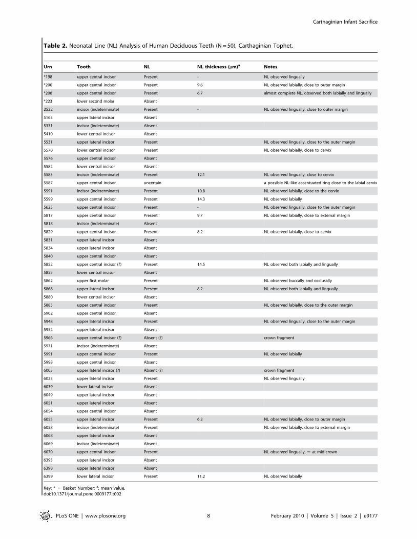
Table 2. Neonatal Line (NL) Analysis of Human Deciduous Teeth (N = 50), Carthaginian Tophet.
Urn Tooth NL NL thickness (mm)a Notes
*198 upper central incisor Present - NL observed lingually
*200 upper central incisor Present 9.6 NL observed labially, close to outer margin
*208 upper central incisor Present 6.7 almost complete NL, observed both labially and lingually
*223 lower second molar Absent
2522 incisor (indeterminate) Present - NL observed lingually, close to outer margin
5163 upper lateral incisor Absent
5331 incisor (indeterminate) Absent
5410 lower central incisor Absent
5531 upper lateral incisor Present NL observed lingually, close to the outer margin
5570 lower central incisor Present NL observed labially, close to cervix
5576 upper central incisor Absent
5582 lower central incisor Absent
5583 incisor (indeterminate) Present 12.1 NL observed lingually, close to cervix
5587 upper central incisor uncertain a possible NL-like accentuated ring close to the labial cervix
5591 incisor (indeterminate) Present 10.8 NL observed labially, close to the cervix
5599 upper central incisor Present 14.3 NL observed labially
5625 upper central incisor Present - NL observed lingually, close to the outer margin
5817 upper central incisor Present 9.7 NL observed labially, close to external margin
5818 incisor (indeterminate) Absent
5829 upper central incisor Present 8.2 NL observed labially, close to cervix
5831 upper lateral incisor Absent
5834 upper lateral incisor Absent
5840 upper central incisor Absent
5852 upper central incisor (?) Present 14.5 NL observed both labially and lingually
5855 lower central incisor Absent
5862 upper first molar Present NL observed buccally and occlusally
5868 upper lateral incisor Present 8.2 NL observed both labially and lingually
5880 lower central incisor Absent
5883 upper central incisor Present NL observed labially, close to the outer margin
5902 upper central incisor Absent
5948 upper lateral incisor Present NL observed lingually, close to the outer margin
5952 upper lateral incisor Absent
5966 upper central incisor (?) Absent (?) crown fragment
5971 incisor (indeterminate) Absent
5991 upper central incisor Present NL observed labially
5998 upper central incisor Absent
6003 upper lateral incisor (?) Absent (?) crown fragment
6023 upper lateral incisor Present NL observed lingually
6039 lower lateral incisor Absent
6049 upper lateral incisor Absent
6051 upper lateral incisor Absent
6054 upper central incisor Absent
6055 upper lateral incisor Present 6.3 NL observed labially, close to outer margin
6058 incisor (indeterminate) Present NL observed labially, close to external margin
6068 upper lateral incisor Absent
6069 incisor (indeterminate) Absent
6070 upper central incisor Present NL observed lingually, < at mid-crown
6393 upper lateral incisor Absent
6398 upper lateral incisor Absent
6399 lower lateral incisor Present 11.2 NL observed labially
Key: * = Basket Number; a: mean value.doi:10.1371/journal.pone.0009177.t002
Carthaginian Infant Sacrifice
PLoS ONE | www.plosone.org 8 February 2010 | Volume 5 | Issue 2 | e9177

Determination of SexSeventy pelvic ilia were sufficiently preserved for visual
assessment of sex, for which we relied on angle and depth of the
greater sciatic notch and, when preserved sufficiently to be
scrutinized, curvature of the iliac crest (Figure 3D). In Schutkows-
ki’s [47] study of a sample of children sexes and ages-at-death were
well-documented, greater sciatic notch angle correctly assigned,
respectively, males 95% and females 71.4%, notch depth 81.2%
and 76.5%, and crest curvature 81.2% and 62.1% of the time. In
our sample of ilia, 26 very probably and one questionably
represented male, and 38 probably and two more questionably
female (Table 1); three specimens were indeterminate. Given the
likelihood that at least some individuals we identified as female
were indeed female, the hypothesis of first-born males being the
focus of a Carthaginian ritual of sacrifice is falsified.
Estimation of Age: Tooth Formation and OsteometricsOnly bones and teeth and tooth crowns that were preserved
sufficiently intact to provide an accurate (not estimated) measure-
ment were used in our estimation of age. Based on skeletal
measurements (of the basilar portion of the occipital or basilaris,
sphenoid, petrosal, ischium, and pubis; Tables S3, S4) [17], as well
as relative states of tooth formation (Table S2) [18], most of the
sample fell within the range of 2 to 12 postnatal months, clustering
between 2 and 5 months at death (Table S2). At least another 20%
of the sample (depending on the representation of the specific
skeletal element) could be identified as prenatal. These results are
consistent with modern infant mortality data [48,49]. We ruled out
misclassifying infants of ‘‘low birth weight’’ (LBW) as prenatal
because, while mortality is 40% higher in perinates ,2500 gm
than infants of normal birth weight [50], LBW is not reflected in
diminished bone length or retarded tooth development [51].
Although experiments on heat-induced bone shrinkage were not
done in the manner of Carthaginian cremation, we nonetheless
thought it prudent to consider them. Most of these studies used
ovens rather than fire as well as dry and defleshed green rather
than fleshed bone [e.g. 52–54]. In all cases, bone shrinkage was
minimal. Richard [55] did, however, cremate parts of human
infant cadavers, but focused only on temperature and degree of
bone carbonation and calcination. Baby [56], who cremated
fleshed adult human remains, concluded that bone size was either
not, or at most only minimally, altered. Buikstra and Swegle [57]
cremated fleshed adult animal remains and found that while bone
shrinkage could be as much as 6%, in general, bone size was
minimally affected. Dokladal [58] compared bones from cremated
halves of five adult cadavers with their uncremated counterparts
and reported shrinkage between 5 and 12%. Muller’s [59]
cremations of defleshed human fetal and newborn bones suggest
shrinkage could reach 10%.
Table 3. Comparison of Ages-at-Death Determined byNeonatal Line [Histological (H)] and Morphological (M)Analyses of Human Deciduous Teeth, Carthaginian Tophet.
Urn Neonatal Line Morphology H versus M
3178 Absent #Birth M = H
5163 Absent #Birth M = H
5331 Absent ? Histological Age Only
5410 Absent Birth M.H
5576 Absent Late Third Trimester M = H
5582 Absent #Birth M = H
5818 Absent Late Third Trimester M = H
5831 Absent Late Third Trimester M = H
5834 Absent #Birth M = H
5840 Absent Late Third Trimester M = H
5855 Absent Late Third Trimester M = H
5880 Absent Late Third Trimester M = H
5902 Absent #Birth M = H
5952 Absent Late Third Trimester M = H
5966 Absent ,Birth M.H
5971 Absent Late Third Trimester M = H
5998 Absent #Birth M = H
6003 Absent Late Third Trimester M = H
6039 Absent Late Third Trimester M = H
6049 Absent Birth to 1 Month M.H
6051 Absent ,Birth M.H
6054 Absent #Birth M = H
6068 Absent ,Birth M = H
6069 Absent Birth M.H
6393 Absent ,Birth M = H
6398 Absent #Birth M = H
3159 Present 1 to 2 months postnatal M = H
3167 Present ,Birth M = H
3163 Present Late Third Trimester M,H
2522 Present ? Histological Age Only
5531 Present ,Birth M = H
5570 Present ,Birth M = H
5583 Present ? Histological Age Only
5587 Present Late Third Trimester M,H
5591 Present ? Histological Age Only
5599 Present ,Birth M = H
5625 Present 2 Months M = H
5817 Present ? Histological Age Only
5829 Present ,Birth M,H
5852 Present Birth M = H
5862 Present #Birth M,H
5868 Present #Birth M,H
5883 Present Late Third Trimester M,H
5948 Present Birth M = H
5991 Present #Birth M,H
6023 Present #Birth M,H
6055 Present Late Third Trimester M,H
6058 Present #Birth M,H
Urn Neonatal Line Morphology H versus M
6070 Present ,Birth M = H
6399 Present #Birth M,H
Key: M = H: Morphological and Histological ages similar; M.H: Morphologicalage advanced compared to Histological age; M,H: Histological age advancedcompared to Morphological age.doi:10.1371/journal.pone.0009177.t003
Table 3. Cont.
Carthaginian Infant Sacrifice
PLoS ONE | www.plosone.org 9 February 2010 | Volume 5 | Issue 2 | e9177

Although some Carthaginian perinates’ bones were barely
charred–and thus their exposure to heat minimal [46]–we
increased all of our measurements by 5, 10 and then an extreme
25% in order to account for any possible shrinkage (Figure 4).
Even at 25% increase in size, most of our analyses still classified
some individuals as prenates and thus not available for sacrifice.
Estimation of Age: Neonatal Line (NL) AnalysisIn the Carthaginian sample, NL thickness ranged from 6.3 to
14.5 mm, with a mean of 10.1 mm (62.76 mm). Comparative
estimates obtained by the same investigative methods on
deciduous teeth of all morphological classes were available from
124 crowns representing 102 modern European children [43,60]
and from 209 crowns representing 109 children (aged 6 months to
9 years) buried at the Imperial Roman cemetery of Isola Sacra
[31,60]. In the modern sample, NL thickness ranged from 6.5 to
50.9 mm and the mean value corresponded to 17.3 mm
(67.97 mm). In the archaeological sample, the range of variation
range 9–36 mm with a mean of 16.7 mm (64.40 mm). Additional
values from a modern sample of 147 children ranged from 10 to
24 mm [27].
An NL results from perturbation in matrix deposition of enamel
prisms reflecting stress in the transition from an intra- to extra-uterine
environment (Figure 2), which does not always correspond to
parturition following a full-term pregnancy [61]. Given the
periodicity of enamel deposition, a newborn must survive at least 7
and even as many as 10 to 15 extra-uterine days in order for an NL to
emerge fully. A definitive NL was observed in 24 Carthaginian
specimens (Table 2); the amount of subsequent enamel deposition
suggests these individuals survived at least 2 weeks postpartum. An
NL was absent in 26 Carthaginian specimens (Table 2), which
suggests that these individuals were either stillborn, spontaneously
aborted, or died during the first extra-uterine week. Unambiguous
counts and measurements of daily enamel cross-striations, which
provide information on the timing and rate of enamel deposition and
thus indirect evidence of gestation length [31,33], could not be
obtained on this sample. However, because other analyses in our
study indicate the presence of individuals who had not reached full
term, we suggest that individuals lacking an NL probably fall into the
prenatal category because comparison of morphological/metric and
NL age estimates demonstrates that when they differed, the
histological (NL) age more frequently over-aged individuals than
did morphological age (M,H 22%, M.H 10%; see Table 3).
Consequently, if we include with the prenates those individuals who
did not survive beyond one or even two weeks postpartum, we must
conclude that a significant number of individuals could not have been
sacrificed because they were either not alive or not yet old enough to
be considered viable sacrificial entities [7,8,10,13] (Figure 5).
Discussion
The identification of prenatal individuals in the Carthaginian
Tophet sample is consistent with current data from modern-day
studies on the incidence of stillbirth and spontaneous abortion as
being the primary contributors to ‘‘reproductive wastage’’ [62], as
well as with recent data on infant mortality [48,49]. For example,
in England and Wales from 1969 to 1976, 48.4% of 6517 deaths
within two weeks of live birth occurred between 30 minutes and
24 hours and 39.3% between 7 and 13 days [61]. These statistics
easily accommodate our results.
Infectious diseases known to lead to stillbirth include smallpox,
vaccinia, and listeriosis; those resulting in prematurity and
perinatal mortality include severe viral infections and malaria
[49]. Noninfectious diseases resulting in stillbirth, abortion, or
preterm delivery include cholestasis, hypertension, toxemia, and
renal disease [50]. The Carthaginians were probably exposed to
and susceptible to all of these afflictions. If conditions of sanitation
at Carthage, including management of water supply and human
and animal excreta, were similar to those at Pompeii, Ostia, and
Rome [63], the Carthaginians would also have been potential
victims to and vectors of cholera, dysentery, gastroenteritis,
infectious hepatitis, leptospirosis, typhoid, and parasitic intestinal
infestations, most of which result in severe dehydration, which is a
common cause of infant death [50].
In sum, while the Carthaginians may occasionally have
practiced human sacrifice, as did other circum-Mediterranean
societies [1,63,64], our analyses do not support the contention that
all humans interred in the Tophet had been sacrificed. Rather, it
would appear that the Carthaginian Tophet, and by extension
Tophets at Carthaginian settlements in general, were cemeteries
for the remains of human prenates and infants who died from a
variety of causes and then cremated and whose remains,
sometimes on a catch-as-catch-can basis, interred in urns.
Following widespread practice at this time in history, it is likely
that at least some, if not all, of the cremated animal remains
represent sacrificial offerings.
Supporting Information
Table S1 Species Identification of Skeletal Remains from Urns,
Carthaginian Tophet.
Figure 5. Distribution of Ages-at-Death Based on Analysis of Human Remains, Carthaginian Tophet.doi:10.1371/journal.pone.0009177.g005
Carthaginian Infant Sacrifice
PLoS ONE | www.plosone.org 10 February 2010 | Volume 5 | Issue 2 | e9177

Found at: doi:10.1371/journal.pone.0009177.s001 (0.33 MB
DOC)
Table S2 Demographic Profile of Human Remains, Carthagi-
nian Tophet.
Found at: doi:10.1371/journal.pone.0009177.s002 (0.93 MB
DOC)
Table S3 Dimensions of Human Cranial Bones (in mm.),
Carthaginian Tophet.
Found at: doi:10.1371/journal.pone.0009177.s003 (0.33 MB
DOC)
Table S4 Dimensions of Human Pelvic Bones (in mm.),
Carthaginian Tophet.
Found at: doi:10.1371/journal.pone.0009177.s004 (0.12 MB
DOC)
Acknowledgments
JHS thanks L. E. Stager (Director, American Team to Carthage) for the
invitation to undertake this study and the Tunisian Department of
Antiquities for granting permission for the excavation and analysis of
Tophet urns and their contents.
Author Contributions
Recovered and did preliminary analysis of specimens: JHS. Collaborated
with the second author (Houghton) on the refined analysis: JHS. Wrote the
bulk of the manuscript: JHS. Collaborated on detailed analysis of remains:
FH. Did the statistics: FH. Created the tables, graphs of figure plates: FH.
Collaborated with L. Bondioli on the neonatal line analysis and
interpretation: RM. Collaborated with R. Macchiarelli on analysis and
interpretation of neonatal line data: LB. Contributed to the Methods
section on this technique: RM LB.
References
1. Brown S (1991) Late Carthaginian Child Sacrifice and Sacrificial Monuments in
their Mediterranean Context. Sheffield: JSOT Press.
2. Harden D (1963) The Phoenicians. New York: Frederick A. Praeger.
3. Mosca PG (1975) Child Sacrifice in Canaanite and Israelite Religion.
Cambridge, MA: Harvard.
4. Stager LE, Greene JA (2007) Child sacrifice: yes, children of Phoenician/Punic
Carthage were sacrificed to the gods. http://phoeniciaorg/childsacrificehtml. pp2–3.
5. Stager LE, Wolf SR (1984) Child sacrifice at Carthage: Religious rite or
population control. Biblical Archaeology Review 10: 31–51.
6. Agelarakis AP, Kanta A, Stampolidis N (1998) The osseous record in the
Western Necropolis of Amathous: an archaeo-anthropological investigation. In:Karageorghis V, Stampolidis N, eds. Eastern Mediterranean Cyprus-Dode-
canses-Crete 16th-6th cent BC. Heraklion: University of Crete. pp 217–232.
7. Benichou-Safar H (1981) A propos des ossements humains du tophet de Carthage.Rivista di Studi Fenici 5: 5–9.
8. Bartoloni P (2006) Il Tophet: un pietoso rito offuscato da troppo miti. DarwinQuaderni 1: 68–75.
9. Conte S (2007) Child sacrifice: children of Phoenician Punic Carthage were not
sacrificed to the gods. http://phoeniciaorg/childsacrificehtml. pp 4–6.
10. Fantar MH (2007) Child sacrifice: child of Pheonician Punic Carthage were not
sacrificed. http://phoeniciaorg/childsacrificehtml. pp 2–4.
11. Fedele F, Foster C (1988) Tharros ovicaprini sacrificiali e rituale del Tofet.Rivista di Studi Fenici 16: 29–42.
12. Lancel S (1995) Carthage: a history. Oxford: Oxford University Press.
13. Moscati S (1987) Il sacrificio punico dei fanciulli: realta or invenzione. Quadernidell’Accademia Nazionale dei Lincei 261: 4–15.
14. Simonetti A (1983) Sacrifici umani e uccisioni rituali nel mondo Fenicio-Punico:
il contributo delle fonti letterarie. Rivista di Studi Fenici 11: 91–111.
15. Schwartz JH (1993) What the Bones Tell Us. New York: Henry Holt & Co.
16. Schwartz JH (1989) The Tophet and ‘‘sacrifice’’ at Phoenician Carthage: an
osteologist’s perspective. Terra 28: 16–25.
17. Fazekas IG, Kosa F (1979) Forensic Fetal Osteology. Budapest: Akademiai
Kiado.
18. Schwartz JH (2007) Skeleton Keys: an introduction to human skeletalmorphology, development, and analysis. New York: Oxford University Press.
19. Rushton A (1939) The birifrangence of deciduous tooth enamel formed before
and after birth. Britannic Dental Journal 67: 1–10.
20. Kronfeld R, Schour I (1939) Neonatal dental hypoplasia. Journal of the
American Dental Association 16: 18–20.
21. Weber DF, Eisenmann D (1971) Microscopy of the neonatal line in developing
human enamel. American Journal of Anatomy 132: 375–392.
22. Whittaker DK, Richards D (1978) Scanning electron microscopy of the neonatalline in human enamel. Archives of Oral Biology 23: 45–50.
23. Levine RS, Turner EP, Dobbing J (1979) Deciduous teeth contain histories of
developmental disturbances. Early Human Development 3: 211–220.
24. Skinner MF (1992) Gestation length and location of the neonatal line in human
enamel. Journal of Paleopathology 2: 41–50.
25. Seow WK (1986) Oral implication of premature birth. Australian Dental Journal31: 23–29.
26. Ranggard L, Noren JG, Nelson N (1994) Clinical and histologic appearance inenamel of primary teeth in relation to neonatal blood ionized calcium values.
Scandinavian Journal of Dental Research 102: 254–259.
27. Eli I, Sarnat H, Talmi E (1989) Effect of the brith process on the neonatal line in
primary tooth enamel. Pediatric Dentistry 11: 220–223.
28. Skinner MF, Dupras T (1993) Variation in birth timing and location of theneonatal line in human enamel. Journal of Forensice Sciences 38: 1383–1390.
29. Teivens A, Mornstad H, Noren JG, Gidlund E (1996) Variation in birth timing
and location of the neonatal line in human enamel. Forensic Science
International 81: 175–183.
30. Shellis RP (1984) Variations in growth of the enamel crown in human teeth and
a possible relationship between growth and enamel structure. Archives of OralBiology 29: 697–705.
31. Rossi PF, Bondioli L, Geusa G, Macchiarelli R (1999) Osteodental Biology of
the People of Portus Romae (Necropolis of Isola Sacra, 2nd–3rd Cent. AD). I.
Enamel Microstructure and Developmental Defects of the Primary Dentition.Rome: Museo Nazionale L. Pigorini.
32. Smith P, Avishai G (2005) The use of dental criteria for estimating postnatal
survival in skeletal remains of infants. Journal of Archaeological Science 32:83–89.
33. FitzGerald C, Saunders S, Bondioli L, Macchiarelli R (2006) Health of infants in
an Imperial Roman skeletal sample: perspective from dental microstructure.American Journal of Physical Anthropology 130: 179–189.
34. Macchiarelli R, Bondioli L, Caropreso S, Mazurier A, Merceron G, et al. (2006)
The oldest human remains from the Beagle Channel, Tierra del Fuego.International Journal of Osteoarchaeology 16: 328–337.
35. Macchiarelli R, Bondioli L, Debenath A, Mazurier A, Merceron G (2006) How
Neanderthal teeth grew. Nature 444: 748–751.
36. Smith TM, Toussaint M, Reid DJ, Olejniczak AJ, Hublin J-J (2007) Rapiddental development in a Middle Paleolithic Begian Neanderthal. Proceedings of
the National Academy of Sciences (USA) 104: 20220–20225.
37. Caropreso S, Bondioli L, Capannolo D, Cerroni L, Macchiarelli R, et al. (2000)Thin sections for hard tissues biology: a new procedure. Journal of Microscopy
199: 244–247.
38. Gejvall N-G (1969) Cremations. In: Brothwell D, Higgs E, Clark G, editors.
Science in Archaeology. 2nd ed. London: Thames and Hudson. pp 468–479.
39. McKinley JI (1994) The Anglo-Saxon Cemetery at Spong Hill, North Elmham.Part VIII: the Cremations. Dereham: Field Archaeology Division, Norfolk
Museums Service.
40. Hillson S (1996) Dental Anthropology. Cambridge: Cambridge University Press.
41. Gatto E (2003) La Place de la Cremation dans le Traitement des Defunts a la Findu Neolithique en France. Outils Methodologiques et Etudes de Sites. Bordeaux:
Universite de Bordeaux 1.
42. Susini A (2003) Etude des Caracteristiques Biophysiques des Tissus CalcifiesHumains Soumis a des Traitements Thermiques: Applications Anthropologi-
ques et Medicales. Geneve: Universite de Geneve.
43. Macchiarelli R, Petrone PP, Bondioli L (1996) I resti ossei combusti. Analisimorfologica ed istomorfometrica. Atti e Memorie della Societa Magna Grecia 3:
101–104.
44. Bondioli L, Macchiarelli R (1999) Indagini istomorfometriche sullo scheletro esui denti di San Paolo Belsito. In: Fedele F, Petrone PP, eds. Un’Eruzione
Vesuviana 4000 Anni Fa. Napoli: Fridericiana Editrice Universitaria. pp 67–68.
45. McKinley JI (1989) Cremations: expectations, methodologies and realities. In:Roberts CA, Lee F, Bintliff J, eds. Burial Archaeology: Current Research,
Methods and Developments. London: BAR British Series. pp 65–76.
46. Gejvall N-G (1963) Cremations. In: Brothwell D, Higgs E, eds. Science in
Archaeology: A Comprehensive Survey of Progress and Research. New York:Basic. pp 379–390.
47. Schutkowski H (1993) Sex determination of infant and juvenile skeletons I.
Morphognostic features. American Journal of Physical Anthropology 90:199–205.
48. Saunders SR, Barrans L (1999) What can be done about the infant category in
skeletal sample? In: Hoppa RD, Firzgerald CM, eds. Cambridge: CambridgeUniversity Press. pp 183–209.
49. Taylor CM, Pernoll ML (1987) Normal pregnancy & prenatal care. In:
Pernoll ML, Benson RC, eds. Current Obstetric & Gynecologic Diagnosis &Treatment 1987. Norwalk, CT: Appleton & Lange. pp 161–177.
50. Behrman RE, Shiono PH (1997) Neonatal risk factors. In: Fanaroff AA,
Martin RJ, eds. Neonatal-Perinatal Medicine: Diseases of the Fetus and Infant.
St. Loius: Mosby. pp 3–12.
Carthaginian Infant Sacrifice
PLoS ONE | www.plosone.org 11 February 2010 | Volume 5 | Issue 2 | e9177

51. Jaya DS, Kumar NS, Bai LS (1995) Anthropometric indices, cord length and
placental weight in newborns. Indian Pediatrics 32: 1183–1188.
52. Pointek J (1976) The process of cremation and its influence on the morphology
of bones in light of results of experimental research. Achaeologia Polski 21:
254–280.
53. Shipman P, Forster G, Schoeninger M (1984) Burnt bones and teeth: an
experimental study of colour, morphology, crystal structure and shrinkage.
Journal of Archaeological Science 11: 307–325.
54. Thurman MD, Willmore LJ (1981) A replicative cremation experiment. North
American Archaeologist 2: 1980–1981.
55. Richard J (1961) Etude Medico-Legale des Urnes Sacrificielles Puniques et de
leur Contenu. Lille: Lille.
56. Baby RS (1954) Hopewell Cremation Practices. Columbus, OH: Ohio History
Society.
57. Buikstra JE, Swegle M (1989) Bone modification due to burning: experimental
evidence. In: Bonnischen R, Sorg MH, eds. Bone Modification. Orono, ME:
University of Maine. pp 247–258.
58. Dokladal M (1971) A further contribution to the morphology of burned bones.
In: Novotny N, ed. Proceedings of the Anthropological Congress Dedicated toAles Hrdlicka. Prague: Czechoslovak Academy of Science. pp 561–568.
59. Muller M (1938) La calcinations du foetus en medicine legale. Bonn:
Verhandlungsbericht des Internationalen Kongresses fur gerichtliche undsociale Medizin.
60. Bondioli L, Macchiarelli R (1999) Neonatal line thickness and delivery at IsolaSacra (2nd–3rd cent. AD, Rome, Italy). American Journal of Physical
Anthropology Suppl. 28: 94–95.
61. Chalmers J, Macfarlane A (1980) Interpretation of perinatal statistics. In:Wharton BA, ed. Topics in Perinatal Medicine. Tunbridge Wells, UK: Pitman
Medical. pp 1–11.62. Durfee RB (1987) Obstetric complications of pregnancy. In: Wharton BA, ed.
Topics in Perinatal Medicine. Tunbridge Wells, UK: Pitman Medical. pp255–278.
63. Scobie A (1986) Slums, sanitation, and mortality in the Roman world. Klio 2:
399–433.64. Moscati S (1965) The World of the Phoenicians. London: Weidenfeld and
Nicolson.
Carthaginian Infant Sacrifice
PLoS ONE | www.plosone.org 12 February 2010 | Volume 5 | Issue 2 | e9177