Single‐dose immunisation with a multimerised SARS‐CoV‐2 ...
18
Single-dose immunisation with a multimerised SARS-CoV-2 receptor binding domain (RBD) induces an enhanced and protective response in mice Ralf Salzer 1 , Jordan J. Clark 2 , Marina Vaysburd 1 , Veronica T. Chang 1 , Anna Albecka 1 , Leo Kiss 1 , Parul Sharma 2 , Andres Gonzalez Llamazares 1 , Anja Kipar 2,3 , Julian A. Hiscox 2 , Andrew Owen 4 , A. Radu Aricescu 1 , James P. Stewart 2 , Leo C. James 1 and Jan L € owe 1 1 MRC Laboratory of Molecular Biology, Cambridge, UK 2 Institute of Infection, Veterinary and Ecological Sciences, University of Liverpool, UK 3 Laboratory for Animal Model Pathology, Institute of Veterinary Pathology, Vetsuisse Faculty, University of Zurich, Switzerland 4 Department of Pharmacology and Therapeutics, Centre of Excellence in Long-acting Therapeutics (CELT), University of Liverpool, UK Correspondence L. C. James and J. L€ owe, MRC Laboratory of Molecular Biology, Cambridge Biomedical Campus, Francis Crick Avenue, Cambridge CB2 0QH, UK Tel: +44 (0)1223 267162 (LCJ); +44 (0)1223 267064 (JL) E-mails: [email protected] (LCJ); [email protected] (JL) Ralf Salzer, Jordan J. Clark, Marina Vaysburd, and Veronica T. Chang contributed equally to this work (Received 10 June 2021, revised 2 July 2021, accepted 13 July 2021) doi:10.1002/1873-3468.14171 Edited by Urs Greber The COVID-19 pandemic, caused by the SARS-CoV-2 coronavirus, has trig- gered a worldwide health emergency. Here, we show that ferritin-like Dps from hyperthermophilic Sulfolobus islandicus, covalently coupled with SARS- CoV-2 antigens via the SpyCatcher system, forms stable multivalent dode- cameric vaccine nanoparticles that remain intact even after lyophilisation. Immunisation experiments in mice demonstrated that the SARS-CoV-2 recep- tor binding domain (RBD) coupled to Dps (RBD-S-Dps) elicited a higher antibody titre and an enhanced neutralising antibody response compared to monomeric RBD. A single immunisation with RBD-S-Dps completely pro- tected hACE2-expressing mice from serious illness and led to viral clearance from the lungs upon SARS-CoV-2 infection. Our data highlight that multi- merised SARS-CoV-2 subunit vaccines are a highly efficacious modality, par- ticularly when combined with an ultra-stable scaffold. Keywords: coronavirus; COVID-19; Dps; RBD; SARS-CoV-2; subunit vaccine On 11 March 2020, the World Health Organisation declared the COVID-19 outbreak, caused by the SARS-CoV-2 virus, a pandemic [1]. Since then, COVID-19 and the efforts to contain it have changed the lives of unprecedented numbers of people. For example, in April 2020 3.9 billion people were affected by lockdown measures aimed to cut or at least reduce the chain of transmission with widespread negative impacts on employment, education, and other health issues. According to the Johns Hopkins University, there have so far been 151 million confirmed COVID- 19 cases globally (May 2021) and virtually every coun- try has been affected. Officially, 3.2 million people have died from SARS-CoV-2 infection [2,3]. SARS-CoV-2 belongs to the family of Coronaviri- dae, which contain a positive-stranded RNA genome [4]. The RNA is enveloped by a membrane that har- bours four coat proteins (Fig. 1A). On the inside of the virus, the nucleocapsid protein (NP) is crucial for RNA packaging and viral release from host cells [5]. Abbreviations hACE2, human angiotensin-converting enzyme 2; NP, nucleocapsid protein; RBD, receptor binding domain. 1 FEBS Letters (2021) ª 2021 MRC Laboratory of Molecular Biology. FEBS Letters published by John Wiley & Sons Ltd on behalf of Federation of European Biochemical Societies. This is an open access article under the terms of the Creative Commons Attribution License, which permits use, distribution and reproduction in any medium, provided the original work is properly cited.
Transcript of Single‐dose immunisation with a multimerised SARS‐CoV‐2 ...

Single-dose immunisation with a multimerisedSARS-CoV-2 receptor binding domain (RBD) induces anenhanced and protective response in miceRalf Salzer1 , Jordan J. Clark2 , Marina Vaysburd1 , Veronica T. Chang1 ,Anna Albecka1 , Leo Kiss1 , Parul Sharma2 , Andres Gonzalez Llamazares1 ,Anja Kipar2,3 , Julian A. Hiscox2 , Andrew Owen4 , A. Radu Aricescu1 ,James P. Stewart2 , Leo C. James1 and Jan L€owe1
1 MRC Laboratory of Molecular Biology, Cambridge, UK
2 Institute of Infection, Veterinary and Ecological Sciences, University of Liverpool, UK
3 Laboratory for Animal Model Pathology, Institute of Veterinary Pathology, Vetsuisse Faculty, University of Zurich, Switzerland
4 Department of Pharmacology and Therapeutics, Centre of Excellence in Long-acting Therapeutics (CELT), University of Liverpool, UK
Correspondence
L. C. James and J. L€owe, MRC Laboratory
of Molecular Biology, Cambridge Biomedical
Campus, Francis Crick Avenue, Cambridge
CB2 0QH, UK
Tel: +44 (0)1223 267162 (LCJ); +44 (0)1223
267064 (JL)
E-mails: [email protected] (LCJ);
[email protected] (JL)
Ralf Salzer, Jordan J. Clark, Marina
Vaysburd, and Veronica T. Chang
contributed equally to this work
(Received 10 June 2021, revised 2 July
2021, accepted 13 July 2021)
doi:10.1002/1873-3468.14171
Edited by Urs Greber
The COVID-19 pandemic, caused by the SARS-CoV-2 coronavirus, has trig-
gered a worldwide health emergency. Here, we show that ferritin-like Dps
from hyperthermophilic Sulfolobus islandicus, covalently coupled with SARS-
CoV-2 antigens via the SpyCatcher system, forms stable multivalent dode-
cameric vaccine nanoparticles that remain intact even after lyophilisation.
Immunisation experiments in mice demonstrated that the SARS-CoV-2 recep-
tor binding domain (RBD) coupled to Dps (RBD-S-Dps) elicited a higher
antibody titre and an enhanced neutralising antibody response compared to
monomeric RBD. A single immunisation with RBD-S-Dps completely pro-
tected hACE2-expressing mice from serious illness and led to viral clearance
from the lungs upon SARS-CoV-2 infection. Our data highlight that multi-
merised SARS-CoV-2 subunit vaccines are a highly efficacious modality, par-
ticularly when combined with an ultra-stable scaffold.
Keywords: coronavirus; COVID-19; Dps; RBD; SARS-CoV-2; subunit
vaccine
On 11 March 2020, the World Health Organisation
declared the COVID-19 outbreak, caused by the
SARS-CoV-2 virus, a pandemic [1]. Since then,
COVID-19 and the efforts to contain it have changed
the lives of unprecedented numbers of people. For
example, in April 2020 3.9 billion people were affected
by lockdown measures aimed to cut or at least reduce
the chain of transmission with widespread negative
impacts on employment, education, and other health
issues. According to the Johns Hopkins University,
there have so far been 151 million confirmed COVID-
19 cases globally (May 2021) and virtually every coun-
try has been affected. Officially, 3.2 million people
have died from SARS-CoV-2 infection [2,3].
SARS-CoV-2 belongs to the family of Coronaviri-
dae, which contain a positive-stranded RNA genome
[4]. The RNA is enveloped by a membrane that har-
bours four coat proteins (Fig. 1A). On the inside of
the virus, the nucleocapsid protein (NP) is crucial for
RNA packaging and viral release from host cells [5].
Abbreviations
hACE2, human angiotensin-converting enzyme 2; NP, nucleocapsid protein; RBD, receptor binding domain.
1FEBS Letters (2021) ª 2021 MRC Laboratory of Molecular Biology. FEBS Letters published by John Wiley & Sons Ltd on behalf of
Federation of European Biochemical Societies.
This is an open access article under the terms of the Creative Commons Attribution License, which permits use,
distribution and reproduction in any medium, provided the original work is properly cited.
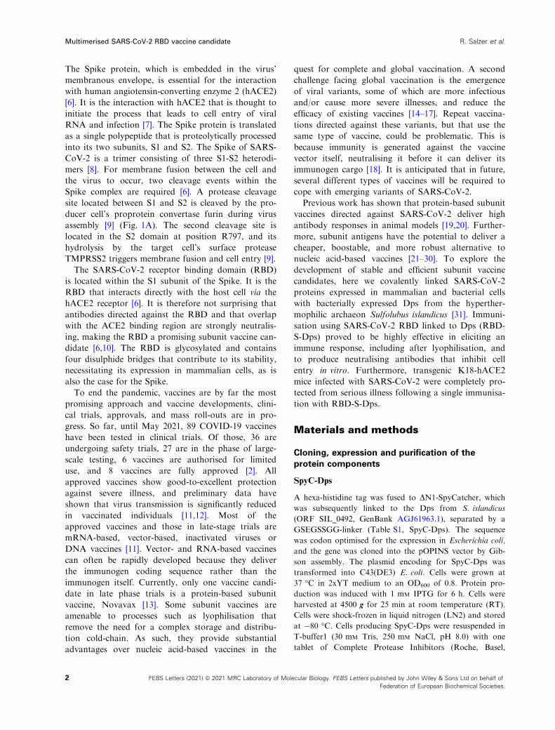
The Spike protein, which is embedded in the virus’
membranous envelope, is essential for the interaction
with human angiotensin-converting enzyme 2 (hACE2)
[6]. It is the interaction with hACE2 that is thought to
initiate the process that leads to cell entry of viral
RNA and infection [7]. The Spike protein is translated
as a single polypeptide that is proteolytically processed
into its two subunits, S1 and S2. The Spike of SARS-
CoV-2 is a trimer consisting of three S1-S2 heterodi-
mers [8]. For membrane fusion between the cell and
the virus to occur, two cleavage events within the
Spike complex are required [6]. A protease cleavage
site located between S1 and S2 is cleaved by the pro-
ducer cell’s proprotein convertase furin during virus
assembly [9] (Fig. 1A). The second cleavage site is
located in the S2 domain at position R797, and its
hydrolysis by the target cell’s surface protease
TMPRSS2 triggers membrane fusion and cell entry [9].
The SARS-CoV-2 receptor binding domain (RBD)
is located within the S1 subunit of the Spike. It is the
RBD that interacts directly with the host cell via the
hACE2 receptor [6]. It is therefore not surprising that
antibodies directed against the RBD and that overlap
with the ACE2 binding region are strongly neutralis-
ing, making the RBD a promising subunit vaccine can-
didate [6,10]. The RBD is glycosylated and contains
four disulphide bridges that contribute to its stability,
necessitating its expression in mammalian cells, as is
also the case for the Spike.
To end the pandemic, vaccines are by far the most
promising approach and vaccine developments, clini-
cal trials, approvals, and mass roll-outs are in pro-
gress. So far, until May 2021, 89 COVID-19 vaccines
have been tested in clinical trials. Of those, 36 are
undergoing safety trials, 27 are in the phase of large-
scale testing, 6 vaccines are authorised for limited
use, and 8 vaccines are fully approved [2]. All
approved vaccines show good-to-excellent protection
against severe illness, and preliminary data have
shown that virus transmission is significantly reduced
in vaccinated individuals [11,12]. Most of the
approved vaccines and those in late-stage trials are
mRNA-based, vector-based, inactivated viruses or
DNA vaccines [11]. Vector- and RNA-based vaccines
can often be rapidly developed because they deliver
the immunogen coding sequence rather than the
immunogen itself. Currently, only one vaccine candi-
date in late phase trials is a protein-based subunit
vaccine, Novavax [13]. Some subunit vaccines are
amenable to processes such as lyophilisation that
remove the need for a complex storage and distribu-
tion cold-chain. As such, they provide substantial
advantages over nucleic acid-based vaccines in the
quest for complete and global vaccination. A second
challenge facing global vaccination is the emergence
of viral variants, some of which are more infectious
and/or cause more severe illnesses, and reduce the
efficacy of existing vaccines [14–17]. Repeat vaccina-
tions directed against these variants, but that use the
same type of vaccine, could be problematic. This is
because immunity is generated against the vaccine
vector itself, neutralising it before it can deliver its
immunogen cargo [18]. It is anticipated that in future,
several different types of vaccines will be required to
cope with emerging variants of SARS-CoV-2.
Previous work has shown that protein-based subunit
vaccines directed against SARS-CoV-2 deliver high
antibody responses in animal models [19,20]. Further-
more, subunit antigens have the potential to deliver a
cheaper, boostable, and more robust alternative to
nucleic acid-based vaccines [21–30]. To explore the
development of stable and efficient subunit vaccine
candidates, here we covalently linked SARS-CoV-2
proteins expressed in mammalian and bacterial cells
with bacterially expressed Dps from the hyperther-
mophilic archaeon Sulfolubus islandicus [31]. Immuni-
sation using SARS-CoV-2 RBD linked to Dps (RBD-
S-Dps) proved to be highly effective in eliciting an
immune response, including after lyophilisation, and
to produce neutralising antibodies that inhibit cell
entry in vitro. Furthermore, transgenic K18-hACE2
mice infected with SARS-CoV-2 were completely pro-
tected from serious illness following a single immunisa-
tion with RBD-S-Dps.
Materials and methods
Cloning, expression and purification of the
protein components
SpyC-Dps
A hexa-histidine tag was fused to DN1-SpyCatcher, which
was subsequently linked to the Dps from S. islandicus
(ORF SIL_0492, GenBank AGJ61963.1), separated by a
GSEGSSGG-linker (Table S1, SpyC-Dps). The sequence
was codon optimised for the expression in Escherichia coli,
and the gene was cloned into the pOPINS vector by Gib-
son assembly. The plasmid encoding for SpyC-Dps was
transformed into C43(DE3) E. coli. Cells were grown at
37 °C in 2xYT medium to an OD600 of 0.8. Protein pro-
duction was induced with 1 mM IPTG for 6 h. Cells were
harvested at 4500 g for 25 min at room temperature (RT).
Cells were shock-frozen in liquid nitrogen (LN2) and stored
at �80 °C. Cells producing SpyC-Dps were resuspended in
T-buffer1 (30 mM Tris, 250 mM NaCl, pH 8.0) with one
tablet of Complete Protease Inhibitors (Roche, Basel,
2 FEBS Letters (2021) ª 2021 MRC Laboratory of Molecular Biology. FEBS Letters published by John Wiley & Sons Ltd on behalf of
Federation of European Biochemical Societies.
Multimerised SARS-CoV-2 RBD vaccine candidate R. Salzer et al.

Switzerland) per 10 g cells wet weight. Cell disruption was
carried out using sonication for 7.5 min ‘on’ time, using a
50% duty cycle. Cell debris were removed by centrifugation
at 20 000 g for 30 min at RT. The supernatant was loaded
onto a HisTrap FF affinity chromatography column
(Cytiva, Freiburg im Breisgau, Germany). Washing was
carried out for 17 column volumes (CV) with T-buffer1
plus 110 mM imidazole. The protein was eluted with T-
buffer1 containing 400 mM imidazole. Purity of fractions
was examined by SDS/PAGE, and the purest fraction were
pooled and concentrated using a Vivaspin Turbo centrifu-
gal concentrator (100 000 MWCO, Sartorius, G€ottingen,
Germany). Concentrated sample was loaded onto a size
exclusion column (SEC, Sephacryl S-400, Cytiva), with
PBS as the running buffer. Purity was examined by SDS/
PAGE, and the sample was frozen in LN2 and stored at
�80 °C.
SpyT2-NP
The NP (amino acids 48–364; GenBank: MN908947; NP)
was cloned into the vector pOP-TH and N terminally
equipped with a hexa-histidine tag [32]. A SpyTag2 sequence
separated by GS-rich linkers was inserted between the hexa-
histidine tag and NP (Fig. 1 and Table S1, SpyT2-NP). The
vector encoding for SpyT2-NP was transformed into E. coli
C41(DE3) cells. For protein expression, cells were grown at
37 °C in 2xYT medium to an OD600 of 0.7. Protein produc-
tion was induced with 1 mM IPTG for 6 h. Cells were har-
vested at 4500 g for 25 min at 4 °C. Cells were frozen in
LN2 and stored at �80 °C. SpyT2-NP-producing cells were
resuspended in T-buffer2 (50 mM Tris, 1 M NaCl, 10 mM
imidazole, 2 mM DTT, pH 8.0) with Complete Protease Inhi-
bitor added (1 tablet per 10 g cells wet weight). Cells were
lysed by sonication (3 min total ‘on’ time, duty cycle 50%).
SpikeReceptor bindingdomain (RBD)
M-protein
Hemagglutinin-esterase
RNAE-proteinNucleocapsid protein (NP)Envelope (E)
TMPRSS2ACE2
(A)
Ag
Ag
Ag
Ag
Ag
Ag
Ag
AgAg
Ag
Ag
Ag
self-assembly
covalent coupling of SpyTag2-antigens with
SpyCatcher-Dps particles
RBD-SpyTag2SpyTag2-NP
trimeric Spike-SpyTag2
SpyCatcher-Dpsproduced in E. coli
DodecamericSpyCatcher-Dps
particles
SpyTag2-fusedantigens produced in HEK and E. coli cells:
Dodecameric SpyCatcher-Dps particles with coupled antigens: Ag-S-Dps
(B)
Antigens
ST2
Avi
His
332 529
His
ST2
Avi
Dps
RBD
SpyCHis
332 529
2 193
ST2
His
48 364
His
ST2 NP
DpsSpyCHis
48 364
2 193
ST2
Avi
His
16
Avi
His
Scaffold protein:
RBD-SpyT2
SpyT2-Nucleocapsid Protein
Spike-SpyT2
Spike
16
RBD
NP
1208 Spike
1208 Spike
DpsSpyCHis
His
His
ST2
Avi
Avi
Avi
1208
1208
120816
16
16
His 2 193
His
Multivalent constructs
RBD-S-Dps
NP-S-Dps
Spike-S-Dps
Spike
Spike
Spike
SpyC SpyC-DpsDps2 193(C)Fig. 1. Overview of the multimerisation
strategy employed and the antigens and
scaffold used. (A) Cartoon representation
of SARS-CoV-2 binding to a human cell
membrane. (B) Schematic diagram of the
Sulfolobus islandicus Dps and SpyCatcher-
based display and multimerisation strategy
employed in this study. (C) Diagram of the
proteins used in this work. SpyC is the
DN1-SpyCatcher domain, and SpyT2 is the
peptidic SpyTag2 that becomes covalently
linked to SpyC upon simple mixing.
Stabilised, trimeric Spike/Spike-SpyT2
contained on average only one SpyT2 tag
in order to avoid uncontrolled
oligomerisation when coupled to Dps.
3FEBS Letters (2021) ª 2021 MRC Laboratory of Molecular Biology. FEBS Letters published by John Wiley & Sons Ltd on behalf of
Federation of European Biochemical Societies.
R. Salzer et al. Multimerised SARS-CoV-2 RBD vaccine candidate

Precipitated proteins and cell debris were removed by cen-
trifugation (40 000 g, 1 h, 4 °C). The supernatant was
loaded onto a HisTrap FF affinity chromatography column
and washed with 20 CV T-Buffer3 (50 mM Tris, 300 mM
NaCl, 1 mM DTT, pH 8.0) containing 20 mM imidazole. Elu-
tion was carried out in T-buffer3 containing 400 mM imida-
zole. Elution fractions containing NP were loaded onto
20 mL HiTrap Heparin HP column equilibrated in T-buffer4
(50 Tris, 1 mM DTT, pH 8.0). The column was washed with
3 CV T-buffer4. Elution was carried out with a linear gradi-
ent of 0–2 M NaCl. Elution fractions containing SpyT2-NP
were examined by SDS/PAGE and pooled, and concentrated
using a Vivaspin Turbo concentrator with a 10 000 MWCO
(Sartorius). Concentrated sample was loaded onto a SEC
column (Sephacryl S-200) (Cytiva) in PBS + 250 mM addi-
tional NaCl. Purity was checked by SDS/PAGE, and sam-
ples were frozen in LN2 and stored at �80 °C.
Spike-SpyT2 and Spike
To express the ectodomain of the stabilised prefusion Spike
protein trimer [33] with only one subunit carrying the Spy-
Tag2 tag, two constructs – one with and one without a
SpyTag2 – were made. First, a gene encoding residues 16–1208 of SARS-CoV-2 Spike protein (GenBank: MN908947)
with proline substitutions at residues 986 and 987, a GSAS
substitution at the furin cleavage site (residue 682–685), aC-terminal T4 fibritin trimerisation motif, a GGSGGS lin-
ker, an HRV3C protease cleavage site, a GGS linker and
an AviTag, was synthesised and cloned into the lentiviral
expression vector pHR-SFFV [34–36] downstream of the
sequence encoding the chicken RPTPr secretion signal pep-
tide (cRPTPrSP) [37]. Then, either a GGS linker and a
hexa-histidine tag, or a GGS linker, an octa-histidine tag, a
GGSGGSGGS linker and a SpyTag2 were inserted after
the AviTag sequence to form two Spike constructs, with
and without a SpyTag2 (Table S1, Spike-SpyT2 and Spike,
respectively). For protein expression and purification, see
the next paragraph.
RBD-SpyT2
A gene encoding residue 332–529 of SARS-CoV-2 Spike pro-
tein (constituting the receptor binding domain, RBD) was
synthesised and cloned downstream of cRPTPr of the pHR-
SFFV vector and a GGSGGS linker, an AviTag, a GGS lin-
ker, an octa-histidine tag, a GGSGGSGGS linker and a Spy-
Tag2 were inserted at the 30 end of the gene (Table S1, RBD-
SpyT2). The vectors for Spike-SpyT2, Spike and RBD-
SpyT2 were used for protein production in the mammalian
lentiviral expression system [34–36]. The DNA of the con-
structs was mixed with the lentiviral envelope and packaging
vectors pMD2-G and psPAX2c (Addgene, Watertown, MA,
USA) and polyethylenimine (PEI, Sigma Aldrich, St. Louis,
MO, USA) to transiently transfect HEK 293T Lenti-X cells
(Takara Bio, Kusatsu, Japan) to make lentiviral particles. To
make Spike trimer protein with only one subunit carrying a
SpyTag2, the DNAs of constructs Spike and Spike-SpyT2
were used at a molar ratio 3 : 1. The virus particles produced
were used to infect HEK 293S GnT1�/� cells (for Spike-
SpyT2) or Expi 293 cells (for RBD-SpyT2). The infected cells
were then expanded to obtain 3 L cultures, and conditioned
media were harvested and sterile filtered (0.22 lm). The
supernatant was concentrated and the buffer exchanged to
25 mM Tris pH 8.0, 300 mM NaCl using an €Akta flux tangen-
tial flow system (Cytiva). The conditioned supernatant was
then loaded onto a HisTrap column (Cytiva) and washed
and eluted with 50 and 250 mM imidazole in the same buffer,
respectively. Eluted fractions were checked by SDS/PAGE,
pooled and further purified in PBS buffer by SEC on Super-
dex 200 for RBD and Superose 6 for trimeric Spike protein
(both Cytiva). Peak fractions were checked by SDS/PAGE
again and frozen in LN2 and stored at �80 °C.
Coupling and purification of multimerised
complexes
For the preparations of Ag-S-Dps complexes, comprising
RBD-S-Dps, NP-S-Dps and Spike-S-Dps the antigens: RBD-
SpyT2, SpyT2-NP and Spike/Spike-SpyT2, and the scaffold
protein SpyC-Dps were diluted in PBS buffer + 250 mM NaCl
to 0.2–1 mg�mL�1 and mixed. To achieve full occupancy of
SpyC-Dps with the antigens, the molar ratio for SpyC-Dps to
RBD-SpyT2 was 1 : 1.3, for SpyT2-NP 1 : 2 and for trimeric
Spike/Spike-SpyT2 1 : 2.5. Reactions were left for ~ 5 min at
RT, and covalent coupling between SpyCather2 and SpyTag2
was checked by SDS/PAGE. Subsequently, samples were con-
centrated using Vivaspin Turbo concentrators (100 000
MWCO). Antigen-decorated SpyC-Dps complexes were sepa-
rated from the excess antigens by SEC in PBS + 250 mM NaCl
on a Superose 6 Increase column (Cytiva). Fractions were
checked again for purity by SDS/PAGE, frozen in LN2 and
stored at�80 °C.
Negative-stain electron microscopy
Proteins were diluted in PBS to concentrations of
~ 0.012 mg�mL�1. Three microlitre of the solution was
applied to a glow-discharged carbon-coated grid and imme-
diately blotted. For the staining, 10 lL of 2% (w/v) uranyl
formate were applied and removed immediately by blotting
the grid with filter paper. Images were collected on a FEI
Tecnai Spirit 120 kV electron microscope, equipped with a
CCD detector.
In vitro human plasma stability assay
The in vitro stability of RBD-S-Dps was studied in clotted
human plasma (MD Biomedicals, Taipei City, Taiwan; cat.
4 FEBS Letters (2021) ª 2021 MRC Laboratory of Molecular Biology. FEBS Letters published by John Wiley & Sons Ltd on behalf of
Federation of European Biochemical Societies.
Multimerised SARS-CoV-2 RBD vaccine candidate R. Salzer et al.

#2930149). Stocks of the RBD-S-Dps samples (751.7 kDa
per dodecameric complex) were diluted in PBS to a final
concentration of ~ 0.8 µM and subsequently mixed with
prewarmed human plasma in a 1 : 3 (protein : plasma, v/v)
ratio. The mixtures were incubated at 37 °C for seven days.
Samples were taken after 0, 1, 24, 48, 72, 96, 120 and
168 h, and immediately mixed with denaturing gel-loading
buffer, followed by 30-min incubation at 99 °C. Inactivatedsamples were stored at �20 °C before the samples were
diluted 1 : 10 with 19 SDS sample buffer and 5 µL per
sample were analysed by SDS/PAGE and western blotting.
The Ag-S-Dps complexes were detected using the
HisProbe-HRP (Thermo Fisher Scientific, Waltham, MA,
USA), and human transferrin was used as a loading control
and detected using transferrin antibodies from chicken and
chicken-HRP conjugated antibodies (Thermo Fisher Scien-
tific, TFS, cat. #PA1-9525 and cat. # 31401).
Lyophilisation of samples
An aliquot of RBD-S-Dps of 120 µL (at a protein con-
centration of 1.4 mg�mL�1, in PBS buffer plus additional
250 mM NaCl) was divided into a 40 µL control and a
second aliquot of 80 µL. The 80 µL aliquot was lyophi-
lised for 4 h at 30 °C with the aid of a vacuum concen-
trator (Eppendorf Concentrator 5301) attached to a
Savant refrigerated vapor trap (Thermo Fisher Scientific).
After lyophilisation to complete dryness, the sample was
resuspended in 80 µL Milli-Q water (Merck KGaA,
Darmstadt, Germany). The sample was not centrifuged or
processed in any other way after rehydration. EM grids
were prepared by staining 1 : 20 and 1 : 100 dilutions (in
PBS plus 250 mM NaCl) of lyophilised and resuspended
sample with 2% uranyl formate solution on carbon-
coated CF400-CU-UL grids (Electron Microscopy
Sciences, Hatfield, PA, USA) as described earlier. Imaging
was also performed as described earlier. Ten microlitre of
lyophilised and rehydrated sample and the untreated
control were compared by SDS/PAGE followed by
Coomassie staining.
Mouse immunisation (Fig. 3 and Fig. S1E)
Six-week-old C57BL/6J mice (Jackson) were used in immu-
nisation experiments, which were conducted in accordance
with the E7 moderate severity limit protocol and the UK
Home Office Animals (Scientific Procedures) Act (ASPA,
1986), and approved by the UKRI Animal Welfare and
Ethical Review Body. Mice were initially (prime) immu-
nised subcutaneously with 50 µg of the antigens in PBS,
mixed with 10 µg CpG ODN 1668 adjuvant (InvivoGen,
San Diego, CA, USA). The following antigens were used:
RBD-S-DPS, RBD-SpyT2; NP-S-DPS, SpyT2-NP; Spike-S-
DPS, Spike/Spike-SpyT2 and SpyC-Dps. Mice were subcu-
taneously boosted with 50 µg antigens at day 23 and with
25 µg antigens at day 64. Tail bleeds for ELISA analyses
were collected on days 13 and 34.
Preparation of SARS-CoV-2 Spike-pseudotyped
HIV-1 virions
Replication deficient SARS-CoV-2 pseudotyped HIV-1 viri-
ons were prepared as described previously [38]. Briefly, viri-
ons were produced in HEK 293T cells by transfection with
1 µg of the plasmid encoding SARS-CoV-2 Spike protein
(pCAGGS-SpikeDc19), 1 µg pCRV GagPol and 1.5 lgGFP-encoding plasmid (CSGW). Viral supernatants were
filtered through a 0.45 lm syringe filter at 48 and 72 h
post-transfection and pelleted for 2 h at 28 000 g. Pelleted
virions were drained and then resuspended in DMEM
(Gibco, Thermo Fisher Scientific).
Spike-pseudotyped neutralisation assays with
mouse sera
HEK 293T-hACE2-TMPRSS2 cells were described previ-
ously [9]. Cells were plated into 96-well plates at a density
of 0.75 9 103 cells per well and allowed to attach over-
night. Twenty microlitre pseudovirus-containing super-
natant was mixed with 2 µL dilutions of heat-inactivated
mouse sera and incubated for 40 min at RT. Ten microlitre
of this mixture was added to cells. Seventy-two hour later,
cell entry was detected through the expression of GFP by
visualisation on an Incucyte S3 live cell imaging system
(Sartorius). The per cent of cell entry was quantified as
GFP positive areas of cells over the total area covered by
cells. Entry inhibition by the sera was calculated as per cent
virus infection relative to virus only control.
ELISA assays
96-well plates (Nunc) were coated overnight with
5 µg�mL�1 of the indicated antigens. Plates were blocked
with MPBST: 2% (v/v) milk in PBS, 0.05% Tween-20.
Polyclonal sera from individual mice (challenge experiment)
or mouse sera pooled within the same group (mouse immu-
nisation) were diluted as indicated with MPBST and incu-
bated for 45 min on antigen-coated plates. Plates were
washed with MPBST, and bound antibodies were detected
with goat anti-mouse IgG-HRP (Jackson Immunoresearch,
West Grove, PA, USA, #115-035-071).
Cell culture and virus
UK strain of SARS-CoV-2 (hCoV-2/human/Liverpool/
REMRQ0001/2020; PANGO lineage B) was used and
grown to P4 in Vero E6 cells [39]. The intracellular viral
genome sequence and the titre of virus in the stock were
determined by direct RNA sequencing (GenBank:
5FEBS Letters (2021) ª 2021 MRC Laboratory of Molecular Biology. FEBS Letters published by John Wiley & Sons Ltd on behalf of
Federation of European Biochemical Societies.
R. Salzer et al. Multimerised SARS-CoV-2 RBD vaccine candidate

MW041156). The virus stock did not contain a deletion of
the furin cleavage that has been described previously during
passage [40].
Mouse SARS-CoV-2 challenge experiment
Animal work was approved by the local University of
Liverpool Animal Welfare and Ethical Review Body and
performed under UK Home Office Project Licence
PP4715265. Mice carrying the human ACE2 gene under
the control of the keratin 18 promoter (K18-hACE2; for-
mally B6.Cg-Tg(K18-ACE2)2Prlmn/J) were purchased from
Jackson Laboratories, Bar Harbor, ME, USA. Mice were
maintained under SPF barrier conditions in individually
ventilated cages. Animals were randomly assigned into mul-
tiple cohorts and given 25 µg antigen (RBD-S-DPS or
RBD-SpyT2) and 10 µg CpG or PBS via subcutaneous
injection. On day 28 postimmunisation, mice were anaes-
thetised lightly with isoflurane and inoculated intranasally
with 50 µL containing 104 PFU SARS-CoV-2 in PBS. They
were culled on day 35 postimmunisation by an overdose of
pentabarbitone. Tissues were removed immediately for
downstream processing.
RNA extraction and DNase treatment
The upper lobes of the right lung were dissected and homo-
genised in 1 mL of TRIzol reagent (TFS) using a Bead
Ruptor 24 (Omni International, Kennesaw, GA, USA) at
2 m�s�1 for 30 s. The homogenates were clarified by cen-
trifugation at 12 000 g for 5 min before full RNA extrac-
tion was carried out according to manufacturer’s
instructions. RNA was quantified and quality assessed
using a Nanodrop (TFS) before a total of 1 lg was DNase
treated using the TURBO DNA-free kit (TFS) as per man-
ufacturer’s instructions.
qRT-PCR for viral load
Viral loads were quantified using the GoTaq� Probe 1-Step
RT-qPCR System (Promega, Madison, WI, USA). For
quantification of SARS-COV-2, the nCOV_N1 primer/
probe mix from the SARS-CoV-2 (2019-nCoV) CDC
qPCR Probe Assay (IDT) was utilised whilst the standard
curve was generated via 10-fold serial dilution of the 2019-
nCoV_N_Positive Control (IDT) from 106 to 0.1 copies/re-
action. The E sgRNA primers and probe have been previ-
ously described [41] and were utilised at 400 and 200 nM,
respectively. Murine 18S primers and probe sequences were
utilised at 400 and 200 nM, respectively. The IAV primers
and probe sequences were published as part of the CDC
IAV detection kit (20403211; Centre for Disease Control
and Prevention, Atlanta, GA, USA). The IAV reverse
genetics plasmid encoding the NS segment was diluted 10-
fold from 106 to 0.1 copies/reaction to serve as a standard
curve. The thermal cycling conditions for all qRT-PCRs
were as follows: 1 cycle of 45 °C for 15 min and 1 cycle of
95 °C followed by 40 cycles of 95 °C for 15 s and 60 °Cfor 1 min The 18S standard was generated by the amplifi-
cation of a fragment of the murine 18S cDNA using the
primers F: ACCTGGTTGATCCTGCCAGGTAGC and
R: GCATGCCAGAGTCTCGTTCG. Similarly, the E
sgRNA standard was generated by PCR using the qPCR
primers. cDNA was generated using the SuperScript IV
reverse transcriptase kit (TFS) and PCR carried out using
Q5 High-Fidelity 2X Master Mix (New England Biolabs,
Ipswich, MA, USA) as per manufacturer’s instructions.
Both PCR products were purified using the QIAquick PCR
Purification Kit (Qiagen, Hilden, Germany) and serially
diluted 10-fold from 1010 to 104 copies/reaction to form the
standard curve.
Histology and immunohistology
The left lung lobes were fixed in formal saline for 24 h and
routinely paraffin wax embedded. Consecutive sections (3–5 µm) were either stained with haematoxylin and eosin
(HE) or used for immunohistology (IH). IH was performed
to detect SARS-CoV-2 antigen and leukocyte subtypes,
that is T cells (CD3+, CD4+, CD8+), B cells (CD45R/
B220+) and macrophages (Iba1+), using the horseradish
peroxidase (HRP) method and the following primary anti-
bodies: rabbit anti-SARS-CoV NP (Rockland Immuno-
chemicals, Limerick, PA, USA, 200-402-A50), rabbit anti-
mouse CD3 (clone SP7; Spring Bioscience Corp., Pleasan-
ton, CA, USA), rabbit anti-mouse CD4 (clone #1; SinoBio-
logical, Beijing, China), rabbit anti-mouse CD8 (D4W2Z;
Cell Signaling Technology, Danvers, MA, USA), rat anti-
mouse CD45R (clone B220; BD Pharmingen Inc, San
Diego, CA, USA) and rabbit anti-human Iba1/AIF1
(FUJIFILM Wako Pure Chemical Corporation, Osaka,
Japan; 019-19741). Briefly, after deparaffination, sections
underwent antigen retrieval in citrate buffer (pH 6.0; Agi-
lent Technologies, Santa Clara, CA, USA) (anti-SARS-
CoV-2, -CD8, -CD45R, -Iba1) or Tris/EDTA buffer, pH
9.0 (anti-CD3, anti-CD4) for 20 min at 98 °C and for
20 min at 37 °C, respectively, followed by incubation with
the primary antibody overnight at 4 °C (anti-SARS-CoV-
2), 60 min at RT (anti-CD3, anti-CD8, anti-CD45R, anti-
Iba1) or 60 min at 37 °C (anti-CD3, anti-CD4). This was
followed by blocking of endogenous peroxidase (peroxidase
block; Agilent Technologies) for 10 min at RT and incuba-
tion with the secondary antibody, EnVision+/HRP, Rabbit
and Rat respectively (Agilent Technologies) for 30 min at
RT (anti-SARS-CoV, anti-CD8, anti-CD45R, anti-Iba1) or
the Omni-Map anti-Rb HRP (Ventana Medical Systems,
Oro Valley, AZ, USA) for 16 min at 37 °C (anti-CD3,
anti-CD4), followed by EnVision FLEX DAB+ Chromogen
in Substrate buffer (Agilent Technologies; anti-SARS-CoV-
6 FEBS Letters (2021) ª 2021 MRC Laboratory of Molecular Biology. FEBS Letters published by John Wiley & Sons Ltd on behalf of
Federation of European Biochemical Societies.
Multimerised SARS-CoV-2 RBD vaccine candidate R. Salzer et al.

2, anti-CD8, anti-CD45R, anti-Iba1) for 10 min at RT or
the DAB-Map-Kit (Ventana; anti-CD3, -CD4), all in an
autostainer (Dako Agilent, Santa Clara, CA, USA or Ven-
tana). Sections were subsequently counterstained with
haematoxylin.
ELISpot
ELISpot plates containing PVDF membranes were acti-
vated with 15 µL of 35% ethanol for 30 s and washed with
distilled water. Plates were then coated overnight at 4 °Cwith 100 µL of monoclonal antibodies against IFN-c5 µg�mL�1 of clone 1-D1K. ELISpot plates were washed
and then blocked with 200 µL R-10 media for at least 3 h.
R-10 media: RPMI 1640 supplemented with 10% (v/v),
FBS, 2 mM L-glutamine, 100 units penicillin, 0.1 mg�mL�1
streptomycin, 10 mM HEPES buffer and 1 mM sodium
pyruvate. At the end of incubation, media was discarded
and triplicates of 200 000 splenocytes were grown in the
presence or absence of Spike peptide pool (PepTivator
SARS-CoV-2 S peptide pool; Miltenyi Biotec, Bergisch
Gladbach, Germany) at 1.5 lg�mL�1 final concentration in
100 µL of R-10 media. After 16-h incubation at 37 °C, theELISpot plate was washed followed by incubation with
50 µL biotinylated mouse anti-mouse IFNc monoclonal
antibody diluted to 0.5 µg�mL�1 in 0.5% BSA/PBS for 3 h.
Captured IFNc was detected with 50 µL of anti-biotin
monoclonal antibody and diluted 1 : 750 mL in 0.5%
BSA/PBS. After 2 h, the plate was washed, 50 µL of nitro
blue tetrazolium/5-bromo-4-chloro-3-indolyl-phosphate was
added; purple spots appeared within 10 min. Spot numbers
were analysed by an ELISpot reader. Frequencies of Cov-2
Spike-specific IFNc producing cells were calculated by sub-
tracting the number of detected spots in the unstimulated
sample from the number of spots detected in the presence
of PepTivator SARS-CoV-2 protein S peptide pool (aver-
age of triplicates), and were given as IFNc spot forming
cells (SFC)/1 9 106 splenocytes.
Results
Three multimerised SARS-CoV-2 antigen
complexes
We aimed to find a stable, convenient, and nonbacte-
rial display scaffold that would allow the display and
multimerisation of a range of SARS-CoV-2 antigens
(Fig. 1A). Multimerisation has been used for many
years to increase the immunogenicity of different anti-
gens through multivalency, and this approach has also
been shown recently to work well with SARS-CoV-2
antigens [20,21,25,28].
For the purpose of stable multimerisation, we identi-
fied Dps (ORF SIL_0492) from S. islandicus. The
source organism is an archaeon, which prefers pH ~ 3
and, as a hyperthermophile, has adapted to grow opti-
mally at temperatures of around 80 °C. The intrinsic
thermostability and environmental robustness of S. is-
landicus Dps make it an outstanding candidate for the
development of a multimerisation scaffold. Dps, a
member of the ferritin-like protein family, self-
assembles into hollow, dodecameric spheres with 12
subunits, which are roughly 10 nm across [31]. Most
ferritins assemble larger spheres with 24 subunits.
Also, in contrast to bona fide ferritin scaffolds, both
the N and the C termini of Dps are accessible on the
outside of the sphere.
We aimed to test whether Dps could efficiently dis-
play Spike, RBD and also NP antigens of SARS-
CoV-2 (Fig. 1A). Spike and RBD could not be
expressed in folded form in E. coli, whereas NP as
well as Dps expressed and folded well in E. coli.
Expression of soluble and multimeric antigens geneti-
cally fused to Dps in mammalian cells (or E. coli)
was unsuccessful, and therefore, we decided to employ
the SpyCatcher/SpyTag system to attach Dps to dif-
ferent antigens. The SpyCatcher/SpyTag system forms
isopeptide bonds between amino acid side chains of
the catcher domain and the peptidic tag [42,43]. DN1-
SpyCatcher [44] was fused genetically to the N termi-
nus of Dps, separated by an eight amino acid long
GS linker and a hexa-histidine tag added for purifica-
tion purposes (SpyC-Dps, Fig. 1B,C). We chose N-
terminal linkage to Dps, SpyC-Dps, rather than Dps-
SpyC since the coupling reactions were more efficient,
but we did not explore this in any detail. Both the N
and C terminus of Dps are on the outside of the
sphere and are accessible for covalent coupling. For
the antigens, SpyTag2 sequences were fused either at
the N or C termini, based on steric considerations
(RBD-SpyT2, SpyT2-NP, Spike-SpyT2). Conjugation
of stabilised and trimeric Spike-SpyT2 to the dode-
cameric SpyC-Dps could potentially lead to unwanted
polymerisation due to the multivalency of both part-
ners. To overcome this problem, and to obtain a bio-
chemically defined sample, we co-transfected HEK
293T Lenti-X cells with two different plasmids in a 3
to 1 ratio, one expressing a SpyT2 version and one
without SpyT2. This favoured the expression of Spike
trimers in which only one of the monomers contains
the SpyTag. Stabilised, trimeric, and on average
monovalent Spike-SpyT2 and also RBD-SpyT2 were
purified from conditioned media of HEK 293S
GnT1�/� (for Spike-SpyT2) or Expi 293 (for RBD-
SpyT2) cell cultures. SpyC-Dps and SpyT2-NP were
purified from the cytosol of E. coli cells transformed
with the appropriate plasmids. All constructs possess
7FEBS Letters (2021) ª 2021 MRC Laboratory of Molecular Biology. FEBS Letters published by John Wiley & Sons Ltd on behalf of
Federation of European Biochemical Societies.
R. Salzer et al. Multimerised SARS-CoV-2 RBD vaccine candidate

histidine tags and were purified by immobilised metal
affinity chromatography and at least one additional
size exclusion step (SEC). Sequences of all proteins
used can be found in Table S1. Expression yields were
excellent in all cases: SpyC-Dps yielded ~ 120 mg�L�1
culture, RBD-SpyT2 ~ 40 mg�L�1 culture, stabilised
trimeric and monovalent Spike-SpyT2 ~ 13 mg�L�1
culture and SpyT2-NP ~ 60 mg�L�1 culture of pure
proteins (Fig. 2A).
To achieve efficient coupling of scaffold and antigens,
a molar excess of each of the three purified antigens
(RBD-SpyT2, SpyT2-NP, Spike-SpyT2) was mixed with
SpyC-Dps to facilitate covalent coupling. Subsequent
removal of excess antigens was accomplished by SEC
using a Superose 6 column (Fig. 2B). Coupling
efficiency was analysed by SDS/PAGE, followed by
Coomassie staining (Fig. 2C). When the coupled sam-
ples were mixed with denaturating SDS sample buffer
without additional heating, we detected high molecular
weight complexes that we suggest represent dodecameric
assemblies caused by Dps that survive SDS treatment
(‘RT’ lanes). Heating the samples to 99 °C led to the dis-
appearance of the higher bands (‘99’ lanes), confirming
both the (SDS-) stability and the purity of the coupled
and multimerised protein samples. Note that there were
no bands showing uncoupled SpyC-Dps in any of the
three Ag-S-Dps samples, meaning that coupling used all
12 available Dps subunits.
Next, we analysed the integrity of the scaffold after
the coupling reactions, as well as homogeneity by
RBD NP Spike
130
100
70
55
35
25
250kDa
15
(A) (B) (C)
99 99 RT
*
RT
SpyC-Dps
RTRBD-S-Dps
RT
NP-S-Dps
99Spike-S-Dps
250
130
1007055
35
25
kDa
15
(D)
Abs
orba
nce
at28
0nm
(mA
U)
10 15 200
500
1000
1500
2000
2500
Elution volume [ml]
RBD-S-DpsNP-S-Dps
Spike-S-Dps
Excess antigens
Ag-S-Dps complexes
SpyC-Dps (scaffold)
200 nm
RBD-S-Dps
200 nm
NP-S-Dps
200 nm
Spike-S-Dps
200 nm
**
99
-SpyT2-
*
Fig. 2. Preparation and quality control of
coupled antigen–Dps complexes (Ag-S-
Dps). (A) SDS/PAGE of the three
expressed and purified antigens as
introduced in Fig. 1C, Coomassie stained.
Glycosylation of Spike leads to a fuzzy
appearance of its band. RBD-SpyT2 and
Spike-SpyT2 were expressed in
mammalian cells, and SpyT2-NP was
expressed in bacteria, as was the SpyC-
Dps scaffold. (B) Size exclusion
chromatography to separate excess
antigens after the SpyCatcher/Spytag2
coupling reactions; Superose 6 Increase in
PBS. (C) SDS/PAGE of the coupled and
purified Ag-S-Dps complexes. ‘RT’, no
heating; ‘99’, heated to 99 °C. The SpyC-
Dps scaffold alone, as well as all the three
coupled complexes show high-molecular
weight complexes, presumably
dodecameric, that disappear only after
heating of the samples in SDS loading
buffer (Coomassie stained). (D) Negative-
stain electron microscopy analyses of the
three multimeric Ag-S-Dps complexes,
showing that all samples form defined and
monodisperse spheres that display the
antigens on their surface, leading to
particles of different sizes for the three
differently sized antigens.
8 FEBS Letters (2021) ª 2021 MRC Laboratory of Molecular Biology. FEBS Letters published by John Wiley & Sons Ltd on behalf of
Federation of European Biochemical Societies.
Multimerised SARS-CoV-2 RBD vaccine candidate R. Salzer et al.

electron microscopy (Fig. 2D). For the scaffold alone,
SpyC-Dps, we observed the expected small and well-
dispersed ~ 10 nm Dps spheres. Similar homogeneity
and monodispersity were observed for all three cou-
pled Ag-S-Dps versions, RBD-S-Dps, NP-S-Dps and
Spike-S-Dps. The Ag-S-Dps complexes were larger
than the scaffold alone as the Dps spheres were den-
sely surrounded by extra densities, indicating the suc-
cess of the coupling and the structural integrity of Ag-
S-Dps complexes after the coupling reactions. We note
that no aggregation was observed for Spike-S-Dps,
indicating that the co-transfection approach produced
mostly trimeric Spike proteins with only one SpyTag2
present. Taken together, we showed that the scaffold
and the three antigens could be produced easily and at
high yields and resulted in biochemically pure and
defined Ag-S-Dps proteins that display 12 antigens on
each Dps scaffold.
To determine whether the coupled Ag-S-Dps com-
plexes were stable in blood plasma for immunisations,
we mixed the RBD-S-Dps complex with human serum
(clotted, not heat inactivated, at a 1 : 3 volume ratio).
The RBD-S-Dps complex was remarkably stable, with
50% remaining intact after ~ 40 h at 37 °C (Fig. S1A,
B). Given the stability of the Dps scaffold both in
serum and when exposed to denaturing conditions
(SDS/PAGE, ‘RT’ lane) (Fig. 2C), we next investi-
gated whether the coupled RBD-S-Dps sample would
survive lyophilisation and subsequent re-solubilisation.
A lyophilised, dry sample would facilitate prolonged
storage even in the absence of refrigeration. We there-
fore freeze-dried RBD-S-Dps and after rehydration
found no evidence of precipitation or significantly
reduced protein concentration by SDS/PAGE
(Fig. S1C). There was also no disappearance of the
SDS-stable high molecular weight band, indicating
Dps sphere integrity was maintained after re-
hydration. Finally, electron microscopy analysis
showed the rehydrated sample to be indistinguishable
from the starting material with no evidence of disinte-
gration or aggregation (Fig. S1D).
Multimerisation by Dps greatly enhances
immunogenicity, especially for RBD
Having obtained the three multimerised antigen-Dps
complexes (Ag-S-Dps), we tested whether they induce
a stronger immune response than their monomeric
equivalents. We immunised mice with the following
protocol: five male C57BL/6J mice per group were
given 50 µg protein subcutaneously on days zero and
23, and 25 µg on day 64 (using CpG 1668 as an adju-
vant) (Fig. 3A). Blood samples were taken on days 13
(1st bleed), 34 (2nd bleed), and 74 (3rd bleed). After
the 1st boost on day 34, antigen-specific antibodies
were detected in the sera from the mice by ELISA
(Fig. 3B). Substantially higher antibody titres were
detected with multimerised Dps-fused RBD and NP.
Multimerisation improved Spike titres only modestly,
which may be expected given that Spike is already a
trimer without Dps. After 74 days, and the second
boost, the specific antibody titres were further
increased. Spike induced the weakest response and
multimerisation had the smallest effect. In contrast,
RBD-S-Dps and NP-S-Dps induced substantial
increases in antibody titres compared to the nonmulti-
merised versions. We also analysed sera for antibodies
against the scaffold protein itself (SpyC-Dps). Sera
from mice immunised with coupled Ag-S-Dps com-
plexes showed measurable but low antibody titres
against SpyC-Dps. Anti-SpyC-Dps responses remained
low even after the second boost, suggesting that the
scaffold itself is poorly immunogenic and that in the
context of the fusions the antibody response is largely
directed against the viral antigens displayed on the sur-
face. Furthermore, we tested if lyophilisation affects
the ability of RBD-S-Dps to induce an anti-Spike
response. Figure S1E shows that single or double
lyophilisation do not systematically affect the immuno-
genicity of RBD-S-Dps. Taken together, the data show
that multimerised Ag-S-Dps complexes produce sub-
stantial improvements in antibody titres over the
uncoupled antigens. Overall, the strongest response
was observed for RBD-S-Dps and the strength of the
response was not affected by lyophilisation.
Next, we tested the neutralisation activity of anti-
bodies produced by the mice immunised with RBD-S-
Dps, RBD-SpyT2, Spike-S-Dps and Spike-SpyT2. The
mouse sera within each group were pooled at day 34
(2nd bleed) or 74 (3rd bleed) and analysed using a
pseudovirus infection assay (note that NP-directed sera
will not have an effect in this assay because pseudo-
typed viruses do not contain NP). In this assay, a len-
tiviral vector expressing GFP is pseudotyped with
Spike protein from SARS-CoV-2 to obtain virions that
display Spike in their envelope and infect cells in an
ACE2-dependent manner. As seen in Fig. 3C, the day
34 sera pool of the multimerised RBD-S-Dps group
protected against pseudovirus infection up to a dilu-
tion of 1 : 400, whereas the monomeric RBD-SpyT2
only showed a protective effect at a 1 : 100 dilution,
and even then, it only reduced infection by ~ 50%.
Sera from mice immunised with multimeric Spike-S-
Dps also protected against infection, whilst Spike-
SpyT2 sera were unable to neutralise at any of the
dilutions tested. The sera taken after 74 days had
9FEBS Letters (2021) ª 2021 MRC Laboratory of Molecular Biology. FEBS Letters published by John Wiley & Sons Ltd on behalf of
Federation of European Biochemical Societies.
R. Salzer et al. Multimerised SARS-CoV-2 RBD vaccine candidate

substantially increased neutralisation activity
(Fig. 3D). The sera from RBD-S-Dps-immunised mice
gave the strongest protection: even at a 1 : 6400 dilu-
tion only ~ 10% infection could be detected. At this
1 : 6400 dilution, the monomeric RBD-SpyT2 and
Spike-S-Dps sera gave very little neutralisation. Whilst
pseudoviruses are widely used to test the neutralisation
activity of SARS-CoV-2 antisera, they are based on a
lentiviral rather than coronavirus particle and do not
recapitulate live virus replication. We therefore tested
whether antibodies raised against multimeric RBD-S-
Dps were capable of blocking a spreading infection of
a primary clinical isolate of SARS-CoV-2. Viral repli-
cation was measured by RT-qPCR using probes
X
1st bleed 2nd bleed 3rd bleed, spleen harvest
0 2313 34 64 74DayPrime 1st boost 2nd boost
150
Spike-S-Dps
Spike-SpyT2
RBD-S-Dps
RBD-SpyT2
SpyC-Dps
1 : 10
0
1 : 20
0
1 : 40
0
1 : 80
0
1 : 16
00
1 : 10
0
1 : 20
0
1 : 40
0
1 : 80
0
1 : 16
00
1 : 10
0
1 : 20
0
1 : 40
0
1 : 80
0
1 : 16
00
1 : 10
0
1 : 20
0
1 : 40
0
1 : 80
0
1 : 16
00
1 : 10
0
1 : 20
0
1 : 40
0
1 : 80
0
1 : 16
000
50
100
Serum dilution
Rel
ativ
e in
fect
ion
(in %
)
Spike d19 pseudovirus neutralisation, 2nd bleed
150
1 : 40
0
1 : 80
0
1 : 16
00
1 : 32
00
1 : 64
00
1 : 40
0
1 : 80
0
1 : 16
00
1 : 32
00
1 : 64
00
1 : 40
0
1 : 80
0
1 : 16
00
1 : 32
00
1 : 64
00
1 : 40
0
1 : 80
0
1 : 16
00
1 : 32
00
1 : 64
00
1 : 40
0
1 : 80
0
1 : 16
00
1 : 32
00
1 : 64
000
50
100
Rel
ativ
e in
fect
ion
(in %
)
Spike d19 pseudovirus neutralisation, 3rd bleed
Serum dilution
(A)
Serum dilutions
2nd bleed , bleed after 1st boost (day 34) :
1 : 200K 1 : 400K 1 : 800K1 : 1600K0
1
2
3
4
Plate coated with : Spike
RBD-SpyT2RBD-S-Dps
1 : 200K 1 : 400K 1 : 800K1 : 1600K0
1
2
3
4
Plate coated with : Spike
Serum dilutions
Spike-S-DpsSpike-SpyT2
1 : 200K 1 : 400K 1 : 800K1 : 1600K0
1
2
3
4
Plate coated with : NP
Serum dilutions
NP-S-DpsSpyT2-NP
Serum dilutions1 : 200K 1 : 400K 1 : 800K1 : 1600K
0
1
2
3
4
Plate coated with : Dps
RBD-SpyT2RBD-S-DpsSpike-S-Dps Spike-SpyT2NP-S-Dps SpyT2-NP
3rd bleed, bleed after 2nd boost (day 74) :
1 : 200K 1 : 400K 1 : 800K1 : 1600K0
1
2
3
4
Serum dilutions
Plate coated with : Spike
RBD-S-Dps
1 : 200K 1 : 400K 1 : 800K1 : 1600K0
1
2
3
4
Serum dilutions
Plate coated with : Spike
Spike-S-Dps
1 : 200K 1 : 400K 1 : 800K1 : 1600K0
1
2
3
4
Plate coated with : NP
Serum dilutions
NP-S-Dps
1 : 200K 1 : 400K 1 : 800K1 : 1600K0
1
2
3
4
Plate coated with : Dps
Serum dilutions
RBD-SpyT2RBD-S-DpsSpike-S-Dps Spike-SpyT2NP-S-Dps SpyT2-NP
(B)
(C)
(D)
(A45
0 nm
)
(A45
0 nm
)
(A45
0 nm
)
(A45
0 nm
)(A
450
nm)
(A45
0 nm
)
(A45
0 nm
)
(A45
0 nm
)
Spike-SpyT2RBD-SpyT2 SpyT2-NP
10 FEBS Letters (2021) ª 2021 MRC Laboratory of Molecular Biology. FEBS Letters published by John Wiley & Sons Ltd on behalf of
Federation of European Biochemical Societies.
Multimerised SARS-CoV-2 RBD vaccine candidate R. Salzer et al.

against NP (gRNA) or E (sgRNA). RBD-S-Dps antis-
era from five different animals all potently inhibited
SARS-CoV-2 (Fig. S2A,B). In contrast, the potency of
antisera raised against RBD-SpyT2 varied consider-
ably between mice. We conclude that immunisation
with RBD-S-Dps not only produces the highest titre
antibodies (Fig. 3B), but also the most neutralising
(Fig. 3C,D) and with reliable potency against live virus
(Fig. S2A,B). We also used ELISpot to test spleno-
cytes from immunised mice culled on day 74 for their
T-cell responses. We observed a statistically significant
increase in the proportion of Spike-specific splenocytes
in RBD-S-Dps immunised mice versus SpyC-Dps
alone, but not between RBD-SpyT2 and SpyC-Dps
alone, or between RBD-S-Dps and RBD-SpyT2
(Fig. S2C). However, given the relatively weak
response, we reasoned it would not be possible to
properly assess whether multimerisation improves T-
cell immunity with this version of the Dps scaffold
and so focussed on the antibody response.
Single-shot immunisation with multimerised
RBD-S-Dps protects mice against SARS-CoV-2
infection
Encouraged by these results, we wanted to know
whether antigen display on our Dps scaffold would
induce a sufficiently strong antibody response to pro-
tect animals from SARS-CoV-2 infection. We selected
our most potent immunogen, RBD-S-Dps, and used it
to immunise mice transgenic for human ACE2 (K18-
hACE2) [45]. As a single-dose vaccination regime
offers many downstream logistical and practical bene-
fits, we opted to immunise only once and then chal-
lenge with SARS-CoV-2 on day 28 (Fig. 4A). We
immunised subcutaneously six K18-hACE2 mice with
RBD-S-Dps, six with RBD-SpyT2 and six with PBS
(always three female and three male mice), each with
25 µg of the immunogens (except PBS control), plus
CpG adjuvant. The anti-Spike antibody response fol-
lowing immunisation was measured by ELISA on days
13 and 24 (before challenge) and on day 35 (7 days
postchallenge). A strong anti-Spike antibody titre was
detected in RBD-S-Dps-immunised mice, but almost
none for either RBD-SpyT2 or PBS (Fig. 4D). Anti-
body titres remained high for RBD-S-Dps at days 24
and 35.
On day 28, animals were challenged with 104 PFU
SARS-CoV-2. Mice in the PBS control and RBD-
SpyT2-immunised groups began to show clinical signs
of illness and a decline in body weight from day four
postinfection (Fig. 4B), consistent with previous
reports of infection in na€ıve animals [45]. In contrast,
mice immunised with multimerised RBD-S-Dps main-
tained body weight until the day seven end point.
There was a statistically significant difference in
weights between the RBD-S-Dps-immunised and PBS
control groups from day four, and between RBD-S-
Dps- and RBD-SpyT2-immunised mice from day five
(Fig. 4C). There was no significant difference in
weight loss between the RBD-SpyT2-immunised mice
and PBS controls at any time point, suggesting that,
unlike RBD-S-Dps, nonmultimerised RBD does not
provide protection after only a single vaccination. All
animals were culled on day seven postinfection and
tissues collected for analysis. As mentioned, there were
no significant changes in anti-Spike antibody levels
pre- versus postchallenge, indicating that mostly anti-
bodies raised during the immunisation contributed to
the immune response during the infection (Fig. 4D).
SARS-CoV-2 infection of the lung was quantified by
plaque assay and genomic and subgenomic qPCR,
using probes against the viral genes NP and E, respec-
tively. There were significantly lower levels of infec-
tious virus in the lungs of mice immunised with RBD-
S-Dps, compared to either RBD-SpyT2 immunised or
PBS control groups (Fig. 4E). A broadly similar pat-
tern was observed when quantifying virus using probes
Fig. 3. Mouse immunisation – multimeric Ag-S-Dps complexes elicit a powerful and neutralising antibody response in mice. (A)
Immunisation protocol. (B) Bleeds on day 34 and 74 were tested for binding activity by ELISA against Spike-SpyT2, NP-SpyT2 or polymeric
scaffold, SpyC-Dps. In all cases did the multimerised Ag-S-Dps complexes produce more antibodies than the nonmultimerised versions.
RBD-S-Dps and NP-S-Dps produced very strong responses. (C) Pseudoviral cell entry neutralisation assay with sera from the 2nd bleed.
Sera from immunised mice were tested for neutralisation activity against a Spike-pseudotyped lentiviral GFP vector (hence NP-S-Dps sera
will not neutralise). This is a lentiviral vector that incorporates a mutant Spike protein lacking 19 residues from the C terminus (D19) into the
envelope of the budding particle (see Materials and methods). Infection was measured 72 h after vector addition by quantifying GFP
expression in HEK 293T ACE2/TMPRSS2 target cells. The data were normalised to the mean infection level in the absence of virus, which
was set to 100%. This is a relative measure and 100% does not indicate that all cells are infected. The multimerised RBD-S-Dps and Spike-
S-Dps showed strong neutralisation, in contrast to their nonmultimerised precursors. (D) Same as (C) but using sera from the 3rd bleed.
Neutralisation activity is enhanced in all sera, and the differential between multimerised and nonmultimerised antigens remains. Overall,
RBD-S-Dps showed the strongest neutralisation activity. Data were measured in triplicates and are given as the mean with error bars
representing the standard error of the mean.
11FEBS Letters (2021) ª 2021 MRC Laboratory of Molecular Biology. FEBS Letters published by John Wiley & Sons Ltd on behalf of
Federation of European Biochemical Societies.
R. Salzer et al. Multimerised SARS-CoV-2 RBD vaccine candidate

RBD-S-Dps RBD-SpyT2 PBS
X
3rd bleedhACE2
1st bleed 2nd bleed
0 35DayPrime Challenge
2413 28(A)
70 1 2 3 4 5 680
85
90
95
100
105
110
Wei
ght l
oss
(% o
f sta
rting
wei
ght)
Days post infection
RBD-SpyT2RBD-S-Dps PBS(B)
RBD-S-Dps RBD-SpyT2 PBS
0
0.5
1.0
1.5 ns
13 days after prime
24 daysafter prime
ns ns
35 daysafter prime
Sera
bin
ding
affi
nity
(1 :
10 0
00 d
ilutio
n) (A
450
nm)
(D)
ns
ns
ns ns
ns
ns
ns
ns ns
ns ns
ns
ns
ns
Wei
ght l
oss
(% o
f sta
rting
wei
ght)
8085
90
95
100
105
110
1 2 3 4 5 6 7Days post infection
(C)
pfu/
ml (
log 10
)
0
1
2
3
4
ns
ns
Male Female
(E)
0
2
4
6
8
10
Male Female
ns nsSu
bgen
omic
E c
opie
sre
lativ
e to
18S
(log
10)
(F)
0
2
4
6
8ns
ns
ns
ns
Male Female
Tota
l NP
copi
esre
lativ
e to
18S
(log
10)
(G)
RBD-S-Dps RBD-SpyT2 PBS
1 mm
RBD-S-Dps
1 mm
RBD-SpyT2
1 mm
PBS
(H)
Fig. 4. Single-shot immunisation and Sars-CoV-2 challenge experiment using hACE2 mice. (A) Immunisation and challenge protocol. (B) K18-
hACE2 mice were immunised with 25 µg of RBD-S-Dps, RBD-SpyT2 or given PBS, plus 10 µg CpG adjuvant. The animals were challenged
on day 28 with 104 PFU SARS-CoV-2 and changes in weight recorded. The animals in the PBS control group and those who had been given
RBD-SpyT2 showed the characteristic weight loss after four days post infection. RBD-S-Dps-immunised mice showed no such weight loss.
(C) Two-way ANOVA tests on the weight changes between groups, as plotted in (B). Data are shown as the mean of three independent
measurements with error bars representing the standard error of the mean. (D) Sera from days 13, 24 and 35 were tested for anti-RBD
antibodies by ELISA. Only RBD-S-Dps mice showed significant antibody. (E) Plaque assay using lung homogenates from mice culled 7 days
postinfection. RBD-S-Dps-immunised mice contained very low amounts of infectious SARS-CoV-2 in their lungs. (F, G) qPCR on RNA
extracted from lung homogenates, using probes against NP or E, respectively. Both genomic and subgenomic RNA (gRNA, sgRNA) can be
detected by NP qPCR, whilst E qPCR detects only subgenomic RNA. Two-way ANOVA tests were carried out with significance levels of:
P = < 0.05 (*), P = < 0.05 (**), P = < 0.005 (***), P = < 0.0005 (****). (H) Representative lung sections from animals (n = 6) taken seven
days postchallenge, stained by immunohistology for SARS-CoV-2 NP protein.
12 FEBS Letters (2021) ª 2021 MRC Laboratory of Molecular Biology. FEBS Letters published by John Wiley & Sons Ltd on behalf of
Federation of European Biochemical Societies.
Multimerised SARS-CoV-2 RBD vaccine candidate R. Salzer et al.

against NP or E (Fig. 4F,G). However, we noted a
marked difference in the amounts detected between
male vs female mice. Female RBD-S-Dps-immunised
mice had significantly fewer genomic and subgenomic
transcripts, compared to mice from other groups and
their male equivalents (Fig. 4F,G and Fig. S3A,B).
We attempted to correlate this with differences in anti-
body titres, but whilst there was a trend towards lower
titres in male mice, particularly just before and just
after the challenge, this was not significant (Fig. S3C).
A larger group size would be needed to confirm this
result. Despite these sex-dependent differences in the
qPCR data, the near-absence of infectious virus in
both male and female RBD-S-Dps immunised mice, as
measured by plaque assay (Fig. S3D), suggests they
were both highly protected. Finally, we examined the
lungs of mice from the different groups for
histopathological changes and for viral antigen expres-
sion using an anti-NP antibody to reveal sites of repli-
cation (Fig. 4H, Fig. S4) and immune cell infiltration
(Fig. S5). A detailed description is provided in the
Appendix S1. In summary, lungs from RBD-SpyT2-
immunised mice or PBS control mice showed substan-
tial and widespread NP expression mainly in pneumo-
cytes (Fig. 4H and Fig. S4), indicative of viral
replication throughout the lobe and consistent with
the high virus levels measured in these animals
(Fig. 4E–G). There was also evidence of pneumocyte
degeneration and syncytial cell formation, as has been
reported in COVID-19 cases postmortem [46]. Multi-
focal leukocyte infiltration was observed, particularly
in PBS control animals, dominated by macrophages,
followed by T cells (mainly CD4+ and lesser CD8+cells), B cells and neutrophils (Figs S4 and S5). This is
reminiscent of the hyperinflammation in postmortem
reports of lethal COVID-19 associated with
immunopathology [47]. In contrast, the lungs of mice
protected by multimerised RBD-S-Dps were either
almost or entirely clear of NP expression (Fig. S4)
and pathological changes (female mice), or showed
only mild changes consistent with those observed in
the PBS control animals (Fig. S5), and markedly
reduced NP expression. Taken together, these data
indicate that immunisation with RBD-S-Dps is highly
protective against SARS-CoV-2 in hACE2-expressing
mice, even after a single dose, whilst monomeric
RBD-SpyT2 is not.
Discussion
Here, we have shown that the ferritin-like protein Dps
from the hyperthermophilic archaeon S. islandicus pos-
sesses exceptional qualities as a SARS-CoV-2 subunit
vaccine scaffold. We combined Dps with the Spy-
Catcher/SpyTag system in order to create a ‘plug-
and-play’ system that allows the rapid and facile syn-
thesis of highly stable multimeric subunit vaccines.
Mixing the SpyCatcher-Dps protein with any com-
patible SpyTag antigen leads to the assembly of
highly monodisperse nanoparticles displaying exactly
12 antigens. Using this approach, we have produced
subunit vaccines based on Spike, RBD or NP from
SARS-CoV-2 and tested them in immunisation and
viral challenge experiments. In each case, the Dps-
displayed antigens out-performed their nonmulti-
merised equivalents and induced a more rapid and
potent antibody response.
Subunit vaccines offer distinct advantages in cost,
simplicity, production capacity, storage, transport and
administration over nucleic-acid based vaccines [48].
Principle amongst these considerations is stability, with
currently used vaccines such as those from Pfizer-
BioNtech (Pfizer, New York City, New York, USA;
BioNTech, Mainz, Germany), Moderna (Moderna,
Cambridge, MA, USA), And Oxford-AstraZeneca
(Oxford University, Oxford, UK; AstraZeneca, Cam-
bridge, UK) requiring a �80 °C or �20 °C cold-chain.
In countries without a highly developed logistical and
medical infrastructure, this represents a significant
impediment to vaccination. Whilst subunit vaccine
development currently lags behind nucleic-acid based
equivalents, there is good evidence that such vaccines
are nevertheless effective at inducing a protective
response. SARS-CoV-2 RBD by itself [49] or in simple
fusions such as to IgG Fc [50] has been shown to elicit
SARS-CoV-2-neutralising antibodies. Antigen multi-
merisation increases neutralising titres, for instance
when using ferritin as a scaffold [23,28,29]. Our finding
of a 3-log increase in the neutralisation of live SARS-
CoV-2 in vitro upon RBD multimerisation compares
favourably with the 2-log increase in neutralisation of
Spike-pseudotyped lentivirus observed by these earlier
studies. However, neither study examined whether
their multimerised scaffolds provided protection
against SARS-CoV-2 in a live virus in vivo infection
model. A live virus challenge experiment in mice
was performed by Ma et al. [27], who tested their
ferritin-multimerised RBD vaccine using a prime/boost
strategy and found that whilst monomeric RBD
dramatically reduced viraemia upon SARS-CoV-2
challenge, multimerisation decreased it 1-log further. A
direct comparison with our data is difficult but the
Dps scaffold presented here performs at least as good,
if not better, as the difference in viraemia after vacci-
nation with monomeric vs multimeric antigen we
observed was in the order of several logs. Moreover,
13FEBS Letters (2021) ª 2021 MRC Laboratory of Molecular Biology. FEBS Letters published by John Wiley & Sons Ltd on behalf of
Federation of European Biochemical Societies.
R. Salzer et al. Multimerised SARS-CoV-2 RBD vaccine candidate

our data suggest that multimerisation is particularly
beneficial when a single vaccine dose is used.
Nonferritin scaffolds have also been used to multi-
merise antigens. For instance, virus-like icosahedral
particles display 60 antigen copies (e.g. I3-01) [51].
When fused directly to viral antigens [52], or using the
SpyCatcher/SpyTag system [19,53], the I3-01 scaffold
has been shown to induce a neutralising antibody
response. Our scaffold differs from both ferritin and
nonferritin scaffolds used previously to deliver SARS-
CoV-2 immunogens in several important aspects. First,
because we have used a thermostable protein it is
intrinsically more stable, providing potential benefits
to vaccine transport and storage and also to immuno-
gen stability in vivo. Second, it is smaller than other
scaffolds (< 10 nm vs > 10 nm for ferritin or 25 nm
for the I3-01 nanoparticle), making it an easier cargo
for cellular uptake. Third, it displays fewer copies than
ferritin or I3-01 (12 vs 24 or 60, respectively), allowing
the selection of higher-affinity B cells and avoiding the
activation of off-target (and possibly cross-reactive) B-
cell competitors [54]. Fourth, in contrast to bona fide
ferritin scaffolds, the N and the C termini of Dps are
both accessible on the outside of the sphere. This
allows, at east in principle, for the conjugation of two
discrete antigens onto a single scaffold, for example
both SARS-CoV-2 Spike/RBD and NP to be displayed
simultaneously.
Importantly, we have provided here data demon-
strating the benefit of antigen multimerisation in
inducing not just neutralising antibodies but an
immune response capable of providing in vivo protec-
tion. In our SARS-CoV-2 challenge experiments, we
found that RBD alone failed to protect mice, which
displayed continued weight loss and high viral loads in
the lungs. In contrast, our Dps-based vaccine display-
ing RBD completely protected mice from SARS-CoV-
2-associated pneumonia and disease after only a single
immunisation. We noted however a difference in Dps-
RBD induced protection between male and female
mice, with the latter having lower viral loads and
hardly any pulmonary changes. Trial data for both
mRNA and vector-based vaccines have not been disag-
gregated by sex but data on SARS-CoV-2 infection
show that men are more at risk of severe adverse con-
ditions, hospitalisation, and death [55,56]. Our results
support the consideration of sex as a variable in vac-
cine trials [57].
Further research is needed to develop the Dps scaf-
fold into a bona fide vaccine for SARS-CoV-2 and
other viruses. Replicating the robust neutralising anti-
body response and high level of protection achieved in
mice from a single dose in primates and humans will
be crucial, especially given that the hACE2 transgenic
mouse model is not perfect. It does better recapitulate
severe infection in humans than the Syrian hamster
model but does not allow respiratory transmission
[58]. The mouse model also has the advantage that it
shows signs of neurological spread of the virus, some-
thing that is underappreciated in the human disease.
Whilst most studies of subunit vaccines have focused
on antibodies, long-lasting protection is likely to be
dependent upon stimulating CD4+ and CD8+ T cell
immunity [59,60]. Data from current vaccine trials and
roll-outs have yet to be fully analysed but a correctly
balanced T-cell response appears associated with
recovery from acute infection and the avoidance of
hospitalisation and severe virus-induced
immunopathology [61]. Fortunately, the analysis of T-
cell epitopes from SARS-CoV-2 convalescents [62] pro-
vides a basis for engineering subunit vaccines specifi-
cally to engage both B and T cells. In this context, the
ability of our Dps scaffold to display antigens at both
termini may prove particularly beneficial. In addition
to ensuring a well-balanced immune response in
humans, a more thorough investigation into the long-
term stability, storage, and reconstitution of lyophi-
lised material is required to demonstrate that a Dps-
based vaccine is suitable for use in regions with limited
infrastructure.
Future work notwithstanding, our data add to a
body of evidence that subunit-based vaccines represent
a viable choice as a vaccine modality for SARS-CoV-
2. Whilst other vaccine formats are significantly more
advanced, subunit approaches such as Dps offer dis-
tinct advantages in simplicity of production, requiring
no proprietary technology, robustness of material and
potency of protection.
Acknowledgements
We thank Mark Wing and Kevin O’Connell (MRC-
LMB, Cambridge). We would especially like to thank
the biomedical research staff at Liverpool and the
LMB’s Ares facility for their help and support, as well
as the technical staff of the Histology Laboratory,
Institute of Veterinary Pathology, Vetsuisse Faculty
Zurich. Work at the MRC-LMB was funded by the
Medical Research Council, UK (U105184326 to JL,
U105181010 to LCJ, MC_UP_1201/15 and MR/
L009609/1 to ARA) and Wellcome, UK (203276/C/16/
Z to JL, 200594/Z/16/Z and 214344/A/18/Z to LCJ).
Work in Liverpool was funded in by the Medical
Research Council, UK (MR/W005611/1 to JPS and
JAH) and the US Food and Drug Administration,
USA (75F40120C00085 to JAH).
14 FEBS Letters (2021) ª 2021 MRC Laboratory of Molecular Biology. FEBS Letters published by John Wiley & Sons Ltd on behalf of
Federation of European Biochemical Societies.
Multimerised SARS-CoV-2 RBD vaccine candidate R. Salzer et al.

Author contributions
Study design and conception: RS, ARA, JPS, LCJ, JL,
with further study design by JAH, AO. Bacterial
expression, purification, coupling reactions, EM analy-
sis and stability assays: RS, with AG-L (EM, lyophili-
sation) and LK (NP protein). Design of Spike and
RBD constructs, protein production in HEK cells,
purification: VTC. In vivo challenge experiment and
downstream analysis: JJC and PS. Antibody analysis
and neutralisation assays: MV and AA. Histopathol-
ogy: AK. Manuscript preparation and editing: RS,
JJC, PS, AK, JPS, LCJ, JL, with further editing by
JAH, AO, and ARA.
Data accessibility
The data that support the findings of this study are
available from the corresponding authors (lcj@mrc-
lmb.cam.ac.uk; [email protected]) upon reason-
able request.
References
1 Cucinotta D and Vanelli M (2020) WHO declares
COVID-19 a pandemic. Acta Biomed 91, 157–160.2 The New York Times, Coronavirus Vaccine Tracker
(The New York Times) (2021).
3 World Health Organization, COVID-19 situation
reports (2021).
4 Pal M, Berhanu G, Desalegn C and Kandi V (2020)
Severe acute respiratory syndrome coronavirus-2
(SARS-CoV-2): an update. Cureus 12, e7423.
5 Zeng W, Liu G, Ma H, Zhao D, Yang Y, Liu M,
Mohammed A, Zhao C, Yang Y, Xie J et al. (2020)
Biochemical characterization of SARS-CoV-2
nucleocapsid protein. Biochem Biophys Res Comm 527,
618–623.6 Ke Z, Oton J, Qu K, Cortese M, Zila V, McKeane L,
Nakane T, Zivanov J, Neufeldt CJ, Cerikan B et al.
(2020) Structures and distributions of SARS-CoV-2
spike proteins on intact virions. Nature 588, 498–502.7 Shang J, Wan Y, Luo C, Ye G, Geng Q, Auerbach A
and Li F (2020) Cell entry mechanisms of SARS-CoV-
2. Proc Natl Acad Sci USA 117, 11727–11734.8 Huang Y, Yang C, Xu XF, Xu W and Liu SW (2020)
Structural and functional properties of SARS-CoV-2
spike protein: potential antivirus drug development for
COVID-19. Acta Pharmacol Sin 41, 1141–1149.9 Papa G, Mallery DL, Albecka A, Welch LG, Cattin-
Ortola J, Luptak J, Paul D, McMahon HT, Goodfellow
IG, Carter A et al. (2021) Furin cleavage of SARS-
CoV-2 Spike promotes but is not essential for infection
and cell-cell fusion. PLoS Pathog 17, e1009246.
10 Seydoux E, Homad LJ, MacCamy AJ, Parks KR,
Hurlburt NK, Jennewein MF, Akins NR, Stuart AB,
Wan YH, Feng J et al. (2020) Characterization of
neutralizing antibodies from a SARS-CoV-2 infected
individual. BioRxiv [PREPRINT].
11 Mahase E (2020) Covid-19: what do we know about
the late stage vaccine candidates? BMJ 371, m4576.
12 Thompson MG, Burgess JL, Naleway AL, Tyner HL,
Yoon SK, Meece J, Olsho LEW, Caban-Martinez AJ,
Fowlkes A, Lutrick K et al. (2021) Interim estimates of
vaccine effectiveness of BNT162b2 and mRNA-1273
COVID-19 vaccines in preventing SARS-CoV-2
infection among health care personnel, first responders,
and other essential and frontline workers – eight U.S.
locations, December 2020-March 2021. MMWR Morb
Mortal Wkly Rep 70, 495–500.13 Mahase E (2021) Covid-19: Novavax vaccine efficacy is
86% against UK variant and 60% against South
African variant. BMJ 372, n296.
14 Davies NG, Abbott S, Barnard RC, Jarvis CI,
Kucharski AJ, Munday JD, Pearson CAB, Russell TW,
Tully DC, Washburne AD et al. (2021) Estimated
transmissibility and impact of SARS-CoV-2 lineage
B.1.1.7 in England. Science 372, eabg3055.
15 Ferreira I, Datir R, Papa G, Kemp S, Meng B, Rakshit
P, Singh S, Pandey R, Ponnusamy K, Radhakrishnan
VS et al. (2021) SARS-CoV-2 B.1.617 emergence and
sensitivity to vaccine-elicited antibodies. BioRxiv
2021.05.08.443253 [PREPRINT].
16 Kupferschmidt K (2021) New mutations raise specter of
“immune escape”. Science 371, 329–330.17 Zhang W, Davis BD, Chen SS, Sincuir Martinez JM,
Plummer JT and Vail E (2021) Emergence of a novel
SARS-CoV-2 variant in Southern California. JAMA
325, 1324–1326.18 Bottermann M, Foss S, van Tienen LM, Vaysburd M,
Cruickshank J, O’Connell K, Clark J, Mayes K,
Higginson K, Hirst JC et al. (2018) TRIM21 mediates
antibody inhibition of adenovirus-based gene delivery
and vaccination. Proc Natl Acad Sci USA 115, 10440–10445.
19 Tan TK, Rijal P, Rahikainen R, Keeble AH,
Schimanski L, Hussain S, Harvey R, Hayes JWP,
Edwards JC, McLean RK et al. (2021) A COVID-19
vaccine candidate using SpyCatcher multimerization of
the SARS-CoV-2 spike protein receptor-binding domain
induces potent neutralising antibody responses. Nat
Commun 12, 542.
20 Wang W, Huang B, Zhu Y, Tan W and Zhu M (2021)
Ferritin nanoparticle-based SARS-CoV-2 RBD vaccine
induces a persistent antibody response and long-term
memory in mice. Cell Mol Immunol 18, 749–751.21 Dalvie NC, Rodriguez-Aponte SA, Hartwell BL,
Tostanoski LH, Biedermann AM, Crowell LE, Kaur K,
Kumru O, Carter L, Yu J et al. (2021) Engineered
15FEBS Letters (2021) ª 2021 MRC Laboratory of Molecular Biology. FEBS Letters published by John Wiley & Sons Ltd on behalf of
Federation of European Biochemical Societies.
R. Salzer et al. Multimerised SARS-CoV-2 RBD vaccine candidate

SARS-CoV-2 receptor binding domain improves
immunogenicity in mice and elicits protective immunity
in hamsters. BioRxiv 2021.03.03.433558 [PREPRINT].
22 Gu M, Torres JL, Greenhouse J, Wallace S, Chiang C-
I, Jackson AM, Porto M, Kar S, Li Y, Ward AB et al.
(2021) One dose of COVID-19 nanoparticle vaccine
REVC-128 provides protection against SARS-CoV-2
challenge at two weeks post immunization. BioRxiv
2021.04.02.438218 [PREPRINT].
23 He L, Lin X, Wang Y, Abraham C, Sou C, Ngo T,
Zhang Y, Wilson IA and Zhu J (2021) Single-
component, self-assembling, protein nanoparticles
presenting the receptor binding domain and stabilized
spike as SARS-CoV-2 vaccine candidates. Science.
Advances 7, eabf1591.
24 Joyce MG, King HAD, Naouar IE, Ahmed A,
Peachman KK, Cincotta CM, Subra C, Chen RE,
Thomas PV, Chen WH et al. (2021) Efficacy of a
broadly neutralizing SARS-CoV-2 ferritin nanoparticle
vaccine in nonhuman primates. BioRxiv [PREPRINT].
25 Kalathiya U, Padariya M, Fahraeus R, Chakraborti S
and Hupp TR (2021) Multivalent display of SARS-
CoV-2 spike (RBD domain) of COVID-19 to
nanomaterial, protein ferritin nanocages. Biomolecules
11, 297.
26 Koenig PA, Das H, Liu H, Kummerer BM, Gohr
FN, Jenster LM, Schiffelers LDJ, Tesfamariam YM,
Uchima M, Wuerth JD et al. (2021) Structure-guided
multivalent nanobodies block SARS-CoV-2 infection
and suppress mutational escape. Science 371,
eabe6230.
27 Ma X, Zou F, Yu F, Li R, Yuan Y, Zhang Y, Zhang
X, Deng J, Chen T, Song Z et al. (2020) Nanoparticle
vaccines based on the receptor binding domain (RBD)
and heptad repeat (HR) of SARS-CoV-2 elicit robust
protective immune responses. Immunity 53, 1315–1330e9.
28 Powell AE, Zhang K, Sanyal M, Tang S, Weidenbacher
PA, Li S, Pham TD, Pak JE, Chiu W and Kim PS
(2021) A single immunization with spike-functionalized
ferritin vaccines elicits neutralizing antibody responses
against SARS-CoV-2 in mice. ACS Cent Sci 7, 183–199.
29 Saunders KO, Lee E, Parks R, Martinez DR, Li D,
Chen H, Edwards RJ, Gobeil S, Barr M, Mansouri K
et al. (2021) Neutralizing antibody vaccine for
pandemic and pre-emergent coronaviruses. Nature 594,
553–559.30 Xiang Y, Nambulli S, Xiao Z, Liu H, Sang Z, Duprex
WP, Schneidman-Duhovny D, Zhang C and Shi Y
(2020) Versatile and multivalent nanobodies efficiently
neutralize SARS-CoV-2. Science 370, 1479–1484.31 Gauss GH, Benas P, Wiedenheft B, Young M, Douglas
T and Lawrence CM (2006) Structure of the DPS-like
protein from Sulfolobus solfataricus reveals a
bacterioferritin-like dimetal binding site within a DPS-
like dodecameric assembly. Biochemistry 45, 10815–10827.
32 Pickering S, Betancor G, Galao RP, Merrick B, Signell
AW, Wilson HD, Kia Ik MT, Seow J, Graham C,
Acors S et al. (2020) Comparative assessment of
multiple COVID-19 serological technologies supports
continued evaluation of point-of-care lateral flow assays
in hospital and community healthcare settings. PLoS
Pathog 16, e1008817.
33 Wrapp D, Wang N, Corbett KS, Goldsmith JA, Hsieh
CL, Abiona O, Graham BS and McLellan JS (2020)
Cryo-EM structure of the 2019-nCoV spike in the
prefusion conformation. Science 367, 1260–1263.34 Chang VT, Spooner RA, Crispin M and Davis SJ
(2015) Glycan remodeling with processing inhibitors
and lectin-resistant eukaryotic cells. Methods Mol Biol
1321, 307–322.35 Chang VT, Fernandes RA, Ganzinger KA, Lee SF,
Siebold C, McColl J, Jonsson P, Palayret M, Harlos K,
Coles CH et al. (2016) Initiation of T cell signaling by
CD45 segregation at “close contacts”. Nat Immunol 17,
574–582.36 Elegheert J, Behiels E, Bishop B, Scott S, Woolley RE,
Griffiths SC, Byrne EFX, Chang VT, Stuart DI, Jones
EY et al. (2018) Lentiviral transduction of mammalian
cells for fast, scalable and high-level production of
soluble and membrane proteins. Nat Protoc 13, 2991–3017.
37 Aricescu AR, Lu W and Jones EY (2006) A time- and
cost-efficient system for high-level protein production in
mammalian cells. Acta Crystallogr D Biol Crystallogr
62, 1243–1250.38 Morecroft JA and Thomas MH (1988) Radionuclide
ejection fraction. Br J Surg 75, 188.
39 Patterson EI, Prince T, Anderson ER, Casas-Sanchez
A, Smith SL, Cansado-Utrilla C, Solomon T, Griffiths
MJ, Acosta-Serrano A, Turtle L et al. (2020) Methods
of inactivation of SARS-CoV-2 for downstream
biological assays. J Infect Dis 222, 1462–1467.40 Davidson AD, Williamson MK, Lewis S, Shoemark D,
Carroll MW, Heesom KJ, Zambon M, Ellis J, Lewis
PA, Hiscox JA et al. (2020) Characterisation of the
transcriptome and proteome of SARS-CoV-2 reveals a
cell passage induced in-frame deletion of the furin-like
cleavage site from the spike glycoprotein. Genome Med
12, 68.
41 W€olfel R, Corman VM, Guggemos W, Seilmaier M,
Zange S, M€uller MA, Niemeyer D, Jones TC, Vollmar
P, Rothe C et al. (2020) Virological assessment of
hospitalized patients with COVID-2019. Nature 581,
465–469.42 Brune KD and Howarth M (2018) New routes and
opportunities for modular construction of particulate
vaccines: stick, click, and glue. Front Immunol 9, 1432.
16 FEBS Letters (2021) ª 2021 MRC Laboratory of Molecular Biology. FEBS Letters published by John Wiley & Sons Ltd on behalf of
Federation of European Biochemical Societies.
Multimerised SARS-CoV-2 RBD vaccine candidate R. Salzer et al.

43 Zakeri B, Fierer JO, Celik E, Chittock EC, Schwarz-
Linek U, Moy VT and Howarth M (2012) Peptide tag
forming a rapid covalent bond to a protein, through
engineering a bacterial adhesin. Proc Natl Acad Sci
USA 109, E690–E697.44 Bruun TUJ, Andersson A-MC, Draper SJ and Howarth
M (2018) Engineering a rugged nanoscaffold to enhance
plug-and-display vaccination. ACS Nano 12, 8855–8866.45 Zheng J, Wong LR, Li K, Verma AK, Ortiz ME,
Wohlford-Lenane C, Leidinger MR, Knudson CM,
Meyerholz DK, McCray PB Jr et al. (2021) COVID-19
treatments and pathogenesis including anosmia in K18-
hACE2 mice. Nature 589, 603–607.46 Bussani R, Schneider E, Zentilin L, Collesi C, Ali H,
Braga L, Volpe MC, Colliva A, Zanconati F, Berlot G
et al. (2020) Persistence of viral RNA, pneumocyte
syncytia and thrombosis are hallmarks of advanced
COVID-19 pathology. EBioMedicine 61, 103104.
47 Schurink B, Roos E, Radonic T, Barbe E, Bouman
CSC, de Boer HH, de Bree GJ, Bulle EB, Aronica EM,
Florquin S et al. (2020) Viral presence and
immunopathology in patients with lethal COVID-19: a
prospective autopsy cohort study. Lancet Microbe 1,
e290–e299.48 Pollet J, Chen WH and Strych U (2021) Recombinant
protein vaccines, a proven approach against
coronavirus pandemics. Adv Drug Deliv Rev 170, 71–82.49 Yang J, Wang W, Chen Z, Lu S, Yang F, Bi Z, Bao L,
Mo F, Li X, Huang Y et al. (2020) A vaccine targeting
the RBD of the S protein of SARS-CoV-2 induces
protective immunity. Nature 586, 572–577.50 Liu Z, Xu W, Xia S, Gu C, Wang X, Wang Q, Zhou J,
Wu Y, Cai X, Qu D et al. (2020) RBD-Fc-based
COVID-19 vaccine candidate induces highly potent
SARS-CoV-2 neutralizing antibody response. Signal
Transduct Target Ther 5, 282.
51 Hsia Y, Bale JB, Gonen S, Shi D, Sheffler W, Fong
KK, Nattermann U, Xu C, Huang PS, Ravichandran R
et al. (2016) Design of a hyperstable 60-subunit protein
dodecahedron [corrected]. Nature 535, 136–139.52 Walls AC, Fiala B, Schafer A, Wrenn S, Pham MN,
Murphy M, Tse LV, Shehata L, O’Connor MA, Chen
C et al. (2020) Elicitation of potent neutralizing
antibody responses by designed protein nanoparticle
vaccines for SARS-CoV-2. Cell 183, 1367–1382 e17.
53 Cohen AA, Gnanapragasam PNP, Lee YE, Hoffman
PR, Ou S, Kakutani LM, Keeffe JR, Wu HJ, Howarth
M, West AP et al. (2021) Mosaic nanoparticles elicit
cross-reactive immune responses to zoonotic
coronaviruses in mice. Science 371, 735–741.54 Kato Y, Abbott RK, Freeman BL, Haupt S, Groschel
B, Silva M, Menis S, Irvine DJ, Schief WR and Crotty
S (2020) Multifaceted effects of antigen valency on B
cell response composition and differentiation in vivo.
Immunity 53, 548–563, e8.
55 Klein SL, Dhakal S, Ursin RL, Deshpande S, Sandberg
K and Mauvais-Jarvis F (2020) Biological sex impacts
COVID-19 outcomes. PLoS Pathog 16, e1008570.
56 Scully EP, Haverfield J, Ursin RL, Tannenbaum C and
Klein SL (2020) Considering how biological sex impacts
immune responses and COVID-19 outcomes. Nat Rev
Immunol 20, 442–447.57 Bischof E, Wolfe J and Klein SL (2020) Clinical trials
for COVID-19 should include sex as a variable. J Clin
Investig 130, 3350–3352.58 Jia W, Wang J, Sun B, Zhou J, Shi Y and Zhou Z
(2021) The mechanisms and animal models of SARS-
CoV-2 infection. Front Cell Dev Biol 9, 578825.
59 Sauer K and Harris T (2020) An effective COVID-19
vaccine needs to engage T cells. Front Immunol 11,
581807.
60 Zuo J, Dowell AC, Pearce H, Verma K, Long HM,
Begum J, Aiano F, Amin-Chowdhury Z, Hallis B,
Stapley L et al. (2021) Robust SARS-CoV-2-specific T
cell immunity is maintained at 6 months following
primary infection. Nat Immunol 22, 620–626.61 Chen Z and John Wherry E (2020) T cell responses in
patients with COVID-19. Nat Rev Immunol 20, 529–536.
62 Nelde A, Bilich T, Heitmann JS, Maringer Y, Salih
HR, Roerden M, Lubke M, Bauer J, Rieth J, Wacker
M et al. (2021) SARS-CoV-2-derived peptides define
heterologous and COVID-19-induced T cell recognition.
Nat Immunol 22, 74–85.
Supporting information
Additional supporting information may be found
online in the Supporting Information section at the end
of the article.Fig. S1. A) Plasma stability assay. RBD-S-Dps was
incubated with non-heated human blood plasma for
the amount of time indicated. SDS-PAGE and Wes-
tern blot against the histidine tag on the protein.
Transferrin was used as loading control and also
detected by Western blot. B) Quantification of the data
in A) and a replicate experiment, with a one phase
exponential decay fitted. The red line indicates the esti-
mated half-life of RBD-S-Dps of 39.7 h. C) SDS-
PAGE of RBD-S-Dps before and after lyophilisation.
(Coomassie staining). D) The lyophilised sample from
C) was diluted to two different concentrations to
demonstrate monodispersity and subjected to negative
stain electron microscopy (left 20 9 dilution and right
100 9 dilution). E) Antibody response in mice immu-
nised with lyophilised RBD-S-DPS. Using the same
immunisation protocol as in Fig 3 (see methods, only
change: 2nd bleed at day 40), five mice in each group
were injected with untreated, once- and twice-
17FEBS Letters (2021) ª 2021 MRC Laboratory of Molecular Biology. FEBS Letters published by John Wiley & Sons Ltd on behalf of
Federation of European Biochemical Societies.
R. Salzer et al. Multimerised SARS-CoV-2 RBD vaccine candidate
lyophilised RBD-S-Dps. After collecting 1st and 2nd
bleeds, ELISA assays on pooled samples per experi-
mental group detected and quantified antibodies
against Spike-SpyT2 (assayed in duplicate). Error bars
represent the standard error of the mean (n = 2).
Fig. S2. A, B): Vero cells expressing ACE2 and
TMPRSS2 were infected with SARS-CoV-2 in the
presence of serial dilutions of antisera. Viral replica-
tion was then determined after 24 h by RT-qPCR
using probes for gRNA (A) or sgRNA (B). Each point
represents sera from an individual mouse. C) ELISpot
measuring IFNg production in splenocytes from mice
74 days post-immunisation upon stimulation with a
peptide library covering Spike protein. Error bars
depict the mean +/- standard error of the mean.
Fig. S3. Mice were immunised with RBD-S-Dps,
RBD-SpyT2 or given PBS control on day 1 and then
challenged with SARS-CoV-2 on day 28.
Fig. S4. Lung, left lobe, K-18 hACE2 mice at day 7
post infection. Histological changes and SARS-CoV-2
antigen expression.
Fig. S5. Lung, K18-hACE2 mice. Composition of the
inflammatory infiltrates.
Table S1. Amino acid sequences of the proteins used
in this work.
Appendix S1. Supplemental results.
18 FEBS Letters (2021) ª 2021 MRC Laboratory of Molecular Biology. FEBS Letters published by John Wiley & Sons Ltd on behalf of
Federation of European Biochemical Societies.
Multimerised SARS-CoV-2 RBD vaccine candidate R. Salzer et al.