Simplified Microscopy Detection Significant …jcm.asm.org/content/7/3/286.full.pdf · Simplified...
6
JOURNAL OF CLINICAL MICROBIOLOGY, Mar. 1978, p. 286-291 0095-1 137/78/0007-0286$02.00/0 Copyright (© 1978 American Society for Microbiology Vol. 7, No. 3 Printed in U.S.A. Simplified Microscopy for Rapid Detection of Significant Bacteriuria in Random Urine Specimens GARY K. BARBIN, JEFFREY D. THORLEY,* AND JAMES A. REINARZ Department of Medicine, University of Texas Medical Branch, Galveston, Texas 77550 Received for publication 1 August 1977 Simplified urine microscopy, nitrite testing, and dipstick culture were compared with urine loop streak culture colony counts in 219 random voided specimens to determine the accuracy of the three rapid screening techniques. Nitrite testing resulted in 65% false negative results, which could not be significantly improved by incubation at 37°C but which could be improved by adding nitrate substrate before incubation. Dipstick culture could not be quantitated until after 18 h of incubation. A new, simplified microscopy technique, using unspun, unstained urine, resulted in 4% false negative results and 4% false positive results in specimens containing over 105 organisms per ml and was the best method. Centrifuges, Gram staining reagents, and counting chambers are not necessary for accurate microscopic screening of random urine specimens for the presence of bacteriuria by this technique, and the results are immediately available. The human urinary tract is normally sterile, with the exception of the distal few millimeters of the urethra. Bacteriuria is abnormal and has been associated with acute urinary tract infec- tions (24), chronic pyelonephritis (28), high blood pressure (10), excess fetal prematurity, and perinatal death (10). Since only 1 to 7% of females and 0.04 to 0.05% of males (11, 15) have bacteriuria, and since the majority are asymp- tomatic (9), screening methods must be rapid, simple, and accurate. Detection and treatment of persons with bacteriuria may decrease mor- bidity. Many tests have been proposed to screen for bacteriuria in voided urine (8), including micro- scopic examination of urine (centrifuged or un- centrifuged) (3, 13, 16, 18, 22, 23, 26), chemical testing of urine (by nitrite, catalase, tetrazolium, glucose, or enzymes) (2, 12, 17, 19, 25), and rapid culture techniques (dipstick culture, agar cup, or dip slide) (4, 7, 20). The most advantageous test would be both sensitive and specific, utilize a randomly voided urine specimen, and give rapid results *hile the patient was still present. We have prospectively studied and compared the most promising rapid tests -for bacteriuria in random urine specimens: simplified urine mi- croscopy, nitrite test, and dipstick cultures. MATERIALS AND METHODS Specimens. Two hundred nineteen unselected ur- ine samples, which had been submitted to the clinical laboratory as clean catch, midstream, voided speci- mens for culture, were studied. Mean age of patients was 36 years (range 1 to 85); 60 were male and 159 female. No attempt was made to identify treatment at the time of specimen collection. Specimens were re- frigerated upon receipt, and all procedures were per- formed within 24 h. Except for an occasional technical problem, every specimen was quantitatively cultured, examined by simplified microscopy, tested for the presence of nitrite, and cultured by dipstick. Quantitative urine culture. Each urine specimen was cultured in our laboratory by the calibrated loop (0.01 ml) streak method using eosin-methylene blue agar and sheep blood agar. Colony counts were done from sheep blood agar and were the standard with which other techniques were compared. Bacteria in numbers greater than 105/ml were identified to species by using API strips (Analytab Products, Carle Place, N.Y.) and other media and reagents as necessary. No pour plate cultures were performed. Simplified microscopy of urine. Because of the complexity of reported procedures, we used a simpli- fied technique suggested by in vitro studies of Kunin (13). A drop of manually agitated, uncentrifuged urine from each specimen was placed on a glass slide, cov- ered with a glass slip, and examined for bacteria under oil immersion (magnification x1,000, field diameter 190 ,um) (Fig. 1). The following guidelines established a test as positive: (i) identification of bacteria with smooth surfaces without adherent material, and (ii) demonstration of at least one bacterium per oil field in each of five fields. Care was taken not to misinter- pret crystalline material as bacteria. Nitrite test with incubation. A 2-ml sample of urine from each specimen was pipetted into a sterile tube and tested by dipstick for the presence of nitrite. The urine was then incubated at 37°C and retested after 2, 4, 6, and 24 h. Two different commercially available nitrite indicator dipsticks were utilized si- multaneously (N-Uristix, Ames Co., Elkhart, Ind., and Bac-U-Dip, Warner-Chilcott Laboratories, Morris Plains, N.J.). 286 on August 27, 2018 by guest http://jcm.asm.org/ Downloaded from
Transcript of Simplified Microscopy Detection Significant …jcm.asm.org/content/7/3/286.full.pdf · Simplified...

JOURNAL OF CLINICAL MICROBIOLOGY, Mar. 1978, p. 286-2910095-1 137/78/0007-0286$02.00/0Copyright (© 1978 American Society for Microbiology
Vol. 7, No. 3
Printed in U.S.A.
Simplified Microscopy for Rapid Detection of SignificantBacteriuria in Random Urine Specimens
GARY K. BARBIN, JEFFREY D. THORLEY,* AND JAMES A. REINARZ
Department ofMedicine, University of Texas Medical Branch, Galveston, Texas 77550
Received for publication 1 August 1977
Simplified urine microscopy, nitrite testing, and dipstick culture were comparedwith urine loop streak culture colony counts in 219 random voided specimens todetermine the accuracy of the three rapid screening techniques. Nitrite testingresulted in 65% false negative results, which could not be significantly improvedby incubation at 37°C but which could be improved by adding nitrate substratebefore incubation. Dipstick culture could not be quantitated until after 18 h ofincubation. A new, simplified microscopy technique, using unspun, unstainedurine, resulted in 4% false negative results and 4% false positive results inspecimens containing over 105 organisms per ml and was the best method.Centrifuges, Gram staining reagents, and counting chambers are not necessary
for accurate microscopic screening of random urine specimens for the presence ofbacteriuria by this technique, and the results are immediately available.
The human urinary tract is normally sterile,with the exception of the distal few millimetersof the urethra. Bacteriuria is abnormal and hasbeen associated with acute urinary tract infec-tions (24), chronic pyelonephritis (28), highblood pressure (10), excess fetal prematurity,and perinatal death (10). Since only 1 to 7% offemales and 0.04 to 0.05% of males (11, 15) havebacteriuria, and since the majority are asymp-tomatic (9), screening methods must be rapid,simple, and accurate. Detection and treatmentof persons with bacteriuria may decrease mor-bidity.Many tests have been proposed to screen for
bacteriuria in voided urine (8), including micro-scopic examination of urine (centrifuged or un-centrifuged) (3, 13, 16, 18, 22, 23, 26), chemicaltesting of urine (by nitrite, catalase, tetrazolium,glucose, or enzymes) (2, 12, 17, 19, 25), and rapidculture techniques (dipstick culture, agar cup, ordip slide) (4, 7, 20). The most advantageous testwould be both sensitive and specific, utilize arandomly voided urine specimen, and give rapidresults *hile the patient was still present. Wehave prospectively studied and compared themost promising rapid tests -for bacteriuria inrandom urine specimens: simplified urine mi-croscopy, nitrite test, and dipstick cultures.
MATERIALS AND METHODSSpecimens. Two hundred nineteen unselected ur-
ine samples, which had been submitted to the clinicallaboratory as clean catch, midstream, voided speci-mens for culture, were studied. Mean age of patientswas 36 years (range 1 to 85); 60 were male and 159
female. No attempt was made to identify treatment atthe time of specimen collection. Specimens were re-frigerated upon receipt, and all procedures were per-formed within 24 h. Except for an occasional technicalproblem, every specimen was quantitatively cultured,examined by simplified microscopy, tested for thepresence of nitrite, and cultured by dipstick.Quantitative urine culture. Each urine specimen
was cultured in our laboratory by the calibrated loop(0.01 ml) streak method using eosin-methylene blueagar and sheep blood agar. Colony counts were donefrom sheep blood agar and were the standard withwhich other techniques were compared. Bacteria innumbers greater than 105/ml were identified to speciesby using API strips (Analytab Products, Carle Place,N.Y.) and other media and reagents as necessary. Nopour plate cultures were performed.
Simplified microscopy of urine. Because of thecomplexity of reported procedures, we used a simpli-fied technique suggested by in vitro studies of Kunin(13). A drop of manually agitated, uncentrifuged urinefrom each specimen was placed on a glass slide, cov-ered with a glass slip, and examined for bacteria underoil immersion (magnification x1,000, field diameter190 ,um) (Fig. 1). The following guidelines establisheda test as positive: (i) identification of bacteria withsmooth surfaces without adherent material, and (ii)demonstration of at least one bacterium per oil fieldin each of five fields. Care was taken not to misinter-pret crystalline material as bacteria.
Nitrite test with incubation. A 2-ml sample ofurine from each specimen was pipetted into a steriletube and tested by dipstick for the presence of nitrite.The urine was then incubated at 37°C and retestedafter 2, 4, 6, and 24 h. Two different commerciallyavailable nitrite indicator dipsticks were utilized si-multaneously (N-Uristix, Ames Co., Elkhart, Ind., andBac-U-Dip, Warner-Chilcott Laboratories, MorrisPlains, N.J.).
286
on August 27, 2018 by guest
http://jcm.asm
.org/D
ownloaded from
BACTERIURIA MICROSCOPY 287
Nitrite test with supplemental nitrate. Ten ur-
ine samples were selected because they containedmore than 105 organisms per ml but tested nitritenegative. Duplicate 1-ml samples of urine were incu-bated at 370C with and without addition of 0.04 ml ofa sterile 5% solution of potassium nitrate. Sampleswere tested immediately for the presence of nitrite,then incubated at 37°C and retested after 2, 4, 6, and24 h. These patients were comparable to the generaltest group. Organisms cultured were six Escherichiacoli and one each of Klebsiella pneumoniae, Proteusmirabilis, Pseudomonas aeruginosa, and Staphylo-coccus aureus.Urine nitrate concentration and culture. Urine
nitrate concentration was measured in paired urinespecimens from nine healthy laboratory personnel,four male and five female. Subjects did not alter theirnormal activities. One specimen was collected at thefirst voiding after a night's sleep and the second atmidafternoon of the same day. All specimens con-
tained less than 103 bacteria per ml by quantitativeculture. Urine nitrate and nitrite were determinedsimultaneously in all specimens by utilizing the spec-
trophotometric method of Wegner (27). A sample con-
taining 106 E. coli in sterile saline, previously isolatedfrom a nitrite-positive urine specimen, was added to 1ml of each specimen, and the urine was incubated at37°C. Nitrite testing was performed by dipstick at 15-min intervals until positive.
Dipstick culture. A commercially available dip-stick was used (Microstix, Ames). A strip was dippedin each urine specimen, placed in a plastic pouch, andobserved during incubation at 37°C. After 18 h thenumber of bacteria was estimated by comparing thepads with pictures provided with the strips.
RESULTS
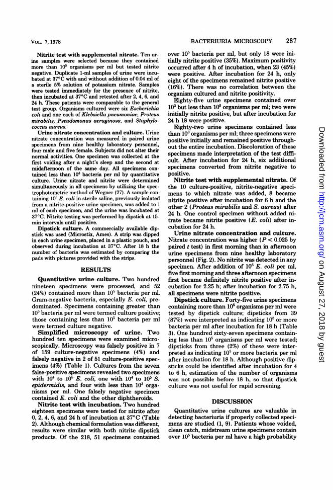
Quantitative urine culture. Two hundrednineteen specimens were processed, and 52(24%) contained more than 105 bacteria per ml.Gram-negative bacteria, especially E. coli, pre-dominated. Specimens containing greater than105 bacteria per ml were termed culture positive;those containing less than 105 bacteria per mlwere termed culture negative.Simplified microscopy of urine. Two
hundred ten specimens were examined micro-scopically. Microscopy was falsely positive in 7of 159 culture-negative specimens (4%) andfalsely negative in 2 of 51 culture-positive spec-imens (4%) (Table 1). Cultures from the seven
false-positive specimens revealed two specimenswith 104 to 105 E. coli, one with 104 to 105 S.epidermidis, and four with less than 103 orga-nisms per ml. One falsely negative specimencontained E. coli and the other diphtheroids.
Nitrite test with incubation. Two hundredeighteen specimens were tested for nitrite after0, 2, 4, 6, and 24 h of incubation at 37°C (Table2). Although chemical formulation was different,results were similar with both nitrite dipstickproducts. Of the 218, 51 specimens contained
over 105 bacteria per ml, but only 18 were ini-tially nitrite positive (35%). Maximum positivityoccurred after 4 h of incubation, when 23 (45%)were positive. After incubation for 24 h, onlyeight of the specimens remained nitrite positive(16%). There was no correlation between theorganism cultured and nitrite positivity.
Eighty-five urine specimens contained over103 but less than 105 organisms per ml; two wereinitially nitrite positive, but after incubation for24 h 18 were positive.
Eighty-two urine specimens contained lessthan 103 organisms per ml; three specimens werepositive initially and remained positive through-out the entire incubation. Discoloration of thesespecimens made interpretation of the test diffi-cult. After incubation for 24 h, six additionalspecimens converted from nitrite negative topositive.
Nitrite test with supplemental nitrate. Ofthe 10 culture-positive, nitrite-negative speci-mens to which nitrate was added, 8 becamenitrite positive after incubation for 6 h and theother 2 (Proteus mirabilis and S. aureus) after24 h. One control specimen without added ni-trate became nitrite positive (E. coli) after in-cubation for 24 h.Urine nitrate concentration and culture.
Nitrate concentration was higher (P < 0.025 bypaired t test) in first morning than in afternoonurine specimens from nine healthy laboratorypersonnel (Fig. 2). No nitrite was detected in anyspecimen. After addition of 106 E. coli per ml,five first morning and three afternoon specimensfirst became definitely nitrite positive after in-cubation for 2.25 h; after incubation for 2.75 h,all specimens were nitrite positive.Dipstick culture. Forty-five urine specimens
containing more than 105 organisms per ml weretested by dipstick culture; dipsticks from 39(87%) were interpreted as indicating 105 or morebacteria per ml after incubation for 18 h (Table3). One hundred sixty-seven specimens contain-ing less than 105 organisms per ml were tested;dipsticks from three (2%) of these were inter-preted as indicating 105 or more bacteria per mlafter incubation for 18 h. Although positive dip-sticks could be identified after incubation for 4to 6 h, estimation of the number of organismswas not possible before 18 h, so that dipstickculture was not useful for rapid screening.
DISCUSSIONQuantitative urine cultures are valuable in
detecting bacteriuria if properly collected speci-mens are studied (1, 9). Patients whose voided,clean catch, midstream urine specimens containover 105 bacteria per ml have a high probability
VOL. 7, 1978
on August 27, 2018 by guest
http://jcm.asm
.org/D
ownloaded from
* .., WP, . ' A, ., b .,_4. w * S{> 5 * > sx; wP ?;-.,a... ,8 /qft.. , . , .... .. ; .,. . , < .s,
S ::
.:. ':
; i;
*e'k'M
...,.~~ ~ ~ ~ ~ ~ ~ ~ ~ ~
:~ ~ ~ ~ ~4
* .U2.....
: X.
am~~~c.. ., < . sf4i:p
on August 27, 2018 by guest
http://jcm.asm
.org/D
ownloaded from

VOL. 7, 1978
TABLE 1. Urine microscopy and urine culture
Bacterial No. of cul- Microscopy results (%)concn (orga- tures exam-nism/ml) in ined Positive Negative
samples tested
>105 51 49 (96) 2 (4)<105 159 7 (4) 152 (96)
TABLE 2. Presence of nitrite in urine samples ofvarying bacterial concentrations
No. (%) of nitrite-positive samples by concn:Incubation (h)
>105/ml 103 to 105/ml <103/ml
0 18 (35) 2 (2) 3 (4)4 23 (45) 4 (5) 3 (4)
24 8 (16) 18 (21) 9 (11)
of having significant bacteriuria, whereas thosewhose specimens contain fewer than 105 bacteriaper ml do not. However, since 24 h is required toobtain results from quantitative urine culture,more rapid methods have been sought to screenfor the presence of urinary bacteria.The results of urine microscopy have been
reported using stained, unspun urine (3, 9),stained sediment (13, 23), or both (16, 26, 28).Robins et al. studied unstained urine utilizing acounting chamber (22). None of these reportsstates exact criteria for positive microscopy. Us-ing these methods, processing is time-consumingand may complicate interpretation, since it ismore difficult to separate bacteria from amor-phous debris in sediment, and staining proce-dures introduce the possibility of false negativeresults, from lost bacteria if the specimen isinadequately fixed on the slide, and false posi-tive results, from artifacts or contaminatedstains. We have demonstrated that a new sim-plified urine microscopy technique, omitting theuse of stains, centrifuges, or counting chambers,is remarkably accurate for rapid screening forbacteriuria in random urine specimens. Thismethod might also be useful for determiningresponse to therapy and could replace the moreexpensive quantitative urine culture for this pur-pose.
Nitrite is not normally detected in humanurine, but bacteria have enzymes capable ofreducing urinary nitrate to nitrite. Cruickshankand Moyes were the first to report urinary nitriteas a marker for urinary bacteria, but found ni-
BACTERIURIA MICROSCOPY 289
15
:~10
0
54
0
4,
4J
z
0.
FIG. 2. Urinary nitrate concentration in pairedfirst morning voided urine (0) and afternoon voidedurine (0) from nine healthy individuals.
trite was not present in all infected urine speci-mens (5). This may be because not all bacteriareduce nitrate, e.g., enterococci, acinetobacter,or alcaligenes. Sleigh improved the accuracy ofthe test by adding nitrate and incubating sam-ples for 6 h before testing for nitrite (25). Czer-winski et al. first observed that nitrite testingwas more sensitive in infected first morningvoided specimens than in random voided speci-mens (6). This difference in nitrite sensitivitydepending upon timing of specimen collectionhas been confirmed by Craig et al. (4) and ex-plains why some investigators have had goodresults (21) and others poor results (20) whenusing urinary nitrite as a marker for bacteriuria.
In an effort to increase the sensitivity of nitritetesting in random voided urine specimens, whichare more practical for screening, to the levelreported in first morning voided specimens, we
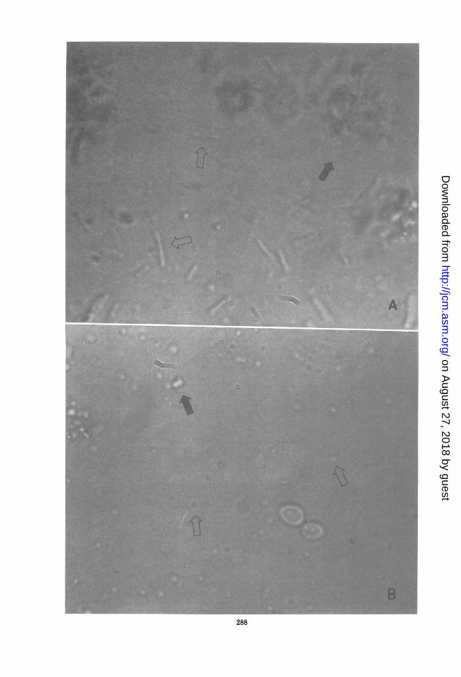
FIG. 1. Uncentrifuged, unstained urine (x1000) containing (A) E. coli and (B) Streptococcus faecalis.Organisms are marked by open arrows, amorphous crystalline material by closed arrows. Notice multipleplanes offocus in wetpreparations and smoothness of bacterial outline. Crystalline material is more refractilethan bacteria. Note yeast in bottom photograph.
on August 27, 2018 by guest
http://jcm.asm
.org/D
ownloaded from

290 BARBIN, THORLEY, AND REINARZ
TABLE 3. Dipstick culture techniqueBacterial Interpreted dipstick results
concn (orga- (:nisms/ml) in
samples >105/ml <105/mltested
>105 39 (87) 6 (13)<10-5 3 (2) 164 (98)
utilized two techniques, incubation at 37°C andaddition of nitrate substrate. Incubation of spec-imens at 37°C failed to increase the sensitivityof the nitrite test, suggesting that, contrary tothe manufacturers' suggestion (package inserts),the difference in sensitivity between random andfirst morning specimens is not due to durationof incubation of urine within the bladder. Sup-plemental nitrate converted culture-positive, ni-trite-negative specimens to positive during in-cubation at 37°C, confirming Sleigh's results(25). Although nitrite testing may be useful inselected patients using first morning voidedspecimens (14), addition of nitrate followed byincubation of random voided specimens is cum-bersome and negates the usefulness for office orhospital bacteriuria screening.
Craig et al. (4) and Moffat et al. (20) havereported good results from using the dipstickculture test to screen for bacteriuria. While our
results were similar, our purpose in using thistest was to determine whether it might beadapted for rapid screening. The manufacturerrecommends interpretation of the dipstick afterincubation for 12 to 18 h, but we hoped thatinterpretation might be possible after a shorterincubation. This was not possible. The timerequired for incubation limits the usefulness ofthe dipstick culture test for rapid bacteriuriascreening.
In summary, we found neither the urinarynitrite test nor the dipstick culture techniqueuseful for rapid detection of significant bacteri-uria in random urine specimens. However, sim-plified microscopic detection of bacteria in un-
stained, uncentrifuged urine accurately detectedbacteriuria. We suggest that when beginningmicroscopy, and periodically thereafter, a labo-ratory should compare the results of urine mi-croscopy with simultaneous quantitative cul-tures. Additionally, including known positives inmicroscopy studies, such as frozen diluted brothcultures or quantities of positive urine, wouldserve as a control to ensure that bacteria can berecognized. We suggest that both E. coli andenterococci be used, since cocci are more difficultto identify than rods. Since microscopy is quickand inexpensive and can be applied to random,clean catch, midstream urine samples, the new
technique of simplified urine microscopy ap-pears to be the best screening technique forbacteriuria currently available.
ACKNOWLEDGMENTS
The Clinical Microbiology Laboratory, supervised byElaine C. Tacquard, helpfully provided urine specimens. Dip-sticks were kindly donated by Warner-Chilcott Laboratoriesand the Ames Co. We express appreciation to Nancy Millsapsfor technical assistance, to Susan Menotti, Valerie Williams,and Bonnie Rhew for manuscript preparation, and to MichaelJ. Megna for expert photographic assistance.
LITERATURE CITED1. Boshell, B. R., and J. P. Sanford. 1958. A screening
method for the evaluation of urinary tract infections infemale patients without catheterization. Ann. Intern.Med. 48:1040-1045.
2. Brenner, B., and V. E. Gilbert. 1963. Elevated levels oflactic dehydrogenase, glutamicoxalacetic transaminase,and catalase in infected urine. Am. J. Med. Sci.245:31-42.
3. Bulger, R. J., and W. W. Kirby. 1963. Simple tests forsignificant bacteriuria. Arch. Intern. Med. 112:742-746.
4. Craig, W. A., C. M. Kunin, and J. DeGroot. 1973.Evaluation of new urinary tract infection screeningdevices. Appl. Microbiol. 26:196-201.
5. Cruickshank, J., and J. M. Moyes. 1914. The presenceand significance of nitrates in the urine. Br. Med. J.2:712-713.
6. Czerwinski, A. W., R. G. Wilkerson, J. A. Merrill, B.Braden, and J. P. Colmore. 1971. Further evaluationof the Griess test to detect significant bacteriuria. Am.J. Obstet. Gynecol. 110:677-681.
7. Edwards, B., R. H. R. White, H. Maxted, I. Deverill,and P. A. White. 1975. Screening methods for covertbacteriuria in schoolgirls. Br. Med. J. 1:463-467.
8. Gillenwater, J. Y. 1975. Diagnosis of urinary tract infec-tion: appraisal of diagnostic procedures. Kidney Int.8:S3-S1 1.
9. Kass, E. H. 1956. Asymptomatic infections of the urinarytract. Trans. Assoc. Am. Physicians 69:56-63.
10. Kass, E. H. 1962. Prevention of apparently non-infectiousdisease by detection and treatment of infections of theurinary tract. J. Chronic Dis. 15:665-673.
11. Kass, E. H. 1962. Pyelonephritis and bacteriuria. Ann.Intern. Med. 56:46-53.
12. Kincaid-Smith, P., M. Bullen, J. Mills, V. Fussell, N.Huston, and F. Goon. 1964. The reliability of screen-ing tests for bacteriuria in pregnancy. Lancet ii:61-62.
13. Kunin, C. M. 1961. The quantitative significance of bac-teria visualized in the unstained urinary sediment. N.Engl. J. Med. 265:589-592.
14. Kunin, C. M., and J. DeGroot. 1975. Self-screening forsignificant bacteriuria: evaluation of a dip-strip combi-nation nitrite/culture test. J. Am. Med. Assoc.231:1349-1353.
15. Kunin, C. M., E. Zacha, and A. J. Paquin. 1962. Urinarytract infections in schoolchildren. I. Prevalence of bac-teriuria and associated urologic findings. N. Engl. J.Med. 266:1287-1296.
16. Lewis, J. F., and J. Alexander. 1976. Microscopy ofstained urine smears to determine the need for quanti-tative culture. J. Clin. Microbiol. 4:372-374.
17. Lie, J. T. 1968. Evaluation of a nitrite test-kit (stat-test)for the detection of significant bacteriuria. J. Clin. Pa-thol. 21:443-444.
18. McGeachie, J., and A. C. Kennedy. 1963. Simplifiedquantitative methods for bacteriuria and pyuria. J. Clin.Pathol. 16:32-38.
19. Matsaniotis, N., C. Danelatou-Athanassiadou, C. Ka-
J. CLIN. MICROBIOL.
on August 27, 2018 by guest
http://jcm.asm
.org/D
ownloaded from

BACTERIURIA MICROSCOPY 291
terelos, P. Hartokalis, and E. Apostolopoulou.1971. Low urinary glucose concentration: reliable index.of urinary tract infection. J. Pediatr. 78:851-858.
20. Moffat, C. M., M. R. Britt, and J. P. Burke. 1974.Evaluation of miniature test for bacteriuria using de-hydrated media and nitrite pads. Appl. Microbiol.28:95-99.
21. Randolph, M. F., and K. Morris. 1974. Instant screeningfor bacteriuria in children: analysis of a dipstick. J.Pediatr. 84:246-248.
22. Robins, D. G., R. H. R. White, K. B. Rogers, and M.S. Osman. 1975. Urine microscopy as an aid to detec-tion of bacteriuria. Lancet i:476-478.
23. Sacks, T. G., and J. H. Abramson. 1967. Screening testsfor bacteriuria: a validity study. J. Am. Med. Assoc.
201:1-4.24. Savage, W. E., S. N. Hajj, and E. H. Kass. 1967.
Demographic and prognostic characteristics of bacteri-uria in pregnancy. Medicine 46:385-407.
25. Sleigh, J. D. 1965. Detection of bacteriuria by a modifi-cation of the nitrite test. Br. Med. J. 1:765-767.
26. Thysell, H. 1969. Evaluation of chemical and microscop-ical methods for mass detection of bacteriuria. ActaMed. Scand. 186:393-400.
27. Wegner, T. N. 1972. Simple and sensitive procedure fordetermining nitrate and nitrite in mixtures of biologicalfluids. J. Dairy Sci. 55:642-644.
28. Zinner, S. H., and E. H. Kass. 1971. Long-term (10 to 14years) follow up of bacteriuria of pregnancy. N. Engl. J.Med. 285:820-824.
VOL. 7, 1978
on August 27, 2018 by guest
http://jcm.asm
.org/D
ownloaded from