Simian Virus 40 Origin DNA-Binding Domain on Large T Antigen
15
Vol. 57, No. 1 JOURNAL OF VIROLOGY, Jan. 1986, p. 50-64 0022-538X/86/010050-15$02.00/0 Copyright ©D 1986, American Society for Microbiology Simian Virus 40 Origin DNA-Binding Domain on Large T Antigen EVA PAUCHA,t* DANIEL KALDERON,t ROBERT W. HARVEY,§ AND ALAN E. SMITH§ Biochemistry Division, National Institute for Medical Research, Mill Hill, London NW7 JAA, United Kingdom Received 28 May 1985/Accepted 3 September 1985 Fifty variant forms of simian virus 40 (SV40) large T antigen bearing point, multiple point, deletion, or termination mutations within a region of the protein thought to be involved in DNA binding were tested for their ability to bind to SV40 origin DNA. A number of the mutant large T species including some with point mutations were unable to bind, whereas many were wild type in this activity. The clustering of the mutations that are defective in origin DNA binding both reported here and by others suggests a DNA-binding domain on large T maps between residues 139 and approximately 220, with a particularly sensitive sequence between amino acids 147 and 166. The results indicate that the domain is involved in binding to both site I and site II on SV40 DNA, but it remains unclear whether it is responsible for binding to cellular DNA. Since all the mutants retain the ability to transform Rat-1 cells, we conclude that the ability of large T to bind to SV40 origin DNA is not a prerequisite for its transforming activity. Simian virus 40 (SV40) large T antigen, the product of the viral A gene, is required for replication of the virus as well as for the transformation of nonpermissive cells in culture (59). A number of biochemical activities have been shown to be intrinsic to the protein including an ATPase activity (4, 58) and the ability to form complexes with the host phosphopro- tein p53 (24, 26). Large T has also been shown to bind with high affinity to viral (8, 10, 31, 55-57) and cellular (2, 11, 41) DNA sequences. The role of the DNA-binding activity in both replicative and transforming functions of large T has been widely investigated. From analysis of a number of mutants isolated in several laboratories, it appears that the binding of large T to SV40 origin DNA is necessary (30, 32, 50, 54) though not sufficient (5, 29, 43) for replication, but not essential for efficient transformation of cells in culture (29, 43, 54). In mapping the domain of large T involved in DNA binding, two types of activity have been measured in dif- ferent studies: the ability to bind to cellular double-stranded (ds) DNA-cellulose (2, 11, 41) and the ability to bind to SV40 origin DNA sequences (8, 10, 31, 55-57). It is not yet clear whether these assays measure the activity of one or more domains. The variants used to map the DNA-binding domain include: (i) fragments of large T produced by in vitro translation of unspliced SV40 cRNA (42); (ii) various trun- cated forms of large T present in different SV40-transformed cell lines (3, 46); (iii) various chimeric proteins coded for by adeno-SV40 hybrid viruses containing defined portions of large T (33, 41, 57); (iv) defective large T species isolated from permissive or nonpermissive transformed cells (12, 28, 29, 43, 49, 54); (v) pseudorevertant forms of large T that are able to replicate from a defective origin sequence (30, 50); (vi) proteolytic fragments of wild-type large T (34); and (vii) large T variants encoded by constructed SV40 A gene deletion mutants (5, 39). Almost all of the data are consistent in suggesting that amino acids between 83 (corresponding to * Corresponding author. t Present address: Dana-Farber Cancer Institute, Boston, MA 02115. t Present address: Department of Biochemistry, University of California, Berkeley, CA 94720. § Present address: Integrated Genetics, Framingham, MA 01701. the splice site in the large T mRNA) and about 270 constitute part, if not all of a DNA-binding domain. We have previously reported the isolation of over 50 large T variants, most of which carry mutations in the DNA coding for amino acids 106 to 158 of large T (19). Almost all of the mutants retain the ability to transform Rat-1 cells, but most are reduced or defective in their ability to replicate. Some of the mutations affect residues that are normally phosphorylated in vivo, and others are located within a sequence that is important in localizing large T to the nucleus (17, 18, 25). Some of the mutants isolated had a super- transforming activity in that they produced transformed foci in greater number and more rapidly than wild-type large T. A further series of mutants have been produced by introducing termination codons at different positions throughout the large T coding region (18; B. Roberts, unpublished data). Here we report the DNA-binding properties of the large T species present in cells transformed by the different large T variants we have isolated. The data show clearly that point, multiple point, deletion, and termination mutations within the target area used here can abolish the ability to bind to SV40 origin DNA sequences, as measured by an immuno- precipitation assay. This, together with data from other laboratories, allows us to define precisely those amino acids that constitute the amino-terminal portion of a DNA-binding domain on large T. The data also confirm that SV40 large T does not require the ability to bind to SV40 origin DNA to transform established cells. It remains to be seen whether the ability to bind to cellular DNA plays a part in this process. MATERIALS AND METHODS Cells. All cells were maintained in Dulbecco modified Eagle medium supplemented with 10% fetal calf serum. The rat cells (RE52) transformed by microinjected Taq-BamHI fragment of SV40 large T, called 70K, were the gift of A. Graessman. SVA31E7 cells, SV40-transformed mouse cells, have been described previously (13, 38). The generation of cell lines transformed by each of the mutant DNAs has been described in detail previously (19). The wild-type SV40-transformed rat line used in these experiments was generated as described (19) by transfection with the wild-type SV40-containing plasmid pPVU-0. Cell lines transformed by the termination 50
Transcript of Simian Virus 40 Origin DNA-Binding Domain on Large T Antigen

Vol. 57, No. 1JOURNAL OF VIROLOGY, Jan. 1986, p. 50-640022-538X/86/010050-15$02.00/0Copyright ©D 1986, American Society for Microbiology
Simian Virus 40 Origin DNA-Binding Domain on Large T AntigenEVA PAUCHA,t* DANIEL KALDERON,t ROBERT W. HARVEY,§ AND ALAN E. SMITH§
Biochemistry Division, National Institute for Medical Research, Mill Hill, London NW7 JAA, United Kingdom
Received 28 May 1985/Accepted 3 September 1985
Fifty variant forms of simian virus 40 (SV40) large T antigen bearing point, multiple point, deletion, ortermination mutations within a region of the protein thought to be involved in DNA binding were tested fortheir ability to bind to SV40 origin DNA. A number of the mutant large T species including some with pointmutations were unable to bind, whereas many were wild type in this activity. The clustering of the mutationsthat are defective in origin DNA binding both reported here and by others suggests a DNA-binding domain onlarge T maps between residues 139 and approximately 220, with a particularly sensitive sequence betweenamino acids 147 and 166. The results indicate that the domain is involved in binding to both site I and site IIon SV40 DNA, but it remains unclear whether it is responsible for binding to cellular DNA. Since all themutants retain the ability to transform Rat-1 cells, we conclude that the ability of large T to bind to SV40 originDNA is not a prerequisite for its transforming activity.
Simian virus 40 (SV40) large T antigen, the product of theviral A gene, is required for replication of the virus as well asfor the transformation of nonpermissive cells in culture (59).A number of biochemical activities have been shown to beintrinsic to the protein including an ATPase activity (4, 58)and the ability to form complexes with the host phosphopro-tein p53 (24, 26). Large T has also been shown to bind withhigh affinity to viral (8, 10, 31, 55-57) and cellular (2, 11, 41)DNA sequences. The role of the DNA-binding activity inboth replicative and transforming functions of large T hasbeen widely investigated. From analysis of a number ofmutants isolated in several laboratories, it appears that thebinding of large T to SV40 origin DNA is necessary (30, 32,50, 54) though not sufficient (5, 29, 43) for replication, but notessential for efficient transformation of cells in culture (29,43, 54).
In mapping the domain of large T involved in DNAbinding, two types of activity have been measured in dif-ferent studies: the ability to bind to cellular double-stranded(ds) DNA-cellulose (2, 11, 41) and the ability to bind to SV40origin DNA sequences (8, 10, 31, 55-57). It is not yet clearwhether these assays measure the activity of one or moredomains. The variants used to map the DNA-binding domaininclude: (i) fragments of large T produced by in vitrotranslation of unspliced SV40 cRNA (42); (ii) various trun-cated forms of large T present in different SV40-transformedcell lines (3, 46); (iii) various chimeric proteins coded for byadeno-SV40 hybrid viruses containing defined portions oflarge T (33, 41, 57); (iv) defective large T species isolatedfrom permissive or nonpermissive transformed cells (12, 28,29, 43, 49, 54); (v) pseudorevertant forms of large T that areable to replicate from a defective origin sequence (30, 50);(vi) proteolytic fragments of wild-type large T (34); and (vii)large T variants encoded by constructed SV40 A genedeletion mutants (5, 39). Almost all of the data are consistentin suggesting that amino acids between 83 (corresponding to
* Corresponding author.t Present address: Dana-Farber Cancer Institute, Boston, MA
02115.t Present address: Department of Biochemistry, University of
California, Berkeley, CA 94720.§ Present address: Integrated Genetics, Framingham, MA 01701.
the splice site in the large T mRNA) and about 270 constitutepart, if not all of a DNA-binding domain.We have previously reported the isolation of over 50 large
T variants, most of which carry mutations in the DNAcoding for amino acids 106 to 158 of large T (19). Almost allof the mutants retain the ability to transform Rat-1 cells, butmost are reduced or defective in their ability to replicate.Some of the mutations affect residues that are normallyphosphorylated in vivo, and others are located within asequence that is important in localizing large T to the nucleus(17, 18, 25). Some of the mutants isolated had a super-transforming activity in that they produced transformed fociin greater number and more rapidly than wild-type large T. Afurther series of mutants have been produced by introducingtermination codons at different positions throughout thelarge T coding region (18; B. Roberts, unpublished data).Here we report the DNA-binding properties of the large Tspecies present in cells transformed by the different large Tvariants we have isolated. The data show clearly that point,multiple point, deletion, and termination mutations withinthe target area used here can abolish the ability to bind toSV40 origin DNA sequences, as measured by an immuno-precipitation assay. This, together with data from otherlaboratories, allows us to define precisely those amino acidsthat constitute the amino-terminal portion of a DNA-bindingdomain on large T. The data also confirm that SV40 large Tdoes not require the ability to bind to SV40 origin DNA totransform established cells. It remains to be seen whetherthe ability to bind to cellular DNA plays a part in thisprocess.
MATERIALS AND METHODS
Cells. All cells were maintained in Dulbecco modified Eaglemedium supplemented with 10% fetal calf serum. The rat cells(RE52) transformed by microinjected Taq-BamHI fragmentof SV40 large T, called 70K, were the gift of A. Graessman.SVA31E7 cells, SV40-transformed mouse cells, have beendescribed previously (13, 38). The generation of cell linestransformed by each of the mutant DNAs has been describedin detail previously (19). The wild-type SV40-transformed ratline used in these experiments was generated as described(19) by transfection with the wild-type SV40-containingplasmid pPVU-0. Cell lines transformed by the termination
50

SV40 ORIGIN DNA-BINDING DOMAIN 51
mutants H23, dH, and HHpa and by the deletion mutantsRL75-RL88 and RL12-S24 were also generated by transfec-tion of Rat-1 cells with plasmid DNAs by the procedurepreviously described (19). The construction of the termina-tion mutants is given in Kalderon et al. (18). The deletionmutants were created by ligating BamHI-EcoRI-cut DNAfrom the appropriate pairs of linker insertion mutants (19).
Labeling and extraction of cells. The procedures for label-ing and extraction of cells were carried out essentially asdescribed previously (38, 51, 52). Cells were labeled withmethionine for 5 h by replacing the medium on confluentmonolayers of cells growing on 50- or 90-mm dishes (Nunc,Roskilde, Denmark) with growth medium minus methioninesupplemented with 1% complete medium (containing 10%fetal calf serum) and 200 ,uCi of [35S]methionine (specificactivity, >1,000 Ci/mmol; Amersham International) per ml.32P04 labeling was for 2 h in growth medium minus phos-phate containing 250 ,uCi of 32p, (Amersham International)per ml. After labeling, the cells were rinsed twice withice-cold Tris-buffered saline and then lysed by the additionof 1 ml/90-mm dish or 0.5 ml/50-mm dish of cell extractionbuffer containing 50 mM Tris (pH 8.0), 120 mM NaCl, and0.5% Nonidet P-40 (NP-40). The samples were spun at10,000 rpm in an Eppendorf microcentrifuge for 10 min at4°C, and the supernatants were used directly or stored at-700C.Immunoprecipitation of cell extracts. Samples of lysates
containing 3 x 10⁶ trichloroacetic acid-precipiptable 35S cpm(generally corresponding to 40 to 100 ,ul of extract) weremixed with 0.5 ml of buffer containing 20 mM Tris (pH 7.0),100 mM NaCl, 2 mM dithiothreitol, 1 mM EDTA and 0.5%NP-40. Portions (10 ,ul) of anti-SV40 tumor cell serum wereadded, and the mixture was incubated for 60 min at 18 to20°C. A 10% suspension of washed Staphylococcus aureusbacteria (50 ,lI) was added, and incubation continued for 10min. The bacterial pellet was washed twice in a buffercontaining 20 mM Tris (pH 8.0), 2 mM dithiothreitol, 1 mMEDTA, 0.5% NP-40, and 0.5 M NaCl. After a third wash inthe same buffer containing 0.1 M NaCl, the bound proteinswere eluted with gel-loading buffer (0.0625 M Tris [pH 6.8],2% sodium dodecyl sulfate, 10% glycerol, 0.1 M dithiothrei-tol, 0.01% bromophenol blue). Sample preparation and poly-acrylamide gel electrophoresis were done as described pre-viously (51, 52). Gels containing [35S]methionine-labeledsamples were soaked for 1 h in 10 volumes of 1 M sodiumsalicylate before being dried. Autoradiography was for 1 to 4days at -700C on Fuji Rx film with an Ilford fast tungstateintensifying screen.SV40 origin binding. Four DNAs were used in these
experiments. pSV328 (a gift from G. C. Grosveld) containsthe larger BamHI-EcoRI fragment of SV40 inserted betweenthe BamHI and EcoRI sites of pBR322. RL18 and 034 areboth derivatives of pPVU-0 (16, 19), a plasmid that containsthe BamHI to PvuII early-region fragment of SV40 (SVSstrain) between the BamHI and PvuII sites in pBR328. RL18has an EcoRI linker (CGGAATTCCG) inserted betweenpositions 5194 and 5186, while 034 contains the linker inplace of SV40 sequences between positions 5215 and 4739.These DNAs were digested with the restriction enzymeBstNI, and in the case of RL18 also with EcoRI. Therestricted DNAs were treated with calf intestinal phospha-tase (Boehringer Mannheim Biochemicals, Indianapolis,Ind.), and then end-labeled with [-y-32PIATP (specific activ-ity, 5 to 7,000 Ci/mmol; Amersham International) and T4polynucleotide kinase by the method of Maniatis et al. (27).The specific activity of DNA obtained was generally be-
tween 1 x 10⁷ and 3 x 10⁷ cpm/,ug. The DNA bindingprocedure, essentially that of McKay (31), has been de-scribed in detail previously (38). In these experiments,samples of cell extracts containing equivalent amounts of35S-labeled large T, as judged from experiments such as thatshown in Fig. 1, generally 40 to 100 ,ul, were made up to 100,ul with extraction buffer and mixed with 0.4 ml of bindingbuffer (20 mM Tris [pH 7.0], 1 mM EDTA, 2.5 mM dithio-threitol, 125 ,ug of bovine serum albumin, 0.05% NP-40) and10 ng of the appropriate labeled DNA. After incubation at18°C for 40 to 60 min, 5 p.l of a mixture of the monoclonalantibodies pAb419 and pAb413 was added. Incubation wascontinued for a further 40 to 60 min, and 25 ,u1 of washed S.aureus bacteria was added. After an additional 5 to 10-minincubation, the pellets were washed three times in bindingbuffer without bovine serum albumin containing 0.2 M NaCl.The DNA was eluted in 15 p.1 of gel loading buffer (10 mMTris [pH 7.5], 10 mM EDTA, 2% sodium dodecyl sulfate)and loaded directly onto 2% agarose gels in Tris-acetatebuffer (27). After electrophoresis, the gels were fixed in 2volumes of ethanol before drying. Autoradiography wasgenerally for 1 to 2 days.
Binding to ds calf thymus DNA immobilized on cellulose.Confluent monolayers of cells on 50-mm dishes (Nunc) werelabeled with 32p, for 2 h as described above. The monolayerswere washed with Tris-saline and lysed in 200 ,ul of buffer A(10 mM HEPES[N-2-hydroxyethylpiperazine-N'-2-ethane-sulfonic acid], 1 mM MgCI2, 0.5% NP-40, 0.3 mg ofphenylmethylsulfonyl fluoride per ml) containing 0.45 MNaCl. The lysate was removed and immediately diluted bythe addition of 2 volumes of buffer without salt. The pH wasthen adjusted to 6.5 by the addition of 0.01 M HCI, and theextract was mixed with 0.5 ml of packed native calf thymusDNA-cellulose (P-L Biochemicals, Inc., Milwaukee, Wis.)which had been equilibrated in buffer A at pH 6.5 with 0.15M NaCl. The mixture was kept at 4°C for 1 h on a rotarymixer. The DNA-cellulose was then pelleted by centrifuga-tion for 1 min at 4°C in an Eppendorf microcentrifuge, andthe supernatant was removed. The DNA-cellulose pellet waswashed three times with buffer A at pH 6.5 with 0.15 M NaCland then incubated for 1 h at 4°C with 0.5 ml of buffer A atpH 8.5 with 0.15 M NaCl to elute bound protein. After threewashes with this buffer, a second elution step was thencarried out with buffer A at pH 8.5 with 1.0 M NaCl. Both ofthe eluted fractions and 20-,ul samples of starting materialand of the unbound fraction were adjusted to pH 8.5 and 0.5M NaCl before overnight incubation with 10 p.1 of anti-SV40tumor cell serum. Immunoprecipitates were then collected,washed, and analyzed as described above.
RESULTSAs part of our efforts to assign biochemical functions to
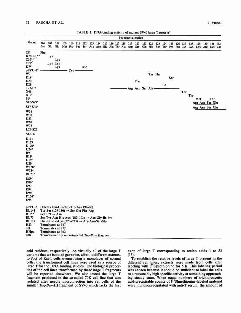
domains of large T, we examined many of the large Tvariants we have isolated (19) for their ability to bind toSV40 origin DNA. The mutants used fall into two groups.The larger group consists of those variants carrying pointmutations or small deletions in the DNA coding for aminoacids 106 to 158 of large T. The sequence of the SV40variants encoded by most of the mutants used has beenreported in detail (19) and is summarized in Table 1.The second group contains the so-called termination mu-
tants which encode truncated large T protein (17, 19;Roberts, unpublished data). Three of these have been usedin the studies reported here: H23, dH (previously referred toas AHindlIl [18]), and HHpa, which encode amino-terminalfragments of large T consisting of 147, 272, and 362 amino
VOL. 57, 1986
52 PAUCHA ET AL.
TABLE 1. DNA-binding activity of mutant SV40 large T proteinaSequence alteration
Mutant 106 107 108 109 110 111 112 113 114 115 116 117 118 119 120 121 122 123 124 125 126 127 128 129 130 131 132Ser Glu Glu Met Pro Ser Ser Asp Asp Glu Ala Thr Ala Asp Ser Gln His Ser Thr Pro Pro Lys Lys Lys Arg Lys Val
C8 PheK79(Kl)cd LysC37c,d LysC22d Lys LysK7d Lys AsnpPVU_1d TyrW7D19D20D29T23-L7X46X12esifS17-S29eS17-S36eW34W38U33W67W75L27-S26S1-S32DlllD119D1209U249B9cBllgU199U20W1289W134RL239D889D899D90D94D96cD104CD98
pPVU-2 Deletes Glu-Gln-Trp-Trp-Asn (92-96)RL148 Tyr-Ser (179-180) - Ser-Glu-Phe-ArgB18cd Ser 189 -. AsnRL75 Ser-Tyr-Asn-His-Asn (189-193) -- AsiRL115 Phe-Leu-Ile-Cys (220-223) -* Arg-AsrH23 Terminates at 147dH Terminates at 272HHpa Terminates at 36270K Transformed by microinjected Taq-Ba
Tyr Phe
PheIle
Arg Asn Ser Ala
Ser
ThrThr
Met ThrArg Asn Ser GluArg Asn Ser Glu
;n-Gly-Ile-Pron-Ser-Gly
rm fragment
acid residues, respectively. As virtually all of the large Tvariants that we isolated gave rise, albeit to different extents,to foci of Rat-1 cells overgrowing a monolayer of normalcells, the transformed cell lines were used as a source oflarge T for the DNA binding studies. The biological proper-ties of the cell lines transformed by these large T fragmentswill be reported elsewhere. We also tested the large Tfragment produced in the so-called 70K cell line that wasisolated after needle microinjection into rat cells of thesmaller Taq-BamHI fragment of SV40 which lacks the first
exon of large T corresponding to amino acids 1 to 82(13).To establish the relative levels of large T present in the
different cell lines, extracts were made from cells afterlabeling with [35Slmethionine for 5 h. This labeling periodwas chosen because it should be sufficient to label the cellsto a reasonably high specific activity at something approach-ing steady state. When equal numbers of trichloroaceticacid-precipitable counts of [35S]methionine-labeled materialwere immunoprecipitated with anti-T serum, the amount of
J. VIROL.

SV40 ORIGIN DNA-BINDING DOMAIN
TABLE 1-Continued
SV40DNA-
133 134 135 136 137 138 139 140 141 142 143 144 145 146 147 148 149 150 151 152 153 154 155 156 157 158 bindingGlu Asp Pro Lys Asp Phe Pro Ser Glu Leu Leu Ser Phe Leu Ser His Ala Val Phe Ser Asn Arg Thr Leu Ala Cys activityb
++
++++++
++
++++++
++
++++++
++
++++
LeuArg Asn Ser Val
LysLys
Ala Glu Phe Arg
AsnAsn
AsnAsnAsn
Tyr ValAsn
ThrVal
MetThr Met
Ile
MetPro Glu Phe Arg
ThrAsn Thr
TyrAsn
Asn Lys
Asn LysPhe
++
++
++
++
++
+
Tr+
++
(+)
(+)
++
++
Asn Thr
t+
(+)
+
++
a The alterations predicted in the amino acid sequence of the mutant large T proteins are indicated. The superscripts refer to the following properties of themutant plasmids as demonstrated by Kalderon and Smith (19). c, Mutant plasmids can replicate in CV1 cells as well as wild-type SV40, both as plasmids and as re-
constructed virus. d, Plasmids are defective in transformation. Fewer foci are produced per microgram of DNA with a longer latent period than with wild-typeDNA. e, Mutant large T is detected only in the cytoplasm by immunofluorescent staining.f, Mutant large T is found in both the cytoplasm and the nucleus of trans-formed cells. g, Mutant DNA produces more foci per microgram with a shorter latent period than does wild-type DNA. This phenotype has been calledsupertransforming (19).
b The ability of the mutant large Ts to bind SV40 origin DNA has been expressed as: + +, binding equivalent to wild type; +, decreased ability to bind; (+) weakbinding detected with very small site I-containing DNA fragments; -, no detectable binding under any conditions with any DNA tested.
LeuLys Asn
Asn
VOL. 57, 1986 53

54 PAUCHA ET AL.
labeled large T detected was reasonably consistent betweenthe different extracts, but some cells clearly containedsignificantly more than others (Fig. 1). Each of the cell lineswas developed from a randomly picked dense focus aftertransformation by each of the mutant DNAs under equiva-lent conditions. Differences in the amount of large T betweenindividual cell lines might therefore reflect their nonclonalnature. In addition, the stability of the mutant proteins or theextent to which the mutant proteins are expressed mightvary between the different transformed lines. Further exper-iments would be required to distinguish between thesepossibilities for individual mutants. It is striking, however,that all of the large T variants that contain mutations withinthe sequence 127-Lys-Lys-Lys-Arg-Lys-Val-132 are over-produced 6 to 20-fold relative to wild type. We have alreadyshown that these mutant large Ts are located entirely (X12,S17-S29, S17-S36) or partly (dl) in the cytoplasm of trans-formed or microinjected cells (17, 18). It is possible, there-fore, that the observed overproduction might be caused bythe inability of cytoplasmic large T to autoregulate its ownproduction. Lack of autoregulation might explain why someof the other large T species appear to be present in largeamounts.The large T fragments immunoprecipitated from Rat-1
cells transformed by the termination mutants H23, dH, andHHpa are also shown in Fig. 1. The apparent molecularweights of the 147, 272, and 362 amino acid fragments are14,000, 30,000, and 45,000, respectively. The large T foundin 70K cells is shown in Fig. 1. Since these rat cells lackcoding sequences corresponding to the first exon of large T,the large T variant expressed cannot include any of theresidues 1 through 82. It is thought that translation begins atthe first in-frame methionine of the large T gene, Met-109(13). The predicted molecular weight of the resulting proteinfragment is about 70,000; however, the mutant large Tmigrates with an apparent molecular weight of 84,000 (Fig.1). Although large T species are known to migrate anoma-lously on sodium dodecyl sulfate-polyacrylamide gels, somedoubt is cast on the exact nature of 70K large T as a result ofthe difference between the predicted and the observedmolecular weights.For the experiments described here we assumed that the
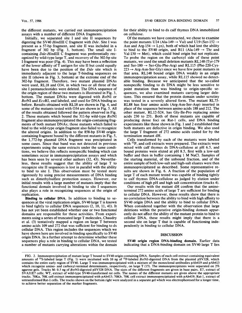
total amount of large T present in the different transformedcells was proportional to that detected after a 5-h labelingperiod. Samples of extracts of the different cell lines con-taining equivalent amounts of large T were incubated with amixture of 32P-end-labeled fragments of SV40 DNA whichhad been produced by cleavage with the restriction enzymeBstNI. Digestion with this enzyme produces among othersan SV40 fragment of 311 base pairs (bp) which contains allthree of the sites to which SV40 large T is known to bind (5,8, 9, 35, 55-57). After incubation for 60 min, any large Tbound to the SV40 DNA was recovered by immunoprecipi-tation with a mixture of the monoclonal antibodies PAb413and PAb419 which recognize determinants localized at thecarboxyl and the amino termini, respectively, of large T (15).The labeled DNA was then fractionated by agarose gelelectrophoresis. Figure 2 shows the results of the so-calledMcKay assay (31), using extracts from the cells shown inFig. 1. Some of the mutants coimmunoprecipitated wild-typelevels of the 311-bp BstNI fragment, whereas others hadreduced levels of binding and some gave none. Addition ofmore extract from cells which were negative for binding inthe experiment in Fig. 2 still failed to give any signal,emphasizing that the large T species present in these extractswere unable to recognize SV40 origin DNA in this assay. For
such mutants, DNA-binding activity was consistently absentin all independently derived transformed cell lines exam-ined.These data, together with the predicted amino acid se-
quences of the different mutant large Ts, are summarized inTable 1. These data show that the mutations which affect theability of large T to bind to SV40 origin-containing DNA aretightly clustered. Small deletions or point mutations up toand including amino acid 137 have little effect on the abilityof the mutated large T to bind to SV40 origin sequences. Thisincludes the region around the Lys-128 mutations whichproduces cytoplasmic large Ts (17, 25) and all of the phos-phorylation sites mapped at the amino terminus of large T(Ser-106, Ser-111, Ser-112, Ser-123, and Thr-124 [48, 60]).Mutation of Pro-139, however, or deletions encompassingthis residue result in large T which is defective in SV40 originbinding. Likewise, mutation of Ser-147 -- Asn (D119) ofAla-149 -- Val (U24) and of Ser-152+Arg-154 -* Asn+Lys(U19) all abolish the ability of large T to bind to SV40 originDNA. A number of multiple and deletion mutants encom-passing these residues were also negative for DNA binding.Other mutants in the region between residues 140 and 160had a severely reduced ability to bind to DNA. The cluster-ing of all of these mutations suggests that they all lie withina functional domain whose amino-terminal end is defined byPro-139.There are few mutants in our collection that map down-
stream of residue 158; consequently, mapping of the extentof the domain is difficult. However, two mutants in thedownstream sequences (RL148 and RL75) were reduced intheir origin-binding activity, and the mutant RL115 (whichhas altered amino acids 220 to 223) was unable to bind originDNA at all. This may mean that the domain extends at leastas far as amino acid 223.The data obtained with the other mutants tested is consis-
tent with this interpretation. The 70K large T, which lacks atleast amino acids 1 to 82, exhibited wild-type levels of originbinding (Fig. 2), indicating that the domain lies within thesecond exon of large T. The 147-amino acid fragment of largeT coded for by the termination mutant H23 failed to bindDNA in this assay (data not shown). dH large T, which is 272amino acids long, and therefore includes the sequencesmaking up the region defined above, did bind to origin DNA,though weakly. HHpa large T bound DNA better, perhapssuggesting that the conformation of the 362-amino acid-longfragment more closely resembles that of wild-type large Tthan do the shorter forms. Taken together, these datasupport the notion that a region involved in binding to SV40origin DNA begins at Pro-139 and extends at least as far asamino acid 223 of large T. Within this domain, a regionbetween residue 147 and 154 appears to be particularlysensitive to mutation.
Ability of the mutants to bind to the SV40 site II originregion. The EcoRII G fragment of SV40 DNA contains threesites to which large T is known to bind. Site II spans theorigin of SV40 DNA replication. Site I is located on the earlyside of the origin, and site III is on the late side. Binding oflarge T to site I is believed to block transcription of SV40early mRNA resulting in autoregulation of large T synthesis(1, 9, 44). Binding to site II is presumably essential for theinitiation of replication of viral DNA (8, 9, 50, 55, 56), whilethe consequences of site III binding are not yet understood.De Lucia et al. (8) have established that each site containsseveral copies of the consensus pentanucleotide 5'-GAGGC-3' which are protected from methylation by dimethylsulfatewhen large T is bound. The sequence of the SV40 origin-
J. VIROL.

SV40 ORIGIN DNA-BINDING DOMAIN
OnN 1-r-C½ CNJr~ r~J rN<WWCOCYCm CO j- ci W.- v- X q< n U
NCJ. > OOO 0) X)CD'0 C.q r N P.NNa.. N CCY3 CYCN t t ---CL2Q zC>)]C X X Xx lo CO
ora~~~0oo00~~~~-xcx~*,,,w'w w
gO
N rZ -IzI W N
* - S - Large-T.a
44=p53
a0
Small-t -
aw < inJ- 'f-NM 4 7
- v- - T- C-4 04 0)3: a O N:
_r Cm <:
m m:D
C: n
in ci c-i cmcD 0; a) o_r _r _r CM
N CY CN N
vNCo C c_ r-
- o c0oDo31,.QCi
6 t, Q o 0(ao ai)a c
X
kC]QC
cuJ N NThu (0%rN Ch_ - T-U) Cl) Cl) 00
t0 ., NNII 1-
i
- XCi COo teIONNN'-_J¢ -j -iwCcO
Large-T- * W. .0*S*4 " 0 *o s.0 *
*h *
p53 -
Small-t
FIG. 1. Immunoprecipitation of mutant cell extracts with SV40 anti-'1 serum. Kat-I cells transformed by each of the mutant DNAs werelabeled for 5 h with 100 ,Ci of [35S]methionine per ml, and cell extracts were prepared. Equal numbers of TCA-precipitable [35S]methioninecounts per minute of each extract were immunoprecipitated with hamster anti-SV40 tumor cell serum. The positions of large T, small t, andp53 are indicated. WT, wild-type SV40-transformed rat cell extract; E7, extract of the SV40-transforitied mouse cell line SVA31E7. Note thatmouse p53 migrates more rapidly in these gels than does rat p53 in the adjacent 70K track. The name of each mutant is given above theappropriate track. The predicted amino acid sequence of the large T encoded by the mutants is shown in Table 1. The letters a and b followingthe name indicate two separately prepared extracts of the same cell line, e.g., K79.21a and K79.21b. Different numbers following the nameindicate extracts prepared from cell lines derived from independent foci produced by transformation with the same DNA, e.g., D20.1 andD20.2. In two cases, D20.1 and S17-S36.1, no large T could be detected upon immunoprecipitation of the cell extracts. Further experimentswere carried out with cell lines derived from different foci (D20.2 and S17-S36.2) which did contain large T.
spanning regions I and II is shown in Fig. 3 (top) with therecognition pentanucleotide boxed. Measurements of therelative affinity of large T binding to the different sites at 4°Chave led to the conclusion that the protein binds most tightlyto the two perfect pentanucleotides in site I, then to the
sequences in region II, and lastly to region III (8). It is likelythat binding of large T to the 311-bp BstNI fragment in theimmunoprecipitation assay used here largely reflects bindingto the highest-affinity site I (32). In an attempt to assesswhether binding to site I and site II was affected in parallel in
.0
CM CM C
*a 0) cO Na3.(.
eIC
Large-T - . ,
p53 -
CU4
,
N _
eacc ii
.0 040.16.00* AMIJAL00 00
VOL. 57, 1986 55

56 PAUCHA ET AL.
co m lq* CYb- C4 N > a) o o:, cn A s ON0) COco N r 00 4 n
Y N C\Y < U) CO1772_404030404_- r '-CM <CON NO~r-~C'4 N N 4oc) 04Nr-'~~w~oe~~eoor a~~~~OoQQHx>c>CV00~6 CY
1772~~~Lj
1046-823-592-
444-311-249-200-
4111_a4 -0_00 C_ a*b Am
cddCO)04 \ _ _ 4 CO 04'- '-v-C/)COl~ co 0 <0 04 40 0
F~~~~~~~~~~~~T C\3 1< im C\ I\ C\j CX)o (,0Vi Ln I CO'- 0)O I ?-N (V, (QNNN T- - \ C -r S-0 ) - - CM lL 3i2 3 J cn) 0 c a D D CD Go co 0 0 D :D 3: 2
-o eo--
04CNJC\j N C\j~~~~~~~0£6Cj C) IN\ Njt L
04004000C)c) 0 t(D O 'r- aCX COC3M 0 o O o DO C0 0 m : -
C5
O 0Nl N cc~
.4
:Ipke_ _
_ _
823
-6 7 3,n. -5 9 2,-n - 5 5 2:g - 486
444
-31 1
-- 249
1 26
VP
CY)
000
m..I.
J. VIROL.
SV40 ORIGIN DNA-BINDING DOMAIN 57
the different mutants, we carried out immunoprecipitationassays with a number of different DNA fragments.
Initially, we separated site I and site II sequences bydigesting the SV40 HindlIl C fragment with Dde. Site I waspresent as a 57-bp fragment, and site II was included in afragment of 302 bp (Fig. 3, bottom). The small site I-containing Dde-HindIII C fragment was preferentially rec-ognized by wild-type large T, and recovery of the 302-bp siteI fragment was poor (Fig. 4). This may have been a reflectionof the lower affinity of T antigen for site II but could equallyhave been due to the position of the Dde site which isimmediately adjacent to the large T-binding sequences onsite II (shown in Fig. 3, bottom) at the extreme end of the302-bp fragment. Therefore, two mutant plasmid DNAswere used, RL18 and 034, in which two or all three of thesite I pentanucleotides were deleted. The DNA sequence ofthe origin region of these two mutants is illustrated in Fig. 3,bottom. The mutant DNAs were digested with BstNI orBstNI and EcoRI, end labeled, and used for DNA binding asbefore. Results obtained with RL18 are shown in Fig. 4, andsome of the mutants tested with 034 are shown in Fig. 5. Allof the results obtained followed the pattern observed in Fig.2. Those mutants which bound the 311-bp wild-type BstNIfragment also immunoprecipitated the origin-containing frag-ments of both mutant DNAs; those mutants which failed tobind to the wild-type origin fragment also failed to recognizethe altered origins. In addition to the 858-bp SV40 origin-containing fragment bound by the different mutants in Fig. 5,the 1,772-bp plasmid DNA fragment was precipitated insome cases. Since that band was not detected in previousexperiments using the same extracts under the same condi-tions, we believe this reflects nonspecific binding which cansometimes occur in immunoprecipitation assays and whichhas been seen by several other authors (32, 43). Neverthe-less, these results suggest that the ability of large T torecognize site II sequences is lost in parallel with the abilityto bind to site I. This observation must be tested morerigorously by using precise measurements of DNA bindingsuch as dimethylsulfate protection assays. However, ourinitial results with a large number of mutants imply that thefunctional domain involved in binding to site I sequencesalso plays a role in recognizing sequences at the origin ofreplication.
Binding to cellular DNA. In addition to binding to se-quences at the viral replication origin, SV40 large T is knownto bind tightly to cellular DNA sequences (2, 10, 11, 41). Ithas not yet been established whether one or two functionaldomains are responsible for these activities. From experi-ments using a series of truncated large T molecules, Chaudryet al. (3) tentatively mapped a region on large T betweenamino acids 109 and 272 that was sufficient for binding to dscellular DNA. This region includes the sequences which wehave shown here are involved in binding specifically to SV40origin DNA. In a further attempt to determine whether thesesequences play a role in binding to cellular DNA, we testeda number of mutants carrying alterations within the domain
for their ability to bind to ds calf thymus DNA immobilizedon cellulose.Of the mutants we have constructed, we chose to examine
the point mutants U24 (Ala-149 -) Val) and U19 (Ser-152 -Asn and Arg-154 -> Lys), both of which had lost the abilityto bind to the SV40 origin, and Bl1 (Ala-149 -- Thr andVal-150 -* Met), which could bind origin but not replicate.To probe the region on the carboxyl side of these pointmutants, we used the small deletion mutants RL148 (Tyr-179and Ser-180 -* Ser-Glu-Phe-Arg) and RL115 (Phe-220-Cys-223 -* Arg-Asn-Ser-Gly) since we have few point mutants inthat area. RL148 bound origin DNA weakly in an originimmunoprecipitation assay, while RL115 showed no detect-able binding. Because we anticipated that the so-callednonspecific binding to ds DNA might be less sensitive topoint mutation than was binding to origin-specific se-quences, we also examined mutants carrying larger dele-tions. This ensured that the protein domain under scrutinywas tested in a severely altered form. The mutant RL75-RL88 has four amino acids (Arg-Asn-Ser-Asp) inserted inplace of the sequence between amino acids 188 and 212. Theother mutant, RL12-S24, has Gly-Ile-Pro replacing aminoacids 230 to 251. Both of these mutants are capable ofproducing dense foci on Rat-1 cells, and DNA bindingexperiments like those shown in Fig. 1 and 2 established thatboth mutants are defective in origin binding. We also usedthe large T fragment of 272 amino acids coded for by thetermination mutant dH.
Cells transformed by each of the mutants were labeledwith 32p, and cell extracts were prepared. The extracts weremixed with calf thymus ds DNA-cellulose at pH 6.5, andbound proteins were eluted at pH 8.5, first with a low-saltbuffer and then in buffer containing 1 M NaCl. Samples ofthe starting material, of the unbound fraction, and of theentire sample of both low-salt and high-salt eluates were thenimmunoprecipitated as described. Some representative re-sults are shown in Fig. 6. A fraction of the population oflarge T of each mutant tested was capable of binding tightlyto calf thymus DNA-cellulose as judged by elution underconditions of high pH and high salt concentration (tracks 4).Our results with the mutant dH confirm that the amino-
terminal 272 amino acids of large T are sufficient for bindingto cellular DNA. However, these results show that there isno correlation between the ability to bind with high affinity toSV40 origin DNA and the ability to bind to cellular DNA.When considered together with the observation that largedeletions within the putative origin-binding domain appar-ently do not affect the ability of the mutant protein to bind tocellular DNA, these results might imply that there is asecond site on large T that is capable of functioning inde-pendently in binding to cellular DNA.
DISCUSSION
SV40 origin region DNA-binding domain. Earlier dataindicating that a DNA-binding domain on SV40 large T lies
FIG. 2. Immunoprecipitation of mutant large T bound to SV40 origin-containing DNA. Samples of each cell extract containing equivalentamounts of I'S-labeled large T (Fig. 1) were incubated with 10 ng of _P-labeled BstNI-digested DNA from the plasmid pSV328, whichcontains the entire early region of SV40. Large T was immunoprecipitated with a mixture of the monoclonal antibodies pAb419 and pAb413which recognize amino- and carboxyl-terminal determinants, respectively, on large T (15). The immunoprecipitates were separated on 2%agarose gels. Tracks M: 0.5 ng of BstNI-digested pSV328 DNA. The sizes of the different fragments are given in base pairs. E7, extract ofSVA31E7 cells; WT, extract of wild-type SV40-transformed rat cells. The names of the different mutants are given above the appropriatetracks. 70Ka, 70K cell extract immunoprecipitated with pAb413; 70Kb, 70K cell extract immunoprecipitated with pAb419; Rat-1, extract ofuntransformed Rat-1 cells. The tracks shown on the bottom right were analyzed in a separate gel which was electrophoresed for a longer time,to achieve better separation of the marker fragments.
VOL. 57, 1986

58 PAUCHA ET AL.
Site E Site I
4 3 2 1 3 2
ATTTATGCAAGGqCI AGAGCCTCCTCPCCCTCTGAGCTATTCCAGAAGTAGTGAGAGG TTTTTTGGAGGC;rkTGGqTTTTGCAAAAAT AA AT ACGTICTCCCiGGILCrGGAGlqCGGAdAC.TCGAT AAGGTCTTCAT CAAC TrCCT CCG|A AAAA AC¢CT CCGIGAT CC GA AAACGT T T T T20 5243 55230 5220 5200 5180
A. Dde 1 of wt.Kind 111-C
ORI Dd 1
C T 1 " _ 871OCTC 17
20 S7
-302 57
B. Bst Nl/Eco Rl of RL 18
Eco RI
510 4 5168S104 4 Sias
C. Bet Ni of 034
t .----CZZHZ:: 3c :i I-C21 GCTTAAR9C " '"
5215 4739
FIG. 3. (Top) Sequence of SV40 DNA about the origin of replication. The nucleotides are numbered according to Tooze (59). Theconsensus pentanucleotide recognition sequence which De Lucia et al. (8) have shown to be protected from methylation by the binding ofSV40 large T are boxed and numbered as described by those authors. Arrows beneath the boxes indicate the 5' -+ 3' polarity of thepentanucleotides. (Bottom) Structure of the origin-containing DNA fragments produced by digestion of wild-type and mutant SV40 DNAswith different enzymes. The sequence between nucleotides 20 and 5171, shown in the top panel, is presented here in a diagrammatic form withthe boxes representing the recognition pentanucleotide. The position of the origin of replication is indicated. (A) Fragments produced bydigestion of the purified wild-type HindIII C fragment with DdeI. (B) Fragments produced by digestion of RL18 DNA with both BstNI andEcoRI. RL18 has an EcoRI linker (CGGAATTCCG) inserted between positions 5194 and 5186 of wild-type SV40. (C) Fragments producedby digestion of 034 with BstNI. Mutant 034 has the SV40 sequences between positions 5215 and 4739 deleted and replaced with the EcoRIlinker.
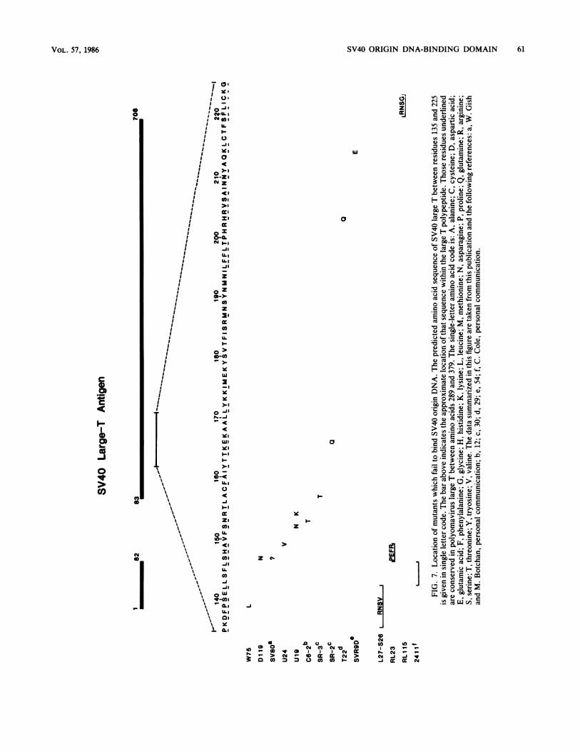
between amino acids 83 and 250 was summarized in theIntroduction. The data presented here allow a more precisemap to be deduced. These data, together with relevantinformation from other laboratories, are summarized in Fig.7. The most amino-terminal of the point mutations weexamined that is negative in the DNA binding assay isPro-139 -- Leu (W75). Deletion of the surrounding regionalso produces SV40 origin DNA-binding-negative large T(L27-S26). Mutations at residues 147 (Ser-- Asn; D119), 149(Ala -- Val; U24), and 152 plus 154 (Ser -* Asn, Arg -- Lys;U19) also abolish origin DNA binding as do a number of
multiple point and deletion mutations in the same region(Dlll, W134, RL23, D89, D98; listed in Table 1). Several ofthe point mutations in this region are reduced in their bindingactivity relative to wild type (W67, D120, Bl, U20, D88,D90, D94). It is striking that mutation of Ala-149 to Thr(D120) retains some binding activity, whereas mutation toVal (U24) does not. Even more noticeably, conversion to theadjacent residue Val-150 -+ Met (B9) generates a virus that isable to replicate and has a wild-type SV40 origin DNA-binding phenotype.The close clustering of mutations with a common defect
J. VIROL.
ani
t----.C:Dmc::30cz:oc::?

SV40 ORIGIN DNA-BINDING DOMAIN 59
L a) N- N > C0 CD N 't
W2 0 OO a X XvQ N
0C4 ) 0Co CD) U)
Lot N N NU)r 1 5N
4 CM r--
CY)C/) V- C5) 0
I r- Ir- 04r W rC 0 Q
2 7 0 -1
209-S b b
126-
0O
It rf0) C
i10 0o) ,- 1- Cj2 t_cnD
aM
1t0)CY)04%
00 L)0 O ~qt tO O 0
00 00C)0) 0) cJ m000 00ECDD ) 112z a
dB_FIG. 4. Binding of large T to SV40 origin DNA containing a deleted site I. (Upper panel and lower left panel) Samples of different cell
extracts containing equivalent amounts of "S-labeled large T were incubated with 10 ng of RL18 DNA which had been digested with EcoRIand Bst NI and then end labeled with 32P. Large T bound to DNA was immunoprecipitated and analyzed as described in the text. Tracks Ml,0.5 ng of EcoRl-BstNI-digested RL18 DNA. The sizes of the different fragments are given in base pairs. (Lower right panel) Extracts wereincubated with 2.5 ng of purified HindIII-C which had been digested with DdeI before labeling.
EllJ XC 'IT
M w ~- 3'
v- 0)C\ CM
M D N cu
1772-
1046_858- _673-
475-
270.
249-
126-
FIG. 5. Binding of large T to SV40 origin DNA lacking site
Samples of the different cell extracts were incubated with 10 ng ofBstNI-cut, end-labeled 034 DNA. Track M, 0.5 ng of starting DNA.The sizes of the different fragments are given in base pairs.
strongly suggests that they lie within a functional domain.This is made even more likely by findings from two separatestudies. By recovering the integrated SV40 DNA fromtransformed permissive monkey cells and sequencing theearly regions, the defects that prevent the expected produc-tive infection of these cells by the virus have been identified(12). In the case of the C6-2 mutation, the defect appears tobe in SV40 origin binding (43), and the lesion maps to residue153. Similarly, the SV40 insert coding for the large T inhuman SV80 cells has been isolated and found to be repli-cation defective. Again, the protein appears to be reduced inits SV40 origin DNA-binding activity (14). The lesion in thiscase maps to amino acid 147 (W. Gish and M. Botchan,personal communication). Using a totally different ap-proach, Nathans and colleagues (50) have isolatedpseudorevertant (sr) forms of SV40 large T that are able toreplicate SV40 virus containing a defective origin of replica-tion. Presumably, the pseudorevertant large T species arealtered in those amino acid residues intimately involved inthe sequence-specific interaction with origin DNA. sr-3maps to amino acid 157, and sr-2 maps to amino acid 166(30). We believe, therefore, that the amino-terminal end ofthe origin DNA-binding domain on large T can now be
1 7 7 2 -
1 046-
823-673 -
475-
0
a4-
cri1-:C,v p.- 4-
2 Uj .:
VOL. 57, 1986
435
302

60 PAUCHA ET AL.
Wt.rat AI I1 2 3 4
Wt.ratf-1 2 3 4
U 1 9[ I
1 23 4
I.I...** _ F**._ _~~~~
.1iI-:.1;li,~~ ~ ~ ~ ~ ~ ~
4..
0
0-.,.0_-
RL148 RL75-RL88 RL1 15 RL 1 2-S24I I
12 3 4I
n
1 2 3 4
FIG. 6. Binding of mutant large T to calf thymus DNA-cellulose. Samples (500 ,ul) of 32P-labeled lysates of different mutant cell lines weremixed with calf thymus DNA-cellulose, washed, and eluted as described in the text. All samples were immunoprecipitated with anti-SV40T serum before analysis on SDS-containing 15% acrylamide gels. Extracts of wild-type SV40-transformed rat cells were also analyzed onunderivatized cellulose as a control (WT rat A). Lanes: 1, 20-,ul sample of starting material; 2, 20 ,ul of unbound supernatant fraction; 3,material eluted at pH 8.5 with 0.15 M NaCl; 4, material eluted at pH 8.5 with 1.0 M NaCl. The positions of migration of large T and p53 areindicated.
localized to a region beginning at Pro-139 with a particularlyimportant sequence between amino acids 147 and 166.We do not know how far the putative domain extends to
the carboxy-terminal side because we have not made manymutants mapping to this region. However, the mutant RL115which affects residues 220 to 223 is origin-binding defective,and two other SV40 point mutants recovered from trans-formed permissive or nonpermissive cells that map to resi-due 203 (T22) (29) and 214 (SVR 9D) (54) also lack originDNA-binding activity. Since domains mapped on procary-otic DNA-binding proteins are usually of the order of 70 to90 residues (37), these mutations may lie at the extremecarboxyl end of the same domain.
The data obtained with the other SV40 large T variants (70K, H23, dH) reported here are also consistent with thislocation. Thus, with the exception of one published reportclaiming that a proteolytic fragment of large T comprisingonly residues 1 to 130 has origin DNA-binding activity (34),almost all other data are consistent with the site identifiedhere between residues 139 and 223 being a major componentin defining origin-specific DNA binding.Comparison of the SV40 large T amino acid sequence
between residues 135 and 225 with the region between aminoacids 289 and 379 of large T of the related papovavirus,polyomavirus, is shown in Fig. 7. The underlined aminoacids are conserved between the two viruses. It is striking
U24
1 2 3 4
Large-T
p53 -
B1 1
1 2 3 4
A.W
1 2 3 4
.4 -#
Large-T
p53 -
1 2 3 4
dH
1 2 3 4
0.a 4
J. VIROL.
A& 41i itIV
SV40 ORIGIN DNA-BINDING DOMAIN
N Y
>1a _'-,
II
I>l
a I-
a o,O 0
XZa o ,
I ,,-zI ca:
I'I
. .,0i
a ILI
IU.,
aIILL
a oZ
a ~0
Z
,-U.a 0>~
a -:I
! ~~~~>1
oILl
* b~~~~~~~-IZi
I,-
\ ~~~~ZI
\ ~~~~~~IL<~~~\ Ow
1. a..
(aZ
wi
0
a
I-
xI..
Z
U.Z c,
1-J
0
@a°a co ), , -
.9 0 o0aII N g
rw. > N 0 m = N > N .Ji-i3: O0 3 30 00 I-0 -i
VOL. 57, 1986
a
61
02
CI-
0
CO)qI0
0

62 PAUCHA ET AL.
that all the mutants defective in origin binding shown in Fig.7 map to residues shared with polyomavirus large T. Thehigh degree of conservation of amino acids suggests that theregion is essential to each virus and has a similar function. Ithas recently been shown (40) that polyomavirus large T canbind to the same pentanucleotide recognized by SV40 largeT.DNA-binding domains in procaryotic proteins have been
characterized in some detail (37). For example, they com-prise 66, 73, and 92 residues, respectively, for the Cro, CAP,and X repressor proteins. Although the domain describedhere could be of approximately this size our preliminarysearches have failed to find sequence homology between theSV40 amino acid sequences and those of the 20-residuehelix-bend-helix motif characteristic of the procaryotic pro-teins.
Site I, site II, and dsDNA binding domains. The datareported here do not allow us to distinguish whether thedomain has different specificities for binding sites I and II inthe SV40 origin. As far as it is possible to tell, the site IIbinding can be detected in such an immunoprecipitationassay by using DNA which lacks site I sequences appears toparallel the site I binding results, suggesting that the domainhas activity for both sites. Furthermore, the pseudorevertantlarge T antigens are presumably effective in origin, i.e., siteII, binding since they are capable of initiating viral DNAreplication and yet they map to the site I domain definedhere. Thus, it seems likely that the region between residues139 and 223 recognizes both sites I and II in the SV40 originregion, but this conclusion must be tested more rigorouslybefore being regarded as established.
It remains to be seen whether other regions on large Tform another DNA-binding domain. One report suggests thata second domain with a specificity for non-SV40 DNA liesbetween residues 245 and 325 (42). The results reported hereare consistent with that hypothesis since mutations withinthe SV40 origin binding region, including some large dele-tions, do not seem to affect the ability of the mutant proteinsto bind to ds cellular DNA. These results must be interpretedwith some caution, however, because of the severe limita-tions of the DNA-cellulose binding assay itself. In our hands,large T binds efficiently to underivatized cellulose (compareFig. 6, panel WT rat A, tracks 1 and 2) although apparentlywith low affinity as the bulk of the protein can be elutedunder low-salt conditions (Fig. 6, WT rat A, track 3).Furthermore, we can only recover 10 to 20% of the 32p_labeled protein bound to calf thymus DNA-cellulose in theelution steps. Any conclusions reached on DNA bindingmay, therefore, apply to only a small fraction of the entireprotein population. The existence of a separate domaincapable of binding independently to cellular DNA thusremains an open question.SV40 origin binding and replication. Very few (5 of 50) of
the mutants described in this study are capable of viral DNAreplication. All others are severely defective or deficient inthat function. Those mutants which can replicate (K79, C37,B9, and D96/D104) bind efficiently to SV40 origin DNA asexpected. The only exception to this rule is mutant B18,which can replicate but shows weak DNA binding in theseassays. B18 transforms cells very poorly, so the possibilityexists that the large T found in the B18-transformed line usedhere was modified in some way, perhaps by a second sitemutation or by gene rearrangement. It is easy to rationalizethe inability to replicate of those mutants whose originbinding activity is severely impaired. Similarly, the replica-tion defect in those mutants around Lys-128 which produce
a cytoplasmic large T could perhaps be explained by aninability to accumulate functional large T in the nucleus.This idea is supported by the apparent failure of thesemutants to autoregulate the levels of large T synthesized. Itis more difficult to understand why the remaining mutantsfail to replicate, even though they appear to bind well toSV40 origin DNA, site II as well as site I, as judged by theimmunoprecipitation assays reported here. Perhaps virtuallyany mutation in this region of large T, even a point mutation40 to 50 residues removed from the DNA-binding domain,disrupts the structure sufficiently to affect the intimateassociation reported to exist between the protein and SV40site II DNA (6). Such mutations might prevent replicationbut still allow an interaction measureable by the McKayassay.SV40 origin binding and transformation. All of the mutants
described here were able to transform Rat-1 cells regardlessof their ability to bind to SV40 origin DNA. In fact, somemutants which cannot bind DNA produced foci more rapidlyand in greater numbers than wild-type virus. Kalderon andSmith (19) suggested that this supertransforming phenotypemight be caused by an overproduction of large T since allmutants showing this property displayed brighter than nor-mal large T fluorescence. Although not consistently reflectedin the amount of large T immunoprecipitated from individualcell lines, a modest overproduction of most mutants display-ing the supertransforming phenotype (Bli, U19, U20, U24,W128, RL23, D88, D89) is seen here. More significantly, allof the supertransforming mutants show a discernible defectin SV40 origin DNA binding, with the mutants U19 and U24displaying the most extreme phenotype in both respects.Furthermore, mutants mapping to the same region (B9, D96,D104) that are able to bind to SV40 origin DNA and toreplicate do not discernibly overproduce or transform withan efficiency greater than wild type. Thus, the hypothesisthat the observed supertransforming activity of some of themutants can be explained in terms of an overproduction ofprotein is consistent with the deficiencies observed in thisstudy in binding to site I, the site of transcriptional controlby large T.The work described here supports the conclusion that
large T does not need to be able to interact with SV40 originDNA to transform established Rat-1 cells (29, 43, 54).However, any suggestion that transformation by large Tdoes not involve an interaction with cellular DNA must betempered by the finding that all of the mutants retain theability to bind to cellular ds DNA-cellulose. Scheller et al.(49) have already described a similar phenotype for the C6-2mutant. If transformation of established cell lines by SV40does not involve binding to DNA, it might imply that thisaspect of transformation is a cytoplasmic event catalyzed bythe membrane-associated forms of large T detected by anumber of workers (7, 20, 21, 47 [and references therein];53). This would be consistent with the emerging view thattransformation, as opposed to immortalization, is catalyzedby cytoplasmic events (22, 23, 36, 45). It would also explainwhy several of our mutant large T species that appear unableto localize to the nucleus of cells still retain the ability totransform established cells (17).Domain structure of large T. From previous studies on the
biological properties of the many mutants we have con-structed and from the data reported in this paper, it hasbecome apparent that the linear amino acid sequence of largeT can be divided into discrete blocks each of which isassociated with a distinct function or property.
All of the phosphorylation sites in the amino-terminal half
J. VIROL.

SV40 ORIGIN DNA-BINDING DOMAIN 63
of large T are clustered between amino acids 106 and 124:Ser-106, Ser-111, Ser-112, Ser-123, and Thr-124 (48). Ourstudies have shown that mutants carrying alterations inamino acids 106 to 114 generally produced foci on Rat-1 cellsmore slowly and in reduced numbers than did wild-type largeT (19). These mutants, therefore, define a domain whichmust play a role in the transformation process. We do not yetknow whether mutations within the putative transformationdomain alter the phosphorylation patterns of large T orwhether some other function is responsible for their reducedtransforming ability. The next domain includes the sequence125-Pro-Pro-Lys-Lys-Lys-Arg-Lys-Val-132 which weshowed constitutes a nuclear location signal. Mutationswithin this region result in large Ts which are wholly orpartially located in the cytoplasm (17, 18, 25). These mutantsretain the ability to transform Rat-1 cells to continuousgrowth. Finally, in this publication, we defined a DNA-binding region which begins at Pro-139 and which mayextend as far as Cys-223. Whether each of these functionaldomains corresponds to a structural domain remains to bedetermined. Nevertheless, the existence of mutations withineach of the different functional domains facilitates the sys-tematic analysis of large T functions and the role they play inits biological activities, particularly in transformation.
ACKNOWLEDGMENTS
We thank A. Graessmann for the gifts of the 70K cell line, CarolPrives for information and advice on calf thymus DNA binding, andour colleagues at Mill Hill and elsewhere for helpful discussions. Weare also grateful to Lydia Pearson for help with the tables and to AnnDesai for typing the manuscript.
LITERATURE CITED1. Alwine, J. C., S. I. Reed, and G. R. Stark. 1977. Characteriza-
tion of the autoregulation of simian virus 40 gene A. J. Virol.24:22-27.
2. Carroll, R. B., L. Hager, and R. Dulbecco. 1974. Simian virus 40T antigen binds to DNA. Proc. Natl. Acad. Sci. USA71:3754-3757.
3. Chaudry, F., R. Harvey, and A. E. Smith. 1982. The structureand biochemical functions of four SV40 truncated large Tantigens. J. Virol. 44:54-66.
4. Clark, R., D. P. Lane, and R. Tjian. 1981. The use of monoclo-nal antibodies as probes of simian virus 40 T antigen ATPaseactivity. J. Biol. Chem. 256:11854-11858.
5. Clark, R., K. Peden, J. M. Pipas, D. Nathans, and R. Tjian.1983. Biochemical activity of T-antigen proteins encoded bysimian virus 40 A gene deletion mutants. Mol. Cell. Biol.3:220-228.
6. Cohen, G. L., P. W. Wright, A. De Lucia, B. A. Lewton, M. E.Anderson, and P. Tegtmeyer. 1984. Critical spatial requirementwithin the origin of simian virus 40 DNA replication. J. Virol.51:91-96.
7. Covey, L., Y. Choi, and C. Prives. 1984. Association of simianvirus 40 T antigen with the nuclear matrix of infected andtransformed monkey cells. Mol. Cell. Biol. 4:1384-1392.
8. De Lucia, A. L., B. A. Lewton, R. Tjian, and P. Tegtmeyer. 1983.Topography of simian virus 40 A protein-DNA complexes:arrangement of pentanucleotide interaction sites at the origin ofreplication. J. Virol. 46:143-150.
9. DiMaio, D., and D. Nathans. 1982. Regulatory mutants of simianvirus 40: effect of mutations at a T antigen binding site on DNAreplication and expression of viral genes. J. Mol. Biol.156:531-548.
10. Fanning, E., K.-H. Westphal, D. Brauer, and D. Corlin. 1982Subclasses of simian virus 40 large T antigen: differentialbinding of two subclasses of T antigen from productivelyinfected cells to viral and cellular DNA. EMBO J. 1:1023-1028.
11. Gidoni, D., A. Scheller, B. Barnet, R. Hantzopoulos, M. Oren,
and C. Prives. 1982. Different forms of simian virus 40 largetumor antigen varying in their affinities for DNA. J. Virol.42:456-466.
12. Gluzman, Y., and B. Ahrens. 1982. SV40 early mutants that aredefective for viral DNA synthesis but competent for transfor-mation of cultured rat and simian cells. Virology 123:78-92.
13. Graessmann, M., and A. Graessmann. 1982. Simian virus 40cRNA is processed into functional mRNA in microinjectedmonkey cells. EMBO J. 1:1081-1088.
14. Gruss, C., E. Baumann, and R. Knippers. 1984. DNA bindingproperties of a mutant T antigen from the simian virus 40-transformed human cell line SV80. J. Virol. 50:943-946.
15. Harlow, E., L. V. Crawford, D. C. Pim, and N. M. Williamson.1981. Monoclonal antibodies specific for simian virus 40 tumorantigens. J. Virol. 39:861-869.
16. Kalderon, D., B. A. Oostra, B. K. Ely, and A. E. Smith. 1982.Deletion-loop mutagenesis: a novel method for the constructionof point mutations using deletion mutants. Nucleic Acids Res.10:5161-5171.
17. Kalderon, D., W. D. Richardson, A. Markham, and A. E. Smith.1984. Sequence requirements for nuclear location of SV40 largeT. Nature (London) 311:33-38.
18. Kalderon, D., B. L. Roberts, W. D. Richardson, and A. E.Smith. 1984. A short amino acid sequence able to specifynuclear location. Cell 39:499-509.
19. Kalderon, D., and A. E. Smith. 1984. In vitro mutagenesis of aputative DNA binding domain on SV40 large T. Virology139:109-137.
20. Klockmann, U., and W. Deppert. 1983. Acylated simian virus 40large T antigen: a new subclass associated with a detergent-resistant lamina of the plasma membrane. EMBO J. 2:1151-1157.
21. Klockmann, U., M. Staufenbiel, and W. Deppert. 1984. Mem-brane interactions of simian virus 40 large T antigen: influenceof protein sequences and fatty acid acylation. Mol. Cell. Biol.4:1542-1550.
22. Land, H., L. Parada, and R. A. Weinberg. 1983. Cellularoncogenes and multistep carcinogenesis. Science 222:771-778.
23. Land, H., L. F. Parada, and R. A. Weinberg. 1983. Tumorigenicconversion of primary embryo fibroblasts requires at least twocooperating oncogenes. Nature (London) 304:596-602.
24. Lane, D. P., and L. V. Crawford. 1979. Large T antigen is boundto a host protein in SV40 transformed cells. Nature (London)278:262-263.
25. Lanford, R. E., and J. S. Butel. 1984. Construction and charac-terization of an SV40 mutant defective in nuclear transport of Tantigen. Cell 37:801-813.
26. Linzer, D., and A. Levine. 1979. Characterization of a 54Kcellular SV40 tumor antigen present in SV40 transformed cellsand uninfected embryonal carcinoma cells. Cell 17:43-52.
27. Maniatis, T., E. F. Fritsch, and J. Sambrook. 1982. Molecularcloning, a laboratory manual. Cold Spring Harbor Laboratory,Cold Spring Harbor, N.Y.
28. Manos, M. M., and Y. Gluzman. 1984. Simian virus 40 large Tantigen point mutants that are defective in viral DNA replicationbut competent in oncogenic transformation. Mol. Cell. Biol.4:1125-1133.
29. Manos, M. M., and Y. Gluzman. 1985. Genetic and biochemicalanalysis of transformation-competent, replication-defectivesimian virus 40 large T antigen mutants. J. Virol. 53:120-127.
30. Margoiskee, P. F., and D. Nathans. 1984. Simian virus 40 mutantT antigens with relaxed specificity for the nucleotide sequenceat the viral DNA origin of replication. J. Virol. 49:386-393.
31. McKay, R. D. G. 1981. Binding of SV40 T antigen relatedprotein to DNA. J. Mol. Biol. 145:471-488.
32. McKay, R. D. G., and D. Di Maio. 1981. Binding of an SV40 Tantigen related protein to the DNA of SV40 regulatory mutants.Nature (London) 289:810-813.
33. Montenarh, M., W. Deppert, and R. Henning. 1982. Mapping ofa DNA-binding domain of simian virus 40 T antigen usingnon-defective adenovirus-2-simian virus 40 hybrid viruses.FEBS Lett. 142:129-132.
34. Morrison, B., M. Kress, G. Khoury, and G. Jay. 1983. SV40
VOL. 57, 1986

64 PAUCHA ET AL.
tumor antigen: isolation of the origin specific DNA bindingdomain. J. Virol. 47:106-114.
35. Myers, R. R., and R. Tjian. 1980. Construction and analysis ofsimian virus 40 origins defective in tumor antigen binding andDNA replication. Proc. Natl. Acad. Sci. USA 77:6491-6495.
36. Newbold, R. F., and R. W. Overell. 1983. Fibroblast immortalityis a prerequisite for transformation by EJ c-HA-ras oncogene.Nature (London) 304:648-651.
37. Pabo, C. O., and R. T. Sauer. 1984. Protein DNA recognition.Annu. Rev. Biochem. 53:293-321.
38. Paucha, E., R. W. Harvey, and A. E. Smith. 1984. Antibodies tosynthetic peptides that immunoprecipitate some forms of SV40large T antigen. J. Virol. 51:670-682.
39. Pipas, J. M., K. W. C. Peden, and D. Nathans. 1983. Mutationalanalysis of simian virus 40 T antigen: isolation and character-ization of mutants with deletions in the T-antigen gene. Mol.Cell. Biol. 3:204-213.
40. Pomerantz, B. J., and J. Hassel. 1984. Polyomavirus and simianvirus 40 large T antigens bind to common DNA sequences. J.Virol. 49:925-937.
41. Prives, C., B. Barnet, A. Scheller, G. Khoury, and J. Gilbert.1982. Discrete regions of simian virus 40 large T antigen arerequired for nonspecific and viral origin-specific DNA binding.J. Virol. 43:73-82.
42. Prives, C., Y. Beck, and H. Shure. 1980. DNA binding proper-ties of simian virus 40 T antigen synthesized in vivo and in vitro.J. Virol. 33:689-696.
43. Prives, C., L. Covey, E. Scheller, and Y. Gluzman. 1983. DNAbinding properties of simian virus T antigen mutants defective inviral DNA replication. Mol. Cell. Biol. 3:1958-1966.
44. Rio, D., A. Robbins, R. Myers, and R. Tjian. 1980. Regulation ofsimian virus 40 early transcription in vitro by purified tumorantigen. Proc. Nati. Acad. Sci. USA 77:5706-5710.
45. Ruley, H. E. 1983. Adenovirus early region 1A enables viral andcellular transforming genes to transform primary cells in cul-ture. Nature (London) 304:602-606.
46. Rundell, K., J. K. Collins, P. Tegtmeyer, H. Ozer, C. J. Lai, andD. Nathans. 1977. Identification of simian virus 40 A protein. J.Virol. 21:636-646.
47. Santos, M., and J. S. Butel. 1984. Antigenic structure of simianvirus 40 large tumor antigen and association with cellularprotein p53 on the surfaces of simian virus 40-infected and
-transformed cells. J. Virol. 51:376-383.48. Scheidtmann, K. H., B. Echle, and G. Walter. 1982. SV40 large
T antigen is phosphorylated at multiple sites clustered in twoseparate regions. J. Virol. 44:116-133.
49. Scheller, G. E., L. Covey, B. Barnet, and C. Prives. 1982. Asmall subclass of SV40 T antigen binds to the viral origin ofreplication. Cell 29:375-383.
50. Shortle, D. R., R. F. Margolskee, and D. Nathans. 1979. Muta-tional analysis of the simian virus 40 replicon: pseudorevertantsof mutants with a defective replication origin. Proc. Natl. Acad.Sci. USA 76:6128-6131.
51. Smith, A. E., R. Smith, and E. Paucha. 1978. Extraction andfingerprint analysis of simian virus 40 large T and small tantigens. J. Virol. 28:140-153.
52. Smith, A. E., R. Smith, and E. Paicha. 1979. Characterization ofdifferent tumor antigens present in cells transformed by simianvirus 40. Cell 18:335-346.
53. Staufenbiel, M., and W. Deppert. 1983. Different structuralsystems of the nucleus are targets for SV40 large T antigen. Cell33:173-181.
54. Stringer, J. R. 1982. Mutant of simian virus 40 large T antigenthat is defective for viral DNA synthesis but competent fortransformation of cultured rat cells. J. Virol. 42:854-864.
55. Tegtmeyer, P., B. A. Lewton, A. L. D)e Lucia, V. G. Wilson, andK. Ryder. 1983. Topography of simian virus 40 A protein-DNAcomplexes: arrangement of protein bound to the origin ofreplication. J. Virol. 46:151-161.
56. Tenen, D. G., T. S. Taylor, L. L. Haines, M. K. Bradley, R. G.Martin, and D. M. Livingston. 1983. Binding of simian virus 40large T antigen from virus-infected monkey cells to wild-typeand mutant viral replication origins. J. Mol. Biol. 168:791-808.
57. Tjian, R. 1978. The binding site on SV40 DNA for a Tantigen-related protein. Cell 13:165-179.
58. Tjian, R., and R. Robbins. 1979. Enzymatic activity associatedwith purified simian virus 40 T antigen-related protein. Proc.Natl. Acad. Sci. USA 76:610-615.
59. Tooze, J. 1980. DNA tumor viruses, 2nd ed., part 2. Cold SpringHarbor Laboratory, Cold Spring Harbor, N.Y.
60. Van Roy, F., L. Fransen, and W. Fiers. 1983. Metabolic turn-over of phosphorylation sites in simian virus 40 large T antigen.J. Virol. 45:442-446.
J. VIROL.