Regulation of Tumor Antigen Synthesis by Simian
11
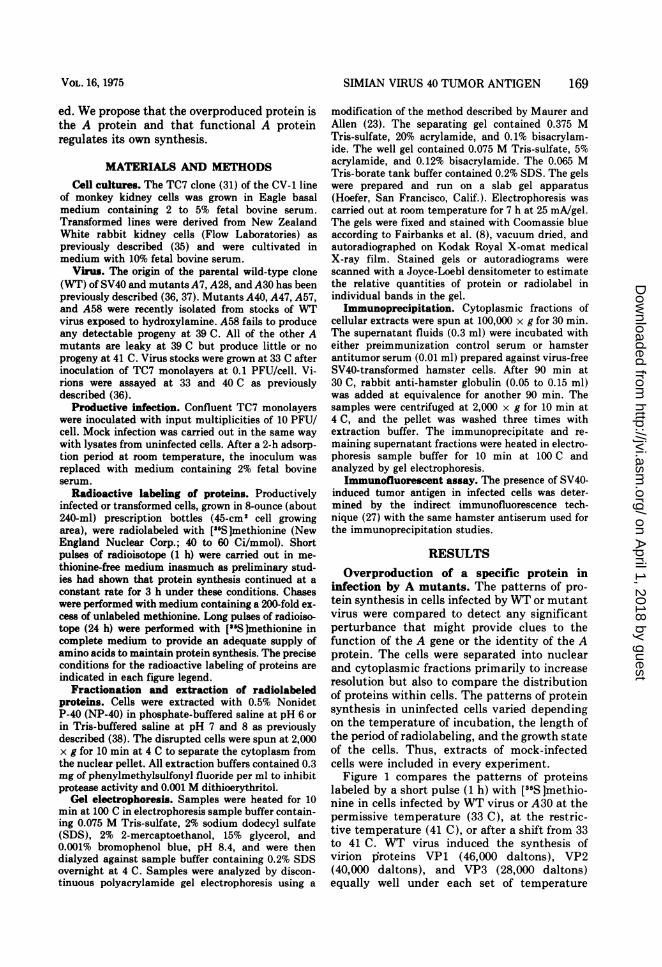
JOURNAL OF VIROLOGY, JUlY 1975, P. 168-178 Copyright i 1975 American Society for Microbiology Vol. 16, No. 1 Printed in U.SA. Regulation of Tumor Antigen Synthesis by Simian Virus 40 Gene A PETER TEGTMEYER,'* MICHAEL SCHWARTZ, JOHN K. COLLINS, AND KATHLEEN RUNDELL Departments of Pharmacology,* Microbiology, and Anatomy, Case Western Reserve University School of Medicine, Cleveland, Ohio 44106 Received for publication 28 February 1975 Simian virus 40 gene A has previously been shown to promote the replication of viral DNA and the transcription of late viral RNA in productive infection and to maintain the growth characteristics of some transformed cells. The present study examines the effect of the A function on proteins synthesized during productive and transforming infections. Under restrictive conditions, temperature-sensitive A mutants induce the overproduction of a 100,000-dalton protein both in productively infected monkey cells and in transformed rabbit cells. Immuno- precipitation of the induced protein with antisera, prepared against simian virus 40-induced tumors in hamsters, was used to identify the induced protein as tumor antigen. The same protein can be precipitated from extracts of cells infected by wild-type virus but not from uninfected cells. Furthermore, the mutant-induced protein is more rapidly degraded in vivo and is less tightly bound to intranuclear components than the protein induced by wild-type virus. The presence of the same virus-induced protein in infected cells from different species and the altered behavior of that protein in mutant infection strongly suggest that the protein is virus coded. Because the protein is large enough to account for the entire coding capacity in the early region of the simian virus 40 genome, the 100,000-dalton protein may well be the primary product of the only early gene identified by complementation studies, the A gene. If the 100,000-dal- ton protein that is overproduced in mutant infection is the A protein and the only early protein, then functional wild-type A protein must regulate its own synthesis in both productive and transforming infections. In productive infection by simian virus 40 (SV40), gene A function continuously regulates the initiation of viral DNA replication (5, 21, 34) and transiently controls the synthesis of late viral RNA (6, 21). In restrictive infection, the A function is required to establish the stable transformation of cells and also to maintain the growth characteristics of some transformed cell lines (3, 16, 22, 25, 35). Thus, it seems quite possible that the A protein may directly interact with specific recognition sites on either viral or cellular DNA to regulate the replication or transcription of either DNA. A satisfactory testing of this hypothesis will require the iden- tification, isolation, and characterization of the A protein and a study of its interaction with different DNA molecules. These studies could eventually lead to a better understanding of growth control mechanisms in mammalian cells at the molecular level. ' Present address: Department of Microbiology, State University of New York at Stony Brook, Stony Brook, N.Y. 11794. 168 A major obstacle to these studies has long been the difficulty encountered in identifying the A protein within the large background of cellular proteins (1, 15, 38, 39). We reasoned that the synthesis, location, or processing of the A protein could be perturbed in infection by temperature-sensitive A mutants and that the resultant alteration might be useful in identify- ing the A protein. Thus a study of in vivo protein synthesis in productive and transform- ing infections by the A mutants was under- taken. Each of the A mutants tested induced the overproduction of a 100,000-dalton protein at the restrictive temperature in both monkey and rabbit cells. The induced protein was specifi- cally and efficiently precipitated from extracts of productively infected or transformed cells by antisera prepared against SV40-induced tumors in hamsters. A similar protein could not be identified in uninfected cells. This and other studies (2, 9, 13, 20, 26, 27, 29, 30, 33) suggest that SV40-induced tumor antigen is virus cod- on April 1, 2018 by guest http://jvi.asm.org/ Downloaded from
Transcript of Regulation of Tumor Antigen Synthesis by Simian

JOURNAL OF VIROLOGY, JUlY 1975, P. 168-178Copyright i 1975 American Society for Microbiology
Vol. 16, No. 1Printed in U.SA.
Regulation of Tumor Antigen Synthesis by SimianVirus 40 Gene A
PETER TEGTMEYER,'* MICHAEL SCHWARTZ, JOHN K. COLLINS, AND KATHLEEN RUNDELL
Departments ofPharmacology,* Microbiology, and Anatomy, Case Western Reserve University School ofMedicine, Cleveland, Ohio 44106
Received for publication 28 February 1975
Simian virus 40 gene A has previously been shown to promote the replication ofviral DNA and the transcription of late viral RNA in productive infection and tomaintain the growth characteristics of some transformed cells. The present studyexamines the effect of the A function on proteins synthesized during productiveand transforming infections. Under restrictive conditions, temperature-sensitiveA mutants induce the overproduction of a 100,000-dalton protein both inproductively infected monkey cells and in transformed rabbit cells. Immuno-precipitation of the induced protein with antisera, prepared against simian virus40-induced tumors in hamsters, was used to identify the induced protein astumor antigen. The same protein can be precipitated from extracts of cellsinfected by wild-type virus but not from uninfected cells. Furthermore, themutant-induced protein is more rapidly degraded in vivo and is less tightlybound to intranuclear components than the protein induced by wild-type virus.The presence of the same virus-induced protein in infected cells from differentspecies and the altered behavior of that protein in mutant infection stronglysuggest that the protein is virus coded. Because the protein is large enough toaccount for the entire coding capacity in the early region of the simian virus 40genome, the 100,000-dalton protein may well be the primary product of the onlyearly gene identified by complementation studies, the A gene. If the 100,000-dal-ton protein that is overproduced in mutant infection is the A protein and the onlyearly protein, then functional wild-type A protein must regulate its own synthesisin both productive and transforming infections.
In productive infection by simian virus 40(SV40), gene A function continuously regulatesthe initiation of viral DNA replication (5, 21,34) and transiently controls the synthesis of lateviral RNA (6, 21). In restrictive infection, the Afunction is required to establish the stabletransformation of cells and also to maintain thegrowth characteristics of some transformed celllines (3, 16, 22, 25, 35). Thus, it seems quitepossible that the A protein may directlyinteract with specific recognition sites on eitherviral or cellular DNA to regulate the replicationor transcription of either DNA. A satisfactorytesting of this hypothesis will require the iden-tification, isolation, and characterization of theA protein and a study of its interaction withdifferent DNA molecules. These studies couldeventually lead to a better understanding ofgrowth control mechanisms in mammalian cellsat the molecular level.
' Present address: Department of Microbiology, StateUniversity of New York at Stony Brook, Stony Brook, N.Y.11794.
168
A major obstacle to these studies has longbeen the difficulty encountered in identifyingthe A protein within the large background ofcellular proteins (1, 15, 38, 39). We reasonedthat the synthesis, location, or processing of theA protein could be perturbed in infection bytemperature-sensitive A mutants and that theresultant alteration might be useful in identify-ing the A protein. Thus a study of in vivoprotein synthesis in productive and transform-ing infections by the A mutants was under-taken.Each of the A mutants tested induced the
overproduction of a 100,000-dalton protein atthe restrictive temperature in both monkey andrabbit cells. The induced protein was specifi-cally and efficiently precipitated from extractsof productively infected or transformed cells byantisera prepared against SV40-induced tumorsin hamsters. A similar protein could not beidentified in uninfected cells. This and otherstudies (2, 9, 13, 20, 26, 27, 29, 30, 33) suggestthat SV40-induced tumor antigen is virus cod-
on April 1, 2018 by guest
http://jvi.asm.org/
Dow
nloaded from
SIMIAN VIRUS 40 TUMOR ANTIGEN 169
ed. We propose that the overproduced protein isthe A protein and that functional A proteinregulates its own synthesis.
MATERIALS AND METHODSCell cultures. The TC7 clone (31) of the CV-1 line
of monkey kidney cells was grown in Eagle basalmedium containing 2 to 5% fetal bovine serum.Transformed lines were derived from New ZealandWhite rabbit kidney cells (Flow Laboratories) aspreviously described (35) and were cultivated inmedium with 10% fetal bovine serum.
Virus. The origin of the parental wild-type clone(WT) of SV40 and mutants A7, A28, and A30 has beenpreviously described (36, 37). Mutants A40, A47, A57,and A58 were recently isolated from stocks of WTvirus exposed to hydroxylamine. A58 fails to produceany detectable progeny at 39 C. All of the other Amutants are leaky at 39 C but produce little or noprogeny at 41 C. Virus stocks were grown at 33 C afterinoculation of TC7 monolayers at 0.1 PFU/cell. Vi-rions were assayed at 33 and 40 C as previouslydescribed (36).
Productive infection. Confluent TC7 monolayerswere inoculated with input multiplicities of 10 PFU/cell. Mock infection was carried out in the same waywith lysates from uninfected cells. After a 2-h adsorp-tion period at room temperature, the inoculum wasreplaced with medium containing 2% fetal bovineserum.
Radioactive labeling of proteins. Productivelyinfected or transformed cells, grown in 8-ounce (about240-ml) prescription bottles (45-cm2 cell growingarea), were radiolabeled with ['"SJmethionine (NewEngland Nuclear Corp.; 40 to 60 Ci/mmol). Shortpulses of radioisotope (1 h) were carried out in me-thionine-free medium inasmuch as preliminary stud-ies had shown that protein synthesis continued at aconstant rate for 3 h under these conditions. Chaseswere performed with medium containing a 200-fold ex-cess of unlabeled methionine. Long pulses of radioiso-tope (24 h) were performed with ["S]methionine incomplete medium to provide an adequate supply ofamino acids to maintain protein synthesis. The preciseconditions for the radioactive labeling of proteins areindicated in each figure legend.
Fractionation and extraction of radiolabeledproteins. Cells were extracted with 0.5% NonidetP-40 (NP-40) in phosphate-buffered saline at pH 6 orin Tris-buffered saline at pH 7 and 8 as previouslydescribed (38). The disrupted cells were spun at 2,000x g for 10 min at 4 C to separate the cytoplasm fromthe nuclear pellet. All extraction buffers contained 0.3mg of phenylmethylsulfonyl fluoride per ml to inhibitprotease activity and 0.001 M dithioerythritol.
Gel electrophoresis. Samples were heated for 10min at 100 C in electrophoresis sample buffer contain-ing 0.075 M Tris-sulfate, 2% sodium dodecyl sulfate(SDS), 2% 2-mercaptoethanol, 15% glycerol, and0.001% bromophenol blue, pH 8.4, and were thendialyzed against sample buffer containing 0.2% SDSovernight at 4 C. Samples were analyzed by discon-tinuous polyacrylamide gel electrophoresis using a
modification of the method described by Maurer andAllen (23). The separating gel contained 0.375 MTris-sulfate, 20% acrylamide, and 0.1% bisacrylam-ide. The well gel contained 0.075 M Tris-sulfate, 5%acrylamide, and 0.12% bisacrylamide. The 0.065 MTris-borate tank buffer contained 0.2% SDS. The gelswere prepared and run on a slab gel apparatus(Hoefer, San Francisco, Calif.). Electrophoresis wascarried out at room temperature for 7 h at 25 mA/gel.The gels were fixed and stained with Coomassie blueaccording to Fairbanks et al. (8), vacuum dried, andautoradiographed on Kodak Royal X-omat medicalX-ray film. Stained gels or autoradiograms werescanned with a Joyce-Loebl densitometer to estimatethe relative quantities of protein or radiolabel inindividual bands in the gel.
Immunoprecipitation. Cytoplasmic fractions ofcellular extracts were spun at 100,000 x g for 30 min.The supernatant fluids (0.3 ml) were incubated witheither preimmunization control serum or hamsterantitumor serum (0.01 ml) prepared against virus-freeSV40-transformed hamster cells. After 90 min at30 C, rabbit anti-hamster globulin (0.05 to 0.15 ml)was added at equivalence for another 90 min. Thesamples were centrifuged at 2,000 x g for 10 min at4 C, and the pellet was washed three times withextraction buffer. The immunoprecipitate and re-imaining supernatant fractions were heated in electro-phoresis sample buffer for 10 min at 100 C andanalyzed by gel electrophoresis.
Immunofluorescent assay. The presence of SV40-induced tumor antigen in infected cells was deter-mined by the indirect immunofluorescence tech-nique (27) with the same hamster antiserum used forthe immunoprecipitation studies.
RESULTS
Overproduction of a specific protein ininfection by A mutants. The patterns of pro-tein synthesis in cells infected by WT or mutantvirus were compared to detect any significantperturbance that might provide clues to thefunction of the A gene or the identity of the Aprotein. The cells were separated into nuclearand cytoplasmic fractions primarily to increaseresolution but also to compare the distributionof proteins within cells. The patterns of proteinsynthesis in uninfected cells varied dependingon the temperature of incubation, the length ofthe period of radiolabeling, and the growth stateof the cells. Thus, extracts of mock-infectedcells were included in every experiment.
Figure 1 compares the patterns of proteinslabeled by a short pulse (1 h) with [35S ]methio-nine in cells infected by WT virus or A30 at thepermissive temperature (33 C), at the restric-tive temperature (41 C), or after a shift from 33to 41 C. WT virus induced the synthesis ofvirion piroteins VP1 (46,000 daltons), VP2(40,000 daltons), and VP3 (28,000 daltons)equally well under each set of temperature
VOL. 16, 1975
on April 1, 2018 by guest
http://jvi.asm.org/
Dow
nloaded from
100K- *.t,
s 4
; 4 e,
fi _ :!
I w
VP1- It
a b c
t i t. 4; $
* X* #- - &
f If If I
de e
d e f
f t 100K
w. la ,. .10 e
41p 4 dip... . -...* 9
t ''fg hif -VPi
g h i
VPI- j41vpt.: '. y
L ;-tI I
2 l- a
i1
4*x-oet- f- -
*w -VP3
170
100K- -100K
VP3-
4 ...-. _.m&
00 ::,..Vpl
I jqf. 11
on April 1, 2018 by guest
http://jvi.asm.org/
Dow
nloaded from

SIMIAN VIRUS 40 TUMOR ANTIGEN 171
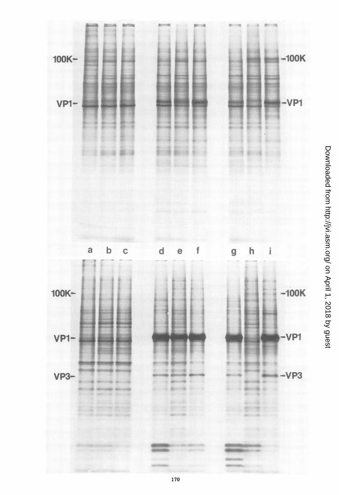
conditions. Most of the capsid proteins werefound in the nuclear fraction of the cells eventhough approximately 80% of the total labeledprotein was present in the cytoplasmic fractionof the cells. Cells infected by A30 producedvirion proteins during continuous incubation at33 C but failed to produce capsid proteinsduring continuous incubation at 41 C. In con-trast, when cells infected by A30 for 72 h at 33 Cwere shifted to 41 C for 24 h and then labeledwith [35S]methionine for 1 h, capsid proteinswere synthesized at the same rate as in cellsinfected by WT virus under the same condi-tions. These findings support previous studies(6, 21) showing that the A function is onlytransiently required for the transcription of lateviral RNA.The most significant finding in infection by
A30 at 41 C was the overproduction of a100,000-dalton (1OOK) protein. This protein hadno apparent counterpart in uninfected cells andwas difficult to identify in cells infected by WTvirus. After extraction at pH 7, most of theA-induced protein was found in the cytoplasmicfraction of cells, but smaller quantities werepresent in the nuclear fraction as well. Theoverproduction of the .100K protein by A30 wasnot the result of a high multiplicity of infectionbecause the same protein was not present inincreased quantities in infection by the samestock of virus at 33 C. Nor was the overproduc-tion of the protein caused by an absence ofcapsid proteins in cells infected by A mutants,since the protein is also overproduced in cellssynthesizing capsid proteins after a shift from33 to 41 C (Fig. 1). Further, the excess synthesisof the A-induced protein is a general phenome-non in infection by A mutants at 41 C inasmuchas each of seven independently isolated mu-tants (A7, A28, A30, A40, A47, A57, A58)induced the synthesis of the same protein to asimilar extent. The excess synthesis is not theresult of a nonspecific inhibition of viral DNAsynthesis because inhibition of the replicationof DNA by 150 ,ug of cytosine arabinoside per mlin cells infected by WT virus does not causeoverproduction of the protein. The 100K proteinis also made in excess 24 and 72 h as well as 48 h
after infection by the A mutants at 41 C. Thisfinding excludes the possibility that the lOOKprotein is produced in similar quantities in WTand mutant infection but with an altered tem-poral sequence.
Identification of the A-induced protein astumor antigen. Cellular extracts, known tocontain the lOOK protein induced by the Amutants, were exposed either to serum fromhamsters bearing SV40-induced tumors or toserum taken from the same hamsters beforeimmunization. Antigen-antibody complexeswere then precipitated by the addition of rabbitimmunoglobulin prepared against hamsterglobulin. The precipitated and non-precipitatedproteins were analyzed by SDS-gel electropho-resis (Fig. 2). The A-induced protein was specif-ically and efficiently precipitated by antitumorantibody but not to a significant extent bypreimmunization control serum. Further, thesame protein could also be identified unam-biguously in immunoprecipitates of cells in-fected by WT virus but not in uninfected cells.The amount of radioactivity in the lOOK bandsin gels of the supernatant and pellet fractions ofthe immunoprecipitation mixture was com-pared by excision of the appropriate bands andliquid scintillation counting. More than 80% ofthe lOOK protein was precipitated by antitumorserum whereas less than 2% was precipitated bythe control preimmunization serum. A similarquantitation can be seen in densitometer trac-ings of an autoradiogram of the same gel (Fig.3). Several other proteins with molecularweights ranging from 66,000 to 88,000 daltonswere also immunoprecipitated in small quan-tities from extracts of WT or mutant-infectedcells but not from extracts of control cells.
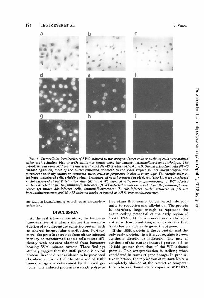
Intracellular localization of the A-inducedprotein. Carroll et al. have recently shown that"T" antigen binds to DNA in vitro at pH 6 butelutes at pH 8 (4). On the basis of this informa-tion, the intracellular localization and in vivobinding characteristics of tumor antigen andthe 100K protein were compared in infection byWT or mutant virus. The location of tumorantigen was determined by immunofluorescentstaining (Fig. 4). Intact cells showed a more
FIG. 1. Altered patterns of protein synthesis in cells infected by wild-type (WT) virus and A30 at 33 C, at41 C, and after a shift from 33 to 41 C. The cultures were labeled with 50 uCi of [35S]methionine per ml ofmethionine-free medium 70 to 72 h after infection at 33 C, 47 to 48 h after infection at 41 C, and 23 to 24 h aftera shift from 33 C (72-h preincubation) to 41 C. SDS-polyacrylamide (20%o) gel autoradiograms of fractionatedcell proteins are shown. The sample order is: (a) control cells, 33 C; (b) control cells, 41 C; (c) control cells,temperature shift; (d) WT-infected cells, 33 C; (e) WT-infected cells, 41 C; (f) WT-infected cells, temperatureshift; (g) A30-infected cells, 33 C; (h) A30-infected cells, 41 C; and (i) A30-infected cells, temperature shift. Theupper and lower panels show the cytoplasmic and nuclear extracts, respectively.
VOL. 16, 1975
on April 1, 2018 by guest
http://jvi.asm.org/
Dow
nloaded from

172 TEGTMEYER ET AL.
Supernatea b c d
Precipitatea b c de f e t A58
FIG. 2. Virus-induced proteins specifically precipitated by antitumor serum. Cultures were labeled with 30
'UCi Of [35SjImethionine per ml of methionine-free medium 47 to 48 h after infection. Soluble proteins extracted
from cells with 0.5% NP-40 at pH 7 were precipitated with serum from hamsters bearing SV40-induced tumors
or with preimmunization control serum using the indirect immunoprecipitation technique described in the text.
The precipitated proteins were identified by SDS-polyacrylamide (20%) gel electrophoresis. Both the
supernatant and the pelleted fractions of the immunoprecipitation reactions are shown in the slab gel
autoradiogram. The sample order is: (a) control cells, control serum; (b) WT-infected cells, control serum; (c)
A58-infected cells, control serum; (d) control cells, antitumor serum; (e) WVT-infected cells, antitumor serum;
and (f) A58-infected cells, antitumor serum. The side wells are whole cytoplasmic extracts of WT- and
A58-infected cells. The arrows indicate the position of the overproduced 100,000-dalton protein.
uniform staining of nuclei in WT infection thanin rnrlant infection. Further, the cytoplasm ofcells infected by A58, but not by WT virus, was
distinctly immunofluroescent in most but notall infected cells. Extraction with NP-40 at pH 6removed most of the tumor antigen from thenuclei of cells infected by A58 but not from thenuclei infected by WT virus. Extraction at pH8, however, efficiently removed tumor antigenfrom the nuclei infected by either WT or mu-
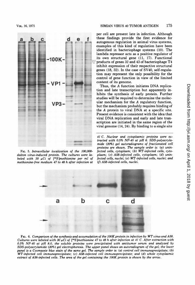
tant virus.The location of the lOOK protein was deter-
mined directly by the electrophoresis of proteinsfrom cellular extracts (Fig. 5). After extractionat pH 6, most of the lOOK protein was present inthe cytoplasmic extract of cells infected by A58
but in the nuclei of cells infected by WT virus.After extraction at pH 8, the lOOK protein couldno longer be identified in the nuclei of cellsinfected by either virus. The data are consistentwith the interpretation that the WT-inducedprotein binds to intranuclear DNA with a
greater affinity than the A-induced protein.Temperature-sensitive degradation of the
lOOK protein. The lOOK proteins induced byWT virus or A58 and radiolabeled by a 1-h pulsewith [35S ]methionine were compared in thesame slab gel both by staining with Coomassieblue and autoradiography (Fig. 6). Although thelOOK protein of the A mutant was more heavilyradiolabeled than the corresponding proteininduced by WT virus in immunoprecipitated
WT
J. VIROL.
on April 1, 2018 by guest
http://jvi.asm.org/
Dow
nloaded from

SIMIAN VIRUS 40 TUMOR ANTIGEN 173
-T
D
E
F
MIGRATION
FIG. 3. Densitometer tracings of selected samplesof the gel autoradiogram shown in Fig. 2 to comparethe relative quantities of proteins precipitated byantitumor serum. The sample order is: (A) A58-infected cell extracts without immunoprecipitation;(B) A58 precipitate, antitumor serum; (C) WTprecip-itate, antitumor serum; (D) control cell precipitate,antitumor serum; (E) A58 precipitate, control serum;(F) WT precipitate, control serum; and (G) controlcell precipitate, control serum.
material, the protein from cells infected by WTvirus was more densely stained with Coomassieblue. The stained gels and autoradiograms ofthe same gels were traced with a densitometerand the relative amounts of accumulated,stained protein and newly synthesized, pulse-labeled protein were quantitated by measuringthe appropriate areas in the tracings. Themutant-induced protein contained 8.2 times asmuch radiolabel but only 0.6 times as muchstain as the WT-induced protein. Thus, themutant-induced protein had a specific activity(radioactivity/stain) more than 10-fold greaterthan the WT-induced protein. These observa-tions indicate that the A-induced protein is notonly more rapidly synthesized than the WT-induced protein but also more rapidly degraded.To confirm these findings, the stability of thelOOK protein was determined by pulse-chaseradiolabeling techniques. After the lOOK pro-tein was labeled with [35S ]methionine for 1 h at41 C, infected cultures were incubated in me-dium containing a 200-fold excess of unlabeledmethionine for 12 h at 41 C. The 100K proteinwas extracted with 0.5% NP-40 at pH 8.0,precipitated with antitumor serum, and quan-titated by SDS-gel electrophoresis (Fig. 7).After a short pulse, radiolabeled, mutant-induced 100K protein was degraded more rap-idly than the WT-induced protein during a 12-hchase at 41 C. A 24-h pulse with [35S]methio-nine without a subsequent chase resulted inapproximately the same degree of labeling ofthe 100K protein in infection by WT or mutantvirus. These findings strongly support the con-clusion that the induced protein is both over-produced and more rapidly turned over ininfection by the A mutants and that the 100Kprotein is temperature sensitive in its behaviorand stability in vivo.Tumor antigen in transformed cells. After
stable transformation of rabbit cells by WT ormutant virus at 33 C, transformed and untrans-formed control cultures were shifted to 41 C for24 h and then radiolabeled for either 1 or 24 h.When proteins were extracted at pH 8 andexamined directly by electrophoresis, no 100Kprotein could be identified in the transformedcells. After immunoprecipitation, however, the100K protein could be easily identified in trans-formed cells but not in control rabbit cells (Fig.8). As in productive infection, the mutant-induced protein was more heavily labeled aftera short but not after a long pulse with[35S]methionine. These findings indicate thatthe same protein is induced by SV40 in twodistinct species of host cells and that the Afunction regulates the production of tumor
VOL. 16, 1975
bg
T
on April 1, 2018 by guest
http://jvi.asm.org/
Dow
nloaded from
174 TEGTMEYER ET AL.
a b C
.A
f
h
FIG. 4. Intracellular localization of SV40-induced tumor antigen. Intact cells or nuclei of cells were stainedeither with toluidine blue or with antitumor serum using the indirect immunofluorescent technique. Thecytoplasm was removed from the nuclei with 0.5% NP-40 at either pH 6.0 or 8.0. During extraction with NP-40without agitation, most of the nuclei remained adherent to the glass surface so that morphological andfluorescent antibody studies on extracted nuclei could be performed in situ on cover slips. The sample order is:(a) intact uninfected cells, toluidine blue; (b) uninfected nuclei extracted at pH 6, toluidine blue; (c) uninfectednuclei extracted at pH 8, toluidine blue; (d) intact WT-infected cells, immunofluorescence; (e) WT-infectednuclei extracted at pH 6.0, immunofluorescence; (f) WT-infected nuclei extracted at pH 8.0, immunofluores-cence; (g) intact A58-infected cells, immunofluorescence; (h) A58-infected nuclei extracted at pH 6.0,immunofluorescence; and (i) A58-infected nuclei extracted at pH 8, immunofluorescence.
antigen in transforming as well as in productiveinfection.
DISCUSSIONAt the restrictive temperature, the tempera-
ture-sensitive A mutants induce the overpro-duction of a temperature-sensitive protein withan altered intracellular distribution. Further-more, the protein extracted from either infectedmonkey or transformed rabbit cells reacts effi-ciently with antisera obtained from hamstersbearing SV40-induced tumors. These findingsstrongly suggest that the 100K protein is a viralprotein. Recent direct evidence to be presentedelsewhere confirms that the structure of 100Ktumor antigen is determined by the viral ge-nome. The induced protein is a single polypep-
tide chain that cannot be converted into sub-units by reduction and alkylation. The proteinis, therefore, large enough to represent theentire coding potential of the early region ofSV40 DNA (14). This observation is also con-sistent with accumulating genetic evidence thatSV40 has a single early gene, the A gene.
If the 100K protein is the A protein and theonly early protein, then it must regulate its ownsynthesis directly or indirectly. The rate ofsynthesis of the mutant-induced protein is 5- to10-fold greater than that of the WT-inducedprotein. This overproduction is striking whencnnsidered in terms of gene dosage. In produc-tive infection, the replication of mutant DNA iscompletely blocked at the restrictive tempera-ture, whereas thousands of copies of WT DNA
d
.J. VIROL.
on April 1, 2018 by guest
http://jvi.asm.org/
Dow
nloaded from
SIMIAN VIRUS 40 TUMOR ANTIGEN 175
per cell are present late in infection. Although
d e f these findings provide the first evidence fort I or autogenous regulation in animal virus systems,
examples of this kind of regulation have beenidentified in bacteriophage systems (10). The
f"̂F lambda repressor acts as a positive regulator of1OOK-1 !. its own structural gene (11, 17). Functional
products of genes 32 and 43 of bacteriophage T4inhibit expression of their respective structural
MM - i+ genes (18, 32). In the case of SV40, self-regula-tion may represent the only possibility for thecontrol of gene function in view of the limited
VPi-4,C " content of its genome.Thus, the A function initiates DNA replica-
tion and late transcription but apparently in-hibits the synthesis of early protein. Furtherstudies will be required to determine the molec-
VP3- ular mechanism for the A regulatory function,but the mechanism probably requires binding ofthe A protein to viral DNA at a specific site.Present evidence is consistent with the idea thatviral DNA replication and early and late tran-scription are initiated in the same region of theviral genome (14, 24). By binding to a single site
FIG. 5. Intracellular localization of the 100,000-dalton virus-induced protein. The cultures were la-beled with 30 uCi of [35S]methionine per ml ofmethionine-free medium 47 to 48 h after infection at
*B_ ~~~S
a b
41 C. Nuclear and cytoplasmic proteins were ex-
tracted with 0.5% NP-40 at pH 6. SDS-polyacryla-mide (20%) gel autoradiograms of fractionated cellproteins are shown. The sample order is: (a) unin-fected cells, cytoplasm; (b) WT-infected cells, cyto-plasm; (c) A58-infected cells, cytoplasm; (d) unin-fected cells, nuclei; (e) WT-infected cells, nuclei; and(f) A58-infected cells, nuclei.
c d
FIG. 6. Comparison of the synthesis and accumulation of the 100K protein in infection by WT virus and A58.Cultures were labeled with 30 gCi of [35S]methionine 47 to 48 h after infection at 41 C. After extraction with0.5% NP-40 at pH 8.0, the soluble proteins were precipitated with antitumor serum and analyzed bySDS-polyacrylamide (20%) gel electrophoresis. The upper panel shows an autoradiogram of the gel; the lowerpanel is a Coomassie blue stain of the same gel. The sample order is: (a) control cell immunoprecipitate; (b)WT-infected cell immunoprecipitate; (c) A58-infected cell immunoprecipitate; and (d) whole cytoplasmicextract of A58-infected cells. The area of the gel containing the 100K protein is shown by the arrow.
* .Or. 4*, #zr J; i
VOL. 16, 1975
on April 1, 2018 by guest
http://jvi.asm.org/
Dow
nloaded from
176 TEGTMEYER ET AL.
a b c d e f
-* 1OOK
FIG. 7. Temperature lability of the virus-induced 100,000-dalton protein as determined by pulse chasestudies. Cultures were labeled with 30 /Ci of [3S6S]methionine per ml of methionine-free medium 47 to 48 h afterinfection at 41 C (short pulse). Some cultures were collected after the short pulse; others were chased for 12 h at41 C in the presence of a 200-fold excess of unlabeled methionine. Alternatively cultures were labeled with[ 5S]methionine from 48 to 72 h after infection (long pulse). The samples were extracted with 0.5% NP-40 atpH 8. Soluble proteins were precipitated with antitumor serum and analyzed by SDS-polyacrylamide (20%) gelelectrophoresis and autoradiography. The sample order is: (a) control cells, short pulse; (b) WT-infected cells,short pulse; (c) A58-infected cells, short pulse; (d) control cells, pulse chase; (e) WT-infected cells, pulse chase;(f) A58-infected cells, pulse chase; (g) control cells, long pulse; (h) WT-infected cells, long pulse; and (i)A58-infected cells, long pulse.
on viral DNA, the A protein could both repress
early transcription and induce DNA replicationor late transcription.Antitumor sera precipitated at least four
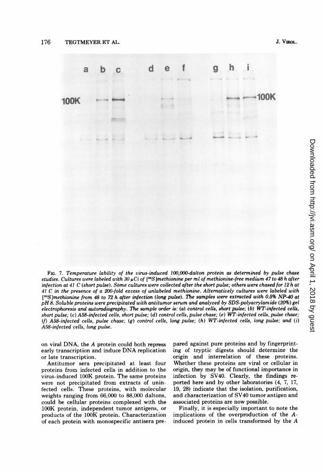
proteins from infected cells in addition to thevirus-induced lOOK protein. The same proteinswere not precipitated from extracts of unin-fected cells. These proteins, with molecularweights ranging from 66,000 to 88,000 daltons,could be cellular proteins complexed with the100K protein, independent tumor antigens, or
products of the lOOK protein. Characterizationof each protein with monospecific antisera pre-
pared against pure proteins and by fingerprint-ing of tryptic digests should determine theorigin and interrelation of these proteins.Whether these proteins are viral or cellular inorigin, they may be of functional importance ininfection by SV40. Clearly, the findings re-
ported here and by other laboratories (4, 7, 17,19, 28) indicate that the isolation, purification,and characterization of SV40 tumor antigen andassociated proteins are now possible.
Finally, it is especially important to note theimplications of the overproduction of the A-induced protein in cells transformed by the A
100K
g h i
J. VIROL.
on April 1, 2018 by guest
http://jvi.asm.org/
Dow
nloaded from

SIMIAN VIRUS 40 TUMOR ANTIGEN 177
a b c dtS ,.w __
_.. $1 .w vl .1 "
e_. _-- ..
f g h
ML 40
FIG. 8. Overproduction of the 100 K protein in rabbit cells transformed by A mutants. After stabletransformation of the rabbit cells by WT or mutant virus at 33 C, transformed and untransformed controlcultures were shifted to 41 C. After 24 h of incubation at 41 C, the cultures were labeled with 30 uCi of['5Sjmethionine per ml for either 1 h (short pulse) or 24 h (long pulse). After extraction with 0.5% NP-40 atpH8, the soluble proteins were precipitated with antitumor serum and analyzed by SDS-polyacrylamide (20%) gelelectrophoresis and autoradiography. The sample order is: (a) control cells, short pulse; (b) WT-transformedcells, short pulse; (c) A28-transformed cells, short pulse; (d) A58-transformed cells, short pulse; (e) control cells,long pulse; (f) WT-transformed cells, long pulse; (g) A28-transformed cells, long pulse; and (h) A58-trans-formed cells, long pulse.
mutants. First, these studies show that the Aprotein can regulate the production of tumorantigen when the viral genome is in the inte-grated state. Second, they strongly confirmprevious findings showing that the A proteinmay have a direct effect on the physiologicalstate of transformed cells (3, 16, 22, 25, 35).
ACKNOWLEDGMENTSThis investigation was supported by grant PRA-113 from
the American Cancer Society, grant 1256 from the Damon
Runyon Fund, and Public Health Service grant CA 16497from the National Cancer Institute.We are grateful for the skillful assistance of Judith
Kohout.
LITERATURE CITED
1. Anderson, C. W., and R. F. Gesteland. 1972. Pattem ofprotein synthesis in monkey cells infected by simianvirus 40. J. Virol. 9:758-765.
2. Black, P. H., W. P. Rowe, H. C. Turner, and R. J.Huebner. 1963. A specific complement-fixing antigenpresent in SV40 tumor and transformed cells. Proc.Natl. Acad. Sci. U.S.A. 50:1148-1156.
lOOK- -100K
VOL. 16, 1975
m ._-
I
t. I 11 ila... ...A
on April 1, 2018 by guest
http://jvi.asm.org/
Dow
nloaded from
178 TEGTMEYER ET AL.
3. Brugge, J. S., and J. S. Butel. 1975. Involvement of thesimian virus 40 gene A function in the maintenance oftransformation. J. Virol. 15:619-635.
4. Carroll, R. B., L. Hager, and R. Dulbecco. 1974. SV40 Tantigen binds to DNA. Proc. Natl. Acad. Sci. U.S.A.71:3754-3757.
5. Chou, J. Y., J. Avila, and R. G. Martin. 1974. Viral DNAsynthesis in cells infected by temperature-sensitivemutants of simian virus 40. J. Virol. 14:116-124.
6. Cowan, K., P. Tegtmeyer, and D. D. Anthony. 1973.Relationship of replication and transcription of simianvirus 40 DNA. Proc. Natl. Acad. Sci. U.S.A.70: 1927-1930.
7. Del Villano, B. C., and V. Defendi. 1973. Characteriza-tion of SV40 T antigen. Virology 51:34-46.
8. Fairbanks, G., T. L. Steck, and D. F. H. Wallach. 1971.Electrophoretic analysis of the polypeptides of thehuman erythrocyte membrane. Biochemistry10:2606-2617.
9. Gilden, R. V., R. I. Carp, F. Taguchi, and V. Defendi.1965. The nature and localization of the SV40-inducedcomplement-fixing antigen. Proc. Natl. Acad. Sci.U.S.A. 53:684-692.
10. Goldberger, R. F. 1974. Autogenous regulation of gene
expression. Science 183:810-816.11. Heinemann, S. F., and W. G. Spiegelman. 1970. Control
of transcription of the repressor gene in bacteriophagelambda. Proc. Natl. Acad. Sci. U.S.A. 67:1122-1129.
12. Henderson, I. C., and D. M. Livingston. 1974. Partialpurification and characterization of the SV40 T anti-gen. Cell 3:65-70.
13. Hoggan, M. D., W. P. Rowe, P. H. Black, and R. J.Huebner. 1965. Production of "tumor-specific" anti-gens by oncogenic viruses during acute cytolytic infec-tions. Proc. Natl. Acad. Sci. U.S.A. 53:12-19.
14. Khoury, G., P. Howley, D. Nathans, and M. Martin.1975. Post-transcriptional selection of simian virus40-specific RNA. J. Virol. 15:433-437.
15. Kiehn, E. D. 1973. Protein metabolism in SV40-infectedcells. Virology 56:313-333.
16. Kimura, G., and A. Itagaki. 1975. Initiation and mainte-nance of cell transformation by simian virus 40: a viralgenetic property. Proc. Natl. Acad. Sci. U.S.A.72:673-677.
17. Kourilsky, O., M. F. Bourginon, M. Bouquet, F. Gros.1970. Early transcription controls after induction ofprophage lambda. Cold Spring Harbor Symp. Quant.Biol. 35:305-314.
18. Krisch, H. M., A. Bolle, and R. H. Epstein. 1974.Regulation of the synthesis of bacteriophage T4 gene 32protein. J. Mol. Biol. 88:89-104.
19. Lazarus, H. M., M. B. Sporn, J. M. Smith, and W. R.Henderson. 1967. Purification of T antigen from nucleiof simian virus 40-induced hamster tumors. J. Virol.1:1093-1095.
20. Lewis, A. M., and W. P. Rowe. 1971. Studies on nondefec-tive adenovirus-simian 40 hybrid viruses. I. A newlycharacterized simian virus 40 antigen induced by theAd2+ND, virus. J. Virol. 7:189-197.
21. Manteuil, S., and M. Girard. 1974. Inhibitors of DNA
synthesis: their influence on the replication and tran-scription of simian virus 40 DNA. Virology 60:438-454.
22. Martin, R. G., and J. Y. Chou. 1975. Simian virus 40functions required for the establishment and mainte-nance of malignant transformation. J. Virol.15:599-612.
23. Maurer, H. R., and R. C. Allen. 1972. Useful buffer and gelsystems for polyacrylamide gel electrophoresis. Z. Klin.Chem. Klin. Biochem. 10:220-225.
24. Nathans, D., and K. J. Danna. 1972. Specific origin inSV40 DNA replication. Nature (London) New Biol.236:200-202.
25. Osborn, M., and K. Weber. 1975. Simian virus 40 gene Afunction and maintenance of transformation. J. Virol.15:636-644.
26. Oxman, M. N., S. Baron, P. H. Black, K. K. Takemoto,K. Habel, and W. P. Rowe. 1967. The effect ofinterferon on SV40 T antigen production in SV40transformed cells. Virology 32:122-127.
27. Pope, J. H., and W. P. Rowe. 1964. Detection of specificantigen in SV40 transformed cells by immunofluores-cence. J. Exp. Med. 120:121-128.
28. Potter, C. W., B. C. McLaughlin, and J. S. Oxford. 1969.Simian virus 40-induced T and tumor antigens. J.Virol. 4:574-579.
29. Rapp, F., J. S. Butel, and J. L. Melnick. 1964. Virus-induced intranuclear antigen in cells transformed bypapovavirus SV40. Proc. Soc. Exp. Biol. Med. 116:1131-1142
30. Rapp, F., T. Kitahara, J. S. Butel, and J. L. Melnick.1964. Synthesis of SV40 tumor antigen during replica-tion of simian papovavirus (SV40). Proc. Natl. Acad.Sci. U.S.A. 52:1138-1142.
31. Robb, J. A., and K. Huebner. 1973. Effect of cellchromosome number on simian virus 40 replicationExp. Cell. Res. 81:120-126.
32. Russel, M. 1973. Control of bacteriophage T4 polymerasesynthesis. J. Mol. Biol. 79:83-94.
33. Sabin, A. B., and M. A. Koch. 1964. Source of geneticinformation for specific complement-fixing antigens inSV40 virus-induced tumors. Proc. Natl. Acad. Sci.U.S.A. 52:1131-1138.
34. Tegtmeyer, P. 1972. Simian virus 40 deoxyribonucleicacid synthesis: the viral replicon. J. Virol. 10:591-598.
35. Tegtmeyer, P. 1975. Function of simian virus 40 gene A intransforming infection. J. Virol. 15:613-618.
36. Tegtmeyer, P., C. Dohan, and C. Reznikoff. 1970. Inac-tivating and mutagenic effects of nitrosoguanidine on
simian virus 40. Proc. Natl. Acad. Sci. U.S.A.66:745-752.
37. Tegtmeyer, P., and H. L. Ozer. 1971. Temperature-sensi-tive mutants of simian virus 40: infection of per-missive cells. J. Virol. 8:516-524.
38. Tegtmeyer, P., J. A. Robb, C. Widmer, and H. L. Ozer.1974. Altered protein metabolism in infection by thelate tsBll mutant of simian virus 40. J. Virol.14:997-1007.
39. Walter, G., R. Roblin, and R. Dulbecco. 1972. Proteinsynthesis in simian virus 40-infected monkey cells.Proc. Natl. Acad. Sci. U.S.A. 69:921-924.
J. VIROL.
on April 1, 2018 by guest
http://jvi.asm.org/
Dow
nloaded from