Shock in Polytrauma Patient
63
-
Upload
umesh-yadav -
Category
Health & Medicine
-
view
469 -
download
2
Transcript of Shock in Polytrauma Patient
How do you define Shock?
Systemic imbalance between oxygen supply and demand…
“Shock is inadequate tissue perfusion with oxygenated blood”
Shock is not a blood pressure diagnosis!!

Pathophysiology of Shock
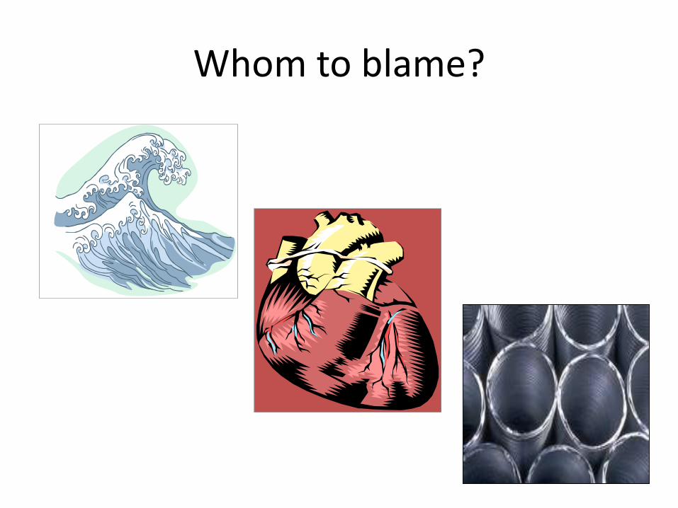
Whom to blame?
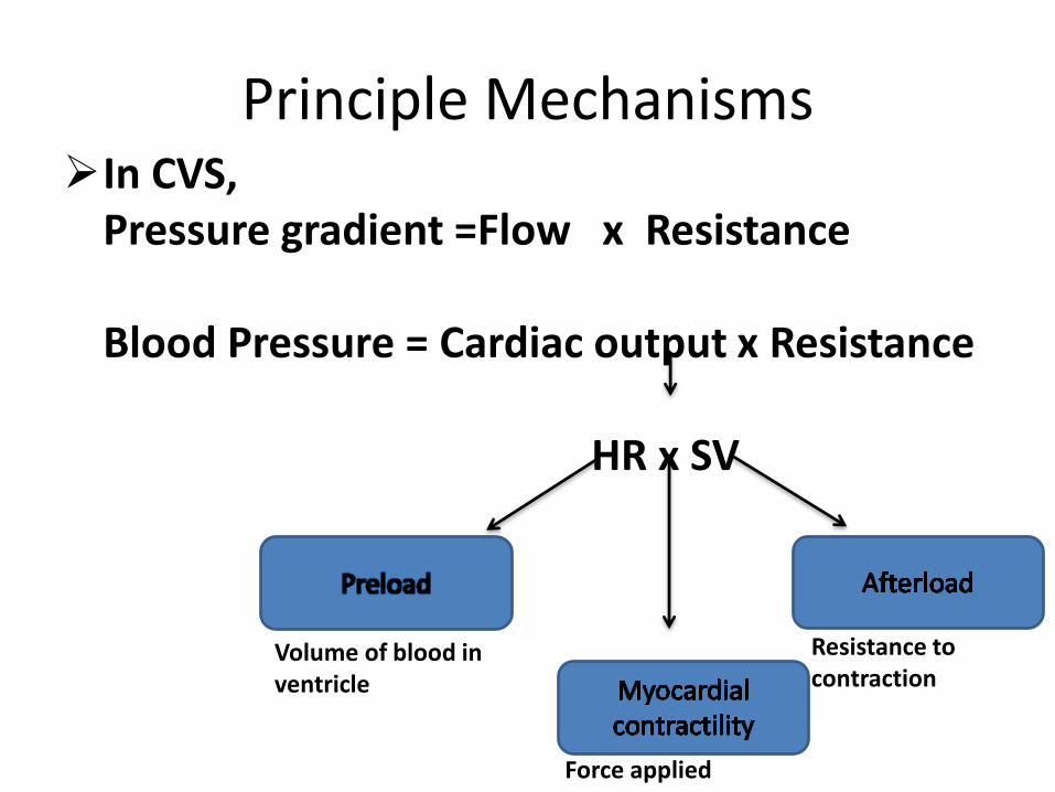
Principle MechanismsIn CVS,
Pressure gradient =Flow x Resistance
Blood Pressure = Cardiac output x Resistance
HR x SV
Volume of blood in ventricle
Resistance to contraction
Force applied
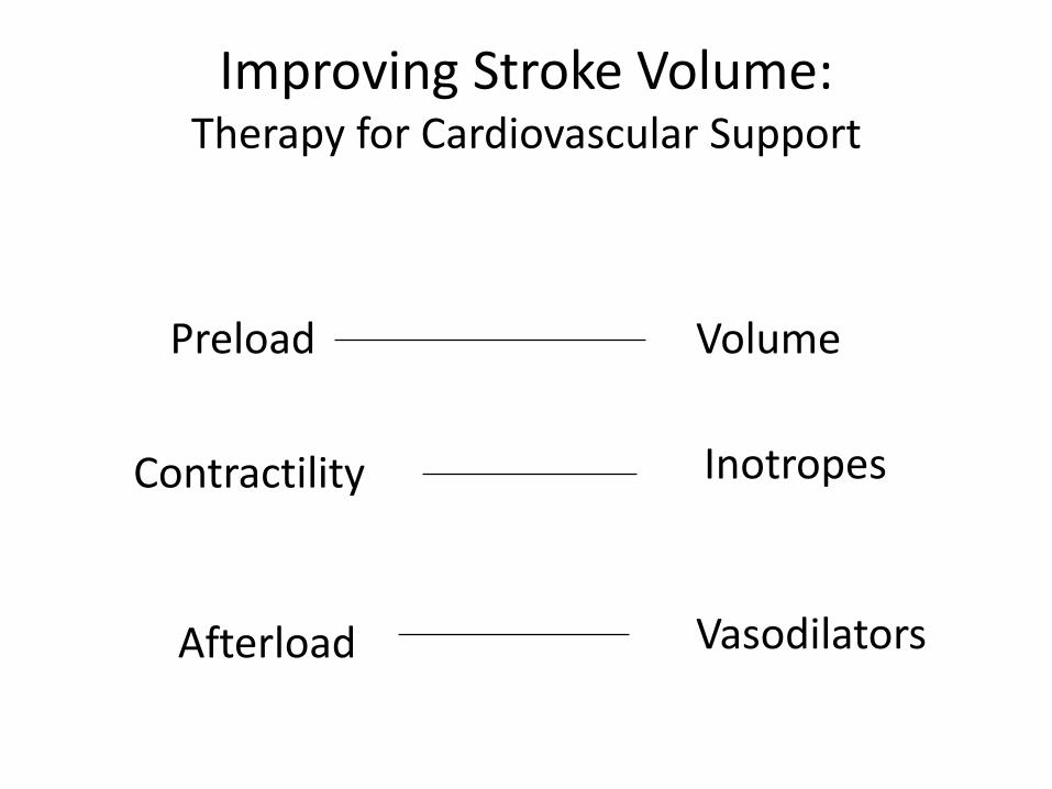
Improving Stroke Volume:Therapy for Cardiovascular Support
Preload Volume
Contractility Inotropes
Afterload Vasodilators
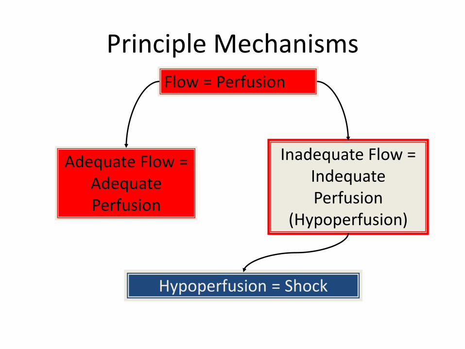
Principle MechanismsFlow = Perfusion
Adequate Flow = Adequate Perfusion
Inadequate Flow = IndequatePerfusion
(Hypoperfusion)
Hypoperfusion = Shock

Types of Shock• Hypovolemic Shock
• Distributive Shock
– Neurogenic shock
– Septic shock
– Anaphylactic shock
• Cardiogenic Shock
Stages of Shock
• Initial Stage
• Compensatory Stage
• Progressive Stage
• Irreversible Stage
Stages of Shock
• Initial Stage
• Compensatory Stage
• Progressive Stage
• Irreversible Stage
Initial Stage
• Initially, the body compensates with the onset of shock.
• No changes are noted clinically.
• Changes are beginning to occur on the cellular level.
Stages of Shock
• Initial Stage
• Compensatory Stage
• Progressive Stage
• Irreversible Stage
Compensatory Stage
• Fluid shift from insterstital to intravascular space.
• Activation of SNS - activation of epinephrine and norepinephrine.
• Kidneys release renin into blood formation of angiotension & release of aldosterone, ADH
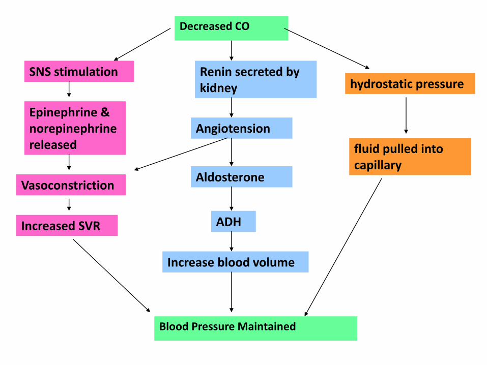
Decreased CO
SNS stimulation
Epinephrine & norepinephrine released
Vasoconstriction
Increased SVR
Renin secreted by kidney
Angiotension
Aldosterone
ADH
Increase blood volume
hydrostatic pressure
fluid pulled into capillary
Blood Pressure Maintained
• Initial Stage
• Compensatory Stage
• Progressive Stage
• Irreversible Stage
Stages of Shock
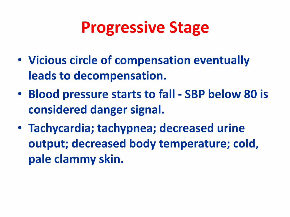
Progressive Stage
• Vicious circle of compensation eventually leads to decompensation.
• Blood pressure starts to fall - SBP below 80 is considered danger signal.
• Tachycardia; tachypnea; decreased urine output; decreased body temperature; cold, pale clammy skin.
• Initial Stage
• Compensatory Stage
• Progressive Stage
• Irreversible Stage
Stages of Shock
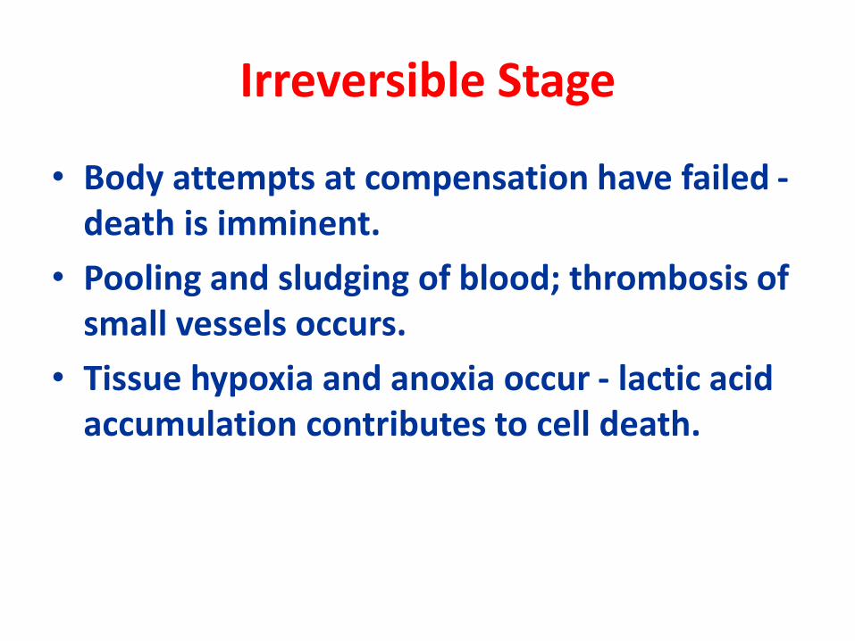
Irreversible Stage
• Body attempts at compensation have failed -death is imminent.
• Pooling and sludging of blood; thrombosis of small vessels occurs.
• Tissue hypoxia and anoxia occur - lactic acid accumulation contributes to cell death.
HYPOVOLEMIC SHOCK IN POLYTRAUMA PATIENT
• Shock is a common and frequently treatable cause of death in injured patients and is second only to traumatic brain injury as the leading cause of death from trauma.
• ATLS (ADVANCED TRAUMA LIFE SUPPORT)
WHEN A PATIENT ARRIVES….
• Primary survey-• ●Airway assessment and protection (maintain cervical
spine stabilization when appropriate)• ●Breathing and ventilation assessment (maintain
adequate oxygenation)• ●Circulation assessment (control hemorrhage and
maintain adequate end-organ perfusion)• ●Disability assessment (perform basic neurologic
evaluation)• ●Exposure, with environmental control (undress
patient and search everywhere for possible injury, while preventing hypothermia)
Is the patient in SHOCK?????
• BLOOD PRESSURE- Not reliable ….
( BP falls when 30% blood volume lost )
Look for- Pulse Rate
Respiratory rate
Skin circulation
Pulse pressure (SBP-DBP)
ANY INJURED PATIENT WHO IS COOL & HAS TACHYCARDIA IS CONSIDERED TO BE IN SHOCK UNTIL PROVEN OTHERWISE.
• Haematocrit / Haemoglobin- Not reliable in Acute blood loss.
• MASSIVE BLOOD LOSS MAY PRODUCE ONLY MINIMAL DECREASE.
• SO, a VERY LOW HAEMATOCRIT SHORTLY AFTER INJURY- MASSIVE BLOOD LOSS OR PRE EXISTING ANAEMIA while a normal doesn’t exclude significant blood loss.
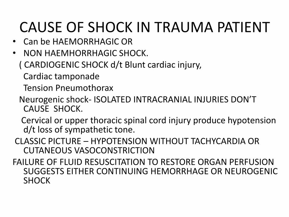
CAUSE OF SHOCK IN TRAUMA PATIENT• Can be HAEMORRHAGIC OR • NON HAEMHORRHAGIC SHOCK.
( CARDIOGENIC SHOCK d/t Blunt cardiac injury, Cardiac tamponadeTension Pneumothorax
Neurogenic shock- ISOLATED INTRACRANIAL INJURIES DON’T CAUSE SHOCK.
Cervical or upper thoracic spinal cord injury produce hypotension d/t loss of sympathetic tone.
CLASSIC PICTURE – HYPOTENSION WITHOUT TACHYCARDIA OR CUTANEOUS VASOCONSTRICTION
FAILURE OF FLUID RESUSCITATION TO RESTORE ORGAN PERFUSION SUGGESTS EITHER CONTINUING HEMORRHAGE OR NEUROGENIC SHOCK
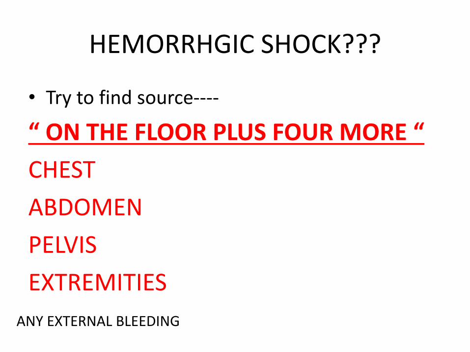
HEMORRHGIC SHOCK???
• Try to find source----
“ ON THE FLOOR PLUS FOUR MORE “
CHEST
ABDOMEN
PELVIS
EXTREMITIES
ANY EXTERNAL BLEEDING
WHAT TO DO
• Xray abdomen , chest and pelvis
• Focussed Assesment Sonography in Trauma (FAST)
• Diagnostic Peritoneal Lavage (DPL)
• Computed Tomography (CT)
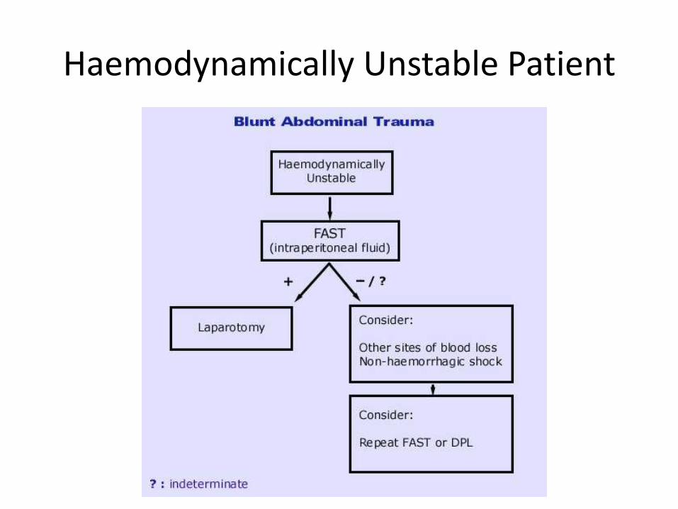
Haemodynamically Unstable Patient
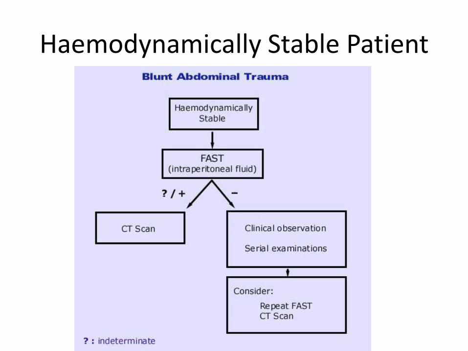
Haemodynamically Stable Patient
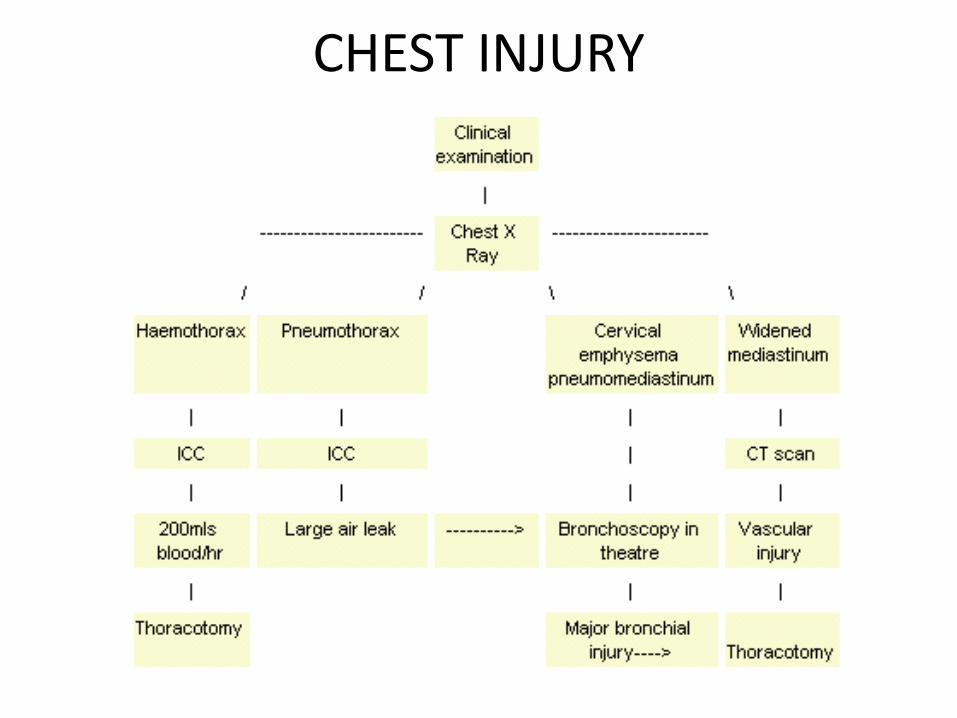
CHEST INJURY
HEMORRHAGIC SHOCK
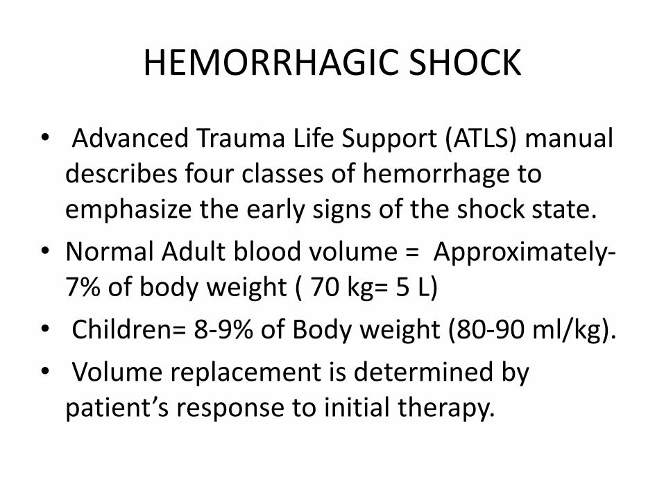
• Advanced Trauma Life Support (ATLS) manual describes four classes of hemorrhage to emphasize the early signs of the shock state.
• Normal Adult blood volume = Approximately-7% of body weight ( 70 kg= 5 L)
• Children= 8-9% of Body weight (80-90 ml/kg).
• Volume replacement is determined by patient’s response to initial therapy.
• Hemhorrhage control & balanced fluid resusicitation must be initiated when early S/S of blood loss are apparent or suspected- NOT WHEN BP IS FALLING OR ABSENT.
• BLEEDING PATIENT NEEDS BLOOD….otherwise
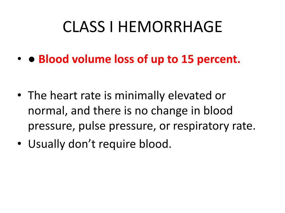
CLASS I HEMORRHAGE
• ● Blood volume loss of up to 15 percent.
• The heart rate is minimally elevated or normal, and there is no change in blood pressure, pulse pressure, or respiratory rate.
• Usually don’t require blood.
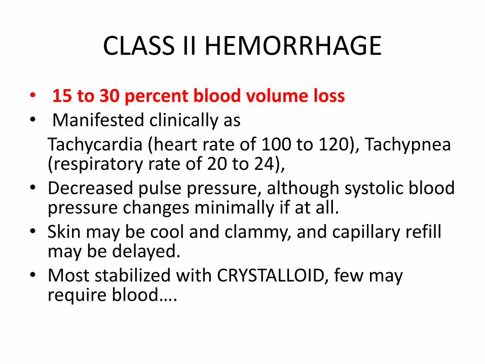
CLASS II HEMORRHAGE
• 15 to 30 percent blood volume loss• Manifested clinically as
Tachycardia (heart rate of 100 to 120), Tachypnea(respiratory rate of 20 to 24),
• Decreased pulse pressure, although systolic blood pressure changes minimally if at all.
• Skin may be cool and clammy, and capillary refill may be delayed.
• Most stabilized with CRYSTALLOID, few may require blood….
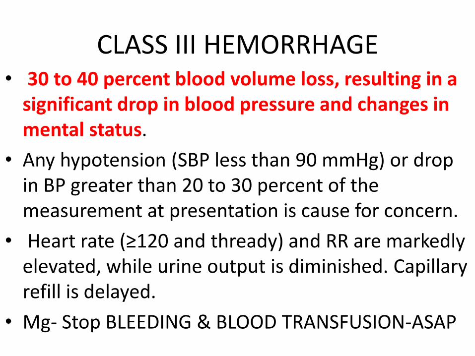
CLASS III HEMORRHAGE• 30 to 40 percent blood volume loss, resulting in a
significant drop in blood pressure and changes in mental status.
• Any hypotension (SBP less than 90 mmHg) or drop in BP greater than 20 to 30 percent of the measurement at presentation is cause for concern.
• Heart rate (≥120 and thready) and RR are markedly elevated, while urine output is diminished. Capillary refill is delayed.
• Mg- Stop BLEEDING & BLOOD TRANSFUSION-ASAP
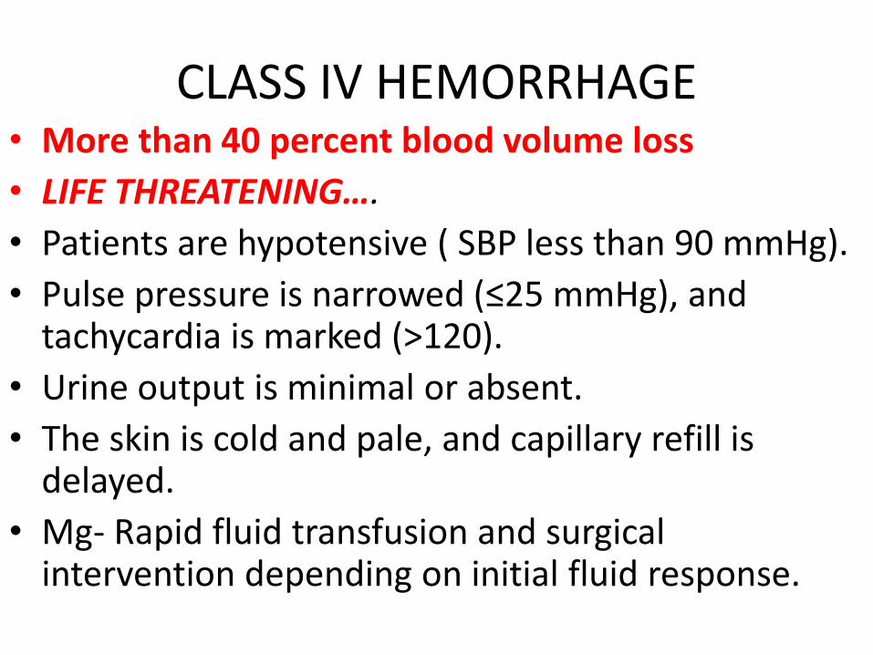
CLASS IV HEMORRHAGE• More than 40 percent blood volume loss
• LIFE THREATENING….
• Patients are hypotensive ( SBP less than 90 mmHg).
• Pulse pressure is narrowed (≤25 mmHg), and tachycardia is marked (>120).
• Urine output is minimal or absent.
• The skin is cold and pale, and capillary refill is delayed.
• Mg- Rapid fluid transfusion and surgical intervention depending on initial fluid response.
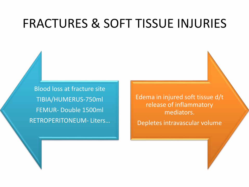
FRACTURES & SOFT TISSUE INJURIES
Blood loss at fracture site
TIBIA/HUMERUS-750ml
FEMUR- Double 1500ml
RETROPERITONEUM- Liters…
Edema in injured soft tissue d/t release of inflammatory
mediators.
Depletes intravascular volume
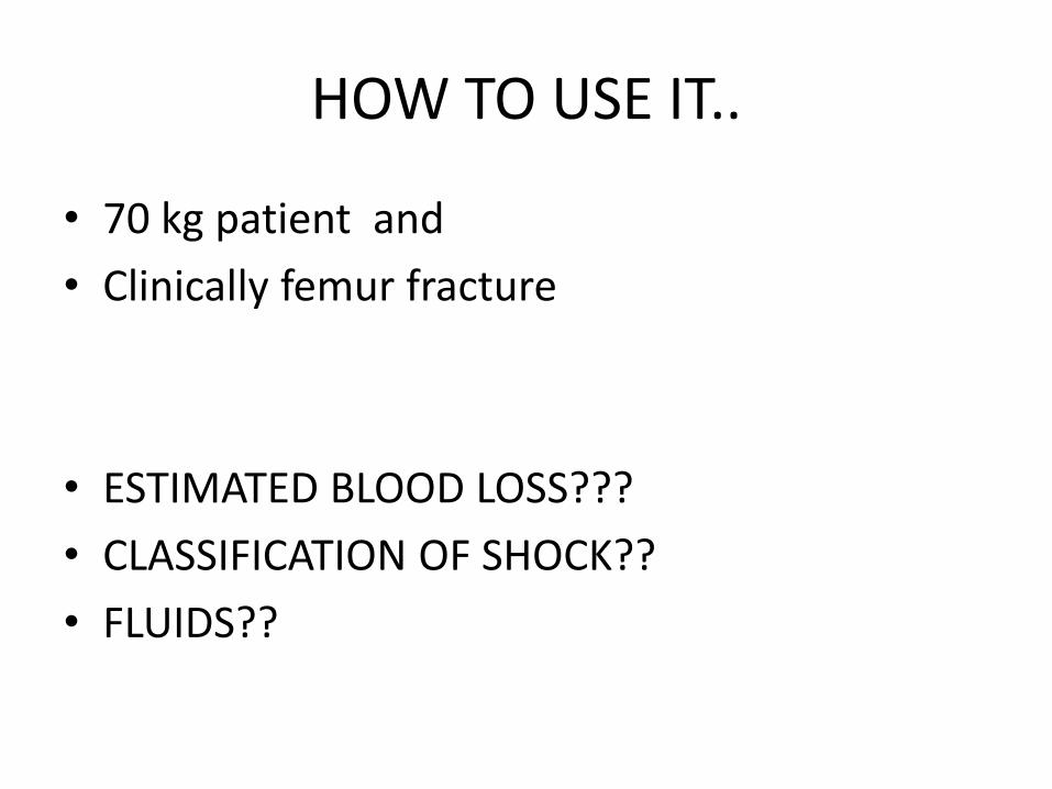
HOW TO USE IT..
• 70 kg patient and
• Clinically femur fracture
• ESTIMATED BLOOD LOSS???
• CLASSIFICATION OF SHOCK??
• FLUIDS??
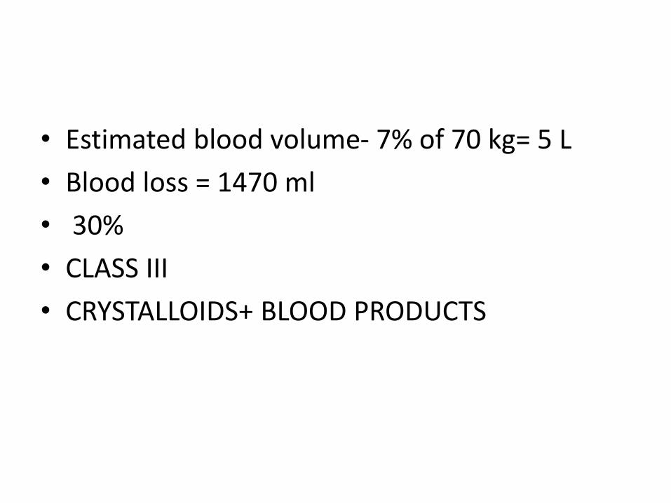
• Estimated blood volume- 7% of 70 kg= 5 L
• Blood loss = 1470 ml
• 30%
• CLASS III
• CRYSTALLOIDS+ BLOOD PRODUCTS
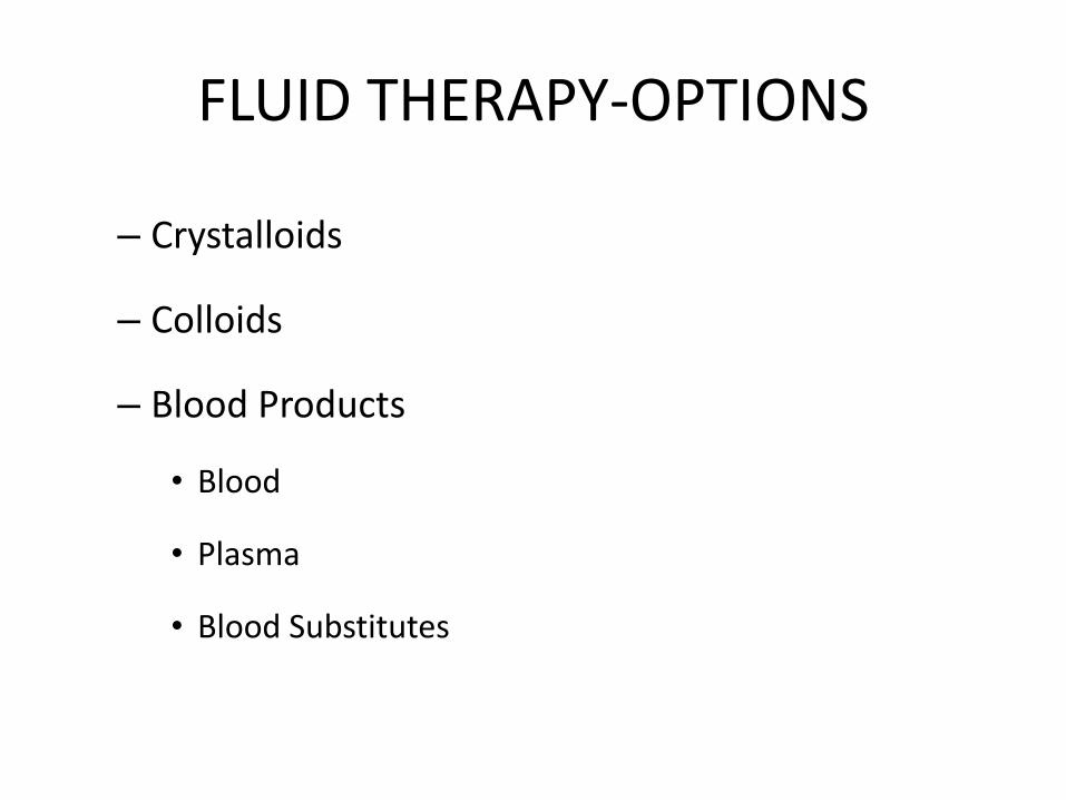
FLUID THERAPY-OPTIONS
– Crystalloids
– Colloids
– Blood Products
• Blood
• Plasma
• Blood Substitutes
CRYSTALLOIDS
Solutions that contain small molecules that flow easily across the cell membranes, allowing for transfer from the bloodstream into the cells and body tissues.
This will increase fluid volume in both the interstitial and intravascular spaces (Extracellular)
Commonly used CRYSTALLOIDS
0.9% sodium chloride (0.9% NaCl)
lactated Ringer's solution
5% dextrose in water (D5W)
Ringer's solution-RL with out lactate
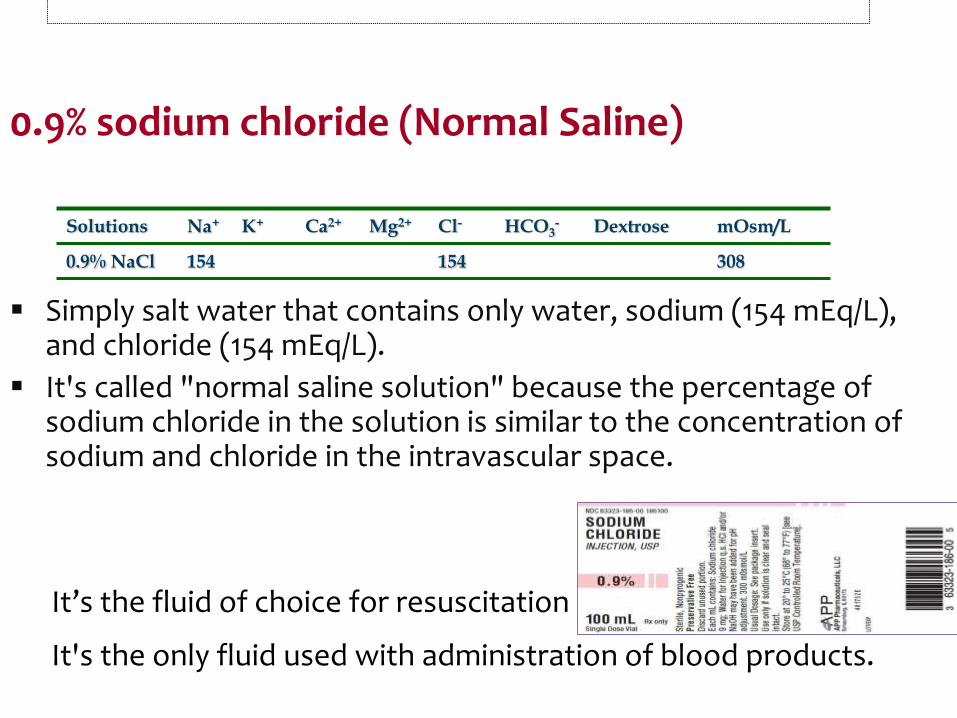
0.9% sodium chloride (Normal Saline)
Simply salt water that contains only water, sodium (154 mEq/L), and chloride (154 mEq/L).
It's called "normal saline solution" because the percentage of sodium chloride in the solution is similar to the concentration of sodium and chloride in the intravascular space.
It’s the fluid of choice for resuscitation efforts.
It's the only fluid used with administration of blood products.
Solutions Na+ K+ Ca2+ Mg2+ Cl- HCO3- Dextrose mOsm/L
0.9% NaCl 154 154 308
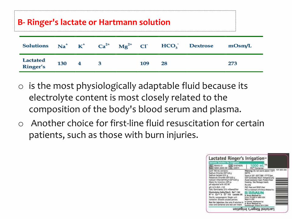
o is the most physiologically adaptable fluid because its electrolyte content is most closely related to the composition of the body's blood serum and plasma.
o Another choice for first-line fluid resuscitation for certain patients, such as those with burn injuries.
B- Ringer's lactate or Hartmann solution
Solutions Na +K +
Ca 2+Mg 2+
Cl - HCO3 - Dextrose mOsm/L
Lactated
Ringer’s 130 4 3
109 28
273
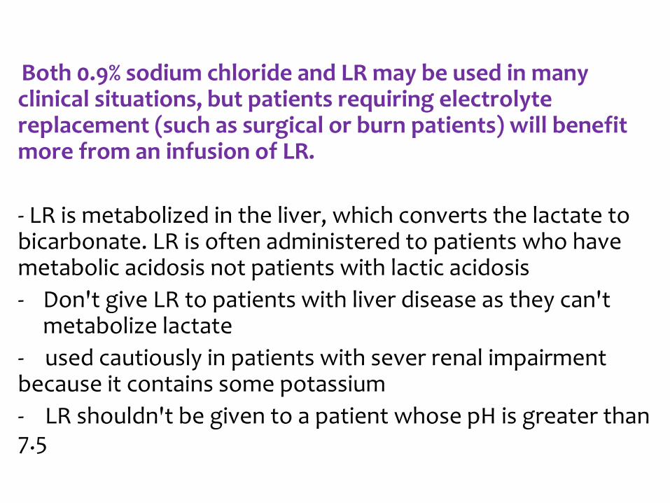
Both 0.9% sodium chloride and LR may be used in many clinical situations, but patients requiring electrolyte replacement (such as surgical or burn patients) will benefit more from an infusion of LR.
- LR is metabolized in the liver, which converts the lactate to bicarbonate. LR is often administered to patients who have metabolic acidosis not patients with lactic acidosis
- Don't give LR to patients with liver disease as they can't metabolize lactate
- used cautiously in patients with sever renal impairment because it contains some potassium
- LR shouldn't be given to a patient whose pH is greater than 7.5
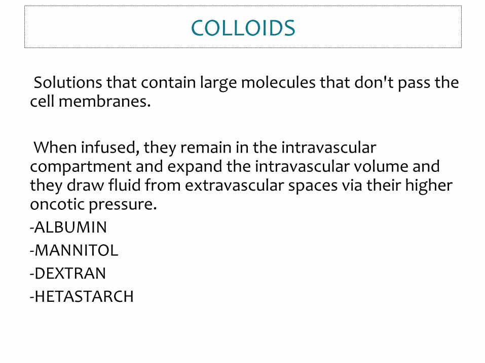
Solutions that contain large molecules that don't pass the cell membranes.
When infused, they remain in the intravascular compartment and expand the intravascular volume and they draw fluid from extravascular spaces via their higher oncotic pressure.
-ALBUMIN
-MANNITOL
-DEXTRAN
-HETASTARCH
COLLOIDS
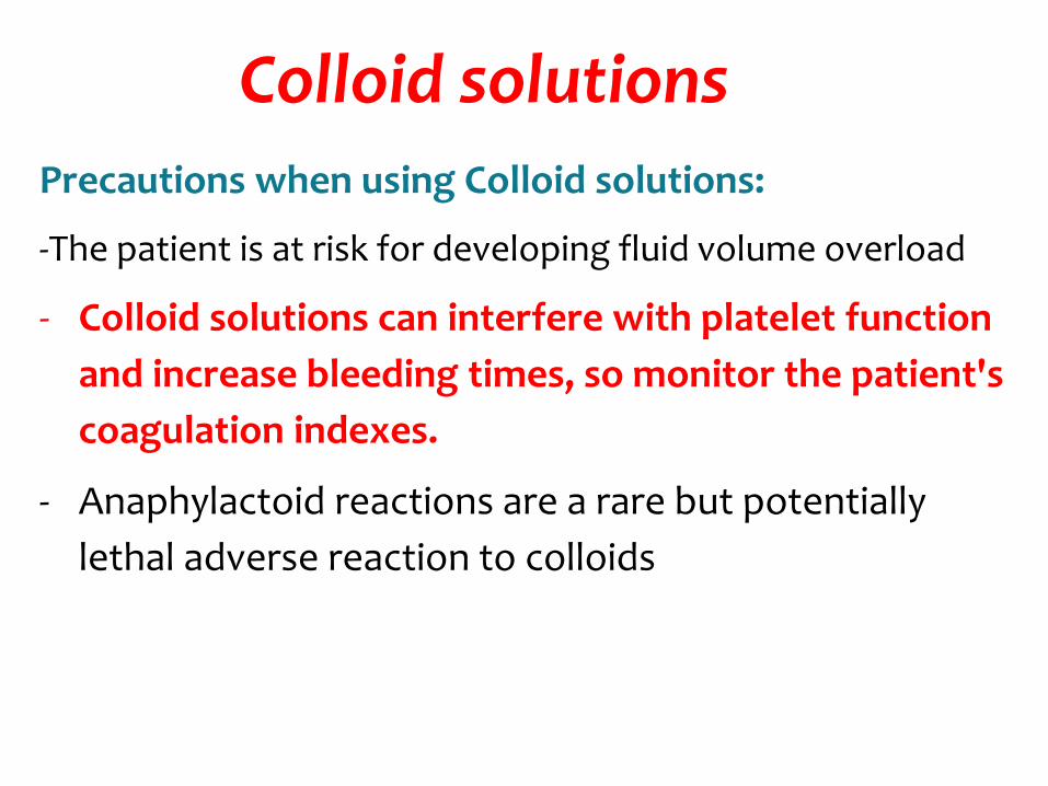
Colloid solutions
Precautions when using Colloid solutions:
-The patient is at risk for developing fluid volume overload
- Colloid solutions can interfere with platelet function
and increase bleeding times, so monitor the patient's
coagulation indexes.
- Anaphylactoid reactions are a rare but potentially
lethal adverse reaction to colloids
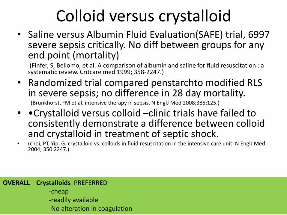
Colloid versus crystalloid• Saline versus Albumin Fluid Evaluation(SAFE) trial, 6997
severe sepsis critically. No diff between groups for any end point (mortality)(Finfer, S, Bellomo, et al. A comparison of albumin and saline for fluid resuscitation : a
systematic review. Critcare med 1999; 358-2247.)
• Randomized trial compared penstarchto modified RLS in severe sepsis; no difference in 28 day mortality.(Brunkhorst, FM et al. intensive therapy in sepsis, N EnglJ Med 2008;385:125.)
• •Crystalloid versus colloid –clinic trials have failed to consistently demonstrate a difference between colloid and crystalloid in treatment of septic shock.
• (choi, PT, Yip, G. crystalloid vs. colloids in fluid resuscitation in the intensive care unit. N EnglJ Med 2004; 350:2247.)
OVERALL Crystalloids PREFERRED-cheap-readily available-No alteration in coagulation
INITIAL FLUID
• Initial fluid resuscitation for trauma patients in hemorrhagic shock consist of 2 L of isotonic saline (ie, normal saline, NS) given as rapidly as possible through short, large gauge (16 or larger) peripheral IVs.
• Central venous catheters are used when peripheral IVs are not available.
• Monitor patient response to initial fluids resuscitation and identify evidence of end organ perfusion.
DANGERS OF EXCESSIVE FLUIDS
• Infusions of large volumes of NS can lead to the development of a nonanion gap hyperchloremicmetabolic acidosis.
• PERSISTANT INFUSION OF LARGE VOLUME OF FLUID AND BLOOD IN AN ATTEMPT TO ACHIEVE NORMAL BP IS NOT SUBSTITUTE FOR DEFINITIVE CONTROL OF BLEEDING.
• TRIAD- COAGULOPATHY- ACIDOSIS- HYPOTHERMIA
• SO MONITOR…..
WHAT TO MONITOR• BP, PP PR-A mean arterial pressure (MAP) around 65
mmHg or a systolic blood pressure (SBP) around 90 mmHg is a reasonable goal in penetrating trauma (MAP = [(2 x diastolic) + systolic]/3).
• CVP STATUS & SKIN PERFUSION- Improvement is evidence of evidence of enhanced perfusion but difficult to quantitate.
• URINE OUTPUT- 0.5 ml/kg/hr in adults1 ml/kg/hr in children
ACID BASE BALANCE- Initially resp alkalosis-d/t tachypnea f/b metabolic acidosis in long standing shock d/t anaerobic metabolism.
CVP• Reflects the pressure in the central veins.
• CVP (right atrial pressure) indicates pressure and not volume.
• DIFFICULTY-
• Shocked patients: low intravascular volume with compensatory vasoconstriction: CVP low.
• Rapid resuscitation: fluid poured into a constricted patient will increase BP & push CVP up rapidly, but the vasculature may still be constricted.
• Redistribution: normally the patient's vasculature will dilate a little and the fluid will redistribute slowly. CVP falls to zero but the patient remains constricted.
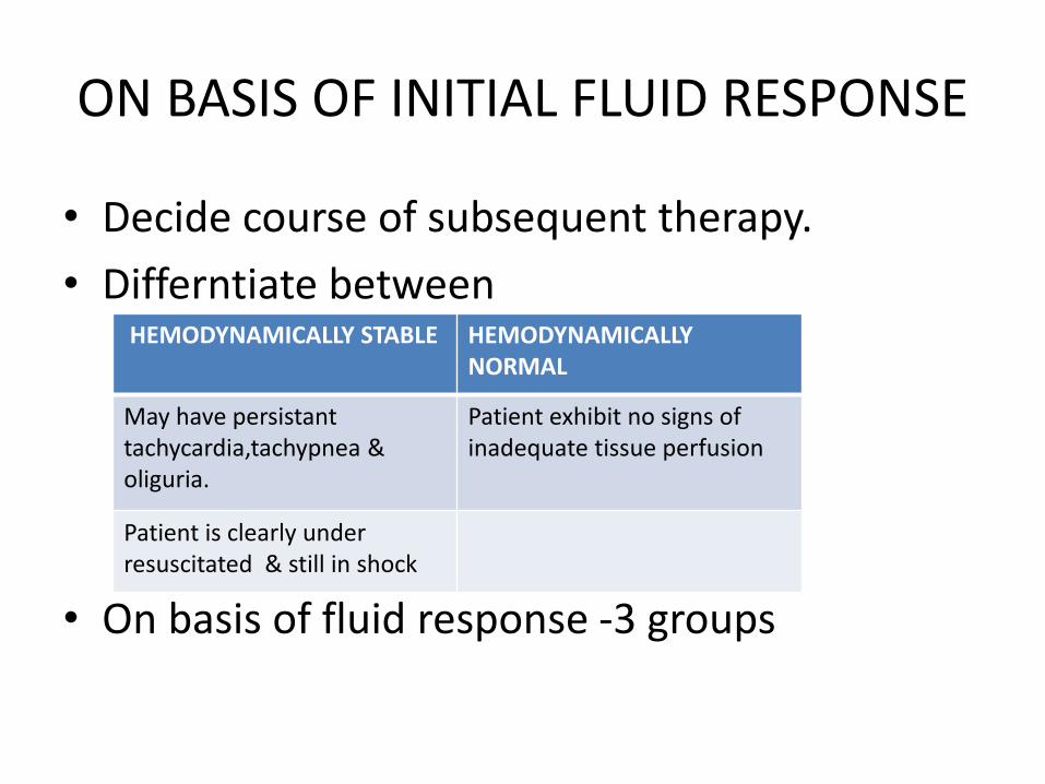
ON BASIS OF INITIAL FLUID RESPONSE
• Decide course of subsequent therapy.
• Differntiate between
• On basis of fluid response -3 groups
HEMODYNAMICALLY STABLE HEMODYNAMICALLY NORMAL
May have persistanttachycardia,tachypnea & oliguria.
Patient exhibit no signs of inadequate tissue perfusion
Patient is clearly under resuscitated & still in shock
RAPID RESPONSERS
• Responds to initial fluid bolus & remains hemodynamically NORMAL in maintaiencephase.
• Estimated blood loss- 20%
• Blood & further surgical evaluation may be needed
TRANSIENT RESPONDERS
• Initially improve but deteriorate when fluids slowed to maintenance level d/t- Ongoing blood loss or inadequate resuscitation.
• Estimated blood loss- 20-40%
• Blood transfusion & in transiently respond to blood- Urgent surgical intervention.
NON RESPONDERS
• Failure to respond to crystalloids or Blood-IMMEDIATE &URGENT SURGICAL INTERVENTION (OPERATION /ANGIOEMBOLIZATION ) to control exsanguinating hemorrhage.
• ALSO CONSIDER- NON HEMOHRRGIC SHOCK (CVP Monitoring and Cardiac ultrasound rules out)
BLOOD TRANSFUSION
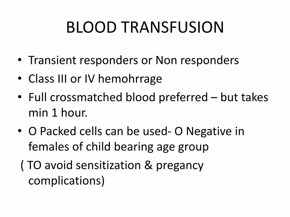
• Transient responders or Non responders
• Class III or IV hemohrrage
• Full crossmatched blood preferred – but takes min 1 hour.
• O Packed cells can be used- O Negative in females of child bearing age group
( TO avoid sensitization & pregancycomplications)
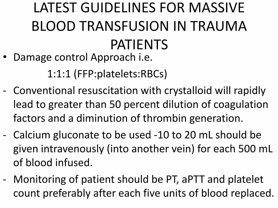
LATEST GUIDELINES FOR MASSIVE BLOOD TRANSFUSION IN TRAUMA
PATIENTS• Damage control Approach i.e.
1:1:1 (FFP:platelets:RBCs)
- Conventional resuscitation with crystalloid will rapidly lead to greater than 50 percent dilution of coagulation factors and a diminution of thrombin generation.
- Calcium gluconate to be used -10 to 20 mL should be given intravenously (into another vein) for each 500 mLof blood infused.
- Monitoring of patient should be PT, aPTT and platelet count preferably after each five units of blood replaced.
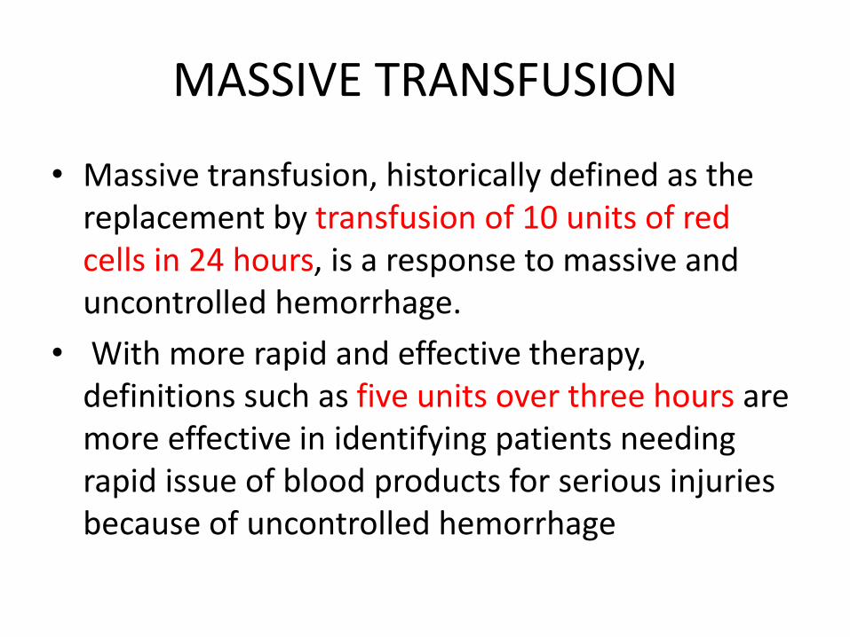
MASSIVE TRANSFUSION
• Massive transfusion, historically defined as the replacement by transfusion of 10 units of red cells in 24 hours, is a response to massive and uncontrolled hemorrhage.
• With more rapid and effective therapy, definitions such as five units over three hours are more effective in identifying patients needing rapid issue of blood products for serious injuries because of uncontrolled hemorrhage
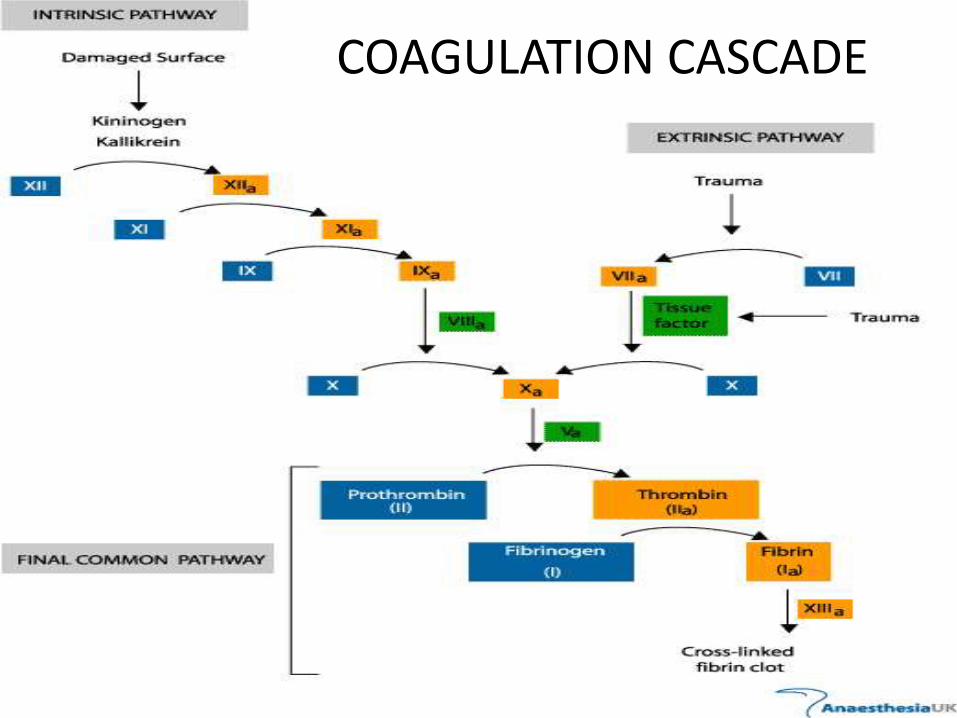
COAGULATION CASCADE
Risk associated• COAGULOPATHY-
Activation and consumption of coagulation factors secondary to tissue trauma, such as massive head injury or muscle damage
Reduced activity of coagulation factors from prolonged shock, hypoxia, hypothermia, or failure to clear activation peptides that act as competitive inhibitors.
Such trauma-associated coagulopathy can be diagnosed as Acute disseminated intravascular coagulation (acute DIC).
Other explanations for COAGULOPATHY
• Tissue injury/trauma and shock, and their associated physiologic changes (ie, acidosis, hypothermia, consumption of coagulant proteins, and fibrinolysis) combined with extensive blood loss and the dilutional effects of physiologic vascular refill and fluid replacement therapy.
• Acidosis and Hypothermia also alters coagulation.
• Decrease platelet count-
• D/t dilutional effect of massive transfusion.
• In an adult, each 10 to 12 units of transfused RBCs are associated with a 50 percent fall in the platelet count; thus, significant thrombocytopenia can be seen after 10 to 20 units of blood, with platelet counts below 50,000/microL
Reed RL Jr, Ciavarella D, Heimbach DM, et al. Prophylactic platelet administration during massive transfusion. A prospective, randomized, double-blind clinical study. Ann Surg 1986; 203:48.
MONITORING
• PT, aPTT, fibrinogen concentration, and platelet count