Serum Myoglobln Determination: Laboratory and Clinical Evaluation
6
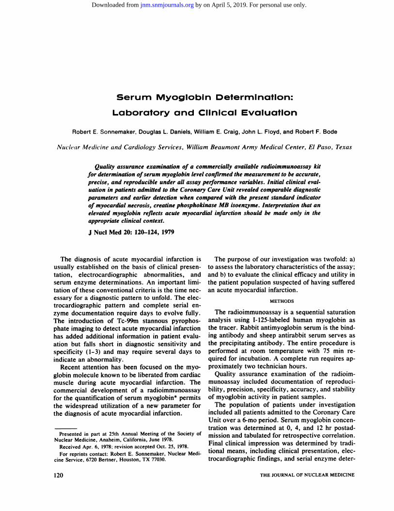
Serum Myoglobln Determination: Laboratory and Clinical Evaluation Robert E. Sonnemaker, Douglas L. Daniels, William E. Craig, John L. Floyd, and Robert F. Bode Nuclear Medicine and Cardiology Services, William Beaumont Army Medical Center, El Paso, Texas Quality assurance examination of a commercially available radioimmunoassay kit for determination ofserum myoglobin level confirmed the measurement to be accurate, precise, and reproducible under all assay performance variables. Initial clinical eval uation in patients admitted to the Coronary Care Unit revealed comparable diagnostic parameters and earlier detection when compared with the present standard indicator of myocardial necrosis, creatine phosphokinase MB isoenzyme. Interpretation that an elevated myoglobin reflects acute myocardial infarction should be made only in the appropriate clinical context. J Nuci Med 20: 120—124, 1979 The diagnosis of acute myocardial infarction is usually established on the basis of clinical presen tation, electrocardiographic abnormalities, and serum enzyme determinations. An important limi tation of these conventional criteria is the time nec essary for a diagnostic pattern to unfold. The elec trocardiographic pattern and complete serial en zyme documentation require days to evolve fully. The introduction of Tc-99m stannous pyrophos phate imaging to detect acute myocardial infarction has added additional information in patient evalu ation but falls short in diagnostic sensitivity and specificity (1-3) and may require several days to indicate an abnormality. Recent attention has been focused on the myo globin molecule known to be liberated from cardiac muscle during acute myocardial infarction. The commercial development of a radioimmunoassay for the quantification of serum myoglobin* permits the widespread utilization of a new parameter for the diagnosis of acute myocardial infarction. Presented in part at 25th Annual Meeting of the Society of Nuclear Medicine, Anaheim, California, June 1978. Received Apr. 6, 1978; revision accepted Oct. 25, 1978. For reprints contact: Robert E. Sonnemaker, Nuclear Mcdi cine Service, 6720 Bertner, Houston, TX 77030. The purpose of our investigation was twofold: a) to assess the laboratory characteristics of the assay; and b) to evaluate the clinical efficacy and utility in the patient population suspected of having suffered an acute myocardial infarction. METHODS The radioimmunoassay is a sequential saturation analysis using 1-125-labeled human myoglobin as the tracer. Rabbit antimyoglobin serum is the bind ing antibody and sheep antirabbit serum serves as the precipitating antibody. The entire procedure is performed at room temperature with 75 mm re quired for incubation. A complete run requires ap proximately two technician hours. Quality assurance examination of the radioim munoassay included documentation of reproduci bility, precision, specificity, accuracy, and stability of myoglobin activity in patient samples. The population of patients under investigation included all patients admitted to the Coronary Care Unit over a 6-mo period. Serum myoglobin concen tration was determined at 0, 4, and 12 hr postad mission and tabulated for retrospective correlation. Final clinical impression was determined by tradi tional means, including clinical presentation, dcc trocardiographic findings, and serial enzyme deter 120 THE JOURNAL OF NUCLEAR MEDICINE by on April 5, 2019. For personal use only. jnm.snmjournals.org Downloaded from
Transcript of Serum Myoglobln Determination: Laboratory and Clinical Evaluation
Serum Myoglobln Determination:
Laboratory and Clinical Evaluation
Robert E. Sonnemaker, Douglas L. Daniels,William E. Craig, John L. Floyd, and Robert F. Bode
Nuclear Medicine and Cardiology Services, William Beaumont Army Medical Center, El Paso, Texas
Quality assurance examination of a commercially available radioimmunoassay kitfor determination ofserum myoglobin level confirmed the measurement to be accurate,precise, and reproducible under all assay performance variables. Initial clinical evaluation in patients admitted to the Coronary Care Unit revealed comparable diagnosticparameters and earlier detection when compared with the present standard indicatorof myocardial necrosis, creatine phosphokinase MB isoenzyme. Interpretation that anelevated myoglobin reflects acute myocardial infarction should be made only in theappropriate clinical context.
J Nuci Med 20: 120—124,1979
The diagnosis of acute myocardial infarction isusually established on the basis of clinical presentation, electrocardiographic abnormalities, andserum enzyme determinations. An important limitation of these conventional criteria is the time necessary for a diagnostic pattern to unfold. The electrocardiographic pattern and complete serial enzyme documentation require days to evolve fully.The introduction of Tc-99m stannous pyrophosphate imaging to detect acute myocardial infarctionhas added additional information in patient evaluation but falls short in diagnostic sensitivity andspecificity (1-3) and may require several days toindicate an abnormality.
Recent attention has been focused on the myoglobin molecule known to be liberated from cardiacmuscle during acute myocardial infarction. Thecommercial development of a radioimmunoassayfor the quantification of serum myoglobin* permitsthe widespread utilization of a new parameter forthe diagnosis of acute myocardial infarction.
Presented in part at 25th Annual Meeting of the Society ofNuclear Medicine, Anaheim, California, June 1978.
Received Apr. 6, 1978; revision accepted Oct. 25, 1978.For reprints contact: Robert E. Sonnemaker, Nuclear Mcdi
cine Service, 6720 Bertner, Houston, TX 77030.
The purpose of our investigation was twofold: a)to assess the laboratory characteristics of the assay;and b) to evaluate the clinical efficacy and utility inthe patient population suspected of having sufferedan acute myocardial infarction.
METHODS
The radioimmunoassay is a sequential saturationanalysis using 1-125-labeled human myoglobin asthe tracer. Rabbit antimyoglobin serum is the binding antibody and sheep antirabbit serum serves asthe precipitating antibody. The entire procedure isperformed at room temperature with 75 mm required for incubation. A complete run requires approximately two technician hours.
Quality assurance examination of the radioimmunoassay included documentation of reproducibility, precision, specificity, accuracy, and stabilityof myoglobin activity in patient samples.
The population of patients under investigationincluded all patients admitted to the Coronary CareUnit over a 6-mo period. Serum myoglobin concentration was determined at 0, 4, and 12 hr postadmission and tabulated for retrospective correlation.Final clinical impression was determined by traditional means, including clinical presentation, dcctrocardiographic findings, and serial enzyme deter
120 THE JOURNAL OF NUCLEAR MEDICINE
by on April 5, 2019. For personal use only. jnm.snmjournals.org Downloaded from
Among runs (n = 24) 7.22 5.89 6.48Among tech's (n = 3) 4.92 2.91 4.17Among lots (n = 5) 2.86 4.18 3.42
Coefficient of variation(%)
Low Highserum serum All
esdCVSerumsample (ng/mI)(ng/ml)(%)
CLINICAL SCIENCESIN VITRO NUCLEAR MEDICINE
minations independent of serum myoglobin levels.Diagnoses were classified as acute myocardial infarction (23 patients) or no acute myocardial infarction (113 patients), the latter including angina documented by ECG (20 patients) and typical oratypical chest pain without ECG abnormalities (93patients). Acute myocardial infarctions were further subclassified as transmural or subendocardial.The normal distribution, mean value, and estimatedstandard deviation of serum myoglobin were derived from the patient group with no acute myocardial infarction. Myoglobin results were comparedwith those of the MB isoenzyme of creatine phosphokinase (CPK-MB) as determined by the selective activation technique.t Serum CPK-MB concentrations were determined at admission andevery 8 hr thereafter for 3 consecutive days. Comparison included time of appearance of the initialabnormal values and time to peak abnormal valuesof both myoglobin and CPK-MB in patients withacute myocardial infarction. Additionally, diagnostic parameters were derived.
RESULTS
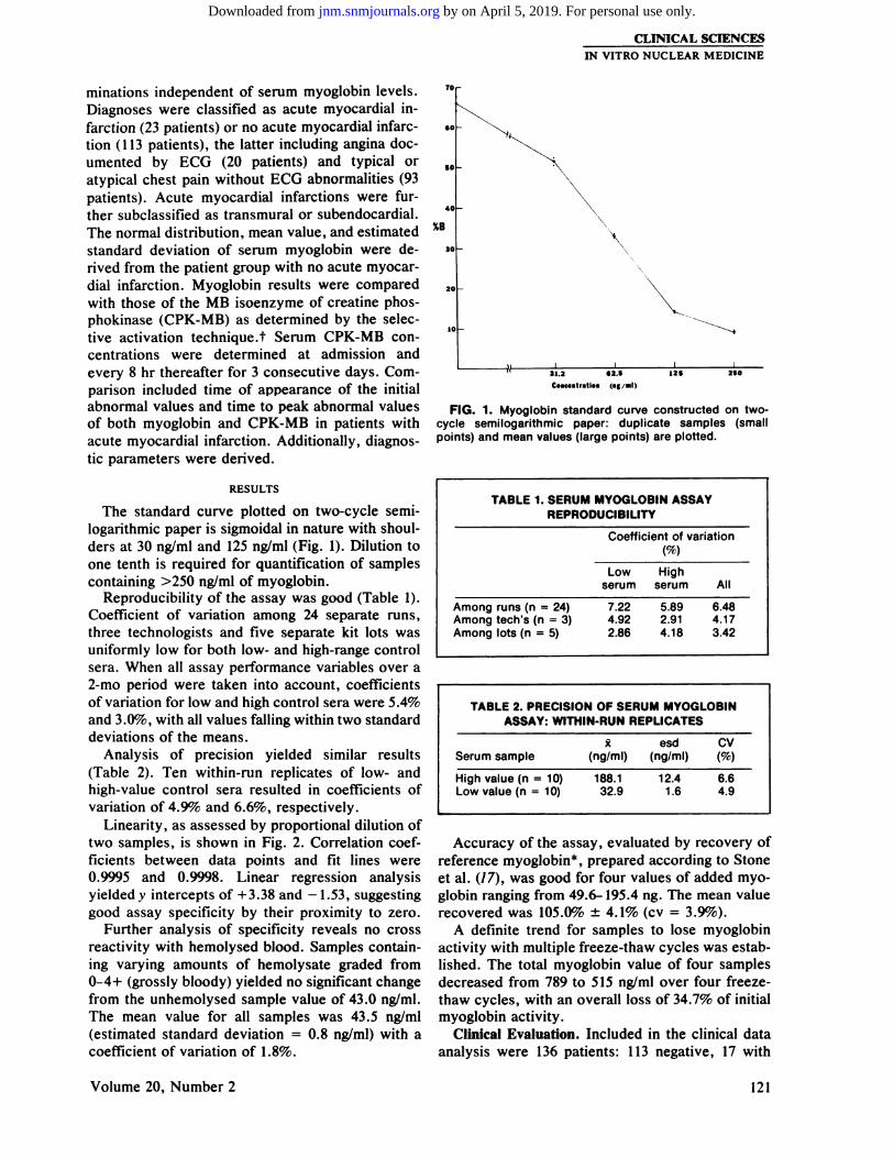
The standard curve plotted on two-cycle semilogarithmic paper is sigmoidal in nature with shoulders at 30 ng/ml and 125 ng/ml (Fig. 1). Dilution toone tenth is required for quantification of samplescontaining >250 ng/ml of myoglobin.
Reproducibility of the assay was good (Table 1).Coefficient of variation among 24 separate runs,three technologists and five separate kit lots wasuniformly low for both low- and high-range controlsera. When all assay performance variables over a2-mo period were taken into account, coefficientsof variation for low and high control sera were 5.4%and 3.0%, with all values falling within two standarddeviations of the means.
Analysis of precision yielded similar results(Table 2). Ten within-run replicates of low- andhigh-value control sera resulted in coefficients ofvariation of 4.9% and 6.6%, respectively.
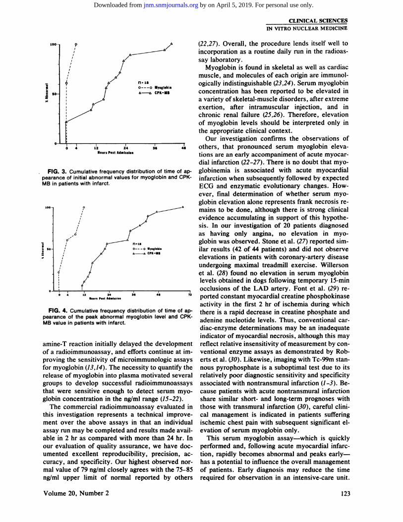
Linearity, as assessed by proportional dilution oftwo samples, is shown in Fig. 2. Correlation coefficients between data points and fit lines were0.9995 and 0.9998. Linear regression analysisyielded y intercepts of +3.38 and —1.53, suggestinggood assay specificity by their proximity to zero.
Further analysis of specificity reveals no crossreactivity with hemolysed blood. Samples containing varying amounts of hemolysate graded from0-4+ (grossly bloody) yielded no significant changefrom the unhemolysed sample value of 43.0 ng/ml.The mean value for all samples was 43.5 ng/ml(estimated standard deviation = 0.8 ng/ml) with acoefficient of variation of 1.8%.
70
@@::@
20
10
11 31.2 62.5 125 250C..c.atratt.u (ug/1)
FIG. 1. Myoglobin standard curve constructed on twocycle semilogarithmic paper: duplicate samples (smallpoints) and meanvalues(large points) are plotted.
TABLE 1. SERUM MYOGLOBIN ASSAYREPRODUCIBILITY
TABLE 2. PRECISION OF SERUM MYOGLOBINASSAY: WIThIN-RUN REPLICATES
High value (n = 10)Low value (n = 10)
188.1 12.4 6.632.9 1.6 4.9
Accuracy of the assay, evaluated by recovery ofreference myoglobin* , prepared according to Stoneet al. (17), was good for four values of added myoglobin ranging from 49.6- 195.4 ng. The mean valuerecovered was 105.0% ±4.1% (cv = 3.9%).
A definite trend for samples to lose myoglobinactivity with multiple freeze-thaw cycles was established. The total myoglobin value of four samplesdecreased from 789 to 515 ng/ml over four freezethaw cycles, with an overall loss of 34.7% of initialmyoglobin activity.
Clinical Evaluation. Included in the clinical dataanalysis were 136 patients: 113 negative, 17 with
Volume 20, Number 2 121
by on April 5, 2019. For personal use only. jnm.snmjournals.org Downloaded from

Result Myoglobin* CPK-MBt
SONNEMAKER,DANIELS, CRAIG, FLOYD, AND BODE
CPK-MB diagnostic parameters are as expectedconsidering the overlap of values from normal andabnormal patient groups. Sensitivity = 100%, specificity = 90%, positive predictive value = 68%,negative predictive value = 100%, and overall diagnostic accuracy = 92% (Table 3). Chi-squareanalysis with Yate's correction yields a high degreeof association with final clinical diagnosis.
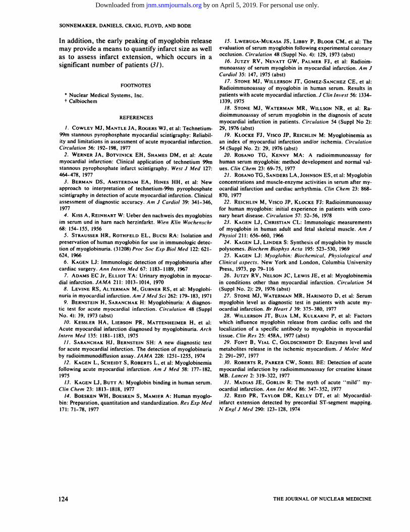
Serial determinations of both myoglobin andCPK-MB permitted examination of both the timeof first appearance and the time of appearance ofpeak abnormal values. The cumulative frequencydistribution of the appearance of the first abnormalvalues for myoglobin and CPK-MB demonstratesthat myoglobin abnormalities precede CPK-MB abnormalities by 12-24 hr in the great majority ofcases (Fig. 3). Likewise, peak abnormal myoglobinvalues preceded peak abnormal CPK-MB values byat least 24 hrs in the majority of cases (Fig. 4).
DISCUSSION
Myoglobin, an oxygen-binding hememolecule oflow molecular weight, was first reported in 1956 tobe present in the serum and urine of patients withacute myocardial infarctions (4). Myoglobinuria accompanying infarction was substantiated by Strausser et al. (5) in 1966 and later by others (6-10);however, the relative insensitivity of measurementusing precipitin and hemagglutination-inhibitionprocedures limited detection of myoglobin to unnary appearance. Although myoglobinuria had beensuggested as the earliest parameter detectable inacute myocardial infarction (8-11), Kagel et al. (12),using a complement-fixation technique with increased sensitivity of measurement, showed thatmyoglobinuria was not a good indicator of the degree of myoglobinemia. Difficulties with radioiodination of human myoglobin by the traditional chlor
1
I
S
SU
FIG. 2. Proportional dilution of two myoglobin samplesconfirms excellent test linearity. Ordinate shows measuredmyoglobin concentration; abscissa gives reciprocal of dilution factor, as percentage of original.
transmural infarction, and six with subendocardialinfarction. Patients with trauma, intramuscular injection, or defibrillation were excluded. There wereI 13 patients identified as having no evidence for anacute myocardial infarction. The 339 serum myoglobin values from these patients ranged from 6.2 to79 ng/ml. The normal value distribution approximated a standard normal curve with the mean at33.0 ng/ml (kurtosis = 3.88, skewness = 0.72). Estimated standard deviation was 13.8 ng/ml, whichplaced mean + 3 s.d. at 74.4 ng/ml, very near the79 ng/ml of the highest value obtained. Four ofthese patients demonstrated myoglobin valuesgreater than the mean + 3 s.d. level (76.5, 78.0,79.0, 79.0). Peak myoglobin values from 17 patientswith transmural infarction ranged from 176 to 1100ng/ml. Peak values from six patients with subendocardial infarction ranged from 120 to 250 ng/ml.
Myoglobin diagnostic parameters were all 100%.The high specificity reflects the exclusion of knowncauses for false-positive elevation. Chi-square analysis with Yate's correction demonstrates a highlysignificant association of myoglobin results with thefinal clinical impression (Table 3).
CPK-MB values from the 113 negative patientsranged from 0 to 24 lUll. The mean value was 4.6lUll (s.d. = 3.4 lUll, kurtosis = 5.84, skewness =1.46). Eleven of the negative patients demonstratedCPK-MB values greater than the mean + 3 s.d.level of 14.8 (range: 15-24 lUll) with the highestvalue (24 lUll) 2.7 s.d. above the mean + 3 s.d.value. Peak CPK-MB values from patients withtransmural infarction ranged from 27 to 490 lU/i,whereas peak values from patients with subendocardial infarction ranged from 16 to 70 lU/i. In contrast to the clear separation of abnormal from normal myoglobin values, there was considerableoverlap between normal and abnormal CPK-MBvalues.
VItIUC (I.peSSwd.s%)
TABLE 3. DIAGNOSTIC PARAMETERS OFMYOGLOBIN AND CPK-MB ASSAYS
Ture positive 23 23True negative 113 102Falsepositive 0 11False negative 0 0Sensitivity 100% 100%Specificity 100% 90%Positive predictive value 100% 68%Negative predictive value 100% 100%Accuracy 100% 92%
* Chi square with Yate's<.0005)
@ Chi square with Yates correction = 78.6 (p<.0005)
correction = 128.6 (p
122 THE JOURNAL OF NUCLEAR MEDICINE
by on April 5, 2019. For personal use only. jnm.snmjournals.org Downloaded from
CLINICAL SCIENCESIN VITRO NUCLEAR MEDICINE
(22,27). Overall, the procedure lends itself well toincorporation as a routine daily run in the radioassaylaboratory.
Myoglobin is found in skeletal as well as cardiacmuscle, and molecules of each origin are immunologically indistinguishable (23,24). Serum myoglobinconcentration has been reported to be elevated ina variety of skeletal-muscle disorders, after extremeexertion, after intramuscular injection, and inchronic renal failure (25,26). Therefore, elevationof myoglobin levels should be interpreted only inthe appropriate clinical context.
Our investigation confirms the observations of3@6 4@ others, that pronounced serum myoglobin eleva
tions are an early accompaniment of acute myocardial infarction (22-27). There is no doubt that myoglobinemia is associated with acute myocardialinfarction when subsequently followed by expectedECG and enzymatic evolutionary changes. However, final determination of whether serum myoglobin elevation alone represents frank necrosis remains to be done, although there is strong clinicalevidence accumulating in support of this hypothesis. In our investigation of 20 patients diagnosedas having only angina, no elevation in myoglobin was observed. Stone et al. (27) reported similar results (42 of 44 patients) and did not observeelevations in patients with coronary-artery diseaseundergoing maximal treadmill exercise. Willersonet al. (28) found no elevation in serum myoglobinlevels obtained in dogs following temporary 15-mmocclusions of the LAD artery. Font et al. (29) re
72 ported constant myocardial creatine phosphokinase
activity in the first 2 hr of ischcmia during whichthere is a rapid decrease in creatine phosphate andadenine nucleotide levels. Thus, conventional cardiac-enzyme determinations may be an inadequateindicator of myocardial necrosis, although this mayreflect relative insensitivity of measurement by conventional enzyme assays as demonstrated by Roberts et al. (30). Likewise, imaging with Tc-99m stannous pyrophosphate is a suboptimal test due to itsrelatively poor diagnostic sensitivity and specificityassociated with nontransmural infarction (1-3). Because patients with acute nontransmural infarctionshare similar short- and long-term prognoses withthose with transmural infarction (30), careful clinical management is indicated in patients sufferingischemic chest pain with subsequent significant cievation of serum myoglobin only.
This serum myoglobin assay—which is quicklyperformed and, following acute myocardial infarction, rapidly becomes abnormal and peaks earlyhas a potential to influence the overall managementof patients. Early diagnosis may reduce the timerequired for observation in an intensive-care unit.
p A
A
/A
IA'
K'A@ —0MyOglObtiA—A CPK-MI
100
50
I
1,
t 4 12 24HoursPost Admissio.
FIG. 3. Cumulativefrequencydistributionof timeofappearance of initial abnormal values for myoglobin and CPKMB inpatientswithinfarct.
100
so
.0
9-
Q
S
A―
I0———0 MpsgI.bi.I,,@A7 A—A
ft/
—@-@-@ @4 3@Is.,. P.it Ad.Issto.
FIG. 4. Cumulative frequency distribution of time of appearance of the peak abnormal myoglobin level and CPKMB value in patients with infarct.
amine-T reaction initially delayed the developmentof a radioimmunoassay, and efforts continue at improving the sensitivity of microimmunologic assaysfor myoglobin (13,14). The necessity to quantify therelease of myoglobin into plasma motivated severalgroups to develop successful radioimmunoassaysthat were sensitive enough to detect serum myoglobin concentration in the ng/ml range (15-22).
The commercial radioimmunoassay evaluated inthis investigation represents a technical improve
ment over the above assays in that an individualassay run may be completed and results made available in 2 hr as compared with more than 24 hr. Inour evaluation of quality assurance, we have documented excellent reproducibility, precision, accuracy, and specificity. Our highest observed normal value of 79 ng/ml closely agrees with the 75-85ng/ml upper limit of normal reported by others
Volume 20, Number 2 123
by on April 5, 2019. For personal use only. jnm.snmjournals.org Downloaded from
SONNEMAKER, DANIELS, CRAIG, FLOYD, AND BODE
In addition, the early peaking of myoglobin releasemay provide a means to quantify infarct size as wellas to assess infarct extension, which occurs in asignificant number of patients (31).
FOOTNOTES
* Nuclear Medical Systems, Inc.
t Calbiochem
REFERENCES
I. COWLEY MJ, MANTLE JA, ROGERS Wi, et al: Technetium99m stannous pyrophosphate myocardial scintigraphy: Reliability and limitations in assessment of acute myocardial infarction.Circulation 56: 192-198, 1977
2. WERNERJA, B0TvINIcK EH, SHAMESDM, et al: Acutemyocardial infarction: Clinical application of technetium 99mstannous pyrophosphate infarct scintigraphy. West J Med 127:464-478,1977
3. BERMANDS, AMSTERDAMEA, HINESHH, Ct al: Newapproach to interpretation of technetium-99m pyrophosphatescintigraphy in detection of acute myocardial infarction. Clinicalassessment of diagnostic accuracy. Am J Cardiol 39: 341-346,1977
4. Kiss A, REINHART W: Ueber den nachweis des myoglobinsim serum und in ham nach herzinfarkt. Wien Klin Wochenschr68: 154—155,1956
5. STRAUSSERHR, ROTHFELDEL, Bucsi RA: Isolationandpreservation of human myoglobin for use in immunologic detection of myoglobinuna. (31208) Proc Soc Exp Biol Med 122: 621-624, 1966
6. KAGENU: Immunologicdetectionof myoglobinuriaaftercardiac surgery. Ann Intern Med 67: 1183- 1189, 1967
7. ADAMSEC Jr, ELLIOTTA: Urinary myoglobin in myocardial infarction.JAMA 211: 1013-1014, 1970
8. LEVINERS, ALTERMANM, GUBNERRS, et al: Myoglobinuria in myocardial infarction. Am'.! Med Sci 262: 179- 183, 1971
9. BERNSTEINH, SARANCHAKH: Myoglobinuria:A diagnostic test for acute myocardial infarction. Circulation 48 (SupplNo. 4): 39, 1973 (abst)
10. KESSLER HA, LIEB50N PR, MATTENHEIMER H, Ct al:Acute myocardial infarction diagnosed by myoglobinuria. ArchInternMed 135:1181—1183,1975
II. SARANCHAK HJ, BERNSTEIN SH: A new diagnostic testfor acute myocardial infarction. The detection of myoglobinuriaby radioimmunodiffusion assay. JAMA 228: 1251- 1255, 1974
/2. KAGENL, SCHEIDTS, ROBERTSL, et al: Myoglobinemiafollowing acute myocardial infarction. Am J Med 58: 177-182,1975
13. KAGEN U, Burr A: Myoglobin binding in human serum.Cli,,C/tern23: 1813-1818,1977
14. BOESKEN WH, BOE5KEN S, MAMIER A: Human myoglobin: Preparation, quantitation and standardization. Res Exp Med171:71-78,1977
15. LWEBUGA-MUKA5A JS, LIBBY P. BLOOR CM, et al: The
evaluation of serum myoglobin following experimental coronaryocclusion. Circulation 48 (Suppl No. 4): 129, 1973 (abst)
16. JUTZY RV, NEVATT OW, PALMER FJ, et al: Radioimmunoassay of serum myoglobin in myocardial infarction. Am JCardiol35: 147,1975(abst)
17. STONE MJ, WILLER50N JT, GOMEZ-SANCHEZ CE, et al:
Radioimmunoassay of myoglobin in human serum. Results inpatients with acute myocardial infarction. J Clin Invest 56: 1334—1339,1975
18. STONE MJ, WATERMAN MR. WILL5ON NR, et al: Ra
dioimmunoassay of serum myoglobin in the diagnosis of acutemyocardial infarction in patients. Circulation 54 (Suppl No 2):29, 1976 (abst)
19. KLOCKE FJ, Vlsco JP, REICHLIN M: Myoglobinemia asan index of myocardial infarction and/or ischemia. Circulation54 (Suppl No. 2): 29, 1976 (abst)
20. ROSANO TO, KENNY MA: A radioimmunoassay for
human serum myoglobin: method development and normal values. Clin Chem 23: 69—75,1977
21. RosANo TO, SANDERS LA, JOHNSON ES, et al: Myoglobinconcentrations and muscle-enzyme activities in serum after myocardial infarction and cardiac arrhythmia. Clin Chem 23: 868-870, 1977
22. REICHLINM, Vlsco JP, KLOCKEFJ: Radioimmunoassayfor human myoglobin: initial experience in patients with coronary heart disease. Circulation 57: 52-56, 1978
23. KAGEN U, CHRISTIAN CL: Immunologic measurements
of myoglobin in human adult and fetal skeletal muscle. Am JPhysiol 211: 656-660, 1966
24. KAGEN II, LINDER S: Synthesis of myoglobin by muscle
polysomes. Biochem Biophys Ada 195: 523-530, 196925. KAGENLI: Myoglobin:Biochemical,Physiologicaland
Clinical aspects. New York and London, Columbia UniversityPress, 1973, pp 79-116
26. JUTZY RV, NELSON JC, LEwis JE, et al: Myoglobinemia
in conditions other than myocardial infarction. Circulation 54(Suppl No. 2): 29, 1976 (abst)
27. STONEMJ, WATERMANMR. HARIMOTOD, et al: Serummyoglobin level as diagnostic test in patients with acute myocardial infarction. Br Heart J 39: 375-380, 1977
28. WILLERSON JT, BUM LM, KULKARNI P, et al: Factors
which influence myoglobin release from cardiac cells and thelocalization of a specific antibody to myoglobin in myocardialtissue. Clin Res 25: 458A, 1977 (abst)
29. FONT B, VIAL C, GOLDSCHMIDTD: Enzymes level andmetabolites release in the ischemic myocardium. J Molec Med2:291-297,1977
30. ROBERTSR, PARKERCW, SOBELBE: Detection of acutemyocardial infarction by radioimmunoassay for creatine kinaseMB. Lancet 2: 319-322, 1977
3!. MADIASJE, GORLINR: The myth of acute “mild―myocardial infarction. Ann ml Med 86: 347-352, 1977
32. REID PR, TAYLORDR. KELLY DT, et al: Myocardialinfarct extension detected by precordial ST-segment mapping.NEnglJMed29O: 123-128,1974
124 THE JOURNAL OF NUCLEAR MEDICINE
by on April 5, 2019. For personal use only. jnm.snmjournals.org Downloaded from
1979;20:120-124.J Nucl Med. Robert E. Sonnemaker, Douglas L. Daniels, William E. Craig, John L. Floyd and Robert F. Bode Serum Myoglobin Determination: Laboratory and Clinical Evaluation
http://jnm.snmjournals.org/content/20/2/120This article and updated information are available at:
http://jnm.snmjournals.org/site/subscriptions/online.xhtml
Information about subscriptions to JNM can be found at:
http://jnm.snmjournals.org/site/misc/permission.xhtmlInformation about reproducing figures, tables, or other portions of this article can be found online at:
(Print ISSN: 0161-5505, Online ISSN: 2159-662X)1850 Samuel Morse Drive, Reston, VA 20190.SNMMI | Society of Nuclear Medicine and Molecular Imaging
is published monthly.The Journal of Nuclear Medicine
© Copyright 1979 SNMMI; all rights reserved.
by on April 5, 2019. For personal use only. jnm.snmjournals.org Downloaded from