Separation of Renal Medullary Cells: Isolation of Cells...
10
Separation of Renal Medullary Cells : Isolation of Cells from the Thick Ascending Limb of Henle's Loop JILL EVELOFF, W. HAASE, and ROLF KINNE Max-Planck-Institut für Biophysik, 6000 Frankfurt am Main 71, Germany. Dr. EvelofPs and Dr. Kinne's present address is the Department of Physiology, Albert Einstein College of Medicine, Bronx, New York 10461 . ABSTRACT A homogeneous population of single cells from the thick ascending limb of Henle's loop (TALH) has been isolated from the rabbit kidney medulla . A total medullary cell suspension was prepared by a series of collagenase, hyaluronidase, and trypsin digestions and separated on a Ficoll gradient (2 .6-30 .7% wt/wt) . Morphologically, the cells isolated from the TALH were homogeneous and showed polarity within their plasma membrane structure, with a few blunt microvilli on their apical surface and deep infoldings of the basal-lateral membrane . Biochemically, the TALH cells were highly enriched in calcitonin-sensitive adenylate cyclase and Na, K-ATPase . Alkaline phosphatase and arginine vasopressin-sensitive adenylate cyclase, highly concentrated in proximal tubule and collecting duct, were present only in low concen- trations in the TALH cells . Additionally, furosemide, a diuretic inhibiting sodium chloride transport in the TALH in vivo, inhibited oxygen consumption of the TALH cells in a close- dependent manner . The TALH cells were viable, as judged by morphological appearance, trypan blue exclusion, the response of oxygen consumption to 2,4-dinitrophenol, succinate and ouabain, and the cellular Na, K and ATP levels . The kidney is a heterogeneous organ and each nephron is composed of a multitude of cell types having widely differing structures and functions . To understand the function of each cell type at the cellular and subcellular level, the isolation and separation of homogeneous cell populations in preparative amounts are necessary . This has been achieved to date only in the cortex of the kidney (9, 15, 16, 20) . Using free-flow electro- phoresis and density gradient centrifugation, it has been pos- sible to isolate proximal tubule cells, distal tubule cells, and renin-rich cells . However, the preparation of homogeneous cell populations from the medulla has, heretofore, not been de- scribed . The medulla contains at least six to seven cell types : cells of the pars recta of the proximal tubule, cells of the thin descend- ing limb of Henle's loop, cells of the thick ascending limb of Henle's loop (TALH), cells of the collecting ducts as well as the interstitial cells, and cells of the blood vessels . Out of the medullary nephron segments, the TALH represents an espe- cially interesting and important tubular segment for the anal- ysis of the molecular mechanisms of chloride transport and the 672 mode of action of the diuretics . The TALH functions integrally in the operation of the countercurrent mechanism for urinary concentration and dilution, which is effected by the transport of sodium chloride out of the tubular lumen in the absence of water movement . Chloride is transported actively in the TALH and its transport is inhibited by many diuretics, such as furo- semide (4, 6, 10, 21) . This paper describes the attempt to isolate a homogeneous population of cells derived from the medullary TALH of the rabbit kidney . As judged by morphological, biochemical, and functional criteria, a population of viable cells was obtained which contained primarily cells from the TALK MATERIALS AND METHODS Isolation of TALH Cells Rabbits were killed by a blow to the head, and the kidneys were rapidly removed and placed in ice-cold Joklik's buffer, pH 7 .4, a commercial isotonic saline medium enriched with the essential amino acids and substrates (Grand Island Biological Co. [GIBCOI-Biocult, Karlsruhe, Germany) which was used THE JOURNAL of CELL BIOLOGY - VOLUME 87 DECEMBER 1980 672-681 © The Rockefeller University Press - 0021-9525/80/12/0672/10 $1 .00 on May 2, 2018 jcb.rupress.org Downloaded from http://doi.org/10.1083/jcb.87.3.672 Published Online: 1 December, 1980 | Supp Info:
Transcript of Separation of Renal Medullary Cells: Isolation of Cells...
Separation of Renal Medullary Cells :
Isolation of Cells from the Thick
Ascending Limb of Henle's Loop
JILL EVELOFF, W. HAASE, and ROLF KINNEMax-Planck-Institut für Biophysik, 6000 Frankfurt am Main 71, Germany. Dr. EvelofPs and Dr. Kinne'spresent address is the Department of Physiology, Albert Einstein College of Medicine, Bronx, New York10461 .
ABSTRACT
A homogeneous population of single cells from the thick ascending limb of Henle'sloop (TALH) has been isolated from the rabbit kidney medulla. A total medullary cellsuspension was prepared by a series of collagenase, hyaluronidase, and trypsin digestions andseparated on a Ficoll gradient (2.6-30.7% wt/wt) . Morphologically, the cells isolated from theTALH were homogeneous and showed polarity within their plasma membrane structure, witha few blunt microvilli on their apical surface and deep infoldings of the basal-lateral membrane .Biochemically, the TALH cells were highly enriched in calcitonin-sensitive adenylate cyclaseand Na, K-ATPase . Alkaline phosphatase and arginine vasopressin-sensitive adenylate cyclase,highly concentrated in proximal tubule and collecting duct, were present only in low concen-trations in the TALH cells . Additionally, furosemide, a diuretic inhibiting sodium chloridetransport in the TALH in vivo, inhibited oxygen consumption of the TALH cells in a close-dependent manner . The TALH cells were viable, as judged by morphological appearance,trypan blue exclusion, the response of oxygen consumption to 2,4-dinitrophenol, succinateand ouabain, and the cellular Na, K and ATP levels .
The kidney is a heterogeneous organ and each nephron iscomposed of a multitude of cell types having widely differingstructures and functions . To understand the function of eachcell type at the cellular and subcellular level, the isolation andseparation of homogeneous cell populations in preparativeamounts are necessary . This has been achieved to date only inthe cortex of the kidney (9, 15, 16, 20). Using free-flow electro-phoresis and density gradient centrifugation, it has been pos-sible to isolate proximal tubule cells, distal tubule cells, andrenin-rich cells . However, the preparation of homogeneous cellpopulations from the medulla has, heretofore, not been de-scribed .The medulla contains at least six to seven cell types: cells of
the pars recta of the proximal tubule, cells of the thin descend-ing limb of Henle's loop, cells of the thick ascending limb ofHenle's loop (TALH), cells of the collecting ducts as well asthe interstitial cells, and cells of the blood vessels . Out of themedullary nephron segments, the TALH represents an espe-cially interesting and important tubular segment for the anal-ysis of the molecular mechanisms of chloride transport and the
672
mode of action ofthe diuretics. TheTALH functions integrallyin the operation of the countercurrent mechanism for urinaryconcentration and dilution, which is effected by the transportof sodium chloride out of the tubular lumen in the absence ofwater movement . Chloride is transported actively in the TALHand its transport is inhibited by many diuretics, such as furo-semide (4, 6, 10, 21).
This paper describes the attempt to isolate a homogeneouspopulation of cells derived from the medullary TALH of therabbit kidney . As judged by morphological, biochemical, andfunctional criteria, a population of viable cells was obtainedwhich contained primarily cells from the TALK
MATERIALS AND METHODS
Isolation of TALH CellsRabbits were killed by a blow to the head, and the kidneys were rapidly
removed and placed in ice-cold Joklik's buffer, pH 7 .4, a commercial isotonicsaline medium enriched with the essential amino acids and substrates (Grand
Island Biological Co. [GIBCOI-Biocult, Karlsruhe, Germany) which was used
THE JOURNAL of CELL BIOLOGY - VOLUME 87
DECEMBER 1980 672-681© The Rockefeller University Press - 0021-9525/80/12/0672/10 $1 .00
on May 2, 2018jcb.rupress.org Downloaded from http://doi.org/10.1083/jcb.87.3.672Published Online: 1 December, 1980 | Supp Info:
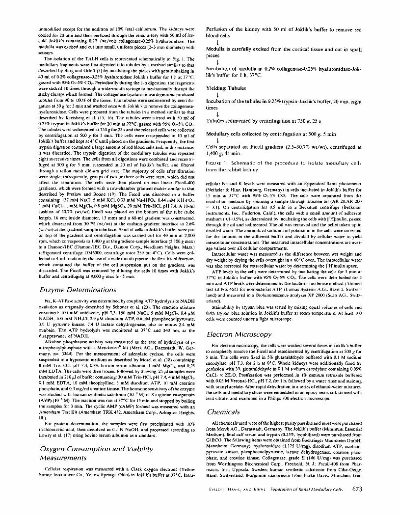
unmodified except for the addition of 10% fetal calf serum. The kidneys werecooled for 20 min and then perfused through the renal artery with 50 ml of ice-cold Joklik's containing 0.2% (wt/vol) collagenase-0 .25% hyaluronidase. Themedulla was excised and cut into small, uniform pieces (2-3 mm diameter) withscissors.The isolation of the TALH cells is represented schematically in Fig. l . The
medullary fragments were first digested into tubules by a method similar to thatdescribed by Burg and Orloff (5) by incubating the pieces with gentle shaking in40 ml of 0.2% collagenase-0.25% hyaluronidase Joklik's buffer for I h at 37°C,gassed with 95% 02-5% CO-2- Periodically during the I-h digestion, the fragmentswere sucked 10 times through a wide-mouth syringe to mechanically disrupt thesticky clumps which formed . The collagenase-hyaluronidase digestions producedtubules from 90 to 100% of the tissue. The tubules were sedimented by centrifu-gation at 50 g for 3 min and washed oncewith Joklik's to remove the collagenase-hyaluronidase. Cells were prepared from the tubules in a method similar to thatdescribed by Kreisberg et al. (15, I6). The tubules were stirred with 50 ml of0.25% trypsin in Joklik's buffer for 20 min at 22°C, gassed with 95% Ot-5% CO..The tubules were sedimented at 750 g for25 s and the released cells were collectedby centrifugation at 500 g for 5 min. The cells were resuspended in 10 ml ofJoklik's buffer and kept at 4°C until placed on the gradients. Frequently, the firsttrypsin digestion contained a large amount of red blood cells and, in this instance,it was discarded. The trypsin digestion of the medullary tubules was repeatedeight successive times. The cells from all digestions were combined and recentri-fuged at 500 g for 5 min, suspended in 20 ml of Joklik's buffer, and filteredthrough a teflon mesh (36-pm grid size) . The majority of cells after filtrationwere single; infrequently, groups of two or three cells were seen, which did notaffect the separation . The cells were then placed on two linear Ficoll-400gradients, which were formed with a two-chamber gradient maker similar to thatdescribed by Pretlow and Boone (l9) . The Ficoll was dissolved in a buffercontaining : 137 mM NaCl, 5 mM KCI, 0.33 mM Na 2HP0,, 0.44 mM KH=P0,,1 mM CaC12 , 1 MM MgCl2, 0.8 MM MgSO,, 20 mM Tris-HCI, pH 7.4 . A l0-mlcushion of 30.7% (wt/wt) Ficoll was placed on the bottom of the tube (tubelength, 16 cm ; inside diameter, 13 mm) and a 60-ml gradient was constructed,which decreased from 30 .7% (wt/wt) at the cushion-gradient interface to 2.6%(wt/wt) at the gradient-sample interface. 10 mlof cells in Joklik's buffer were puton top of the gradient and centrifugation was carried out for 40 min at 2,800rpm, which corresponds to 1,400 gat thegradient-sample interface (2,100g max)in a Damon/IEC (Damon/IEC Div., Damon Corp., Needham Heights, Mass.)refrigerated centrifuge DM6000, centrifuge rotor 259 (at 4°C) . Cells were col-lected in 4-ml fraction by the use ofa wide mouth pipette, the first 10-ml fraction,which contained the buffer of the cell suspension put on the gradient, wasdiscarded . The Ficoll was removed by diluting the cells 10 times with Joklik'sbuffer and centrifuging at 4,000 g max for 5 min.
Enzyme DeterminationsNa, K-ATPase activity was determined by coupling ATP hydrolysis to NADH
oxidation as originally described by Schoner et al . (23) . The reaction mixturecontained : 100 mM imidazole, pH 7.3, 150 mM NaCl, 5 MM MgCl,. 0.4 AMNADH, 100 mM NH,CI, 2.9 AM disodium ATP, 0.6 AM phosphoenolpyruvate,3.9 U pyruvate kinase, 7.4 U lactate dehydrogenase, plus or minus 2.4 mMouabain. The ATP hydrolysis was monitored at 37°C and 340 nm, as thedisappearance of NADH .
Alkaline phosphatase activity was measured as the rate of hydrolysis of p-nitrophenylphosphate with a Merckotest" kit (Merk AG ., Darmstadt, W. Ger-many, no. 3344). For the measurement of adenylate cyclase, the cells weresuspended in a hypotonic medium as described by Morel et al. (18) containing :8 mM Tris-HCI, pH 7.4, 0.8% bovine serum albumin, I mM MgCl., and 0.25mM EDTA . The cells were then frozen, followed by thawing. 25-Al samples wereincubated in 210 Al of buffer containing: 30 mM Tris-HCI, pH 7.4, 4mM MgC12 ,0.1 mM EDTA, 10 mM theophylline, 3 mM disodium ATP, 10 mM creatinephosphate, and 0.3 mg/ml creatine kinase . The hormonesensitivity of the enzymewas studied with human synthetic calcitonin (10-M) or 8-arginine vasopressin(AVP) (10-M). The reaction was run at 37°C for 15 min and stopped by boilingthe samples for 3 min. The cyclic AMP(cAMP) formed was measured with anAmersham Test Kit (Amersham TRK 432, Amersham Corp., Arlington Heights,Ill .).
For protein determination, the samples were first precipitated with 10%trichloracetic acid, then dissolved in 0.1 N NaOH, and processed according toLowry et al . (17) using bovine serum albumin as a standard .
Oxygen Consumption and ViabilityMeasurements
Cellular respiration was measured with a Clark oxygen electrode (YellowSpring Instrument Co ., Yellow Springs, Ohio) in Joklik's buffer at 37 °C. Intra-
Perfusion of the kidney with 50 ml of Joklik's buffer to remove redblood cells
Medulla is carefully excised from the cortical tissue and cut in smallpieces
Incubation of medulla in 0.2% collagenase-0.25% hyaluronidase-Jok-lik's buffer for 1 h, 37°C .
Yielding : Tubules
Incubation ofthe tubules in 0.25% trypsin-Joklik's buffer, 20 min, eighttimes
Tubules sedimented by centrifugation at 750 g, 25 s
Medullary cells collected by centrifugation at 500 g, 5 min
Cells separated on Ficoll gradient (2.5-30.7% wt/wt), centrifuged at1,400 g, 45 min.
FIGURE 1 Schematic of the procedure to isolate medullary cellsfrom the rabbit kidney .
cellular Na and K levels were measured with an Eppendorf flame photometer(Netheler & Hinz, Hamburg, Germany) in cells incubated in Joklik's buffer for15 min at 37 °C with 95% 0-5% CO, The cells were separated from theincubation medium by spinning a sample through silicone oil (AR 20:AR 200= 3:1) . On centrifugation for 0.5 min in a Beckman centrifuge (BeckmanInstruments, Inc., Fullerton, Calif), the cells with a small amount of adherentmedium (0 .1-0 .5%), as determined by incubating the cells with [''H]inulin, passedthrough the oil and sedimented . The oil was removed and the pellet taken up indistilled water. The amounts ofsodium and potassium in the cells were correctedfor the amount in the adherent buffer and divided by cellular water to yieldintracellular concentrations . The measured intracellular concentrations are aver-age values over all cellular compartments .
Intracellular water was measured as the difference between wet weight anddry weight by drying the cells overnight in a 60°C oven . The intracellular waterwas also corrected for extracellular water by determining the [''H]inulin space.ATP levels in the cells were determined by incubating the cells for 5 min at
37°C in Joklik's buffer with 95% 0.-5% C02. The cells were then boiled for 3min and ATP levels were determined by the luciferin-luciferase method (Abimedtest kit No . 4613 for nonbacterial ATP, (Lumac Systems A.G ., Basel 2, Switzer-land) and measured in a Bioluminescence analyzer XP 2000 (Scan AG ., Switz-erland) .
Stainability by trypan blue was tested by mixing equal volumes of cells and0.4% trypan blue solution in Joklik's buffer at room temperature. At least 100cells were counted under a light microscope.
Electron MicroscopyFor electron microscopy, the cells were washed several times in Joklik's buffer
to completely remove the Ficoll and resedimented by centrifugation at 500 g for5 min. The cells were fixed in 5% glutaraldehyde buffered with 0.1 M sodiumcacodylate, pH 7.3, for 2 h at 0°C. Whole kidneys were additionally fixed byperfusion with 3% glutaraldehyde in 0.1 M sodium cacodylate containing 0.05%CaCl, x 2H=0. Postfixation was performed in 1% osmium tetroxide bufferedwith 0.05 M Veronal-HCI, pH 7.2, for I h, followed by a water rinse and stainingwith uranyl acetate . After rapid dehydration in a series of ethanol-watermixtures,the cells and medullary slices were embedded in an epoxy resin, cut, stained withlead citrate, and examined in a Philips 300 electron microscope.
ChemicalsAll chemicals used wereof thehighest purity possible and mostwere purchased
from Merck AG ., Darmstadt, Germany. The Joklik's buffer (Minimum EssentialMedium), fetal calf serum and trypsin (0 .25%, lyophilized) were purchased fromGIBCO. The following items were obtained from Boehringer Mannheim GmbH,Mannheim, Germany) : hyaluronidase (1,175 U/mg), disodium ATP, ouabain,pyruvate kinase, phosphoenolpyruvate, lactate dehydrogenase, creatine phos-phate, and creatine kinase . Collagenase, grade 11 (146 U/mg) was purchasedfrom Worthington Biochemical Corp ., Freehold . N. J . ; Ficoll-400 from Phar-macia, Inc., Uppsala, Sweden; human synthetic calcitonin from Ciba-Geigy,Basel, Switzerland ; 8-arginine vasopressin from Parke-Davis, Munchen, Ger-
EvEtorr, HAASE, AND KINNE
Separation of Renal Medullary Cells
673
many . Furosemide was a gift from Hoechst Pharmaceuticals, Frankfurt, Ger-
many .
RESULTS
During the isolation of the TALH cells, the various tissuefractions were identified morphologically, enzymatically, andby their response to furosemide, which is known to interferewith the chloride transport system in the TALH in vivo. Thesethree criteria will be discussed separately in the followingsections.
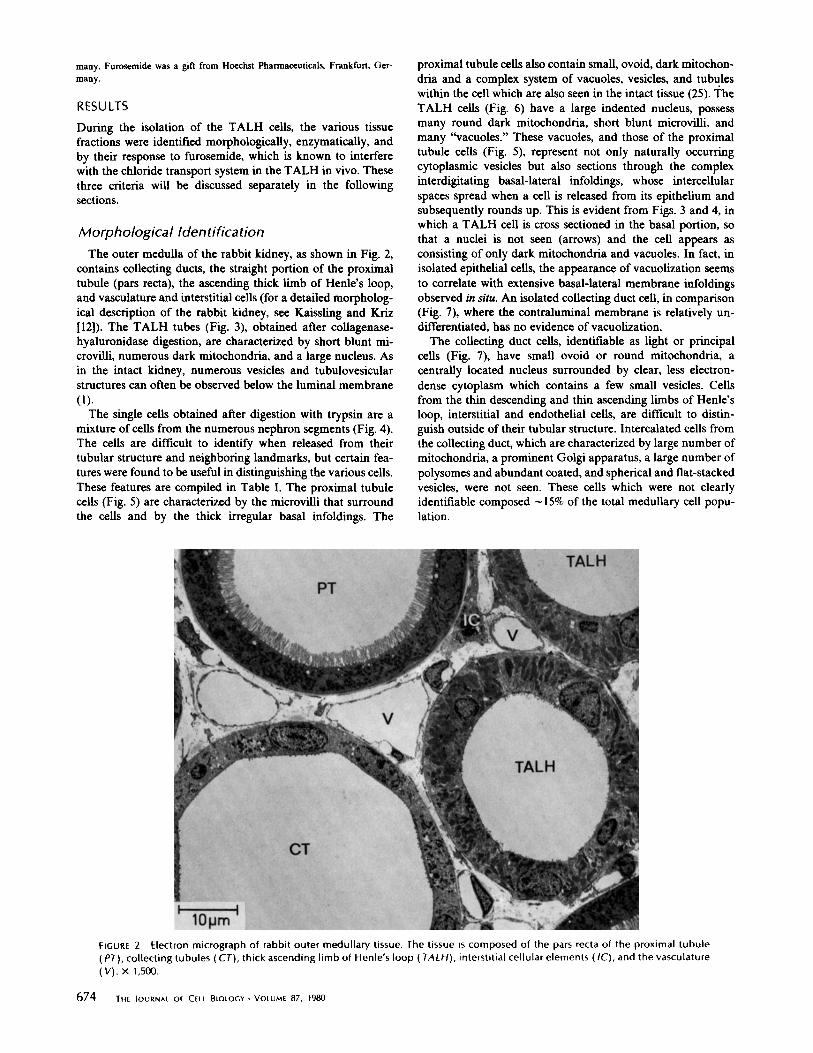
Morphological IdentificationThe outer medulla of the rabbit kidney, as shown in Fig . 2,
contains collecting ducts, the straight portion of the proximaltubule (pars recta), the ascending thick limb of Henle's loop,and vasculature and interstitial cells (for a detailed morpholog-ical description of the rabbit kidney, see Kaissling and Kriz[12]) . The TALH tubes (Fig. 3), obtained after collagenase-hyaluronidase digestion, are characterized by short blunt mi-crovilli, numerous dark mitochondria, and a large nucleus . Asin the intact kidney, numerous vesicles and tubulovesicularstructures can often be observed below the luminal membrane(1) .The single cells obtained after digestion with trypsin are a
mixture of cells from the numerous nephron segments (Fig. 4) .The cells are difficult to identify when released from theirtubular structure and neighboring landmarks, but certain fea-tures were found to be useful in distinguishing the various cells.These features are compiled in Table 1 . The proximal tubulecells (Fig . 5) are characterized by the microvilli that surroundthe cells and by the thick irregular basal infoldings . The
proximal tubule cells also contain small, ovoid, dark mitochon-dria and a complex system of vacuoles, vesicles, and tubuleswithin the cell which are also seen in the intact tissue (25) . TheTALH cells (Fig. 6) have a large indented nucleus, possessmany round dark mitochondria, short blunt microvilli, andmany "vacuoles ." These vacuoles, and those of the proximaltubule cells (Fig . 5), represent not only naturally occurringcytoplasmic vesicles but also sections through the complexinterdigitating basal-lateral infoldings, whose intercellularspaces spread when a cell is released from its epithelium andsubsequently rounds up. This is evident from Figs. 3 and 4, inwhich a TALH cell is cross sectioned in the basal portion, sothat a nuclei is not seen (arrows) and the cell appears asconsisting of only dark mitochondria and vacuoles . In fact, inisolated epithelial cells, the appearance of vacuolization seemsto correlate with extensive basal-lateral membrane infoldingsobserved in situ. An isolated collecting duct cell, in comparison(Fig . 7), where the contraluminal membrane is relatively un-differentiated, has no evidence of vacuolization .The collecting duct cells, identifiable as light or principal
cells (Fig . 7), have small ovoid or round mitochondria, acentrally located nucleus surrounded by clear, less electron-dense cytoplasm which contains a few small vesicles . Cellsfrom the thin descending and thin ascending limbs of Henle'sloop, interstitial and endothelial cells, are difficult to distin-guish outside of their tubular structure . Intercalated cells fromthe collecting duct, which are characterized by large number ofmitochondria, a prominent Golgi apparatus, a large number ofpolysomes and abundant coated, and spherical and flat-stackedvesicles, were not seen . These cells which were not clearlyidentifiable composed - 15% of the total medullary cell popu-lation.
FIGURE 2
Electron micrograph of rabbit outer medullary tissue . The tissue is composed of the pars recta of the proximal tubule(PT), collecting tubules (CT), thick ascending limb of Henlé s loop ( TALH), interstitial cellular elements (IC), and the vasculature(V) . X 1,500 .
674
THE JOURNAL OF CELL BIOLOGY " VOLUME 87, 1980

FIGURE 3
Electron micrograph of an isolated TALH tubule from the rabbit medulla after incubation of the renal medulla in 0.2%collagenase-0.25% hyaluronidase for 1 h at 37 °C . The arrow indicates a TALH cell sectioned without a nucleus, thus, the cellappears to consist of only mitochondria and vacuoles . x 3,600.
FIGURE 4 Electron micrograph of the total medullary cell population before being centrifuged on the Ficoll gradient . Therecognizable cells are derived from the proximal tubule (PT), collecting duct (CT), and the thick ascending limb of Henle's loop( TALH). The arrow indicates a TALH that was sectioned through the basal portion of the cell and can be compared with the cellindicated by the arrow in Fig . 3. a, x 340; b, x 1,800.
EVELOFF, HAASE, AND KINNE
Separation ofRenal Medullary Cells
675

TABLE I
Structural Markers for Identification of Single Isolated Cells from the Rabbit Renal Medulla
Cells*
Plasma membrane
Nucleus
Proximal tubule (pars Long microvilli, thickrecta)
basal-lateral membraneinfoldings
TALH
Fewshort microvillus pro-
Elongatedjections, basal-lateral tationsmembrane is highly in-folded and appear asvesicles
Collecting duct (light or
Afew short microvilli, no
Large roundprincipal cells)
infoldings
Thin
loop,
interstitial,
Irregular surface with mi-
Irregular shapeendothelial cells
crovillous
like
extru-sions
In an electron micrograph of the cells from the dense fraction10 (Fig . 8), the majority of cells are morphologically identifia-ble as TALH cells. Occasionally, a proximal tubule cell orprincipal cell of the collecting duct is seen . The former isespecially evident if, in the first step of isolation procedure, thekidney medulla is not very carefully dissected from the corticaltissue . The TALH cells (Fig . 9) have intact triple-layeredplasma membranes and their polarity is apparently maintained .Short microvilli are evident on one side of the cell, the formerapical membrane, whereas the other cell pole is characterizedby deep infolding of the plasma membrane, the former basal-lateral membrane .
676
THE JOURNAL Of CELL BIOLOGY - VOLUML 87, 1980
with inden-
Very few
' All cells have a more or less round shape on separation .$ Vesicles may include naturally occurring vesicle structures and cross sections of basal-lateral membranes.
MitochondriaRound, elongated, of-
Small, ovoid, darkten indented
Numerous, large, dark,densly packed, fillingup most of cell, mostlyround, larger than inproximal and collectingtubule cells, cristae vis-ible
FIGURE 5
Electron micrograph of a proximal tubule cell from the total cell population . x 6,200 .
Enzymatic Identification
Cytoplasmic inclusionsNumerous vesicles (mean
diameter "0.7 pm), elec-tron-dense material$
Vesicles mostly smallerthan mitochondria$
Dark, small less numerous
Small, light vesicles (meanthan in proximal tubule
--0.3 turn diameter)
Some vesicles, cytoplasm isonly a small ring aroundnucleus
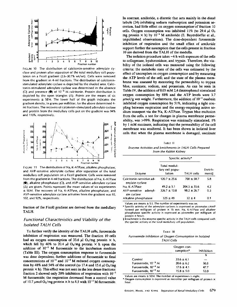
The enzymatic criteria used to identify the medullary TALHcells were the enrichment of calcitonin-sensitive adenylatecyclase activity and Na, K-ATPase activity . Both enzymeactivities have been shown by microdissection to be highlyconcentrated in the TALH (7, 13, 22) . As shown in Fig. 10, ina linear Ficoll gradient ranging in density from 0.22 to 1 .145gm/ml, the protein was distributed approximately evenly . Thecalcitonin-sensitive adenylate cyclase activity was, however,concentrated in the densest portion of the gradient (fraction10), the maximum enzyme activity being 700 pmol cAMP
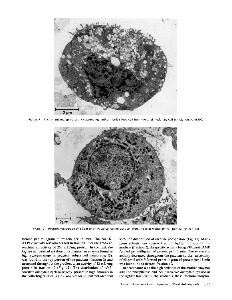
FIGURE 6
Electron micrograph of a thick ascending limb of Henle's loop cell from the total medullary cell population . x 10,800 .
FIGURE 7
Electron micrograph of a light or principal collecting duct cell from the total medullary cell population . x 6,400 .
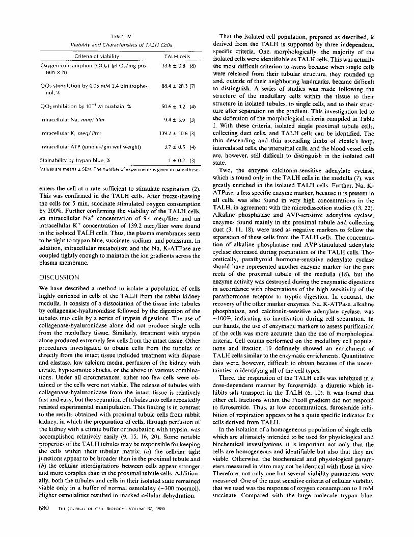
formed per milligram of protein per 15 min . The Na, K-ATPase activity was also highest in fraction 10 of the gradient,reaching an activity of 250 mU/mg protein . In contrast, thehighest activities of alkaline phosphatase, an enzyme found inhigh concentrations in proximal tubule cell membranes (3),was found in the top portion of the gradient (fraction 2) anddecreased throughout the gradient to an activity of 32 mU/mgprotein in fraction 10 (Fig . 11) . The distribution of AVP-sensitive adenylate cyclase activity, present in high amounts inthe collecting duct cells (18), was similar to, but not identical
with, the distribution of alkaline phosphatase (Fig . 11) . Maxi-mum activity was achieved in the lighter portions of thegradient (fraction 2), the specific activity being 890 pmol CAMPformed per milligram of protein per 15 min . The enzymaticactivity decreased throughout the gradient so that an activityof 98 pmol CAMP formed per milligram of protein per 15 minwas found in the densest fraction 10 .
In accordance with the high activities of the marker enzymesalkaline phosphatase and AVP-sensitive adenylate cyclase inthe lighter fractions of the gradients, these fractions morpho-
EVEEOFF, HAASE, AND KINNE
Separation of Renal Medullary Cells
677
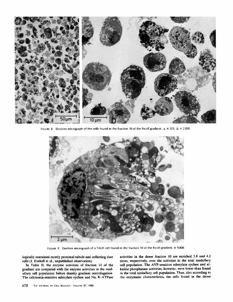
FIGURE 8
Electron micrograph of the cells found in the fraction 10 of the Ficoll gradient . a, x 325 ; b, x 2,000 .
FIGURE 9
Electron micrograph of a TALH cell found in the fraction 10 of the Ficoll gradient . x 9,800 .
logically contained mostly proximal tubule and collecting ductcells (J. Evelofi et al ., unpublished observation) .
In Table II, the enzyme activities of fraction 10 of thegradient are compared with the enzyme activities in the med-ullary cell population before density gradient centrifugation .The calcitonin-sensitive adenylate cyclase and Na, K-ATPase
678
THE JOURNAL OF CELL BIOLOGY " VOLUME 87, 1980
activities in the dense fraction 10 are enriched 5.8 and 4.2times, respectively, over the activities in the total medullarycell population . The AVP-sensitive adenylate cyclase and al-kaline phosphatase activities, however, were lower than foundin the total medullary cell population. Thus, also according tothe enzymatic characteristics, the cells found in the dense
Ë
TCO -
a
c 2._.w .;n E
p Cw_ v-mm
FIGURE 10 The distribution of calcitonin-sensitive adenylate cy-clase and protein after separation of the total medullary cell popu-lation on a Ficoll gradient (2 .6-30.7% wt/wt) . Cells were removedfrom the gradient in 4-ml fractions . The distribution of calcitonin-stimulated adenylate cyclase is depicted by the shaded area . Calci-tonin-stimulated adenylate cyclase was determined in the absence(O) and presence (0) of 10-5 M calcitonin . Protein distribution isdepicted by the open triangles (A). Points are the means of sixexperiments t SEM. The lower half of the graph indicates thegradient density, in grams per milliliter, for the above determined 4-ml fractions. The recovery of calcitonin-stimulated adenylate cyclaseand protein from the medullary cells put on the gradient was 94%and 110%, respectively .
uit
6 7 8 9 16Fraction number
3 4 5
ó O3 n
FIGURE 11
The distribution of Na, K-ATPase, alkaline phosphatase,and AVP-sensitive adenylate cyclase after separation of the totalmedullary cell population on a Ficoll gradient. Cells were removedfrom the gradient in 4-ml fractions. The distribution of Na, K-ATPase("), alkaline phosphatase (O), and AVP-sensitive adenylate cyclase(A) are given. Points represent the mean values of six experimentst SEM. The recovery of Na, K-ATPase, alkaline phosphatase, andAVP-sensitive adenylate cyclase activities from the gradient was 93,102, and 92%, respectively .
fraction of the Ficoll gradient are derived from the medullaryTALK
Functional Characteristics and Viability of theIsolated TALH CellsTo further verify the identity of the TALH cells, furosemide
inhibition of respiration was measured . The fraction 10 cellshad an oxygen consumption of 33 .6 11 02/mg protein x h,which fell by 40% to 20.4 1il 02/mg protein x h upon theaddition of 10-5 M furosemide to the incubation medium(Table III) . The oxygen consumption response to furosemidewas dose dependent; further additions of furosemide to fmalconcentrations of 10-4 and 10-3 M reduced oxygen consump-tion by 48% and 54% of the control (to 17 .4 and 15.61102/mgprotein x h) . This effect was not seen in the less dense fractions :fraction 2 showed only 20% inhibition of respiration with 10 -3M furosemide; the respiration decreased from a control valueof 10.71mo1 O2/mg protein x h to 8 .5 with 10-3 M furosemide .
In contrast, amiloride, a diuretic that acts mainly in the distaltubule (24) inhibiting sodium reabsorption and potassium se-cretion, had little effect on oxygen consumptiowof fraction 10cells . Oxygen consumption was inhibited 1l% (to 29.4 11 02mg protein x h) by 10-` M amiloride (E . Bayerd6rffer et al .,unpublished observations) . The dose-dependent furosemideinhibition of respiration and the small effect of amiloridesupport further the assumption that the cells present in fraction10 are derived from the TALH of the medulla .The isolation procedure takes -6 h with exposure of the cells
to collagenase, hyaluronidase, and trypsin. Therefore, the via-bility of the isolated cells was measured using the followingcriteria : the metabolic state of the cells was estimated by theeffect of uncouplers on oxygen consumption and by measuringthe ATP levels of the cell; and the state of the plasma mem-brane was assessed by measuring the permeability to trypanblue, succinate, sodium, and potassium. As can be seen inTable IV, the addition of0.05 mM 2,4 dinitrophenol stimulatedoxygen consumption by 88% and the ATP levels were 3 .71mo1/g wet weight . Furthermore, the addition of 10-" ouabaininhibited oxygen consumption by 51%, indicating a tight cou-pling between respiration and the energy-requiring active so-dium transport via the Na, K-ATPase . Trypan blue exclusionfrom the cells, a test for changes in plasma membrane perme-ability, was >99% . Respiration was minimally stimulated, 1%by 1 mM succinate, indicating that the permeability ofthe cellmembrane was unaltered . It has been shown in isolated livercells that when the plasma membrane is damaged, succinate
TABLE II
Enzyme Activities and Enrichments in TALH Cells Preparedfrom the Rabbit Kidney
Specific activity*
Total medul-
Values are means tS.E. The number of experiments was six .* Specific activity of the adenylate cyclases is expressed as picomoles cAMPformed per milligram of protein x 15 min. Na, K-ATPase and alkalinephosphatase specific activity is expressed as picomoles per milligram ofprotein x hours.
$ Enrichment is the enzyme specific activity in the TALH cells compared withthe specific activity in the total medullary cell population .
TABLE III
Furosemide Inhibition of Oxygen Consumption in IsolatedTALH Cells
Values are means tSEM. The number of experiments is eight.' Oxygen consumption is expressed as microliter per milligram of protein xhour .
EVELOFF, HAASE, AND KINNE
Separation ofRenalMedullaryCells
679
Enzymelary cell popu-
lation TALH cellsEnrich-ment$
Calcitonin-sensitive ad- 120.7 t 25 .6 700 t58 .7 5.8enylate cyclase
Na, K-ATPase 49 .2 t 5.1 204.5 t35 .6 4.2AVP-sensitive adenyl- 326.7 t 13 .8 98 .2 t26 .7 0.3ate cyclase
Alkaline phosphatase 325 t 49 32 ±4 0.1
Oxygen con-sumption* Inhibition
Control 33.6 t 6.1 -Furosemide, 10-5 M 20.6 t 6.2 38.8Furosemide, 10-` M 17 .6 t 4.1 47 .7Furosemide, 10-3 M 15 .8 t 5.0 53 .0
Values are means ±SEM. The number of experiments is given in parentheses .
enters the cell at a rate sufficient to stimulate respiration (2) .This was confirmed in the TALH cells . After freeze-thawingthe cells for 5 min, succinate stimulated oxygen consumptionby 200% . Further confirming the viability of the TALH cells,an intracellular Na' concentration of 9.4 meq/liter and anintracellular K+ concentration of 139.2 meq/liter were foundin the isolated TALH cells. Thus, the plasma membranes seemto be tight to trypan blue, succinate, sodium, and potassium. Inaddition, intracellular metabolism and the Na, K-ATPase arecoupled tightly enough to maintain the ion gradients across theplasma membrane.
DISCUSSION
TABLE IV
Viability and Characteristics of TALH Cells
680
TfIE JOURNAL Of CELL BIOLOGY - VOLUME 87, 1980
We have described a method to isolate a population of cellshighly enriched in cells of the TALH from the rabbit kidneymedulla. It consists of a dissociation of the tissue into tubulesby collagenase-hyaluronidase followed by the digestion of thetubules into cells by a series of trypsin digestions. The use ofcollagenase-hyaluronidase alone did not produce single cellsfrom the medullary tissue . Similarly, treatment with trypsinalone produced extremely few cells from the intact tissue . Otherprocedures investigated to obtain cells from the tubules ordirectly from the intact tissue included treatment with dispaseand elastase, low calcium media, perfusion of the kidney withcitrate, hypoosmotic shocks, or the above in various combina-tions . Under all circumstances, either too few cells were ob-tained or the cells were not viable . The release of tubules withcollagenase-hyaluronidase from the intact tissue is relativelyfast and easy, but the separation oftubules into cells repeatedlyresisted experimental manipulation . This finding is in contrastto the results obtained with proximal tubule cells from rabbitkidney, in which the preparation of cells, through perfusion ofthe kidney with a citrate buffer or incubation with trypsin, wasaccomplished relatively easily (9, 15, 16, 20). Some notableproperties ofthe TALH tubules may be responsible for keepingthe cells within their tubular matrix ; (a) the cellular tightjunctions appear to be broader than in the proximal tubule and(b) the cellular interdigitations between cells appear strongerand more complex than in the proximal tubule cells. Addition-ally, both the tubules and cells in their isolated state remainedviable only in a buffer of normal osmolality (-300 mosmol) .Higher osmolalities resulted in marked cellular dehydration .
That the isolated cell population, prepared as described, isderived from the TALH is supported by three independent,specific criteria . One, morphologically, the majority of theisolated cells were identifiable as TALH cells . This was actuallythe most difficult criterion to assess because when single cellswere released from their tubular structure, they rounded upand, outside of their neighboring landmarks, became difficultto distinguish. A series of studies was made following thestructure of the medullary cells within the tissue to theirstructure in isolated tubules, to single cells, and to their struc-ture after separation on the gradient . This investigation led tothe definition of the morphological criteria compiled in TableI. With these criteria, isolated single proximal tubule cells,collecting duct cells, and TALH cells can be identified . Thethin descending and thin ascending limbs of Henle's loop,intercalated cells, the interstitial cells, and the blood vessel cellsare, however, still difficult to distinguish in the isolated cellstate.Two, the enzyme calcitonin-sensitive adenylate cyclase,
which is found only in the TALH cells in the medulla (7), wasgreatly enriched in the isolated TALH cells. Further, Na, K-ATPase, a less specific enzyme marker, because it is present inall cells, was also found in very high concentrations in theTALH, in agreement with the microdissection studies (13, 22).Alkaline phosphatase and AVP-sensitive adenylate cyclase,enzymes found mainly in the proximal tubule and collectingduct (3, 11, 18), were used as negative markers to follow theseparation of these cells from the TALH cells. The concentra-tion of alkaline phosphatase and AVP-stimulated adenylatecyclase decreased during preparation of the TALH cells . The-oretically, parathyroid hormone-sensitive adenylate cyclaseshould have represented another enzyme marker for the parsrecta of the proximal tubule of the medulla (18), but theenzyme activity was destroyed during the enzymatic digestionsin accordance with observations of the high sensitivity of theparathormone receptor to tryptic digestion. In contrast, therecovery ofthe other marker enzymes, Na, K-ATPase, alkalinephosphatase, and calcitonin-sensitive adenylate cyclase, was100%, indicating no inactivation during cell separation . In
our hands, the use of enzymatic markers to assess purificationof the cells was more accurate than the use of morphologicalcriteria . Cell counts performed on the medullary cell popula-tions and fraction 10 definitely showed an enrichment ofTALH cells similar to the enzymatic enrichments . Quantitativedata were, however, difficult to obtain because of the uncer-tainties in identifying all of the cell types.
Three, the respiration of the TALH cells was inhibited in adose-dependent manner by furosemide, a diuretic which in-hibits salt transport in the TALH (6, 10) . It was found thatother cell fractions within the Ficoll gradient did not respondto furosemide . Thus, at low concentrations, furosemide inhi-bition of respiration appears to be a quite specific indicator forcells derived from TALH .
In the isolation of a homogeneous population of single cells,which are ultimately intended to be used for physiological andbiochemical investigations, it is important not only that thecells are homogeneous and identifiable but also that they areviable . Otherwise, the biochemical and physiological param-eters measured in vitro may not be identical with those in vivo .Therefore, not only one but several viability parameters weremeasured. Oneofthe most sensitive criteria of cellular viabilitythat we used was the response ofoxygen consumption to 1 mMsuccinate. Compared with the large molecule trypan blue,
Criteria of viability TALH cells
Oxygen consumption (Q02) (ul 02/mg pro- 33 .6 ± 0.8 (8)tein x h)
Q02 stimulation by 0.05 mM 2,4 dinitrophe- 88 .4 ± 28 .3 (7)nol, q
Q02 inhibition by 10-° M ouabain, % 50 .6 ± 4.2 (4)
Intracellular Na, meq/liter 9.4 ± 3.9 (3)
Intracellular K, meq/lifer 139.2 ± 10 .6 (3)
Intracellular ATP (,umoles/gm wet weight) 3.7 ± 0.5 (4)
Stainability by trypan blue, % 1 ± 0.2 (3)

succinate, with a molecular weight of 162, has a small molecularradius and, therefore, small changes in membrane permeabilitycan be more easily detected . Similarly, the sodium and potas-sium gradients across the cell membrane are very sensitiveindicators of membrane integrity and the functional couplingof the cell .The lack of effect of succinate on the respiration of TALH
is an interesting experimental observation because the respi-ration of renal cortical slices is stimulated normally by succi-nate (8) . This discrepancy is probably caused by the presenceofsuccinate transport systems in the plasma membranes of thecells in the proximal tubules but not in the TALH (14) .
As a last point, the present method yields -r4 mg of proteinfrom two rabbit kidneys in -6 h. If a calculation is made tocompare this method with the teased tubule technique, thefollowing results are obtained . ATALH tubule of --1 .5 mm inlength can be dissected by microdissection in 5 min. The tubulehas a protein content of 0.06 ttg/mm (11) ; thus, a maximalyield of ^-6.8 p,g protein could be maximally expected in anequivalent 6-h period . The present method, therefore, yields atleast 1,000 times more protein, allowing for flexibility in theapplication of many biochemical and physiological techniquesin the study of the mechanisms of ion transport to this nephronsegment.
The assistance ofDr. Eva Kinne-Saffran and Dr . K. Ring in measuringthe cellular ATP levels and Dr . Muschaweck and Dr. Metzger ofHoechst Pharmaceuticals in providing diuretics and other chemicals isgratefully acknowledged .
This work was partially supported by U. S. Public Health Servicegrant AM 05841.
Receivedforpublication 25 February 1980, and in revisedform 25 July1980.
REFERENCES
I . Allen, F ., and C . C. Tisher. 1976 . Morpholog y of the ascending thick limb of Henle .Kidney Int . 9:8-22 .
2 . Baur, H ., S . Kasperek, and S . Pfaff. 1975 . Criteria of viability of isolated liver cells.Hoppe-Seylers Z. Physiol. Chem . 356:827-838.
3 . Bunting, S. L . . V . E. Pollack, R . C. Muehrcke, and R. M . Kark . 1958 . Quantitativ ehistochemistry of the nephron . Science (Wash . D. C.) 127:1342-1343 .
4 . Burg, M ., and N . Green . 1973 . Function of the thick ascending limb of Henle's loop. Am.J. Physiol. 224:659-668.
5 . Burg, M . B ., and J . Orloff. 1962. Oxygen consumption and active transport in separatedrenal tubules. Am. J . Physiol. 203 :327-330.
6 . Burg, M ., L . Stoner, 1 . Cardinal, and N . Green . 1973 . Furosemide effect on isolatedperfused tubules. Am. J . Physiol. 225 :119-124.
7 . Charbades, D., M. Imbert-Teboul, M . Montegut, A. Clique, and F . Morel . 1976 . Distri-bution of calcitonin-sensitive adenylate cyclase along the rabbit kidney tubule. Proc. Nall.Acad. Sci. U . S . A . 73 :3608-3612.
8. Cross, R . 1 ., and J . V . Taggart . 1950. Rena l tubular transport : accumulation of p-aminohippurate by rabbit kidney slices. Am. J. Physiol. 161 :181-190.
9. Heidrich, H .-G ., and M . E . Dew . 1977 . Homogenou s cell populations from rabbit kidneycortex . Proximal, distal tubule, and renin-active cell vs isolated by free-flow electropho-resis . J. Cell Biol. 74:780.788 .
10 . Imai . M . 1977 . Effect of bumetanide and furosemide on the thick ascending limb ofHenlés loop of rabbits and rats perfused in vitro. Eur. J. Pharmacol. 41 :409-416.
11 . Imbert. M ., D . Charbardes, M . Montegut, A . Clique, and F . Morel . 1975 . Adenylatecyclase activity along the rabbit nephron as measured in single isolated segments . PfhigersArch. IV 354 :213-228 .
12 . Kaissling, B ., and W . Kriz . 1979 . Structural analysis of the rabbit kidney . Adv . Anai .Embryol. Cell Biol . 56 :1-123 .
13 . Katz, A . 1 ., A . Doucet, and F . Morel . 1979 . Na . K-ATPase activity along the rabbit, ratand mouse nephron . Am . J. Physiol. 237:1`114
14-F120.
14 . Kippen, L, B . Hirayama, 1 . R . Klinenberg, and E. M . Wright . 1979 . Transport oftricarboxylic acid cycle intermediated by membrane vesicles from renal brush border .Proc . Nail. Acad. Sci. U. S. A . 76 :3397-3400.
15 . Kreisberg, 1 . L, A. M . Pitts, and T . G. Pretlow II. 1977 . Separation of proximal tubulecells from suspensions of rat kidney cells in density gradients of ficoll in tissue culturemedium . Am. J. Pathol. 86:591-602.
16 . Kreisberg, 1 . I ., G . Sachs, T. G . Pretlow II, and R . A . McGuire. 1977. Separation ofproximal tubule cells from suspensions of rat kidney cells by free flow electrophoresis . J.Cell Physiol. 93:169-172 .
17 . Lowry, 0. H ., N . T . Rosebrough, A . L. Farr, and R. T. Randall. 1951 . Protei n measurementwith the folin phenol reagent . J. Biol. Chem . 193 :265-275 .
18. Morel, F ., D . Charbardes, and M . Imbert . 1976 . Functional segmentation of the rabbitdistal tubule by microdetermination of hormone-dependent adenylate cyclase activity .Kidney Int. 9:264-277 .
19 . Pretlow, T. G . It, and C . W. Boone . 1969 . Separation of mammalian cells using pro-grammed gradient sedimentation. Exp . Mal. Parhol. 11 :139-152 .
20 . Pretlow, T. G . II, J . Jones, and S. Dow . 1974 . Separation of cells having histochemicaBydemonstrable glucose-6-phosphatase from suspensions of hamster kidney cells in anisokinelic density gradient of ficoll in tissue culture medium . Am. J. Pathol. 74:275-286 .
21 . Rocha, A . S ., and 1 . P. Kokko . 1973. Sodiu m chloride and water transport in the medullarythick ascending limb of Henle. Evidence for active chloride transport . J. Clin . Invest. 52 :612-623.
22 . Schmidt, U . . and U . C . Dubach. 1969 . Activit y of (Na' K')-stimulated adenosine triphos-phatase in the rat nephron . Pflügers Arch . IV 306 :219-226.
23 . Schoner, W ., C . Von Ilberg, R . Kramer, and W . Seubert . 1967 . On the mechanism of Na'-and K'-stimulated hydrolysis of adenosine triphosphate. 1 . Purification and properties ofNa'- and K'-activated ATPase from ox brain . Eur. J . Biochem. 1 :334-343 .
24 . Stoner, L . C ., M . B . Burg, and J . Orloff. 1974 . Ion transport in cortical collecting tubuleeffect of amiloride . Am . J. Physiol . 227 :453-459 .
25 . Tisher, C . C ., R . E. Bulger, and B . F. Trump . 1966 . Huma n renal ultrastructure. I .Proximal tubule of healthy individuals . Lab. Invest. 15 :1357-1394 .
EVE[OFF, HAASE, AND KINNE
Separation of Renal Medullary Cells
681