Altered CLC-5 expression in mouse inner medullary ... · Altered CLC-5 expression in mouse inner...
27
Altered CLC-5 expression in mouse inner medullary collecting duct cells disrupts endocytosis and promotes calcium oxalate crystal adhesion Georgina Carr*, J.A. Sayer† and N.L. Simmons* Schools of *Biosciences and †Medicine, Medical School, Framlington Place, University of Newcastle upon Tyne, Newcastle upon Tyne NE2 4HH, UK Dent’s disease, characterised by proteinuria, hypercalciuria, and renal stone formation, results from mutation of the Cl _ channel CLC-5. We have previously demonstrated that mCLC-5 is expressed in inner medullary collecting duct cells (mIMCD-3 cell line) within acidic endosomes (Sayer et al. 2001). Here we report the consequence of ablation of mCLC-5 expression upon endocytosis of wheatgerm agglutin (WGA) and upon binding of Ca-oxalate crystals. mIMCD-3 cells were transiently transfected with expression vector alone (control) (pCDNA3.1/CT-GFP, Invitrogen) or combined with vector containing sense full-length mCLC-5 (with C-terminal stop codon) or antisense mCLC-5, using Lipofectamine 2000 (Life Technologies). Positive transfectants were identified using a Leica confocal laser imaging microscope equipped (CLSM) with a Kr-Ar laser by their GFP fluorescence and analysed 24–48 h post-transfection. Binding and endocytosis of TRITC-wheatgerm agglutinin (WGA) was followed over a 1 h time course in phosphate-buffered saline (PBS). Optical sections of positive transfectants were collected to allow identification of membrane-bound and internalised WGA. In 3–5 separate experiments, after 1 h, internalisation of WGA occurred in 17/21 of control transfectants and 13/17 of sense mCLC-5 transfectants. With antisense mCLC-5 transfectants endocytosis was disrupted with the majority of cells showing only membrane-bound WGA; only 1/13 of cells showed WGA internalisation (P < 0.001 antisense vs. combined control/sense, Fisher’s exact test). Calcium oxalate monohydrate crystals were grown in a high calcium (50 mM CaCl 2 ) medium exposed to diethyloxalate vapour. Crystals were harvested and overlaid onto mIMCD-3 cultures for 30 min prior to a brief wash in PBS and CLSM imaging of unfixed cells. In 4–5 separate experiments, in the majority of control or sense transfectants (34/44 and 38/44 cells, respectively), no crystal adhesion was observed. The remaining cells (5/44 or 3/44) showed adhesion of single crystals (< 10 mM size) or agglomerates (5/44 and 3/44 >10 mM size). For antisense mCLC-5 transfectants the majority of cells were associated (33/50, P < 0.001 vs. control/sense cells, x 2 test) with crystal agglomerates, whilst 1/50 showed adhesion of single crystals and 16/50 showed no crystal adhesion. Transfection of antisense mCLC-5 is therefore associated with disruption of endocytosis and aggregation of agglomerates of Ca- oxalate crystals on inner medullary collecting duct cells. Crystal retention and agglomeration at the point of maximal urinary concentration are likely to be key factors in renal stone formation. Sayer, J.A. et al. (2001). J. Physiol. 536, 769–783. This work was supported by the NCKRF and the NKRF. How do CFTR blockers inhibit the growth of MDCK cysts? H. Li and D.N. Sheppard Department of Physiology, University of Bristol, Bristol BS8 1TD, UK Polycystic kidney disease is characterised by massive enlargement of fluid-filled epithelial cysts that involves cAMP-dependent cell proliferation and fluid secretion driven by the cystic fibrosis transmembrane conductance regulator (CFTR) Cl _ channel (Sullivan et al. 1998). Using MDCK cysts, we previously demonstrated that CFTR blockers inhibit cyst growth (Findlay et al. 2001). In this study, we investigated the effects of CFTR blockers on cell proliferation and cAMP-stimulated Cl _ currents to understand how drugs inhibit the growth of MDCK cysts. We grew cysts as described previously (Findlay et al. 2001) with the exception that the effects of drugs were tested between days 6 and 12. For cell proliferation assays, MDCK cells were grown in media containing 0.01 % FBS and cAMP agonists in the absence and presence of drugs over a 6 day period and cell numbers determined using a haemocytometer. Using MDCK epithelia and the Ussing chamber technique, we quantified the effects of drugs on cAMP-stimulated Cl _ currents. We tested the effects of four drugs that inhibit CFTR including the open-channel blockers glibenclamide (100 mM) and NPPB (50 mM) and the allosteric blocker genistein (100 mM). As controls, we used first, tamoxifen (10 mM), calix[4]arene (10 mM) and DIDS (200 mM), which inhibit other Cl _ channels but not CFTR, and second, TEA (10 mM), bumetanide (100 mM) and ouabain (1 mM), which block basolateral membrane channels and transporters in MDCK cells. There were three sets of results in this study. First, CFTR inhibitors, bumetanide and ouabain reduced dramatically the volume of cysts (n = 20–36; P < 0.05; one-way ANOVA), whereas TEA, calix[4]arene and DIDS were without effect on cyst growth (n = 31–39; P > 0.05). Second, with the exception of bumetanide, all the drugs tested inhibited potently cell proliferation (n = 4–10; P < 0.0.1). Third, CFTR inhibitors, TEA, bumetanide and ouabain all inhibited cAMP-stimulated Cl _ currents (n = 5–7; P < 0.01; Student’s paired t test), whereas calix[4]arene, DIDS and tamoxifen were without effect. To analyse further these data, we compared the magnitude of inhibition of cyst growth, cell proliferation and cAMP- stimulated Cl _ currents by different drugs. Inhibition of cyst growth and cAMP-stimulated Cl _ currents were well correlated (correlation coefficient = 0.71; P < 0.01; Pearson’s correlation test), whereas inhibition of cyst growth and cell proliferation was not well correlated (correlation coefficient = 0.40; P > 0.05; Pearson’s correlation test). We interpret these data to suggest that CFTR blockers probably inhibit cyst growth by preventing fluid accumulation within the cyst lumen. Findlay, I.A. et al. (2001). J. Physiol. 531.P, 74P. Sullivan, L.P. et al. (1998). Physiol. Rev. 78, 1165–1191. This work was supported by the NKRF and Royal Society. Epithelia & Membrane Transport – Renal Physiology 87P J. Physiol. (2002). 544.P
-
Upload
nguyenxuyen -
Category
Documents
-
view
216 -
download
0
Transcript of Altered CLC-5 expression in mouse inner medullary ... · Altered CLC-5 expression in mouse inner...

Altered CLC-5 expression in mouse inner medullarycollecting duct cells disrupts endocytosis and promotescalcium oxalate crystal adhesion
Georgina Carr*, J.A. Sayer† and N.L. Simmons*
Schools of *Biosciences and †Medicine, Medical School, FramlingtonPlace, University of Newcastle upon Tyne, Newcastle upon TyneNE2 4HH, UK
Dent’s disease, characterised by proteinuria, hypercalciuria, andrenal stone formation, results from mutation of the Cl_ channelCLC-5. We have previously demonstrated that mCLC-5 isexpressed in inner medullary collecting duct cells (mIMCD-3 cellline) within acidic endosomes (Sayer et al. 2001). Here we reportthe consequence of ablation of mCLC-5 expression uponendocytosis of wheatgerm agglutin (WGA) and upon binding ofCa-oxalate crystals.
mIMCD-3 cells were transiently transfected with expressionvector alone (control) (pCDNA3.1/CT-GFP, Invitrogen) orcombined with vector containing sense full-length mCLC-5(with C-terminal stop codon) or antisense mCLC-5, usingLipofectamine 2000 (Life Technologies). Positive transfectantswere identified using a Leica confocal laser imaging microscopeequipped (CLSM) with a Kr-Ar laser by their GFP fluorescenceand analysed 24–48 h post-transfection. Binding and endocytosisof TRITC-wheatgerm agglutinin (WGA) was followed over a 1 htime course in phosphate-buffered saline (PBS). Optical sectionsof positive transfectants were collected to allow identification ofmembrane-bound and internalised WGA. In 3–5 separateexperiments, after 1 h, internalisation of WGA occurred in 17/21of control transfectants and 13/17 of sense mCLC-5transfectants. With antisense mCLC-5 transfectants endocytosiswas disrupted with the majority of cells showing onlymembrane-bound WGA; only 1/13 of cells showed WGAinternalisation (P < 0.001 antisense vs. combined control/sense,Fisher’s exact test).
Calcium oxalate monohydrate crystals were grown in a highcalcium (50 mM CaCl2) medium exposed to diethyloxalatevapour. Crystals were harvested and overlaid onto mIMCD-3cultures for 30 min prior to a brief wash in PBS and CLSMimaging of unfixed cells. In 4–5 separate experiments, in themajority of control or sense transfectants (34/44 and 38/44 cells,respectively), no crystal adhesion was observed. The remainingcells (5/44 or 3/44) showed adhesion of single crystals (< 10 mM
size) or agglomerates (5/44 and 3/44 >10 mM size). For antisensemCLC-5 transfectants the majority of cells were associated(33/50, P < 0.001 vs. control/sense cells, x2 test) with crystalagglomerates, whilst 1/50 showed adhesion of single crystals and16/50 showed no crystal adhesion.
Transfection of antisense mCLC-5 is therefore associated withdisruption of endocytosis and aggregation of agglomerates of Ca-oxalate crystals on inner medullary collecting duct cells. Crystalretention and agglomeration at the point of maximal urinaryconcentration are likely to be key factors in renal stoneformation.
Sayer, J.A. et al. (2001). J. Physiol. 536, 769–783.
This work was supported by the NCKRF and the NKRF.
How do CFTR blockers inhibit the growth of MDCK cysts?
H. Li and D.N. Sheppard
Department of Physiology, University of Bristol, Bristol BS8 1TD, UK
Polycystic kidney disease is characterised by massive enlargementof fluid-filled epithelial cysts that involves cAMP-dependent cellproliferation and fluid secretion driven by the cystic fibrosistransmembrane conductance regulator (CFTR) Cl_ channel(Sullivan et al. 1998). Using MDCK cysts, we previouslydemonstrated that CFTR blockers inhibit cyst growth (Findlay etal. 2001). In this study, we investigated the effects of CFTRblockers on cell proliferation and cAMP-stimulated Cl_ currentsto understand how drugs inhibit the growth of MDCK cysts.
We grew cysts as described previously (Findlay et al. 2001) withthe exception that the effects of drugs were tested between days 6and 12. For cell proliferation assays, MDCK cells were grown inmedia containing 0.01 % FBS and cAMP agonists in the absenceand presence of drugs over a 6 day period and cell numbersdetermined using a haemocytometer. Using MDCK epithelia andthe Ussing chamber technique, we quantified the effects of drugson cAMP-stimulated Cl_ currents. We tested the effects of fourdrugs that inhibit CFTR including the open-channel blockersglibenclamide (100 mM) and NPPB (50 mM) and the allostericblocker genistein (100 mM). As controls, we used first, tamoxifen(10 mM), calix[4]arene (10 mM) and DIDS (200 mM), whichinhibit other Cl_ channels but not CFTR, and second, TEA(10 mM), bumetanide (100 mM) and ouabain (1 mM), whichblock basolateral membrane channels and transporters in MDCKcells.
There were three sets of results in this study. First, CFTRinhibitors, bumetanide and ouabain reduced dramatically thevolume of cysts (n = 20–36; P < 0.05; one-way ANOVA),whereas TEA, calix[4]arene and DIDS were without effect on cystgrowth (n = 31–39; P > 0.05). Second, with the exception ofbumetanide, all the drugs tested inhibited potently cellproliferation (n = 4–10; P < 0.0.1). Third, CFTR inhibitors, TEA,bumetanide and ouabain all inhibited cAMP-stimulated Cl_
currents (n = 5–7; P < 0.01; Student’s paired t test), whereascalix[4]arene, DIDS and tamoxifen were without effect. Toanalyse further these data, we compared the magnitude ofinhibition of cyst growth, cell proliferation and cAMP-stimulated Cl_ currents by different drugs. Inhibition of cystgrowth and cAMP-stimulated Cl_ currents were well correlated(correlation coefficient = 0.71; P < 0.01; Pearson’s correlationtest), whereas inhibition of cyst growth and cell proliferation wasnot well correlated (correlation coefficient = 0.40; P > 0.05;Pearson’s correlation test). We interpret these data to suggestthat CFTR blockers probably inhibit cyst growth by preventingfluid accumulation within the cyst lumen.
Findlay, I.A. et al. (2001). J. Physiol. 531.P, 74P.
Sullivan, L.P. et al. (1998). Physiol. Rev. 78, 1165–1191.
This work was supported by the NKRF and Royal Society.
Epithelia & Membrane Transport – Renal Physiology 87PJ. Physiol. (2002). 544.P

Effects of hypothermia on renal function in normothermicand cold-acclimated anaesthetised rats
R. Sabharwal, E.J. Johns and S. Egginton
Department of Physiology, University of Birmingham, BirminghamB15 2TT, UK
Acute hypothermia causes a reduction in renal blood flow (RBF),glomerular filtration rate (GFR), natriuresis and diuresis, eventhough mean arterial blood pressure (MABP) is largelyunchanged (Sabharwal et al. 2001). This implies an increase inrenal vascular resistance (RVR) occurring primarily at theafferent arteriole, and a possible rise in the neurally mediatedvascular tone (Broman et al. 1995). This study aimed to correlatethe changes in the pattern of renal sympathetic nerve activity(RSNA) to cardiovascular and renal functions cooling to 25 °Cand on rewarming back to 37 °C, and to compare the response innormothermic and cold-acclimated rats.
Male Wistar rats, 290–320 g (n = 14 normothermic, n = 12 cold-acclimated), were anaesthetised with fluothane (2.5 % in O2 ) andmaintained with a-chloralose/urethane (32/450 mg kg_1) via afemoral vein cannula. MABP and heart rate (HR) were measuredvia a femoral artery cannula interfaced with a digital recordingdevice. An infusion of 1.5 % inulin in saline (150 mM NaCl at3 ml h_1) was used for the estimation of glomerular filtration rate(GFR), while urine flow (UV) and absolute sodium excretion(UNaV) were determined from the outflow of the left ureteralcannula. For RSNA experiments, a renal nerve bundle of the leftkidney was isolated, placed on bipolar electrodes and the signalsubjected to power spectral analysis. Core (deep oesophageal)temperature (Tb) was regulated by means of a thermostattedplate. At the end of experiments, rats were killed with anoverdose of sodium pentabarbitone. Cold-acclimated rats wereexposed to progressively lower environmental temperature(20 °C to 4 °C) and photoperiod (12 h to 1 h light day_1) over8 weeks. Data (means ± S.E.M.) were analysed using ANOVA andsignificance taken at P < 0.05.
At Tb = 25 °C, there was a ~50 % reduction in HR (418 ± 9 b.p.m.at 37 °C, P < 0.01) but only a ~15 % fall in MABP (111 ± 2 mmHgat 37 °C, P < 0.05). On cooling GFR decreased by ~50 %(4.6 ± 1 ml kg_1 min_1 at 37 °C, P < 0.05) in normothermic and by5 % (3.8 ± 1 ml kg_1 min_1 at 37 °C) in cold-acclimated rats. A cold-induced diuresis (40 ± 5 vs. 21 ± 2 µl kg_1 min_1, P < 0.05, innormothermic and 47 ± 10 vs. 38 ± 4 µl kg_1 min_1 in cold-acclimated at 37 °C) and natriuresis (5 ± 1 vs. 1 ± 1 µmolkg_1 min_1, P < 0.05, in normothermic and 5 ± 1 vs. 3 ± 1 µmolkg_1 min_1, P < 0.05, in cold-acclimated at 37 °C) was evident atTb = 25 °C. There was a loss in pulsatility in the RSNA signal whileintegrated RSNA increased by 20 % (P < 0.05) in normothermicbut decreased by 20 % (P < 0.05) in cold-acclimated rats atTb = 25 °C. There was a progressive fall in proportion of RSNApower at HR frequency with cooling (20 % in normothermic and80 % in cold-acclimated rats, P < 0.05). On rewarming all variables,in both groups of rats, returned to basal levels. We conclude thatthe natriuresis and diuresis in normothermic rats duringhypothermia is a consequence of a reduction in nephronreabsorption and possibly due to the altered patterning of RSNA. Incold-acclimated rats it may be due to altered renal haemodynamicsand/or hormonal influences induced by chronic cold exposure.
Broman, M. et al. (1995). Acta Physiol. Scand. 153, 179–184.
Sabharwal, R. et al. (2001). J. Physiol. 531.P, 216P.
All procedures accord with current UK legislation.
Regulation of intracellular [Ca2+] in equine chondrocytes
J.S. Gibson*, M.E. Davies*, E.A. Bowe* and R.J. Wilkins†
*Centre for Veterinary Science, Madingley Road, Cambridge CB3 0ESand †University Laboratory of Physiology, Parks Road, OxfordOX1 3PT, UK
Articular chondrocytes survive in an extracellular matrix rich innegatively charged proteoglycans, subject to substantialmechanical stress. This environment presents the challenge ofhigh, fluctuating levels of hydrostatic pressure, osmolality, and[H+], and relative hypoxia (Wilkins et al. 2000). During gallopingor jumping, the equine metacarpophalangeal (or fetlock) jointmust withstand particularly high pressures. We report the firstexperiments to investigate the effect of some of thesephysicochemical variables on free [Ca2+]i in chondrocytes fromthis joint.
Articular chondrocytes were isolated by collagenase digestion ofcartilage slices taken aseptically from the equine fetlock joint(animal was killed humanely for other purposes, by barbiturateoverdose). Cells were loaded with the Ca2+-sensitive fluorophorefura-2 (fura-2 AM, 5 mM). [Ca2+]i was measured in cellsuspensions incubated in a thermostatically regulated (37 °C)cuvette fluorimeter equipped with a magnetic stirrer(Ex. 340 nm/380 nm, Em. 510 nm; Browning & Wilkins, 2002).Following hypotonic shock (osmolality reduced from 290 to145 mosmol kg_1), [Ca2+]i increased by 216 ± 66 nM (Fig. 1,trace a). The rise in free [Ca2+]i was: (i) dependent onhypotonicity, trace b; (ii) greater when free [Ca2+]o was increasedfrom 2 to 5 mM, trace c; (iii) inhibited in Ca2+-free solutions(nominally 0 [Ca2+]i plus 100 mM EGTA), trace d; and(iv) retarded by gadolinium (10 µM, an inhibitor of stretch-activated channels), trace e. In addition, thapsigargin(intracellular store Ca2+ pump inhibitor) and rises in pHo bothelevated free [Ca2+]i.
Figure 1. Effect of hypotonic shock on free [Ca2+]i in equinechondrocytes. Free [Ca2+]i was determined spectro-photometrically with fura-2 emission. See text forexperimental conditions.
These findings indicate that free [Ca2+]i in equine chondrocytesresponds to changes in osmolality, pH and [Ca2+]o, in a similarway to that observed previously in human chondrocytes(Browning & Wilkins, 2002). The response appears to requireentry through channels, although intracellular stores are alsopresent and provide an additional source of Ca2+. Free [Ca2+]i inchondrocytes is an important parameter, a change in whosemagnitude affects many cell parameters including matrixsynthesis. The equine preparation described here represents avaluable one in which to study further the mechanismsresponsible.
Browning, J.A. & Wilkins, R.J. (2002). Eur. J. Physiol. (in the Press).
Wilkins, R.J. et al. (2000). J. Memb. Biol. 177, 95–108.
Epithelia & Membrane Transport – Renal Physiology88P J. Physiol. (2002). 544.P

This work was funded by the Arthritis Research Campaign, UK.
All procedures accord with current UK legislation.
Noradrenaline reverses the SH-NaCl hydro-osmoticresponse in Rana temporaria urinary bladder
A.T. Hanna-Mitchell and E.M. Gebruers
Department of Physiology, National University of Ireland, Cork,Ireland
Hypertonic serosal medium (SH) invokes a hydro-osmoticresponse in anuran urinary bladder similar to that induced byantidiuretic hormone (ADH) (Bentley, 1964). We have reportedthat the hydro-osmotic response of bladders to medium madehypertonic by addition of 100 mosmol NaCl (SH-NaCl) isreversed by noradrenaline (NA) (Hanna-Mitchell & Gebruers,2001). Ripoche et al. (1973) reported that NA failed to reverse thehydro-osmotic response to medium made hypertonic byaddition of mannitol (220 mosmol). This was interpreted asindicating that the SH-hydro-osmotic response did not dependon cAMP for its maintenance, in contrast to ADH-induced waterflow.
In this study, we examined the mode of reversal by noradrenaline(50 mM) of SH-NaCl-induced water permeability increases. Ranatemporaria urinary bladders from humanely killed males wereused in gravimetric experiments, employing a modification ofthe Bentley method. Bath addition of NA was 15 min followingimposition of SH-NaCl. Flux (Jw) is expressed as meancumulative fluid loss (ml 20 min_1), commencing 5 min post-addition of agents to the bath. Statistical analysis employedStudent’s unpaired t test and results are expressed asmeans ± S.E.M.
Noradrenaline failed to reverse the SH–water response in thepresence of 100 mM yohimbine (a specific a2 -antagonist).Neither 100 mM prazosin (a specific a1-antagonist) nor 100 mM
propranolol (a non-specific b-antagonist) inhibited NA(Table 1).
Table 1. Mean Jw (ml 20 min_1) in bladders exposed to NA in thepresence and absence of adrenergic receptor antagonist
Mean Jw
(ml 20 min_1)
SH-NaCl (control) 334.5 ± 69.4 n = 5SH+NA 88.7 ± 23.6 n = 5, P < 0.005SH+Yohimbine+NA 242.7 ± 88.6 n = 5, P > 0.05SH+Prazosin+NA 71.9 ± 32.9 n = 6, P < 0.005SH+Propranolol+NA 142.0 ± 32.0 n = 4, P < 0.005
P values are statistical comparisons with control.
To exclude a role for prostaglandins, bladders were incubated inindomethacin (10_5
M) for 2 h before exposure to SH. The hydro-osmotic response was still reversed by NA (244.2 ± 40.8 ml15 min_1 (n = 5) compared with 521.0 ± 89.5 ml 15 min_1 (n = 5)in control bladders, P < 0.005).
Successful RT-PCR using RNA extracted from isolated urinarybladder epithelium, resulted in a product of approximately520 bp, suggesting that the frog a2-receptor gene (Hunter &Elgar, 2001) is expressed in Rana temporaria bladder.
These results indicate that similar to the ADH-hydro-osmoticresponse, the SH-NaCl-water response is dependent on cAMPfor its maintenance.
Bentley, P.J. (1964). Comp. Biochem. Physiol. 12, 233–239.
Hanna-Mitchell, A.T. & Gebruers, E.M. (2001). J.Physiol. 533.P, 9P.
Hunter, C. & Elgar, G. (2001). Genbank Accession No. AL606551 (bases1–697.
Ripoche, P. et al. (1973). J. Gen. Physiol. 61, 110–124.
We acknowledge support of HRB, Ireland and UCC Foundation.
All procedures accord with current National guidelines.
Sheep choroid plexus cells in culture: expression ofepithelial phenotype, barrier properties and apicalsecretion of the CSF
Zoran B. Redzic and Malcolm B. Segal
Neuroscience Research Centre, GKT School of Biomedical Sciences,King’s College, London, UK
The aim of this study was to develop and evaluate a primaryculture of choroid plexus (CP) epithelial cells as an in vitro modelfor studying transport processes between the plasma and theCSF. Epithelial cells were disseminated from sheep (Clun Forest)CPs by enzymatic digestion. The animals were anaesthetised withthiopenthone-Na+ (20–25 mg kg_1
I.V.), killed humanely andthen fourth ventricle and lateral ventricle CPs removed and keptin warm (37 °C) CO2-independent medium. The CPs werewashed and incubated with DMEM (w/o fetal calf serum (FCS),Ca2+, Mg2+) containing proteolytic enzymes and released cellswere finally resuspended in DMEM/F12 medium supplementedwith FCS, antibiotic/antimicotic solution and hormones and2 w 105 cells cm_2 seeded on polyester inserts (0.4 mm pore)which were uncoated or coated with different basal laminacomponents. The cells were left in the incubator at 37 °C and 5 %CO2 for 4–72 h for the attachment to the surface, then themedium was changed first time and subsequently each 2–3 days.The results showed that the digestion of tissue with 0.25 %trypsin (30 min) yielded the maximal number of the cells(3.4 ± 0.9 w 106 cells (100 mg)_1, mean ± S.D.) but < 5 % platingefficiency (PE), while mild and short digestion with pronase andtrypsin released less cells but the PE was 12.2 ± 4.3 %(mean ± S.D.) (although no difference in the viability of the cellswas observed). Cellular attachment and formation of themonolayer depended also on the coating of inserts. Although thelowest PE and longest time for the initial attachment (more than24 h) was observed on laminin-coated inserts, the cells spreadsubsequently more rapidly on laminin, with the populationdoubling time 3–4 days and they made optical confluence atday 4 after seeding showing typical cobblestone-likearrangement. The plated cells maintained epithelial-likemorphology and showed positive staining with a mixture of anti-cytokeratin antibodies. The electrical resistance across themonolayer increased with time and reached 87 ± 6V cm_2
(mean ± S.D.) at day 8, after which no further increase wasobserved, while the permeability for 14C sucrose and 3H mannitoldecreased from 15.0 ± 2.5 and 15.9 ± 3.0 to 3.5 ± 1.0 and4.9 ± 1.4 w 10_4 cm min_1 (means ± S.E.M.), respectively,indicating that the tight junctions were developed, which wasalso proved by positive staining with anti-occludin antibodies.These cells seem to be highly differentiated since theimmunocytochemical study revealed strong positive stain oftransthyretin in the cytoplasma (mostly around the nucleus).Another sign of differentiation of these cells was the CSFsecretion from the apical side that was detected by measuring thedilution of 125I-albumin or blue dextran over 18 h incubation.When natural sheep CSF was present in the apical (CSF)compartment 12 h before and during the incubation CSF
Epithelia & Membrane Transport – Renal Physiology 89PJ. Physiol. (2002). 544.P

secretion was 2.32 ± 0.98 ml cm_2 h_1 (125I-albumin, n = 4) and1.46 ± 0.66 ml cm_2 h_1 (blue dextran, n = 5) (means ± S.E.M.),respectively, and the presence of medium with serum in theapical chamber affected that secretion. These results suggest thatthis primary cell culture system possesses typical choroidalepithelial characteristics and appears to be a suitable model for invitro studies.
The Wellcome Trust supported this research.
All procedures accord with current UK legislation.
Signalling pathways involved in rapid non-genomic effectsof 17b-oestradiol on the Na+–H+ exchanger in female ratdistal colon
Céline Renard, Brian J. Harvey and Christina M. Doolan
Wellcome Trust Cellular Physiology Research Unit, Department ofPhysiology, University College Cork, Ireland
Rapid activation of the Na+–H+ exchanger, in response to steroidhormones, has been demonstrated in a wide variety of tissuesincluding epithelia, vascular smooth muscle and lymphocytes.Non-genomic stimulation of the Na+–H+ exchanger caninfluence the activity of other ionic transporters, which in turndetermine cell volume, secretion and absorption. Previousstudies from our laboratory have shown that 17b-oestradiol (E2)activates the Na+–H+ exchanger in female rat distal colon.
Here, we investigated the signalling pathways involved in rapidE2 effects on the Na+–H+ exchanger. The acute response of theNa+–H+ exchanger to E2 was measured on pHi recovery ratefollowing an ammonium chloride acid load in female Sprague-Dawley rats (killed by cervical dislocation). The distal colon wasremoved, crypts were isolated, loaded with the pH-sensitivefluorescent dye, 2‚,7‚-bis(carboxyethyl)carboxyfluorescein (BCECF)and finally pHi was recorded in the absence of bicarbonate. Datarepresent means ± S.E.M. of six independent experiments.
We have previously shown (unpublished data) that E2 induces arapid (< 10 min), non-concentration-dependent activation ofthe pHi recovery rate basal = 0.46 ± 0.05 DpH units min_1; E2(10 nM) = 0.96 ± 0.04 DpH units min_1 (P < 0.05, ANOVA test).The rapidity of the E2 action and its insensitivity to the classicalsteroid receptor antagonist, ICI 182,780, indicate a non-genomicaction of E2 on the Na+–H+ exchanger. Evidence from otherstudies supports the involvement of a membrane receptor innon-genomic responses. To test the involvement of a membranereceptor in our system, we used an impeded form of E2 linked toBSA (E2-CMO-BSA). Both E2 (10 nM) and E2-CMO-BSA(10 nM) induced the same activation of the pHi recovery rate (E2(10 nM) = 0.96 ± 0.04 DpH units min_1, E2-CMO-BSA (10 nM) =0.89 ± 0.06 DpH units min_1; P > 0.05). These results areconsistent with the presence of a membrane receptor. Wesubsequently investigated G protein-coupled receptor involve-ment in our rapid E2 response. The involvement of twoG protein subtypes, Gai and Gas, were assessed using pertussistoxin (100 ng ml_1) and cholera toxin (100 ng ml_1), respectively.Inhibition of Gas led to a significant reduction of the E2-inducedpHi recovery rate (E2 = 0.98 DpH units min_1, E2 + choleratoxin = 0.60 DpH units min_1; P < 0.05). Inhibition of Gai waswithout effect. Phospholipase C (PLC) involvement in E2 actionwas assessed using the specific inhibitor, U73122 (800 nM). Pre-incubation with U73122 significantly reduced the E2-inducedactivation of pHi recovery rate by 30 %. The role of intracellularcalcium was demonstrated using the cell-permeant calciumchelator, BAPTA AM (50 mM). Pretreatment with BAPTA AM
inhibited the E2-induced activation of pHi recovery rate by 40 %(E2 = 0.98 ± 0.04 DpH units min_1, E2 + BAPTA AM = 0.59 ± 0.04DpH units min_1; P < 0.05).
This study demonstrates a rapid non-genomic activation of theNa+–H+ exchanger in isolated female rat distal colonic crypts byE2. This early response to E2 appears to involve a cholera toxin-sensitive G protein-coupled membrane receptor, the identity ofwhich is unknown. Phospholipase C and intracellular calciumare also involved in the rapid E2 effect on the Na+–H+ exchanger.This early response to E2 may result in an increase in the NaClabsorptive capacity of the colon and/or modulation of pHi-sensitive ion channels.
This work was funded by the Higher Education Authority of Ireland.
All procedures accord with current National guidelines.
Activity of Na+–H+ exchanger isoforms in thesyncytiotrophoblast of the term human placenta
P.F. Speake, K.J. Mynett, J.D. Glazier and C.P. Sibley
Academic Unit of Child Health, University of Manchester,Manchester M13 0JH, UK
Western blotting data suggest that the Na+–H+ exchanger (NHE)isoforms, NHE1, NHE2 and NHE3, are expressed on thematernal-facing microvillous plasma membrane (MVM) of thehuman placental syncytiotrophoblast (Hughes et al. 2000; Pepe etal. 2001). NHE1 and NHE 3 are expressed on the fetal-facingbasal plasma membrane (BM) (Pepe et al. 2001). Here, we usedspecific inhibitors of NHE1/NHE2 and NHE3 to investigate andcompare the functional activity of NHE isoforms in MVM andBM vesicles from the term placental syncytiotrophoblast (TPS).
Initial rate of 22Na uptake into vesicles was measured at roomtemperature in the presence of an outwardly directed protongradient, without or with NHE inhibitors and expressed as pmol(mg protein)_1 min_1, mean ± S.E.M., n = number of placentas.Inhibitor experiments were performed over 30 s and resultsexpressed as pmol (mg protein)_1 (30 s)_1 (mean ± S.E.M.). Initialrate in MVM was 289 ± 66 (n = 5), a significantly higher ratethan BM, 71 ± 21 (n = 4, P < 0.05, Student’s unpaired t test). InMVM vesicles, uptake at 30 s was 336 ± 52 (n = 6); amiloride(500 mM), a non-specific inhibitor of NHE transport(Mahnensmith & Aronson, 1985), significantly reduced this to116 ± 47 (n = 6, P < 0.05, ANOVA followed by Dunnett’s post-hoc test). HOE 694 (100 mM inhibits NHE1; Counillon et al.1993), reduced uptake to 51 ± 23 (n = 5, P < 0.01) with an EC50
of 0.12 mM. S3226 at 1 mM (inhibits NHE3; Schwark et al. 1998)had no effect (262 ± 54, n = 5; n.s.). At higher concentrations,S3226 (inhibits NHE1; Schwark et al. 1998) uptake was inhibitedwith an EC50 of 4.04 mM. Control uptake of 22Na into BM vesiclesat 30 s was 84 ± 11 (n = 4), which was unaffected by inhibitors(amiloride, 63 ± 18, n = 4; HOE 694, 63 ± 13, n = 4; S3226,95 ± 12, n = 4).
In conclusion, these data suggest that NHE3 is not a functionalisoform in MVM vesicles isolated from the TPS. 22Na uptakeactivity in the BM is 25 % of that seen in the MVM and wasinsensitive to all inhibitors, suggesting that NHE is not active inthis membrane under these conditions. The polarizationobserved in this study may be of physiological significance withregard to maternofetal Na+ and proton exchange and possiblysyncytiotrophoblast pH regulation.
Counillon, L. et al. (1993). Mol. Pharmacol. 44, 1041–1045.
Hughes, J.L. et al. (2000). Pediatr. Res. 48, 652–659.
Epithelia & Membrane Transport – Renal Physiology90P J. Physiol. (2002). 544.P

Mahnensmith, R.L. & Aronson, P.S. (1985). Circ. Res. 56, 773–788.
Pepe, G.J. et al. (2001). Endocrinol. 142, 3685–3692.
Schwark, J.R. et al. (1998). Pflügers Arch. 436, 797–800.
This work was supported by The Wellcome Trust. HOE 694 and S3226were kindly donated by Dr Jurgen Punter, Aventis Pharma.
All procedures accord with current local guidelines.
Sodium and bicarbonate reabsorption in NHE3 null mice:evidence for upregulation of NHE2
M.A. Bailey, T. Wang, T. Abbiati, G. Giebisch and P.S. Aronson
Departments of Cellular and Molecular Physiology and InternalMedicine, Yale University School of Medicine, New Haven, USA
NHE3 null mice are only mildly acidotic, despite a profounddefect in sodium bicarbonate reabsorption in the proximaltubule (Schultheis et al. 1998). Recent evidence suggests that asignificant fraction of bicarbonate reabsorption in the distalconvoluted tubule is mediated by NHE2 (Wang et al. 2001). Toassess the possible role of distal tubule NHE2 in compensatingfor the proximal defect in NHE3 null mice, we have assessed theimpact of HOE694, an inhibitor with affinity for NHE2, onexcretion of sodium and bicarbonate in both NHE3 null andwild-type mice.
Mice (n = 12 in each group) were anaesthetised (Inactin,100 mg kg_1
I.P.) and infused intravenously with a saline solutioncontaining [3H]inulin for the measurement of glomerularfiltration rate (GFR). After 1 h of measurements, mice in eachgroup received either HOE694 (3 mg kg_1; 3 mg kg_1 h_1) orvehicle (1 % DMSO) alone, and measurements were performedduring the subsequent hour. At the end of the experiment,animals were killed by an overdose of anaesthetic. Data aremeans ± S.E.M. Comparisons were made by ANOVA.
GFR (0.62 ± 0.07 vs. 1.05 ± 0.05 ml min_1 100 g_1; P < 0.05) andsodium excretion (0.14 ± 0.02 vs. 0.21 ± 0.03 mmol min_1;P < 0.05) were lower in null mice than in wild-type. Net acidexcretion was also reduced in NHE3 null mice, reflecting areduction in phosphate excretion (0.6 ± 0.3 vs.73.7 ± 8.4 nmol min_1; P < 0.05) and an increase in bicarbonateexcretion (15.0 ± 2.2 vs. 2.5 ± 0.7 nmol min_1; P < 0.05).Ammonium excretion was similar in both groups. In wild-typemice, HOE694 had no effect on electrolyte excretion rate. Incontrast, both sodium excretion (0.24 ± 0.05 mmol min_1;P < 0.05) and bicarbonate excretion (33.5 ± 4.9 nmol min_1)were higher in NHE3 null mice receiving HOE694 than in timecontrols. The drug did not affect GFR.
These data indicate that the acidosis observed in NHE3 null micereflects impaired renal acid-base handling. Our results suggestthat increased NHE2 activity contributes to a compensatoryincrease in renal sodium bicarbonate reabsorption although themechanism of upregulation remains unclear.
Schultheis, P.J. et al. (1998). Nat. Genet. 19, 282–285.
Wang, T. et al. (2001). Am. J. Physiol. 281, F1117–1122.
M.A.B. was funded by The Wellcome Trust.
All procedures accord with current National and local guidelines.
Regulatory volume decrease in a-cells isolated from the ratpancreas involves K+–Cl_ cotransporters
Sarah L. Davies, Katie S. Williams, Edward G. Syer, Len Best andPeter D. Brown
School of Biological Sciences, University of Manchester, ManchesterM13 9PT, UK
Regulatory volume decrease (RVD) in a-cells isolated from ratpancreatic islets was examined using video-imaging methods.Rats were humanely killed by stunning and cervical dislocation.Pancreatic islets were isolated by collagenase digestion, and theislets dispersed into single cells in Ca2+-free medium (Miley et al.1997). a-Cells were selected from the islet cell population on thebasis of volume, i.e. < 0.8 pl (see Majid et al. 2001).
Cells were bathed in isotonic, Hepes-buffered solutions(302 mosmol (kg H2O)_1). On exposure to hypotonic solutions(197 mosmol (kg H2O)_1 by removal of NaCl), relative cellvolume increased to a maximum of 1.30 ± 0.04 (mean ± S.E.M.)in nine a-cells. Cell volume then decreased over the remainder ofthe 15 min exposure to the hypotonic solution, i.e. they exhibiteda RVD. The volume recovery (maximum volume _ minimumvolume) was 0.21 ± 0.02 in control conditions. The RVD ina-cells was inhibited by 10 mM R-(+)-([2-n-Butyl-6,7-dichloro-2-cyclopentyl-2,3-dihydro1-oxo-1H-inden-5-yl]oxy)acetic acid(DIOA; a K+–Cl_ cotransporter inhibitor, Shen et al. 2000). Thevolume recovery in the presence of DIOA was 0.12 ± 0.03 (n = 6;significantly less than control by Student’s t test for unpaireddata, P < 0.05). The RVD in pancreatic b-cells, which involves K+
and Cl_ channels, was not affected by 10 mM DIOA (P > 0.1).These data suggest that K+–Cl_ cotransporters contribute to theRVD in pancreatic a-cells.
The relative volume of eight a-cells increased from 0.49 ± 0.08 plin control conditions, to a maximum of 0.52 ± 0.03 pl (n = 8)when they were exposed to isotonic solutions containing 10 mM
DIOA for 15 min (P < 0.05 by paired t test). Cell volume did notchange in time-matched isotonic control experiments (P > 0.1).These data suggest that K+–Cl_ cotransporters may be active ina-cells at normal cell volumes in isotonic solutions.
In conclusion, the data are consistent with the expression ofK+–Cl_ cotransporters in pancreatic a-cells.
Majid, A. et al. (2001). Pflügers Arch. 442, 570–576.
Miley, H.E. et al. (1997). J. Physiol. 504, 191–198.
Shen, M.R. et al. (2000). Pflügers Arch. 440, 51–60.
All procedures accord with current UK legislation.
The N-terminal blocked amino acid N-acetyl-L-phenylalanine is a non-translocated substrate for themammalian peptide transporter PepT1 expressed inXenopus laevis oocytes
D. Meredith
Department of Human Anatomy and Genetics, South Parks Road,Oxford OX1 3QX, UK
The N-blocked amino acid N-acetyl-L-phenylalanine (Ac-Phe)has previously been shown to be a competitive inhibitor of thepeptide transporter PepT1 with a Ki of 1.8 mM (Guha et al. 1999;Meredith et al. 2000). However, Ac-Phe would not be predictedto bind in the same configuration as other substrates by ourcurrent template model of the PepT1 substrate-binding site
Epithelia & Membrane Transport – Renal Physiology 91PJ. Physiol. (2002). 544.P

(Bailey et al. 2000) and therefore was not expected to be asubstrate. To test this we have used a trans-stimulation of effluxassay to ascertain whether Ac-Phe is a translocated substrate.
PepT1-expressing Xenopus laevis oocytes were injected with4.6 nl 3H-D-Phe-L-Gln (56 MBq ml_1). Efflux studies wereperformed by placing five oocytes into 100 µl of medium (95 mM
NaCl, 2 mM KCl, 1 mM CaCl2, 20 mM Tris/Mes; pH 5.5) plus theappropriate test compound (at the Ki or Km concentration value)for 90 min. After this time, an aliquot of the incubation mediumwas taken and the oocytes were washed in ice-cold medium andlysed with 2 % (w/v) SDS. The oocytes and the aliquot ofmedium were scintillation counted. All data are means ± S.E.M.,with n = 5 oocytes per data point.
As can be seen in Fig. 1, the control compound Gly-L-Gln trans-stimulated the efflux of D-Phe-L-Gln with 78.6 ± 3.1 % of thepeptide remaining in the oocyte after 90 min, whereas for Ac-Phethere was no trans-stimulation (109.6 ± 11.7 % remaining). Insupport of this the amount of radiolabelled peptide in theincubation medium was increased 7.8- and 1.2-fold, respectively.In comparison, the C-terminal blocked dipeptide L-phenyl-alanyl-L-tyrosine-amide (Phe-Tyr-NH2) stimulated a similarlevel of trans-stimulation to Gly-L-Gln, consistent withelectrophysiological evidence for its translocation into PepT1-expressing oocytes (Beattie & Boyd, 2000). No efflux was seen innon-injected oocytes under the same conditions.
Figure 1. Percentage of injected 3H-D-Phe-L-Gln remainingin the oocytes after 90 min in either incubation medium(control) or medium containing Gly-L-Gln (1.1 mM),Ac-Phe (1.8 mM) or Phe-Tyr-NH2 (0.95 mM). *P < 0.05,Student’s unpaired t test.
These data, when taken with the previously available evidence,are consistent with Ac-Phe being a non-translocated competitiveinhibitor for PepT1. This finding validates the prediction of thetemplate binding model that Ac-Phe is binding in an atypicalmanner, and hence should not be translocated.
Bailey, P.D. et al. (2000). Ang. Chemie. Int. Ed. 39, 505–508.
Beattie, L.A. & Boyd, C.A.R. (2000). J. Physiol. 528.P, 99P.
Guha, N. et al. (1999). J. Physiol. 517.P, 27P.
Meredith, D. et al. (2000). 2000 Eur. J. Biochem. 267, 3723–3728.
We thank M.A. Hediger for the PepT1 clone and The Wellcome Trustfor their generous support.
All procedures accord with current UK legislation.
Cell-specific manipulation of second messengers inprecisely defined cells by targeted ectopic expression oftransgenic receptors
Martin Kerr, Shireen A. Davies and Julian A.T. Dow
Institute of Biomedical and Life Sciences, University of Glasgow,Glasgow G11 6NU, UK
The combination of physiology and genetics affordsopportunities to perform experiments with greater precisionthan heretofore possible (Dow & Davies, 2001). We illustrate thisby selective manipulation of the second messengers cyclic AMP(cAMP), cyclic GMP (cGMP) and calcium (Ca2+) in the renal(Malpighian) tubule of the genetic model organism, Drosophilamelanogaster (Dow & Davies, 2001). Pharmacologicalintervention typically involves bathing a whole tissue in a drug,or the use of agents with broad specificities that might confoundinterpretation. However, ectopic expression of receptors in a cellrenders it sensitive to the cognate ligand, and – provided that thenecessary internal machinery is present – gives it the potential torespond. In Drosophila, it is possible to target such transgeneswith great precision, to specific cells in an organotypic context,using GAL4/UAS enhancer traps (Brand & Perrimon, 1993).
Flies were generated that were transgenic for the Drosophila5-HT7 receptor or the rat atrial natriuretic peptide (rANP)receptor, under control of UAS or heat-shock promoters. Fliestransgenic for the Drosophila 5-HT1A receptor (Saudou et al.1992) were a gift from L. Maroteaux. Fluid secretion could bestimulated in tubules dissected from such flies by application of5-HT or rANP, whereas control tubules showed no response.The 5-HT7 receptor acted to raise cAMP levels, and the rat ANPreceptor raised cGMP levels.
UAS-targeted aequorin to measure Ca2+ levels in intact tubulesshowed that 5-HT1A increases Ca2+, with corresponding increasesin fluid transport. Furthermore, 5-HT-and rANP-induced signaltransduction also increased Ca2+ in only principal cells,consistent with the presence of cyclic-nucleotide gated Ca2+
channels in this cell-type (MacPherson et al. 2001).
The robust tubule transport phenotype makes this system an idealtest-bed for integrative physiology, for example, in investigationsof the role of signalling and transport proteins in renal function,and of the in vivo function of novel vertebrate receptors. However,this generic technology has potential beyond this specific tissue: inprinciple, such transgenes can be expressed specifically in anypopulation of cells that can be delineated by a GAL4 driver line.
Brand, A.H. & Perrimon, N. (1993). Development 118, 401–415.
Dow, J.A.T. & Davies, S.A. (2001). Adv. Insect Physiol. 28, 1–83.
MacPherson, M.R. et al. (2001). Am. J. Physiol. 280, C394–407.
Saudou, F. et al. (1992). EMBO J. 11, 7–17.
This work was supported by the BBSRC.
All procedures accord with current UK legislation.
Epithelia & Membrane Transport – Renal Physiology92P J. Physiol. (2002). 544.P

Selective blockade of vasopressin V1a receptors does notreduce fractional sodium excretion in anaesthetized rats
M.F. Walter*, N.J. Waters*, B.D. Keeler*, S.J. Walter* andD.G. Shirley†
*Division of Biomedical Sciences, Imperial College School of Medicine,London SW7 2AZ and †Centre for Nephrology, PhysiologyDepartment, Royal Free & University College Medical School, LondonNW3 2PF, UK
Administration of the V1a receptor antagonistd(CH2)5[Tyr(Me)2]AVP to rats with high vasopressin levelsreduces sodium excretion (Musabayane et al. 1997; Walter et al.2000), suggesting that stimulation of V1a receptors is natriuretic.However, this antagonist cross-reacts to some extent withoxytocin receptors (Chan et al. 2000), and oxytocin is known toincrease sodium excretion (Forsling et al. 1994; Walter et al.2001). In the present study we have used a recently developed,highly selective V1a receptor antagonist to test the hypothesis thatprevious findings with the less selective analogue might haveresulted from blockade of oxytocin receptors.
Male Sprague-Dawley rats were anaesthetized with Intraval (May& Baker; 100 mg kg_1, I.P.), prepared for clearance studies andlaparotomized, as described previously (Walter et al. 2001). Aftera 1 h control period, one group of animals (n = 10) received theV1a antagonist d(CH2)5[Tyr(Me)2, Dab5]AVP (Chan et al. 2000;100 mg bolus, 50 mg h_1; I.V.) for 2 h, while a time-control group(n = 10) continued to receive saline alone. This dose ofantagonist had previously been shown to block the pressor effectof 10 mU of vasopressin. At the end of each experiment, the ratwas killed with an overdose of Intraval. Table 1 shows glomerularfiltration rate (GFR), sodium excretion (UNaV) and fractionalsodium excretion (FENa) during the control period and duringthe final hour of antagonist or vehicle infusion (experimentalperiod). The V1a antagonist had no significant effect on thesevariables; in particular, there was no evidence for a reduction inFENa.
Table 1. GFR and absolute and fractional sodium excretion(means ± S.E.M.) during the control (C) and experimental (E)periods
GFR UNaV FENa
(ml min_1) (mmol min_1) (%)
Time controls C 2.4 ± 0.1 8.3 ± 1.1 2.6 ± 0.4Time controls E 2.5 ± 0.1 7.8 ± 0.9 2.3 ± 0.2V1a antagonist C 2.4 ± 0.1 8.1 ± 0.9 2.5 ± 0.3V1a antagonist E 2.2 ± 0.1 6.5 ± 0.9 2.1 ± 0.3
There were no significant changes in either group of animals(Student’s paired t test).
These findings with a highly selective V1a antagonist call intoquestion previous claims that V1a receptor stimulation isnatriuretic.
Chan, W.Y. et al. (2000). Exp. Physiol. 85S, 7–18S.
Forsling, M.L. et al. (1994). J. Endocrinol. 141, 59–67.
Musabayane, C.T. et al. (1997). Renal Failure 19, 23–32.
Walter, M.F. et al. (2000). J. Physiol. 527.P, 11–12P.
Walter, M.F. et al. (2001). J. Physiol. 535.P, 19P.
The V1a antagonist was a gift from Professor M. Manning.
All procedures accord with current UK legislation.
Renal calcium homeostasis, calbindin-D28K and plasmacalcium ATPase (PMCA) expression in the offspring ofdiabetic rats
H. Bond, K. Hamilton, J. Glazier, C.P. Sibley and R.J. Balment
Academic Unit of Child Health, School of Biological Sciences,University of Manchester, Manchester M13 0JH, UK
Offspring from mothers with diabetes mellitus are at risk ofaltered calcium homeostasis and bone mineral metabolism.Using the streptozotocin rat model of diabetic pregnancy wehave previously shown that diabetic mothers show markedhypercalciuria (Birdsey et al. 1995). We have also shown that theadult offspring of these rats have, by constrast, reduced urinarycalcium output and a remodelling of bone (Hamilton et al.1999). Previous work has also shown altered calbindin-D28K
mRNA in kidneys of pregnant diabetic rats and adult offspringfrom diabetic mothers (Hamilton et al. 2000). The aim of thisstudy was to establish whether reduced urinary calcium outputwas associated with a change in protein expression ofcalbindin-D28K and PMCA in neonatal offspring of diabetic rats.
Two rat groups were studied: offspring born to control mothers(OC) and offspring born to streptozotocin-induced diabeticmothers (OD). Urine samples were collected following 2 hincubation from neonates and every 24 h over a 3 day periodfrom 8-, 12- and 16-week-old rats. Urinary calcium output wasmeasured using atomic absorption spectrophotometry andexpressed as µmol 24 h_1 g_1 (mean ± S.E.M., n = number oflitters); see Table 1. Calbindin-D28K and PMCA proteinexpression was measured in neonate kidneys using Westernblotting and signal was measured in arbitrary density units,normalised to a single standard sample and expressed as apercentage (mean ± S.E.M., n = number of litters).
Table 1.
Treatment Neonates 8 12 16
OC 0.29 ± 0.04 34.2 ± 4.0 25.0 ± 2.1 26.0 ± 1.0
(n = 18) (n = 6) (n = 6) (n = 6)
OD 0.21 ± 0.02 22.1 ± 1.9 15.9 ± 2.2 21.9 ± 0.9
(n = 12) (n = 6) (n = 6) (n = 6)
Reduced urinary calcium output was observed in OD whencompared with OC in neonates, and 8-, 12- and 16-week rats.Calbindin-D28K (OC = 12.9 ± 2.2 % vs. OD = 65.9 ± 11.9 %,n = 6, P < 0.01, ANOVA) and PMCA (OC = 101.2 ± 4.5 % vs.OD = 153.4 ± 8.4 %, n = 6, P < 0.001, ANOVA) proteinexpression was significantly higher in neonatal OD than neonatalOC.
In conclusion, these data provide support for previous evidencesuggesting that diabetic pregnancy leads to reduced calciumoutput in offspring and indicate that increased calbindin-D28K
and PMCA expression may be partly responsible for this alteredrenal calcium homeostasis in rats.
Birdsey, T.J. et al. (1995). J. Endocrinol. 145, 11–18.
Hamilton, K. et al. (1999). J. Soc. Gynecol. Invest. 6 (supplement), 491,172A.
Hamilton, K. et al. (2000). J. Endocrinol. 164, 67–76.
This work was supported by the Sir Jules Thorn Charitable Trust.
All procedures accord with current UK legislation.
Epithelia & Membrane Transport – Renal Physiology 93PJ. Physiol. (2002). 544.P
Phloretin-inhibitable urea transport in the mouse colon
G.S. Stewart*, R.A. Fenton†, F. Thevenod* and C.P. Smith*
*School of Biological Sciences, University of Manchester, OxfordRoad, Manchester M13 9PT, UK and †Laboratory of Kidney andElectrolyte Metabolism, NHLBI, NIH, Bethesda, USA
Commensal bacteria that live in the colon express urease and usehost-derived urea as a nitrogen source (Fuller & Reeds, 1998).We have previously reported the possible role in this host-microbial relationship of facilitative UT-A urea transporters inthe mouse gastrointestinal tract, using an antibody raised againstrat UT-A1 (Stewart et al. 2001). The aim of this study was tocharacterise two novel antibodies specifically raised againstmouse UT-A proteins, and use them to further investigate thefunction of UT-A transporters in the mouse colon. Tissue wasobtained from humanely killed male adult MF1 mice. Northernblot analysis revealed that four UT-A transcripts were present inmouse colon. These transcripts were different from the knownrenal mouse UT-A (mUT-A) isoforms, but had similarmolecular weights to those in mouse testes. Two antisera, ML446and ML194, targeted to the N- and C-termini of mUT-A1,respectively, were raised in rabbits. ML446 detected proteins at34 and 48 kDa in the colon, as well as mUT-A1 (89 and 119 kDa)and mUT-A3 (48–53 kDa) in mouse kidney medulla. ML194detected proteins at 48, 75 and 100 kDa in both testes and colon,in addition to mUT-A1 and mUT-A2 (43–55 kDa) in kidneymedulla. Immunolocalisation in mouse colon using ML446showed the presence of UT-A proteins in the cytoplasm of cellsin the lower third of all colonic crypts. In contrast, ML194specifically stained the plasma membranes of cells located in thelower portion of colonic crypts, in the proliferative and stemregions that extended to the beginning of the goblet cells.Refractive light flux experiments using colonic plasmamembrane vesicles revealed a significant urea flux (n = 7,P < 0.01, ANOVA). This urea flux was completely inhibited by500 µM phloretin (n = 5, P < 0.01, ANOVA), a known inhibitorof facilitative urea transporters.
Our results show that functional UT-A transporters areexpressed in the plasma membranes of mouse colonic crypts andare thus ideally situated to transport urea into the colon.
Fuller, M.F. & Reeds, P.J. (1998). Annu. Rev. Nutr. 18, 385–411.
Stewart, G.S. et al. (2001). J. Physiol. 533.P, 61P.
This work was funded by the BBSRC and The Royal Society.
All procedures accord with current UK legislation.
Acid and base secretion by intact distal airways
S.K. Inglis and S.M. Wilson
Tayside Institute of Child Health, University of Dundee, DundeeDD1 9SY, UK
Secretion of HCO3_ by airway submucosal glands is essential for
normal liquid and mucus secretion (Inglis et al. 1998). Since theliquid bathing the airway surface is acidic, we have proposed thatthe surface epithelium may acidify HCO3
_-rich glandular fluid.The aim of this study was to investigate the mechanisms by whichintact distal airways modify pH of luminal fluid. Porcine distalbronchi were isolated from humanely killed pigs, cannulated in abath containing HCO3
_-buffered solution and perfused(3 ml min_1) with similar solution, in which NaCl replaced
NaHCO3_, which was lightly buffered (buffer capacity 0.6 mM
pH unit_1) with KH2PO4 and NaOH to pH ~7, gassed with 100 %O2 to eliminate dissolved CO2 and stirred vigorously. The pH ofthis circulating luminal solution (10 ml) was monitoredcontinuously. A luminal microelectrode was used to monitortransepithelial potential difference (PD) (Inglis et al. 1996).Upon perfusion through the airway lumen pH initially fell by0.053 ± 0.005 pH units ([H+] increase 1.56 ± 0.19 mmol h_1
(mean ± S.E.M., n = 22)) before stabilising (see Fig. 1).
Figure 1. Effect of passing unbuffered solution throughairway lumen and addition of ACh to the bathing solutionon pH of luminal solution.
Acetylcholine (ACh, 10 mM), a gland secretagogue, increased pH,consistent with HCO3
_ secretion (see Fig. 1). Treatment withDMA (100 mM), a Na+–H+ exchanger inhibitor that inhibitsgland HCO3
_ secretion (Inglis et al. 1998), significantly reducedthe rate of alkalinisation induced by ACh (from 0.103 ± 0.023 to0.028 ± 0.023 pH units h_1, n = 8, P < 0.05, Student’s pairedt test used throughout). Removal of HCO3
_ and CO2 from thebathing solution and treatment with acetazolamide (1 mM) alsosignificantly reduced ACh-induced alkalinisation (0.054 ± 0.006to 0.0084 ± 0.0008 pH units h_1, n = 4, P < 0.05). Bumetanide(100 mM), which inhibits Cl_ secretion, significantly increasedboth resting and ACh-stimulated rates of alkalinisation,consistent with earlier studies suggesting that HCO3
_ secretion isincreased after Cl_ secretion is inhibited (Inglis et al. 1996).Treatment with NPPB (300 mM) to block the anion channelthought to be required for both Cl_ and HCO3
_ secretionsignificantly inhibited ACh-induced alkalinisation(0.099 ± 0.024 to 0.023 ± 0.0009, n = 7, P < 0.05). ACh evoked adepolarisation that was unaffected by DMA, bumetanide orHCO3
_ removal but was abolished by NPPB.
These studies demonstrate that distal airways can both acidifyand alkalinise luminal fluid. Stimulation of submucosal glandsinduces secretion of HCO3
_ by a mechanism that includesgeneration of intracellular HCO3
_ and concomitant efflux of H+
by Na+–H+ exchange to maintain intracellular pH, and secretionof HCO3
_ through NPPB-sensitive anion channels. Secretion ofHCO3
_ is increased following inhibition of Cl_ secretion.
Inglis, S.K. et al. (1996). Am. J. Physiol. 270, L289–297.
Inglis, S.K. et al. (1998). Am. J. Physiol. 274, L762–766.
This work was supported by The Wellcome and Tenovus Trusts.
All procedures accord with current UK legislation.
Epithelia & Membrane Transport – Renal Physiology94P J. Physiol. (2002). 544.P

Multiple P2Y receptors present on apical and basolateralmembranes of Calu-3 cells
L.A. Chambers, M.T. Clunes, S.K. Inglis, R.E. Olver andS.M. Wilson
Lung Membrane Transport Group, Tayside Institute of Child Health,Ninewells Hospital and Medical School, University of Dundee,Dundee DD1 9SY, UK
Previous studies of the human airway-derived Calu-3 cell linesuggested that P2Y1 receptors were the only phospholipase C-coupled P2Y receptor subclass present (Communi et al. 1999).However, experiments in which nucleotide-evoked changes in[Ca2+]i were recorded from cells grown on permeable supports,suggested that at least two P2Y receptor subtypes are present andindicated that these were confined to the basolateral membrane(Clunes et al. 2002). We have therefore now explored the effectsof apical and basolateral nucleotides upon the short-circuitcurrent generated by Calu-3 cells cultured (10 days) onpermeable membranes.
Figure 1. Changes in ISC evoked by adding nucleotides(100 mM) to the solution bathing the basolateral side ofCalu-3 cells. The continuous lines shows the mean ISC andvertical bars denote S.E.M.
Basolateral ATP and UTP (100 mM) increased ISC although theintegrated response to UTP was only 33.0 ± 14.5 % of thatevoked by ATP (Fig. 1). This contrasts with data from theexperiments in which [Ca2+]i was measured, which indicated thatbasolateral ATP and UTP acted with equal efficacy (Clunes et al.2002). Moreover, the electrometric response to ATP consists ofan initial transient followed by a second, slower component,whilst UTP evokes only a monophasic increase (Fig. 1).Experiments in which cells were stimulated with differentconcentrations of nucleotides showed that the EC50 values forATP and UTP were 48.5 ± 18.1 and 19.8 ± 9.5 mM, respectively.Cross-desensitisation experiments using concentrations of ATPand UTP known to produce essentially complete (> 95 %)autologous desensitisation, showed that ATP-prestimulated cellsretained little (22.4 ± 5.5 %, n = 11) sensitivity to UTP but thatUTP-stimulated cells maintained substantial (59.9 ± 4.0 %,n = 12, P < 0.05, unpaired t test) sensitivity to ATP. At least twoP2Y receptor subtypes are thus present on the basolateralmembrane. ATP and UTP (both 100 mM) also increased ISC whenadded to the apical solution, but the integrated responses weresmaller than the response to basolateral ATP (ATP: 33.9 ± 8.4 %,n = 5; UTP: 23.8 ± 9.0 %, n = 5). Cross-desensitisationexperiments indicated that multiple P2Y receptor subtypes werealso present in this membrane. Rather than simply expressingP2Y1 receptors (Communi et al. 1999), multiple P2Y receptorsubtypes seem to be present in both the apical and basolateralmembranes of Calu-3 cells, although complementary studies(Clunes et al. 2002) suggest that only the basolateral receptors arecoupled to changes in [Ca2+]i.
Clunes, M.T. et al. (2002). J. Physiol. 544.P, 97P.
Communi, D. et al. (1999). Br. J. Pharmacol. 127, 562–568.
The authors thank The Wellcome Trust for a Prize Studentship (L.A.C).
Increased glomerular angiotensin II binding in the in uteroprotein-restricted rat
Vandana Sahajpal and Nick Ashton
School of Biological Sciences, University of Manchester, ManchesterM13 9PT, UK
Exposure to a low protein diet during pregnancy results in anincrease in offspring blood pressure as early as 4 weeks of age.Previously we have reported that in utero protein restriction isassociated with a reduction in glomerular number and increasedAT1 receptor expression (Sahajpal & Ashton, 2002). These jointlyenhance renal haemodynamic sensitivity in rats exposed to low(9 %) protein, resulting in a significantly greater reduction inGFR than in control (18 %) rats (Sahajpal & Ashton, 2001). Wenow report differences in glomerular angiotensin II (Ang II)binding in preparations isolated from the renal cortex of ratsexposed to 9 and 18 % protein diets.
In utero protein restriction was induced by feeding female Wistarrats a diet containing 9 % protein compared with an isocalorific18 % protein diet for control animals, from the day of conceptionuntil birth. Immediately after birth, dams and pups were fed witha standard maintenance diet. At 4 weeks of age the offspring werehumanely killed and kidneys were harvested, decapsulated andchilled in ice-cold phosphate-buffered saline, pH 7.4 (PBS).Cortical tissue was minced, washed through grade sieves withPBS, and centrifuged at 120 g at 4°C for 5 min. The pellet wasresuspended in PBS, passed under pressure through a 23-gaugeneedle to remove Bowman’s capsule and recentrifuged for afurther 5 min. The final yield was > 80 % glomeruli.
Glomerular binding of Ang II was determined by incubating20 mg of glomerular protein with 125I-labelled Ang II (1 w 10_10 to1 w 10_7
M) for 45 min in BSA-coated tubes. Non-specificbinding was determined by the addition of unlabelled Ang II(5 w 10_5
M) to the incubation medium. After incubation, alltubes were centrifuged at 10 000 g for 10 min and thesupernatant was aspirated. Each sample was then washed in PBS,recentrifuged and bound activity in the pellet counted in agamma counter.
Binding of 125I-labelled Ang II by glomeruli from the 9 % proteinrats was significantly greater than that by glomeruli from thecontrol 18 % protein rats (18 %, n = 9, 159 ± 49 vs. 9 %, n = 7,452 ± 75 fmol (mg protein)_1, mean ± S.E.M., unpaired t test,P < 0.01). The binding affinity constant (KD) was comparablebetween both groups (18 %, n = 9, 1.82 ± 1.06 vs. 9 %, n = 7,0.99 ± 0.26 pmol 125I-labelled Ang II).
These data show that rats exposed to low protein in utero havemore Ang II binding sites in their glomeruli, although the affinityof these receptors remains unchanged. This supports ourprevious observation that protein restriction results in asignificant increase in AT1 receptor protein expression (Sahajpal& Ashton, 2002). Increased sensitivity to Ang II coupled withfewer glomeruli may account for the elevated blood pressureobserved in these rats.
Sahajpal, V. & Ashton, N. (2001). J. Physiol. 535.P, 18–19P.
Sahajpal, V. & Ashton, N. (2002). J. Hypertens. (in the Press).
All procedures accord with current UK legislation.
Epithelia & Membrane Transport – Renal Physiology 95PJ. Physiol. (2002). 544.P

.
Reduction of the expression and function of a greenfluorescent protein-tagged calcium-sensing receptor by anantisense cDNA construct
David Maldonado Perez and Daniela Riccardi
School of Biological Sciences, G.38 Stopford Building, Oxford Road,Manchester M13 9PT, UK
The extracellular calcium-sensing receptor (CaR) is the firstreceptor to be identified that has an inorganic cation as itsphysiological agonist. It was initially isolated from parathyroidglands (Brown et al. 1993), where it plays a major role inmaintaining normocalcaemia by regulating parathyroidhormone secretion. Subsequent studies have identified the CaRin many other tissues not involved in mineral ion metabolism,where its role is unclear. It was the purpose of this work todevelop tools to understand the role of the CaR outside thecalcium homeostatic system. Initially, a green fluorescentprotein-tagged CaR construct (CaR-GFP) was produced andtransiently transfected in an established cell line, humanembryonic kidney (HEK)-293 cells. Western blotting andimmunofluorescence microscopy performed using anti-GPFpeptide and anti-CaR polyclonal antibodies showed that theexpression of the GFP-tagged receptor was comparable to that ofthe non-GFP-tagged CaR. Functional activation of the GFP andnon-GFP-tagged receptors was assessed by measuring agonist-induced changes in intracellular calcium concentration ([CaA])with the calcium-sensitive fluorescent dye fura-2. Our resultsfrom three independent experiments show comparable increasesin CaA levels in CaR-GFP and CaR-transfected cells.
We then assessed the ability of a CaR antisense cDNA constructto reduce CaR expression and function in HEK-293 cells. Cellswere transiently transfected with CaR-GFP together with: (1) acDNA construct coding for the antisense sequence of amino acid1–332 of the rat kidney CaR (CaR-antisense), (2) a cDNAconstruct coding for the sense sequence of amino acid 1–332 ofthe rat kidney CaR (CaR-sense), (3) empty vector (pcDNA3.1).24 h after transfection, confocal microscopy experiments showeda remarkable reduction in the expression of CaR-GFP in cellsco-transfected with CaR-antisense cDNA, but not in thoseco-transfected with the sense-CaR cDNA or with the emptyvector. The reduction in CaR-GFP immunoreactivity, measuredby Western analysis, amounted to ~70 % in the antisense.Co-transfection of the antisense-CaR cDNA with CaR-GFP in amolar ratio of 1:3 significantly reduced (n = 3, P < 0.05, pairedt test) the expected induction of an increase in [CaA] (fura-2fluorescence) evoked by CaR agonists.
The present results show that a CaR-antisense cDNA can be usedas a tool to modulate the expression and function of CaR in cellsexpressing the receptor. The availability of GFP-CaR and CaRantisense constructs will allow us to characterise several CaR-mediated processes such as signalling, internalisation andtrafficking.
Brown, E.M. et al. (1993). Nature 366, 575–580.
This work was funded by The Wellcome Trust and the MRC.
Gene regulation of renal Ca2+ transport proteins by dietaryCa2+ in 1a-OHase knock-out mice
J. Hoenderop*, O. Dardenne†, M. van Abel*, A. van der Kemp*,C. van Os*, R. St-Arnaud† and R. Bindels*
*Department of Cell Physiology, UMCN, Nijmegen, The Netherlandsand †Genetics Unit, Shriners Hospital for Children, Montreal,Canada
Pseudovitamin D-deficiency rickets (PDDR) is an autosomaldisease, characterized by growth retardation, hyperpara-thyroidism, rickets and undetectable levels of 1,25-dihydroxy-vitamin D3 (1,25(OH)2D3). Mice homozygous for the25-hydroxyvitamin D3-1a-hydroxylase (1a-OHase) genepresented the same clinical phenotype as patients with PDDR. Todetermine whether the severe hypocalcaemia in PDDR issecondary to disturbed 1,25(OH)2D3-stimulated Ca2+
reabsorption, the effect of 1a-OHase gene disruption wasexamined on serum Ca2+ and renal expression of the epithelialCa2+ channel (ECaC1), the calbindins, Na+–Ca2+ exchanger(NCX1) and Ca2+-ATPase (PMCA1b) in 1a-OHase knockoutmice. Animals were humanely killed according to the guidelinesof the Animal Welfare committee. Both serum Ca2+
(1a-OHase_/_ mice: 1.20 ± 0.05 versus 1a-OHase+/_ mice:2.19 ± 0.01 mM) and mRNA/protein expression of ECaC1(41 ± 3 %, mean ± S.E.M., n = 6), calbindin-D28K (31 ± 2 %),calbindin-D9K (58 ± 7 %), NCX1 (10 ± 2 %), PMCA1b(69 ± 3 %) were significantly decreased in 1a-OHase_/_ mice.Feeding the 1a-OHase_/_ mice a Ca2+-enriched diet normalizedexpression of these Ca2+ proteins except for the calbindin-D9Kexpression that was not significantly altered. In contrast to 1a-OHase+/_ mice in which dietary Ca2+ reduced (ECaC1, calbindin-D28K, calbindin-D9K), or did not change (NCX1, PMCA1b) theexpression levels of the Ca2+ transport proteins. Elevation of theCa2+ transport proteins by dietary Ca2+ was accompanied bynormalization of the serum Ca2+ concentration in 1a-OHase_/_
mice from 1.20 ± 0.05 to 2.33 ± 0.10 mM. Like dietary Ca2+,1,25(OH)2D3 repletion resulted in increased expression of theCa2+ transport proteins and normalization of serum Ca2+
concentrations. Immunohistochemistry showed that ECaC1,calbindin-D28K and calbindin-D9K are co-expressed in themajority of the distal tubules of 1a-OHase_/_ mice. In addition,1a-OHase_/_ mice, exposed to the high Ca2+-enriched diet,contained immunopositive distal tubules expressing calbindin-D28K, whereas these tubules did not contain calbindin-D9K.Importantly, confocal microscopy demonstrated that thelocalization of the Ca2+ transport proteins is clearly polarized inthe renal cell in which ECaC1 is localized along the apicalmembrane, calbindin-D28K in the cytoplasm and calbindin-D9K
along the basolateral membrane resulting in a comprehensivemechanism facilitating renal transcellular Ca2+ transport.
All procedures accord with current National and local guidelines.
Epithelia & Membrane Transport – Renal Physiology96P J. Physiol. (2002). 544.P

Immunolocalisation of P2Y2 receptor in human cysticfibrosis eccrine sweat glands.
S.L. Lindsay*, B. Stack†, F. Campbell‡, J. Wilkinson§ andD.L. Bovell*
*School of Biological & Biomedical Sciences, Glasgow CaledonianUniversity, Glasgow, †West of Scotland CF Clinic, ‡Glasgow RoyalInfirmary, Glasgow and §Royal Hospital for Sick Children, Glasgow,UK
The purinoceptor agonist ATP has been shown to increase[Ca2+]i levels in a sweat gland cell line derived from humans(Wilson et al. 1994) and in cells derived from primary sweatgland cultures (Bovell et al. 2000). ATP can also induce sweatingin isolated glands (Sato et al. 1991); however, the mechanism andphysiological relevance is not entirely understood. These studiessuggest the presence of a P2Y receptor, which we have recentlylocalised in normal human glands (Lindsay et al. 2002).Extracellular nucleotides have also been proposed as a means ofalleviating some of the symptoms of the autosomal recessiveillness, cystic fibrosis (CF), although the localisation of such apurinoceptor in CF sweat glands has never been investigated.Therefore, immunohistochemistry was employed to investigatethe localisation of the P2Y2 receptor in human CF eccrine sweatglands and compare this with the expression in normal glands.
Skin biopsies were obtained with informed consent and localmedical ethical committee approval from patients suffering fromCF (n = 4) and from patients with no apparent skin disease(n = 11). Samples were fixed, processed and sectioned usingstandard techniques. Immunohistochemical staining wasperformed using rabbit antibodies raised against the P2Y2
receptor (Alomone Labs), employing the avidin-biotin complex(ABC) procedure (Vector Labs). Sections were haematoxylincounterstained, dehydrated, cleared, mounted and viewed usinglight microscopy.
The eccrine reabsorptive duct of CF glands contained dark P2Y2-like immunoreactivity localised to the apical membrane, whichwas similar to the staining found in normal glands. However, CFglands exhibited staining on the basolateral membranes, whichwas not seen in normal glands. The secretory coil of CF glandsexhibited no staining using both citrate buffer antigen retrievaland no antigen retrieval methods. However, normal glandsshowed P2Y2-like immunoreactivity localised to themyoepithelial cells of the secretory coil, with some staining seenin the coil itself. Preabsorption of the antibody with theappropriate control peptide abolished all specific staining.
The presence of apical P2Y2-like immunoreactivity in the eccrinesweat gland duct of both normal and CF glands suggests thatapical regulation of absorption is important, which has beenshown to be the case in colonic epithelia (Cliff & Frizzell, 1990).The reabsorptive duct of CF sweat glands elicited a widerdistribution of the P2Y2 receptor compared with normal glands.Those receptors present on the duct may be involved in saltreabsorbtion, as CF glands cannot reabsorb any salt throughCFTR. Although the myoepithelial cells of the sweat gland arenot regarded as cells involved in either secretion or reabsorptionof sweat, the presence of P2Y2 receptor in normal glands wouldsuggest that they do play a role. The absence of any staining in thesecretory coil of CF glands may be a result of the condition.
Bovell, D. et al. (2000). Eur. J. Pharmacol. 403, 45–48.
Cliff, W.H. & Frizzell, R.A. (1990). Proc. Natl Acad. Sci. USA 87,4956–4960.
Lindsay, S.L. et al. (2002). J. Physiol. 543.P, 92P.
Sato et al. (1991). J. Am. Acad. Dermatol. 24, 1010–1014.
Wilson et al. (1994). J. Exp. Physiol. 79, 445–459.
S.L.L. would like to thank GCU studentship.
All procedures accord with current local guidelines.
Nucleotide-evoked Ca2+ transients in monolayer cultures ofCalu-3 cells reveal multiple P2Y receptor subtypes on thebasolateral membrane
M.T. Clunes, R.E. Olver and S.M. Wilson
Lung Membrane Transport Group, Tayside Institute of Child Health,Ninewells Hospital and Medical School, University of Dundee,Dundee DD1 9SY, UK
P2Y-mediated Ca2+ signals are well documented in culturedepithelial cells but most such studies have been undertaken usingcells grown on impermeable supports, which often fail to becomepolarised. As cell polarity can be an important determinant ofP2Y receptor expression (Clunes et al. 1998; Wilson et al. 1998),we now explore the effects of nucleotides upon intracellular freeCa2+ ([Ca2+]i) in Calu-3 cells grown to confluence (3–4 days) oncollagen-coated, permeable membranes (Costar Transwell-Col).These cells were loaded with fura-2 by incubation in Hepes-buffered physiological saline containing 3 mM fura-2 AM and2.5 mM probenecid, and mounted in a chamber that allowed theapical and basolateral sides of the cultured epithelium to besuperfused independently (2–3 ml min, 37 °C). Fura-2fluorescence ratios were recorded using an inverted microscopeequipped with extra long working distance optics. All numbersare means ± S.E.M.
Apical ATP (100 mM, n = 6) and UTP (300 mM, n = 6) failed toevoke discernible responses but were effective basolaterally.Experiments (n > 4) in which increasing concentrations of P2Yreceptor agonists (0.1_300 mM) were delivered to the basolateralmembrane as 30 s pulses revealed a rank order of potency (EC50)of ADP-b-S (8.4 ± 1.1 mM) ∆ ATP (10.7 ± 1.9 mM) > UTP(53.7 ± 6.0 mM). The maximal response to ADP-b-S was only~50 % of that evoked by ATP, suggesting that this P2Y1 receptoragonist acts as a partial agonist. ATP-stimulated cells (100 mM)became essentially insensitive (6 ± 1 % of control sensitivity,n = 4) to UTP, whereas cells exposed to a maximally effectiveconcentration of UTP (300 mM) retained 30 ± 10 % of theirsensitivity to ATP. Calu-3 cells thus seem to express a complexpopulation of P2Y receptors in the basolateral, but not the apicalmembrane. This is surprising as functional studies have shownthat apical nucleotides do evoke increased ion transport in thesecells (Chambers et al. 2002). Moreover our findings also contrastwith earlier data from cells grown on impermeable substrateswhich indicated that UTP-sensitive receptors are not expressedby Calu-3 cells (Communi et al. 1999).
Chambers, L. et al. (2002). J. Physiol. 544.P, 95P.
Clunes, M.T. et al. (1998). Br. J. Pharmacol. 124, 845–847.
Communi, D. et al. (1999). Br. J. Pharmacol. 127, 562–568.
Wilson, S.M. et al. (1998). Br. J. Pharmacol. 124, 832–838.
The authors thank The Wellcome Trust and Tenovus for their financialsupport.
Epithelia & Membrane Transport – Renal Physiology 97PJ. Physiol. (2002). 544.P

Role of P2X receptors in volume regulation in renalproximal tubule cells isolated from frog
J.P. Davies and L. Robson
Department of Biomedical Science, University of Sheffield, WesternBank, Sheffield S10 2TN, UK
Extracellular ATP activates P2X purinoceptors, a class ofreceptors that form Ca2+-permeable channels. It has previouslybeen observed that primary cultures of human renal proximaltubule cells release ATP in response to cell swelling, and that thisATP may then activate P2X receptors leading to a rise inintracellular Ca2+ (Wilson et al. 1999). A previous study has alsoreported that single proximal tubule cells isolated from frogundergo an extracellular Ca2+-dependent regulatory volumedecrease (RVD) in response to hyposmotic challenge (Robson &Hunter, 1994). The aim of the following study was to investigatewhether release of ATP and activation of P2X receptors may playa role in RVD in the renal proximal tubule.
Frogs were killed humanely by cervical dislocation and singleproximal tubule cells isolated from the kidneys by enzymedigestion (Hunter, 1989). Cell length was measured using anoptical technique (Robson & Hunter, 1994). The bath containeda high Na+, low K+ amphibian Ringer solution that contained89 mM mannitol. This Ringer solution was made hypotonic bythe removal of 40 mM mannitol. RVD was examined under thecontrol circumstance and in the presence of (i) 3 units ml_1
ADP/ATP apyrase, (ii) 2 mM brilliant blue G (BBG) (a P2X7
antagonist) and (iii) 120 nM KN-62 (a P2X7 antagonist). Data areexpressed as means ± S.E.M. Statistical analysis was performedusing ANOVAs and significance was assumed at the 5 % level.
Under the control circumstance cell length was 21.23 ± 0.34 mm(n = 40). This increased by 0.8 ± 0.04 mm on exposure to ahypotonic shock and subsequently decreased by 0.68 ± 0.06 mmon RVD. At steady state the length was 0.13 ± 0.05 mm above theinitial control level. In the presence of ATP/ADP apyrase, BBG orKN-62 RVD was inhibited. Steady-state lengths were 0.52 ± 0.12(n = 7), 0.46 ± 0.08 (n = 9) and 0.36 ± 0.1 mm (n = 24) above thecontrol level for apyrase, BBG and KN-62, respectively.
In summary, these data support the hypothesis that swelling ofrenal proximal tubule cells leads to the release of ATP andsubsequent activation of purinoceptors. The inhibitory effect ofBBG and KN-62, both of which are P2X7 antagonists, supports arole for P2X7 in the RVD response. Activation of these P2Xreceptors may provide the entry pathway for extracellular Ca2+
during RVD in the renal proximal tubule.
Hunter, M. (1989). J. Physiol. 416, 13P.
Robson, L. & Hunter, M. (1994). Pflügers Arch. 428, 60–68.
Wilson, P. et al. (1999). J. Am. Soc. Nephrol. 10, 218–229.
This work was supported by The Wellcome Trust.
All procedures accord with current UK legislation.
Chronic hypoxia upregulates expression of adenosine A1
receptors in DDT1 MF-2 cells
L.C. Hammond, P.J. Kemp, M.S. Yates and C.J. Bowmer
School of Biomedical Sciences, University of Leeds, Leeds LS2 9JT, UK
Some forms of acute renal failure (ARF) are associated withupregulation of renal adenosine A1 receptors (Smith et al. 2000)
and this may account for the enhanced renal vasoconstrictorresponse to adenosine noted in ARF induced bymyohaemoglobinuria (Gould et al. 1995). Since hypoxia/ischaemia are initiating factors for ARF, we have investigatedwhether chronic hypoxia alters the expression of adenosine A1
receptor in DDT1 MF-2 cells, a hamster smooth muscle cell line.
Binding characteristics of [3H]1,3-dipropyl-8-cyclopentyl-xanthine ([3H]DPCPX), a selective adenosine A1 receptorantagonist, were determined at 37 °C with membranes and wholecells. Binding studies were conducted following 1–16 h culture ineither normoxic (PO2
~142 mmHg) or hypoxic (PO2~18 mmHg)
conditions. Data are given as means ± S.E.M. (n = 3).
Hypoxia of 4–16 h resulted in significant (P < 0.05, Student’sunpaired t test) increases of up to 60 % in the percentage of[3H]DPCPX bound to intact DDT1 MF-2 cells. Analysis ofbinding isotherms with membranes isolated from cells exposedto 16 h of hypoxia (Fig. 1) showed a significant increase(P < 0.05) in Bmax (1.7 ± 0.5 vs. 0.48 ± 0.02 pmol (mg protein)_1)with no detectable change in A1 receptor affinity as indicated bysimilar Kd values (1.2 ± 0.3 vs. 0.84 ± 0.2 nM). The findings withisolated membranes suggest that the increase in binding notedwith intact cells is a result of increased receptor number.Incubation of cells for 4 h with either actinomycin D (5–20 mgml_1) or cycloheximide (1–10 mM) markedly attenuated thehypoxia-induced increase in percentage binding of [3H]DPCPX.For example, 4 h of hypoxia resulted in an increase in[3H]DPCPX binding of 67 ± 14 %, whereas in the presence of1 mM cycloheximide the increase in binding was only 17 ± 9 %(P < 0.01).
Figure 1. Effect of 16 h of hypoxia on the specific binding of[3H]DPCPX (0.1–12 nM) to membranes from DDT1 MF-2cells. Mean data are shown (n = 3) and S.E.M. bars have beenomitted for clarity.
In summary, this study has shown that chronic hypoxiaupregulates adenosine A1 receptors in smooth muscle cells. Thisresponse to hypoxia appears to be a consequence of increasedgene transcription and subsequent synthesis of receptor protein.These findings suggest that hypoxia associated with ARF maytrigger the upregulation of adenosine A1 receptors in renalvascular smooth muscle.
Gould, J. et al. (1995). Nephron 59, 184–189.
Smith, J.A. et al. (2000). Biochem. Pharmacol. 59, 727–732.
Epithelia & Membrane Transport – Renal Physiology98P J. Physiol. (2002). 544.P

Effect of cross-fostering on blood pressure and renalfunction in the New Zealand genetically hypertensive rat
Nick Ashton*, Philip Kelly† and Janet M. Ledingham†
*School of Biological Sciences, University of Manchester, Manchester,UK and †Department of Pharmacology, School of Medical Sciences,University of Otago, Dunedin, New Zealand
Cross-fostering lowers adult blood pressure in the spontaneouslyhypertensive rat (SHR) (Gouldsborough & Ashton, 2001) andthe Dahl salt-sensitive rat (Murphy & McCarty, 1989). Themechanisms involved are unclear, but involve the kidney and therenin-angiotensin system in the SHR (Gouldsborough & Ashton,2001). As both of these rat models show salt sensitivity, the aimof this study was to determine whether exposure to anormotensive dam is effective in lowering adult blood pressurein a salt-insensitive model, the New Zealand geneticallyhypertensive (GH) rat (Ledingham et al. 1990).
GH and normotensive (N) rat pups were cross-fostered to a damof the opposite strain on the day of birth. Systolic blood pressurewas measured by tail cuff plethysmography from the age of6–17 weeks. At 19–20 weeks rats were anaesthetised (thiopentonesodium 100 mg kg_1) and infused with 0.9 % saline and clearancemarkers (inulin and para-aminohippuric acid, 100 ml min_1
I.V.).Following a 3 h equilibration period, urine samples werecollected every 20 min and plasma samples (0.5 ml) werecollected every hour for a further 3 h. Animals were humanelykilled at the end of the experiment.
Cross-fostering GH rats (GHX) significantly reduced theirsystolic blood pressure compared with naturally reared GH ratsup to the age of 9 weeks (GH 192 ± 5 vs. GHX 174 ± 3 mmHg,P < 0.05, repeated measures ANOVA); thereafter blood pressuredid not differ. Cross-fostering N rats (NX) increased their bloodpressure compared with naturally reared N rats up to the age of10 weeks (N 124 ± 3 vs. NX 136 ± 3 mmHg, P < 0.001);thereafter blood pressure did not differ.
Cross-fostering had little effect on renal function in adult rats,although GHX rats had a significantly lower (P < 0.05)glomerular filtration rate (Table 1) and renal blood flow than Nrats. Electrolyte handling and fluid output were unaltered ineither GHX or NX rats compared with naturally reared rats.
Table 1. Glomerular filtration rate (GFR), urine flow rate (UV)and sodium excretion (UNaV) in naturally reared and cross-fostered N and GH rats
GFR UV UNaV (ml min_1) (µl min_1) (µmol min_1)
N (n = 6) 1.2 ± 0.2 7.1 ± 2.5 0.31 ± 0.17NX (n = 6) 1.1 ± 0.2 10.1 ± 2.5 0.48 ± 0.15GH (n = 5) 1.0 ± 0.1 7.9 ± 2.2 0.33 ± 0.14GHX (n = 5) 0.5 ± 0.1* 4.8 ± 1.0 0.10 ± 0.05
All values are means ± S.E.M. corrected to 100 g body weight.*P < 0.05 vs. control N rats, one-way ANOVA.
These data show that cross-fostering is only effective in loweringblood pressure in the young GH rat, in contrast to SHR and Dahlrats in which cross-fostering permanently lowers blood pressure.Interestingly, cross-fostering increased blood pressure in N rats,which is not seen in control WKY and Dahl salt-resistant strains.Renal function appeared to be unaffected in adult rats in whichblood pressure had returned to levels comparable with naturallyreared animals.
Gouldsborough, I. & Ashton, N. (2001). Clin. Exp. Pharmacol. Physiol.28, 504–509.
Ledingham, J.M. et al. (1990). J. Cardiovasc. Pharmacol. 16, S6–8.
Murphy, C.A. & McCarty, R. (1989). Am. J. Physiol. 257, H1396–1401.
All procedures accord with current National and local guidelines.
Effect of relaxin on renal electrolyte handling in theanaesthetised rat
Alsadek H. Bogzil and Nick Ashton
School of Biological Sciences, University of Manchester, Manchester,UK
Pregnancy is associated with changes in renal haemodynamicsand electrolyte handling. Recent evidence suggests that theovarian hormone relaxin (RLX) may contribute to these changes.Infusion of RLX for 5 days resulted in a 20–40 % increase inglomerular filtration rate (GFR) and effective renal plasma flowin conscious male rats (Danielson et al. 1999). However, thispaper did not report the effects of RLX on electrolytes, hence theaim of this study was to determine whether RLX alters renalelectrolyte handling.
Under isoflurane anaesthesia, male Sprague-Dawley rats wereimplanted with an osmotic minipump (Alzet model 2001)loaded with either recombinant human RLX (rhRLX, 4 mg h_1,n = 9, a gift from Dr E. Unemori, Connetics Corp., USA) orvehicle (20 mM sodium acetate, n = 7). After 7 days animals wereprepared for renal function study. Under Intraval anaesthesia(100 mg kg_1 sodium thiopentone) rats received euvolaemic fluidreplacement (0.9 % saline, [3H]inulin, para-aminohippuric acid)of spontaneous urine output using a servo-controlled system(Burgess et al. 1993). After a 3 h equilibration period, urine andplasma samples were taken over a further 3 h. Animals werehumanely killed at the end of the experiment.
After 7 days administration of rhRLX effective renal blood flow(ERBF) and urine flow rate were significantly higher than incontrol rats (P < 0.05, ANOVA), but GFR was unaltered. Sodiumexcretion tended to be higher, while potassium excretion tendedto be lower in rhRLX-treated rats, but these differences did notachieve statistical significance. However the Na+:K+ ratio wassignificantly increased (P < 0.05). Plasma sodium and potassiumconcentrations and plasma osmolality were significantly lower(P < 0.05) in rhRLX-treated rats (Table 1).
Table 1. Renal function and plasma electrolytes in rats treatedwith rhRLX
Control rhRLX P(n = 7) (n = 9)
ERBF (ml min_1 100 g_1) 1.2 ± 0.2 2.5 ± 0.3 0.002GFR (ml min_1 100 g_1) 0.57 ± 0.05 0.66 ± 0.05 0.206UV (ml min_1 100 g_1) 3.1 ± 0.3 4.8 ± 0.6 0.031UNaV (mmol min_1 100 g_1) 0.75 ± 0.14 1.23 ± 0.18 0.07UKV (mmol min_1 100 g_1) 0.93 ± 0.03 0.83 ± 0.04 0.064Na+:K+ ratio 0.8 ± 0.2 1.5 ± 0.3 0.044PNa (mmol l_1) 150.0 ± 1.6 143.5 ± 2.1 0.034PK (mmol l_1) 3.8 ± 0.1 3.5 ± 0.1 0.038POsm (mosmol kg_1) 303.6 ± 3.4 293.1 ± 3.2 0.045
Data are shown as means ± S.E.M.
rhRLX induced changes in renal function in male rats whichresemble those seen in pregnancy, namely an increase in ERBFand urine output, coupled with a reduction in plasma sodiumand osmolality. The lack of apparent effect on GFR may reflectthe influence of anaesthesia. These data suggest that RLX mayaffect renal electrolyte handling in addition to its previouslyreported effects on haemodynamics.
Epithelia & Membrane Transport – Renal Physiology 99PJ. Physiol. (2002). 544.P
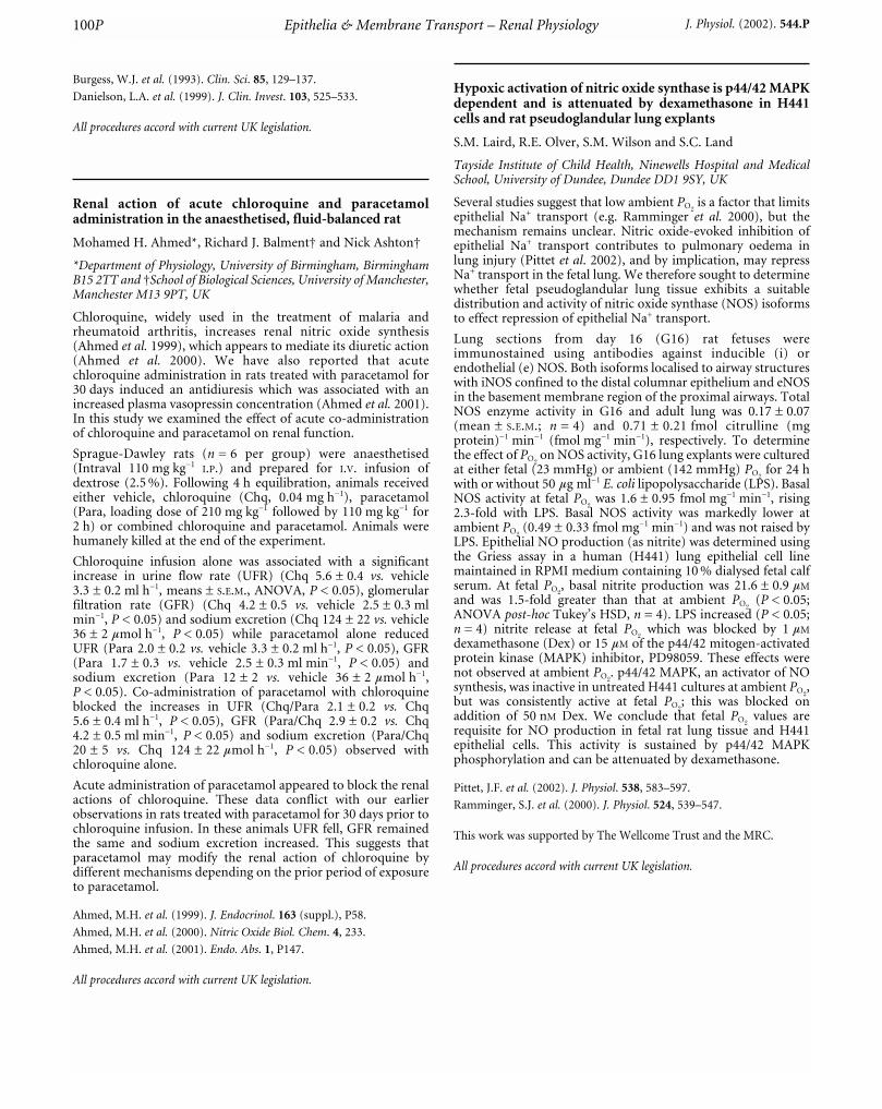
Burgess, W.J. et al. (1993). Clin. Sci. 85, 129–137.
Danielson, L.A. et al. (1999). J. Clin. Invest. 103, 525–533.
All procedures accord with current UK legislation.
Renal action of acute chloroquine and paracetamoladministration in the anaesthetised, fluid-balanced rat
Mohamed H. Ahmed*, Richard J. Balment† and Nick Ashton†
*Department of Physiology, University of Birmingham, BirminghamB15 2TT and †School of Biological Sciences, University of Manchester,Manchester M13 9PT, UK
Chloroquine, widely used in the treatment of malaria andrheumatoid arthritis, increases renal nitric oxide synthesis(Ahmed et al. 1999), which appears to mediate its diuretic action(Ahmed et al. 2000). We have also reported that acutechloroquine administration in rats treated with paracetamol for30 days induced an antidiuresis which was associated with anincreased plasma vasopressin concentration (Ahmed et al. 2001).In this study we examined the effect of acute co-administrationof chloroquine and paracetamol on renal function.
Sprague-Dawley rats (n = 6 per group) were anaesthetised(Intraval 110 mg kg_1
I.P.) and prepared for I.V. infusion ofdextrose (2.5 %). Following 4 h equilibration, animals receivedeither vehicle, chloroquine (Chq, 0.04 mg h_1), paracetamol(Para, loading dose of 210 mg kg_1 followed by 110 mg kg_1 for2 h) or combined chloroquine and paracetamol. Animals werehumanely killed at the end of the experiment.
Chloroquine infusion alone was associated with a significantincrease in urine flow rate (UFR) (Chq 5.6 ± 0.4 vs. vehicle3.3 ± 0.2 ml h_1, means ± S.E.M., ANOVA, P < 0.05), glomerularfiltration rate (GFR) (Chq 4.2 ± 0.5 vs. vehicle 2.5 ± 0.3 mlmin_1, P < 0.05) and sodium excretion (Chq 124 ± 22 vs. vehicle36 ± 2 mmol h_1, P < 0.05) while paracetamol alone reducedUFR (Para 2.0 ± 0.2 vs. vehicle 3.3 ± 0.2 ml h_1, P < 0.05), GFR(Para 1.7 ± 0.3 vs. vehicle 2.5 ± 0.3 ml min_1, P < 0.05) andsodium excretion (Para 12 ± 2 vs. vehicle 36 ± 2 mmol h_1,P < 0.05). Co-administration of paracetamol with chloroquineblocked the increases in UFR (Chq/Para 2.1 ± 0.2 vs. Chq5.6 ± 0.4 ml h_1, P < 0.05), GFR (Para/Chq 2.9 ± 0.2 vs. Chq4.2 ± 0.5 ml min_1, P < 0.05) and sodium excretion (Para/Chq20 ± 5 vs. Chq 124 ± 22 mmol h_1, P < 0.05) observed withchloroquine alone.
Acute administration of paracetamol appeared to block the renalactions of chloroquine. These data conflict with our earlierobservations in rats treated with paracetamol for 30 days prior tochloroquine infusion. In these animals UFR fell, GFR remainedthe same and sodium excretion increased. This suggests thatparacetamol may modify the renal action of chloroquine bydifferent mechanisms depending on the prior period of exposureto paracetamol.
Ahmed, M.H. et al. (1999). J. Endocrinol. 163 (suppl.), P58.
Ahmed, M.H. et al. (2000). Nitric Oxide Biol. Chem. 4, 233.
Ahmed, M.H. et al. (2001). Endo. Abs. 1, P147.
All procedures accord with current UK legislation.
Hypoxic activation of nitric oxide synthase is p44/42 MAPKdependent and is attenuated by dexamethasone in H441cells and rat pseudoglandular lung explants
S.M. Laird, R.E. Olver, S.M. Wilson and S.C. Land
Tayside Institute of Child Health, Ninewells Hospital and MedicalSchool, University of Dundee, Dundee DD1 9SY, UK
Several studies suggest that low ambient PO2is a factor that limits
epithelial Na+ transport (e.g. Ramminger et al. 2000), but themechanism remains unclear. Nitric oxide-evoked inhibition ofepithelial Na+ transport contributes to pulmonary oedema inlung injury (Pittet et al. 2002), and by implication, may repressNa+ transport in the fetal lung. We therefore sought to determinewhether fetal pseudoglandular lung tissue exhibits a suitabledistribution and activity of nitric oxide synthase (NOS) isoformsto effect repression of epithelial Na+ transport.
Lung sections from day 16 (G16) rat fetuses wereimmunostained using antibodies against inducible (i) orendothelial (e) NOS. Both isoforms localised to airway structureswith iNOS confined to the distal columnar epithelium and eNOSin the basement membrane region of the proximal airways. TotalNOS enzyme activity in G16 and adult lung was 0.17 ± 0.07(mean ± S.E.M.; n = 4) and 0.71 ± 0.21 fmol citrulline (mgprotein)_1 min_1 (fmol mg_1 min_1), respectively. To determinethe effect of PO2
on NOS activity, G16 lung explants were culturedat either fetal (23 mmHg) or ambient (142 mmHg) PO2
for 24 hwith or without 50 mg ml_1 E. coli lipopolysaccharide (LPS). BasalNOS activity at fetal PO2
was 1.6 ± 0.95 fmol mg_1 min_1, rising2.3-fold with LPS. Basal NOS activity was markedly lower atambient PO2
(0.49 ± 0.33 fmol mg_1 min_1) and was not raised byLPS. Epithelial NO production (as nitrite) was determined usingthe Griess assay in a human (H441) lung epithelial cell linemaintained in RPMI medium containing 10 % dialysed fetal calfserum. At fetal PO2
, basal nitrite production was 21.6 ± 0.9 mM
and was 1.5-fold greater than that at ambient PO2(P < 0.05;
ANOVA post-hoc Tukey’s HSD, n = 4). LPS increased (P < 0.05;n = 4) nitrite release at fetal PO2
which was blocked by 1 mM
dexamethasone (Dex) or 15 mM of the p44/42 mitogen-activatedprotein kinase (MAPK) inhibitor, PD98059. These effects werenot observed at ambient PO2
. p44/42 MAPK, an activator of NOsynthesis, was inactive in untreated H441 cultures at ambient PO2
,but was consistently active at fetal PO2
; this was blocked onaddition of 50 nM Dex. We conclude that fetal PO2
values arerequisite for NO production in fetal rat lung tissue and H441epithelial cells. This activity is sustained by p44/42 MAPKphosphorylation and can be attenuated by dexamethasone.
Pittet, J.F. et al. (2002). J. Physiol. 538, 583–597.
Ramminger, S.J. et al. (2000). J. Physiol. 524, 539–547.
This work was supported by The Wellcome Trust and the MRC.
All procedures accord with current UK legislation.
Epithelia & Membrane Transport – Renal Physiology100P J. Physiol. (2002). 544.P

Differential mechanisms of intracellular pH (pHi) recoveryfollowing acid load to in situ bovine articular chondrocyteswithin different cartilage zones
J.R. O’Neill, M.J. Kerrigan and A.C. Hall
Department of Biomedical Sciences, University Medical School,George Square, Edinburgh EH8 9XD, UK
The regulation of pHi by isolated articular chondrocytes is almostexclusively by the Na+–H+ antiporter (Wilkins & Hall, 1995).There are, however, significant variations in the environment ofin situ chondrocytes with cartilage depth from synovial surface tobone. Here, we have investigated whether there are differences inthe ability of chondrocytes within three cartilage zones(superficial, mid, deep; SZ, MZ, DZ) to recover their pHi
following an intracellular acid load.
Explants of bovine articular cartilage were obtained frommetacarpophalangeal joints of animals freshly slaughtered at thelocal abattoir and cultured in Dulbecco’s modified Eagle’smedium (DMEM, 280 mosmol, pH 7.4). When required,explants were incubated with BCECF-AM (10 mM, 30 min, 37 °C)and chondrocytes within the zones imaged (Ex. 490/439 nm; Em.535 nm) using a Photon Technology International ImageMastersystem (Hall & Bush, 2001). pHi was altered using theammonium pre-pulse technique (Thomas, 1984) and recovery influorescence ratio studied in the presence of NaHCO3 (23 mM)or its absence (NaCl substituted; both pH 7.4). There was nosignificant difference (ANOVA) to the change of fluorescenceratio following ammonium pre-pulse of cells within the threezones in the presence or absence of HCO3 (P = 0.68; n = 14). InHCO3-free media, recovery of pHi (in units of change influorescence ratio s_1 w 105) of cells within the MZ and DZ wasrapid (32 ± 2 and 56 ± 2, respectively) and completely inhibitedby ethylisopropyl amiloride (EIPA, 100 mM; _2 ± 6 and _1 ± 1,respectively). In contrast, cells within the SZ demonstrated nosignificant (P > 0.05; Student’s unpaired t test) recovery of pHi
over the same time period in the absence of HCO3, although inits presence recovery was markedly stimulated (49 ± 1; data aremeans ± S.E.M., n = 3).
These data suggest that chondrocytes within the SZ of bovinearticular cartilage utilise a HCO3-dependent system for pHi
recovery from acid load, whereas cells within the MZ and DZ ofcartilage largely utilise the Na+–H+ antiporter. These differencesin the mechanisms of chondrocyte pHi regulation may relate tothe environment experienced by the cells within the threecartilage zones. It is possible that this property of SZchondrocytes has not previously been observed in isolated cellpreparations, because they represent a relatively small (< 10 %)proportion of the total cell population.
Hall, A.C. & Bush, P.G. (2001). Eur. J. Physiol. 442, 771–781.
Thomas, R.C. (1984). J. Physiol. 354, 3–22P.
Wilkins, R.J. & Hall, A.C. (1995). J. Cell. Physiol. 164, 474–481.
This work was supported by the Arthritis Research Campaign (H0621),The Wellcome Trust (045925/Z/95/A) and the MRC.
All procedures accord with current UK legislation.
Measuring the binding kinetics of myosin X PH domains atthe plasma membrane by total internal reflectionfluorescence microscopy
G. Mashanov*, D. Tacon†, M. Peckham* and J.E. Molloy†
*Department of Biology, University of York, York YO10 5DD and†School of Biomedical Sciences, University of Leeds, Leeds LS2 9JT,UK
The myosin superfamily contains over 18 family members(Hodge & Cope, 2000). Of these myosins, myosin X is unique inthat it is the only myosin found in mammals that containspleckstrin homology (PH) domains in its tail (Berg et al. 2000).PH domains are found in over 250 different proteins in thehuman genome and selectively bind to phosphoinositidephospholipids. Myosin X is expressed by several cell types,including cultured mouse myoblasts prior to their differentiationinto myotubes (Wells et al. 1997). The PH domains of myosin X,of which there are three, are thought to bind to either Pi(3,4,5)P3
or Pi(3,4)P2, produced following phosphorylation of Pi(4,5)P2 byPi3kinase.
To measure the binding kinetics of the myosin X PH domains atthe plasma membrane in cultured mouse myoblasts, a constructcontaining all three PH domains from myosin X fused to greenfluorescent protein (PH123-GFP) was transiently expressed incultured myoblasts, and single fluorophores were imaged in livecells using total internal reflection fluorescence microscopy(TIRFM). Single fluorescent spots (single PH123-GFPmolecules) were identified using a set of D.I.S.H. criteria: theyhad Diffraction limited size, average Intensity of spotscorresponding to single fluorophore intensity, spots disappeared(photobleached) in a Single step manner, and average Half-lifewas proportional to illumination intensity. Purified GFPmolecules attached to a glass surface by antibodies were used as acontrol.
We found that single fluorophores disappeared suddenly eitherbecause they photobleached or because they detached from themembrane and diffused away rapidly. By varying illuminationintensity we determined the rates of these different processes andwe were able to measure the apparent binding and detachmentrates of the pleckstrin homology domains. These experimentsdemonstrate that steady-state observation of single fluorophoresin live cells gives information about binding and release kineticsof proteins at the plasma membrane.
Berg, J.S. et al. (2000). J. Cell Sci. 113, 3439–3451.
Hodge, T. & Cope, M.J. (2000). J. Cell. Sci. 113, 3353–3354.
Wells, C. et al. (1997). J. Mus. Res. Cell Motil. 18, 501–515.
This work was supported by the BBSRC.
Effects of extracellular calcium and pH upon a chloridechannel in the early distal convoluted tubule of the mouse
Stéphane Lourdel, Marc Paulais and Jacques Teulon
CNRS FRE 2468, Institut des Cordeliers, Paris, France
We investigated the properties of basolateral Cl_ channels in theearly distal convoluted tubule (DCT), using the cell-attached andexcised configurations of the patch-clamp technique.Microdissected DCTs were isolated from collagenase-treatedkidneys after mice were humanely killed. The pipette and bathsolutions contained (mM): 140 NaCl, 4.8 KCl, 1.2 MgCl2,1 CaCl2, 10 glucose and 10 Hepes (pH 7.4). We recorded one
Epithelia & Membrane Transport – Renal Physiology 101PJ. Physiol. (2002). 544.P

channel of 9.4 ± 0.3 pS (n = 23, mean ± S.E.M.). In the conditionsspecified above, the reversal potential (Er) of the I–V relationshipis close to zero (2.0 ± 2.2 mV). Replacing 100 mM NaCl withsodium gluconate shifted Er by 30.2 ± 3.7 mV (n = 4) towardsthe right, whereas substituting N-methyl-D-glucamine forNa+ had no effect (_1 ± 1.9 mV, n = 5). This indicates Cl_
selectivity. We estimated the influence of extracellular Ca2+ andpH on channel activity by measuring the averaged current andNPo on separate patches under various conditions of Ca2+ andpH. For this purpose, as the channel is inhibited at acidintracellular pH (Marvao & Teulon, 2000), the closed currentlevel was assessed on the cell by inhibiting the channels viasodium acetate superfusion. The NPo measured with zero Ca2+ inthe pipette was 7.6 ± 1.6 (n = 13). It significantly increased to33.2 ± 5.6 (n = 10) with 5 mM Ca2+ in the pipette. The influenceof external pH was dramatic since NPo significantly increasedfrom 2.8 ± 0.1 (n = 9) at pH 6.4 to 44.7 ± 8.8 at pH 8.0 (n = 10).In contrast, the percentages of patches with active chloridechannels were similar under the four conditions (71 and 62 %active patches at 0 and 5 mM Ca2+; 58 and 54 % active patches atpH 6.4 and 8.0). Upon excision, it was possible to assessquantitatively the anionic selectivity of the channel in the earlyDCT despite frequent channel rundown. The channel had therelative permeability sequence Cl_ > Br_ ~ NO3
_ ~ I_ > F_
(n = 4–5). The properties of the channel described here arecompatible with the currently known properties of the ClC-Kchannels.
Student’s unpaired t test was used. P < 0.05 was consideredsignificant.
Marvao, P. & Teulon, J. (2000). Nephrol. Dial. Transpl. 15, 28A.
All procedures accord with current National guidelines.
Carbachol-stimulated anion secretion in rat distal colonoccurs predominantly through cystic fibrosistransmembrane conductance regulator (CFTR)
M. Bellingham, A.D. Corbett and D.L. Bovell
School of Biological and Biomedical Sciences, Glasgow CaledonianUniversity, Cowcaddens Road, Glasgow G4 0BA, UK
The muscarinic receptor agonist carbachol is believed to evokeCl_ secretion in rat colonic epithelial cells by elevation ofintracellular Ca2+ (Greger et al. 1997) and although Ca2+-activated Cl_ channels have been identified in colonic epithelialcell lines (Merlin et al. 1998) controversy exists over theirpresence/function in native colonic epithelia. Previous studiesfrom this laboratory have shown that carbachol evokes Cl_ andHCO3
_ secretion in rat distal colon (Bellingham et al. 2002). Herewe have examined the effects of both cAMP- and Ca2+-activatedCl_ channel inhibitors on carbachol-evoked anion secretion inrat distal colon.
The terminal colon (5 cm) was removed from male Wistar ratskilled by cervical dislocation. The mucosal portion was mountedin an Ussing chamber (area 0.5 cm2) containing Krebs solution(mM: NaCl 119, KCl 4.7, NaHCO3 24.8, MgSO4 1.2, KH2PO4 1.2,CaCl2 2.5 and glucose 11.1) at 37 °C, gassed with 95 % O2 and 5 %CO2. Short-circuit current (ISC) was recorded and measurementswere taken from baseline to peak. All values representmeans ± S.E.M. and Student’s unpaired t test was used to testsignificance (P < 0.05).
Basal ISC was 18.4 ± 5.2 mA cm_2 with a resistance of102.3 ± 6.8 V cm_2 (n = 17). Enhancing cAMP levels by addingthe phosphodiesterase inhibitor isobutylmethylxanthine (IBMX,100 mM basolaterally) increased ISC by 45.0 ± 12.1 mA cm_2
(n = 3), suggesting tonic activation of cAMP-sensitive channels.Addition of the CFTR channel blocker diphenylamine-2-carboxylic acid (DPC, 100 mM apically) reduced basal ISC by9.9 ± 4.0 mA cm_2 (n = 3), whereas addition of the Ca2+-activatedCl_ channel inhibitor 4,4-diisothiocyanatostilbene-2,2-disulfonicacid (DIDS, 1 mM apically) had no effect on basal ISC (n = 4).Furthermore, in the presence of DPC (100 mM apically) all threephases of the typical carbachol response (Fig. 1A) weresignificantly attenuated (P < 0.05, n = 3). However, in thepresence of DIDS, carbachol-evoked ISC was not significantlydifferent from control (n = 4). Inhibition of endogenous cAMPproduction via the prostaglandin pathway, with the cyclo-oxygenase inhibitor indomethacin (10 mM basolaterally andapically), resulted in a markedly altered carbachol response(Fig. 1B, n = 11). Under these conditions, carbachol evoked adecrease in ISC (_5.7 ± 2.5 mA cm_2, n = 11).
Figure 1. A, typical triphasic short-circuit current (ISC)response to carbachol (100 mM, Bl) in rat distal colon(n = 17). B, monophasic ISC response to carbachol (100 mM,Bl) in the presence of indomethacin (10 mM, Bl/Ap) in ratdistal colon (n = 11). Bl = basolateral, Bl/Ap = basolateraland apical application.
The present results suggest that CFTR is the predominant Cl_
channel involved in carbachol-evoked anion secretion in the ratdistal colon. These results support the findings of Strabel &Diener (1995) and provide further evidence against directactivation of Cl_ channels by carbachol in rat distal colon.
Bellingham, M. et al. (2002). J. Physiol. 543.P, 52P.
Greger, R. et al. (1997). Comp. Biochem. Physiol. 118, 271–275.
Merlin, D. et al. (1998). Am. J. Physiol. 275, C484–495.
Strabel, D. & Diener, M. (1995). Eur. J. Pharmacol. 274, 181–191.
All procedures accord with current UK legislation.
Epithelia & Membrane Transport – Renal Physiology102P J. Physiol. (2002). 544.P
Effects of brefeldin-A upon the b-adrenoceptor-mediatedcontrol of apical Na+ conductance (GNa) in rat fetal distallung epithelial (FDLE) cells
S.J. Ramminger, R.E. Olver and S.M. Wilson
Lung Membrane Transport Group, Tayside Institute of Child Health,Ninewells Hospital and Medical School, University of Dundee,Dundee DD1 9SY, UK
If breathing is to be established at birth then the liquid secretedinto the lung lumen during fetal life must first be removed fromthe potential air spaces. This occurs during the final stages ofpregnancy and is driven by the b-adrenoceptor-mediatedtransport of Na+ out of the alveolar space (Olver et al. 1986). Ithas been suggested that this stimulation of Na+ transport involvesthe insertion of additional Na+ channels into the apical plasmamembrane by an exocytotic mechanism that is sensitive tobrefeldin A (Ito et al. 1997). Recent work from this laboratoryhas shown that b-adrenoceptor agonists do increase GNa in FDLEcells (Collett et al. 2002) and so we now explore the effects ofbrefeldin-A upon this response.
Figure 1. Time-mated pregnant rats were anaesthetised onthe 20th day of gestation, their fetuses delivered by Caesareansection and immediately decapitated and the donor ratshumanely killed without regaining consciousness. FDLE cellswere then isolated from the fetal lung tissue as describedpreviously (Collett et al. 2002). This acquisition of fetal lungtissue accords with current UK legislation. The cells weregrown to confluence (~48 h) on Snapwell membranes(Costar) which were then mounted in Ussing chambers andISC measured. The cells were then basolaterally permeabilised(75 mM nystatin) so that GNa could be estimated bymeasuring the amiloride-sensitive current evoked byimposing an inwardly directed Na+ gradient upon the cells(Collett et al. 2002). Such experiments were undertakenusing control and isoprenaline-stimulated cells that hadeither been maintained under control conditions or exposedto brefeldin-A (1 mM) for 30 min. Data are presented asmeans ± S.E.M. (n = 6). ***P < 0.001 vs. data derived fromthe unstimulated cells; †P < 0.05 vs. data derived fromunstimulated control cells (Student’s paired t test).
These data confirm (Collett et al. 2002) that isoprenaline-evokedincrease in ISC (basal, 7.9 ± 0.7 mA cm_2; post-isoprenaline,11.2 ± 0.9 mA cm_2, P < 0.02) is normally associated with a rise inGNa (Fig. 1). Exposing unstimulated cells to brefeldin-A (1 mM)caused a barely discernible rise in ISC that developed over a periodof ~5 min. The current then declined slowly, reaching a valuethat was 88.7 ± 3.6 % of its initial level (P < 0.05) after 30 min.Application of isoprenaline to brefeldin A-treated cells did notelicit a discernible change in ISC. Data obtained afterpermeabilisation showed that this drug causes a fall in GNa andblocks the isoprenaline-evoked increase in conductance (Fig. 1).Isoprenaline thus causes a brefeldin-A-sensitive rise in GNa inFDLE cells (Ito et al. 1997; Collett et al. 2002).
Collett, A. et al. (2002). Am. J. Physiol. Lung Cell. Mol. Physiol. 282,L621–630.
Ito, Y. et al. (1997). Pflügers Arch. 434, 492–494.
Olver, R.E. et al. (1986). J. Physiol. 376, 321–340.
The authors thank The Wellcome TRust and Tenovus (Scotland) fortheir financial support.
All procedures accord with current UK legislation.
Localisation of the two-pore domain K+ channel TASK-2 inmouse kidney _ RT-PCR of individual segments
Michael J. Morton, Amanda L. Ingham and Malcolm Hunter
School of Biomedical Sciences, Worsley Building, University of Leeds,Leeds LS2 9NQ, UK
Potassium conductances and permeabilities are present in allnephron segments and serve various functions, such as K+
excretion and the maintenance of cell potential and volume.TASK-2 potassium channel has two pore-forming domains intandem in each monomer and is sensitive to extracellular pH.Messenger RNA encoding TASK-2 can be detected in RNAderived from whole kidney (Reyes et al. 1998). As a first step tounderstanding the role of TASK-2 in the kidney, we haveattempted to localise TASK-2 mRNA by RT-PCR of individualnephron segments.
Adult male mice were anaesthetised with sodiumpentobarbitone, 100 mg kg_1
I.P., and killed by decapitation. Thekidneys were removed and divided into cortical and papillarysections by crude dissection, divided into 1 mm segments anddigested with collagenase (0.5 mg ml_1) at 37 °C in a shakingwater bath for 30–60 min. Individual tubules were selected fromthe resulting tubule suspensions. Glomeruli and proximaltubules (PT) were identified by eye. Thick ascending limb (TAL)and cortical collecting tubule (CCT) segments were identified bythe presence of mRNA for Tamm-Horsfall protein (THP) andaquaporin-2 (AQP-2), respectively. Amplification ofendogenous b-actin mRNA was used as a positive control.Tubules were permeabilised with 0.5 % Triton X-100 and heatingto 95 °C and reverse transcribed according to the manufacturer’sinstructions (Promega). Reverse-transcribed material wassubjected to two rounds of PCR: the first with multiplex primersspecific to TASK-2, AQP-2, THP and b-actin, and the secondwith nested primers. PCR products were analysed by gelelectrophoresis, stained with ethidium bromide and visualised byUV illumination. Reverse transcriptions were also performed inthe absence of enzyme to control for contaminating genomicDNA.
Epithelia & Membrane Transport – Renal Physiology 103PJ. Physiol. (2002). 544.P
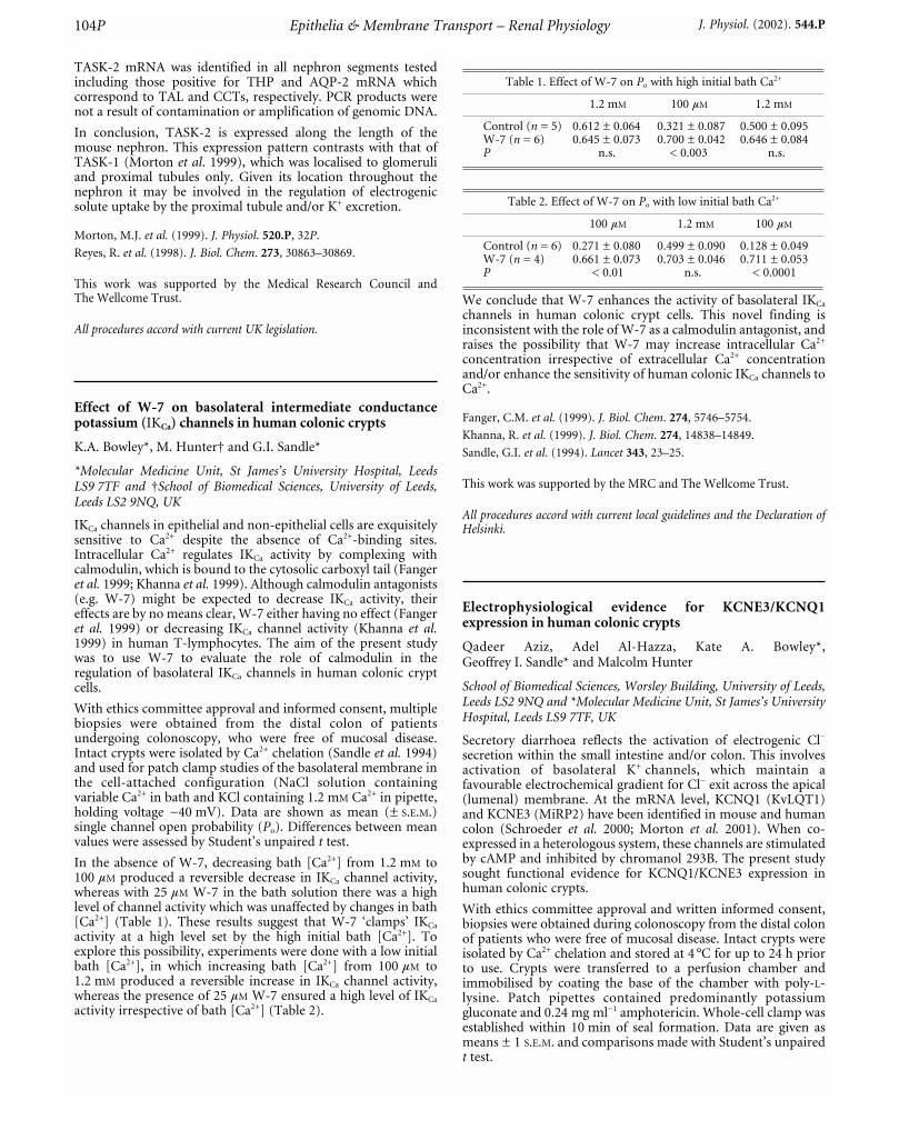
TASK-2 mRNA was identified in all nephron segments testedincluding those positive for THP and AQP-2 mRNA whichcorrespond to TAL and CCTs, respectively. PCR products werenot a result of contamination or amplification of genomic DNA.
In conclusion, TASK-2 is expressed along the length of themouse nephron. This expression pattern contrasts with that ofTASK-1 (Morton et al. 1999), which was localised to glomeruliand proximal tubules only. Given its location throughout thenephron it may be involved in the regulation of electrogenicsolute uptake by the proximal tubule and/or K+ excretion.
Morton, M.J. et al. (1999). J. Physiol. 520.P, 32P.
Reyes, R. et al. (1998). J. Biol. Chem. 273, 30863–30869.
This work was supported by the Medical Research Council andThe Wellcome Trust.
All procedures accord with current UK legislation.
Effect of W-7 on basolateral intermediate conductancepotassium (IKCa) channels in human colonic crypts
K.A. Bowley*, M. Hunter† and G.I. Sandle*
*Molecular Medicine Unit, St James’s University Hospital, LeedsLS9 7TF and †School of Biomedical Sciences, University of Leeds,Leeds LS2 9NQ, UK
IKCa channels in epithelial and non-epithelial cells are exquisitelysensitive to Ca2+ despite the absence of Ca2+-binding sites.Intracellular Ca2+ regulates IKCa activity by complexing withcalmodulin, which is bound to the cytosolic carboxyl tail (Fangeret al. 1999; Khanna et al. 1999). Although calmodulin antagonists(e.g. W-7) might be expected to decrease IKCa activity, theireffects are by no means clear, W-7 either having no effect (Fangeret al. 1999) or decreasing IKCa channel activity (Khanna et al.1999) in human T-lymphocytes. The aim of the present studywas to use W-7 to evaluate the role of calmodulin in theregulation of basolateral IKCa channels in human colonic cryptcells.
With ethics committee approval and informed consent, multiplebiopsies were obtained from the distal colon of patientsundergoing colonoscopy, who were free of mucosal disease.Intact crypts were isolated by Ca2+ chelation (Sandle et al. 1994)and used for patch clamp studies of the basolateral membrane inthe cell-attached configuration (NaCl solution containingvariable Ca2+ in bath and KCl containing 1.2 mM Ca2+ in pipette,holding voltage _40 mV). Data are shown as mean (± S.E.M.)single channel open probability (Po). Differences between meanvalues were assessed by Student’s unpaired t test.
In the absence of W-7, decreasing bath [Ca2+] from 1.2 mM to100 mM produced a reversible decrease in IKCa channel activity,whereas with 25 mM W-7 in the bath solution there was a highlevel of channel activity which was unaffected by changes in bath[Ca2+] (Table 1). These results suggest that W-7 ‘clamps’ IKCa
activity at a high level set by the high initial bath [Ca2+]. Toexplore this possibility, experiments were done with a low initialbath [Ca2+], in which increasing bath [Ca2+] from 100 mM to1.2 mM produced a reversible increase in IKCa channel activity,whereas the presence of 25 mM W-7 ensured a high level of IKCa
activity irrespective of bath [Ca2+] (Table 2).
Table 1. Effect of W-7 on Po with high initial bath Ca2+
1.2 mM 100 mM 1.2 mM
Control (n = 5) 0.612 ± 0.064 0.321 ± 0.087 0.500 ± 0.095W-7 (n = 6) 0.645 ± 0.073 0.700 ± 0.042 0.646 ± 0.084P n.s. < 0.003 n.s.
Table 2. Effect of W-7 on Po with low initial bath Ca2+
100 mM 1.2 mM 100 mM
Control (n = 6) 0.271 ± 0.080 0.499 ± 0.090 0.128 ± 0.049W-7 (n = 4) 0.661 ± 0.073 0.703 ± 0.046 0.711 ± 0.053P < 0.01 n.s. < 0.0001
We conclude that W-7 enhances the activity of basolateral IKCa
channels in human colonic crypt cells. This novel finding isinconsistent with the role of W-7 as a calmodulin antagonist, andraises the possibility that W-7 may increase intracellular Ca2+
concentration irrespective of extracellular Ca2+ concentrationand/or enhance the sensitivity of human colonic IKCa channels toCa2+.
Fanger, C.M. et al. (1999). J. Biol. Chem. 274, 5746–5754.
Khanna, R. et al. (1999). J. Biol. Chem. 274, 14838–14849.
Sandle, G.I. et al. (1994). Lancet 343, 23–25.
This work was supported by the MRC and The Wellcome Trust.
All procedures accord with current local guidelines and the Declaration ofHelsinki.
Electrophysiological evidence for KCNE3/KCNQ1expression in human colonic crypts
Qadeer Aziz, Adel Al-Hazza, Kate A. Bowley*,Geoffrey I. Sandle* and Malcolm Hunter
School of Biomedical Sciences, Worsley Building, University of Leeds,Leeds LS2 9NQ and *Molecular Medicine Unit, St James’s UniversityHospital, Leeds LS9 7TF, UK
Secretory diarrhoea reflects the activation of electrogenic Cl_
secretion within the small intestine and/or colon. This involvesactivation of basolateral K+ channels, which maintain afavourable electrochemical gradient for Cl_ exit across the apical(lumenal) membrane. At the mRNA level, KCNQ1 (KvLQT1)and KCNE3 (MiRP2) have been identified in mouse and humancolon (Schroeder et al. 2000; Morton et al. 2001). When co-expressed in a heterologous system, these channels are stimulatedby cAMP and inhibited by chromanol 293B. The present studysought functional evidence for KCNQ1/KCNE3 expression inhuman colonic crypts.
With ethics committee approval and written informed consent,biopsies were obtained during colonoscopy from the distal colonof patients who were free of mucosal disease. Intact crypts wereisolated by Ca2+ chelation and stored at 4 °C for up to 24 h priorto use. Crypts were transferred to a perfusion chamber andimmobilised by coating the base of the chamber with poly-L-lysine. Patch pipettes contained predominantly potassiumgluconate and 0.24 mg ml_1 amphotericin. Whole-cell clamp wasestablished within 10 min of seal formation. Data are given asmeans ± 1 S.E.M. and comparisons made with Student’s unpairedt test.
Epithelia & Membrane Transport – Renal Physiology104P J. Physiol. (2002). 544.P

Whole-cell currents were activated within 5 min by addition of10 µM forskolin to the bath solution (current at +100 mV:123 ± 21.4 pA control, 410 ± 79.2 pA after forskolin, n = 3). Thisforskolin-activated current was inhibited by 100 µM chromanol293B and 1 mM BaCl2. Prior to forskolin addition, currents wereinsensitive to chromanol 293B. In addition to these whole-cellrecordings, small conductance channels of approximately 7 pShave been observed in cell-attached recordings with K+-richpipette solutions.
In conclusion, small conductance KCNE3/KCNQ1 channels areexpressed in the basolateral membrane of human colonic crypts.These channels are stimulated by forskolin and inhibited bychromanol 293B and Ba2+ and may be expected to play a role inthe pathogenesis of secretory diarrhoea.
Morton, M.J. et al. (2001). J. Am. Soc. Nephrol. 12, A0329.
Schroeder, B.C. et al. (2000). Nature 403, 196–199.
This work was supported by The Wellcome Trust and the MRC.Chromanol 293B was a kind gift from Dr A.E. Busch of Aventis Pharma.
All procedures accord with current local guidelines and the Declaration ofHelsinki.
P-Glycoprotein expression significantly increases thecapability of human placental trophoblast cells to effluxxenobiotics
D.E. Atkinson, J.D. Glazier, C.P. Sibley, L.J. Fairbairn* andS.L. Greenwood
Academic Unit of Child Health, University of Manchester, St Mary’sHospital, Manchester M13 0JH and *Paterson Institute for CancerResearch, Christie Hospital, Manchester M20 4BX, UK
We have previously shown that the trophoblast derived BeWoand JEG choriocarcinoma cell lines express MRP1 (multi-drugresistance related protein), whilst the JAr choriocarcinoma cellline and primary cytotrophoblast cells express both MRP1 andP-glycoprotein (P-gp, coded by the MDR1 gene) (Atkinson et al.2000). The functional consequences of the absence of P-gp havealso been determined and we have shown that the cyclosporin-sensitive accumulation of [3H] vinblastine by cells expressingonly MRP1 is higher than that by cells expressing both drugefflux proteins (Atkinson et al. 2001).
In the present study we tested the hypothesis that the increasedcyclosporin-sensitive [3H]vinblastine accumulation (i.e. reducedefflux) in BeWo cells is due to their lack of expression of MDR1 P-gp.
Using a retroviral vector based on the Moloney murineleukaemia virus we introduced MDR1 into BeWo cells by co-culture with the vector packaging cell line in transwell plates.MDR1 transduced cells (BeWo MDR) were then selected using100 mg ml_1 of colchicine. To establish that transduction hadbeen successful, Western blot analysis was performed using theF4 monoclonal antibody specific for human P-gp. The cells werethen used in accumulation assays as previously described(Atkinson et al. 2001).
Figure 1. Expression of P-gp in native (BeWo, 13.6 mg) andtransduced BeWo (BeWo MDR, 3.4 mg) cells. Microvillous(MVM, 10 mg) and basal (BM 15 mg) membranes ofplacental trophoblast are shown as +ve and _ve controls,respectively.
Figure 2. Cyclosporin-sensitive accumulation of[3H] vinblastine in native BeWo cells and cells followingtransduction with MDR1. Accumulation by cytotrophoblastcells are included for comparison. Data are means ± S.E.M.(P < 0.01, two-way ANOVA)
BeWo MDR cells express P-gp, whilst, as expected, native BeWocells show no expression of the protein (Fig. 1). Cyclosporin-sensitive accumulation of [3H] vinblastine in BeWo MDR cells issignificantly lower than in native BeWo cells and is very similarto that seen in cytotrophoblast cells which naturally expressMDR1 (Fig. 2).
We conclude that the higher cyclosporin-sensitive accumulationin native BeWo cells resulting from a lower capacity to effluxsubstrate drugs is due to their lack of P-gp, and therefore thatP-gp is important in xenobiotic handling by trophoblast cells.The expression of P-gp on the maternal facing plasma membraneof the placenta (Fig. 1, Atkinson et al. 2000) places it in an idealposition to protect the fetus from potentially harmfulcompounds in the maternal circulation.
Atkinson, D.E. et al. (2000). J. Physiol. 528.P, 34P.
Atkinson, D.E. et al. (2001). J. Physiol. 535.P, 38P.
This work was funded by the MRC. MVM and BM were kindly donatedby Dr P. Speake.
All procedures accord with current local guidelines.
Epithelia & Membrane Transport – Renal Physiology 105PJ. Physiol. (2002). 544.P

Regulation of the AE2 anion exchanger by pHi and pHo
requires highly conserved amino acids in the AE2cytoplasmic N-terminal domain
A.K. Stewart, M.N. Chernova, B.E. Shmukler, S. Wilhelm andS.L. Alper
Department of Molecular Medicine and Renal Units, HarvardMedical School, Beth Israel Deaconess Medical Center, East Campus,330 Brookline Avenue, Boston, MA, USA
We recently reported that regulation of the murine Na+-independent Cl_ –HCO3
_ exchanger, AE2, by pH requires aminoacid residues 312–347 of its N-terminal cytoplasmic domain(Stewart et al. 2001). Initial pH-sensitive data employing hexa-alanine bloc mutations across this region prompted closer studyof individual alanine substitutions in AE2 amino acid residues336–347. AE2 function was measured as DIDS-sensitive 36Cl_
efflux from Xenopus oocytes during variation of extracellular pH(pHo) with clamped or unclamped intracellular pH (pHi), orduring variation of pHi at constant pHo. Statistical significancewas assessed by Student’s unpaired t test, where the level ofsignificance was P < 0.05.
Wild-type (WT) AE2-mediated 36Cl_ efflux was inhibited by acidpHo, with a value of pHo(50) = 6.87 ± 0.05 (± S.E.M., n = 36), andwas stimulated up to 10-fold by the intracellular alkalinizationproduced by bath removal of the pre-equilibrated weak acid,butyrate (40 mM; n = 43). Systematic hexa-alanine [(A)6] blocsubstitutions between aa 312–347 revealed that hexa-blocmutation A(6)342–347 exhibited the greatest acid shift in pHo(50)
value of ~0.8 pH units (n = 10, P < 0.05). Two of the six (A)6
mutants retained normal pHi sensitivity of 36Cl_ efflux, whereasthe (A)6 mutants 318–323 (n = 10), 336–341 (n = 9) and342–347 (n = 10) were not stimulated by intracellularalkalinization (P < 0.05).
We further evaluated the highly conserved region betweenaa 336–347 by alanine scan and other mutagenesis of singleresidues. Significant changes in AE2 sensitivity to pHo and to pHi
were found independently and in concert. Interestingly,activation of AE2 E346A by intracellular alkalinisation remainedintact (n = 25, P > 0.05), but was abolished by the mutationE346D (n = 9, P < 0.05). In contrast, E346A acid shifted thepHo(50) value from WT AE2 by ~0.7 pH units (n = 12, P < 0.05).
Alanine substitution of conserved glutamate residues in thecytoplasmic N-terminal domain of two additional pH-sensitivepolypeptides, cAE3 and the AE1cyto/AE2memb chimera, confirmedthe importance of these residues in the regulation by pH of otherAE anion exchangers. We hypothesize that individual amino acidresidues of AE2 aa 336–347 contribute to a local structure thatinteracts, directly or indirectly, with pH-sensor residues of theAE2 transmembrane domain. This regulatory interaction isprobably conserved among members of the SLC4 bicarbonatetransporter gene superfamily.
Stewart, A.K. et al. (2001). Am. J. Physiol. 281, C1344–1354.
We thank The Wellcome Trust for generous support.
All procedures accord with current National and local guidelines.
Oxygen tension may influence NHE1 protein expression invillous explants from human placenta
H.A. Lacey, K. Mynett, M. Cretney, J.D. Glazier, S.L. Greenwoodand C.P. Sibley
Academic Unit of Child Health, University of Manchester,Manchester M13 0JH, UK
We have previously demonstrated that Na+–H+ exchanger 1(NHE1) protein is expressed and activity differentially regulatedthroughout gestation in the microvillous membrane (MVM) ofthe human placenta (Hughes et al. 2000). Furthermore,preliminary data have shown that NHE1 mRNA expression islower in first trimester than term placenta (Lacey et al. 2001).Oxygen tension in the intervillous space of the placenta rapidlyincreases between 8 and 11 weeks of pregnancy, concomitantwith the onset of blood flow through the spiral arteries (Jauniauxet al. 2001). Here we test the hypothesis that NHE1 proteinexpression in the placenta is regulated by oxygen tension. Villousexplants were dissected from first trimester (n = 2, 11+6 and10+5 weeks) and term (n = 3) placental tissue (5 mg wet weight)and maintained in explant culture on Netwell porous supportsfor 4–5 days, by which time term placental villous explants haveundergone resyncytialisation (Siman et al. 2001). Explants weremaintained in 3 or 21 % oxygen in 5 % carbon dioxide at 37 °C.On day 4–5 of culture, several explants were harvested, washed inPBS and protein homogenate prepared at 4 °C in PBS + 1.0 mM
serine protease inhibitor. 20 mg protein homogenate from firsttrimester and term placental explants cultured at 3 and 21 %oxygen, together with MVM (positive control for NHE1; Hugheset al. 2000), were loaded on a 7 % reducing SDS-PAGE gel andelectroblotted. Blotted membranes were probed with 1:100mouse anti-human NHE1 primary monoclonal antibody(Becton Dickenson), followed by incubation with goat anti-mouse-HRP conjugated secondary antibody (1:1000) anddetection by enhanced chemiluminescence. A band of 94 kDarepresenting NHE1 was observed in all samples. Intensity ofNHE1 signal was calculated as optical density w area (MolecularAnalyst Software, BioRad). In first trimester explants (n = 3), themedian change in NHE1 protein expression, quantified as %change in optical density between 3 and 21 %, was _1.83 %(_63.08/+78.2 %, 95 % confidence interval), compared withterm explants, n = 5, 27.85 % (_21.8/+106.1 %, 95 % confidenceinterval). We conclude that NHE1 protein expression in termhuman placenta may be affected by oxygen tension.
Hughes, J.L. et al. (2000). Pediatr. Res. 48, 652–659.
Jauniaux, E. et al. (2001). Am. J. Obstet. Gynecol. 184, 998–1003.
Lacey, H.A. et al. (2001). Placenta 22, A43.
Siman, C.M. et al. (2001). Am. J. Physiol. 280, R1116–1122.
This work was supported by The Wellcome Trust.
All procedures accord with current local guidelines.
Epithelia & Membrane Transport – Renal Physiology106P J. Physiol. (2002). 544.P

The immunocytochemical localisation of anion exchangers(AE) in the reabsorptive duct of the human eccrine sweatgland
D.L. Bovell* and P. Quinton†
*School of Biological and Biomedical Sciences, Glasgow CaledonianUniversity, Glasgow, UK and †UCSD School of Medicine,Department of Pediatrics, La Jolla, California, USA
Sweating carries with it a potential loss of salt and as NaCl isphysiologically important, it must, therefore, be conserved.Functionally, the human eccrine sweat gland has two regions: asecretory coil, which produces an isosmotic primary sweat by theelectrogenic movement of Cl_ and Na+ ions, coupled osmoticallyto the movement of water, and a reabsorptive duct, whichhypertonically reabsorbs NaCl from the primary sweat. Thereabsorption of salt depends upon Na+ being pumped from theduct cells into the interstitial fluid by a Na/K-ATPase on thebasolateral membranes, while Na+ moves passively into the cellsvia sodium channels sited only in the luminal membrane. ATP-dependent Cl_ (CFTR) channels allow Cl_ ions to follow downtheir electrochemical gradient. At low flow rates, sweat can beproduced physiologically at concentrations as low as 5–15 mM
NaCl. However, the electrochemical salt recovery mechanismcannot extract salt from the lumen when luminal Cl_
concentration falls below its electrochemical equilibrium acrossthe apical membrane. Previous findings suggested that anadditional mechanism for salt recovery at low flow rates may bedriven by H+ pumps associated with a Cl _–HCO3
_ exchanger inthe luminal membrane which could lower luminal Cl_ to thelevels observed physiologically (Reddy & Quinton, 1994).
The presence of a H+ pump has been demonstrated (Bovell et al.2000), while that of a Cl_–HCO3
_ exchanger has not beenestablished. A family of anion exchangers (AE1, AE2 and AE3)have been cloned that functionally mediate Cl_–HCO3
_ exchangeand which are expressed in a variety of tissues. AE2 is expressedin many epithelia including kidney, while AE3 has been detectedin the nervous system (bAE3) and cardiac muscle (cAE3), as wellas in the gut. This study investigated the presence of an AE in theluminal cells of the human eccrine gland reabsorptive duct.
Frozen sections were obtained from skin biopsies donated, withinformed consent and ethics committee approval, by malevolunteers (n = 6). Immunocytochemical techniques employedantibodies, raised against AE2 and AE3. Appropriate negativecontrols were performed using pre-absorbed sera.
In this study, immunoperoxidase labelling demonstrated thepresence of anion exchangers and showed a predominantlocalisation of AE2 at the apical plasma membrane of the ductalcells, suggesting that the AE2 is involved in exchanging Cl_ andHCO3
_ ions at slow sweat flow rates through the duct. However,the human sweat gland results differ from those in rat salivaryduct cells (Park et al. 1999), where basolaterally located AE2 may,together with a Na+ –H+ exchanger, control the intracellular pHand contribute to intracellular accumulation of Cl_. Our resultsalso demonstrate the localisation of AE3 in the cells of thereabsorptive duct, which is surprising. AE3 is normally found ineither brain (AE3b) or cardiac tissue (AE3c), although it has beenfound in gut epithelia (Kudrycki et al. 1990). Further work isclearly required to clarify the role of these exchangers.
Bovell, D.L. et al. (2000). Histochem. J. 32, 409–413.
Kudrycki, K.E. et al. (1990). J. Biol. Chem. 265, 462–471.
Park, K. et al. (1999). Am. J. Physiol. 276, G470–478.
Reddy, M.M. & Quinton, P.M. (1994). Am. J. Physiol. 36, C1136–1144.
The authors wish to acknowledge the American Physiological Society fora Research Career Enhancement Award to D.L.B. and Professor S. Alperfor generously providing the AE antibodies.
All procedures accord with current local guidelines and the Declaration ofHelsinki.
Blood glucose threshold for appearance of glucose in nasalsecretions. Evidence for active glucose transport acrosshuman nasal epithelium?
David M. Wood*, Barbara J. Philips† and Emma H. Baker*
*Department of Physiological Medicine and †Department ofAnaesthetics and Intensive Care, St George’s Hospital Medical School,London, UK
Glucose concentrations are low or undetectable in animal airwaysecretions (Barker et al. 1989). Sodium–glucose co-transportersin pulmonary epithelium, identified by RT-PCR in rat andmouse and by functional studies in sheep, could activelymaintain this low glucose concentration (Barker et al. 1989; Leeet al. 1994). In healthy humans glucose is undetectable in airwaysecretions, but is present in secretions from those withhyperglycaemia (King et al. 2001; Meguer et al. 2001). Thedynamic relationship between blood and airway glucose has notpreviously been studied.
Twelve non-diabetic volunteers with healthy nasal mucosaunderwent repeated measurement of nasal glucose (NG) duringelevation of blood glucose by the hyperglycaemic clamptechnique. Nasal glucose was measured using glucose oxidaseindicator sticks wetted by secretions during a 30 s application tothe nasal mucosa under direct vision. Glucose was measured inarterialised whole blood using a glucose oxidase-based analyser(Analox, UK).
In six subjects blood glucose was raised to 12 mmol l_1 over15 min. Glucose appeared in nasal secretions of all subjectswithin 20 min of commencing the infusion and 10 min ofreaching the target blood sugar. NG decreased as the bloodglucose fell on stopping the glucose infusion and was neverhigher than blood glucose. In a further six subjects blood glucoselevels were raised from baseline by 1 mmol l_1 increments andheld at each level for 15 min before NG measurement. The bloodglucose level at which nasal glucose appeared was8.2 ± 1.0 mmol l_1 (mean ± S.D.) (range 7.1–10.0).
Passive diffusion is likely to account for the rapid appearance ofglucose in nasal secretions when blood glucose is raised.However, active transport must account for reabsorption ofglucose against the concentration gradient as blood sugar falls.Our observation of a blood glucose threshold above whichglucose appears in airway secretions is consistent with thepresence of a saturable glucose transporter, such as a sodium–glucose co-transporter, actively reabsorbing glucose acrosshuman airway epithelium. Maintenance of glucose-free airwaysecretions may be important for pulmonary defence againstinfection.
Barker, P.M. et al. (1989). J. Physiol. 409, 15–27.
King, L.A. et al. (2001). Eur. Resp. J. 18, 405s.
Lee, W.S. et al. (1994). J. Biol. Chem. 269, 12032–12039.
Meguer, J.X. et al. (2001). Eur. Resp. J. 18, 366s.
All procedures accord with current local guidelines and the Declaration ofHelsinki.
Epithelia & Membrane Transport – Renal Physiology 107PJ. Physiol. (2002). 544.P
The physiological roles of the KCl cotransporter in humancervical cancer cells
Meng-Ru Shen*†, Cheng-Yang Chou† and J. Clive Ellory*
*University Laboratory of Physiology, Oxford, UK and †Departmentof Obstetrics & Gynecology, National Cheng Kung University, Tainan701, Taiwan
The electroneutral KCl cotransporter (KCC) plays a role in cellvolume regulation, transepithelial transport, and in theregulation of intracellular chloride concentration. Four KCCisoforms (KCC1–4) have been cloned. Human cervicalcarcinogenesis is accompanied by the upregulation of mRNAtranscripts in KCC1, KCC3 and KCC4 (Shen et al. 2000). The up-regulation of volume-sensitive KCC activity is also associatedwith human cervical carcinogenesis. KCC works synergisticallywith ion channels for volume regulation of human cervicalcancer cells. Truncation of the N-terminal 117 amino acids ofKCC1 diminished KCC function and exhibited dominantnegative phenotypes for KCC1, KCC3 and KCC4 (Casula et al.2001). To study the physiological roles of KCC, we developed thestable transfection of N-terminal truncation of KCC1 mutant inhuman cervical cancer SiHa cell lines. KCC activities are studiedusing 86Rb+ as a congener of K+. In the wild-type cervical cancerSiHa cells, the KCC activity is nearly quiescent in normalphysiological conditions but high transport rates are observed inresponse to hypotonic challenge, 1 mM staurosporine and 1 mM
N-ethylmaleimide (NEM). In contrast, KCC1 mutant cervicalcancer cells have KCl cotransport which is also quiescent innormal physiological conditions and is relatively refractory tohypotonic stimulation, and treatment with NEM or staurosporine.Basal intracellular Cl_ concentration in wild-type cervical cancercells is 30 ± 2 mM (mean ± S.E.M., n = 5) estimated by 36Cl_
equilibrium, and KCC1 mutant transfection significantlyincreases the intracellular Cl_ concentration to 46 ± 3 mM (n = 5,P < 0.05, Student’s unpaired t test). Surprisingly, for wild-typecervical cancer cells, the swelling-activated Cl_ current is70 ± 3 pA pF_1 (n = 30) at +100 mV and the current density issignificantly decreased to 30 ± 2 pA pF_1 (n = 20; P < 0.01) forKCC1 mutant cervical cancer cells. In addition to altering thecurrent amplitude, the activation rate of the swelling-activatedCl_ channel is also significantly decreased (wild-type:0.8 ± 0.1 pA pF_1 s_1 at +100 mV, n = 30 vs. KCC1 mutant:0.35 ± 0.08 pA pF_1 s_1, n = 20, P < 0.01). Consistently, theswelling-activated taurine efflux rate constant decreasessignificantly from 0.06 ± 0.002 min_1 (n = 6) in wild-typecervical cancer cells to 0.032 ± 0.001 min_1 (n = 6) in KCC1mutant cells (P < 0.05). KCC1 mutant cervical cancer cells displaya slower growth curve. More importantly, the active forms of Rband cdc2 kinase, two key regulators controlling cell cycleprogression, are decreased in the KCC1 mutant cells. Consideringthe results overall, KCC plays an important role in the volumeregulation and cell proliferation of human cervical cancer cells.Interestingly, reducing regulatory volume decrease (RVD) viainhibition of KCC1 also lowers the swelling-activated Cl_
currents, suggesting an interaction at either the signallings ormembrane activity level for the process of RVD. In addition,overexpression of N-terminal mutant offers a unique non-pharmacological tool for functional inhibition of KCC geneproducts.
Casula, S. et al. (2001). J. Biol. Chem. 276, 41870–41878.
Shen, M.R. et al. (2000). Pflügers Arch. 440, 751–760.
Meng-Ru Shen thanks the Swire Group for the scholarship.
Volume regulation by isolated bovine articularchondrocytes within gels
C.D. Lucas, M.J. Kerrigan and A.C. Hall
Department of Biomedical Science, University Medical School, GeorgeSquare, Edinburgh EH8 9XD, UK
An understanding of the processes involved in volume regulationof cartilage cells (chondrocytes) is of interest because changes canhave a deleterious effect on matrix metabolism (Urban et al.1993). There is growing interest in the use of 3-dimensionalartificial gels (alginate, agarose) for suspending chondrocytes asthis allows more precise control of the chondrocyte’senvironment, and prevents changes to phenotype which canoccur (Guo et al. 1989). Here, we have compared the volumeregulatory response of chondrocytes within gels to hyper- orhypo-osmotic challenge.
Articular chondrocytes isolated into DMEM (380 mosmol,pH 7.4) were obtained from cartilage explants taken from thebovine metacarpal-phalangeal joint of animals slaughtered at thelocal abattoir. Chondrocytes were entrapped in alginate (0.625 %w/v) or agarose (0.2 %) gels within 2 h and volume regulatorybehaviour following osmotic challenges studied usingfluorescence imaging of intracellular calcein (Hall & Bush, 2001).A hypotonic challenge (380–180 mosmol) caused rapid cellswelling and regulatory volume decrease (RVD) for cells in allgels (72 ± 15 % recovery within 10 min; for all results, data aremeans ± S.E.M., n ≥ 3). In contrast, a hypertonic challenge(380–580 mosmol) caused rapid cell shrinkage but no regulatoryvolume increase (RVI) (P > 0.05, Student’s unpaired t test).When cells were exposed to a hypotonic challenge and thenreturned to isotonic saline (the ‘post-RVD/RVI protocol’), cellswithin agarose showed weak post-RVD/RVI (30 ± 5 % recoveryin 10 min; n ≥ 3). In contrast, the cells in alginate showed anovershoot in cell volume (140 ± 18 %). REV5901 (50 mM) andbumetanide (75 mM) blockers of the osmolyte channel andNa+–K+–Cl_ cotransporter, respectively, inhibited RVD and RVIof chondrocytes in alginate by 80 ± 10 and 95 ± 5 %.
The results show that post-RVD/RVI occurred mainly inchondrocytes suspended in alginate, but also in agarose gels. Thisresponse is not observed in freshly isolated chondrocytes(Kerrigan et al. 2002), suggesting that changes to chondrocytephysiology occur as a result of gel formation. The encapsulationof chondrocytes within gels is undoubtably a valuableexperimental model, although these data suggest that this is nota benign process, and there may be longer-term consequences tochondrocytes which might not accurately model the behaviour ofin situ cells.
Guo, J. et al. (1989). Conn. Tiss. Res. 19, 277–297.
Hall, A.C. & Bush, P.G. (2001). Eur. J. Physiol. 442, 771–781.
Kerrigan, M.J. et al. (2002). Eur. J. Physiol. 443, S288–289.
Urban, J.P.G. et al. (1993). J. Cell. Physiol. 154, 262–270.
This work was supported by the Arthritis Research Campaign (H0621),The Wellcome Trust (045925/Z/95/A) and the MRC.
Epithelia & Membrane Transport – Renal Physiology108P J. Physiol. (2002). 544.P
The role of the actin cytoskeleton in regulatory volumedecrease (RVD) in bovine articular chondrocytes
M.J. Kerrigan and A.C. Hall
Department of Biomedical Sciences, Hugh Robson Building, GeorgeSquare, University of Edinburgh, Edinburgh EH8 9XD, UK
In response to a hypo-osmotic challenge, cartilage cells(chondrocytes) regulate their volume primarily by the activationof an ‘osmolyte channel’ (Kerrigan & Hall, 2000). The sensor(s)involved are unclear, and therefore in the present study weexamined whether there is a role for the actin cytoskeleton inmediating chondrocyte RVD.
Full-depth cartilage explants were excised aseptically frombovine metacarpal phalangeal joints (obtained from the localabattoir) and chondrocytes isolated as described into DMEM(380 mosmol; Hall et al. 1996). For measurements of cell volumeand [Ca2+]i, chondrocytes were incubated with fura-2 AM andimaged using a PTI Imagemaster system (Kerrigan & Hall, 2000).The actin cytoskeleton was labelled with Phalloidin-FITC andDNase-Texas Red for F- and G-actin, respectively, and visualisedusing a Zeiss CLSM510 confocal microscope. To examine therole of the intact actin cytoskeleton in RVD, chondrocytes wereincubated with latrunculin B (2–5 mM for up to 30 min;Wakatsuki et al. 2001). Data were collected for 2 min(380 mosmol) under constant perfusion and then a hypo-osmotic challenge (220 mosmol) applied. There was nosignificant difference (P > 0.05; Student’s unpaired t test) ineither the fluorescence change (8.9 ± 1.5 %; 9.5 ± 0.8 %; data aremeans ± S.E.M.) or the time taken for maximal swelling betweencontrol and latrunculin B treated cells (n = 5 joints, 85 cells). Inparallel, there was a transient rise in [Ca2+]i that was not inhibitedby latrunculin. RVD proceeded linearly with no significantdifference (P > 0.05) in either the rate (t Î = 5.2 ± 1.6 and5.2 ± 0.7 min, respectively) or % cells responding (61 ± 6 and61 ± 12 %) with complete volume recovery within 10 min.Confocal imaging of the actin cytoskeleton showed that undercontrol conditions the F-actin was arranged cortically, whereaslatrunculin addition disrupted this arrangement. The addition ofREV 5901 (50 mM), a potent inhibitor of RVD, did not disruptthe actin cytoskeleton.
Rearrangement of the actin cytoskeleton might be a step inactivating chondrocyte RVD. However, the present worksuggests that when the cytoskeleton is disrupted, RVD isunaffected and thus an intact actin cytoskeleton is not necessarilyrequired.
Hall, A.C. et al. (1996). Am. J. Physiol. 270, C1300–1310.
Kerrigan, M.J. & Hall, A.C. (2000). J. Physiol. 527.P, 42P.
Wakatsuki, T. et al. (2001). J. Cell Sci. 114, 1025–1036.
This work was supported by the Arthritis Research Campaign (H0621)and the MRC.
Glucose-induced volume changes in a-cells isolated fromrat pancreas
Sarah L. Davies, Len Best and Peter D. Brown
School of Biological Sciences, University of Manchester, ManchesterM13 9PT, UK
Pancreatic a-cells secrete glucagon when plasma glucoseconcentrations are low. Glucagon secretion declines as glucose
concentrations increase, but the mechanism by which secretionis inhibited is unknown. Recent work from our laboratoryindicates that in pancreatic b-cells, glucose-induced changes incell volume may help couple plasma glucose concentrations toinsulin secretion (Miley et al. 1997). In the present study we haveinvestigated the effects of glucose on the a-cell volume.
Rats were humanely killed by stunning and cervical dislocation.Islets were isolated from the pancreas by collagenase digestion,and dispersed into single cells using a Ca2+-free medium. Cellvolume was measured by video-imaging methods, and a-cellsselected on the basis of volume, i.e. < 0.8 pl (Majid et al. 2001).Single a-cells were initially superfused with a control Hepes-buffered solution containing 5 mM glucose. The superfusate wasthen changed to a solution containing 20 mM glucose (isotonicreplacement of mannitol) for 15 min. In this solution, relativecell volume increased to a maximum of 1.08 ± 0.03(mean ± S.E.M.; n = 8). Cell volume recovered towards controlvalues on return to the 5 mM glucose superfusate.
a-Cell volume was unaffected by superfusion with a solutioncontaining 15 mM 3-O-methyl glucose (3-OMG; a non-metabolisable analogue of glucose) and 5 mM glucose. Thevolume changes (area under a volume versus time plot) in 20 mM
glucose were compared by ANOVA with those with 20 mM
3-OMG or in time-matched controls (5 mM glucose). Cellvolume increased in glucose compared with both 3-OMG (n = 4;P < 0.05) and the controls (n = 6; P < 0.01). There was nosignificant difference between the mean volume changes in3-OMG and the controls (P > 0.1).
In conclusion, an increase in extracellular glucose concentrationcauses pancreatic a-cells to swell. These changes in cell volumemay affect a-cell electrical activity, and help couple increases inglucose concentration to the inhibition of glucagon secretion.
Majid, A. et al. (2001). Pflügers Arch. 442, 570–576.
Miley, H.E. et al. (1997). J. Physiol. 504, 191–198.
S.L.D. is supported by a MRC postgraduate studentship.
All procedures accord with current UK legislation.
Calcium dependency of RVD in ZR-75-1 cells
L.M. Ashes, C.R. Monk and C. Garner
Chemical and Biological Sciences, University of Huddersfield,Queensgate, Huddersfield HD1 3DH, UK
RVD in ZR-75-1 cells, epithelial-derived human breast cancercells, is HCO3
_ dependent and involves the parallel activation ofthe K+ and volume-regulated anion channel (VRAC; Nicholl etal. 2001). Preliminary data indicated that the mechanisms ofRVD were calcium (Ca2+) dependent, although the exact natureis unknown. This study investigated the Ca2+ dependency of RVDin ZR-75-1 cells using the video imaging technique.
ZR-75-1 cells were bathed in isotonic solution at 37 °C for 10 minand allowed to equilibrate. They were subsequently exposed to a23 % hypotonic solution for 15 min and then returned toisotonic solution for 12 min. Photographs were taken everyminute and the area in pixels measured using Scion Imagecomputer software. Cell volume was calculated and expressed asrelative volume (mean ± S.E.M.) with respect to control values.The role of Ca2+ was investigated by exposure to Ca2+-freesolution and Ca2+ entry blockers. The significance of changes incell volume during hypotonic exposure was tested using ANOVAfollowed by the Tukey HSD test.
Epithelia & Membrane Transport – Renal Physiology 109PJ. Physiol. (2002). 544.P
In hypotonic solution control cells swelled to 1.27 ± 0.016(n = 8) in 3 min and recovered to 1.03 ± 0.015 during the15 min. The role of external Ca2+ was investigated using a Ca2+-free hypotonic solution; cells swelled to 1.27 ± 0.004 in 4 minand after 15 min in hypotonic solution the volume was1.26 ± 0.002 (n = 3). In the absence of extracellular Ca2+ themean volume change was significantly less than the control(P ≤ 0.01). The absence of RVD suggests a role for external Ca2+.
The role of Ca2+ entry was investigated using 20 mM cadmium(Cd2+), a non-specific voltage-gated Ca2+ channel blocker. Cellsswelled to 1.27 ± 0.004 (n = 5) and after 15 min in hypotonicsolution the volume was 1.19 ± 0.013. The type of Ca2+ entrypathway was investigated using 50 mM nifedipine, an L-typevoltage-gated Ca2+ channel blocker. Cells swelled to 1.30 ± 0.018(n = 6), and after 15 min in hypotonic solution the volume was1.24 ± 0.048. Ca2+ entry was also investigated using 50 mM
flunarizine, a specific T-type voltage-gated Ca2+ channel blocker.Cells swelled to 1.29 ± 0.029 (n = 5) and after 15 min inhypotonic solution the volume was 1.16 ± 0.038. In each case thevolume change was significantly less than in the control (P ≤ 0.01).
These results support preliminary experiments indicating thatRVD in ZR-75-1 cells is Ca2+ dependent. RVD is inhibited in boththe absence of extracellular Ca2+ and in the presence of Ca2+ entryblockers, Cd2+, nifedipine and flunarizine. These results suggestthat external Ca2+ enters into the cell via voltage-gated Ca2+
channels and favours entry via L-type voltage-gated Ca2+ channels.
Nicholl, A.J. et al. (2001). Pflügers Arch. 443, 875–881.
Modulation of airway morphogenesis in thepseudoglandular lung by endotoxin-induced interleukin 6expression
S.C. Land, K. Muir and F. Darakhshan
Tayside Institute of Child Health, Ninewells Hospital and MedicalSchool, University of Dundee, Dundee DD1 9SY, UK
It is axiomatic that uterine infection raises the risk of pretermlabour and postnatal respiratory disorder; however, recent worksuggests that components of the inflammatory response mayactively promote antenatal lung maturation (Moss et al. 2002).We sought to determine whether an inflammatory stimulus(E. coli lipopolysaccharide, LPS) impacts mesenchyme-epithelium differentiation through cytokine signalling pathwaysin the pseudoglandular lung.
Lung lobes (extracted following humane killing procedures)from gestation day 16 rat fetuses were mounted onto Whatmannucleopore track-etch filters (8 mm pore) floating on 2 ml ofserum-free DMEM/Hams F12 and were maintained in variableO2 incubators at either fetal (23 mmHg) or ambient(142 mmHg) PO2
. Net change relative to 0 h culture in airwayterminal bifurcation, oligonucleosome and lactatedehydrogenase release were used respectively as morphogenic,apoptotic and necrotic indices. TNFa, IL-1b and IL6 weremeasured by enzyme-linked immuno-absorbent sandwich assay.
Over 120 h, explants at fetal PO2showed a net 67.3 ± 2.7 %
(mean ± S.E.M.) increase in airway terminal bifurcation, whereasthose at ambient PO2
showed a net 65.2 ± 6.20 % reduction.Addition of 0.5–50 mg ml_1 LPS did not alter bifurcation atambient PO2
, but caused a significant reduction at fetal PO2at
50 mg ml_1 LPS (P < 0.05, ANOVA; post-hoc Tukey’s HSD,n = 10). No net shift in necrosis or apoptosis occurred,suggesting a regulated reduction in mesenchyme-epitheliumdifferentiation. To manipulate LPS-evoked TNFa, IL6 and IL-1brelease, explants were exposed to 50 mg ml_1 LPS plus
0.1–1000 nM of Zn2+-conjugated thymulin at fetal or ambientPO2
. At either PO2, thymulin significantly suppressed the release of
TNFa at 0.1 nM (P < 0.05, ANOVA; post-hoc Tukey’s HSD,n = 5), but did not alter IL6 or IL-1b release. This was associatedwith conserved mesenchyme mass without net change in necrosisor apoptosis.
We hypothesised that IL6 signalling via its nuclear factor,CCAAT enhancer binding protein (C/EBP)b, modulatesfibroblast growth factor-9 (FGF-9) expression, a regulator ofmesenchyme differentiation (Colvin et al. 2001). FGF-9immunoprecipitate recovery was elevated over ambient PO2controls by 50 mg ml_1 LPS, LPS + 1 mM thymulin, 10 ng ml_1
recombinant rat IL6 and fetal PO2. C/EBPb DNA binding activity
matched the FGF-9 expression pattern and these effects wereabrogated by rat IL6 antisense oligonucleotide. Explantimmunostaining revealed an enhanced mass of FGF-9 andC/EBPb positive mesenchyme in LPS+thymulin-treated explantsover LPS alone. The demonstration that FGF-9 expression maybe modulated by LPS-evoked IL6 signalling suggests that uterineinfection during pregnancy is likely to alter the course of normalfetal lung morphogenesis.
Colvin, J. et al. (2001). Development 128, 2095–2106.
Moss, T. et al. (2002). Am. J. Respir. Crit. Care Med. 65, 805–811.
This work was supported by the MRC (S.C.L.) and the Anonymous Trust.
All procedures accord with current UK legislation.
Amino acid transport by system A in the cat placenta
E.E. Champion*, S.J. Bailey†, S.J. Mann†, J.M. Rawlings†,C.P. Sibley* and S.L. Greenwood*
*Academic Unit of Child Health, University of Manchester, St Mary’sHospital, Manchester M13 0JH and †Waltham Centre for PetNutrition, Waltham on the Wolds, Leicestershire LE14 4RT, UK
The placental transport of amino acids by Na+-dependentsystems A and b is important for growth and development of thefetus (Sibley et al. 1999). There are no studies of amino acidtransport by the cat placenta or other endotheliochorialplacentas. We have recently developed a model to study taurineuptake by cat placental fragments and demonstratedcharacteristics of system b (Champion et al. 2001). Here we usethe fragment model to measure methylaminoisobutyric acid(MeAIB) uptake to examine the functional activity of system A inthe cat placenta.
Cat placentas were collected following natural litter production(one placenta per litter), with full cat welfare considerations inplace. Dissection of the placenta was begun within 45 min ofdelivery. Fragments of the lamellar region (~10 mg wet weight),were isolated and tied to hooks in triplicate. MeAIB was used asa non-metabolisable specific substrate for system A. Uptake of[14C]MeAIB was initiated by immersing the fragments in controlTyrode buffer (containing Na+) or Na+-free Tyrode buffer(choline replaced Na+) containing 0.1 mCi ml_1 [14C]MeAIB at37 °C. Uptake was terminated by washing the fragments in anexcess of control or Na+-free ice-cold Tyrode buffer, after 5, 10,15, 20 or 30 min to give a time course of uptake. Following lysisof the fragments in distilled water for 16–20 h, the lysate wascounted for radioactivity as a measure of the labelled MeAIBtransported into the placental fragments. The tissue was thentransferred to 0.3 M NaOH to dissolve. A sample of each NaOHlysate was assayed to establish the protein content of eachfragment. Na+-dependent MeAIB uptake, attributable to system
Epithelia & Membrane Transport – Renal Physiology110P J. Physiol. (2002). 544.P

A, was calculated as the difference between uptake in thepresence and absence of Na+.
Figure 1. A, Na+-dependent [14C]MeAIB uptake intoplacental fragments was linear over 5–30 min, r 2 = 0.48,P = 0.0001; least squares linear regression. B, over 15 min,10 mM cold MeAIB completely abolished the Na+-dependentcomponent of [14C]MeAIB uptake. *P = 0.0136 vs. control;paired t test. In the absence of Na+, 10 mM cold MeAIB hadno effect on [14C]MeAIB uptake. Values are means ± S.E.M.;n = 5 placentas.
These results show that cat placental fragments exhibit system A-like transport characteristics. Thus system A as well as system bare likely to be important in provision of amino acids to the fetusby the cat placenta.
Champion, E.E. et al. (2001). Placenta 22, 135P.
Sibley, C.P. et al. (1999). Exp. Physiol. 82, 389–402.
All procedures accord with current UK legislation.
Relaxant effect of alkaloid, flavonoid and saponin fractionsof Pycnocycla spinosa on rat isolated ileum
Hassan Sadraei, Golamreza Asghari and Aliakbar Hekmatti
Department of Pharmacology, School of Pharmacy andPharmaceutical Sciences, Isfahan University of Medical Sciences,Isfahan, Iran
Pycnocycla spinosa Decne. exBoiss. var. spinosa (Fam.Umbelliferae) is an essential oil-containing wild plant growing in
central parts of Iran (Mozafarain, 1996). Hydroalcoholic extractof Pycnocycla spinosa has spasmolytic effects in vitro and anti-diarroheal actions in vivo (Sadraei et al. 2002). The aim of thisresearch was to separate fractions of total hydroalcoholic extractof P. spinosa and screen for their spasmolytic activity to find outwhich fraction possesses the most antispasmodic activity. Thehydroalcoholic extract of P. spinosa constituents includesalkaloids, flavonoids and saponins. These components wereseparated by a fractional liberation technique. Male Wistar ratswere killed by a blow to the head followed by exsanguination.A portion of ileum was removed and suspended in Tyrodesolution at 37 °C for contraction studies. An alkaloid fraction ofP. spinosa in a concentration-dependent manner inhibited theileum contraction induced by 80 mM KCl with an IC50 value of4.5 ± 0.9 mg ml_1 (mean ± S.E.M., n = 6). With 10 mg ml_1 bathconcentration the response to KCl was almost abolished. Theinhibitory effect of the alkaloid fraction was reversible afterwashing the tissue with fresh Tyrode solution. The flavonoidextract also concentration dependently inhibited the toniccontraction to KCl (IC50 = 12 ± 2.1 mg ml_1, n = 6). The saponinfraction at relatively higher concentrations had an inhibitoryeffect on ileum contractions due to KCl. The inhibitory effectstarts at 40 mg ml_1 and at 160 mg ml_1 about 18 % of thecontraction still remained. The alkaloid extract of P. spinosa alsoreduced the tissue response to 5-hydroxytryptamine (5-HT,1.28 mM) (IC50 = 4.8 ± 1.2 mg ml_1, n = 6) and acetylcholine(ACh, 320 nM) (IC50 = 6.5 ± 0.8 mg ml_1, n = 6) in aconcentration-dependent manner. Equivalent volume of vehicle(ethanol) had no significant effect on KCl, ACh or 5-HTresponses (ANOVA). From this study it can be concluded thatalkaloid, flavonoid and saponin compounds in the total extractof P. spinosa contribute to the spasmolytic activity. As thealkaloid fraction has the most potent constituents and it caninhibit the ileum contractions induced by three differentspasmogens, it is suggested that active materials in the alkaloidfraction should be further separated in order to identify the mostactive chemical structure, which could be used as anantispasmodic in gastrointestinal disorders such as diarrhoea.
Mozafarian, V. (1996). A Dictionary of Iranian Plant Names,pp. 443–444.
Sadraei, H. et al. (2002). Phytotherapy Res. (in the Press).
All procedures accord with current local guidelines.
A sodium-dependent mechanism responsible for thyroxinetransport has been identified on the apical side of the lateralchoroid plexus of the sheep
N.A. Kassem, M.B. Segal and R. Deane
*Centre for Neurosciences Research, King’s College London,St Thomas’ Hospital, Lambeth Palace Road, London SE1 7EH, UK
The thyroid hormones (THs) are synthesized outside the brainand have important effects on the growth, development andmetabolism of the central nervous system (Moore et al. 1973;Pickard et al. 1987).The entry of thyroxine (T4) into the brain isvia two main barriers, the blood–brain and the blood–cerebrospinal fluid (CSF) barriers, the choroid plexus (CP).However, the transport route of T4 from the CSF compartmentsis still not fully understood. The aim of this research was tocharacterise the mechanism by which T4 is transported from theCSF across the lateral CP. Therefore we investigated the effect ofsodium replacement with choline in ACSF on 125I-labelled T4
transport at the apical side of the CP using the steady-statemethod with 125I-T4 in the ACSF to calculate CSF to blood
Epithelia & Membrane Transport – Renal Physiology 111PJ. Physiol. (2002). 544.P

movement (R %) in the in situ perfused CP of the sheep. Sheepwere anaesthetised with 20 mg kg_1 thiopentone (I.V.),heparinised and exsanguinated, and killed humanely. The brainwas removed and the CP perfused with a modified Ringersolution via the internal carotid arteries. Both ventricles werethen opened and the CP was superfused with ACSF containing0.5 mCi 125I-labelled T4 and 10 mCi 14C-labelled mannitol per100 ml of ACSF. The steady-state uptake of 125I-labelled T4 wasachieved after 1 h perfusion, then the ACSF was replaced with alow Na+ ACSF. The results have shown that the control value forthe 125I-labelled T4 after 1 h across CP was 0.012 ± 0.0013 (n = 9)and in the presence of low Na+ was 0.006 ± 0.0005 (n = 3)(means ± S.E.M., P < 0.05, Student’s unpaired t test).
These preliminary findings indicate that the significant reductionin the transport of T4 from the CSF to the blood across the CPepithelial cells might be largely due to a Na+-dependentmechanism present on the apical side of the CP cells, animportant factor in T4 transport into the CNS. Work is inprogress to determine the mechanism of this effect.
Moore, T.J. et al. (1973). Am. J. Physiol. 225, 925–929.
Pickard, R. et al. (1987). Endocrinology 121, 2018–2026.
This work was funded by Al-Tajir World of Islam Trust.
All procedures accord with current UK legislation.
Rapid increase in urinary sodium excretion in response toaldosterone in the anaesthetised rat
Abolfazl K. Rad, Richard J. Balment and Nick Ashton
School of Biological Sciences, University of Manchester, ManchesterM13 9PT, UK
The classic mode of action of aldosterone involves nucleartranscription and synthesis of aldosterone-induced proteins overa period of hours to days. However, there is growing evidence tosuggest that aldosterone, like other steroid hormones, may exertrapid effects via non-genomic mechanisms over a period ofminutes. These rapid effects have been studied in a number ofcell types, but there is little information about aldosterone’saction on its major target site, the renal epithelium. In vitrostudies have shown that aldosterone stimulates Na+–H+
exchange in MDCK cells within 2–4 min, via an intracellularCa2+-mediated mechanism (Gekle et al. 1997), but it is notknown whether these effects are seen in vivo. Accordingly, theaim of this study was to determine whether aldosteronestimulates rapid changes in renal sodium excretion in vivo.
Under Intraval anaesthesia (100 mg kg_1 sodium thiopentone)male Sprague-Dawley rats received euvolaemic fluid replacement(2.5% dextrose, [3H] inulin) of spontaneous urine output using aservo-controlled system (Burgess et al. 1993). After a 3 hequilibration period, control urine and plasma samples weretaken for 1 h, following which half of the animals receivedaldosterone (42 pmol min_1) for 1 h before returning to dextrosealone for the final hour. Animals were humanely killed at the endof the experiment.
Blood pressure (vehicle, n = 8, 90 ± 1 vs. aldosterone, n = 10,103 ± 1 mmHg, means ± S.E.M.) and glomerular filtration rate(vehicle 0.70 ± 0.05 vs. aldosterone 0.81 ± 0.03 ml min_1 (100 gbody wt)_1) remained steady throughout the experiment. Urineflow rate (vehicle 15.1 ± 0.5 vs. aldosterone 15.4 ± 0.7 mlmin_1 (100 g body wt)_1 over hour of treatment, ANOVA,P < 0.001) and sodium excretion (Fig. 1) increased significantlyduring the period of aldosterone infusion.
Figure 1. Urinary sodium excretion in vehicle (2.5%dextrose, n = 8) and aldosterone (42 pmol min_1, n = 10)treated rats.
These changes were apparent within 15 min of thecommencement of aldosterone infusion and had an equally rapidoffset at the end of aldosterone infusion. The change in thesodium excretion rate from the control hour to aldosteroneinfusion hour was _12.9 ± 11.9% in vehicle infused ratscompared with 404.7 ± 256.7 % in aldosterone infused rats(Mann-Whitney U test, P < 0.01). Potassium and osmolarexcretion were not affected by aldosterone infusion.
These data show that aldosterone is able to induce a rapidincrease in urinary sodium but not potassium excretion in vivo.This contrasts with its classic mode of action in which sodiumexcretion falls and potassium excretion increases over a period ofhours to days. The mechanisms involved in this rapid responseto aldosterone are unclear at this stage, but probably involve anon-genomic pathway.
Burgess, W.J. et al. (1993). Clin. Sci. 85, 129_137.
Gekle, M. et al. (1997). Am. J. Physiol. 42, C1673–1678.
All procedures accord with current UK legislation.
Simultaneous inhibition of the active and facilitatedcomponents of rat intestinal glucose absorption effectivelyabolishes all absorption in high stress perfusions
P.A. Helliwell and G.L. Kellett
Department of Biology, The University of York, PO Box 373, YorkYO10 5YW, UK
We have previously reported that rat jejunal glucose absorptioncomprises an active component mediated by SGLT1 and afacilitated component mediated by GLUT2 (Kellett & Helliwell,2000). At high glucose concentrations, similar to the effectiveconcentrations likely to be present at the apical membrane aftera meal, the facilitated component is as much as three to fivetimes greater than the active. At 50 mM glucose the ratio of thepassive to the active component was 2.1. These results wereobtained by low stress jejunal perfusion in vivo with low flow rate(0.75 ml min_1) and zero pressure head. Under these conditions,most of the facilitated component was dependent on the activecomponent. Here we report the relationship of the twocomponents in high stress perfusions with high flow rate andpressure head.
Jejunal loops of male Wistar rats (from humanely killedanimals), anaesthetized I.P. with 1.0 ml Hypnorm plus 0.4 ml
Epithelia & Membrane Transport – Renal Physiology112P J. Physiol. (2002). 544.P

Hypnovel, were perfused in vivo with 50 mM D-glucose in Krebs-Henseleit buffer in the absence (control) or presence ofinhibitor(s). The perfusate was recirculated at a rate of7.0 ml min_1 and segmented with gas (95 % O2/5 % CO2) at a rateof 2.0 ml min_1: the pressure head of the perfusate reservoir was18 cm, so that the jejunum became distended. Absorption wasmeasured by the luminal disappearance of glucose; n = 4 for allperfusions, values given as means ± S.E.M. and statisticalsignificance assessed by paired t test. The control rate was42.0 ± 3.7 mmol min_1 (g dry wt)_1. Phloridzin (0.5 mM) inhibitedabsorption by 51 ± 4 % (P < 0.001), a value close to that reportedby Debnam & Levin (1975) under similar high stress conditions.Cytochalasin B (0.2 mM) inhibited absorption by 49 ± 8 %(P < 0.001), demonstrating that the large phloridzin-insensitivecomponent was mediated by a facilitative transporter, confirmedby Western blotting to be GLUT2. The ratio of the passive toactive components was therefore close to 1. Phloridzin andcytochalasin B together inhibited absorption by 92 ± 7 %(P < 0.001).
The results provide additional support for our proposal that ratintestinal glucose absorption comprises a facilitated as well as anactive component. They further show that in high stressperfusions the facilitated component is independent of the activecomponent, in contrast to the situation in low stress perfusions.The sum of the two components determined by independentinhibition with cytochalasin B and phloridzin accounts withinexperimental error for all absorption. This finding wasconfirmed by the fact that simultaneous inhibition of bothcomponents abolishes all but a few per cent of absorption. Non-carrier-mediated absorption is therefore minimal.
Debnam, E.S. & Levin, R.J. (1975). J. Physiol. 246, 181–196.
Kellett, G.L. & Helliwell, P.A. (2000). Biochem. J. 350, 155–162.
This work was supported by The Wellcome Trust.
All procedures accord with current UK legislation.
Epithelia & Membrane Transport – Renal Physiology 113PJ. Physiol. (2002). 544.P