SEOM clinical practice guideline: management and ... ·...
12
Vol.:(0123456789) 1 3 Clinical and Translational Oncology (2019) 21:75–86 https://doi.org/10.1007/s12094-018-1983-4 CLINICAL GUIDES IN ONCOLOGY SEOM clinical practice guideline: management and prevention of febrile neutropenia in adults with solid tumors (2018) A. Carmona‑Bayonas 1 · P. Jimenez‑Fonseca 2 · E. M. de Castro 3 · E. Mata 4 · M. Biosca 5 · A. Custodio 6,7 · J. Espinosa 8 · E. G. Vázquez 9,10 · F. Henao 11 · F. Ayala de la Peña 1 Received: 6 November 2018 / Accepted: 8 November 2018 / Published online: 23 November 2018 © The Author(s) 2018 Abstract Febrile neutropenia (FN) is a common dose-limiting toxicity of chemotherapy, with a profound impact on the evolution of patients with cancer, due to the potential development of serious complications, mortality, delays, and decrease in treatment intensity. This article seeks to present an updated clinical guideline, with recommendations regarding the diagnosis, preven- tion, and treatment of febrile neutropenia in adults with solid tumors. The aspects covered include how to properly approach the risk of microbial resistances, epidemiological aspects, considerations about the initial empirical approach adapted to the risk, special situations, and prevention of complications. A decision-making algorithm is included for use in the emergency department based on a new, validated tool, the Clinical Index of Stable Febrile Neutropenia, which can be used in patients with solid tumors who appear stable in the initial phase of neutropenic infections, and can help detect those at high risk for complications in whom early discharge must be avoided. Keywords CISNE score · Febrile neutropenia · SEOM · Clinical practice guideline All of the authors have contributed equally to writing this manuscript. * F. Ayala de la Peña [email protected] A. Carmona-Bayonas [email protected] P. Jimenez-Fonseca [email protected] E. M. de Castro [email protected] E. Mata [email protected] M. Biosca [email protected] A. Custodio [email protected] J. Espinosa [email protected] E. G. Vázquez [email protected] F. Henao [email protected] 1 Hematology and Medical Oncology Department, Hospital Universitario Morales Meseguer, IMIB, Avenida Marqués de los Vélez, 30008 Murcia, Spain 2 Medical Oncology Department, Hospital Universitario Central de Asturias, Oviedo, Spain 3 Medical Oncology Department, Hospital Universitario Marqués de Valdecilla, Santander, Spain 4 Medical Oncology Department, Complejo Hospitalario de Navarra, Pamplona, Spain 5 Medical Oncology Department, Hospital Vall D’Hebron, Barcelona, Spain 6 Medical Oncology Department, Hospital Universitario La Paz, Madrid, Spain 7 CIBERONC (CB16/12/00398), Madrid, Spain 8 Medical Oncology Department, Hospital General de Ciudad Real, Ciudad Real, Spain 9 Infectious Disease Department, Hospital Universitario Virgen de la Arrixaca, Murcia, Spain 10 Departamento de Medicina, Facultad de Medicina, Universidad de Murcia, Murcia, Spain 11 Medical Oncology Department, Complejo Hospitalario Regional Virgen Macarena, Seville, Spain
Transcript of SEOM clinical practice guideline: management and ... ·...

Vol.:(0123456789)1 3
Clinical and Translational Oncology (2019) 21:75–86 https://doi.org/10.1007/s12094-018-1983-4
CLINICAL GUIDES IN ONCOLOGY
SEOM clinical practice guideline: management and prevention of febrile neutropenia in adults with solid tumors (2018)
A. Carmona‑Bayonas1 · P. Jimenez‑Fonseca2 · E. M. de Castro3 · E. Mata4 · M. Biosca5 · A. Custodio6,7 · J. Espinosa8 · E. G. Vázquez9,10 · F. Henao11 · F. Ayala de la Peña1
Received: 6 November 2018 / Accepted: 8 November 2018 / Published online: 23 November 2018 © The Author(s) 2018
AbstractFebrile neutropenia (FN) is a common dose-limiting toxicity of chemotherapy, with a profound impact on the evolution of patients with cancer, due to the potential development of serious complications, mortality, delays, and decrease in treatment intensity. This article seeks to present an updated clinical guideline, with recommendations regarding the diagnosis, preven-tion, and treatment of febrile neutropenia in adults with solid tumors. The aspects covered include how to properly approach the risk of microbial resistances, epidemiological aspects, considerations about the initial empirical approach adapted to the risk, special situations, and prevention of complications. A decision-making algorithm is included for use in the emergency department based on a new, validated tool, the Clinical Index of Stable Febrile Neutropenia, which can be used in patients with solid tumors who appear stable in the initial phase of neutropenic infections, and can help detect those at high risk for complications in whom early discharge must be avoided.
Keywords CISNE score · Febrile neutropenia · SEOM · Clinical practice guideline
All of the authors have contributed equally to writing this manuscript.
* F. Ayala de la Peña [email protected]
A. Carmona-Bayonas [email protected]
P. Jimenez-Fonseca [email protected]
E. M. de Castro [email protected]
E. Mata [email protected]
M. Biosca [email protected]
A. Custodio [email protected]
J. Espinosa [email protected]
E. G. Vázquez [email protected]
F. Henao [email protected]
1 Hematology and Medical Oncology Department, Hospital Universitario Morales Meseguer, IMIB, Avenida Marqués de los Vélez, 30008 Murcia, Spain
2 Medical Oncology Department, Hospital Universitario Central de Asturias, Oviedo, Spain
3 Medical Oncology Department, Hospital Universitario Marqués de Valdecilla, Santander, Spain
4 Medical Oncology Department, Complejo Hospitalario de Navarra, Pamplona, Spain
5 Medical Oncology Department, Hospital Vall D’Hebron, Barcelona, Spain
6 Medical Oncology Department, Hospital Universitario La Paz, Madrid, Spain
7 CIBERONC (CB16/12/00398), Madrid, Spain8 Medical Oncology Department, Hospital General de Ciudad
Real, Ciudad Real, Spain9 Infectious Disease Department, Hospital Universitario Virgen
de la Arrixaca, Murcia, Spain10 Departamento de Medicina, Facultad de Medicina,
Universidad de Murcia, Murcia, Spain11 Medical Oncology Department, Complejo Hospitalario
Regional Virgen Macarena, Seville, Spain

76 Clinical and Translational Oncology (2019) 21:75–86
1 3
AbbreviationsECOG-PS Eastern Cooperative Oncology Group perfor-
mance statusNCI National Cancer InstituteCOPD Chronic obstructive pulmonary disease
Introduction
Febrile neutropenia (FN) is defined as the appearance of fever (temperature > 38 °C) or clinical signs of sepsis, in a patient with a neutrophil count of < 500/mm3 or < 1000/mm3 pre-nadir [1, 2]. It is a common iatrogenic complication, varying in incidence (2–50%), depending on patient-related risk fac-tors, neoplasm, type of chemotherapy, and genetic susceptibil-ity [3–5]. While most patients experience mild episodes, the rate of serious complications (25–30%) and mortality (9–12%) remains elevated in high-risk groups [6]. The physiopathology of neutropenic sepsis covers the disruption of mucosal barriers, the architecture of the gastrointestinal lymphatic system, and the dissemination of bacterial inoculates throughout the blood-stream. Emergency (< 1 h) initiation of broad-spectrum antimi-crobial therapy is indispensable to preserve clinical stability.
In addition to its pharmacoeconomic impact [7, 8], neu-tropenia generates delays and dose reductions [9], which decreases the efficacy of chemotherapy [10, 11]. Primary prophylaxis of FN may be necessary for the most aggressive antineoplastic regimens or, in certain contexts, in subgroups of more vulnerable patients (e.g., the elderly) [12, 13].
The aim of this guideline, developed by the Spanish Soci-ety of Medical Oncology (SEOM, for its acronym in Span-ish), is to set for the updated directives for the evidence-based diagnosis and treatment of FN in patients with solid tumors.
Methods
These guidelines have been developed by SEOM with the con-sensus of a panel of ten experts in the management of immuno-compromised patients with solid tumors. The assignment of a level of evidence and grade of recommendation for each state-ment follows the Infectious Diseases Society of America-US Public Health Service Grading System for Ranking Recom-mendations in Clinical Guidelines. A full list of recommenda-tions for febrile neutropenia is provided in Table 1.
Etiology and antimicrobial susceptibility patterns
In patients with solid tumors, FN has an identifiable clini-cal focus in 65% of the episodes; however, microbiological documentation is only possible in 20–30% of the cases and
blood cultures are positive in 10–25% [14, 15]. Etiology is usually bacterial, due to both Gram-negative bacilli, as well as Gram-positive cocci (approximate ratio of 3:2). Selec-tive pressures that favor Gram-positive cocci infections in hematological patients (central venous catheter [CVC], prophylaxis with quinolones, mucositis grade 3–4, etc.) are less intense in patients with solid cancer [15, 16]. Anaerobic microorganisms and polymicrobial infections are uncom-mon, but can occur in special situations (e.g., abscesses, enteritis, etc.).
In recent years, there has been an increase in strains resistant to extended-spectrum β-lactamase (ESBL) or car-bapenemases [17]. The risk of resistant microorganisms depends on previous colonization, invasive procedures, prior exposure to antibiotics, prior hospitalization, chronic comorbidity, and local pattern of resistances [II] [18–20]. The pattern of resistance recorded in Spain in 2015 for Gram positive is 10–25% to macrolides, in the case of pneumococ-cus; 25–50% to cloxacillin, in the case of Staphylococcus aureus; up to 90% for coagulase-negative staphylococci, and 1–5% resistance to vancomycin in the case of Enterococcus faecium [I, A] [21].
Invasive fungal infections (IFIs) are rare in patients with solid tumors (< 8%) [22]. Risk factors for IFI include prior use of antibiotics, having received multiple lines of chemo-therapy, high-dose steroids (doses equivalent to or exceeding 20 mg/day of prednisone for 4 weeks or more), extensive mucositis, CVC, and prolonged neutropenia (> 7 days). Can-dida albicans is responsible for most candidemias, with a recent increase in infections due to fluconazole-resistant spe-cies (e.g., Candida krusei and Candida glabrata). Seasonal respiratory viruses are common in contact with infected individuals, but reactivation of other latent viruses or rel-evant pathogens, common in the context of acute leukemia or bone marrow transplant, is unusual in the case of solid tumors [II, C].
Initial evaluation and risk stratification
The clinical history seeks to evaluate severity and clarify the clinical focus and epidemiological context (e.g., recent history of antibiotic use and the patient’s microbiologi-cal record). To identify the infectious focus, a systematic physical examination is essential, targeting potential foci of infection (CVC, skin, and soft tissues, perineal and perianal area, respiratory system, oral cavity, pharynx, sinus pressure points, abdominal examination; as well as neurological and eye fundus examination, if symptoms such as scotomas or blurry vision are present).
Before administering empirical antibiotics, two sets of samples for blood cultures from different anatomic sites (if patient has CVC, at least, one of the samples should be taken

77Clinical and Translational Oncology (2019) 21:75–86
1 3
Table 1 General recommendations and levels of evidence
Diagnostic/treatment recommendation Level, Grade
Initial clinical evaluationThe initial assessment should include the clinical history, physical examination, complete blood count, and basic biochemistry,
and chest RxIII, B
Two pairs of blood cultures and microbiological specimens, as clinically indicated, should be obtained prior to initiating empirical antibiotic therapy
III, A
Initial evaluation may include procalcitonin levels for the diagnosis of presumed bacterial infection and prognostic stratification II, BConsider a more comprehensive microbiological study in patients with clinical suspicion or history of specific infections, or with
severe immunosuppressionIII, B
Perform a computerized tomography of the chest in patients with clinically relevant respiratory symptoms and inconclusive chest Rx, or in patients with persistent fever (72 h or more) and risk factors for complications
II, B
Initial prognostic evaluationAn initial prognostic evaluation according to clinical criteria is recommended to identify unstable patients or those at risk of
severe complicationsIII, B
The application of the CISNE score is recommended in patients with apparent clinical stability and solid tumors on moderate-intensity chemotherapy
II, B
Initial empirical treatmentEmpirical antibiotic therapy should be initiated within the first hour of arrival at the ED (after sampling for cultures) II, AInitial parenteral antibiotherapy should include a beta-lactam with activity against Pseudomonas spp. (piperacillin/tazobactam,
meropenem, imipenem–cilastatin, cefepime)I, A
In high-risk patients meeting criteria for severity, initial parenteral antibiotherapy should include a beta-lactam with activity against Pseudomonas spp. (preferably a carbapenem) in combination with amikacin
II, A
In high-risk patients with suspicion of catheter-related infection or infection with a skin focus, pneumonia, or hemodynamic instability, it is recommended to associate vancomycin, linezolid (of choice if the focus is either pulmonary or cutaneous, but not recommended in catheter-related infections), or daptomycin (of choice in severe patients with quick SOFA ≥ 2 points and suspicion of cutaneous or catheter focus) to initial antibiotherapy. Tigecycline should be used only as a last option
II, A
In high-risk patients with enterocolitis or perirrectal infection, metronidazole should be associated to a beta-lactam with antipseu-domonal activity
II, A
In patients who are allergic to penicillin, we recommend aztreonam and vancomycin, with association of amikacin in situations of severity or Pseudomonas aeruginosa infection
II, B
In low-risk patients without prior prophylaxis with fluoroquinolones, after parenteral administration of the first doses, oral treat-ment can be undertaken with the combination of amoxicillin–clavulanic and levofloxacin or ciprofloxacin
II, B
In low-risk patients, oral fluoroquinolones in monotherapy should not be used II, DTherapeutic use of G-CSF is recommended in patients at high risk for infectious complications, with neutropenia < 100 neutro-
phils/mm3 or in the presence of risk factors (age > 65, clinical instability, widespread infection, or severe complication)I, A
Treatment in special situationsAntimicrobial treatment should be adapted to the isolates and patterns of resistance (detailed in Table 3) II, AEmpirical antifungal treatment should be considered if the fever persists after 5–7 days and microbiological diagnoses are nega-
tiveI, A
If the fever persists after 5–7 days and microbiological diagnoses are negative, studies aimed at ruling out invasive fungal infec-tion should be performed (determination of blood galactomannan titers, studies guided by clinical suspicion)
III, A
Hygiene-dietary measuresPrescription of a low-bacteria diet may be considered in patients with febrile neutropenia III, CStrict hand-washing protocols should be followed before and after contact with a patient with febrile neutropenia (FN) I, AOral and skin hygiene should be optimized in patients with FN II, APatients with resistant bacterial infections (methicillin-resistant Staphylococcus aureus, vancomycin-resistant Enterococcus spp,
carbapenemase-resistant Enterococcus spp, or ESBL-producing Enterobacteriaceae, non-fermenting Gram-negative bacilli, such as Pseudomonas aeruginosa or Acznetobacter baumannii, with a multi-resistant pattern) should be isolated
II, A
Reverse isolation is recommended only in patients treated with chemotherapy associated with profound, prolonged neutropenia II, AFollow-up and adjusting treatmentDaily clinical and analytical follow-up, with blood cultures every 48 h (and samples from other sites if applicable) in situations of
persistent fever are recommended in patients with FNIII, A
Prevention and treatment of associated complications (thrombosis, cytopenias, mucositis) should be performed in patients with FN
III, A
78 Clinical and Translational Oncology (2019) 21:75–86
1 3
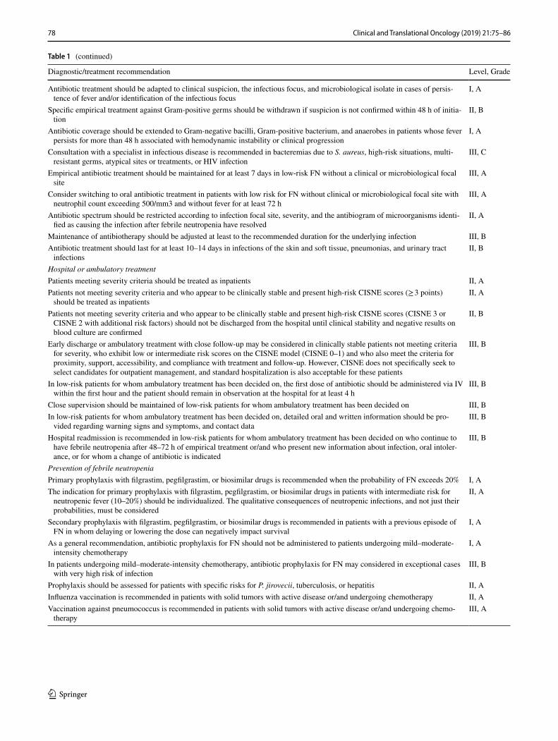
Table 1 (continued)
Diagnostic/treatment recommendation Level, Grade
Antibiotic treatment should be adapted to clinical suspicion, the infectious focus, and microbiological isolate in cases of persis-tence of fever and/or identification of the infectious focus
I, A
Specific empirical treatment against Gram-positive germs should be withdrawn if suspicion is not confirmed within 48 h of initia-tion
II, B
Antibiotic coverage should be extended to Gram-negative bacilli, Gram-positive bacterium, and anaerobes in patients whose fever persists for more than 48 h associated with hemodynamic instability or clinical progression
I, A
Consultation with a specialist in infectious disease is recommended in bacteremias due to S. aureus, high-risk situations, multi-resistant germs, atypical sites or treatments, or HIV infection
III, C
Empirical antibiotic treatment should be maintained for at least 7 days in low-risk FN without a clinical or microbiological focal site
III, A
Consider switching to oral antibiotic treatment in patients with low risk for FN without clinical or microbiological focal site with neutrophil count exceeding 500/mm3 and without fever for at least 72 h
III, A
Antibiotic spectrum should be restricted according to infection focal site, severity, and the antibiogram of microorganisms identi-fied as causing the infection after febrile neutropenia have resolved
II, A
Maintenance of antibiotherapy should be adjusted at least to the recommended duration for the underlying infection III, BAntibiotic treatment should last for at least 10–14 days in infections of the skin and soft tissue, pneumonias, and urinary tract
infectionsII, B
Hospital or ambulatory treatmentPatients meeting severity criteria should be treated as inpatients II, APatients not meeting severity criteria and who appear to be clinically stable and present high-risk CISNE scores (≥ 3 points)
should be treated as inpatientsII, A
Patients not meeting severity criteria and who appear to be clinically stable and present high-risk CISNE scores (CISNE 3 or CISNE 2 with additional risk factors) should not be discharged from the hospital until clinical stability and negative results on blood culture are confirmed
II, B
Early discharge or ambulatory treatment with close follow-up may be considered in clinically stable patients not meeting criteria for severity, who exhibit low or intermediate risk scores on the CISNE model (CISNE 0–1) and who also meet the criteria for proximity, support, accessibility, and compliance with treatment and follow-up. However, CISNE does not specifically seek to select candidates for outpatient management, and standard hospitalization is also acceptable for these patients
III, B
In low-risk patients for whom ambulatory treatment has been decided on, the first dose of antibiotic should be administered via IV within the first hour and the patient should remain in observation at the hospital for at least 4 h
III, B
Close supervision should be maintained of low-risk patients for whom ambulatory treatment has been decided on III, BIn low-risk patients for whom ambulatory treatment has been decided on, detailed oral and written information should be pro-
vided regarding warning signs and symptoms, and contact dataIII, B
Hospital readmission is recommended in low-risk patients for whom ambulatory treatment has been decided on who continue to have febrile neutropenia after 48–72 h of empirical treatment or/and who present new information about infection, oral intoler-ance, or for whom a change of antibiotic is indicated
III, B
Prevention of febrile neutropeniaPrimary prophylaxis with filgrastim, pegfilgrastim, or biosimilar drugs is recommended when the probability of FN exceeds 20% I, AThe indication for primary prophylaxis with filgrastim, pegfilgrastim, or biosimilar drugs in patients with intermediate risk for
neutropenic fever (10–20%) should be individualized. The qualitative consequences of neutropenic infections, and not just their probabilities, must be considered
II, A
Secondary prophylaxis with filgrastim, pegfilgrastim, or biosimilar drugs is recommended in patients with a previous episode of FN in whom delaying or lowering the dose can negatively impact survival
I, A
As a general recommendation, antibiotic prophylaxis for FN should not be administered to patients undergoing mild–moderate-intensity chemotherapy
I, A
In patients undergoing mild–moderate-intensity chemotherapy, antibiotic prophylaxis for FN may considered in exceptional cases with very high risk of infection
III, B
Prophylaxis should be assessed for patients with specific risks for P. jirovecii, tuberculosis, or hepatitis II, AInfluenza vaccination is recommended in patients with solid tumors with active disease or/and undergoing chemotherapy II, AVaccination against pneumococcus is recommended in patients with solid tumors with active disease or/and undergoing chemo-
therapyIII, A

79Clinical and Translational Oncology (2019) 21:75–86
1 3
through the catheter) and microbiological samples should be taken, depending on the clinical orientation (e.g., urine, sputum, mucosal or skin lesions, feces, cerebrospinal fluid, urinary antigens for pneumococcus and/or Legionella spp., nasal swab for flu virus during flu season, etc.) [III, A]. A complete blood count and biochemistry is necessary with liver and kidney function tests, lactate, and a chest X-ray with two projections [III, B]. Procalcitonin (value > 0.5 ng/ml) may be more accurate and earlier than C-reactive protein (value ≥ 90 mg/dl) to guide the diagnosis of bacteremia, and in predicting severity and FN complications, although its benefit is unclear with respect to the information provided by clinical criteria [II, B] [23].
In patients with pulmonary infiltrates and more profound immunosuppression (e.g., steroids, prolonged neutropenia, use of immunosuppressants, etc.), early bronchoscopy with bronchoalveolar lavage may be indicated. A full microbio-logical study (e.g., virus, fungi, atypical bacteria) is indi-cated in selected patients with clinical suspicion, previous infections or immunosuppression in addition to neutropenia [III, B]. In individuals with respiratory symptoms and incon-clusive chest X-ray, or with persistent fever ≥ 72 h and risk factors for complications, a chest computed tomography is useful to detect incipient pneumonias, with 87% sensitivity and a negative predictive value of 88%, surpassing chest Rx [24] [II, B]. Abdominal Rx has low sensitivity and specific-ity, and an abdominal CT is preferable in clinically sugges-tive cases and suspicion of abdominal focus (enterocolitis, typhlitis, etc.).
Severity is graded according to symptoms and signs, and risk assessment scores should only be applied when said signs and symptoms rule out clinical instability (see below) [III, B]. Most (3:1) individuals with FN and solid tumors display clinical stability in the first 3 h after diagnosis [25, 26]. High-risk patients or those who are potentially unstable are those with sepsis (defined as life-threatening acute organ
dysfunction caused by the anomalous response to an infec-tion, identifiable by an increase of 2 or more points on the quick SOFA scale), [27] septic shock, or severe documented infections (pneumonia, empyema, peritonitis, cellulitis > 4.5 cm, suspected typhlitis, enteritis grade 3–4, appendici-tis, cholecystitis, or other complicated abdominal infections, meningitis, encephalitis, catheter infections, endocarditis, and pyelonephritis) [28, 29]. In these unstable patients, mor-tality due to sepsis is 18.1% versus 2.7% of patients without risk factors [29]. Infections entailing greater mortality are chronic obstructive pulmonary disease (COPD) exacerba-tions, pneumonia, bacteremia (specially those caused by Gram-negative bacilli), and invasive fungal infections [6, 14]. Tumor progression and lung cancer are associated with a worse prognosis [6, 30, 31]. Furthermore, other toxici-ties (e.g., mucositis, enteritis, thrombopenia) or coexisting complications (thromboembolic disease, need for transfu-sion, etc.) affect decision-making [25, 26, 32]. Table 2 is a complete list of clinical characteristics that define an episode of FN as being high risk.
Clinical assessment in subjects with inflammatory responses decreased by immunosuppression is complex, and some seemingly stable episodes remain at potential risk of complications, despite not showing evident cri-teria of severity at the onset of the FN episode. In three contemporary series, the frequency of unexpected seri-ous complications in apparently stable individuals in the first 3 h was 7.3–18.6% [25, 29]. The Clinical Index of Stable Febrile Neutropenia (CISNE) (Table 2) was devel-oped and validated for the Western population to predict serious complications (shock, acute organ failure, arrhyth-mia, major bleeding, delirium, disseminated intravascular coagulation, acute abdomen), in the subgroup of patients with solid tumors who have undergone mild–moderate-intensity chemotherapy and who appear to be clinically stable at the onset of the episodes [II, B] [14, 25, 26, 29]. Its application requires having previously ruled out hemo-dynamic instability or clinical risk of complications by means of vital signs, anamnesis, physical examination, and
Table 2 Risk factors for complications that contraindicate ambulatory management
Organ or system Risk factor
Vascular Syncope, systolic blood pressure < 90 mmHg, hypertensive crisis, arrhythmias, heart failure, clinically relevant bleeding, angina pectoris
Hematological Other clinically relevant cytopenias (platelets < 50,000/μL, Hb < 8 g/dL), thromboembolic diseaseGastrointestinal Oral intolerance, vomiting, diarrhea, pain abdominal, jaundice, alteration of liver function testsInfectious Sepsis or severe focus (pneumonia, extensive cellulitis, bacteremia, catheter, pyelonephritis, meningitis, cholecystitis, and
other surgical infections), allergy to antibiotics, recent use of antibioticsNeurological Presence of focal neurological symptoms, suspicion of meningitis, acute confusional syndromePulmonary Abscesses, pneumothorax, pleural effusion, acute respiratory failure, pulmonary infiltrates, or cavitary nodulesOthers Acute kidney failure, dehydration, electrolyte alterations, other alterations of vital signs, other complications considered
severe, pregnancy, fractures

80 Clinical and Translational Oncology (2019) 21:75–86
1 3
laboratory data (Table 3). CISNE should never be used in hematological patients or individuals with lymphoma, those who are unstable, with serious infections, or who have contraindications for ambulatory treatment for other reasons. There is no experience regarding its application to select patients at low risk for direct ambulatory treatment. Online calculators for CISNE are available and provide a categorized (https ://www.mdcal c.com/clini cal-index -stabl e-febri le-neutr openi a-cisne ) or continuous estimate of risk (http://www.irico m.es/progn ostic tools /cisne /inici o.aspx).
The usefulness of the Multinational Association of Supportive Care in Cancer (MASCC) is unknown in predicting complications in patients deemed stable and one must be cognizant of its methodological limitations (coincidence of hypotension as the main predictor and endpoint, heterogeneous population with hematological neoplasms, etc.) [1], and of its low sensitivity in stable patients (34.8–36%) [23, 24, 26, 31–34].
Initial empirical treatment
Empirical treatment should be selected on the basis of the type and severity of the infection, and the probability of antibiotic-resistant microorganisms being involved in its eti-ology. The first dose of empirical antibiotic therapy should be initiated within the first hour and after taking samples for culture; hospitals should, therefore, adopt suitable logistical measures [II, A].
For all patients, intravenous administration of a β-lactam with antipseudomonal activity but that conserves activity against Gram positive is recommended (piperacillin–tazo-bactam 4.5 g/6 h; meropenem 1 g/8 h; imipenem–cilastatin 500 mg/6 h; cefepime 2 g/8 h) [I, A]. This regimen must be modified depending on the patient’s prognosis and charac-teristics, as indicated below:
(a) High-risk patients: parenteral β-lactam with antip-seudomonal activity should be combined with another agent chosen on the basis of the clinical situation:
• In the event that piperacillin–tazobactam is used, amikacin 15–20 mg/kg/day intravenous [IV] should be associated, or substitute the β-lactam for imipe-nem or meropenem due to the growing problem of Pseudomonas spp. infections, particularly in onco-logical patients, and ESBL-producing Gram-nega-tive bacilli [II, A].
• In situations of severity (e.g., quick SOFA ≥ 2 points), associate amikacin 15–20 mg/kg/day IV (higher doses may be appropriate depending on isolation and pharmacokinetic data) [II, A] to the β-lactam (preferably a carbapenem).
• When a catheter-related infection, skin focus, or pneumonia is suspected or if the patient presents hemodynamic instability, associate vancomycin (15–20 mg/kg/8–12 h IV), linezolid (of choice if the focus is pulmonary or cutaneous, but not recom-mended in catheter-related infections), or daptomy-cin (of choice in severe patients with quick SOFA ≥ 2 points and suspicion of cutaneous or catheter focus) to the β-lactam [II, A]. Tigecycline (50–100 mg/12 h IV after an initial dose of 100–200 mg) is an alter-native that may be used as a last option given the increase in all-cause mortality observed in a meta-analysis.
• In case of enterocolitis (typhlitis) or perirrectal infec-tion, the previously mentioned β-lactams are active; however, given the risk of possible resistance, the recommendation is that parenteral metronidazole 500 mg/6 h be associated [II, A].
• In the event of prior carbapenem-resistant Pseu-domonas aeruginosa colonization or of high risk due to high incidence of said strains in the patient’s setting and severity, assess the use of ceftolozane–tazobactam or ceftazidime–avibactam (unless resist-ance is already known) as β-lactam or associate colistimethate.
(b) In patients who are allergic to penicillin, the β-lactam must be substituted and aztreonam 1 g/8 h in associa-tion with vancomycin (or linezolid, tigecycline or dap-tomycin; see previous paragraph) used in its place, assessing the addition of metronidazole and/or amika-cin, according to previous indications [II, B]. Consider the possibility that Pseudomonas aeruginosa may be resistant to aztreonam; therefore, in the previously mentioned situations it is wise to always associate amikacin, and later, with the data from the antibio-gram in hand, assess the patient’s desensitization to the
Table 3 The CISNE score
a A CISNE score ≥ 3, equals ≥ 116 points on the nomogram, or a risk of ≥ 13%, on the online calculator
Characteristics Weight (points)
ECOG-PS ≥ 2 2Stress-induced hyperglycemia 2Chronic obstructive pulmonary disease 1Chronic cardiovascular disease 1Mucositis NCI grade ≥ 2 1Monocytes < 200 per μL 1
CISNE prognostic categories Scorea
Low risk 0Intermediate risk 1–2High risk 3–8
81Clinical and Translational Oncology (2019) 21:75–86
1 3
β-lactam of choice according to the microbiology data and focus of infection [II, B].
(c) Low-risk patients for infection by resistant microor-ganisms and for complications: after the first intrave-nous doses, if the patient has not received prophylaxis with fluoroquinolones, oral treatment (always in com-bination) is an alternative that is deemed effective:
• Amoxicillin–clavulanic 875/175 mg/8 h or 1000/62.5 mg 2 comp/12 h (per os) (clindamycin 300 mg/8 h in case of allergy to penicillin) + Levoflox-acin 750 mg/day P.O. or ciprofloxacin 750 mg/12 h P.O. [II, B]. In patients who are allergic to β-lactams, there is no good oral treatment option. In these cases, the initial treatment should be intravenous; hospital discharge is not recommended until microbiological documentation is available or after 48–72 h of clinical stability.
In our setting, in which enterobacteria resistant to qui-nolones and non-fermenting bacteria are high (up to 50%), initial treatment with fluoroquinolones in monotherapy is not recommended, not even in low-risk patients [II, D]. In general, antibiotic doses must be adjusted whenever neces-sary for the patient’s kidney function.
Therapeutic use of granulocyte-colony stimulating fac-tor (G-CSF) is recommended in episodes of FN in patients at high risk for infectious complications, with profound neutropenia (< 100 neutrophils/mm3), or in the presence of adverse prognostic factors, such as age > 65 years, unstable patients, with serious complications or widespread infec-tions [I, A] [35, 36]. Their routine use is not recommended, since, although it decreases duration and hospital stay, it does not enhance survival [35, 36].
Treatment in special situations
For infections due to certain resistant microorganisms or in special situations, there are specific recommendations to modify the general regimen, collected in Table 4.
As regards fungal infections, empirical antifungal treat-ment must be initially considered (“preemptive therapy”) if, after 5–7 days of antibiotic treatment, the patient continues to be febrile and the initial microbiological studies are nega-tive [I, A]; the most appropriate time depends on the clinical setting and the presence of risk factors for invasive fungal infection (IFI) (prolonged neutropenia, immunosuppressant treatment, corticosteroids, etc.). A galactomannan titer in blood of > 0.5 (optical density index) on two consecutive determinations, or > 0.7 in a single determination, is valu-able for the diagnosis of invasive aspergillosis. Furthermore, the IFI must be characterized by means of targeted stud-ies based on clinical suspicion (CT; ear, nose and throat exploration, lung puncture aspirate, biopsy of skin lesions,
etc.) [III, A]. The choice of antifungal drug (echinocandin, voriconazole, or liposomal amphotericin B) is conditioned by the prior use of azoles, clinical suspicion, and results of the galactomannan test. Echinocandins would only be of choice in cases of suspected Candida spp. infection (patients with widespread mucositis, patients with CVC and/or paren-teral nutrition); in the event that Aspergillus spp. infection is suspected (lung nodules, positive galactomannan test), vori-conazole is the drug of choice [37]. At centers with a high incidence of Mucorales, initiating liposomal amphotericin B treatment must be evaluated.
Hygiene–dietary considerations
Patients with FN are sometimes prescribed a diet low in bacteria, without having confirmed that this diet lowers the risk of infection or mortality [III, C] [38]. Hand hygiene is the most effective procedure for preventing hospital acquired infections; all personnel should, therefore, follow hand-washing protocols before entering and after leaving the room of a neutropenic patient, as well as the “5 moments” approach of hand-washing as per the guidelines of the World Health Organization [I, A] [39]. General protection meas-ures should be adopted (e.g., gloves, gowns, and/or masks) [40] and the contact isolation measures established in each for specific isolations: methicillin-resistant Staphylococcus aureus, vancomycin-resistant Enterococcus spp, carbap-enemase- or ESBL-producing Enterobacteriaceae, non-fermenting Gram-negative bacilli, such as Pseudomonas aeruginosa or Acinetobacter baumannii, with a pattern of multi-resistance [II, A]. Moreover, patients should optimize their oral and skin hygiene [II, A]. In patients with solid tumors, reverse isolation is indicated only for patients being treated with chemotherapy schedules that produce profound and prolonged neutropenia [II, A] [40].
Follow‑up and treatment adjustment
During follow-up of a patient with FN, a physical examina-tion must be performed and their general daily status must be evaluated to rule out the appearance of new infectious foci. Vital signs must be taken every 8 h. Hemogram and biochemistry with daily kidney and liver function tests must be carried out, and blood cultures every 48 h in patients with persistent fever, in addition to sampling of other foci that may appear. Supervision, prevention, and treatment of other complications (thrombosis, anemia, thrombopenia, intestinal dysbacteriosis, mucositis, etc.) are needed [III, A].
The adjustments made to the antibiotic regimen will depend on the patient’s evolution, the infectious focus, and the sensi-tivity profile of the microbiological isolations. Likewise, if

82 Clinical and Translational Oncology (2019) 21:75–86
1 3
Tabl
e 4
Tre
atm
ent i
n sp
ecia
l situ
atio
ns (c
onsu
ltatio
n w
ith a
n in
fect
ious
dis
ease
spec
ialis
t is r
ecom
men
ded)
Situ
atio
nD
iagn
ostic
/trea
tmen
t rec
omm
enda
tion
Leve
l of e
vide
nce
and
reco
mm
enda
-tio
n
ESB
L-pr
oduc
ing
ente
roba
cter
iaTr
eatm
ent w
ith c
arba
pene
ms
III,
AM
ulti-
resi
stan
t Aci
neto
bact
er sp
p. in
seve
re p
atie
nts a
nd th
ose
with
pul
mon
ary
sym
ptom
sA
ssoc
iate
inha
led
colis
timet
hate
(2 M
U/8
h) a
nd p
aren
tera
l: lo
adin
g do
se 6
–9 M
U
IV fo
llow
ed b
y 2–
3 M
U/8
hII
I, B
Sten
otro
phom
onas
mal
toph
ilia,
esp
ecia
lly in
pat
ient
s with
CV
C o
r with
pul
mon
ary
sym
ptom
s or i
nfiltr
ates
Add
cot
rimox
azol
e 5
mg/
kg/6
h IV
III,
B
Pneu
moc
ystis
jiro
veci
i or i
n pa
tient
s with
inte
rstit
ial i
nfiltr
ated
ass
ocia
ted
with
m
arke
d ly
mph
open
ia, p
rolo
nged
use
of s
tero
ids o
r bio
logi
cal t
reat
men
ts (a
nti-
tum
or n
ecro
sis f
acto
r (TN
F) m
onoc
lona
l ant
ibod
ies,
or a
nti-l
ymph
ocyt
es),
if as
soci
ated
resp
irato
ry sy
mpt
oms
Cot
rimox
azol
e 5
mg/
kg/6
h IV
, add
pre
dnis
one
40 m
g/12
h IV
in p
atie
nts w
ith
pO2 <
70 m
mH
g an
d pe
rform
bro
ncho
scop
y or
indu
ced
sput
um fo
r mic
robi
olog
i-ca
l stu
dy
II, B
–C
Prev
ious
Clo
stri
dium
diffi
cile
or i
n pa
tient
s with
dia
rrhe
a, a
fter s
tool
sam
plin
gVa
ncom
ycin
250
–500
mg/
6 h
per o
s as fi
rst c
hoic
e an
d as
soci
ated
with
met
roni
da-
zole
500
mg/
8 h
IV in
hig
h-ris
k pa
tient
s. In
cas
e of
ora
l int
oler
ance
, the
van
com
y-ci
n ca
n be
adm
inist
ered
by
NG
T or
by
mea
ns o
f ene
mas
I, A
Met
hici
llin-
resi
stan
t Sta
phyl
ococ
cus a
ureu
sIn
gen
eral
, add
van
com
ycin
or l
inez
olid
(the
latte
r is t
he d
rug
of c
hoic
e in
the
even
t of
a p
ulm
onar
y fo
cus o
r ski
n an
d so
ft tis
sue
infe
ctio
n)I,
A
In p
atie
nts w
ith p
rimar
y M
RSA
bac
tere
mia
or s
kin
focu
s and
crit
eria
of s
ever
ity, i
n th
e ab
senc
e of
pne
umon
ia, c
onsi
der t
he sw
itch
to d
apto
myc
in (6
–8 m
g/kg
/day
IV)
II, B
–C
Susp
ecte
d or
con
firm
ed G
ram
-pos
itive
bac
tere
mia
rela
ted
with
cen
tral v
enou
s cat
h-et
er, i
n pa
rticu
lar i
f the
cen
tral l
ine
cann
ot b
e re
mov
ed a
nd th
e pa
tient
’s c
linic
al
situ
atio
n is
serio
us
Add
van
com
ycin
II, A
Line
zolid
is n
ot re
com
men
ded
III,
DIn
pat
ient
s mee
ting
seve
rity
crite
rion
or p
ersi
stenc
e of
bac
tere
mia
, con
side
r sw
itch-
ing
to d
apto
myc
in a
nd c
onsu
lting
with
a sp
ecia
list i
n in
fect
ious
dis
ease
for c
om-
bine
d tre
atm
ents
II, B
Ente
roco
ccus
faec
alis
In o
ur se
tting
, the
trea
tmen
t of c
hoic
e is
am
pici
llin
2 g/
4–6
h IV
. It i
s im
porta
nt to
do
cum
ent n
egat
iviz
atio
n of
bac
tere
mia
III,
A
Ente
roco
ccus
faec
ium
and
am
pici
llin-
resi
stan
t spe
cies
Vanc
omyc
in, l
inez
olid
, or d
apto
myc
in d
epen
ding
on
the
focu
s of i
nfec
tion
Car
bape
nem
ase-
prod
ucin
g En
tero
bact
eria
ceae
Cef
tazi
dim
e/av
ibac
tam
(KPC
-2 o
r OX
A-4
8-pr
oduc
ing
strai
ns).
In c
ase
of in
fec-
tion
due
to c
arba
pene
mas
e-pr
oduc
ing
or c
arba
pene
m-r
esist
ant s
train
s due
to th
e lo
ss o
f por
ins t
oget
her w
ith h
yper
-pro
duct
ion
of A
mp-
C, a
ssoc
iatio
ns o
f tw
o or
th
ree
of th
e fo
llow
ing
antib
iotic
s: c
olist
in, t
igec
yclin
e, fo
sfom
ycin
(if t
he st
rain
is
sens
itive
), an
am
inog
lyco
side
(am
ikac
in o
r gen
tam
icin
) and
mer
open
em (i
f the
M
IC ≤
16 m
g/L)
, adm
inist
ered
in h
igh
dose
s and
con
tinuo
us p
erfu
sion
III,
A
Sym
ptom
s sug
gesti
ve o
f flu
durin
g th
e w
inte
r flu
seas
on p
eak
Sam
ples
shou
ld b
e ta
ken
for s
tudy
by
nucl
eic
acid
am
plifi
catio
n te
chni
ques
(NA
ATs
) of
the
flu v
irus.
Empi
rical
trea
tmen
t sho
uld
be a
dmin
ister
ed w
ith o
selta
miv
ir 75
mg/
12 h
P.O
.
II, A
Doc
umen
ted
resp
irato
ry sy
ncyt
ial v
iral i
nfec
tion
in p
atie
nts w
ith m
arke
d ly
mph
ope-
nia
The
ther
apy
with
riba
virin
may
be
bene
ficia
l for
som
e hi
gh-r
isk
patie
nts
III,
C
Sym
ptom
s and
lesi
ons c
linic
ally
com
patib
le w
ith c
hick
en p
ox o
r her
pes z
oste
rEm
piric
al tr
eatm
ent w
ith a
cycl
ovir
(10
mg/
kg/8
h iv
)I,
APa
tient
s rec
eivi
ng p
rolo
nged
, hig
h-do
se st
eroi
ds o
r TN
F an
tago
nists
in th
e pre
senc
e of
sugg
estiv
e sym
ptom
s (pr
olon
ged
feve
r with
out a
focu
s, in
ters
titia
l pne
umon
ia, n
ephr
i -tis
, hep
atiti
s, ch
olan
gitis
, eso
phag
itis,
colit
is, p
ancr
eatit
is, en
ceph
aliti
s, or
uve
itis)
Dia
gnos
tic st
udie
s for
the
dete
ctio
n of
CM
V; i
nitia
te tr
eatm
ent w
ith g
anci
clov
ir if
the
resu
lt is
pos
itive
or i
n ca
se o
f clin
ical
wor
seni
ng w
ith a
hig
h de
gree
of s
uspi
-ci
on
II, A
83Clinical and Translational Oncology (2019) 21:75–86
1 3
the fever persists and the focus of infection is identified, the antibiotherapy should be modified based on a specific sus-picion or on the microbiological isolation [I, A]. In the case of having initiated specific empirical treatment against Gram-positive bacilli (e.g., glycopeptide), it must be withdrawn 48 h after its initiation if the initial suspicion is not confirmed [II, B]. In patients who have been febrile > 48 h without clinical worsening, a wait and see approach can be adopted, actively searching for the clinical source of infection by means of serial blood cultures, stool cultures, and imaging studies depending on the clinical suspicion. Similarly, peripheral venous catheters should also be rotated periodically and non-infectious causes of fever should also be assessed. In patients with persistent fever > 48 h who also present hemodynamic instability or clin-ical progression, the empirical antimicrobial spectrum should be expanded to cover resistant Gram-negative bacilli, Gram-positive bacteria, and anaerobes [I, A]. Antifungal medications may be necessary in patients who meet criteria of severity and have persistent fever > 4–7 days (see treatment in special situations, above). In cases that so require and depending on the patient’s options for recovery and the cancer’s prognosis, the intensive care unit will be contacted [41]. The isolation of pathogens resistant to one or more of the components of the initial antibiotic therapy entails the need to adjust it (Table 4). A specialist in infectious disease or experienced clinician should be consulted when faced with patients with poor evo-lution, S. aureus bacteremia, persistent bacteremia, atypical microorganisms or resistances, highly severity infectious syn-dromes (CNS involvement, endocarditis, etc.), the need for antimicrobials with which the attending oncologist is relatively unfamiliar (Table 3), or HIV infection [III, C].
Empirical antibiotic treatment should last for a minimum of 7 days in low-risk FN without an identified clinical or microbiological focal site. When the neutrophil count exceed 500 cells/mm3 and the patient has been fever free for 72 h, oral administration can commence (as per the same consider-ations as in the previous section) [III, A]. In FN with clinical or microbiologically documented infection, said treatment should be followed in accordance with the recommended duration for the underlying infectious process [III, B]. In the specific case of skin and soft tissue infections, pneumonias, and urinary tract infections, antibiotic treatment should be followed for 10–14 days in most cases [II, B]. In the event of microbiological documentation, the antibiotic spectrum can be decreased depending on the focus and severity of the infection, and the antibiogram of the microorganisms identi-fied as the cause of the infectious disease [II, A].
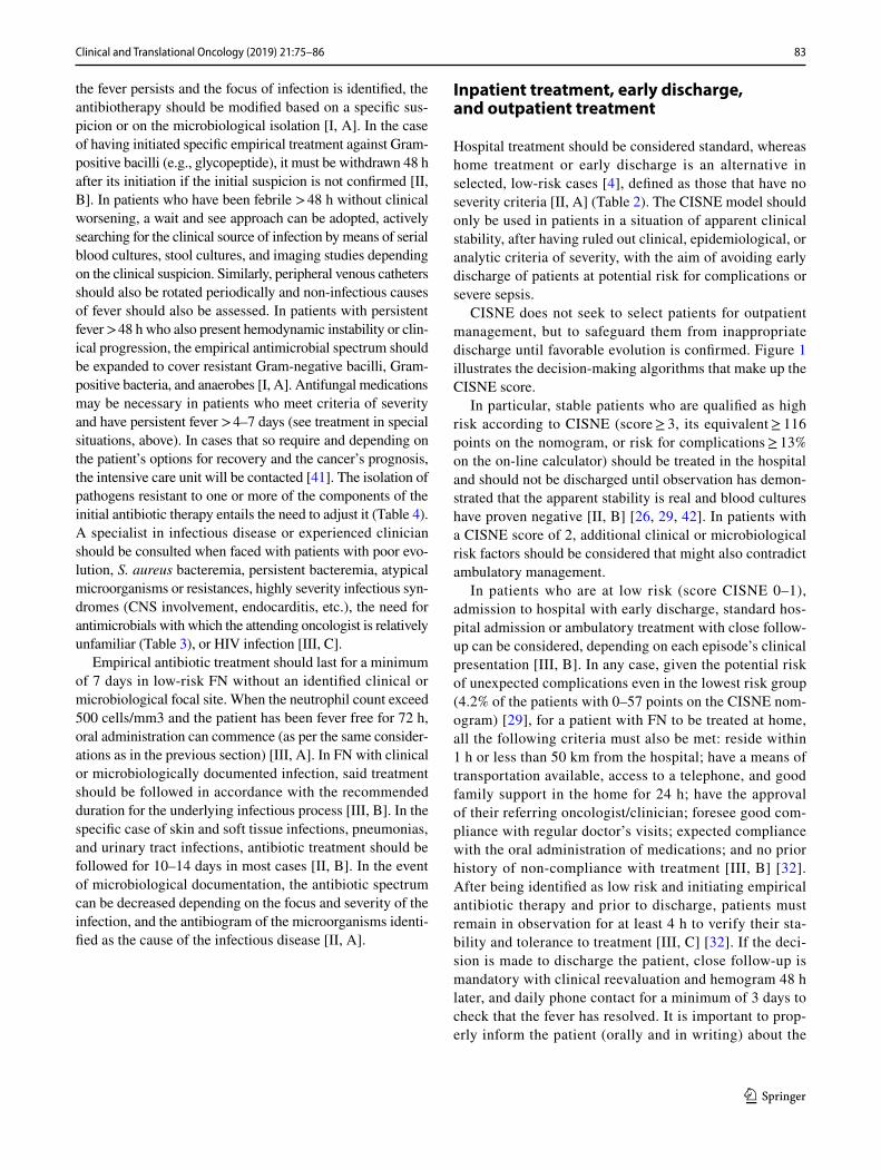
Inpatient treatment, early discharge, and outpatient treatment
Hospital treatment should be considered standard, whereas home treatment or early discharge is an alternative in selected, low-risk cases [4], defined as those that have no severity criteria [II, A] (Table 2). The CISNE model should only be used in patients in a situation of apparent clinical stability, after having ruled out clinical, epidemiological, or analytic criteria of severity, with the aim of avoiding early discharge of patients at potential risk for complications or severe sepsis.
CISNE does not seek to select patients for outpatient management, but to safeguard them from inappropriate discharge until favorable evolution is confirmed. Figure 1 illustrates the decision-making algorithms that make up the CISNE score.
In particular, stable patients who are qualified as high risk according to CISNE (score ≥ 3, its equivalent ≥ 116 points on the nomogram, or risk for complications ≥ 13% on the on-line calculator) should be treated in the hospital and should not be discharged until observation has demon-strated that the apparent stability is real and blood cultures have proven negative [II, B] [26, 29, 42]. In patients with a CISNE score of 2, additional clinical or microbiological risk factors should be considered that might also contradict ambulatory management.
In patients who are at low risk (score CISNE 0–1), admission to hospital with early discharge, standard hos-pital admission or ambulatory treatment with close follow-up can be considered, depending on each episode’s clinical presentation [III, B]. In any case, given the potential risk of unexpected complications even in the lowest risk group (4.2% of the patients with 0–57 points on the CISNE nom-ogram) [29], for a patient with FN to be treated at home, all the following criteria must also be met: reside within 1 h or less than 50 km from the hospital; have a means of transportation available, access to a telephone, and good family support in the home for 24 h; have the approval of their referring oncologist/clinician; foresee good com-pliance with regular doctor’s visits; expected compliance with the oral administration of medications; and no prior history of non-compliance with treatment [III, B] [32]. After being identified as low risk and initiating empirical antibiotic therapy and prior to discharge, patients must remain in observation for at least 4 h to verify their sta-bility and tolerance to treatment [III, C] [32]. If the deci-sion is made to discharge the patient, close follow-up is mandatory with clinical reevaluation and hemogram 48 h later, and daily phone contact for a minimum of 3 days to check that the fever has resolved. It is important to prop-erly inform the patient (orally and in writing) about the

84 Clinical and Translational Oncology (2019) 21:75–86
1 3
detection of warning signs and symptoms, as well as how to contact the healthcare personnel 24 h/day [III, B] [29, 32, 43]. Hospital admission must be contemplated when fever or neutropenia persists after 2–3 days of treatment with empirical antibiotic therapy, new signs or symptoms of infection, oral intolerance, or need to change antibio-therapy [III, B] [32].
Prevention of febrile neutropenia: prophylaxis with G‑CSF
The risk of developing FN must be evaluated on a case-by-case basis at the beginning of each cycle of chemotherapy [44]. Primary prophylaxis with G-CSF is recommended when the likelihood of developing FN is > 20% [2, 11, 45, 46], having demonstrated a decrease in incidence, duration, and severity, without significantly affecting tumor response or survival [I, A] [35, 47]. In patients with an intermediate level of risk (10–20%), the indication of prophylaxis with G-CSF must be individualized depending on the character-istics that increase the risk of FN (age > 65 years, liver or kidney dysfunction, widespread bone marrow involvement, recent extensive surgery, persistent neutropenia, or prior epi-sodes of FN) [II, A] [2, 11, 45, 46]. In special circumstances, regardless of frequency, individuals may also be considered
eligible for primary prophylaxis if the consequences of the neutropenic episode are foreseen to be more severe.
The recommended dose of filgastrim is 5 µg/kg/day subcutaneously administered 24–72 h after administration of chemotherapy until achieving a sufficient, stable post-nadir neutrophil count [I, A]. Pegfilgastrim (a single dose of 100 µg/kg or 6 mg) can be more effective in different contexts and approved biosimilar drugs can also be consid-ered [2, 45].
In patients who have experienced a prior episode of FN or dose-limiting neutropenia [48], the use of secondary prophy-laxis with G-CSF can be considered individually if the dose reductions or delay of antineoplastic treatments can nega-tively affect prognosis [I, A] [2, 11, 45, 46].
Prevention of febrile neutropenia: antimicrobials and vaccination
In patients with solid tumors treated with chemotherapy of mild–moderate intensity, the use of prophylaxis with oral antibiotics to prevent episodes of FN [I, E] is not recom-mended, except for exceptional cases of patients with very high risk of infection in whom antineoplastic therapy cannot be delayed [III, B].
In patients with specific risks, prophylaxis against Pneu-mocystis jirovecii, tuberculosis [49], or viral hepatitis should
Febrile Neutropenia
EMERGENCY ROOM Triage consultation
Neutrophil<0.5 × 10⁹/l, or expected to fall below 0.5 × 10⁹/lFever >38.3°C+ chemotherapy in previous 6 weeks
Initial assessment: Temperature, Heart and Respiratory rate,Blood Presure, O2Sat.Initial blood studies: count, chemistry (urea, creanine, ions, lactate), extractions for culture Venous Access
SIRS or SepsisYes NoIntensive careStart empirical IV broad spectrum antibioticsResuscitation and hemodynamic support Clinical assessment:
ID source of infection
Clear anatomic site of infection: Symptoms of pneumonia, cellulitis, abdominal infectionPositive imaging, Microbial laboratory findings
Hospital admission
Yes
Clinical risk of complications: Expected duration of neutropenia (< 500 neutrophils/mm3) >7 daysComorbidities, acute organ failure (renal, cardiac, respiratory) or decompensation of chronic insufficiencyAdmission criterion on their own (pulmonary thromboembolism, arrhythmias, disseminated intravascular coagulation, and bleeding)Unable to swallow oral medicationsUnfavorable social, logistic, or socio-family factors
Yes
Begin empirical IV broad spectrum antibioticin Emergency Room
No
Apparently stable patients
Interview and medical history: Risk factors for complications
High risk of complications
Low risk of complications
CISNE score ≥3
Hospital admission
Observation in hospital 4-72 h, orOutpatient follow-up, orStandard hospital admission, depending on risk factors and clinical presentation
15 minutes
CISNE score <3
30 minutes
60 minutes
Consider hospital admission if risk factors present, or Observation in hospital 4-72 h, orOutpatient follow-up in selected cases
CISNE score <2 CISNE score =2
Fig. 1 Decision-making algorithm in febrile neutropenia

85Clinical and Translational Oncology (2019) 21:75–86
1 3
be assessed according to recommendations found in other clinical practice guidelines [II, A].
Patients with active solid tumors and those receiving chemotherapy should be vaccinated every year against flu [II, A] and against pneumococcus [III, A] in accordance with guidelines for immunodepressed patients. Other vac-cinations may be advisable depending on the kind of chemo-therapy, the patient’s clinical status, or a specific indication [43].
Compliance with ethical standards
Conflict of interest ACB have received speaker and travel grants from IPSEN, Servier, and Roche, outside the submitted work. PJF has noth-ing to disclose. EMC has nothing to disclose. EM has nothing to dis-close. MB reports personal fees from Leo Pharma, Rovi and Sanofi, outside the submitted work. AC has nothing to disclose. JE reports per-sonal fees from MSD, Novartis, Pfizer, BMS, Astra Zeneca and Roche, outside the submitted work. EGV has nothing to disclose. FH has noth-ing to disclose. FAP reports grants from Roche and Celgene, and other financial support from Roche, Celgene, Eisai, AstraZeneca, Pfizer, Pierre Fabre and Novartis, outside the submitted work.
Ethical standards The current study has been performed in accordance with the ethical standards laid down in the 1964 Declaration of Hel-sinki and its later amendments. This article does not contain any studies with human participants or animals performed by any of the authors.
Informed consent For this type of study, no informed consent is required.
Open Access This article is distributed under the terms of the Crea-tive Commons Attribution 4.0 International License (http://creat iveco mmons .org/licen ses/by/4.0/), which permits unrestricted use, distribu-tion, and reproduction in any medium, provided you give appropriate credit to the original author(s) and the source, provide a link to the Creative Commons license, and indicate if changes were made.
References
1. Klastersky J, Paesmans M, Rubenstein EB, Boyer M, Elting L, Feld R, et al. The multinational association for supportive care in cancer risk index: a multinational scoring system for identify-ing low-risk febrile neutropenic cancer patients. J Clin Oncol. 2000;18(16):3038–51.
2. Klastersky J, de Naurois J, Rolston K, Rapoport B, Maschmeyer G, Aapro M, et al. Management of febrile neutropaenia: ESMO Clinical Practice Guidelines. Ann Oncol. 2016;27(5):v111–8.
3. Weycker D, Barron R, Kartashov A, Legg J, Lyman GH. Inci-dence, treatment, and consequences of chemotherapy-induced febrile neutropenia in the inpatient and outpatient settings. J Oncol Pharm Pract. 2014;20(3):190–8.
4. Taplitz RA, Kennedy EB, Flowers CR. Outpatient management of fever and neutropenia in adults treated for malignancy: American Society of Clinical Oncology and Infectious Diseases Society of America Clinical Practice Guideline update summary. J Oncol Pract. 2018;14:250–5.
5. McCune JS, Sullivan SD, Blough DK, Clarke L, McDermott C, Malin J, et al. Colony-stimulating factor use and impact on febrile
neutropenia among patients with newly diagnosed breast, colo-rectal, or non-small cell lung cancer who were receiving chemo-therapy. Pharmacotherapy. 2012;32(1):7–19.
6. Kuderer NM, Dale DC, Crawford J, Cosler LE, Lyman GH. Mor-tality, morbidity, and cost associated with febrile neutropenia in adult cancer patients. Cancer. 2006;106(10):2258–66.
7. Mayordomo JI, López A, Viñolas N, Castellanos J, Pernas S, Domingo Alonso J, et al. Retrospective cost analysis of manage-ment of febrile neutropenia in cancer patients in Spain. Curr Med Res Opin. 2009;25(10):2533–42.
8. Schilling MB, Parks C, Deeter RG. Costs and outcomes associated with hospitalized cancer patients with neutropenic complications: a retrospective study. Exp Ther Med. 2011;2(5):859–66.
9. Pettengell R, Schwenkglenks M, Leonard R, Bosly A, Paridaens R, Constenla M, et al. Neutropenia occurrence and predictors of reduced chemotherapy delivery: results from the INC-EU pro-spective observational European neutropenia study. Support Care Cancer. 2008;16(11):1299–309.
10. Lyman GH, Michels SL, Reynolds MW, Barron R, Tomic KS, Yu J. Risk of mortality in patients with cancer who experience febrile neutropenia. Cancer. 2010;116(23):5555–63.
11. Aapro MS, Bohlius J, Cameron DA, Lago LD, Donnelly JP, Kear-ney N, et al. 2010 update of EORTC guidelines for the use of granulocyte-colony stimulating factor to reduce the incidence of chemotherapy-induced febrile neutropenia in adult patients with lymphoproliferative disorders and solid tumours. Eur J Cancer. 2011;47(1):8–32.
12. Balducci L, Al-Halawani H, Charu V, Tam J, Shahin S, Dreiling L, et al. Elderly cancer patients receiving chemotherapy benefit from first-cycle pegfilgrastim. Oncologist. 2007;12(12):1416–24.
13. von Minckwitz G, Kümmel S, du Bois A, Eiermann W, Eidt-mann H, Gerber B, et al. Pegfilgrastim ± ciprofloxacin for primary prophylaxis with TAC (docetaxel/doxorubicin/cyclophosphamide) chemotherapy for breast cancer. Results from the GEPARTRIO study. Ann Oncol. 2008;19(2):292–8.
14. Carmona-Bayonas A, Jiménez-Fonseca P, Virizuela J, Antonio M, Font C, Biosca M, et al. Performance of the clinical index of stable febrile neutropenia (CISNE) in different types of infections and tumors. Clin Transl Oncol. 2016;19(3):1–10.
15. Carmona-Bayonas A, Fonseca PJ, Font C, Martínez-García J, Torrella F, Urrego E, et al. Bacteremia in adult cancer patients with apparently stable febrile neutropenia: data from a cohort of 692 consecutive episodes from a single institution. J Community Support Oncol. 2014;12(9):312–20.
16. Viscoli C. Management of infection in cancer patients: studies of the EORTC International Antimicrobial Therapy Group (IATG). Eur J Cancer. 2002;38:82–7.
17. Trecarichi EM, Tumbarello M. Antimicrobial-resistant Gram-negative bacteria in febrile neutropenic patients with cancer: current epidemiology and clinical impact. Curr Opin Infect Dis. 2014;27(2):200–10.
18. Ben-Ami R, Rodríguez-Baño J, Arslan H, Pitout JDD, Quen-tin C, Calbo ES, et al. A multinational survey of risk factors for infection with extended-spectrum β-lactamase-producing Enterobacteriaceae in nonhospitalized patients. Clin Infect Dis. 2009;49(5):682–90.
19. Ram R, Farbman L, Leibovici L, Raanani P, Yeshurun M, Vidal L, et al. Characteristics of initial compared with subsequent bacterial infections among hospitalised haemato-oncological patients. Int J Antimicrob Agents. 2012;40(2):123–6.
20. Furuno JP, McGregor JC, Harris AD, Johnson JA, Johnson JK, Langenberg P, et al. Identifying groups at high risk for carriage of antibiotic-resistant bacteria. Arch Intern Med. 2006;166(5):580–5.

86 Clinical and Translational Oncology (2019) 21:75–86
1 3
21. EARS-Net. Antimicrobial resistance surveillance in Europe 2015. Annual report of the European Antimicrobial Resistance Surveil-lance Network (EARS-Net); 2015.
22. Mensa J, Gatell JM, García-Sánchez JE, Marco E, Letang E, López-Suñé F. Guía de terapéutica antimicrobiana. Barcelona: Antares; 2017.
23. Carmona-Bayonas A, Gómez J, González-Billalabeitia E, Can-teras M, Navarrete A, Gonzálvez ML, et al. Prognostic evaluation of febrile neutropenia in apparently stable adult cancer patients. Br J Cancer. 2011;105(5):612–7.
24. Heussel CP, Kauczor H-U, Ullmann AJ. Pneumonia in neutro-penic patients. Eur Radiol. 2004;14(2):256–71.
25. Carmona-Bayonas A, Gómez J, González-Billalabeitia E, Can-teras M, Navarrete A, Gonzálvez ML, et al. Prognostic evaluation of febrile neutropenia in apparently stable adult cancer patients. Br J Cancer. 2011;105(5):612.
26. Carmona-Bayonas A, Jiménez-Fonseca P, Echaburu JV, Antonio M, Font C, Biosca M, et al. Prediction of serious complications in patients with seemingly stable febrile neutropenia: validation of the clinical index of stable febrile neutropenia in a prospec-tive cohort of patients from the FINITE study. J Clin Oncol. 2015;33(5):465.
27. Singer M, Deutschman CS, Seymour CW, Shankar-Hari M, Annane D, Bauer M, et al. The Third International consen-sus definitions for sepsis and septic shock (Sepsis-3). JAMA. 2016;315(8):801–10.
28. Bone RC, Balk RA, Cerra FB, Dellinger RP, Fein AM, Knaus WA, et al. Definitions for sepsis and organ failure and guidelines for the use of innovative therapies in sepsis. Chest. 1992;101(6):1644–55.
29. Fonseca PJ, Carmona-Bayonas A, García IM, Marcos R, Castañón E, Antonio M, et al. A nomogram for predicting complications in patients with solid tumours and seemingly stable febrile neutro-penia. Br J Cancer. 2016;114(11):1191–8.
30. Caggiano V, Weiss RV, Rickert TS, Linde-Zwirble WT. Incidence, cost, and mortality of neutropenia hospitalization associated with chemotherapy. Cancer. 2005;103(9):1916–24.
31. Talcott JA, Siegel RD, Finberg R, Goldman L. Risk assess-ment in cancer patients with fever and neutropenia: a prospec-tive, two-center validation of a prediction rule. J Clin Oncol. 1992;10(2):316–22.
32. Flowers CR, Seidenfeld J, Bow EJ, Karten C, Gleason C, Hawley DK, et al. Antimicrobial prophylaxis and outpatient management of fever and neutropenia in adults treated for malignancy: Ameri-can Society of Clinical Oncology Clinical Practice Guideline. J Clin Oncol. 2013;31(6):794–810.
33. Carmona-Bayonas A, Jiménez-Fonseca P, Virizuela Echaburu J, Sánchez Cánovas M, de la Peña AF. The time has come for new models in febrile neutropenia: a practical demonstration of the inadequacy of the MASCC score. Clin Transl Oncol. 2017;19(9):1084–90.
34. Carmona-Bayonas A, Jiménez-Fonseca P. CISNE or MASCC, which predictor is really the weakest in febrile neutropenia? Eur J Intern Med. 2018;50:e33–4.
35. Clark OAC, Lyman GH, Castro AA, Clark LGO, Djulbegovic B. Colony-stimulating factors for chemotherapy-induced febrile
neutropenia: a meta-analysis of randomized controlled trials. J Clin Oncol. 2005;23(18):4198–214.
36. García-Carbonero R, Mayordomo JI, Tornamira MV, López-Brea M, Rueda A, Guillem V, et al. Granulocyte colony-stimulating fac-tor in the treatment of high-risk febrile neutropenia: a multicenter randomized trial. J Natl Cancer Inst. 2001;93(1):31–8.
37. Mercier T, Maertens J. Clinical considerations in the early treat-ment of invasive mould infections and disease. J Antimicrob Chemother. 2017;72(suppl_1):i29–38.
38. van Dalen EC, Mank A, Leclercq E, Mulder RL, Davies M, Kersten MJ, et al. Low bacterial diet versus control diet to pre-vent infection in cancer patients treated with chemotherapy causing episodes of neutropenia. Cochrane Database Syst Rev. 2016;4:CD006247.
39. Allegranzi B, Pittet D. Role of hand hygiene in healthcare-asso-ciated infection prevention. J Hosp Infect. 2009;73(4):305–15.
40. Siegel JD, Rhinehart E, Jackson M, Chiarello L. 2007 guide-line for isolation precautions: preventing transmission of infectious agents in health care settings. Am J Infect Control. 2007;35(10):S65–164.
41. Carmona-Bayonas A, Gordo F, Beato C, Castaño Pérez J, Jimé-nez-Fonseca P, Virizuela Echaburu J, et al. Intensive care in can-cer patients in the age of immunotherapy and molecular thera-pies: commitment of the SEOM-SEMICYUC. Med Intensiva. 2018;42(6):363–9.
42. Clinical Index of Febrile Neutropenia WebSite; 2015. http://www.irico m.es/progn ostic tools /Cisne /inici o.aspx. Accessed 15 Oct 2018.
43. Virizuela JA, Carratalà J, Aguado JM, Vicente D, Salavert M, Ruiz M, et al. Management of infection and febrile neutropenia in patients with solid cancer. Clin Transl Oncol. 2016;18(6):557–70.
44. Lyman GH, Kuderer NM, Crawford J, Wolff DA, Culakova E, Poniewierski MS, et al. Predicting individual risk of neutropenic complications in patients receiving cancer chemotherapy. Cancer. 2011;117(9):1917–27.
45. Baden LR, Swaminathan S, Angarone M, Blouin G, Camins BC, Casper C, et al. Prevention and treatment of cancer-related infec-tions, Version 2.2016, NCCN Clinical Practice Guidelines in Oncology. J Natl Compr Cancer Netw. 2016;14(7):882–913.
46. Smith TJ, Khatcheressian J, Lyman GH, Ozer H, Armitage JO, Balducci L, et al. 2006 update of recommendations for the use of white blood cell growth factors: an evidence-based clinical practice guideline. J Clin Oncol. 2006;24(19):3187–205.
47. Mhaskar R, Clark OAC, Lyman G, Botrel TEA, Paladini LM, Djulbegovic B. Colony-stimulating factors for chemotherapy-induced febrile neutropenia. Cochrane Database Syst Rev. 2014;10:CD003039.
48. Crawford J, Glaspy JA, Stoller RG, Tomita DK, Vincent ME, McGuire BW, et al. Final results of a placebo-controlled study of filgrastim in small-cell lung cancer: exploration of risk factors for febrile neutropenia. Support Cancer Ther. 2005;3(1):36–46.
49. Person AK, Pettit AC, Sterling TR. Diagnosis and treatment of latent tuberculosis infection: an update. Curr Respir Care Rep. 2013;2(4):199–207.