Seasonal Prevalence of Gastro-Intestinal Helminths of Sheep fileGenerally lower age groups and nomad...
18
Seasonal Prevalence of Gastro-Intestinal Helminths of Sheep (Ovis Aries) and Goat (Capra Hircus) with Respect to Age and Gender of Gurez Valley, Kashmir Sheikh B. A., Ahmad F. and Sofi T. A. J Anim Sci Adv 2016, 6(3): 1609-1625 DOI: 10.5455/jasa.20160409122017 Journal of Animal Science Advances Online version is available on: www.grjournals.com
Transcript of Seasonal Prevalence of Gastro-Intestinal Helminths of Sheep fileGenerally lower age groups and nomad...
Seasonal Prevalence of Gastro-Intestinal Helminths of Sheep
(Ovis Aries) and Goat (Capra Hircus) with Respect to Age and
Gender of Gurez Valley, Kashmir
Sheikh B. A., Ahmad F. and Sofi T. A.
J Anim Sci Adv 2016, 6(3): 1609-1625
DOI: 10.5455/jasa.20160409122017
Journal of Animal Science Advances
Online version is available on: www.grjournals.com

SHEIKH ET AL.
1609 J. Anim. Sci. Adv., 2016, 6(3): 1609-1625
Seasonal Prevalence of Gastro-Intestinal
Helminths of Sheep (Ovis Aries) and Goat (Capra
Hircus) with Respect to Age and Gender of Gurez
Valley, Kashmir
Sheikh B. A., Ahmad F. and Sofi T. A.
* Parasitology Research Laboratory, Department of Zoology, University of Kashmir, Srinagar- 190 006, India.
Abstract
The present study was carried out with an objective to assess the seasonal dynamics of intestinal helminth
parasites in association with age and gender of sheep (Ovis aries) and goat (Capra hircus) in Gurez valley. Out
of the total of 123 sheep and 96 goats examined through gut examination during May 2013 - May 2015,
25.20%, 17.07%, 27.64% in sheep and 22.91%, 15.62%, 26.04% in goats was the prevalence of trematodes,
cestodes and nematodes respectively. Comparatively goats harbored low infection level as compared to sheep.
There was no significant difference (P>0.05) in gastrointestinal parasite infection in Ovis aries and Capra
hircus examined. Data showed that infection was moderately positive all year round but highest infection was
found in the autumn season. A significant relationship was found between seasons and prevalence of infection
(P<0.05). Generally lower age groups and nomad breed reported high infection. In conclusion, the present study
states that seasons affected the prevalence of intestinal cestodes significantly while as gender and age of host
were not significant factors in the onset of infection under temperate climatic conditions of Gurez valley. These
findings may contribute to the existing epidemiological knowledge of the intestinal cestodes of Sheep and will
also improve the control strategies of intestinal helminthiasis.
Keywords: Age, gender, sheep, goat, intestinal cestodes, prevalence, season.
Corresponding author: Parasitology Research Laboratory, Department of Zoology, University of Kashmir, Srinagar- 190 006, India.
Received on: 02 Jan 2016 Revised on: 12 Jan 2016
Accepted on: 09 Mar 2016
Online Published on: 30 Mar 2016
Original Article
ISSN: 2251-7219
SEASONAL PREVALENCE OF GASTRO-INTESTINAL HELMINTHS OF …
1610 J. Anim. Sci. Adv., 2016, 6(3): 1609-1625
Introduction
The temperate agro-climatic conditions,
traditional animal husbandry practices and poor
veterinary infrastructure, abundance of alpine and
sub alpine pastures are natural determining factors
of incidence and severity of various parasitic
diseases of livestock in Kashmir valley (Tariq,
2007). Gastrointestinal parasitism is the major cause
of damage and decreased productivity in the goat
industry particularly in developing countries.
Soulsby (1982) mentioned that tape worms are
relatively less pathogenic, but in heavy infections
may cause reduced weight gain, diarrhoea and
intestinal obstruction. Numerous epidemiological
studies have been conducted throughout the world
to arrive at the detailed information on the
gastrointestinal parasites of livestock but scanty
references are available on epidemiology and
prevalence of intestinal helminths of sheep and
goats. According to Odoi et al., (2007), the major
risk factors of helminthiasis are broadly classified
as parasite factors (including epidemiology of the
different species), host factors (genetic resistance,
age and physiological status of the animal) and
environmental factors (climate, nutrition, stocking
density and management). No epidemiological
information was available on intestinal helminth of
sheep and goat in Gurez valley. The present study
was carried out with an objective to assess the
seasonal epidemiological prevalence of intestinal
helminths in association with age and sex of sheep
and goat in Gurez valley, India.
Materials and Methods
During this study a total of 123 sheeps and 96
goats were examined over two consecutive years
from May 2013 to May 2015. All intestinal tracts
belonged to sheeps of Bhakarwal (nomad) and
Gurez (local) breed. The animals were of both
genders and age ranged from less than one year to
more than four years. The samples were collected
on monthly basis and later expressed seasonally in
order to analyze the seasonal prevalence. The
collected samples were carefully labelled with
animal identification, sex, dental age and month of
collection.
Gastrointestinal Tract of Animals
The gastrointestinal tracts of freshly
slaughtered animals in various abattoirs of the
valley were collected, tied off at both ends and
brought to the laboratory and immediately
processed to analyze the parasite species present.
The abomasum and the small and large intestines
were thoroughly opened and examined separately
for the presence of tapeworms using standard
procedures and worms were identified using the
descriptions of Soulsby (1982). The faecal samples
were obtained directly from the rectum of the
animals in suitable air tight containers properly
labelled and brought to the laboratory in 4%
formalin and kept at 4oC until processing. The
laboratory procedure was as per the methods of
Soulsby (1982).
Statistical Analysis
Percentages to measure prevalence and chi-
square test to measure association between the
prevalence of infection and the age, gender and
breed were the statistical methods applied. The
association between seasons and prevalence of
infection was analyzed by Pearson’s coefficient of
correlation ‘r’. Means and standard error of means
with their respective 95% confidence intervals were
also calculated for each parameter tested in this
study. The level of significance was statistically
accepted at the 5% level (P≤0.05). The data was
analyzed using Statistical packages MINITAB
software version 13.2 for Windows.
Results and Discussion
Understanding the biology and epidemiology of
gastro-intestinal parasites of sheep and goat, it is
essential to improve the control measures and
decrease in production losses (Pal and Qayyum,
1992). The epidemiology of gastrointestinal helminth
parasites is governed by host-parasite relationship and
reaction with environmental conditions. Tembely et
al., (1997) and Vlassoff et al., (2001) demonstrated
that the effect of helminth infection on production
of particular livestock species depend mostly up on
the age of the animals, the breed, the parasite species
involved and the intensity of the worm populations
within the host. The prevalence and distribution of

SHEIKH ET AL.
1611 J. Anim. Sci. Adv., 2016, 6(3): 1609-1625
parasitic nematodes are largely governed by a
combination of their ecological requirements for
development and survival outside the host and farm
management practice. The epidemiology of GIT
parasites of sheep and goat in Gurez has been
studied taking into consideration the overall
prevalence (overall, seasonal, age-wise, sex wise)
and the associated risk factors with GIT parasites.
Overall Prevalence
Out of the total of 123 sheep and 96 goats
examined through gut examination during the study
period, 25.20%, 17.07%, 27.64% in sheep and
22.91%, 15.62%, 26.04% in goats was the
prevalence of trematodes, cestodes and nematodes
respectively (Table 1, 2, 3 & Fig. 1, 2, 3).
Comparatively goats harbored low infection level as
compared to sheep. There was no significant
difference (P>0.05) in gastrointestinal parasite
infection in Ovis aries and Capra hircus examined.
Table 1: Distribution of Helminths in Ovis aries and Capra hircus.
Host Examined Uninfected Infected %age P-Value
Ovis aries 123 89 34 27.64 P-Value =
0.071 Capra hircus 96 71 25 26.04
Total 219 160 59 26.94
Fig. 1: Graph showing the overall prevalence of sheep and Goat.
Table 2: Distribution of trematodes, cestodes and nematodes in Ovis aries and Capra hircus.
Host Examined Uninfected Infected Parasites collected
Trematodes Cestodes Nematodes P-Value
Ovis aries 123 89 34 31 (25.20) 21 (17.07) 34 (27.64) P-Value = 0.002
Capra hircus 96 71 25 22 (22.91) 15 (15.62) 25 (26.04) P-Value = 0.002
Total 219 160 59 53 (24.20) 36 (16.43) 59 (26.94) (Figures in parentheses indicate percentage)
Fig. 2: Prevalence of recovered Helminth parasites.
Sheep Goat
No. Examined
No. Infected
Sheep Goat
No. Examined
No. Infected
Trematode
Cestode
Nematode

SEASONAL PREVALENCE OF GASTRO-INTESTINAL HELMINTHS OF …
1612 J. Anim. Sci. Adv., 2016, 6(3): 1609-1625
In case of small ruminants, 89 out of 123
(27.64%) sheep and 71 out of 96 (26.04%) goat
were infected with helminth parasites. A mean
intensity & relative abundance of 81.64, 59.07 &
52.78, 39.04 in sheep and goat respectively was
observed. The observed parasites were Haemonchus
contortus, Ostertagia, Chabertia ovina, Fasciola
hepatica, Dicrocoelium dendriticum & Moneizia
expansa in both sheep & goat, and showed mean
intensity & relative abundance of 88.23, 24.39;
32.93, 8.30; 26.79, 5.22; 5.00, 1.05; 82.86, 19.53 &
4.31, 0.56 in sheep and 74.4, 19.37; 23.81, 5.45;
16.82, 2.97; 3.90, 0.85; 43.72, 10.02 & 3.4, 0.35 in
goat respectively.
Table 3: Mean intensity and Relative abundance of Helminths in Ovis aries and Capra hircus.
Host No.
Examined
No.
Infected
% age No. of
parasites
Mean
Intensity
Relative
Abundance
Sheep 123 89 27.64 7266 81.64 59.07
Goats 96 71 26.04 3748 52.78 39.04
Fig. 3: Graph showing the mean intensity and relative abundance of Helminths in sheep and goat.
Table 4: Mean intensity and Relative abundance of different Helminths in Ovis aries.
Parasite No.
Examined
No.
Infected
% age No. of
parasites
Mean
Intensity
Relative
Abundance
Haemonchus
contortus
123
34 27.64 3000 88.23 24.39
Ostertagia 31 25.20 1021 32.93 8.30
Chabertia ovina 24 19.51 643 26.79 5.22
Fasciola hepatica 26 21.13 130 5.00 1.05
Dicrocoelium
dendriticum
29 23.57 2403 82.86 19.53
Moneizia expansa 16 13.00 69 4.31 0.56
Sheep
Goats
SHEIKH ET AL.
1613 J. Anim. Sci. Adv., 2016, 6(3): 1609-1625
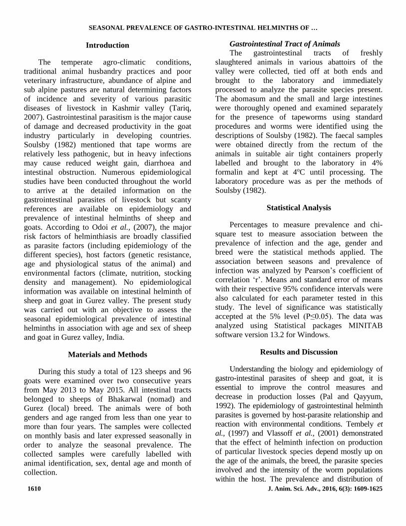
Fig. 4: Graph showing the mean intensity and relative abundance of different Helminths in sheep.
Table 5: Mean intensity and Relative abundance of different Helminths in Capra hircus.
Parasite No.
Examined
No.
Infected
%
age
No. of
parasites
Mean
Intensity
Relative
Abundance
Haemonchus
contortus
96
25 26.04 1860 74.4 19.37
Ostertagia 22 22.91 524 23.81 5.45
Chabertia ovina 17 17.70 286 16.82 2.97
Fasciola hepatica 21 21.87 82 3.90 0.85
Dicrocoelium
dendriticum
22 22.91 962 43.72 10.02
Moneizia expansa 10 10.41 34 3.40 0.35
Fig. 4: Graphg showing the mean intensity and relative abundance of different Helminths in goat.
Based on post mortem examination of sheep
and goats obtained from different areas of Gurez the
present endeavour revealed the presence of variety
of gastrointestinal helminth parasitic fauna in them
(Table 6). The parasites encountered during the
present investigation in sheep were: Haemonchus
contortus (27.64%), Ostertagia ostertagia
(25.20%), Chabertia ovina (19.51%), Fasciola
heptica (21.13%), Dicrocoelium dendriticum
(23.57%), Moneizia expansa (13.008%). In goats
the prevalence was Haemonchus contortus
(26.04%), Ostertagia Ostertagia (22.91%),
No. Examined
No. Infected
No. of parasites
Mean Intensity
Relative Abundance
No. Examined
No. Infected
No. of parasites
Mean Intensity
Relative Abundance

SEASONAL PREVALENCE OF GASTRO-INTESTINAL HELMINTHS OF …
1614 J. Anim. Sci. Adv., 2016, 6(3): 1609-1625
Chabertia ovina (17.70%), Fasciola hepatica;
(21.87%), Dicrocoelium dendriticum (22.91%),
Moneizia expansa (10.41%).
Table 6: Overall prevalence of gastrointestinal helminth parasites in Ovis aries and Capra hircus.
Parasite Ovis aries (123 examined) Capra hircus (96 examined) P-Value
Number infected %age Number infected %age
Haemonchus contortus 34 27.64% 25 26.04% P-Value = 0.071
Ostertagia ostertagia 31 25.20% 22 22.91% P-Value = 0.069
Chabertia ovina 24 19.51% 17 17.70% P-Value = 0.071
Fasciola hepatica 26 21.13% 21 21.87% P-Value = 0.071
Dicrocoelium
dendriticum 29 23.57% 22 22.91%
P-Value = 0.071
Moneizia expansa 16 13.00% 10 10.41% P-Value = 0.071
The epidemiological study of GIH infection
revealed that sheep and goats harbour a spectrum of
GIH fauna in Gurez valley. Sheep and Goats
exhibited similar type of helminth fauna; this can be
attributed to the co-rearing and mixed grazing in
pastures and in other grazing areas. The proportions
of the genera of gastrointestinal parasites identified
in the present study in which Haemonchus was the
most prevalent (27.64% sheep & 26.04% goats) and
Monezia (13.00% sheep & 10.41% goats) the least,
is in conformity with the findings of other studies
carried out in many other climatic areas of India and
other parts of world.
Trematodes
The prevalence rate of trematodes in the
present study was 25.20% in sheep and 22.91% in
goats, among trematodes Dicrocoelium spp.
(23.57% in sheep and 22.91% in goats) was found
most prevalent followed by Faciola spp. (21.13% in
sheep and 21.87% in goats) as shown in Table 4.35.
The prevalence of Dicrocoelium spp. (Sheep: 23.57%;
Goats: 22.91%) which is in conformity with the
observations of other authors who also reported quite
similar prevalences of Dicrocoelium spp. Garg et al.,
(2009) reported the overall abbatoir prevalence of
fasciolosis was 16.54%. The prevalence of infection
was almost similar in sheep (4.78%) and goats
(4.68%).
Cestodes
The prevalence rate of cestodes in the present
study was 17.07% in sheep and 15.62% in goats,
among cestodes Monezia expansa (13.008% in
sheep and 10.41% in goats) was the only cestode
found in sheep and goat during the present study as
shown in Table 4.35. Similar results also have been
reported in other parts of the world although
prevalence varies. Sultan et al., (2010) reported
Monezia expensa was the most prevalent parasite
(19.04%) among cestodes followed by Avitellina
centripuctata (1.6%) in Sheep of Gharbia
Governorate, Egypt. Bashtar et al., (2010) also
reported Moniezia expensa was the highest
prevalent cestode parasite in sheep. Aydenizoz and
Yildiz (2003) observed highest infection of
Anoplocephalidae (M. expansa, Avitellina
centripunctata, Thysaneizia ovilla) infections in sheep
from Kirikkale (Turkey) in July (9.89%) and the
lowest in September (1.32%) in sheep. Muraleedharan
(2005) reported the prevalence of Moneizia spp. in
sheep and goats as 1.65% and 0.94% respectively.
Munib et al., (2004) reported the overall prevalence
rate of 28% of M. expansa, M. benedeni and A.
centripunctata. Kiran et al., (2005) found M. expansa
as the most prevalent helminth in sheep and goats in
Dehradun. Abebe and Esayas (2001) reported 26.75%
prevalence of Moneizia spp.; 33.77% of Avitellina
spp., 31.65% of Stilesia spp. in sheep and 24.20%,
35.13%, and 28.80% in goats. Thangathurai et al.,
(2003) recorded the prevalence of Stilesia as 5% and
2.8% in sheep and goats respectively. Zgardan
(2002) from sheep in Moldova recovered M.
expansa, M. benedeni, T. giardi and A. centripunctata.
Naem et al., (2011) recorded most prevalent cestode
in sheep of Fereidoonkenar city, Iran was Moniezia
expansa (10%). From the necroscopy and
coproscopy observations it is possible to deduct that
there is a continuous but moderate cestode
infestation in sheep and goats. The moderate

SHEIKH ET AL.
1615 J. Anim. Sci. Adv., 2016, 6(3): 1609-1625
prevalence of cestode parasites in sheep and goat
was perhaps due to adequate nonavailability of
oribated mites (intermediate host of cestodes)
(Soulsby, 1986) in the study area. This was found to
be in close agreement with the climatic conditions
of the Gurez Valley as has been observed by other
authours from Kashmir valley and other similar
areas as discussed above. It is pertinent to mention
here that Gurez valley has short growth periods
which hinders the life history of intermediate hosts
directly and consequently the helminth infestations
indirectly. The helminth infections (although low)
in sheep and goat of Gurez in such non supportive
environmental conditions can be attributed to the
fact that during short summers these ruminants
share the high altitude pastures with non-local
ruminants from Kashmir and Poonch areas where
the transmissions might be occuring.
Nematodes
In present study prevalence of nematodes was
27.64% in sheep and 26.04% in goats which is
lower than 81.17% as reported by Pandit et al.,
(2003). He also reported that Haemonchus and
Bunostomum the predominant among all genera.
However, the present study recorded the highest
prevalence of Haemonchus (27.64%) and
Ostertagia (25.20%) in sheep and Haemonchus
(26.04%) and Ostertagia (23.85%) in goats
respectively. Lone et al., (2011) reported the highest
prevalent nematode Haemonchus (60%) followed
by Trichuris (51%) in goats of Barramulla District
of Jammu and Kashmir. Dhar et al., (1982) also
reported H. contortus most prevalent parasite
among nematodes in sheep of Kashmir. Although,
Haemonchus contortus is particularly adapted to
warm climates of tropics, subtropics and temperate
Kashmir valley, it was very much prevalent in
sheep and goats in the cold and dry climate of
Gurez Valley as well. The increase in
temperature due to climatic alterations and global
warming can be one of the possible factors behind its
occurrence and also the transport of small ruminants
from tropical and sub-tropical parts to the Gurez
valley for meat and for other purposes is also
responsible for its predominance in Gurez valley.
Besides the mixed grazing of sheep and goats of
Gurez with those of Kashmir valley, Poonch and other
areas during summers while sharing the same pastures
for grazing may be the other reason for its occurrence
as well. As reported by Kapahi et al., (1993) the
diversity and abundance of medicinal plants of Gurez,
like Artimesia martima (Noorie) found abundantly on
high altitude pastures (2425 meters asl) acts as
anthelmintic agent for the grazing sheep and goat as
well. Other researchers of Kashmir also recorded
Haemonchus as most dominant parasite in small
ruminants (Sheep and Goat) of Kashmir. Nasreen et
al., (2005) recorded the prevalence of Haemonchus
contortus as 20.73% in sheep of Kashmir. Tariq et
al., (2008) observed the prevalence of Haemonchus
contortus (59.6%); Ostertagia circumcincta (38.0%);
Bunostomum trigonocephalum (37.7%); Chabertia
ovina (37.7%); Trichostrongylus spp. (33.9%);
Nematodirus spathiger (29.4%); Oesophagostomum
columbianum (28.4%); Trichuris ovis (23.5%) and
Marshallagia marshalli (22.1%) in sheep managed
under traditional husbandry system in Kashmir
valley. Outside India similar results have been
observed by other researchers. Kumsa and Wossene
(2006) recorded 91.2% and 82.9% prevalence rate
of Haemonchus in sheep and goats. Likewise, an
overall prevalence of 37.7% and 40.2%
Trichostrongylus axei was recorded in sheep and
goats, respectively in Ogada. Okaiyeto et al., (2008)
recorded prevalence of Haemonchus (49.9%);
Cooperia curtecie (39.6%); Oesophagostomum spp.
(14.9%) and Trichostrongylus spp. (1.9%) in
Nomadic Sheep of Northern Nigeria. Mondal et al.,
(2000) in Bangladesh revealed the presence of H.
contortus, T. axei, Mecistocirrus digitatus,
Oesophagostomum spp., Trichuris spp. and
Bunostomum. Jacquiet et al., (1995) reported H.
contortus, O. columbianum, Trichostrongylus spp and
S. globipunctata as the most prevalent species in
sheep and goats. Morales et al., (2001) reported H.
contortus, T. axei, T. colubriformis,
Cooperiafuelleborni, C. pectina, C. curticei, C.
punctata, T. ovis, O. columbianum, Skrjabinema
ovis, and B. trigonocephalum in ewes from
Venezuela. Alani and Yahya (1991) in Iraq
reported Trichostrongylus spp., Nematodirus spp.,
B. trigonocephalum, Strongyloides papillosus,
Cooperia spp., M. mtarshalli, Camelostrongylus spp.,
T. ovis, T. skrjabini, T. discolor, T. globulosa,
Chabertia ovina, O. venulosum and O. columbianum

SEASONAL PREVALENCE OF GASTRO-INTESTINAL HELMINTHS OF …
1616 J. Anim. Sci. Adv., 2016, 6(3): 1609-1625
in sheep. Suarez and Busetti (1995) in Argentina's
Western Pampas reported Haemonchus,
Nematodirus and Trichostrongylus. Borgsteede and
Dercksen (1996) in the Netherlands recovered
Ostertagia, Trichostrongylus, Strongyloides
papillosus, H. contortus and Trichuris from goats.
Nwosu et al., (1996) reported that Haemonchus was
the most common nematode recorded during their
study. Comparing the observations of present study
with those of earlier workers in other parts of the
world it is obvious that differences in prevalence of
nematode parasites is dependent on local
geographical, climatic factors, management and
husbandry strategies.
Age Wise Prevalence
After pooling all the data, age wise
epidemiological observations were made which
revealed highest prevalence rate in lower age
groups in both sheep and goats (Table 7). With the
increase in age, the infection level decreased.
Generally, the <1-year age group was more
infected. The results were significant (P < 0.05) for
all parasites viz; trematodes, Cestodes and
nematodes.
Table 7: Age wise prevalence of GIPs in Ovis aries and Capra hircus.
Host Age group
(in Years)
No.
Examined
Infected P-Value
Trematode Cestode Nematode
Ovis aries
<1 28 9 (32.14) 6 (21.42) 9 (32.14) P-Value =
0.002
1-3 56 15 (26.78) 10 (17.85) 15 (26.78) P-Value =
0.002
>3 39 7 (17.94) 5 (12.82) 10 (25.64) P-Value =
0.002
Total 123 31 (25.20) 21 (17.07) 34 (27.64)
Capra hircus
<1 25 7 (28.00) 4 (16.00) 7 (28.00) P-Value =
0.003
1-3 44 10 (22.72) 8 (18.18) 12 (27.27) P-Value =
0.001
>3 27 5 (18.51) 3 (11.11) 6 (22.22) P-Value =
0.002
Total 96 22 (22.91) 15 (15.62) 25 (26.04)
(Figures in parenthesis indicate percentage).
Fig. 6: Graph showing age wise prevalence of Sheep and Goat.
<1 Year 1-3 Years >3 Years
No. Examined
Trematode (Sheep)
Cestode (Sheep)
Nematode (Sheep)
No. Examined
Trematode (Goat)
Cestode (Goat)
Nematode (Goat)

SHEIKH ET AL.
1617 J. Anim. Sci. Adv., 2016, 6(3): 1609-1625
The lower age groups of animals found to be
infected more with GIHPs in both sheep and goats
is because of the high susceptibility and low
resistance in young animals. Thus age was an
important factor in the onset of infection because
immunity played a great role in the establishment of
parasites in the host body. The more infection
observed in <1 year animals is attributed to the
delay in the development of significant immunity,
which is initially low but increases with intensity
and duration of exposure of infection. When the
animals cross 1 year of age the major part of their
infection is eliminated because of development of
self-cure phenomenon and tend to remain relatively
resistant to reinfection; however, constant exposure
of some level of infection is required to maintain
their resistant status (Vlasoff et al., 2001). Lone et
al., (2011) reported the highest prevalence of
helminth parasites were <1-year age group (58%) as
compared to >1-year age group (34%) in goats of
Barramulla District of Jammu and Kashmir. The
present study revealed that prevalence was higher
(32.14% in sheep and 28.00% in goats) in animals
below 1 year of age than the above 1 year. These
results are closely related to the findings of Pal &
Qayyum, (1992); Maqsood et al., (1996); Vlasoff
et al., (2001); Magona and Musisi, (2002);
Vanimisetti et al., (2004); Lateef et al., (2005).
Maqsood et al., (1996) reported that the prevalence of
haemonchosis was higher in both sheep and goats
less than two years of age (67.1%; 47.8%) compared
with those of above two years (40.4%; 33.3%). Rauf
et al., (2005) in different age groups of sheep in
Government and private farms in Pakistan reported
young animals more prone to infection with tape
worms than adult animals. Lone et al., (2012)
reported that lambs become infected with
nematodes and cestodes very early in life and pass
eggs and ripe proglottids as was witnessed in a
number of clinical cases from Jammu and Kashmir.
According to Soulsby (1986) previous infection and
age of the host afford some protection against
reinfection and hence acute disease is usually seen
in young animals. The high rate of infection with
GINs in young lambs has been observed by Vlasoff
et al., (2001). Young goats were two times more at
risk of infections than adult goats (Magona et al.,
2002). Adult animals harboured low number of
worms, mainly O. ostertagi despite continuous
grazing (Borgsteede and Dercksen 1996). It was
recognized that sheep below 1 year of age are more
susceptible to parasite infection than above 1 year of
age, Wildeus and Zajac, (1992); Watson et al.,
(1991); Colditz et al., (1996). Lateef et al., (2005)
reported higher infection of GIH parasites in young
sheep reared under traditional husbandry system in
Pakistan. Qamar et al., (2011) recorded the
occurrence of haemonchosis was more frequently in
younger (below 9 months) sheep and goats
(39.91%) than in older (above 9 months) animals
(33.23%). Age wise prevalence may be due to the fact
that with the advancement of age, vigor of the animal
becomes better and they develop resistance against
the parasitic diseases. Immune response may not be
fully developed in sheep before 6-10 months (Vlasoff
et al., 2001). The results of the present study are in
proximity with the above-mentioned workers.
Gender Wise Prevalence
After arranging all the data, sex wise
observations were made which revealed that
females were slightly more infected with GIHPs
than males in both sheep and goats (Table 8).
Table 8: Gender wise prevalence in Ovis aries and Capra hircus.
Host Sex No.
Examained
Infected P-Value
Trematodes Cestodes Nematodes
Ovis
aries
Male 56 13 (23.21%) 9 (16.00%) 14 (25.00%) P-Value =
0.001
Female 67 18 (26.86%) 12 (17.91%) 20 (29.85%) P-Value =
0.002
Total 123
31 (25.20%) 21 (17.07%) 34 (27.64%)
P-Value =
0.041
P-Value =
0.050
P-Value =
0.036
Capra Male 50 11 (22.00%) 7 (14.00%) 13 (26.00%) P-Value =
SEASONAL PREVALENCE OF GASTRO-INTESTINAL HELMINTHS OF …
1618 J. Anim. Sci. Adv., 2016, 6(3): 1609-1625
hircus 0.002
Female 46 11 (23.91%) 8 (17.39%) 12 (26.08%) P-Value =
0.001
Total 96
22 (22.91%) 15 (15.62%) 25 (26.04%)
P-Value =
0.034
P-Value =
0.039
P-Value =
0.027
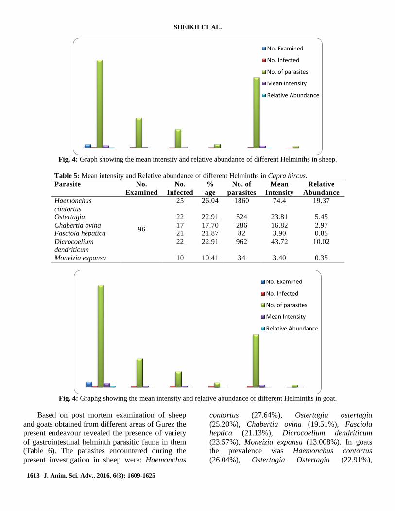
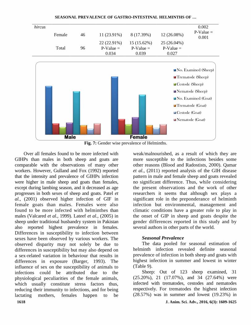
Fig. 7: Gender wise prevalence of Helminths.
Over all females found to be more infected with
GIHPs than males in both sheep and goats are
comparable with the observations of many other
workers. However, Gulland and Fox (1992) reported
that the intensity and prevalence of GIHPs infection
were higher in male sheep and goats than females,
except during lambing season, and it decreased as age
progresses in both sexes of sheep and goats. Patel et
al., (2001) observed higher infection of GIF in
female goats than males. Females were also
found to be more infected with helminthes than
males (Valcared et al., 1999). Lateef et al., (2005) in
sheep under traditional husbandry system in Pakistan
also reported highest prevalence in females.
Differences in susceptibility to infection between
sexes have been observed by various workers. The
observed disparity may not solely be due to
differences in susceptibility but may also depend on
a sex-related variation in behaviour that results in
differences in exposure (Barger, 1993). The
influence of sex on the susceptibility of animals to
infections could be attributed due to the
physiological peculiarities of the female animals,
which usually constitute stress factors thus,
reducing their immunity to infections, and for being
lactating mothers, females happen to be
weak/malnourished, as a result of which they are
more susceptible to the infections besides some
other reasons (Blood and Radostists, 2000). Qamar
et al., (2011) reported analysis of the GIH disease
pattern in male and female sheep and goats revealed
no significant difference. Thus, while considering
the present observations and the work of other
researchers it seems that although sex plays a
significant role in the preponderance of helminth
infection but environmental, management and
climatic conditions have a greater role to play in
the onset of GIP in sheep and goats despite the
gender differences reported in this study and by
several authors in other parts of the world.
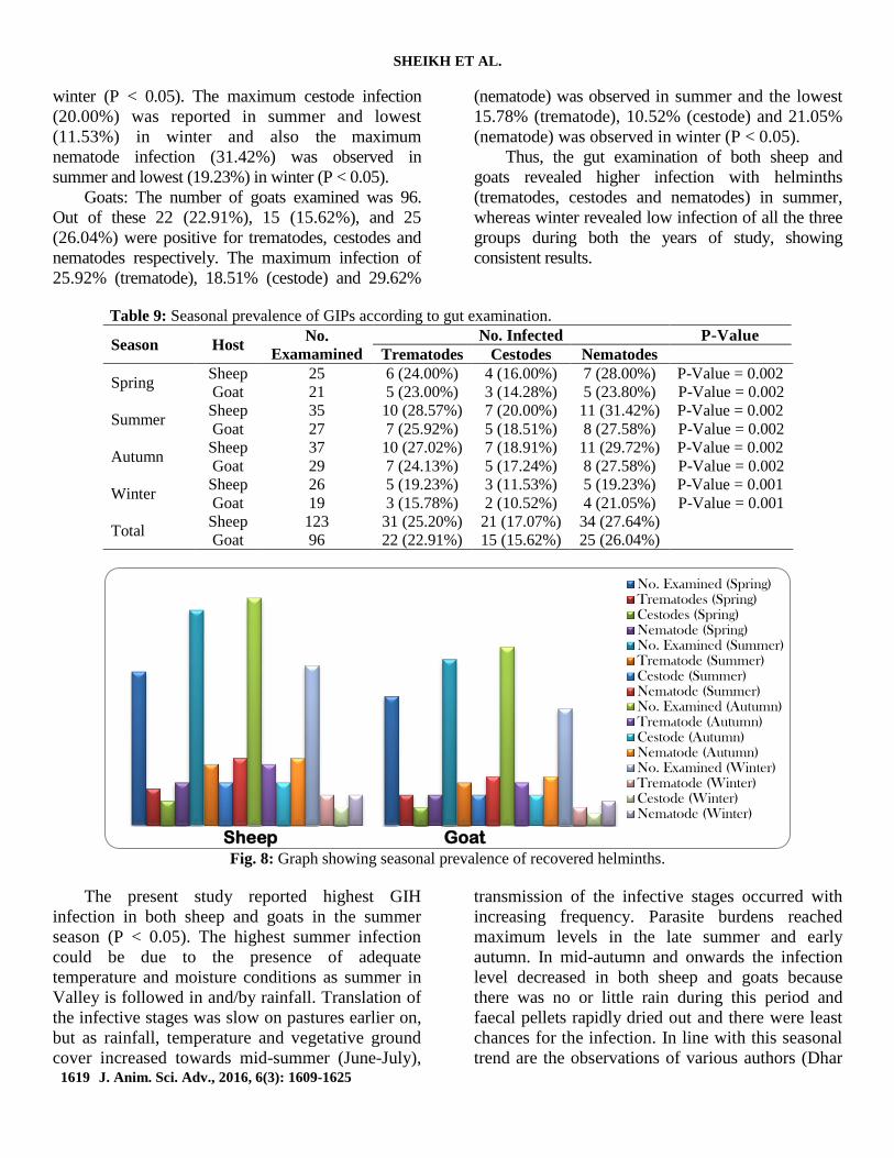
Seasonal Prevalence
The data pooled for seasonal estimation of
helminth infection revealed definite seasonal
prevalence of infection in both sheep and goats with
highest infection in summer and lowest in winter
(Table 9).
Sheep: Out of 123 sheep examined, 31
(25.20%), 21 (17.07%), and 34 (27.64%) were
infected with trematodes, cestodes and nematodes
respectively. For trematodes the highest infection
(28.57%) was in summer and lowest (19.23%) in
Male Female
No. Examined (Sheep)
Trematode (Sheep)
Cestode (Sheep)
Nematode (Sheep)
No. Examined (Goat)
Trematode (Goat)
Cestode (Goat)
Nematode (Goat)
SHEIKH ET AL.
1619 J. Anim. Sci. Adv., 2016, 6(3): 1609-1625
winter (P < 0.05). The maximum cestode infection
(20.00%) was reported in summer and lowest
(11.53%) in winter and also the maximum
nematode infection (31.42%) was observed in
summer and lowest (19.23%) in winter (P < 0.05).
Goats: The number of goats examined was 96.
Out of these 22 (22.91%), 15 (15.62%), and 25
(26.04%) were positive for trematodes, cestodes and
nematodes respectively. The maximum infection of
25.92% (trematode), 18.51% (cestode) and 29.62%
(nematode) was observed in summer and the lowest
15.78% (trematode), 10.52% (cestode) and 21.05%
(nematode) was observed in winter (P < 0.05).
Thus, the gut examination of both sheep and
goats revealed higher infection with helminths
(trematodes, cestodes and nematodes) in summer,
whereas winter revealed low infection of all the three
groups during both the years of study, showing
consistent results.
Table 9: Seasonal prevalence of GIPs according to gut examination.
Season Host No.
Examamined
No. Infected P-Value
Trematodes Cestodes Nematodes
Spring Sheep 25 6 (24.00%) 4 (16.00%) 7 (28.00%) P-Value = 0.002
Goat 21 5 (23.00%) 3 (14.28%) 5 (23.80%) P-Value = 0.002
Summer Sheep 35 10 (28.57%) 7 (20.00%) 11 (31.42%) P-Value = 0.002
Goat 27 7 (25.92%) 5 (18.51%) 8 (27.58%) P-Value = 0.002
Autumn Sheep 37 10 (27.02%) 7 (18.91%) 11 (29.72%) P-Value = 0.002
Goat 29 7 (24.13%) 5 (17.24%) 8 (27.58%) P-Value = 0.002
Winter Sheep 26 5 (19.23%) 3 (11.53%) 5 (19.23%) P-Value = 0.001
Goat 19 3 (15.78%) 2 (10.52%) 4 (21.05%) P-Value = 0.001
Total Sheep 123 31 (25.20%) 21 (17.07%) 34 (27.64%)
Goat 96 22 (22.91%) 15 (15.62%) 25 (26.04%)
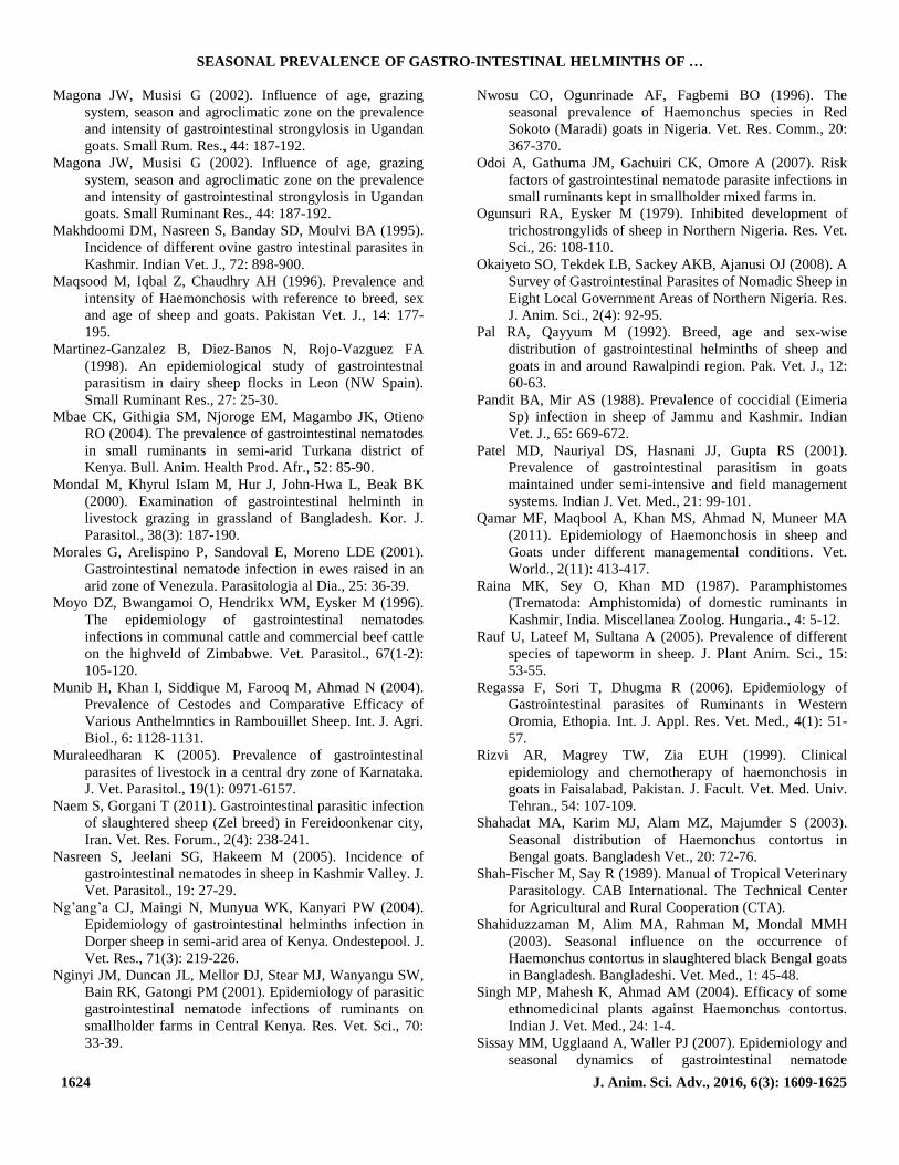
Fig. 8: Graph showing seasonal prevalence of recovered helminths.
The present study reported highest GIH
infection in both sheep and goats in the summer
season (P < 0.05). The highest summer infection
could be due to the presence of adequate
temperature and moisture conditions as summer in
Valley is followed in and/by rainfall. Translation of
the infective stages was slow on pastures earlier on,
but as rainfall, temperature and vegetative ground
cover increased towards mid-summer (June-July),
transmission of the infective stages occurred with
increasing frequency. Parasite burdens reached
maximum levels in the late summer and early
autumn. In mid-autumn and onwards the infection
level decreased in both sheep and goats because
there was no or little rain during this period and
faecal pellets rapidly dried out and there were least
chances for the infection. In line with this seasonal
trend are the observations of various authors (Dhar
Sheep Goat
No. Examined (Spring)Trematodes (Spring)Cestodes (Spring)Nematode (Spring)No. Examined (Summer)Trematode (Summer)Cestode (Summer)Nematode (Summer)No. Examined (Autumn)Trematode (Autumn)Cestode (Autumn)Nematode (Autumn)No. Examined (Winter)Trematode (Winter)Cestode (Winter)Nematode (Winter)

SEASONAL PREVALENCE OF GASTRO-INTESTINAL HELMINTHS OF …
1620 J. Anim. Sci. Adv., 2016, 6(3): 1609-1625
et al., 1982; Pandit and Mir, 1988; Makhdoomi et
at., 1995; Vlassoff et al., 2001; Magona et al.,
2002; Khajuria and Kapoor, 2003; Kaplan et al.,
2004; Nasreen et al., 2005; Kuchai et al., 2011;
Lone et al., 2011).
The low infection reported during the winter
season in the present study could be attributed to
low temperature which helps in AD (hypobiosis) in
host (Ogunsuri and Eysker, 1979; Gibbs, 1986;
Tariq et al., 2008). Moreover, in winter the area
remains mostly snow covered as such there is no
grazing which hinders in loss of contact between
host and parasites. El-azazy (1995) in Saudi
Arabia also reported the overall worm counts and
infection rates lowest in the winter season in goats.
Valcared and Garcia (1999) observed lowest level
of infection in the winter and rose progressively
till the autumn. Shahadat et al., (2003)
recorded lowest prevalence in the month of
January of H. contortus in Bengal goats. The initial
exposure of native animals to infective larvae on
pasture in spring generates egg production
approximately 3-4 weeks after turnout.
There is peak biotic potential of Haemonchus
contortus which results in rapidly assuming
dominance at times when environmental conditions
on pasture are favourable for the development and
survival of the free-living stages. Nwosu et al., (1996)
reported that Haemonchus was the most common
nematode recorded during their study. They also
reported that Haemonchus eggs and adults were high
in prevalence and seasonal fluctuation was common
in the study area. They further reported the high
prevalence during hot humid season. These seasonal
variations in the nematode worm burdens were
similar to studies in other tropical countries with
distinct rainy and dry seasons (Fritsche et al., 1993;
Magona & Musisi, 2002; Nwosu et al., 1996). In
general, moist and warm environmental conditions
are favourable for the development, survival and
transmission of the pre-parasitic stages of parasitic
nematodes (Hansen & Perry, 1994; Urquhart et al.,
1987). Nasreen et al., (2005) also observed highest
infection (33.18 %) in summer and lowest in winter
(15.25%). Makhdoomi et al., (1995) also observed
highest infection in the summer season. Thus our
findings of higher infection in summer are in
agreement with them although the percent prevalence
in different seasons differs which may be due to the
topographic and climatic factors unique to the study
area.
The 1st peak level of infection as recorded from
April onwards is derived from over wintered larvae of
Haemonchus originating from the eggs deposited.
This contributes to what is known as the spring rise
phenomenon. High rainfall in spring also helps in
providing suitable molarity of salt present in soil
which is an important factor for ecdysis (Soulsby,
1986). It also helps in larval dispersion on herbage
which increases the chance of contact between host
and larvae (Nginyi et al., 2001).
The 2nd peak level of infection from June
onwards till autumn is derived from eggs
deposited in the first grazing cycle (Soulsby,
1986). Ingestion of sufficient numbers of larvae
results in type 1 disease in lambs during their first
summer at pasture. In this study, the presence of
sufficient moisture and optimum temperature
conditions during the rainy season favored the
survival of infective larvae in the pasture and higher
probability of uptake of the infective larvae leading
to higher prevalence rate. The rainy season which
started in the spring and early summer made the
environmental conditions more favourable for
the development and survival of preparasitic
stages and led to increased availability of infective
larvae in the rainy and post rainy season. Climatic
conditions influenced the build-up of pasture
larval contamination and elevated levels were
present in late July-August. This is in contrast to the
situation in other parts of world- the tropical
countries where disease primarily is caused due to
infection with L3 derived from early season
contamination (Vercruysse and Claerebout, 2003),
thus climate plays significant role in epidemiology
of GIH parasites.
It is well documented that GIP in grazing
animals is directly related to the availability of
larvae on pasture and seasonal pasture
contamination (Soulsby, 1986). Kates (1950)
demonstrated that hot and humid weather provides
favourable conditions, for the development and
survival of exogenous stages of H. contortus.
Gordon (1953) stated that total monthly rainfall of
more than 50 mm and a mean monthly maximum
temperature of over 18.3 C provide optimum

SHEIKH ET AL.
1621 J. Anim. Sci. Adv., 2016, 6(3): 1609-1625
conditions for the development and survival of
exogenous stages of H. contortus. Therefore, it is
expected that higher incidence of H. contortus and
other nematodes would be restricted to months with
adequate rainfall. H. contortus eggs can withstand
extreme climatic conditions and also survive longer
period during winter season (Blood and Radostits,
1989) resulting in mass hatching of ova and high
summer infection of pasture. Sissay et al., (2007) in
Ethiopia showed distinct seasonal patterns with
high levels of infection during the short and
long rainy seasons with peaks occurring in May
and September of each year.
There are several studies which have reported
highest GIN infection in rainy seasons (Charles,
1989; Ahmad Abdalla et al., 1997; Katoch et al.,
2000; Laha et al., 2001; Fakae, 1990;
Shahiduzzaman et al., 2003; Yadav Khajuria,
2006). Agyei (1997) in coastal Savanna regions of
Ghana observed that the number of infective larvae
of strongylate nematode on pasture was directly
related to the pattern of rainfall. Qamar et al.,
(2011) the overall highest incidence of the year in
sheep and goats was recorded during summer
(43.69%), followed by autumn (38.46%) then
spring (37.12%), while the lowest (28.79%)
prevalence was recorded during winter. Singh et al.,
(2004) at an organized farm in Rajasthan reported
H. contortus as the most predominant parasite and
concluded that the period from July to October
(rainy season) was more favourable for H.
contortus. Tembely et al., (1997) in the highlands
of Ethiopia indicated that rainfall and humidity
seemed to be the most important factors for the
development of eggs and free living stages of
gastrointestinal nematode infections in sheep.
Martinez-Gonzalez et al., (1998) concluded that
environmental factors affected the prevalence and
intensity of GIH infection under semi-intensive
management conditions in dairy sheep flocks in
Leon, Spain. Thamsborg et al., (1998) in Joannina
(Greece) suggested that the factors affecting the
epidemiology of GI nematodes of naturally
infected sheep during grazing include
anthelmintic treatment, host genotype and season.
Rizvi et al., (1999) reported highest Haemonchus
infection in spring season in goats in Pakistan.
Najeeb-ur-Rehman and Akhtar Ali (2001)
observed the highest GIH infection in sheep and
goats in Pakistan in the months of June, July and
August.
Magona and Musisi (2002) indicated that age,
grazing system, season and agro-climatic zone have
significant influence on the level of risk of GIN
infections in Ugandan goats and on the worm egg
output under field conditions. Soundarajan and lyue
(2003) attributed the increasing infection trends of
helminth infection of sheep in Nilgiris hills to
varying climatic factors. Maturation of inhibited
larvae of H. contortus in sheep and the resultant
spring rise in FEC has been related to a loss of
immunity due to the limited exposure to infection in
winter, to stress, to immuno suppression associated
with the late stages of pregnancy and lactation and
to the frequent occurrence of poor nutrition at the
end of winter (Uriarte et al., 2003).
Nganga et al., (2004) indicated that rainfall
distribution was the major factor governing the
development and survival of the pre-parasitic
stages on pastures. Umur and Yukuri (2005)
showed that the parasite counts increased slightly
in spring and summer, and then reached maximum
in autumn. Sreedevi and Murthy (2005) reported the
overall prevalence of GIH in sheep higher in
summer. However, Garg et al., (2009) recorded the
highest incidence of GIN in winter in goats. Thus
seasons have an important role in the prevalence of
helminth parasites of sheep and goats.
From the above discussion it is now clear that
season and the other associated environmental
conditions including temperature and moisture
have a greater role to play in the onset of G1H
infection in sheep and goats. There are several
factors that contribute the disease onset like warm,
humid and wet grazing season, the more time
animals spend on pasture, inefficient selection of
de-wormers, poor husbandry practices and or the
development of anthelmentic resistance. The low
prevalence rate observed in sheep and goats of
Gurez valley could be due to difference in
management system of the animals and more
importantly the environmental conditions prevalent
in the area. However, our results are in close
conformity with Tariq et al., (2006) who reported
that higher parasite prevalence is more common in
sheep than in goats due to the grazing habit of

SEASONAL PREVALENCE OF GASTRO-INTESTINAL HELMINTHS OF …
1622 J. Anim. Sci. Adv., 2016, 6(3): 1609-1625
sheep. The result in this study could be because
most of the goats in this study were from mid and
high altitude areas, which are thought to be suitable
for survival of the larval stage of the parasites.
Likewise, in the highland areas of the Kashmir
where goats are mostly reared, there is poor
veterinary infrastructure and medication to goats.
More importantly, the condition could be due to less
or slow development of immunity in goats to
gastrointestinal parasites compared with the
situation in sheep and cattle (Regassa et al., 2006).
The degree of infection in most of the study animals
was low, agreeing with various works Kuchai et al.,
2011, and 2012 indicating the sub-clinical cases of
gastrointestinal parasites with subsequent subsistent
low degree of pasture contamination. But it is
inconsistent with reports from Kashmir (Dhar et al.,
1988) that could be explained by the difference in
management and breed of the respective animals.
This study revealed that the helminth infection
in ruminants occurs throughout the Gurez valley. It
also disclosed that regardless of the environmental
conditions, the animals are infected with variety of
helminth parasites. The various parasites recovered
during the present study have also been reported
from other two parts of the Jammu and Kashmir
state, as well as from different parts of the world
having almost same geographical locations and
environment (Bali, 1976, Raina et al., 1987,
Troncy, 1989, Nasreen et al., 2005, Fatima et al.,
2012).
However, the various intraspecific
morphological variations observed during the
present study could be due to the variation in age of
parasite, host species, intensity of infection (higher
intensity, smaller parasites), methodology
(fixation), environmental factors of the study area,
body conditions of the host, etc.
The significantly higher prevalence in autumn
than spring is in consent with many reports around
the world, Tembely et al., 1997, 1998; Moyo et al.,
1996; Fritche et al., 1993; Mbae et al., 2004. This
could be due to the existence of a direct relationship
between prevalence with the rainfall, humidity and
temperature. In this study, the presence of sufficient
rainfall and moisture during the autumn season
favored the survival of infective larvae in the
pasture and higher probability of uptake of the
infective larvae leading to higher prevalence rate.
Similarly, a higher prevalence recorded in
younger animals as compared to the adult ones is in
agreement with most literatures Dunn, 1978; Shah-
Fischer and Say, 1989; Keyyu, 2003; Nwosu et al.,
1996; Nganga et al., 2004, from different corner of
the world. This could be due to the fact that younger
animals are more susceptible to infections than
adults. Adult animals may acquire immunity to the
parasites through frequent challenge and expel the
ingested parasite before they establish infection,
(Dunn, 1978; Shah-Fischer and Say, 1989). But the
findings of this study are inconsistent with reports
from Gambia where adults and older animals bear
high worm burden.
Acknowledgments
The authors are greatly thankful to the
Department of Zoology, University of Kashmir for
the facilities they provided. Bashir A. Sheikh is also
thankful to those people of Gurez valley, India who
make them host material available for research
purpose.
References
Abebe W, Esayas G (2001). Survey of ovine and caprine
gastrointestinal helminthosis in eastern part of Ethiopia
during the dry season of the year. Revue Med. Vet., 152:
379-384.
Agyei, AD (1997). Seasonal changes in the level of infective
strongylate larvae on pasture in the coastal savanna
regions of Ghana, Vet. Parasitol., 70: 175-182.
Ahmad Abdalla EA, Elmalik KH (1997). Prevalence of
nematode parasitism in desert sheep brought to
Khartoum state. Sudan J. Vet. Sci. Anim. Husbandry, 36:
44-49.
Alani AJ, Yahya HQ (1991). A study of the epidemiology of
intestinal helminths in sheep in Ninewah (Iraq). J. Vet.
Parasitol., 5: 66-68.
Ali A (2001). Monthwise prevalence of gastrointestinal
trematodes, cestodes and nematodes infecting Damani
sheep and goats in District D. I. Khan. Pak. Vet. J., 21:
111-113.
Bali HS (1976). A survey of helminth parasites of sheep (Ovis
aries) in Jammu and Kashmir. J. Anim. Health Prod., 4:
25-32.
Barger IA (1993). Influence of sex and reproductive status on
the susceptibility of ruminants to nematode parasitism.
Int. J. Parasitol., 23: 463-469.

SHEIKH ET AL.
1623 J. Anim. Sci. Adv., 2016, 6(3): 1609-1625
Bashtar AR, Hassanein M, Abdel-Ghaffar F, Al-Rasheid K,
Hassan S, Mehlhorn H, AL-Mahdi M, Morsy K, Al-
Ghamdi A (2010). Studies on monieziasis of sheep I.
Prevalence and anthelmintics effects of some plant
extracts, a light and electron microscopic study.
Parasitol. Res., Online First, 23 September.
Blood DC, Radostits OM (1989). Veterinary medicine.
Balliere Tindall: London.
Blood DC, Radostits OM (1992). Medicina Veterinarian
Editorial. Interamericana Mc Graw Hill, New York. pp.
1137-1141.
Borgsteede FHM, Dercksen DP (1996). Coccidial and
helminth infections in goats kept indoors in the
Netherlands. Vet. Parasitol., 61: 321-326.
Charles TP (1989). Seasonal prevalence of gaslrointestinal
nematodes of goats in Pernambuco state, Brazil. Vet.
Parasitol., 30: 335-343.
Colditz IG, Watson DL, Gray GD, Eady SJ (1996). Some
relationship between age, immune responsiveness and
resistance to parasites in ruminants. Int. J. Parasitol., 26:
869-877.
Dhar DN, Sharma RL, Bansal GC (1982). Gastrointestinal
nematodes in sheep in Kashmir. Vet. Parasitol., 10(1):
91-5.
Dhar DN, Sharma RL, Raina OK (1988). Fascioliasis in
Animals in Kashmir valley. J. Vet. Parasitol., 2(1): 31-
35.
Dunn AM (1978). Textbook of Veterinary Helminthology,
2nd. Edition William Heinemann Medical Books Ltd.,
London.
El-azazy OME (1995). Seasonal changes and inhibited
development of the abomasal nematodes of sheep and
goats in Saudi Arabia. Vet. Parasitol., 58: 91-98.
Fakae BB (1990). Seasonal changes and hypobiosis in
Haemonchus contortus infection in the wesr African
Dwarf sheep and goats in the Nigerian derived Savanna.
Vet. Parasitol., 36: 123-130.
Fatima M, Ahmad F, Chishti MZ, Lone BA (2012). A Survey
on the Bovine Amphistomiasis in Kashmir Valley, India.
Int. J. Recent Sci. Res., 3(1): 50-52.
Fritsche T, Kaufmann J, Pfister K (1993). Parasite spectrum
and seasonal epidemiology of gastro-intestinal
nematodes of small ruminants in the Gambia. Vet.
Parasitol., 49(2): 271-283.
Garg R, Yadav CL, Kumar RR, Banerjee PS, Vatsya S,
Godara R (2009). The epidemiology of fasciolosis in
ruminants in different geo-climatic regions of north
India. Trop. Anim. Health Prod., 41: 1695-1700.
Gibbs HC (1986). Hypobiosis in parasitic nematodes-an
update. Adv. Parasitol., 25: 129-174.
Gordon HML (1953). The epidemiology of helminthiasis in
sheep in winter rainfall regions of Australia. Aust. Vet.
J., 56: 80-86.
Gulland FMD, Fox M (1992). Epidemiology of nematode
infections of Soay sheep (Ovis. aries L.) on St. Kilda.
Parasitol., 105: 481-492.
Hansen J, Perry B (1994). The epidemiology, diagnosis and
control of helminth parasites of ruminants. Animal
Production and Health Division, Food and Agricultural
organization Rome, Italy. 2nd Ed., Nairobi, Kenya,
ILRAD.
Kaplan RM, Burke JM, Terill TH, Miller JE, Getz WR,
Mobini S, Valencia E, Williams MJ, Williamson LH,
Larsen M, Vatta AF (2004). Validation of the
FAMACHA® eye colour chart for detecting clinical
anaemia in sheep and goats on farms in the southern
United States. Vet. Parasitol., 123: 105-120.
Kates KC (1950). Survival on pasture of free living stages of
some common gastrointestinal nematodes of sheep. Proc.
Helm. Soc. Wash., 17: 39-58.
Katoch R, Chauhan PPS, Johri DK (2000). Seasonal incidence
of gastrointestinal nematodes in goats of Mathura region.
Indian Vet. J., 8: 261-263.
Keyyu JD, Kassuku AA, Kyvsgaard NC, Willingham AL
(2003). Gastrointestinal nematodes in indigenous zebu
cattle under pastoral and nomadic management systems
in the lower plain of Southern highlands of Tanzania.
Vet. Res. Commun., 27(5): 371-380.
Khajuria JK, Kapoor PR (2003). Prevalence of parasites in
sheep and goats at Kathua-Jammu. J. Vet. Parasitol., 17:
121-126.
Kiran S, Kumar S, Kanchan (2005). Prevalence of
gastrointestinal helminths in sheep and goats in
Dehradun. J. Exp. Zool., 8: 281-283.
Kuchai JA, Chishti MZ, Tak H, Lone BA (2012). Faecal
Examinations of Pashmina Goats (Capra siberica) of
Ladakh for Nematode Infections. Global J. Sci. Frontier
Res. Biol. Sci., 12(4): 37-40.
Kuchai JA, Chishti MZ, Zaki MM, Ahmad J, Rasool M, Dar
SA, Tak H (2011). Epidemiology of Helminth Parasites
in Small Ruminants of Ladakh, India. Online J. Anim.
Feed Res., 1(5): 239-242.
Kumsa B, Wossene A (2006). Abomasal nematodes of small
ruminants of Ogaden region, eastern Ethiopia:
prevalence, worm burden and species composition.
Revue Méd. Vét., 157(12): 27-32.
Laha R, Ramakrishna C, Bhattcharya D, Sikdar A (2001).
Seasonal incidence of Haemonchus contortus infection in
goats- a post-mortem study. Indian J. Anim. Sci., 71:
345-346.
Lateef M, Iqbal Z, Jabbar A, Khan MN, Akhtar MS (2005).
Epidemiology of trichostrongylid nematode infections in
sheep under traditional husbandry system in Pakistan.
Int. J. Agr. Biol., 7: 596-600.
Lateef M, Iqbal Z, Jabbar A, Khan MN, Akhter MS (2005).
Epidemiology of trichostrongylid nematode infections in
sheep under Traditional Husbandry System in Pakistan.
Int. J. Agr. Biol., 7(4): 596-600.
Lone BA, Chishti MZ, Ahmad F (2011a). Prevalence of
Coccidia and Gastrointestinal Nematode Infections in
Goats of Barramulla District of Kashmir Valley. Global
Vet., 7(1): 27-30.
Lone BA, Chisti MZ, Ahmad F, Tak H (2012c).
Immunodiagnosis of Haemonchus contortus Infection in
Sheep by Indirect Enzyme Linked Immunosorbent Assay
(ELISA). Iranian J. Vet. Res., 13(1)(Ser. No. 38): 49-53.
SEASONAL PREVALENCE OF GASTRO-INTESTINAL HELMINTHS OF …
1624 J. Anim. Sci. Adv., 2016, 6(3): 1609-1625
Magona JW, Musisi G (2002). Influence of age, grazing
system, season and agroclimatic zone on the prevalence
and intensity of gastrointestinal strongylosis in Ugandan
goats. Small Rum. Res., 44: 187-192.
Magona JW, Musisi G (2002). Influence of age, grazing
system, season and agroclimatic zone on the prevalence
and intensity of gastrointestinal strongylosis in Ugandan
goats. Small Ruminant Res., 44: 187-192.
Makhdoomi DM, Nasreen S, Banday SD, Moulvi BA (1995).
Incidence of different ovine gastro intestinal parasites in
Kashmir. Indian Vet. J., 72: 898-900.
Maqsood M, Iqbal Z, Chaudhry AH (1996). Prevalence and
intensity of Haemonchosis with reference to breed, sex
and age of sheep and goats. Pakistan Vet. J., 14: 177-
195.
Martinez-Ganzalez B, Diez-Banos N, Rojo-Vazguez FA
(1998). An epidemiological study of gastrointestnal
parasitism in dairy sheep flocks in Leon (NW Spain).
Small Ruminant Res., 27: 25-30.
Mbae CK, Githigia SM, Njoroge EM, Magambo JK, Otieno
RO (2004). The prevalence of gastrointestinal nematodes
in small ruminants in semi-arid Turkana district of
Kenya. Bull. Anim. Health Prod. Afr., 52: 85-90.
MondaI M, Khyrul IsIam M, Hur J, John-Hwa L, Beak BK
(2000). Examination of gastrointestinal helminth in
livestock grazing in grassland of Bangladesh. Kor. J.
Parasitol., 38(3): 187-190.
Morales G, Arelispino P, Sandoval E, Moreno LDE (2001).
Gastrointestinal nematode infection in ewes raised in an
arid zone of Venezula. Parasitologia al Dia., 25: 36-39.
Moyo DZ, Bwangamoi O, Hendrikx WM, Eysker M (1996).
The epidemiology of gastrointestinal nematodes
infections in communal cattle and commercial beef cattle
on the highveld of Zimbabwe. Vet. Parasitol., 67(1-2):
105-120.
Munib H, Khan I, Siddique M, Farooq M, Ahmad N (2004).
Prevalence of Cestodes and Comparative Efficacy of
Various Anthelmntics in Rambouillet Sheep. Int. J. Agri.
Biol., 6: 1128-1131.
Muraleedharan K (2005). Prevalence of gastrointestinal
parasites of livestock in a central dry zone of Karnataka.
J. Vet. Parasitol., 19(1): 0971-6157.
Naem S, Gorgani T (2011). Gastrointestinal parasitic infection
of slaughtered sheep (Zel breed) in Fereidoonkenar city,
Iran. Vet. Res. Forum., 2(4): 238-241.
Nasreen S, Jeelani SG, Hakeem M (2005). Incidence of
gastrointestinal nematodes in sheep in Kashmir Valley. J.
Vet. Parasitol., 19: 27-29.
Ng’ang’a CJ, Maingi N, Munyua WK, Kanyari PW (2004).
Epidemiology of gastrointestinal helminths infection in
Dorper sheep in semi-arid area of Kenya. Ondestepool. J.
Vet. Res., 71(3): 219-226.
Nginyi JM, Duncan JL, Mellor DJ, Stear MJ, Wanyangu SW,
Bain RK, Gatongi PM (2001). Epidemiology of parasitic
gastrointestinal nematode infections of ruminants on
smallholder farms in Central Kenya. Res. Vet. Sci., 70:
33-39.
Nwosu CO, Ogunrinade AF, Fagbemi BO (1996). The
seasonal prevalence of Haemonchus species in Red
Sokoto (Maradi) goats in Nigeria. Vet. Res. Comm., 20:
367-370.
Odoi A, Gathuma JM, Gachuiri CK, Omore A (2007). Risk
factors of gastrointestinal nematode parasite infections in
small ruminants kept in smallholder mixed farms in.
Ogunsuri RA, Eysker M (1979). Inhibited development of
trichostrongylids of sheep in Northern Nigeria. Res. Vet.
Sci., 26: 108-110.
Okaiyeto SO, Tekdek LB, Sackey AKB, Ajanusi OJ (2008). A
Survey of Gastrointestinal Parasites of Nomadic Sheep in
Eight Local Government Areas of Northern Nigeria. Res.
J. Anim. Sci., 2(4): 92-95.
Pal RA, Qayyum M (1992). Breed, age and sex-wise
distribution of gastrointestinal helminths of sheep and
goats in and around Rawalpindi region. Pak. Vet. J., 12:
60-63.
Pandit BA, Mir AS (1988). Prevalence of coccidial (Eimeria
Sp) infection in sheep of Jammu and Kashmir. Indian
Vet. J., 65: 669-672.
Patel MD, Nauriyal DS, Hasnani JJ, Gupta RS (2001).
Prevalence of gastrointestinal parasitism in goats
maintained under semi-intensive and field management
systems. Indian J. Vet. Med., 21: 99-101.
Qamar MF, Maqbool A, Khan MS, Ahmad N, Muneer MA
(2011). Epidemiology of Haemonchosis in sheep and
Goats under different managemental conditions. Vet.
World., 2(11): 413-417.
Raina MK, Sey O, Khan MD (1987). Paramphistomes
(Trematoda: Amphistomida) of domestic ruminants in
Kashmir, India. Miscellanea Zoolog. Hungaria., 4: 5-12.
Rauf U, Lateef M, Sultana A (2005). Prevalence of different
species of tapeworm in sheep. J. Plant Anim. Sci., 15:
53-55.
Regassa F, Sori T, Dhugma R (2006). Epidemiology of
Gastrointestinal parasites of Ruminants in Western
Oromia, Ethopia. Int. J. Appl. Res. Vet. Med., 4(1): 51-
57.
Rizvi AR, Magrey TW, Zia EUH (1999). Clinical
epidemiology and chemotherapy of haemonchosis in
goats in Faisalabad, Pakistan. J. Facult. Vet. Med. Univ.
Tehran., 54: 107-109.
Shahadat MA, Karim MJ, Alam MZ, Majumder S (2003).
Seasonal distribution of Haemonchus contortus in
Bengal goats. Bangladesh Vet., 20: 72-76.
Shah-Fischer M, Say R (1989). Manual of Tropical Veterinary
Parasitology. CAB International. The Technical Center
for Agricultural and Rural Cooperation (CTA).
Shahiduzzaman M, Alim MA, Rahman M, Mondal MMH
(2003). Seasonal influence on the occurrence of
Haemonchus contortus in slaughtered black Bengal goats
in Bangladesh. Bangladeshi. Vet. Med., 1: 45-48.
Singh MP, Mahesh K, Ahmad AM (2004). Efficacy of some
ethnomedicinal plants against Haemonchus contortus.
Indian J. Vet. Med., 24: 1-4.
Sissay MM, Ugglaand A, Waller PJ (2007). Epidemiology and
seasonal dynamics of gastrointestinal nematode
SHEIKH ET AL.
1625 J. Anim. Sci. Adv., 2016, 6(3): 1609-1625
infections of sheep in a semi-arid region of eastern
Ethiopia. Vet. Parasitol., 143: 311-321.
Soulsby EJL (1982). Helminths, Arthropods and Protozoa of
Domesticated Animals, 7th Ed., The English Language
Book Society and Bailliere Tindall, London.
Soulsby EJL (1986). Helminthes, Arthopods and Protozoa of
Domesticated animals, 7th Ed, printed in Great Britain by
Williams Clowes Ltd., Beccles and London.
Soundararajan C, lyue M (2003). Seasonal prevalence of
helminthic infection of sheep in Nilgiris hills. Indian J.
Small Ruminants., 9: 144-164.
Sreedevi C, Murthy GSS (2005). A note on prevalence of
gastrointestinal parasitism in sheep. Indian Vet. J., 82:
912.
Suarez VH, Busetti MR (1995). The epidemiology of
Helminth infections of growing sheep in Argentinias
Western Pampas. Int. J. Parasitol., 25: 489-494.
Tariq KA (2007). Epidemiology of GIT helminth parasites in
small ruminants of Kashmir valley and their control by
indigenous herbal anthelmintics. Ph D Thesis to
University of Kashmir, Srinagar-190 006, J. K., India.
Tariq KA, Chishti MZ, Ahmad F, Shawl AS (2008a).
Anthelmintic efficacy of Achillea millifolium against
gastrointestinal nematodes of sheep: in vitro and in vivo
studies. J. Helminthol., 82: 227-233.
Tariq KA, Chishti MZ, Fayaz A, Shawl AS (2006).
Epidemiology of paramphistomosis in small ruminants in
Kashmir valley. Orient. Sci., 11: 11-16.
Tembely S, Lahlou-Kassi K, Rege JE (1998). Breed and
season effects on the peri-parturient rise in nematode egg
output in indigenous ewes in a cool tropical environment.
Vet. Parasitol., 77(2-3): 123-132.
Thamsborg SH, Boa ME, Makundi AE, Kassuki AA (1998).
Lungworm infection (Dictyocaulus viviparous) on Diary
cattle farms in tropical highlands of Tanzania. Trop.
Anim. Health Prod., 30(2): 93-96.
Thangathurai R, Rao DGK, Thimma Reddy PM (2003).
Prevalence of Enteric Parasitism in Sheep and Goats in
and around Bidder. Indian Vet. J., 80: 72-73.
Troncy PM (1989). Helminths of livestock and Poultry in
Tropical Africa. In: Fischer. (1989). Manual of tropical
veterinary Parasitology. CAB Int., UK. pp. 63-73.
Umur S, Yukuri BA (2005). Seasonal Activity of Gastro-
Intestinal Nematodes in Goats in Burdur Region, Turkey.
Turk. J. Vet. Anim. Sci., 29: 441-448.
Uriarte J, Llorente MM, Valderrabano J (2003). Seasonal
changes of gastrointestinal nematode burden in sheep
under an intensive grazing system. Vet. Parasitol., 118:
79-92.
Urquhart GM, Armour J, Duncan JL, Dunn AM, Jennings FW
(1987). Vet. Parasitol., 1st Edn. Longman Group UK.
Valcarcel F, Romero CG (1999). Prevalence and seasonal
pattern of caprine Trichostrongyles in a dry area of
central Spain. J. Vet. Med. Series B., 46: 673-681.
Valcared F, Garcia RC (1999). Prevalence and Seasonal
Pattern of Caprine Trichostrongyles in a Dry Area of
Central Spain. J. Vet. Med. B., 46: 673-681.
Vanimisetti HB, Andrew SL, Zajac AM, Notter DR (2004).
Inheritance of fecal egg count and packed cell volume
and their relationship with production traits in sheep
infected with Haemonchus contortus. J. Anim. Sci., 82:
1602-1611.
Vatta AF, Krecek RC (2000). Trematode infection of goats
farmed under resourcepoor conditions in South Africa.
Vercruysse J, Claerebout E (2003). Assessment of the efficacy
of helminth vaccines. J. Parasitol., 89(Suppl.): 202-209.
Vlassoff A, Leathwick DM, Heath ACG (2001). The
epidmiology of nematodes infections of sheep. New
Zealand Vet. J., 49(6): 213-221.
Watson DL, Gill HS (1991). Effect of weaning on antibody
responses and nematode parasitism in Merino lambs.
Res. Vet. Sci., 51: 128-132.
Yadav A, Khajuria JK, Raina AK (2006). Seasonal prevalence
of gastrointestinal parasites in sheep and goats of Jammu.
J. Vet. Parasitol., 20: 9-12.
Zgardan E (2002). Some aspects of the epidemiology and
prophylaxis of Anoplocephalidosis in sheep in Moldovo.
Revista Romana de Parazitologie., 12: 82-83.