
Intestinal Parasites: Helminths, Cestodes, Protozoa … Parasites: Helminths, Cestodes, Protozoa...
55
Intestinal Parasites: Helminths, Cestodes, Protozoa (and other things you’d rather not share with your body) James Gaensbauer, MD, MPH Kevin Messacar, MD Global Health Conference November 12, 2012
Transcript of Intestinal Parasites: Helminths, Cestodes, Protozoa … Parasites: Helminths, Cestodes, Protozoa...
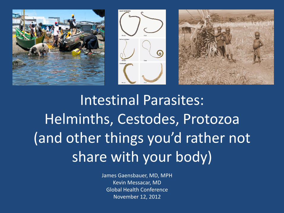
Intestinal Parasites: Helminths, Cestodes, Protozoa
(and other things you’d rather not share with your body)
James Gaensbauer, MD, MPH Kevin Messacar, MD
Global Health Conference November 12, 2012
Learning Objectives 1: Public Health Issues
• Understand the contribution of poverty, sanitation, and clean water on the worldwide prevalence of intestinal parasites
• Recognize the effects of intestinal parasites on child health, nutrition, and development
• Understand the preventative public health measures recommended for intestinal parasite control, including school-based deworming and screening
Learning Objectives 2: Presentation of Specific Parasites
• Understand the basics of transmission, life cycle, clinical manifestations, diagnosis and treatment of the most common intestinal parasites worldwide. – Focus on the unique aspects of each pathogen – Understand the way in which intestinal parasites
in the developing setting can mimic more common clinical diseases in the developed world
• When you hear footsteps, think horses, not zebras
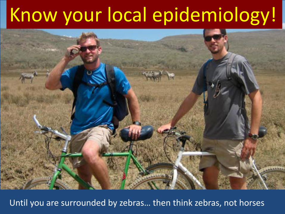
Until you are surrounded by zebras… then think zebras, not horses
Know your local epidemiology!
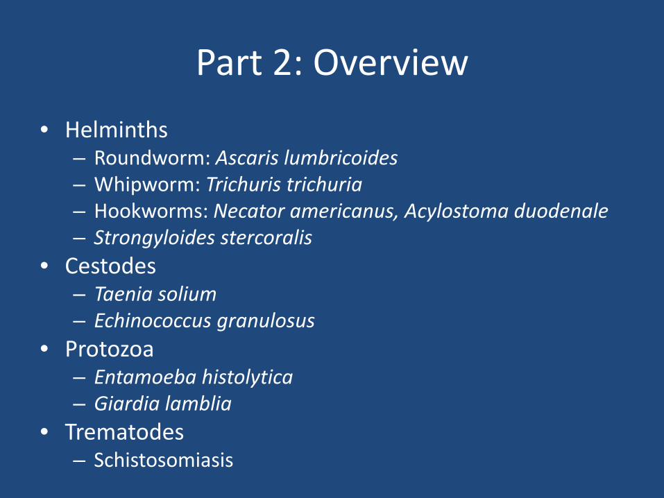
Part 2: Overview • Helminths
– Roundworm: Ascaris lumbricoides – Whipworm: Trichuris trichuria – Hookworms: Necator americanus, Acylostoma duodenale – Strongyloides stercoralis
• Cestodes – Taenia solium – Echinococcus granulosus
• Protozoa – Entamoeba histolytica – Giardia lamblia
• Trematodes – Schistosomiasis
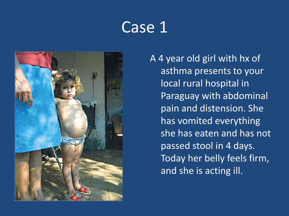
Case 1
A 4 year old girl with hx of asthma presents to your local rural hospital in Paraguay with abdominal pain and distension. She has vomited everything she has eaten and has not passed stool in 4 days. Today her belly feels firm, and she is acting ill.
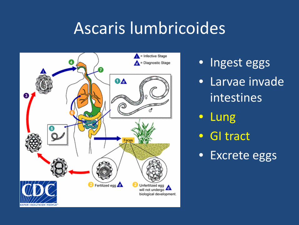
Ascaris lumbricoides
• Ingest eggs • Larvae invade
intestines • Lung • GI tract • Excrete eggs
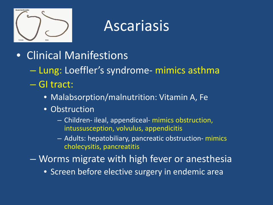
Ascariasis
• Clinical Manifestions – Lung: Loeffler’s syndrome- mimics asthma – GI tract:
• Malabsorption/malnutrition: Vitamin A, Fe • Obstruction
– Children- ileal, appendiceal- mimics obstruction, intussusception, volvulus, appendicitis
– Adults: hepatobiliary, pancreatic obstruction- mimics cholecysitis, pancreatitis
– Worms migrate with high fever or anesthesia • Screen before elective surgery in endemic area
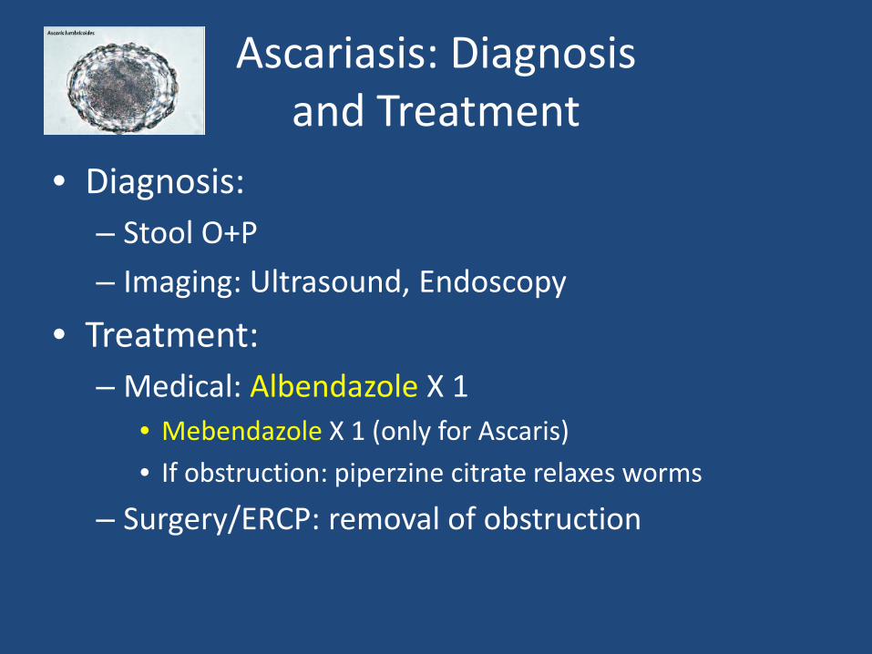
Ascariasis: Diagnosis and Treatment
• Diagnosis: – Stool O+P – Imaging: Ultrasound, Endoscopy
• Treatment: – Medical: Albendazole X 1
• Mebendazole X 1 (only for Ascaris) • If obstruction: piperzine citrate relaxes worms
– Surgery/ERCP: removal of obstruction
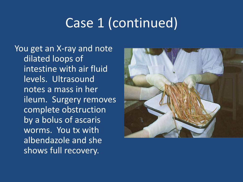
Case 1 (continued)
You get an X-ray and note dilated loops of intestine with air fluid levels. Ultrasound notes a mass in her ileum. Surgery removes complete obstruction by a bolus of ascaris worms. You tx with albendazole and she shows full recovery.
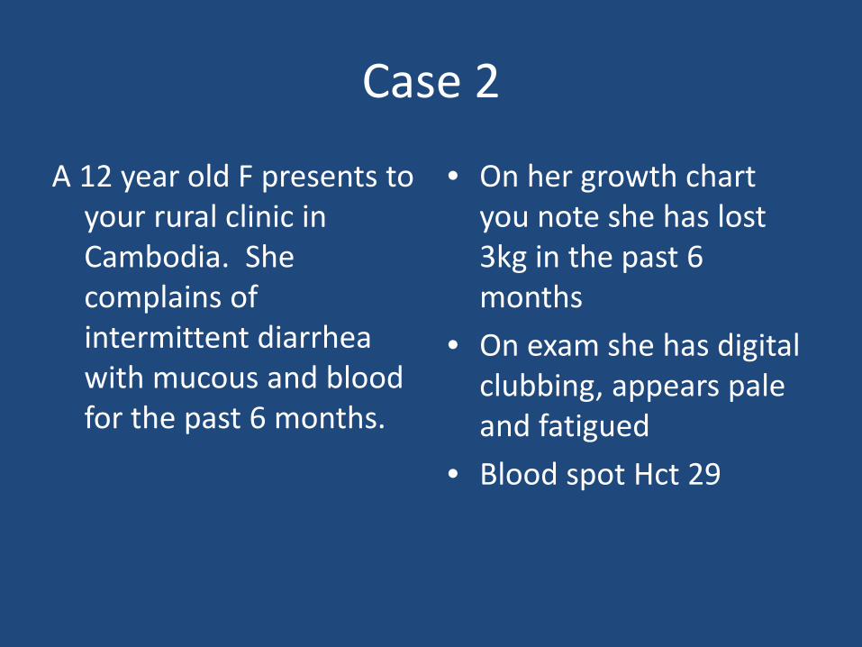
Case 2
A 12 year old F presents to your rural clinic in Cambodia. She complains of intermittent diarrhea with mucous and blood for the past 6 months.
• On her growth chart you note she has lost 3kg in the past 6 months
• On exam she has digital clubbing, appears pale and fatigued
• Blood spot Hct 29
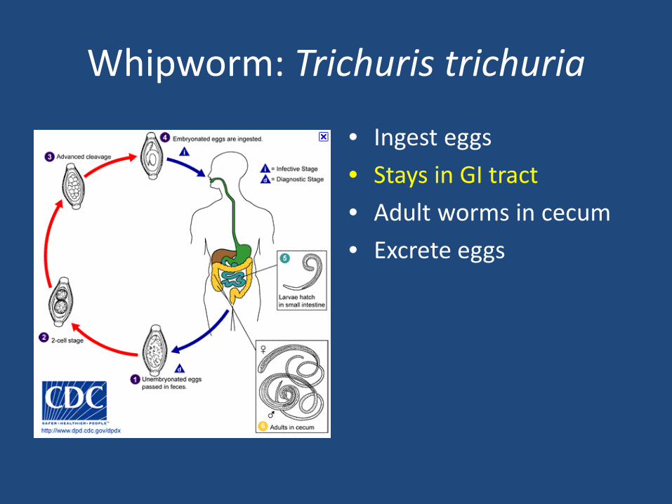
Whipworm: Trichuris trichuria
• Ingest eggs • Stays in GI tract • Adult worms in cecum • Excrete eggs
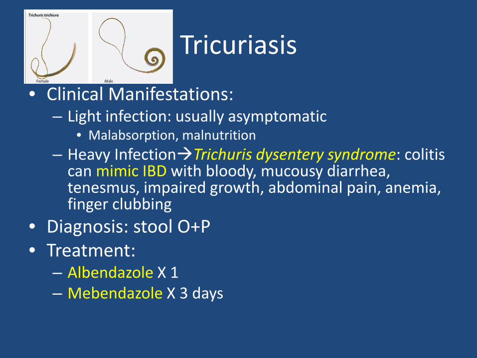
Tricuriasis
• Clinical Manifestations: – Light infection: usually asymptomatic
• Malabsorption, malnutrition – Heavy InfectionTrichuris dysentery syndrome: colitis
can mimic IBD with bloody, mucousy diarrhea, tenesmus, impaired growth, abdominal pain, anemia, finger clubbing
• Diagnosis: stool O+P • Treatment:
– Albendazole X 1 – Mebendazole X 3 days

Case 2 (continued) • Knowing the local
epidemiology, you do a stool O+P which demonstrates barrel shaped eggs of Trichuriasis
• You treat with Mebendazole 100mg twice daily for three days
• You start Iron supplements and give Vitamin A supplementation
• On follow-up 2 months later, her anemia has resolved and she has regained 3kg
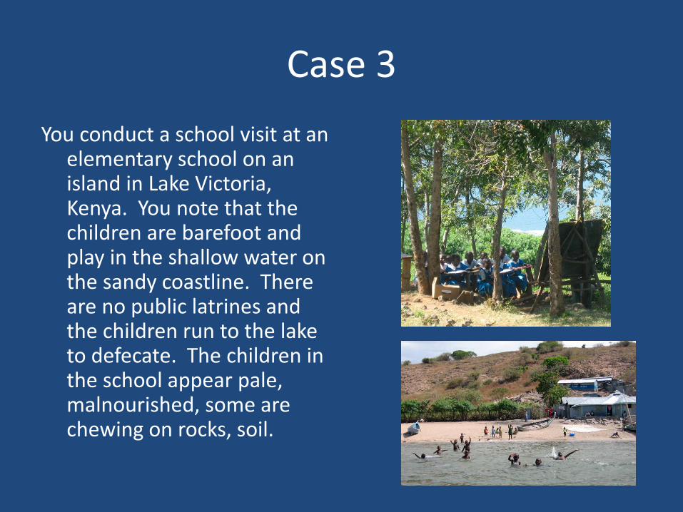
Case 3 You conduct a school visit at an
elementary school on an island in Lake Victoria, Kenya. You note that the children are barefoot and play in the shallow water on the sandy coastline. There are no public latrines and the children run to the lake to defecate. The children in the school appear pale, malnourished, some are chewing on rocks, soil.
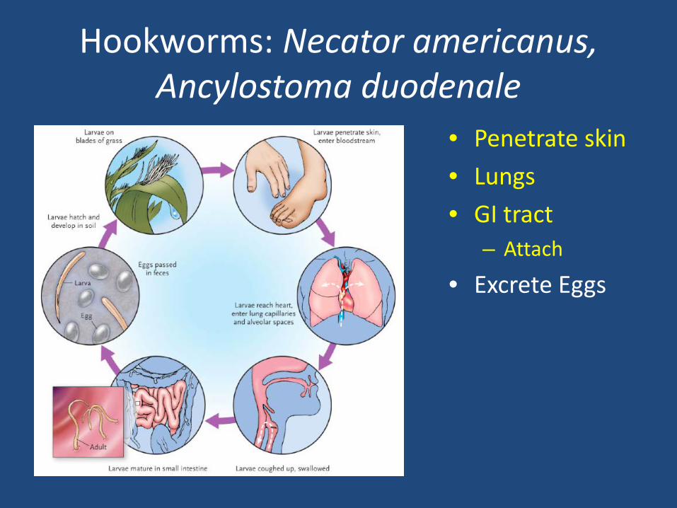
Hookworms: Necator americanus, Ancylostoma duodenale
• Penetrate skin • Lungs • GI tract
– Attach
• Excrete Eggs
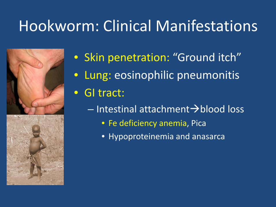
Hookworm: Clinical Manifestations
• Skin penetration: “Ground itch” • Lung: eosinophilic pneumonitis • GI tract:
– Intestinal attachmentblood loss • Fe deficiency anemia, Pica • Hypoproteinemia and anasarca
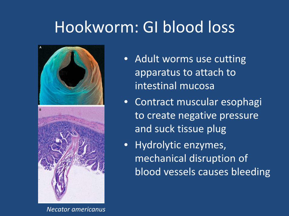
Hookworm: GI blood loss
• Adult worms use cutting apparatus to attach to intestinal mucosa
• Contract muscular esophagi to create negative pressure and suck tissue plug
• Hydrolytic enzymes, mechanical disruption of blood vessels causes bleeding
Necator americanus
Hookworm: Diagnosis and Treatment
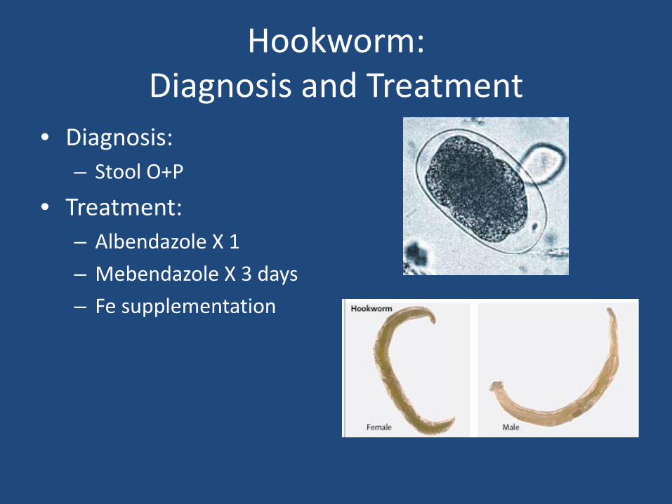
• Diagnosis: – Stool O+P
• Treatment: – Albendazole X 1 – Mebendazole X 3 days – Fe supplementation
Case 3 (continued) • You take stool samples and discover the
majority of children are carrying Ancylostoma Duodenale
• You conduct school deworming with albendazole
• You work with local government to construct latrines for the school and water sanitation education
• Deworming program is established every 6 months
• You follow Hct, weight and height for students over time and note a substantial improvement over the next 2 years
Case 4
• You are evaluating a 6 year old F with recent onset asthma at a referral hospital in Ghana. She began having dyspnea and wheezing 1 month ago and has been treated with a prolonged prednisone course X 3 weeks for refractory symptoms. She is also complaining of abdominal pain, diarrhea, and is now ill appearing and complaining of headaches.
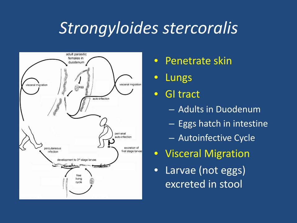
Strongyloides stercoralis
• Penetrate skin • Lungs • GI tract
– Adults in Duodenum – Eggs hatch in intestine – Autoinfective Cycle
• Visceral Migration • Larvae (not eggs)
excreted in stool
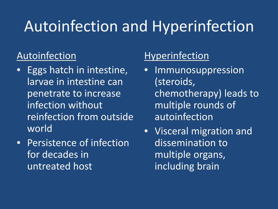
Autoinfection and Hyperinfection
Autoinfection • Eggs hatch in intestine,
larvae in intestine can penetrate to increase infection without reinfection from outside world
• Persistence of infection for decades in untreated host
Hyperinfection • Immunosuppression
(steroids, chemotherapy) leads to multiple rounds of autoinfection
• Visceral migration and dissemination to multiple organs, including brain
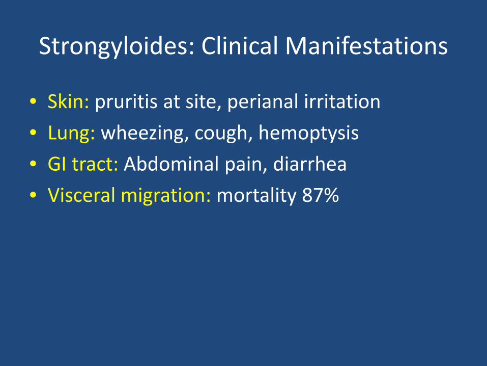
Strongyloides: Clinical Manifestations
• Skin: pruritis at site, perianal irritation • Lung: wheezing, cough, hemoptysis • GI tract: Abdominal pain, diarrhea • Visceral migration: mortality 87%
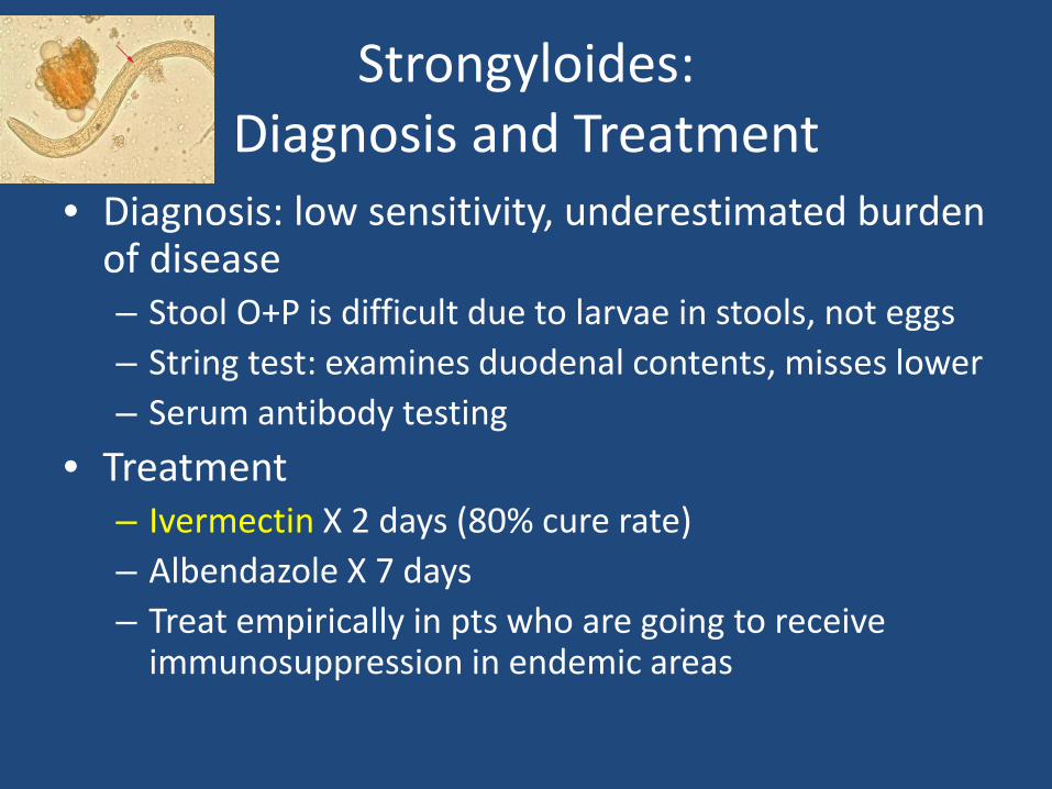
Strongyloides: Diagnosis and Treatment
• Diagnosis: low sensitivity, underestimated burden of disease – Stool O+P is difficult due to larvae in stools, not eggs – String test: examines duodenal contents, misses lower – Serum antibody testing
• Treatment – Ivermectin X 2 days (80% cure rate) – Albendazole X 7 days – Treat empirically in pts who are going to receive
immunosuppression in endemic areas
Case 4 (continued)
Knowing the local epidemiology of her area, you suspect Strongyloidiasis now exacerbated by prolonged steroid course. You send a serum ELISA for Strongyloides which is positive. You stop her steroids and start her on Ivermectin. Despite your best efforts, she passes away after 2 days in the intensive care unit from Stongyloides hyperinfection.
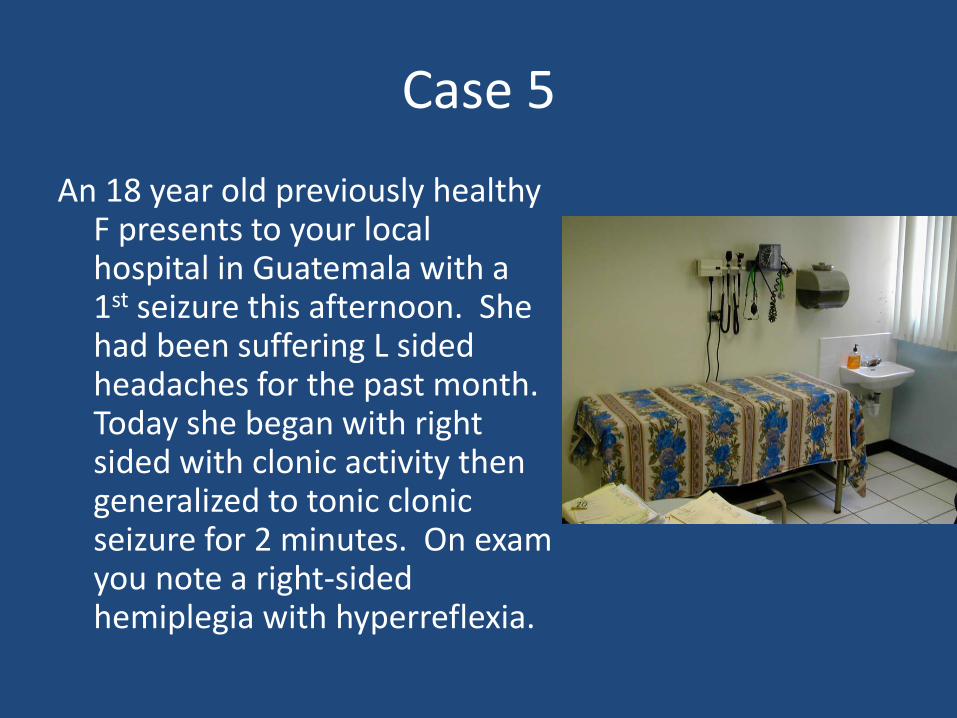
Case 5
An 18 year old previously healthy F presents to your local hospital in Guatemala with a 1st seizure this afternoon. She had been suffering L sided headaches for the past month. Today she began with right sided with clonic activity then generalized to tonic clonic seizure for 2 minutes. On exam you note a right-sided hemiplegia with hyperreflexia.
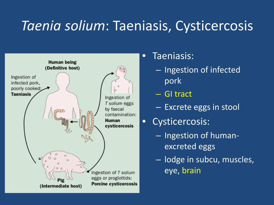
Taenia solium: Taeniasis, Cysticercosis
• Taeniasis: – Ingestion of infected
pork – GI tract – Excrete eggs in stool
• Cysticercosis: – Ingestion of human-
excreted eggs – lodge in subcu, muscles,
eye, brain
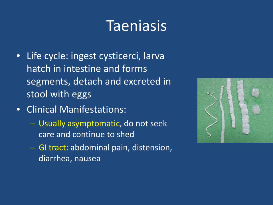
Taeniasis
• Life cycle: ingest cysticerci, larva hatch in intestine and forms segments, detach and excreted in stool with eggs
• Clinical Manifestations: – Usually asymptomatic, do not seek
care and continue to shed – GI tract: abdominal pain, distension,
diarrhea, nausea
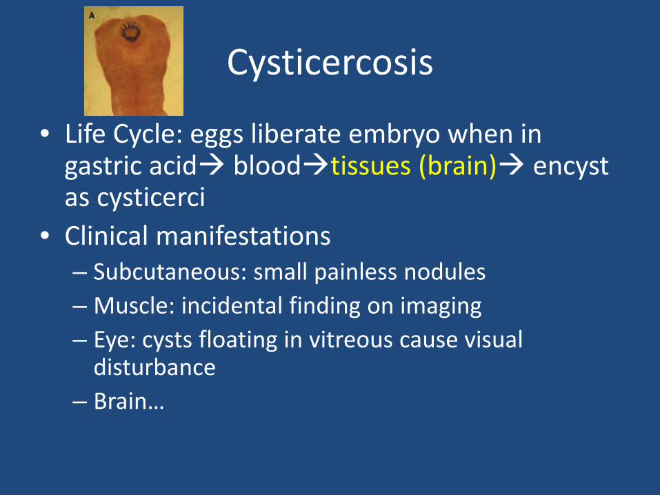
Cysticercosis
• Life Cycle: eggs liberate embryo when in gastric acid bloodtissues (brain) encyst as cysticerci
• Clinical manifestations – Subcutaneous: small painless nodules – Muscle: incidental finding on imaging – Eye: cysts floating in vitreous cause visual
disturbance – Brain…
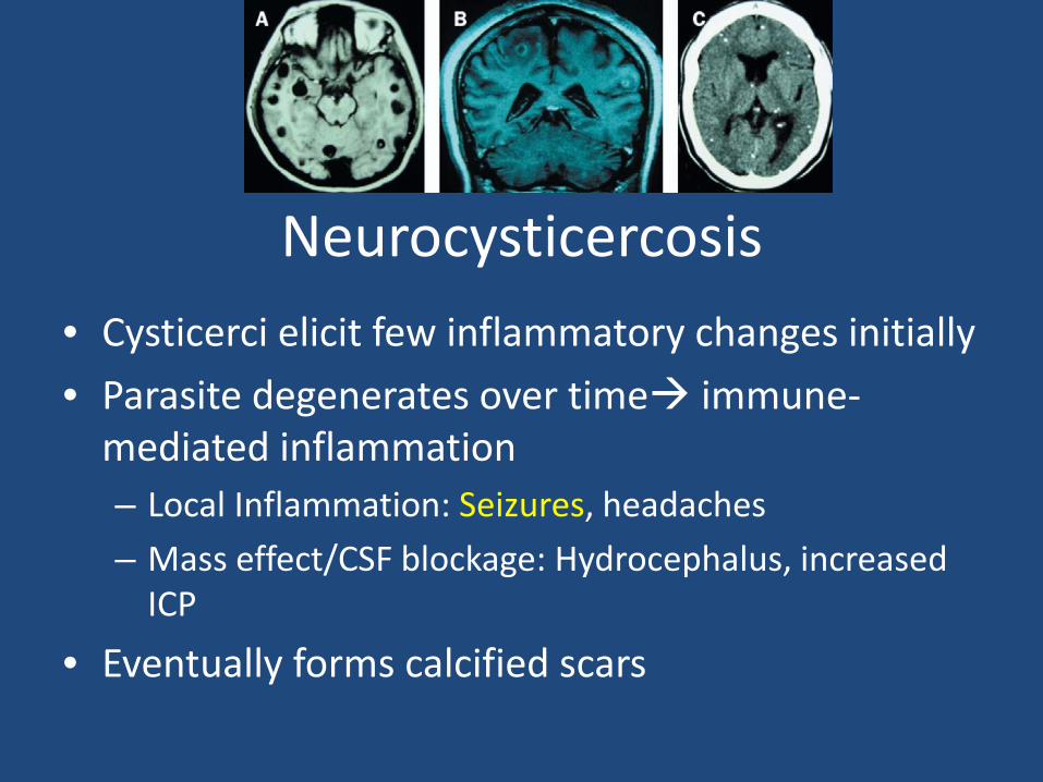
Neurocysticercosis • Cysticerci elicit few inflammatory changes initially • Parasite degenerates over time immune-
mediated inflammation – Local Inflammation: Seizures, headaches – Mass effect/CSF blockage: Hydrocephalus, increased
ICP
• Eventually forms calcified scars

Taenia Solium: Diagnosis and Treatment
Taeniasis: • stool O+P poor, stool
ELISA better • Niclosamide X 1 (not
absorbed, stays in GI tract), or praziquantel X 1
Neurocysticercosis: • Antibody testing of CSF or
serum • Imaging: CT or MRI
– Cystic lesion with mural nodule (scolex)
• Anti-epileptics • Treatment of
symptomatic patients with 1 or more lesions – Albendazole with steroids
• Surgery
Case 5 (continued)
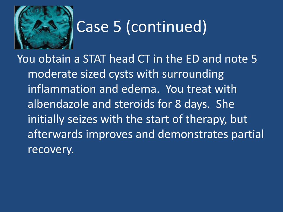
You obtain a STAT head CT in the ED and note 5 moderate sized cysts with surrounding inflammation and edema. You treat with albendazole and steroids for 8 days. She initially seizes with the start of therapy, but afterwards improves and demonstrates partial recovery.
Case 6
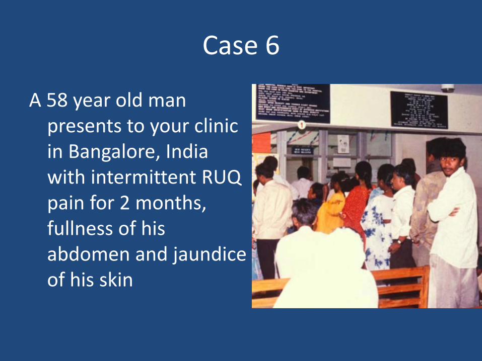
A 58 year old man presents to your clinic in Bangalore, India with intermittent RUQ pain for 2 months, fullness of his abdomen and jaundice of his skin
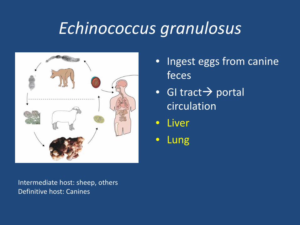
Echinococcus granulosus
• Ingest eggs from canine feces
• GI tract portal circulation
• Liver • Lung
Intermediate host: sheep, others Definitive host: Canines
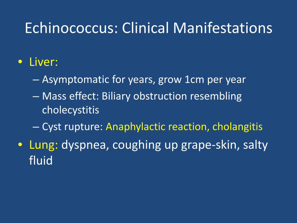
Echinococcus: Clinical Manifestations
• Liver: – Asymptomatic for years, grow 1cm per year – Mass effect: Biliary obstruction resembling
cholecystitis – Cyst rupture: Anaphylactic reaction, cholangitis
• Lung: dyspnea, coughing up grape-skin, salty fluid
Echinococcus: Diagnosis and Treatment
Diagnosis • Serum antibody testing • Imaging Medical Treatment • Albendazole X 3
months, can add praziquantel
Surgical Treatment: • Risk of peritonitis,
anaphylaxis from spill – Pre-operative
albendazole
• PAIR: – Puncture under
ultrasound guidance – Aspirate fluid – Inject protoscolicide – Re-aspirate after 15-20m
Case 6 (continued)
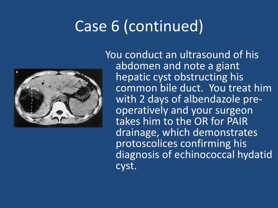
You conduct an ultrasound of his abdomen and note a giant hepatic cyst obstructing his common bile duct. You treat him with 2 days of albendazole pre-operatively and your surgeon takes him to the OR for PAIR drainage, which demonstrates protoscolices confirming his diagnosis of echinococcal hydatid cyst.
Case 7
You are running a new-immigrant clinic and conducting health screenings. You note on stool O+P that many of your Ethiopian immigrant patients have amebic cysts. The children appear to be growing well and do not complain of GI symptoms.
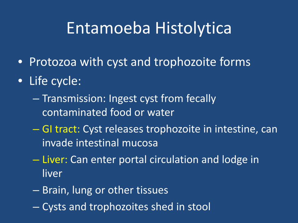
Entamoeba Histolytica
• Protozoa with cyst and trophozoite forms • Life cycle:
– Transmission: Ingest cyst from fecally contaminated food or water
– GI tract: Cyst releases trophozoite in intestine, can invade intestinal mucosa
– Liver: Can enter portal circulation and lodge in liver
– Brain, lung or other tissues – Cysts and trophozoites shed in stool
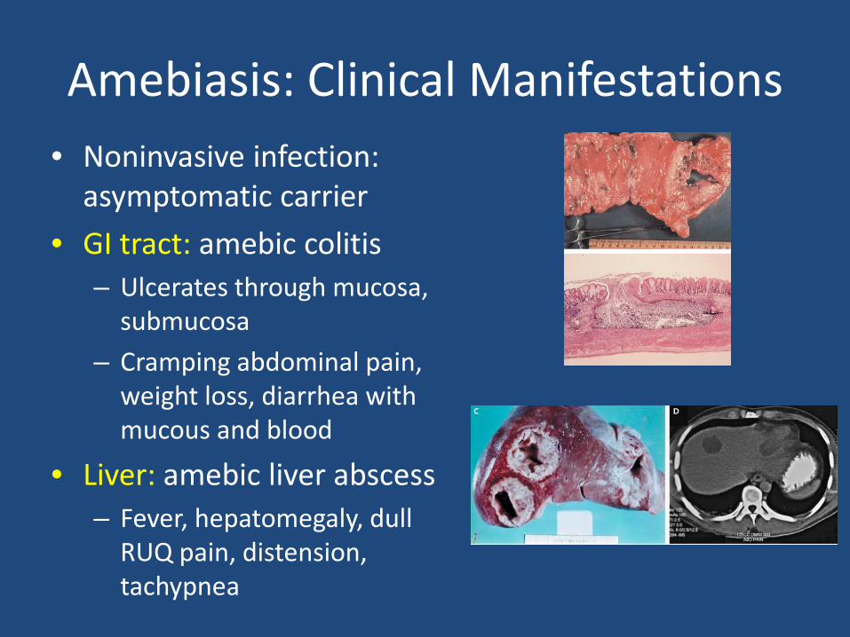
Amebiasis: Clinical Manifestations • Noninvasive infection:
asymptomatic carrier • GI tract: amebic colitis
– Ulcerates through mucosa, submucosa
– Cramping abdominal pain, weight loss, diarrhea with mucous and blood
• Liver: amebic liver abscess – Fever, hepatomegaly, dull
RUQ pain, distension, tachypnea
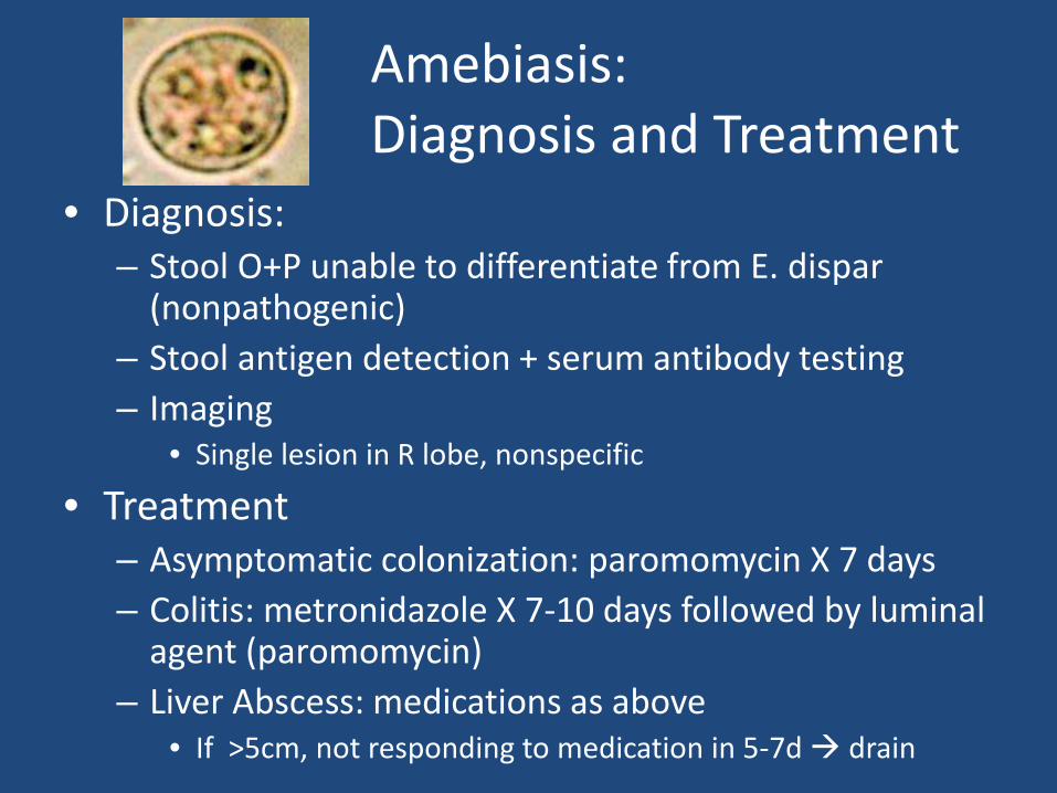
Amebiasis: Diagnosis and Treatment
• Diagnosis: – Stool O+P unable to differentiate from E. dispar
(nonpathogenic) – Stool antigen detection + serum antibody testing – Imaging
• Single lesion in R lobe, nonspecific
• Treatment – Asymptomatic colonization: paromomycin X 7 days – Colitis: metronidazole X 7-10 days followed by luminal
agent (paromomycin) – Liver Abscess: medications as above
• If >5cm, not responding to medication in 5-7d drain
Case 7 (continued)
As you recognize that stool O+P cannot often differentiate E. dispar from E. histolytica, you send stool antigen testing for E. histolytica. All of your patients are negative. You astutely decide not to treat them for the carriage of these non-pathogenic amebas and they continue to do well.
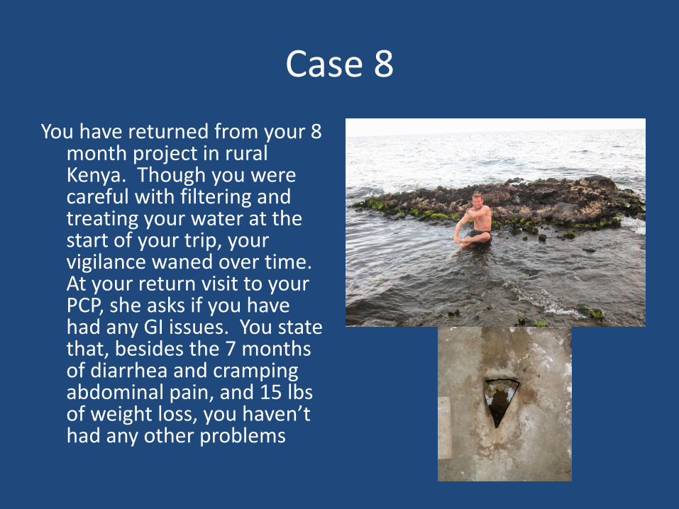
Case 8 You have returned from your 8
month project in rural Kenya. Though you were careful with filtering and treating your water at the start of your trip, your vigilance waned over time. At your return visit to your PCP, she asks if you have had any GI issues. You state that, besides the 7 months of diarrhea and cramping abdominal pain, and 15 lbs of weight loss, you haven’t had any other problems
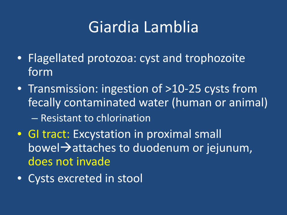
Giardia Lamblia
• Flagellated protozoa: cyst and trophozoite form
• Transmission: ingestion of >10-25 cysts from fecally contaminated water (human or animal) – Resistant to chlorination
• GI tract: Excystation in proximal small bowelattaches to duodenum or jejunum, does not invade
• Cysts excreted in stool
Giardiasis: Clinical Manifestations
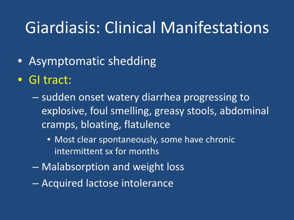
• Asymptomatic shedding • GI tract:
– sudden onset watery diarrhea progressing to explosive, foul smelling, greasy stools, abdominal cramps, bloating, flatulence
• Most clear spontaneously, some have chronic intermittent sx for months
– Malabsorption and weight loss – Acquired lactose intolerance
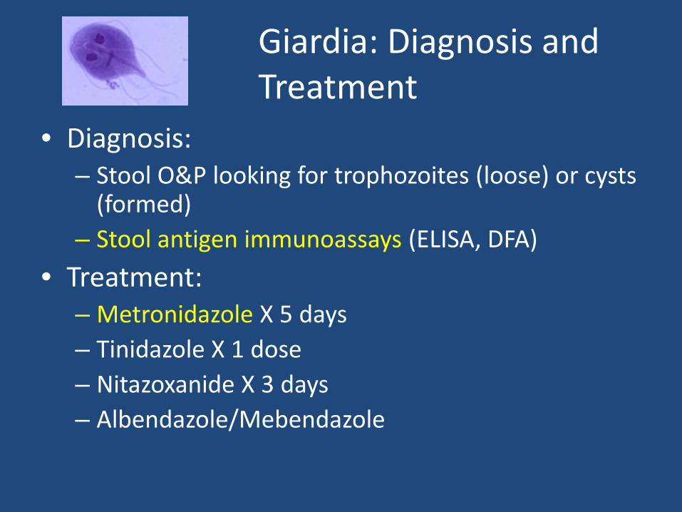
Giardia: Diagnosis and Treatment
• Diagnosis: – Stool O&P looking for trophozoites (loose) or cysts
(formed) – Stool antigen immunoassays (ELISA, DFA)
• Treatment: – Metronidazole X 5 days – Tinidazole X 1 dose – Nitazoxanide X 3 days – Albendazole/Mebendazole
Case 8 (continued)
You leave a stool sample which is sent for O+P, antigen testing. Cysts are seen under the microscope and antigen testing returns positive for Giardia Lamblia. You take 5 days of metronidazole and gain 15 lbs back on some home cooking. Your next trip you decide to filter and treat all of your drinking water….
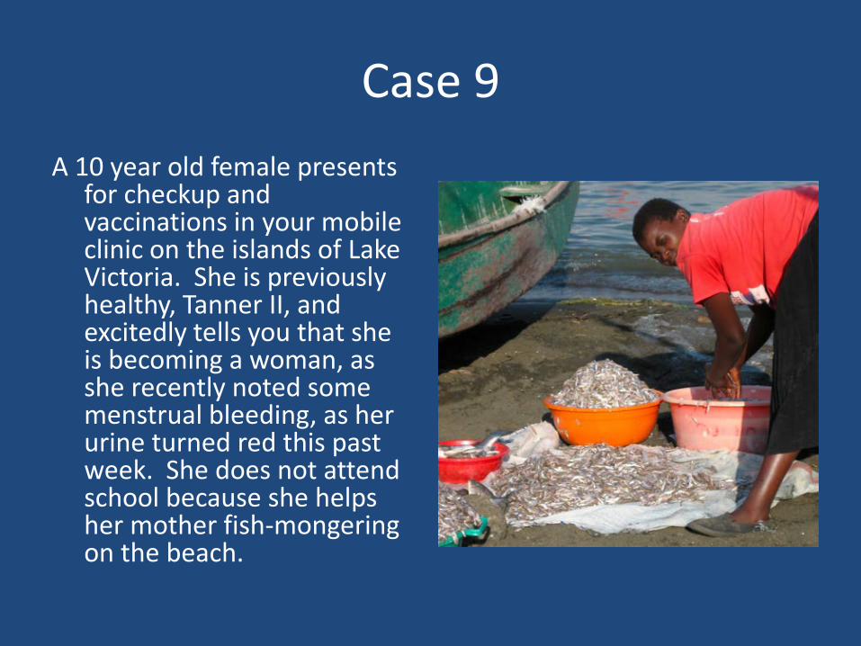
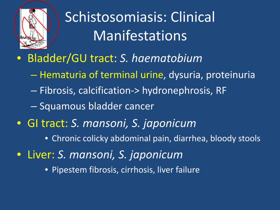
Case 9 A 10 year old female presents
for checkup and vaccinations in your mobile clinic on the islands of Lake Victoria. She is previously healthy, Tanner II, and excitedly tells you that she is becoming a woman, as she recently noted some menstrual bleeding, as her urine turned red this past week. She does not attend school because she helps her mother fish-mongering on the beach.
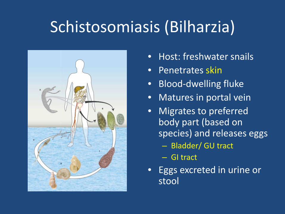
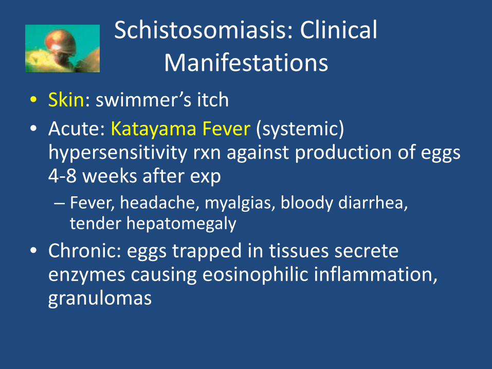
Schistosomiasis (Bilharzia) • Host: freshwater snails • Penetrates skin • Blood-dwelling fluke • Matures in portal vein • Migrates to preferred
body part (based on species) and releases eggs – Bladder/ GU tract – GI tract
• Eggs excreted in urine or stool
Schistosomiasis: Clinical Manifestations
• Skin: swimmer’s itch • Acute: Katayama Fever (systemic)
hypersensitivity rxn against production of eggs 4-8 weeks after exp – Fever, headache, myalgias, bloody diarrhea,
tender hepatomegaly • Chronic: eggs trapped in tissues secrete
enzymes causing eosinophilic inflammation, granulomas
Schistosomiasis: Clinical Manifestations
• Bladder/GU tract: S. haematobium – Hematuria of terminal urine, dysuria, proteinuria – Fibrosis, calcification-> hydronephrosis, RF – Squamous bladder cancer
• GI tract: S. mansoni, S. japonicum • Chronic colicky abdominal pain, diarrhea, bloody stools
• Liver: S. mansoni, S. japonicum • Pipestem fibrosis, cirrhosis, liver failure
Schistosomiasis: Diagnosis and Treatment
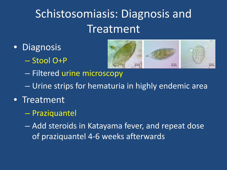
• Diagnosis – Stool O+P – Filtered urine microscopy – Urine strips for hematuria in highly endemic area
• Treatment – Praziquantel – Add steroids in Katayama fever, and repeat dose
of praziquantel 4-6 weeks afterwards
Case 9 (continued)
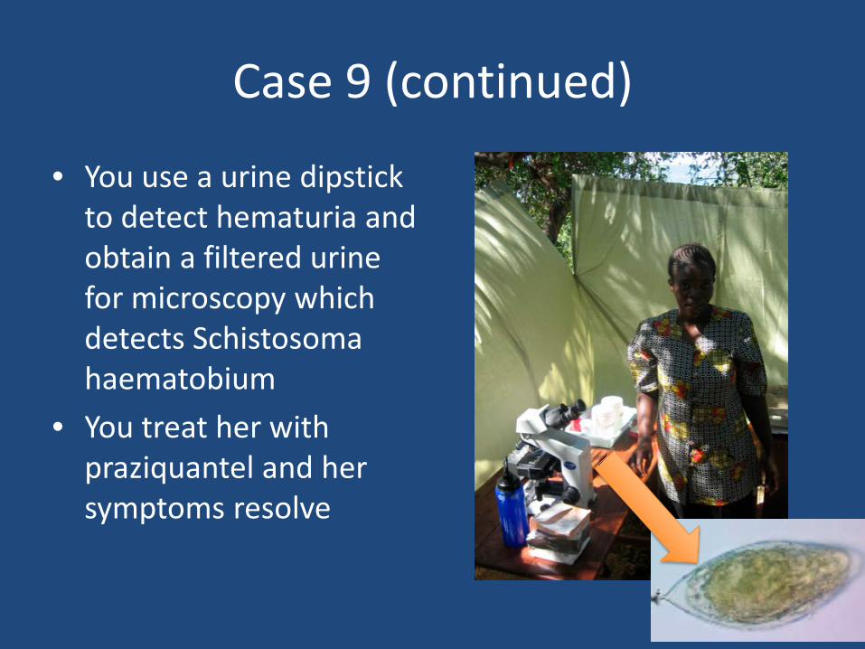
• You use a urine dipstick to detect hematuria and obtain a filtered urine for microscopy which detects Schistosoma haematobium
• You treat her with praziquantel and her symptoms resolve
References • Bethony J, Brooker S, Albonico M, et al. Soil-transmitted helminth
infections: ascariasis, trichuriasis, and hookworm. The Lancet. 2006;367:1521-1532.
• Hotez PJ, Brooker S, Bethony JM, Bottazzi ME, Loukas A, Xiao S. Hookworm Infection. N Engl J Med. 2004;351:799-807.
• Olsen A, van Lieshout L, Marti H, et al. Strongyloidiasis--the most neglected of the neglected tropical diseases? Trans R Soc Trop Med Hyg. 2009;103:967-972.
• García HH, Gonzalez AE, Evans CAW, Gilman RH. Taenia solium cysticercosis. The Lancet. 2003;362:547-556.
• McManus DP, Zhang W, Li J, Bartley PB. Echinococcosis. Lancet. 2003;362:1295-1304.
• Stanley SL. Amoebiasis. Lancet. 2003;361:1025-1034. • Ross AG, Bartley PB, Sleigh AC, et al. Schistosomiasis. N Engl J Med.
2002;346:1212-1220.


