
Searching for Amelia Earhart at the Molecular Level - ResearchGate
15
Journal of Alzheimer’s Disease 28 (2012) 823–837 DOI 10.3233/JAD-2011-110989 IOS Press 823 Extent and Neural Basis of Semantic Memory Impairment in Mild Cognitive Impairment Emmanuel J. Barbeau a,b,∗ , Mira Didic c,d , Sven Joubert e,f , Eric Guedj g , Lejla Koric c , Olivier Felician c,d , Jean-Philippe Ranjeva h , Patrick Cozzone h and Mathieu Ceccaldi c,d a Universit´ e de Toulouse, UPS, Centre de Recherche Cerveau et Cognition, Toulouse, France b CNRS, CerCo, Toulouse, France c APHM, CHU Timone, Service de Neurologie et Neuropsychologie, Marseille Cedex, Marseille, France d Aix-Marseille Univ, Laboratoire Epilepsies et Cognition, INSERM, Marseille Cedex, Facult´ e de M´ edecine, Marseille, France e D´ epartement de psychologie, Universit´ e de Montr´ eal, Montr´ eal, Canada f Centre de Recherche de l’Institut Universitaire de G´ eriatrie de Montr´ eal, Montr´ eal, Canada g Service Central de Biophysique et M´ edecine Nucl´ eaire, CHU Timone and Centre Europ´ een de Recherche en Imagerie M´ edicale, Universit´ e de la M´ editerran´ ee, Marseille, France h Centre d’Exploration M´ etabolique par R´ esonance Magn´ etique (UMR CNRS), Universit´ e de la M´ editerran´ ee, Marseille, France Handling Associate Editor: John Hodges Accepted 21 October 2011 Abstract. An increasing number of studies indicate that semantic memory is impaired in mild cognitive impairment (MCI). However, the extent and the neural basis of this impairment remain unknown. The aim of the present study was: 1) to evaluate whether all or only a subset of semantic domains are impaired in MCI patients; and 2) to assess the neural substrate of the semantic impairment in MCI patients using voxel-based analysis of MR grey matter density and SPECT perfusion. 29 predominantly amnestic MCI patients and 29 matched control subjects participated in this study. All subjects underwent a full neuropsychological assessment, along with a battery of five tests evaluating different domains of semantic memory. A semantic memory composite Z-score was established on the basis of this battery and was correlated with MRI grey matter density and SPECT perfusion measures. MCI patients were found to have significantly impaired performance across all semantic tasks, in addition to their anterograde memory deficit. Moreover, no temporal gradient was found for famous faces or famous public events and knowledge for the most remote decades was also impaired. Neuroimaging analyses revealed correlations between semantic knowledge and perirhinal/entorhinal areas as well as the anterior hippocampus. Therefore, the deficits in the realm of semantic memory in patients with MCI is more widespread than previously thought and related to dysfunction of brain areas beyond the limbic-diencephalic system involved in episodic memory. The severity of the semantic impairment may indicate a decline of semantic memory that began many years before the patients first consulted. Keywords: Long-term memory, mild cognitive impairment, neuroimaging, semantic memory ∗ Correspondence to: E.J. Barbeau, Centre de Recherche Cerveau and Cognition (CerCo), CNRS CERCO UMR 5549, Pavillon Bau- dot, CHU Purpan, BP 25202, 31052 Toulouse Cedex, France. Tel.: +335 81 18 49 56; E-mail: [email protected]. INTRODUCTION At the dementia stage of Alzheimer’s disease (AD), the memory dysfunction is characterized by a severe impairment in episodic memory [1, 2]. There is also ISSN 1387-2877/12/$27.50 © 2012 – IOS Press and the authors. All rights reserved
Transcript of Searching for Amelia Earhart at the Molecular Level - ResearchGate

Journal of Alzheimer’s Disease 28 (2012) 823–837DOI 10.3233/JAD-2011-110989IOS Press
823
Extent and Neural Basis of Semantic MemoryImpairment in Mild Cognitive Impairment
Emmanuel J. Barbeaua,b,∗, Mira Didicc,d, Sven Jouberte,f , Eric Guedjg, Lejla Koricc, Olivier Felicianc,d,Jean-Philippe Ranjevah, Patrick Cozzoneh and Mathieu Ceccaldic,daUniversite de Toulouse, UPS, Centre de Recherche Cerveau et Cognition, Toulouse, FrancebCNRS, CerCo, Toulouse, FrancecAPHM, CHU Timone, Service de Neurologie et Neuropsychologie, Marseille Cedex, Marseille, FrancedAix-Marseille Univ, Laboratoire Epilepsies et Cognition, INSERM, Marseille Cedex, Faculte de Medecine,Marseille, FranceeDepartement de psychologie, Universite de Montreal, Montreal, Canadaf Centre de Recherche de l’Institut Universitaire de Geriatrie de Montreal, Montreal, CanadagService Central de Biophysique et Medecine Nucleaire, CHU Timone and Centre Europeen de Recherche enImagerie Medicale, Universite de la Mediterranee, Marseille, FrancehCentre d’Exploration Metabolique par Resonance Magnetique (UMR CNRS), Universite de la Mediterranee,Marseille, France
Handling Associate Editor: John Hodges
Accepted 21 October 2011
Abstract. An increasing number of studies indicate that semantic memory is impaired in mild cognitive impairment (MCI).However, the extent and the neural basis of this impairment remain unknown. The aim of the present study was: 1) to evaluatewhether all or only a subset of semantic domains are impaired in MCI patients; and 2) to assess the neural substrate ofthe semantic impairment in MCI patients using voxel-based analysis of MR grey matter density and SPECT perfusion. 29predominantly amnestic MCI patients and 29 matched control subjects participated in this study. All subjects underwent a fullneuropsychological assessment, along with a battery of five tests evaluating different domains of semantic memory. A semanticmemory composite Z-score was established on the basis of this battery and was correlated with MRI grey matter density andSPECT perfusion measures. MCI patients were found to have significantly impaired performance across all semantic tasks, inaddition to their anterograde memory deficit. Moreover, no temporal gradient was found for famous faces or famous publicevents and knowledge for the most remote decades was also impaired. Neuroimaging analyses revealed correlations betweensemantic knowledge and perirhinal/entorhinal areas as well as the anterior hippocampus. Therefore, the deficits in the realm ofsemantic memory in patients with MCI is more widespread than previously thought and related to dysfunction of brain areasbeyond the limbic-diencephalic system involved in episodic memory. The severity of the semantic impairment may indicate adecline of semantic memory that began many years before the patients first consulted.
Keywords: Long-term memory, mild cognitive impairment, neuroimaging, semantic memory
∗Correspondence to: E.J. Barbeau, Centre de Recherche Cerveauand Cognition (CerCo), CNRS CERCO UMR 5549, Pavillon Bau-dot, CHU Purpan, BP 25202, 31052 Toulouse Cedex, France. Tel.:+335 81 18 49 56; E-mail: [email protected].
INTRODUCTION
At the dementia stage of Alzheimer’s disease (AD),the memory dysfunction is characterized by a severeimpairment in episodic memory [1, 2]. There is also
ISSN 1387-2877/12/$27.50 © 2012 – IOS Press and the authors. All rights reserved
824 E.J. Barbeau et al. / Semantic Memory Impairment in MCI
evidence that semantic memory is impaired at this stageof the disease, as demonstrated by impaired perfor-mance on tasks that require naming and recognizingfamous people, as well as on category fluency and con-frontation naming tasks [3–5]. In addition, impairedsemantic memory has been well documented in earlystages of AD, i.e., patients with “minimal” AD (e.g.,[6–8]).
Mild cognitive impairment (MCI) refers to a syn-drome that is defined by mild and progressive cognitivedecline, prevailing in the memory domain, and pre-served independence in daily life. Patients with MCIare at high risk for AD. While impaired recall ofrecently learned information is a defining criterion ofMCI [9], patients with MCI also show impaired seman-tic memory, as observed most often on category fluencytasks, as well as on tasks assessing naming of famousfaces and probing semantic knowledge about famouspeople [5–16].
While most studies focused on category fluency andtasks assessing naming or knowledge about famouspeople, it remains unknown whether other types ofsemantic knowledge are also impaired. Some studiesreport impaired ability to name objects [10–13] andfamous monuments [11] in patients with MCI. Knowl-edge about historical facts has also been found to bediminished [14]. Other studies report preliminary dataindicating that MCI patient are also impaired on shortbatteries that assess knowledge about famous publicevents [12, 15, 16] and cultural knowledge [17]. Acqui-sition of new words that entered the dictionary recentlyis likewise poor in these patients [18]. Thus, there isconverging evidence that the semantic impairment inMCI is found across different semantic domains. Thesefindings suggest that the memory difficulties in MCImay be more widespread and more severe than initiallythought. The memory impairment in MCI patients maythus extend beyond classic anterograde amnesia, alsoaffecting semantic knowledge that was acquired overa lifetime (retrograde memory).
Impaired memory in AD is generally attributed tohippocampal damage because of the critical role ofthis structure in memory and because it is affectedby neurofibrillary tangles in the course of the dis-ease [19, 20]. More recently, there has been evidencethat impaired episodic memory in patients with ADis related to dysfunction that extends beyond thehippocampus and involves a limbic-diencephalic net-work [21, 22]. Similar findings have been reportedin patients with MCI [23]. However, the neural sub-strate of the semantic memory impairment observedin MCI patients remains to be determined [11, 24].
Cortical regions implicated in semantic cognition typi-cally include the temporal poles, the inferior and lateraltemporal lobes, regions of the parietal lobes as well asthe left inferior prefrontal cortex [25, 26]. Semanticmemory deficits in MCI patients are therefore likely tobe related to a neural dysfunction that extends beyondthe limbic-diencephalic network supporting episodicmemory, and particularly outside the hippocampus. Ifthis assumption is true, it would suggests that the neuraldysfunction at the MCI stage may be more widespreadthan generally considered.
The aims of the present study were thus twofold:1) To evaluate whether semantic deficits in predomi-nantly amnestic MCI patients affect several semanticdomains including general factual cultural knowledge;knowledge about historical facts; knowledge aboutgeographical facts; knowledge about famous publicevents; famous face naming, and knowledge aboutfamous persons; and 2) To assess the neural substrate ofthe semantic impairment in these patients using voxel-based analysis of grey matter density on MRI and ofbrain perfusion on 99mTc-ECD SPECT.
METHODS
Patients and control subjects
29 patients meeting criteria for MCI [9] from theMarseille Memory study, a study aimed at identify-ing neuropsychological and neuroimaging predictorsof conversion to AD, were included (protocol AP-HMPHRC 2001/54). Inclusion criteria were: normal activ-ities of daily living (assessed during a clinical interviewand requiring an IADL score of 0, [27]); Mini-MentalStatus Exam (MMSE) ≥25 [28], a memory complaint(as assessed during a clinical interview and using arating scale), an objective memory impairment uponformal neuropsychological evaluation, largely normalgeneral cognitive function, essentially intact activi-ties of daily living and no dementia. The memoryimpairment was defined in terms of impaired perfor-mance on either delayed free recall of a word list(the French adaptation of the FCSRT, [29]) or on thedelayed Logical Memory subtest of the WMS-III [30]),using a cut-off score of 1.5 SD below the mean ofmatched control subjects [9]. Prior to the inclusioninto the study, all patients underwent a comprehen-sive neuropsychological assessment that included thefollowing tests: the Modified Wisconsin Card Sort-ing Test, the Stroop test, matrix reasoning of theWAIS-III, the Frontal Assessment Battery, Trail Mak-ing Test A and B, Benton’s facial recognition test,

E.J. Barbeau et al. / Semantic Memory Impairment in MCI 825
Benton’s Judgment of line orientation, and the Hamil-ton depression scale. Detailed results of these testsare not reported in this study since control subjectsdid not complete these tasks (published norms wereused instead). Patients with a clear deficit in one ormore cognitive domains other than memory on thisneuropsychological assessment were excluded. Thus,overall, our patients can be described as being predom-inantly amnestic MCI. Other exclusion criteria werea history of systemic and neurological disease and amodified Hachinski ischemic score ≥2 [31]. Routine,pre-inclusion, brain CT scan and/or MRI, as well asbiological and psychiatric screening were performed inorder to exclude non-degenerative causes of memoryimpairment. Patients who acquired the French lan-guage during adulthood were excluded. This study wasapproved by the local institutional ethics committeeand all patients and control subjects signed informedconsent.
Patients were matched with control subjects on eachof the following features: 1) age; 2) years of educa-tion; and 3) socio-professional status achieved duringlife. Socio-professional status was evaluated using thestandard classification of the main French statisti-cal institute (categories socio-professionnelles, InstitutNational de la Statistique et des Etudes economiques).29 control subjects, strictly matched for age, educa-tion, and socio-professional status were included intothis study. Demographic characteristics of both groupsare presented in Table 1.
Patients with MCI were followed longitudinallyevery 18 months in order to assess conversion to AD.12 of the 29 patients fulfilled criteria for probable AD[32] after either 18 or 36 months follow-up.
Semantic memory tests
The battery used in order to assess semantic memoryin this study consisted of five different tests.
Famous face naming taskThe subjects were presented with photographs of
40 faces of famous people, one by one, presented inshades of gray, and were asked to name them. Theresponse was considered to be correct if the subjectprovided at least the surname, which led to a nam-ing score (max = 40). If unable to find the name of theperson, the subjects were asked to provide as muchsemantic information as they could about each famousface. A famous person was considered to be correctlyidentified if at least two correct semantic details wereprovided [33]. An identification score was derived byadding correctly named faces and correctly identi-fied faces (max = 40), with the underlying assumptionthat correct naming required identification. Subtract-ing the naming score from the identification scoreallowed computing an index of proper name anomia.Famous people belonged to a wide variety of publicdomains (actors, singers, comics, politicians). For eachfamous person, the decade during which they weremost famous was estimated using dictionaries and sub-mitted to consensus among authors (number of famouspeople per decade: 30 s and 40 s: 7; 50 s: 5; 60 s: 8; 70 s:7; 80 s: 7; 90 s and 2000 s: 6). Analyses were then car-ried out per decade to determine if a gradient emergedin both groups of subjects.
Information subtest of the WAIS-III [34]This standard subtest allows assessing knowledge
about general cultural facts that are typically acquiredover a lifetime. It consists of 28 questions on variouscultural topics. Both raw scores and scaled scores wereanalyzed in this study.
Didactic acquisition questionnaire (DAQ)The DAQ was designed in our laboratory in order to
assess basic knowledge about historical and geograph-ical facts learned in primary and secondary school(therefore during childhood, several decades before
Table 1Demographic characteristics of the patients with MCI and control subjects
Patients with MCI Control subjects p
N 29 29Age (mean; SD; min-max) 68.97 (6.61) 58–80 68.79 (6.40) 57–80 0.98Years of education (mean; SD; min-max) 13.17 (4.09) 7–22 12.41 (3.12) 6–17 0.50Men/women 17/12 14/15Socio-professional achievement
- businessmen 2 3- whitecollars and other higher intellectual professions 10 10- intermediary professions 6 6- employee, clerk 9 9- bluecollar 1 0- housewife 1 1

826 E.J. Barbeau et al. / Semantic Memory Impairment in MCI
assessment). One questionnaire was about French his-tory and a second one about French geography. Eachconsisted of 20 questions. The type of knowledge thatwas evaluated was elementary (as indicated by controlsubjects’ close to ceiling performance). Raw scores(max = 20) for each questionnaire were analyzed in thisstudy.
Short-EVEThis test assesses knowledge about public events
[16, 35]. It originally consists of 30 different publicevents, among which we selected 10 (2 for each fromthe past 5 decades). For each event, the structure of thequestionnaire was the following: free recall (2 points),multiple choice questions requiring the selection of thecorrect answer among three choices (1 point). At thispoint, all subjects were informed of the correct answerof the multiple choice question in order to facilitateretrieval. Two closed questions ensued, which focusedon specific details about the event (2 points). The lastquestion concerned the decade during which the eventoccurred (1 point). Subjects were allowed to use a sheetof paper on which a time axis had been represented. Thetotal maximum score was 60 (10 events × 6 points). Wealso analyzed scores for each type of question (freerecall, multiple choice question, closed questions anddecades), as well as for each decade in order to identifya possible temporal gradient.
Semantic memory composite Z-score
In order to obtain an overall estimate of the integrityof semantic memory in each subject, the performanceof each control subject and patient on the main mea-sure of each test was transformed into a Z-score usingthe control group’s mean and standard deviation. TheseZ-scores for each test were then averaged in order toobtain the semantic memory composite Z-score foreach subject. This semantic memory composite Z-score was then used as the variable that was correlatedwith MR density and SPECT perfusion on a voxel byvoxel basis.
Behavioral statistical analyses
Comparisons between two groups (control subjectsversus MCI and non-converters versus converters)were carried out using non-parametric sums of ranksMann-Whitney U tests since homoscedasticity (usingBartlett’s test) or normality (using Kolmogorov-Smirnov’s test) were frequently violated. The Mann-Whitney U test does not rely on normality assumptions
and has the advantage of being insensitive to outliers.We used chi-square statistics to compare performanceof MCI patients and controls on each of the questionof the Information subtest. The level of significancewas set at p < 0.05. Size of effects was computed forall significant differences using Cohen’s d. Standarddeviations were weighted by sample size when n wasnot equal between groups [36]. Values of d are dis-cussed according to recommendations: around 0.5 isconsidered a medium effect and >0.8 a large effect [37].
Furthermore, we provide for the main measure ofeach test a graphical representation of the dispersion ofthe performance of each group using box-plots. Boxesrepresent the 25th and 75th percentiles, the lines in theboxes the medians. Notches display the variability ofthe median between samples. Box plots whose notchesdo not overlap have different medians at the 5% signifi-cance level based on a normal distribution assumption.Comparisons are reasonably robust for other distribu-tions, however, and statistical comparisons reported inthe text were carried out independently of this graphi-cal representation. Upper and lower lines of whiskersrepresent minimum and maximum performance. Cir-cles are outliers in each group, i.e., subjects whoseperformance fall outside minimum or maximum val-ues of ±1.5 the difference between the 25th and 75thpercentile.
Anatomical MRI imaging and analyses
Brain imaging was performed using a 1.5 TMagneton Vision Plus imager (Siemens, Erlangen,Germany) with morphological 3D T1-weighted mag-netization prepared rapid gradient echo (MPRAGE)sequences (TE/TR = 407 ms/9.7 ms, flip angle 12,128 contiguous slices, matrix = 2562, isotropic voxel1.25 mm × 1.25 mm × 1.25 mm).
Perfusion SPECT imaging
Regional cerebral blood flow (rCBF) was studiedat baseline using single-photon emission computedtomography (SPECT). Patients received an injection of740 MBq of technetium-99 m-ethyl cysteinate dimer(99 mTc-ECD), and were kept at rest for 1 h, in aquiet surrounding with their eyes closed. SPECT imageacquisitions were performed using a double-head rotat-ing gamma camera (ECAM, Siemens) equipped with afan beam collimator. Data were collected in 64 projec-tions of 40 s spread through 360 degrees. Tomographic3D reconstructions were performed using a filteredback projection algorithm.

E.J. Barbeau et al. / Semantic Memory Impairment in MCI 827
Neuroimaging statistical analysis
A voxel-by-voxel analysis was performed usingSPM5 (Welcome Department of Cognitive Neurology,University College, London) to study correlations withperformance on semantic memory tasks in the groupof patients with MCI.
Images were initially converted from the DICOM tothe Analyze format using MRIcro (http://www.mricro.com), and transferred to SPM5. MR and SPECT datawere then standardized on the Montreal NeurologicalInstitute (MNI) atlas by using a 12-parameter affinetransformation, followed by nonlinear transformationsand trilinear interpolation. Dimensions of the resultingresampled voxels were 1.5 mm × 1.5 mm × 1.5 mmfor MR images, and 2 × 2 × 2 mm for SPECT images.MR gray matter segmentation was performed on thenormalized T1-weighed images. The images werethen smoothed with a Gaussian filter in order toblur individual variations in gyral anatomy, and toincrease signal-to-noise ratio, according to spatial reso-lution (FWHM = 8 mm for MR images, and 12 mm forSPECT images). The resulting SPECT images weredivided by cerebellar rCBF to control for individualvariations in global SPECT measures.
Age and education level were considered as a nui-sance variables in the different analyses performed,and the SPM maps thresholded at p = 0.001 (uncor-rected for multiple comparisons), with at least 20voxels. MNI coordinates were finally transformed intoTalairach’s coordinates with a nonlinear transforma-tion, and anatomical localization of most significantvoxels identified using Talairach Daemon (http://www.talairach.org).
RESULTS
Neuropsychological data of control subjects andpatients with MCI are reported in Table 2. Performanceof patients with MCI on the delayed free recall of theFrench adaptation of the FCSRT [29] was severelyimpaired. As expected, the performance of the MCIgroup on the MMSE (mean = 27.21, SD = 1.15, [28])was below that of control subjects. There was no dif-ference between MCI patients and control subjectson the WAIS-III digit span subtest [34], indicatingthat working memory was intact. The patient’s perfor-mance on matrix reasoning of the WAIS-III was withinnormal limits using normative data from the Frenchpopulation, as well as their performance on a standardconfrontation naming test [38], overall indicating that
Table 2Neuropsychological performance of control subjects and patientswith MCI on standard tests (mean, SD in brackets). Control subjectsdid not undergo the WAIS-III Matrix reasoning subtest (thus scaledscores with a mean of 10 and SD of 3 are reported in the table)and the confrontation naming subtest (80 pictures to be named, max
performance = 80)
Control MCI p
subjects patients
MMSE 28.83 (1.10) 27.21 (1.15) P < 0.001Lexical fluency “P” in
2 min26.38 (6.40) 18.90 (5.79) P < 0.001
FCSR test-delayedfree recall
12.90 (1.93) 4.41 (2.90) P < 0.001
WAIS-III digit span 10.93 (3.43) 9.72 (3.02) P = 0.21WAIS-III Matrix
reasoning– 9.93 (2.14) –
Confrontation naming(DO80)
– 79.38 (1.24) –
the main cognitive deficit in these patients was in therealm of memory.
Famous faces naming task
The famous face naming score of MCI patients wassignificantly below that of control subjects matched forage and educational level (m = 23.59, SD = 7.61 ver-sus m = 34.34, SD = 4.24, p < 0.001, Cohen’s d = 1.82;Fig. 1A). MCI patients also scored below con-trols on identification (providing semantic informationabout famous people) (m = 32.83, SD = 5.60 versusm = 37.65, SD = 2.91, p < 0.001, Cohen’s d = 1.13;Fig. 1B). In addition, MCI patients showed propername anomia (number of correctly identified famouspersons that could not be named: m = 3.31, SD = 2.54in control subjects versus m = 9.24, SD = 3.86 inMCI patients, p < 0.001, Cohen’s d = 1.85). Differ-ences between groups differed significantly for alldecades of famousness, when considering both namingand identification. Cohen’s d for each period for nam-ing revealed that there was no trend for any gradient(Fig. 1C).
Information subtest of the WAIS-III
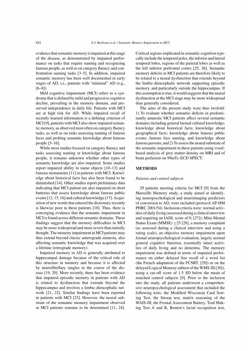
MCI patients performed significantly more poorlythan control subjects (raw scores: m = 13.86, SD = 5.91versus m = 22.48, SD = 4.29, p < 0.001, Cohen’sd = 1.65, Fig. 2A). Results between groups using scaledscores were very similar.
Because the questions on this subtest are supposedto be increasingly difficult, and because we were inter-ested in assessing whether MCI patients would alsobe impaired on “easy” questions, we assessed whether

828 E.J. Barbeau et al. / Semantic Memory Impairment in MCI
Fig. 1. A) Box plot of the distribution of the famous faces naming scores in MCI patients and control subjects. B) Famous faces identificationscore. C) Naming scores according to the period of famousness. Numbers for each period are Cohen’s d. ***p < 0.001, **p < 0.01.
there was a significant difference between controls andMCI subjects for each question. The results indicatethat MCI patients were impaired on most of the ques-tions (See plot, Fig. 2B). We defined “easy” questionsas being correctly answered by more than 90% of con-trol subjects, which seems a reasonable assumptiongiven that it means that only two controls out of 29 wereunable to answer the question. Five questions met thiscriterion (Q8, Q12, Q13, Q14, Q16). The mean numberof control subjects succeeding on these questions was94% (SD = 2%). By contrast, the mean number of MCIpatients succeeding on these same questions was only50% (SD = 7%), indicating difficulties of MCI patientseven on these “easy” questions.
History and geography didactic acquisitionquestionnaire (DAQ)
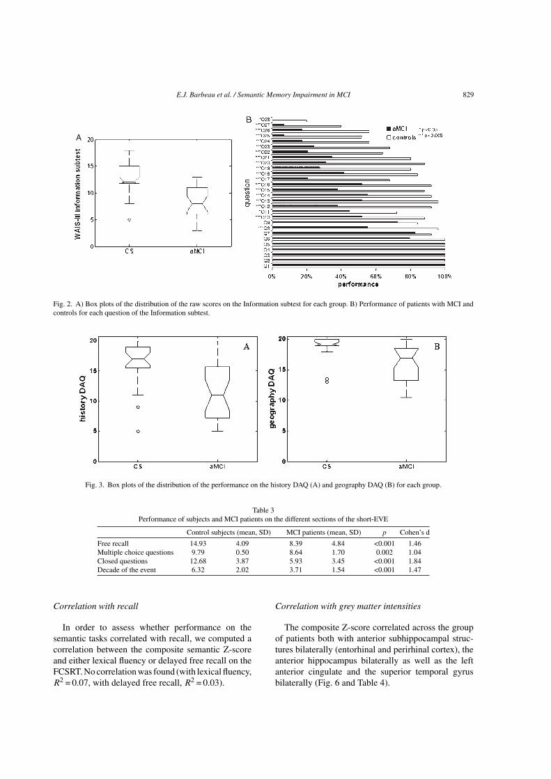
Patients with MCI performed significantly worsethan control subjects on the history DAQ (m = 11.96,SD = 4.75 versus m = 16.54, SD = 3.65, p < 0.01,Cohen’s d = 1.08, Fig. 3A) as well as on the geographyDAQ (m = 15.96, SD = 3.04 versus 19.02, SD = 1.73,p < 0.001, Cohen’s d = 1.24, Fig. 3B).
French public events battery (short-EVE)
We first compared the performance between groupson the short-Eve by averaging scores for all sub-tests. Patients with MCI performed well below controlsubjects (m = 26.68, SD = 10.14 versus m = 43.71,SD = 9.17, p < 0.001, Cohen’s d = 1.76, Fig. 4A). It isnoteworthy that the patients performed significantlybelow control subjects on all individual measures(Table 3), including when they were asked to choose acorrect answer among three in a multiple choice format(Fig. 4B).
The patients’ performance significantly differedfrom that of controls regardless of the decade consid-ered, even for the oldest events (Fig. 4C). Analyses ofCohen’s d indicated that the impairment was similaracross all decades except for the most recent period,which was most impaired.
Semantic memory composite Z-score
Figure 5 plots the semantic memory compositeZ-score at baseline for each subject within each group.
E.J. Barbeau et al. / Semantic Memory Impairment in MCI 829
A
Fig. 2. A) Box plots of the distribution of the raw scores on the Information subtest for each group. B) Performance of patients with MCI andcontrols for each question of the Information subtest.
Fig. 3. Box plots of the distribution of the performance on the history DAQ (A) and geography DAQ (B) for each group.
Table 3Performance of subjects and MCI patients on the different sections of the short-EVE
Control subjects (mean, SD) MCI patients (mean, SD) p Cohen’s d
Free recall 14.93 4.09 8.39 4.84 <0.001 1.46Multiple choice questions 9.79 0.50 8.64 1.70 0.002 1.04Closed questions 12.68 3.87 5.93 3.45 <0.001 1.84Decade of the event 6.32 2.02 3.71 1.54 <0.001 1.47
Correlation with recall
In order to assess whether performance on thesemantic tasks correlated with recall, we computed acorrelation between the composite semantic Z-scoreand either lexical fluency or delayed free recall on theFCSRT. No correlation was found (with lexical fluency,R2 = 0.07, with delayed free recall, R2 = 0.03).
Correlation with grey matter intensities
The composite Z-score correlated across the groupof patients both with anterior subhippocampal struc-tures bilaterally (entorhinal and perirhinal cortex), theanterior hippocampus bilaterally as well as the leftanterior cingulate and the superior temporal gyrusbilaterally (Fig. 6 and Table 4).

830 E.J. Barbeau et al. / Semantic Memory Impairment in MCI
Fig. 4. Performance on the short-EVE questionnaire assessing knowledge about famous public events. A) Total performance on the short EVE ofthe group of control subjects vs the group of patients with MCI. B) Performance of both groups on the multiple choice questions. C) Performanceof both groups across decades. Numbers represent Cohen’s d values. ***p < 0.001.
Fig. 5. Dispersion of the semantic memory composite Z-scores ineach group. Each circle represents one subject. Note that 5 controlsubjects appear to have poor semantic memory.
Correlation with SPECT perfusion
The semantic memory composite Z-score corre-lated with SPECT perfusion in left medial temporallobe structures comprising both a cluster in the
hippocampus (Fig. 7A, Table 5) and a cluster inentorhinal/perirhinal cortices (BA20/BA36, Fig. 7B).
Follow-up
MCI patients were followed longitudinally at 18months and 36 months so that conversion to proba-ble AD could be assessed. Out of the 29 patients, 12converted to probable AD after either 18 or 36 month.Hence, two subgroups of patients were compared, onegroup comprising patients who did not convert duringthis follow-up (“non-converters”) and the other com-prising those who converted (“converters”). Converterswere slightly older than non-converters (m = 70.00,SD = 5.70 versus 68.24, SD = 7.27, p = 0.04) but didnot differ in terms of years of education (m = 12.42,SD = 4.27 versus m = 13.71, SD = 4.00, p = 0.51). Theydid not differ either in terms of their general neuropsy-chological assessment, except for the lexical fluencytask (Table 6). In particular, comparison of eitherdelayed free recall or efficiency of delayed cueing onthe Free and Cued Selective Reminding Test indicatedthat the two subgroups did not differ on this memorytest.

E.J. Barbeau et al. / Semantic Memory Impairment in MCI 831
1
0
2
3
4
Fig. 6. A) Correlations within medial temporal lobe structures between the semantic memory composite Z-score and grey matter intensities foreach voxel within the group of patients. B) Correlations with the left anterior cingulate.
Table 4Regions showing a significant correlation between the semanticmemory composite Z-score and grey matter intensities within thegroup of patients. x, y, z = Talairach coordinates. BA: BrodmannArea. MFG: Middle frontal gyrus, MTG: Middle temporal gyrus,
STG: superior frontal gyrus, L: left, R: Right
t-score Talairach coordinates LocalizationX y Z
4.57 −2 29 31 L anteriorcingulate
BA 32
4.5 −22 −13 −28 L entorhinal/perirhinal
BA 28/36
4.24 24 −6 −30 R perirhinal BA 364.13 30 −25 −6 R entorhinal BA 284.1 −22 2 −21 L entorhinal BA 284.05 51 13 32 R MFG BA 93.09 29 −2 −38 R Uncus BA 203.08 33 −13 −19 R entorhinal BA 283.7 38 6 −17 R STG BA 383.7 −35 13 −25 L STG BA 38
All analyses that were performed on semantictasks were carried out again and performance ofconverters was compared with that of non-converter.Virtually no difference was found. Direct compar-isons of the semantic memory composite Z-scorebetween both groups did not yield any significantdifference (non-converters’ mean = −1.38, SD = 1.03;converters’ mean = −1.87, SD = 1.03, p = 0.24).
Neuroimaging correlation analyses were also carriedout within each group, but no correlations were found,probably because of reduced sample size.
DISCUSSION
In this study, we found that the defining feature ofMCI, impaired anterograde memory, is accompaniedby an extensive impairment in the realm of semanticmemory, affecting various areas of knowledge (famousfaces, cultural knowledge, historical facts, geograph-ical facts, famous public events). This suggests thatthe semantic memory disorder is more widespread andsevere than previously documented. In addition, thelack of a temporal gradient in the semantic memory losssuggests that all periods of knowledge are affected, notonly the more recent ones. This makes it unlikely thatanterograde amnesia underlies the semantic deficits,since this would have lead to a gradient with bet-ter knowledge on remote compared with more recentdecades. Neuroimaging analyses using grey matterdensity and blood-flow measures suggest that impairedsemantic knowledge in these patients is related to dys-function within anterior, extra-hippocampal, temporallobe structures (entorhinal and perirhinal cortices) andthe hippocampus.

832 E.J. Barbeau et al. / Semantic Memory Impairment in MCI
0
1
2
3
4
0
1
2
3
4
Fig. 7. Correlations between the semantic memory composite Z-score and peaks of significant SPECT rCBF (p < 0.001 uncorrected) within thegroup of patients. A) Correlation with the hippocampus. B) correlation with the medial temporal pole (BA28/BA36). L: left hemisphere.
Table 5Talairach coordinates of significant SPECT rCBF findings (p-voxel< 0.001, uncorrected) within the group of patients. x, y and z are the
Talairach coordinates (mm). BA = Brodmann area
t-score Talairach coordinates Localizationx y Z
3.83 −30 −1 −27 Left Perirhinal/Entorhinal Cortex(BA36/BA28)
3.60 −36 −20 −12 Left Hippocampus
Table 6Neuropsychological performance on standard tests of converters vs.
non-converters (mean, SD in brackets)
Non-converters Converters p
MMSE 27.18 (1.19) 27.25 (1.14) P = 0.84Lexical fluency “P” in
2 min20.88 (5.00) 16.08 (5.85) P = 0.03
FCSR test-delayedfree recall
5.00 (3.12) 3.58 (2.43) P = 0.13
FCSR test-delayedcued recall
7.82 (2.38) 7.25 (2.67) P = 0.64
WAIS-III digit span 9.53 (2.79) 10.00 (3.44) P = 0.75WAIS-III Matrix
reasonning10.12 (2.34) 9.67 (1.87) P = 0.68
Confrontation naming(DO80)
79.35 (1.17) 79.42 (1.38) P = 0.69
Evidence for a widespread semantic impairment inMCI
The present study confirms previous findings thatMCI patients are impaired on tests of naming famous
faces and famous person knowledge (e.g., [8, 10,12, 13, 15, 17, 24, 35, 39, 40]). The current studyadditionally indicates that other domains of seman-tic memory are impaired. This is the case for generalfactual knowledge, as assessed with the standardInformation subtest of the WAIS-III [34]. Such knowl-edge is largely context-free and does not rely onepisodic memory. Individuals are regularly exposedto this type of culturally-shared knowledge in every-day conversations in the context of their social andprofessional activities, media exposure, or hobbies.However, MCI patients were impaired on most ques-tions, even the easy ones. We also found MCI patientsto have impaired knowledge about history and geog-raphy. Since all subjects were exposed to this typeof knowledge under similar conditions during formaleducation at school, the period of acquisition of thisknowledge can clearly be dated and dates back sev-eral decades before the assessment (acquired duringchildhood), suggesting that even remote knowledge isaffected, confirming the findings of a previous study[14]. A further domain of semantic memory that wasimpaired in MCI patients was knowledge concerningfamous public events. Such knowledge is shared bymany people and repeatedly presented in newspapers,TV news, and in some instances, in ensuing TV doc-umentaries or movies. Knowledge about such eventswas impaired in MCI regardless of test format, usingfree recall, closed questions, recognition or dating, sup-porting recent preliminary data [15]. In summary, thesemantic memory impairment in MCI patients covers

E.J. Barbeau et al. / Semantic Memory Impairment in MCI 833
multiple domains of knowledge, multiple periods, andis severe, such as indicated by effect sizes that werewell above 1 (Cohen’s d).
It is noteworthy that the MCI patients were impairedat the recognition subtest of the public event task,which does not rely on recall (Fig. 4B). In addition, allsubjects were systematically told the correct answerfollowing this procedure, even if they had answeredcorrectly. Despite this cue, MCI patients performedpoorly compared to control subjects on the subse-quent questions (Cohen’s d = 1.84). Such results areusually interpreted as a genuine memory impairment[41]. Also, the lack of correlation between the com-posite semantic Z-score and lexical fluency or delayedfree recall on the FCSRT suggests that the semanticimpairment found in the current study is unlikely to berelated to impaired recall. Taken together, these resultsprovide converging evidence to support the notion thatthe semantic memory impairment in MCI patients maybe due to impairment at the level of the semantic store.
Finally, it is worth mentioning that findings fromprevious studies suggest that semantic aspects ofautobiographical memory, (e.g., one’s own teacher’snames) are preserved in MCI patients for both child-hood and early adulthood [42, 43]. While this mayappear to be in contradiction with the findings ofthe present study, this could alternatively be relatedto a dissociation between semantic memory for per-sonal facts (also depending on representations fromepisodic memory), and semantic memory for generalfacts, which may not rely on the same neural systems(e.g., [44]).
Is there a temporal gradient in the semanticmemory impairment?
Although a gradient for autobiographical episodicmemory has been reported at the dementia stage ofAD [6, 45], there is evidence that there may be no gra-dient for semantic memory [7, 8]. For example, Greeneand Hodges [7], did not find a gradient for knowl-edge about famous faces at the dementia stage of AD,since all periods of knowledge (over five decades) wereimpaired.
This issue has rarely been investigated in MCIpatients. Seidenberg et al. [46] emphasized that MCIpatients show impaired naming, reaction time, andknowledge about names of people who became famousrecently (in the 1990–2003 period). Although thiswas attributed to impaired anterograde memory, it isnoteworthy that knowledge for people who had beenfamous at a more remote period of time (during the
1950–1965 period) and whose fame did not last after-ward was also impaired in this study. This absenceof a gradient for retrograde knowledge would there-fore be consistent with what is found at the dementiastage of AD. Likewise, no temporal gradient for eitherthe “famous faces” or the “public events” task in MCIpatients was found in the present study, as all periods ofknowledge up to the most remote ones were impaired,compared to controls. Even the most remote knowl-edge acquired at school was impaired as assessed withtasks assessing knowledge on history and geography.Of note, control subjects did not show any gradientin our study (i.e., they showed a rather flat curve), ashas previously been reported for either famous facesor public events [16, 47]. Taken together, these resultssupport the idea that there is no gradient in seman-tic memory in MCI patients. The only exception wasfor the most recent decade (90 s-00 s) of public events,which was more severely impaired, possibly as a resultof an anterograde memory impairment, as has beenpreviously suggested [46].
The standard Consolidation theory and the MultipleTrace Theory [48] both predict that the hippocam-pus plays a crucial role for the acquisition of newsemantic information during a limited period of time,after which semantic knowledge increasingly dependson adjacent neocortical structures acting as semanticmemory stores. Consequently, lesions of the hip-pocampus should lead to a temporal gradient, whilelesions of the hippocampus and the adjacent neocor-tex should lead to an impairment of all periods of time.The findings of the present study may therefore provideadditional evidence that the impairment of semanticmemory stores may be independent from hippocampaldysfunction in MCI patients.
What is the neural substrate of the semanticimpairment in MCI?
Using correlations of a semantic memory com-posite Z-score with grey matter density on MRI, aswell as regional blood flow using SPECT, we foundthat the neural substrate of semantic memory lossinvolves anterior mesiotemporal areas, including theanterior portion of the hippocampus as well as adja-cent structures. Precisely, on MRI the correlations werebilateral for the perirhinal/entorhinal cortices but onlyconcerned the right hippocampus. On PET, only a cor-relation with the left perirhinal/entorhinal cortices andthe left hippocampus was found. These areas are partof a ventral mesiotemporal pathway interlinking theanterior hippocampus with extra-hippocampal areas

834 E.J. Barbeau et al. / Semantic Memory Impairment in MCI
recently identified in high resolution functional con-nectivity studies in the human brain using fMRI [49].Moreover, this network has recently been shown tohave a functional relationship with context-free object-based memory [50]. This network includes the rhinalregion (perirhinal/entorhinal cortices), the anterior lat-eral temporal lobe, the middle temporal gyrus, and thehead of the hippocampus. Therefore, the findings ofa correlation with cortical areas adjacent to the hip-pocampus appear consistently related to a functionalnetwork. These areas are located anterior and underthe head of the hippocampus [51]. In addition, thecomposite Z-score correlated with grey matter densityin the left anterior cingulate (BA32) and in the rightdorso-lateral prefrontal cortex (BA9).
These results are consistent with recent imagingstudies of healthy individuals which report hippocam-pal activation using fMRI during semantic retrieval[52, 53], particularly on the left. Interestingly, a rightdorso-medial area (BA8), adjacent to the area BA9reported in our study, was also involved in semanticretrieval [52]. Burianova and colleagues [54] tried toidentify the network of brain areas that was commonto autobiographical, episodic (lab tests), and semanticmemory retrieval. The anterior cingulate was identi-fied as one of these regions and its activity was foundto correlate with that of the hippocampus. Overall, thebrain areas that were correlated with performance onthe semantic memory composite Z-score appear to beinvolved in a network of brain areas engaged in declar-ative memory.
We also found correlations between semantic mem-ory and the perirhinal/entorhinal cortices. Only twoprevious studies investigated such correlations. Aresult implying a region more lateral to the one foundin our study has been reported recently in patients withMCI using grey matter density and ROI analyses [13].A study in patients with very mild AD (MMSE = 23)likewise identified correlations with anterior temporallobe structures along the parahippocampal gyrus usinga detailed analysis of a lexical fluency task [55]. Over-all, there is therefore converging evidence that changeswithin anterior mesial temporal areas, adjacent to thehippocampus, contribute to impaired semantic mem-ory in MCI patients.
These structures, i.e., the temporal pole, regionswithin the anterior collateral sulcus and the anteriorfusiform gyrus, have repeatedly been suggested to bea crucial site for the representation of semantic rep-resentations at an abstract and amodal level [56–59].A region slightly more lateral to that reported in thepresent study has been identified as a critical node
among three regions that appear to be consistentlyinvolved in semantic retrieval [25]. Among these threeregions, this temporal area is proposed to be involvedin semantic knowledge storage. The present findingsare also consistent with previous studies that exploredthe neural basis of context-free memory or long-termknowledge representations in patients with semanticdementia, which suggested that the rhinal region maybe a central area for the storage of long-term knowledgerepresentations [33, 60]. Interestingly, a recent studywhich investigated the neural correlate of semanticimpairment in patients with semantic dementia iden-tified areas in the anterior (bilateral) fusiform gyrus“subjacent to the head and body of the hippocampus”,a pattern that closely resembles the changes reported inthe present study [51]. This same region has also beenshown to be involved in context-free and single-itemmemory in MCI [61–63]. Importantly, this region isconsistently affected by neuropathological lesions inpatients with MCI who display AD [64, 65].
Overall, our findings of a semantic impairmentrelated to anterior subhippocampal structures thusappear remarkably convergent with previous studies inMCI and with studies on semantic memory in healthysubjects. While it was suggested that the interactionbetween the rhinal region and the temporopolar cortexis the main target of pathology in semantic demen-tia [60], the main target of neurofibrillary tangles inAD is thought to be the interaction between the rhinalregion and the hippocampus in the so-called transen-torhinal cortex [19, 66]. We here provide evidence thatimpaired interaction between the hippocampus and therhinal region in MCI patients not only plays a role inthe acquisition of new knowledge, but also in long-termrepresentation of such knowledge.
Semantic impairment in converters versusnon-converters
There was no clear difference between patientswho converted to AD over the 36 months follow-up period and patients who did not convert over thisperiod of time on the neuropsychological assessment,suggesting that the population of MCI patients wasoverall homogenous. It is likely that many patientswho did not convert over the comparatively shortfollow-up period may in fact have AD, and will con-vert to dementia later. The rate of progression fromMCI to dementia of 41% over 36 month appearscomparable to that of other studies, but some MCIpatients have been shown to convert over longer peri-ods of up to 72 month or more [67]. In addition, the

E.J. Barbeau et al. / Semantic Memory Impairment in MCI 835
inclusion of patients mainly meeting criteria for amnes-tic MCI could lead to an overrepresentation of patientswith AD due to focal mesial temporal lobe dysfunction[68] and pure progressive amnesia [69–71], with veryslow progression. This may explain why there was lit-tle difference between converters and non-convertersregarding semantic tasks, but also regarding otherneuropsychological tests including those that assessepisodic memory.
CONCLUSION
To sum up, we found a significant and widespreadimpairment in the realm of semantic memory in a groupof patients with MCI, related to dysfunction of brainareas beyond the classic limbic-diencephalic networkinvolved in episodic memory. The memory impairmentat the predementia stage of AD is therefore not limitedto episodic memory and the limbic-diencephalic sys-tem as sometimes assumed. Furthermore, the relativeseverity of the semantic memory impairment observedin this group of patients at an early stage of MCI(slow conversion) suggests, in accordance with otherstudies [72], that this impairment began insidiouslymany years before patients first consulted, and possi-bly during the transentorhinal stage in preclinical AD[73]. Semantic memory impairment may therefore bea defining characteristic of very early AD, in additionto the episodic memory impairment.
ACKNOWLEDGMENTS
This study was supported by AP-HM PHRC2001/54 and France Alzheimer. SJ is supported bythe Alzheimer Society of Canada (ASC) and holds aChercheur-Boursier Junior 2 award from the Fonds derecherche en sante du Quebec (FRSQ). We would liketo thank Michele Balzamo, Marie-Luce Royere andCaroline Latger for their contribution in patient selec-tion and neuropsychological assessments, Julie Pelatfor data management, as well as the patients and controlsubjects for their participation in this study.
Authors’ disclosures available online (http://www.j-alz.com/disclosures/view.php?id=1040).
REFERENCES
[1] Joubert S, Joncas S, Barbeau E, Joannette Y, Ska B(2007) Cognition. In: Clinical diagnosis and managementof Alzheimer’s disease. 3rd edition, Gauthier S ed., MartinDunitz & Parthenon publishing, pp. 165-176.
[2] Salmon DP, Bondi MW (2008) Neuropsychological assess-ment of dementia. Annu Rev Psychol 60, 257-282.
[3] Chertkow H, Bub D, Seidenberg M (1989) Priming andsemantic memory loss in Alzheimer’s disease. Brain Lang36, 420-446.
[4] Hodges JR, Patterson K (1995) Is semantic memory consis-tently impaired early in the course of Alzheimer’s disease?Neuroanatomical and diagnostic implications. Neuropsy-chologia 33, 441-459.
[5] Hodges JR, Patterson K (1997) Semantic memory disorders.Trends Cogn Sci 1, 68-72.
[6] Greene JD, Hodges JR (1996) The fractionation of remotememory. Evidence from a longitudinal study of dementia ofAlzheimer type. Brain 119(Pt 1), 129-142.
[7] Greene JD, Hodges JR (1996) Identification of famous facesand famous names in early Alzheimer’s disease. Relationshipto anterograde episodic and general semantic memory. Brain119(Pt 1), 111-128.
[8] Thompson SA, Graham KS, Patterson K, Sahakian BJ,Hodges JR (2002) Is knowledge of famous peopledisproportionately impaired in patients with early and ques-tionable Alzheimer’s disease? Neuropsychology 16, 344-358.
[9] Petersen R, Doody R, Kurz A, Mohs R, Morris J, RabinsP, Ritchie K, Rossor M, Thal L, Winblad B (2001) Currentconcepts in mild cognitive impairment. Arch Neurol 58, 1985-1992.
[10] Adlam AL, Bozeat S, Arnold R, Watson P, Hodges JR (2006)Semantic knowledge in mild cognitive impairment and mildAlzheimer’s disease. Cortex 42, 675-684.
[11] Ahmed S, Arnold R, Thompson SA, Graham KS, HodgesJR (2008) Naming of objects, faces and buildings in mildcognitive impairment. Cortex 44, 746-752.
[12] Joubert S, Felician O, Barbeau EJ, Didic M, Poncet M, Cec-caldi M (2008) Patterns of semantic memory impairment inMild Cognitive Impairment. Behav Neurol 19, 35-40.
[13] Joubert S, Brambati SM, Ansado J, Barbeau EJ, FelicianO, Didic M, Lacombe J, Goldstein R, Chayer C, KergoatMJ (2010) The cognitive and neural expression of semanticmemory impairment in mild cognitive impairment and earlyAlzheimer’s disease. Neuropsychologia 48, 978-988.
[14] Leyhe T, Muller S, Eschweiler GW, Saur R (2010) Deteri-oration of the memory for historic events in patients withmild cognitive impairment and early Alzheimer’s disease.Neuropsychologia 48, 4093-4101.
[15] Cuetos F, Rodriguez-Ferreiro J, Menendez M (2009) Seman-tic markers in the diagnosis of neurodegenerative dementias.Dement Geriatr Cogn Disord 28, 267-274.
[16] Thomas-Anterion C, Collomb K, Borg C, Nevers B, LaurentB (2006) (Evaluation of memory for French public events:EVE-30 in 108 controls, 10 mild cognitively impaired and 10Alzheimer’s disease patients). Rev Neurol (Paris) 162, 1232-1239.
[17] Vogel A, Gade A, Stokholm J, Waldemar G (2005) Seman-tic memory impairment in the earliest phases of Alzheimer’sdisease. Dement Geriatr Cogn Disord 19, 75-81.
[18] Thomas-Anterion C, Borg C, Basaglia-Pappas S, Laroche L,Minvielle B, Bedoin N (2010) (Semantic knowledge in MCIand Alzheimer’s disease: The French version of the NewWords Interview). Rev Neurol (Paris) 166, 419-427.
[19] Braak H, Braak E (1991) Neuropathological stageing ofAlzheimer-related changes. Acta Neuropathol (Berlin) 82,239-259.
[20] Mortimer JA, Gosche KM, Riley KP, Markesbery WR, Snow-don DA (2004) Delayed recall, hippocampal volume andAlzheimer neuropathology: Findings from the Nun Study.Neurology 62, 428-432.

836 E.J. Barbeau et al. / Semantic Memory Impairment in MCI
[21] Desgranges B, Baron JC, Lalevee C, Giffard B, Viader F, de LaSayette V, Eustache F (2002) The neural substrates of episodicmemory impairment in Alzheimer’s disease as revealed byFDG-PET: Relationship to degree of deterioration. Brain 125,1116-1124.
[22] Nestor PJ, Fryer TD, Hodges JR (2006) Declarative memoryimpairments in Alzheimer’s disease and semantic dementia.Neuroimage 30, 1010-1020.
[23] Poettrich K, Weiss PH, Werner A, Lux S, Donix M, Gerber J,von Kummer R, Fink GR, Holthoff VA (2009) Altered neuralnetwork supporting declarative long-term memory in mildcognitive impairment. Neurobiol Aging 30, 284-298.
[24] Dudas RB, Clague F, Thompson SA, Graham KS, HodgesJR (2005) Episodic and semantic memory in mild cognitiveimpairment. Neuropsychologia 43, 1266-1276.
[25] Jefferies E, Lambon Ralph MA (2006) Semantic impairmentin stroke aphasia versus semantic dementia: A case-seriescomparison. Brain 129, 2132-2147.
[26] de Zubicaray GI, Rose SE, McMahon KL (2010) The structureand connectivity of semantic memory in the healthy olderadult brain. Neuroimage 54, 1488-1494.
[27] Lawton M, Brody E (1969) Assessment of older people:Self-maintaining and instrumental activities of daily living.Gerontologist 9, 179-186.
[28] Folstein M, Folstein S, McHugh P (1975) “Mini-mentalstate”. A practical method for grading the cognitive state ofpatients for the clinician. J Psychiatr Res 12, 189-198.
[29] Ergis A-M, Van der Linden M, Deweer B (1994) Investigationof memory performance with a cued recall test in Alzheimer’sdisease. Rev Neuropsychol 4, 47-68.
[30] Wechsler D (2001) Echelle clinique de memoire de WechslerMEM-III (WMS-III), Les editions du Centre de Psychologieappliquee, Paris.
[31] Loeb C, Gandolfo C (1983) Diagnostic evaluation of degen-erative and vascular dementia. Stroke 14, 399-401.
[32] McKhann G, Drachman D, Folstein M, Katzman R, PriceD, Stadlan E (1984) Clinical diagnosis of Alzheimer’s dis-ease: Report of the NINCDS-ADRDA Work Group under theauspices of Department of Health and Human Services TaskForce on Alzheimer’s Disease. Neurology 34, 939-944.
[33] Joubert S, Felician O, Barbeau E, Ranjeva JP, Christophe M,Didic M, Poncet M, Ceccaldi M (2006) The right tempo-ral lobe variant of frontotemporal dementia: Cognitive andneuroanatomical profile of three patients. J Neurol 253, 1447-1458.
[34] Wechsler D (2000) Echelle d’Intelligence de Wechsler pourAdulte III (WAIS-III), Les editions du Centre de Psychologieappliquee, Paris.
[35] Thomas-Anterion C, Jacquin K, Laurent B (2000) Differentialmechanisms of impairment of remote memory in Alzheimer’sand frontotemporal dementia. Dement Geriatr Cogn Disord11, 100-106.
[36] Zakzanis KK (2001) Statistics to tell the truth, the whole truth,and nothing but the truth: Formulae, illustrative numericalexamples, and heuristic interpretation of effect size analysesfor neuropsychological researchers. Arch Clin Neuropsychol16, 653-667.
[37] Cohen J (1988) Statistical Power Analysis for the BehavioralSciences (second ed.), Lawrence Erlbaum Associates.
[38] Deloche G, Hannequin D (1997) Test de denomination oraled’images DO80, Les Editions du Centre de Psychologieappliquee, Paris.
[39] Estevez-Gonzalez A, Garcia-Sanchez C, Boltes A, Otermin P,Pascual-Sedano B, Gironell A, Kulisevsky J (2004) Semanticknowledge of famous people in mild cognitive impairment
and progression to Alzheimer’s disease. Dement Geriatr CognDisord 17, 188-195.
[40] Borg C, Thomas-Anterion C, Bogey S, Davier K, Laurent B(2010) Visual imagery processing and knowledge of famousnames in Alzheimer’s disease and MCI. Neuropsychol DevCogn B Aging Neuropsychol Cogn 17, 603-614.
[41] Grober E, Buschke H, Crystal H, Bang S, Dresner R (1988)Screening for dementia by memory testing. Neurology 38,900-903.
[42] Murphy KJ, Troyer AK, Levine B, Moscovitch M (2008)Episodic, but not semantic, autobiographical memory isreduced in amnestic mild cognitive impairment. Neuropsy-chologia 46, 3116-3123.
[43] Leyhe T, Muller S, Milian M, Eschweiler GW, Saur R(2009) Impairment of episodic and semantic autobiograph-ical memory in patients with mild cognitive impairmentand early Alzheimer’s disease. Neuropsychologia 47, 2464-2469.
[44] Levine B, Turner GR, Tisserand D, Hevenor SJ, GrahamSJ, McIntosh AR (2004) The functional neuroanatomy ofepisodic and semantic autobiographical remembering: Aprospective functional MRI study. J Cogn Neurosci 16, 1633-1646.
[45] Piolino P, Desgranges B, Belliard S, Matuszewski V, LaleveeC, De La Sayette V, Eustache F (2003) Autobiographicalmemory and autonoetic consciousness: Triple dissociation inneurodegenerative diseases. Brain 126, 2203-2219.
[46] Seidenberg M, Guidotti L, Nielson KA, Woodard JL, Durg-erian S, Zhang Q, Gander A, Antuono P, Rao SM (2009)Semantic knowledge for famous names in mild cognitiveimpairment. J Int Neuropsychol Soc 15, 9-18.
[47] Piolino P, Lamidey V, Desgranges B, Eustache F (2007)The semantic and episodic subcomponents of famous personknowledge: Dissociation in healthy subjects. Neuropsychol-ogy 21, 122-135.
[48] Moscovitch M, Nadel L, Winocur G, Gilboa A, RosenbaumRS (2006) The cognitive neuroscience of remote episodic,semantic and spatial memory. Curr Opin Neurobiol 16, 179-190.
[49] Kahn I, Andrews-Hanna JR, Vincent JL, Snyder AZ, Buck-ner RL (2008) Distinct cortical anatomy linked to subregionsof the medial temporal lobe revealed by intrinsic functionalconnectivity. J Neurophysiol 100, 129-139.
[50] Gour N, Ranjeva JP, Caccaldi M, Confort-Gouny S, BarbeauE, Soulier E, Guye M, Didic M, Felician O’ (2011) Basalfunctional connectivity within the anterior temporal networkis associated with performance on declarative memory tasks.Neuroimage 58, 687-697.
[51] Mion M, Patterson K, Acosta-Cabronero J, Pengas G,Izquierdo-Garcia D, Hong YT, Fryer TD, Williams GB,Hodges JR, Nestor PJ (2010) What the left and right ante-rior fusiform gyri tell us about semantic memory. Brain 133,3256-3268.
[52] Burianova H, Grady CL (2007) Common and unique neu-ral activations in autobiographical, episodic, and semanticretrieval. J Cogn Neurosci 19, 1520-1534.
[53] Ryan L, Cox C, Hayes SM, Nadel L (2008) Hippocampalactivation during episodic and semantic memory retrieval:Comparing category production and category cued recall.Neuropsychologia 46, 2109-2121.
[54] Burianova H, McIntosh AR, Grady CL (2010) A commonfunctional brain network for autobiographical, episodic, andsemantic memory retrieval. Neuroimage 49, 865-874.
[55] Venneri A, McGeown WJ, Hietanen HM, Guerrini C, EllisAW, Shanks MF (2008) The anatomical bases of semantic

E.J. Barbeau et al. / Semantic Memory Impairment in MCI 837
retrieval deficits in early Alzheimer’s disease. Neuropsycholo-gia 46, 497-510.
[56] Patterson K, Nestor PJ, Rogers TT (2007) Where do you knowwhat you know? The representation of semantic knowledgein the human brain. Nat Rev Neurosci 8, 976-987.
[57] Vargha-Khadem F, Gadian DG, Watkins KE, Connelly A, VanPaesschen W, Mishkin M (1997) Differential effects of earlyhippocampal pathology on episodic and semantic memory.Science 277, 376-380.
[58] Barbeau EJ, Taylor MJ, Regis J, Marquis P, Chauvel P,Liegeois-Chauvel C (2008) Spatio temporal dynamics of facerecognition. Cereb Cortex 18, 997-1009.
[59] Brambati SM, Benoit S, Monetta L, Belleville S, Joubert S(2010) The role of the left anterior temporal lobe in the seman-tic processing of famous faces. Neuroimage 53, 674-681.
[60] Davies RR, Graham KS, Xuereb JH, Williams GB, HodgesJR (2004) The human perirhinal cortex and semantic memory.Eur J Neurosci 20, 2441-2446.
[61] Eustache F, Desgranges B, Giffard B, de la Sayette V, BaronJC (2001) Entorhinal cortex disruption causes memory deficitin early Alzheimer’s disease as shown by PET. Neuroreport12, 683-685.
[62] Guedj E, Barbeau E, Didic M, Felician O, de Laforte C,Ceccaldi M, Mundler O, Poncet M (2006) Identification ofsubgroups in amnestic mild cognitive impairment. Neurology67, 356-358.
[63] Barbeau EJ, Ranjeva JP, Didic M, Confort-Gouny S, FelicianO, Soulier E, Cozzone PJ, Ceccaldi M, Poncet M (2008) Pro-file of memory impairment and gray matter loss in amnesticmild cognitive impairment. Neuropsychologia 46, 1009-1019.
[64] Delacourte A, David J, Sergeant N, Buee L, Wattez A, Ver-mersch P, Ghozali F, Fallet-Bianco C, Pasquier F, Lebert F,Petit H, Di Menza C (1999) The biochemical pathway of neu-rofibrillary degeneration in aging and Alzheimer’s disease.Neurology 52, 1158-1165.
[65] Mitchell TW, Mufson EJ, Schneider JA, Cochran EJ, Nis-sanov J, Han LY, Bienias JL, Lee VM, Trojanowski JQ,Bennett DA, Arnold SE (2002) Parahippocampal tau pathol-ogy in healthy aging, mild cognitive impairment, and earlyAlzheimer’s disease. Ann Neurol 51, 182-189.
[66] Van Hoesen G, Hyman B, Damasio A (1991) Entorhinal cor-tex pathology in Alzheimer’s disease. Hippocampus 1, 1-8.
[67] Hodges JR, Erzinclioglu S, Patterson K (2006) Evolutionof cognitive deficits and conversion to dementia in patientswith mild cognitive impairment: A very-long-term follow-upstudy. Dement Geriatr Cogn Disord 21, 380-391.
[68] Butters MA, Lopez OL, Becker JT (1996) Focal temporal lobedysfunction in probable Alzheimer’s disease predicts a slowrate of cognitive decline. Neurology 46, 687-692.
[69] Didic M, Ali Cherif A, Gambarelli D, Poncet M,Boudouresques J (1998) A permanent pure amnestic syn-drome of insidious onset related to Alzheimer’s disease. AnnNeurol 43, 526-530.
[70] Caselli RJ, Couce ME, Osborne D, Deen HG, Parisi JP (1998)From slowly progressive amnesic syndrome to rapidly pro-gressive Alzheimer disease. Alzheimer Dis Assoc Disord 12,251-253.
[71] Barbeau EJ, Didic M, Felician O, Tramoni E, Guedj E, Cecca-ldi M, Poncet M (2006) Pure progressive amnesia: An atypicalamnestic syndrome? Cogn Neuropsychol 23, 1230-1247.
[72] Amieva H, Le Goff M, Millet X, Orgogozo JM, Peres K,Barberger-Gateau P, Jacqmin-Gadda H, Dartigues JF (2008)Prodromal Alzheimer’s disease: Successive emergence of theclinical symptoms. Ann Neurol 64, 492-498.
[73] Didic M, Barbeau E, Felician O, Tramoni E, Guedj E, PoncetM, Ceccaldi M (2011) Which memory system is impaired firstin AD? J Alzheimers Dis 27, 11-22.