![Superiority of [68Ga]-DOTATATE PET/CT to other functional imaging … · Room 1E-3140, Bethesda, MD, 20892 2Nuclear Medicine Department, Bichat Hospital, 46 Rue Henri Huchard, 75018](https://static.fdocuments.in/doc/165x107/5f0dc77b7e708231d43c0b62/superiority-of-68ga-dotatate-petct-to-other-functional-imaging-room-1e-3140.jpg)
Sandostatin LAR therapy differentially alters 68Ga...
19
Original Article Sandostatin LAR therapy differentially alters 68 Ga-DOTATATE uptake in normal tissues compared to primary tumors and metastatic lesions Narjess Ayati 1 ,MD; Sze Ting Lee 2 , MD; Rasoul Zakavi 1 ,MD; Kunthi Pathmaraj 2 ,Loai Qatawneh 3 , MD; Aurora Poon 2 , MD, Andrew M. Scott 2,4,5 ,MD 1. Nuclear Medicine Research Centre, Mashhad University of Medical Sciences, Mashhad, Iran 2. Department of Molecular Imaging & Therapy, Austin Health, Heidelberg, Victoria, Australia 3. Nuclear Medicine and Cyclotron Unit, King Hussein Medical Center, Jordanian Royal Medical Services, Amman, Jordan 4. Olivia Newton-John Cancer Research Institute; and School of Cancer Medicine, La Trobe University, Heidelberg, Victoria, Australia 5. Department of Medicine, University of Melbourne, Austin Health, Heidelberg, Victoria, Australia Corresponding Author: Prof Andrew Scott, MD Address: Department of Molecular Imaging & Therapy, Austin Health 145 Studley Road, Heidelberg, Victoria, 3084, Australia Tel: +613 9496 5718 Fax: +613 9496 5892 Email: [email protected] First Author: Narjess Ayati, MD (The first author is Assistant Professor of medical school.) Address: Nuclear Medicine Research Center, Mashhad University of Medical Sciences, Ghaem Hospital, Mashhad, Iran Tel: +989151164941 Fax: +985138676769 Email: [email protected] Running head title: Sandostatin effect on Ga-DOTATATE uptake Word count: 2526 Conflict of interest: The authors declare no conflict of interest. Journal of Nuclear Medicine, published on July 20, 2017 as doi:10.2967/jnumed.117.192203 by on February 14, 2020. For personal use only. jnm.snmjournals.org Downloaded from
Transcript of Sandostatin LAR therapy differentially alters 68Ga...
1
Original Article
Sandostatin LAR therapy differentially alters 68Ga-DOTATATE
uptake in normal tissues compared to primary tumors and
metastatic lesions
Narjess Ayati1,MD; Sze Ting Lee2, MD; Rasoul Zakavi1,MD; Kunthi Pathmaraj2,Loai Qatawneh3, MD; Aurora Poon2, MD, Andrew M. Scott2,4,5,MD
1. Nuclear Medicine Research Centre, Mashhad University of Medical Sciences, Mashhad, Iran
2. Department of Molecular Imaging & Therapy, Austin Health, Heidelberg, Victoria, Australia
3. Nuclear Medicine and Cyclotron Unit, King Hussein Medical Center, Jordanian Royal Medical Services, Amman, Jordan
4. Olivia Newton-John Cancer Research Institute; and School of Cancer Medicine, La Trobe University, Heidelberg, Victoria, Australia
5. Department of Medicine, University of Melbourne, Austin Health, Heidelberg, Victoria, Australia
Corresponding Author:
Prof Andrew Scott, MD
Address: Department of Molecular Imaging & Therapy, Austin Health
145 Studley Road, Heidelberg, Victoria, 3084, Australia
Tel: +613 9496 5718
Fax: +613 9496 5892
Email: [email protected]
First Author:
Narjess Ayati, MD (The first author is Assistant Professor of medical school.)
Address: Nuclear Medicine Research Center, Mashhad University of Medical Sciences, Ghaem Hospital, Mashhad, Iran
Tel: +989151164941
Fax: +985138676769
Email: [email protected]
Running head title: Sandostatin effect on Ga-DOTATATE uptake
Word count: 2526
Conflict of interest: The authors declare no conflict of interest.
Journal of Nuclear Medicine, published on July 20, 2017 as doi:10.2967/jnumed.117.192203by on February 14, 2020. For personal use only. jnm.snmjournals.org Downloaded from

2
ABSTRACT
Synthetic somatostatin analogs have been posed as a potential source of error in
somatostatin receptor imaging by interfering with tumor detection; however, experimental
models and clinical studies have shown a complex mechanism of octreotide effect on
tumors. This study aimed to assess whether 68Ga-DOTATATE uptake differs before and
after treatment with long-acting somatostatin analogs.
Methods: Thirty patients with intermediate to well differentiated neuroendocrine tumors
(NET) who underwent 68Ga-DOTATATE Positron Emission Tomography/Computed
Tomography (PET/CT) scanning before and after receiving long-acting repeatable (LAR)
Sandostatin were included in the study. The maximum and the mean standardized uptake
values (SUVmax and SUVmean) of healthy target organs, residual primary tumor and up to five
lesions with the highest SUVmax in each organ were compared before and after octreotide
treatment.
Results: Thirty patients (15 males) with a mean age of 64.6±13.4 years were studied. The
mean time interval between the two 68Ga-DOTATATE studies was 9.6±7.2 months, and the
mean time gap between the last Sandostatin LAR injection and the second 68Ga-DOTATATE
study was 25.1±14.8 days. The pre-treatment mean SUVmax and SUVmean were both
significantly higher in the thyroid, liver and spleen (P<0.05) than the values measured after
administration of Sandostatin LAR. No significant differences were found among the uptake
indices for residual primary tumor or any metastatic lesions of the liver, bone, lung or lymph
nodes before and after Sandostatin LAR administration (P>0.05).
Conclusion: Long-acting octreotide treatment diminished 68Ga-DOTATATE uptake in the
liver, spleen, and thyroid gland but did not compromise tracer uptake in residual primary
tumor and metastatic lesions. These findings have direct impact on the interpretation of
68Ga-DOTATATE PET/CT scans.
Key Words: neuroendocrine tumor; somatostatin receptor imaging; 68Ga-DOTATATE;
octreotide; somatostatin analogs
by on February 14, 2020. For personal use only. jnm.snmjournals.org Downloaded from

3
INTRODUCTION
68Ga-DOTATATE PET/CT plays a pivotal role in neuroendocrine tumor management (1),
providing incremental data compared to Octreoscan and conventional imaging (2) and better
sensitivity and specificity than 18F-FDG PET imaging for neuroendocrine neoplasms
(3,4).The clinical indications include primary tumor localization (5), metastatic disease
detection and response monitoring (6). In addition, somatostatin receptor imaging has
established value in prediction of response to treatment (7).
Synthetic somatostatin analogs have shown clinical utility in patients with functionally
active NETs by prolonging time to progression (8,9) and controlling symptoms (10).
However, somatostatin analogs may confound interpretation of somatostatin receptor
imaging data because they interfere with tumor detection (11) by saturating somatostatin
receptors (SSTRs) in tumors (12). However, experimental models and some human studies
have shown up-regulation of SSTR expression in tumor cells and alterations in the
internalization of subtype 2 SSTRs following the introduction of somatostatin analog
treatment (12-15). These observations may, in part, result from the complex mechanism of
octreotide has on tumoral lesions, which may be different from the effects the compound has
on normal target tissues (16). Clinical studies in this regard have been limited with respect to
the number of patients evaluated and have mainly assessed the impact of octreotide
treatment on Octreoscan imaging (17,18). Only one study has investigated the effect of
octreotide on PET tracer somatostatin receptor imaging (19).
This study aimed to assess whether 68Ga-DOTATATE uptake in target organs,
residual primary NETs and metastases differs before and after treatment with long-acting
somatostatin analogs.
MATERIALS AND METHODS
All patients with intermediate to well differentiated neuroendocrine tumors who
underwent 68Ga-DOTATATE PET/CT scans before and after receiving somatostatin analog
treatment, referred to the Austin Hospital for restaging from December 2013 to July 2016,
by on February 14, 2020. For personal use only. jnm.snmjournals.org Downloaded from

4
were included in this study. This study was approved by the Austin Health Human Research
Ethics Committee, and all subjects signed a written informed consent. Demographic data
were collected and all disease-related information, including tumor type, grade and primary
site; any treatment received between the two scans; exact somatostatin analog dosing, the
time interval from the second scan and the interval between the two studies were recorded.
If a patient had more than one 68Ga-DOTATATE PET/CT scan, the last scan taken before
somatostatin analog administration and the first scan taken after starting the drug were
evaluated to minimize the interval between the two imaging sessions. All patients received
LAR Sandostatin at a dose of 30 mg every four weeks, except for one patient who received
a 60 mg dose on the same timeline.
68Ga-DOTATATE was synthesized using established techniques (7). 68Ga was eluted
from a 68Ge/68Ga generator (ITG, Germany), and high-performance liquid chromatography
and radio-thin-layer chromatography were used to assess the purity and verify the identity of
the final product. The radiochemical purity of the final product was >90%.
Sixty minutes after the injection of 110-185 mega Becquerel of 68Ga-DOTATATE,
whole body scans were obtained from the skull vertex to the upper thighs on a Gemini Time-
of-Flight PET/CT scanner (Philips Medical Systems, Cleveland, OH). After the transmission
scans, 3D PET acquisition was performed for 2-3 minutes per bed position. Low-dose CT
was performed, and the data were used for attenuation correction and lesion localization. An
iterative reconstruction algorithm was applied for image reconstruction.
Images were analyzed using a dedicated software package (MIM Software Inc.,
Version 6.4.5). The mean and the maximum standardized uptake values (SUVmean and
SUVmax) of healthy target organs, including the liver, spleen, adrenals, thyroid and pituitary
gland, were analyzed in both scans for each patient. If there was evidence of metastasis in
an organ, it was excluded from the analysis, except for the liver. In livers with up to 3 focal
sites of metastasis, a region of interest was drawn distal to the lesions at the same location
in both 68Ga-DOTATATE studies, and the indices were considered to represent the values of
the healthy target organ. SUVmean and SUVmax were also calculated in the primary site of the
by on February 14, 2020. For personal use only. jnm.snmjournals.org Downloaded from
5
tumor (if remaining) and in up to 5 lesions with the highest SUV in each organ that were
present in both scans (Fig. 1). To prevent partial volume effects, sub-centimeter lesions were
excluded from the analysis.
SPSS software (Version 16.0, SPSS Inc. USA) was used for data analysis.
Descriptive statistics are used to describe the data. Quantitative variables were compared
between two groups using paired sample t-test and qualitative variables were assessed
using McNemar test. P<0.05 was considered significant in all comparisons.
RESULTS
Thirty patients were studied (15 female, 15 male) with mean age of 64.6±13.4 years
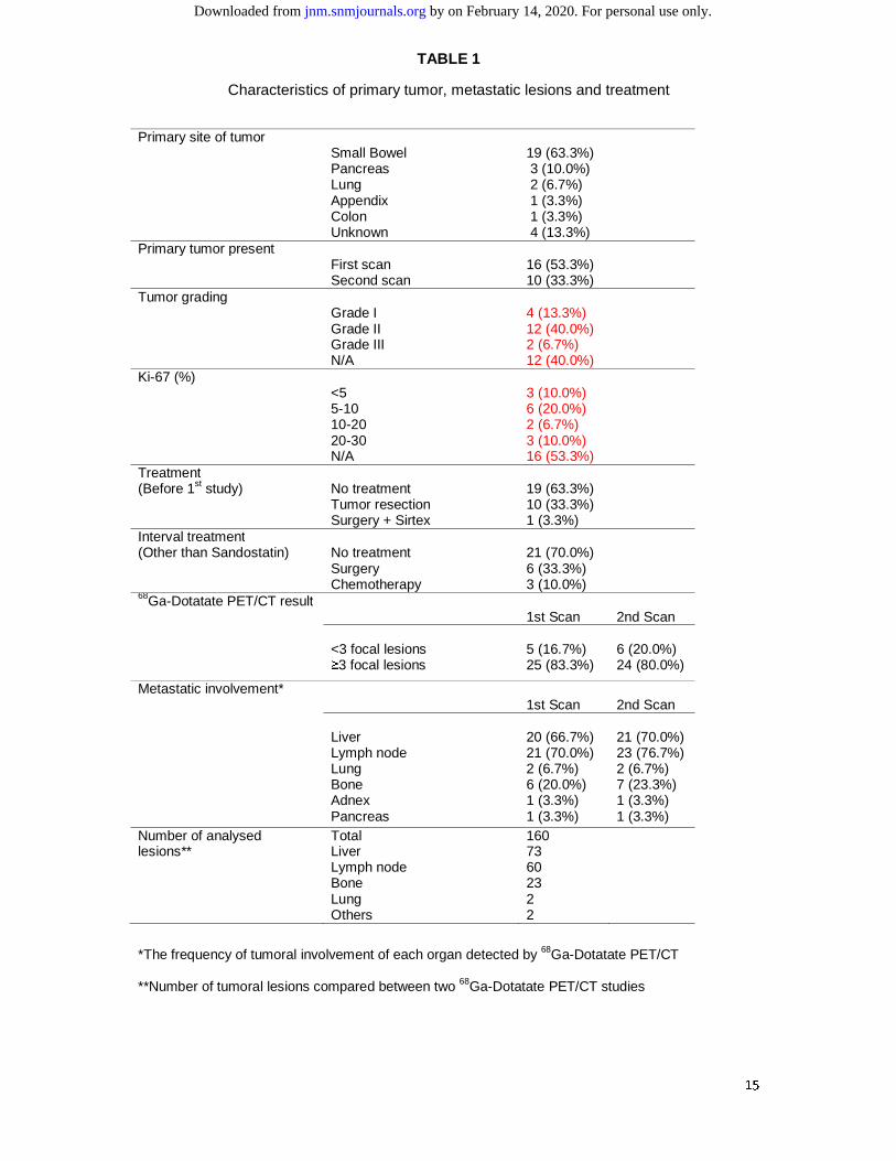
and an age range of 32-86 years. Primary tumor sites are shown in Table 1. Tumor grade
and Ki-67 were formally reported in 18 and 14 patients respectively (Table 1).
The mean time interval between the two studies was 9.6±7.2 months (range, 3-32
months), and the mean time gap between the last Sandostatin LAR injection and the second
study was 25.1±14.8 days (range, 1-59 days). Our analysis of 68Ga-DOTATATE studies in
individual patients did show stable disease in 73.3% (22/30), disease progression in 20.0%
(6/30) and partial response in 6.7% (2/30).
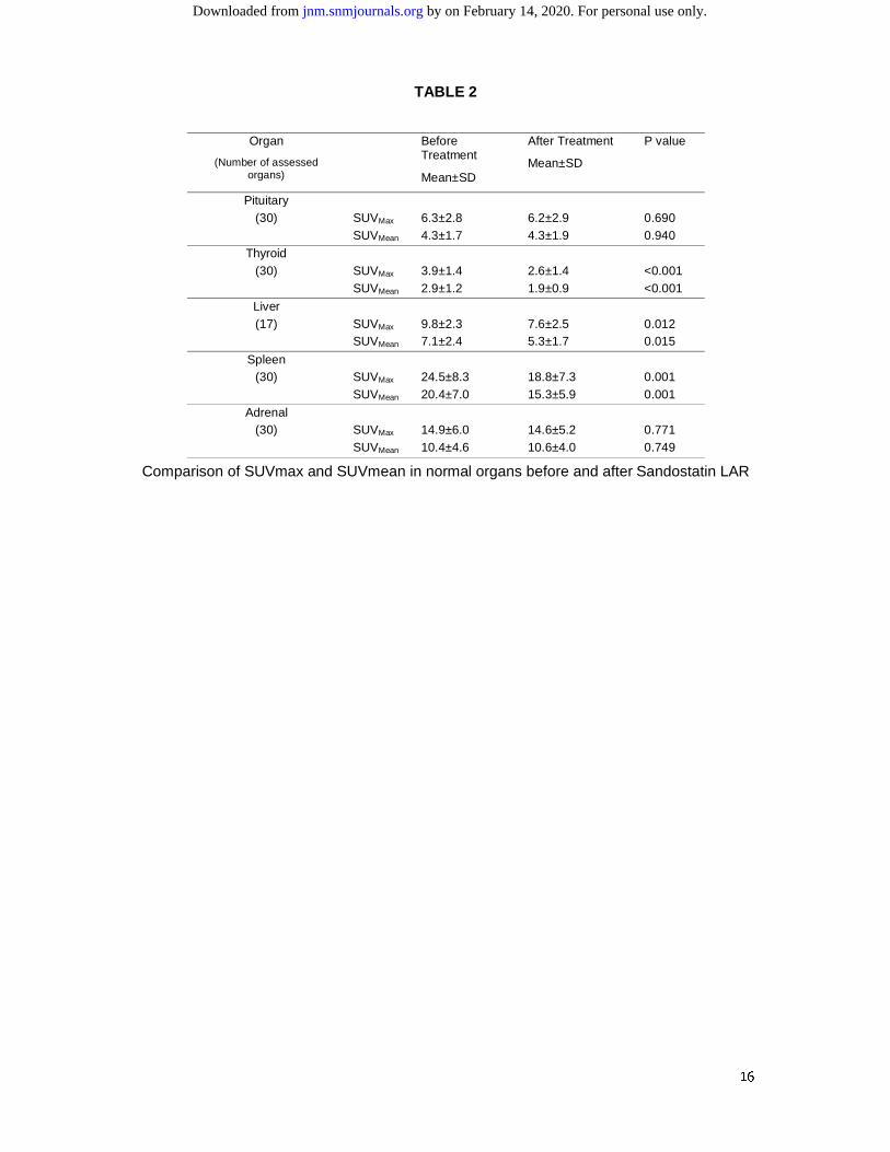
Mean SUVmax and SUVmean values for normal organs were compared before and after
Sandostatin LAR administration; the results are shown in Table 2. After Sandostatin LAR
treatment, the mean SUVmax significantly decreased in the thyroid, liver and spleen (P<0.05),
whereas no significant differences were noted in the pituitary and adrenal glands (P=NS).
Figure 1 shows 68Ga-DOTATATE PET/CT images from a representative patient before and
after Sandostatin LAR treatment (Fig. 1).
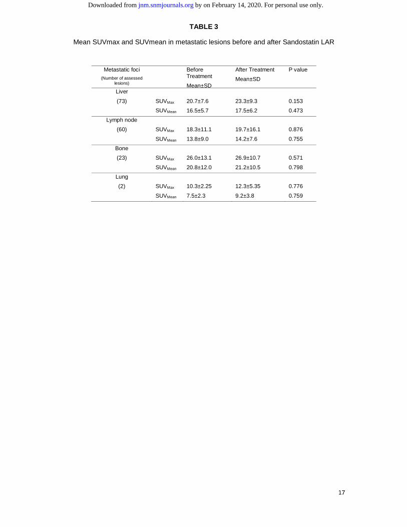
No significant differences were found in the SUVmax values in any metastatic lesion in
the liver, bone, lung or lymph nodes before and after Sandostatin LAR administration
(P>0.05). We further analyzed the SUVmean values of the metastatic lesions before and after
Sandostatin LAR treatment. There were no significant differences in the SUVmean values
before and after Sandostatin LAR treatment (P>0.05). Table 3 shows the mean pre- and
by on February 14, 2020. For personal use only. jnm.snmjournals.org Downloaded from
6
post-treatment SUVmax and SUVmean values for the metastatic lesions examined (Table 3).
We further compared the SUVmax and SUVmean for the hottest lesion (i.e., with the highest
SUVmax) in each scintigraphy before and after Sandostatin LAR therapy. The mean SUVmax
values for the hottest lesion were 26.8±14.7 and 27.8±13.3 before and after Sandostatin
LAR therapy, respectively (P=0.58), while the mean SUVmean values were 19.9±10.2 and
20.3±9.1 (P=0.79). For the residual primary tumors, the mean SUVmax values were 15.3±8.0
and 13.3±6.9 before and after Sandostatin LAR therapy, respectively (P=0.087). The mean
SUVmean values for the residual primary tumors were 12.4±5.9 and 11.0±4.8 before and after
Sandostatin LAR therapy, respectively (P=0.179).
DISCUSSION
In this study, the effect of long-acting somatostatin analog treatment on 68Ga-
DOTATATE uptake in patients harboring NETs was assessed. This is the first study to
assess 68Ga-DOTATATE uptake in the same subset of patients before and after
administration of a long-acting somatostatin analog.
Somatostatin receptor imaging guidelines recommend the withdrawal of octreotide
therapy prior to imaging due to the possibility of interference of cold octreotide with tracer
uptake in tumor cells caused by competition for receptor occupancy as well as SSTR
blockade (11,20). However, in vitro studies (14,21-23), in vivo animal studies (24) and
human studies(25) have all shown that octreotide treatment strongly triggers agonist-induced
internalization of SSRT2 and alters the degree of SSTR expression in NETs.
In the present study, we showed that primary tumor sites and metastatic lesions
exhibit similar 68Ga-DOTATATE uptake before and after administration of a long-acting
somatostatin analog; however, 68Ga-DOTATATE uptake was significantly reduced in the
liver, spleen and thyroid gland following the treatment. Our findings suggest that pre-
treatment with somatostatin analogs does not impact the detection of metastatic disease and
in fact reduces background uptake in normal organs, which enhances the tumor-to-
by on February 14, 2020. For personal use only. jnm.snmjournals.org Downloaded from
7
background ratio and facilitates tumor detection. One possible explanation for our findings is
the difference in the internalization pattern in normal tissues and tumor cells (25) and the
potential up-regulation of SSTR expression that occurs following somatostatin analog
therapy. These mechanisms may compensate for each other and consequently negate
possible decreases in 68Ga-DOTATATE accumulation in metastatic lesions. Previous studies
have shown that NET tumors may be heterogeneous in SSTR expression (25) and respond
differently to somatostatin analog treatment. To address this potentially confounding issue,
we compared 68Ga-DOTATATE uptake in the same lesions in individual patients, and also
by using both mean and max SUV values in our analysis.
It is interesting to note that there was no difference in adrenal and pituitary uptake
before and after Sandostatin LAR treatment, which is in agreement with a prior study (18).
However, significant differences in uptake were found in other normal organs (liver, spleen
and thyroid). This might be due to physiologic variability in SSTR expression among different
organs, as has been previously reported (26,27).
Two previous studies investigated the impact of somatostatin analog treatment on
111In-Octreotide uptake and reported increases in tumor-to-background ratio following
octreotide administration; however, both studies evaluated small populations (5 and 8
patients), and the tumor-to-background ratios measured in the latter study were quite
heterogeneous, ranging from -79% to +0.87% with an average of 50% (17,18). In the only
previously published study on the effect of cold somatostatin analog treatment on the uptake
of 68Ga-labeled somatostatin analogs, Haug et al. compared 68Ga-DOTATATE uptake in two
groups of patients with NETs: one group received long-acting octreotide treatment, while the
other did not. Lower tracer uptake was found in normal liver and spleen tissues in the treated
patients, while no significant difference in radiotracer uptake was found in the metastatic
lesions between the two groups (19). Intraindividual assessments of 9 available patients also
showed that uptake was unaffected after octreotide treatment. Our study agrees with the
above-referenced results for both normal organ uptake and residual primary tumor activity.
by on February 14, 2020. For personal use only. jnm.snmjournals.org Downloaded from

8
To reduce the interfering effect of somatostatin analog treatment on somatostatin
receptor imaging, some guidelines have recommended scheduling the imaging just prior to
the next dose (28-day intervals); however, some previous studies have shown a steady-state
profile for long-acting octreotide (28) with a long maintenance period of greater than 0.01
ng/mL/mg by weeks 12 to 13. Further studies comparing 68Ga-DOTATATE uptake in
patients with less and more than 28-day intervals between the last octreotide injection and
imaging may be needed. While our study has evaluated the impact of initial somatostatin
analog treatment on 68Ga-DOTATATE uptake in NET, in many patients 68Ga-DOTATATE
PET studies are performed throughout the course of their treatment, and at different times
from somatostatin analog administration. The results of our study provide important
information on the potential impact of time post treatment on initial 68Ga-DOTATATE scans,
and which may have relevance to repeat imaging in individual patients.
In our study, all patients were imaged using 68Ga-DOTA-Tyr3-Octreotate (TATE).
Although variable affinity to SSTR subtypes has been reported for differently labeled
somatostatin analogs, the imaging characteristics of each of these radiotracers are similar
(29,30). Therefore, our results might be extended to all PET tracer-radiolabeled somatostatin
molecules. The slow-growing nature of NET tumors as well as the potential anti-proliferative
properties of somatostatin analogs could potentially imply differences in disease status.
However our study was not designed to assess the therapeutic activity of somatostatin
analogs and the ability of 68Ga-DOTATATE to assess or predict response.
Although our study has the advantage of comparing data within the same subset of
patients, due to the retrospective nature of the current study, the time interval between two
PET/CT imaging was quite long (9.6 months). Designing a prospective study with narrow
imaging interval (e.g. SSTR-PET before somatostatin analog and after first or second
administration) would be helpful.
CONCLUSION
Long-acting somatostatin analog treatment decreases 68Ga-DOTATATE uptake in
the liver, spleen, and thyroid gland but does not compromise 68Ga-DOTATATE uptake in
by on February 14, 2020. For personal use only. jnm.snmjournals.org Downloaded from

9
residual primary tumor and metastatic lesions. These findings support the better sensitivity of
somatostatin receptor imaging for the detection of metastasis in patients receiving
somatostatin analog treatment.
FINANCIAL DISCLOSURE
No potential conflict of interest was reported.
by on February 14, 2020. For personal use only. jnm.snmjournals.org Downloaded from

10
REFERENCES
1. Virgolini I, Gabriel M, Kroiss A, et al. Current knowledge on the sensitivity of the
(68)Ga-somatostatin receptor positron emission tomography and the SUVmax reference
range for management of pancreatic neuroendocrine tumours. Eur J Nucl Med Mol Imaging.
2016;43:2072-83.
2. Deppen SA, Liu E, Blume JD, et al. Safety and efficacy of 68Ga-DOTATATE PET/CT
for diagnosis, staging, and treatment management of neuroendocrine tumors. J Nucl Med.
2016;57:708-14.
3. Mojtahedi A, Thamake S, Tworowska I, Ranganathan D, Delpassand ES. The value
of (68)Ga-DOTATATE PET/CT in diagnosis and management of neuroendocrine tumors
compared to current FDA approved imaging modalities: a review of literature. Am J Nucl
Med Mol Imaging. 2014;4:426-34.
4. Janssen I, Chen CC, Millo CM, et al. PET/CT comparing (68)Ga-DOTATATE and
other radiopharmaceuticals and in comparison with CT/MRI for the localization of sporadic
metastatic pheochromocytoma and paraganglioma. Eur J Nucl Med Mol Imaging.
2016;43:1784-91.
5. Pruthi A, Pankaj P, Verma R, Jain A, Belho ES, Mahajan H. Ga-68 DOTANOC
PET/CT imaging in detection of primary site in patients with metastatic neuroendocrine
tumours of unknown origin and its impact on clinical decision making: experience from a
tertiary care centre in India. J Gastrointest Oncol. 2016;7:449-61.
6. Velikyan I, Sundin A, Sorensen J, et al. Quantitative and qualitative intrapatient
comparison of 68Ga-DOTATOC and 68Ga-DOTATATE: net uptake rate for accurate
quantification. J Nucl Med. 2014;55:204-10.
7. Virgolini I, Ambrosini V, Bomanji JB, et al. Procedure guidelines for PET/CT tumour
imaging with 68Ga-DOTA-conjugated peptides: 68Ga-DOTA-TOC, 68Ga-DOTA-NOC,
68Ga-DOTA-TATE. Eur J Nucl Med Mol Imaging. 2010;37:2004-10.
8. Rinke A, Muller HH, Schade-Brittinger C, et al. Placebo-controlled, double-blind,
prospective, randomized study on the effect of octreotide LAR in the control of tumor growth
by on February 14, 2020. For personal use only. jnm.snmjournals.org Downloaded from

11
in patients with metastatic neuroendocrine midgut tumors: a report from the PROMID Study
Group. J Clin Oncol. 2009;27:4656-63.
9. Caplin ME, Pavel M, Cwikla JB, et al. Lanreotide in metastatic enteropancreatic
neuroendocrine tumors. N Engl J Med. 2014;371:224-33.
10. Lancranjan I, Atkinson AB. Results of a European multicentre study with Sandostatin
LAR in acromegalic patients. Sandostatin LAR Group. Pituitary. 1999;1:105-14.
11. Kwekkeboom DJ, Krenning EP, Scheidhauer K, et al. ENETS consensus guidelines
for the standards of care in neuroendocrine tumors: somatostatin receptor imaging with
(111)In-pentetreotide. Neuroendocrinology. 2009;90:184-9.
12. Ben-Shlomo A, Schmid H, Wawrowsky K, et al. Differential ligand-mediated pituitary
somatostatin receptor subtype signaling: implications for corticotroph tumor therapy. J Clin
Endocrinol Metab. 2009;94:4342-50.
13. Arnold R, Trautmann ME, Creutzfeldt W, et al. Somatostatin analogue octreotide and
inhibition of tumour growth in metastatic endocrine gastroenteropancreatic tumours. Gut.
1996;38:430-8.
14. Cescato R, Schulz S, Waser B, et al. Internalization of sst2, sst3, and sst5 receptors:
effects of somatostatin agonists and antagonists. J Nucl Med. 2006;47:502-11.
15. Lesche S, Lehmann D, Nagel F, Schmid HA, Schulz S. Differential effects of
octreotide and pasireotide on somatostatin receptor internalization and trafficking in vitro. J
Clin Endocrinol Metab. 2009;94:654-61.
16. Bakker WH, Krenning EP, Reubi JC, et al. In vivo application of [111In-DTPA-D-
Phe1]-octreotide for detection of somatostatin receptor-positive tumors in rats. Life Sci.
1991;49:1593-601.
17. Dorr U, Rath U, Sautter-Bihl ML, et al. Improved visualization of carcinoid liver
metastases by indium-111 pentetreotide scintigraphy following treatment with cold
somatostatin analogue. Eur J Nucl Med. 1993;20:431-3.
18. Janson ET, Kalkner KM, Eriksson B, Westlin JE, Oberg K. Somatostatin receptor
scintigraphy during treatment with lanreotide in patients with neuroendocrine tumors. Nucl
Med Biol. 1999;26:877-82.
by on February 14, 2020. For personal use only. jnm.snmjournals.org Downloaded from

12
19. Haug AR, Rominger A, Mustafa M, et al. Treatment with octreotide does not reduce
tumor uptake of (68)Ga-DOTATATE as measured by PET/CT in patients with
neuroendocrine tumors. J Nucl Med. 2011;52:1679-83.
20. Balon HR, Goldsmith SJ, Siegel BA, et al. Procedure guideline for somatostatin
receptor scintigraphy with (111)In-pentetreotide. J Nucl Med. 2001;42:1134-8.
21. Liu Q, Cescato R, Dewi DA, Rivier J, Reubi JC, Schonbrunn A. Receptor signaling
and endocytosis are differentially regulated by somatostatin analogs. Mol Pharmacol.
2005;68:90-101.
22. Ginj M, Chen J, Walter MA, Eltschinger V, Reubi JC, Maecke HR. Preclinical
evaluation of new and highly potent analogues of octreotide for predictive imaging and
targeted radiotherapy. Clin Cancer Res. 2005;11:1136-45.
23. Nock BA, Nikolopoulou A, Galanis A, et al. Potent bombesin-like peptides for GRP-
receptor targeting of tumors with 99mTc: a preclinical study. J Med Chem. 2005;48:100-10.
24. Waser B, Tamma ML, Cescato R, Maecke HR, Reubi JC. Highly efficient in vivo
agonist-induced internalization of sst2 receptors in somatostatin target tissues. J Nucl Med.
2009;50:936-41.
25. Reubi JC, Waser B, Cescato R, Gloor B, Stettler C, Christ E. Internalized
somatostatin receptor subtype 2 in neuroendocrine tumors of octreotide-treated patients. J
Clin Endocrinol Metab. 2010;95:2343-50.
26. Kuyumcu S, Ozkan ZG, Sanli Y, et al. Physiological and tumoral uptake of (68)Ga-
DOTATATE: standardized uptake values and challenges in interpretation. Ann Nucl Med.
2013;27:538-45.
27. Shastry M, Kayani I, Wild D, et al. Distribution pattern of 68Ga-DOTATATE in
disease-free patients. Nucl Med Commun. 2010;31:1025-32.
28. Astruc B, Marbach P, Bouterfa H, et al. Long-acting octreotide and prolonged-release
lanreotide formulations have different pharmacokinetic profiles. J Clin Pharmacol.
2005;45:836-44.
by on February 14, 2020. For personal use only. jnm.snmjournals.org Downloaded from
13
29. Johnbeck CB, Knigge U, Kjaer A. PET tracers for somatostatin receptor imaging of
neuroendocrine tumors: current status and review of the literature. Future Oncol.
2014;10:2259-77.
30. Poeppel TD, Binse I, Petersenn S, et al. Differential uptake of (68)Ga-DOTATOC and
(68)Ga-DOTATATE in PET/CT of gastroenteropancreatic neuroendocrine tumors. Recent
Results Cancer Res. 2013;194:353-71.
by on February 14, 2020. For personal use only. jnm.snmjournals.org Downloaded from
14
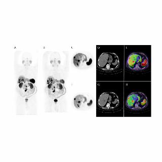
FIGURE LEGEND
FIGURE 1. 68Ga-Dotatate PET whole body MIP images in a patient with small bowel
carcinoid tumor A) before, and B) after 12 months of treatment with Sandostatin LAR (30
mg/4week). The post treatment scan showed reduced uptake of 68Ga-Dotatate in normal
liver and spleen, and thyroid, also seen in transaxial PET, CT and fused images before (C, D
and E) and after (F, G and H) treatment. Both primary and metastatic lesions showed similar
uptake of 68Ga-Dotatate, with the- SUVmean of the hottest lesion 30.6 in the first study and
31.8 in second study.
by on February 14, 2020. For personal use only. jnm.snmjournals.org Downloaded from
15
TABLE 1
Characteristics of primary tumor, metastatic lesions and treatment
*The frequency of tumoral involvement of each organ detected by 68Ga-Dotatate PET/CT
**Number of tumoral lesions compared between two 68Ga-Dotatate PET/CT studies
Primary site of tumor Small Bowel Pancreas Lung Appendix Colon Unknown
19 (63.3%) 3 (10.0%) 2 (6.7%) 1 (3.3%) 1 (3.3%) 4 (13.3%)
Primary tumor present First scan Second scan
16 (53.3%) 10 (33.3%)
Tumor grading
Grade I Grade II Grade III N/A
4 (13.3%) 12 (40.0%) 2 (6.7%) 12 (40.0%)
Ki-67 (%) <5 5-10 10-20 20-30 N/A
3 (10.0%) 6 (20.0%) 2 (6.7%) 3 (10.0%) 16 (53.3%)
Treatment (Before 1st study)
No treatment Tumor resection Surgery + Sirtex
19 (63.3%) 10 (33.3%) 1 (3.3%)
Interval treatment (Other than Sandostatin)
No treatment Surgery Chemotherapy
21 (70.0%) 6 (33.3%) 3 (10.0%)
68Ga-Dotatate PET/CT result 1st Scan
2nd Scan
<3 focal lesions ≥3 focal lesions
5 (16.7%) 25 (83.3%)
6 (20.0%) 24 (80.0%)
Metastatic involvement*
1st Scan
2nd Scan
Liver Lymph node Lung Bone Adnex Pancreas
20 (66.7%) 21 (70.0%) 2 (6.7%) 6 (20.0%) 1 (3.3%) 1 (3.3%)
21 (70.0%) 23 (76.7%) 2 (6.7%) 7 (23.3%) 1 (3.3%) 1 (3.3%)
Number of analysed lesions**
Total Liver Lymph node Bone Lung Others
160 73 60 23 2 2
by on February 14, 2020. For personal use only. jnm.snmjournals.org Downloaded from
16
TABLE 2
Comparison of SUVmax and SUVmean in normal organs before and after Sandostatin LAR
Organ
(Number of assessed organs)
Before Treatment
Mean±SD
After Treatment
Mean±SD
P value
Pituitary (30)
SUVMax 6.3±2.8 6.2±2.9 0.690 SUVMean 4.3±1.7 4.3±1.9 0.940
Thyroid (30)
SUVMax 3.9±1.4 2.6±1.4 <0.001 SUVMean 2.9±1.2 1.9±0.9 <0.001
Liver (17)
SUVMax 9.8±2.3 7.6±2.5 0.012 SUVMean 7.1±2.4 5.3±1.7 0.015
Spleen (30)
SUVMax 24.5±8.3 18.8±7.3 0.001 SUVMean 20.4±7.0 15.3±5.9 0.001
Adrenal (30)
SUVMax 14.9±6.0 14.6±5.2 0.771 SUVMean 10.4±4.6 10.6±4.0 0.749
by on February 14, 2020. For personal use only. jnm.snmjournals.org Downloaded from
17
TABLE 3
Mean SUVmax and SUVmean in metastatic lesions before and after Sandostatin LAR
Metastatic foci
(Number of assessed lesions)
Before Treatment
Mean±SD
After Treatment
Mean±SD
P value
Liver
(73)
SUVMax
SUVMean
20.7±7.6
16.5±5.7
23.3±9.3
17.5±6.2
0.153
0.473
Lymph node
(60)
SUVMax
SUVMean
18.3±11.1
13.8±9.0
19.7±16.1
14.2±7.6
0.876
0.755
Bone
(23)
SUVMax
SUVMean
26.0±13.1
20.8±12.0
26.9±10.7
21.2±10.5
0.571
0.798
Lung
(2)
SUVMax
SUVMean
10.3±2.25
7.5±2.3
12.3±5.35
9.2±3.8
0.776
0.759
by on February 14, 2020. For personal use only. jnm.snmjournals.org Downloaded from
by on February 14, 2020. For personal use only. jnm.snmjournals.org Downloaded from

Doi: 10.2967/jnumed.117.192203Published online: July 20, 2017.J Nucl Med. Narjess Ayati, Sze Ting Lee, Seyed Rasoul Zakavi, Kunthi Pathmaraj, Loai Qatawneh, Aurora Poon and Andrew Scott compared to primary tumors and metastatic lesions
Ga-DOTATATE uptake in normal tissues68Sandostatin LAR therapy differentially alters
http://jnm.snmjournals.org/content/early/2017/07/19/jnumed.117.192203This article and updated information are available at:
http://jnm.snmjournals.org/site/subscriptions/online.xhtml
Information about subscriptions to JNM can be found at:
http://jnm.snmjournals.org/site/misc/permission.xhtmlInformation about reproducing figures, tables, or other portions of this article can be found online at:
and the final, published version.proofreading, and author review. This process may lead to differences between the accepted version of the manuscript
ahead of print area, they will be prepared for print and online publication, which includes copyediting, typesetting,JNMcopyedited, nor have they appeared in a print or online issue of the journal. Once the accepted manuscripts appear in the
. They have not beenJNM ahead of print articles have been peer reviewed and accepted for publication in JNM
(Print ISSN: 0161-5505, Online ISSN: 2159-662X)1850 Samuel Morse Drive, Reston, VA 20190.SNMMI | Society of Nuclear Medicine and Molecular Imaging
is published monthly.The Journal of Nuclear Medicine
© Copyright 2017 SNMMI; all rights reserved.
by on February 14, 2020. For personal use only. jnm.snmjournals.org Downloaded from
![Review The Search for an Alternative to [ Ga]Ga-DOTA-TATE ...thno.org/v09p1336.pdf · [68Ga]Ga-DOTA-TATE, [68Ga]Ga-DOTA-TOC, and [68Ga]Ga-DOTA-NOC allows for NET staging with high](https://static.fdocuments.in/doc/165x107/5e2a1b5b2104573c786ad22c/review-the-search-for-an-alternative-to-gaga-dota-tate-thnoorg-68gaga-dota-tate.jpg)


![[68Ga]NOTA-Galactosyl Human Serum Albumin: a Tracer for ... · 68Ga]NOTA-GSA showed slower uptake in comparison with 68Ga-DTPA-GSA (123 ± 10 vs. 89 ± 3 s, p G 0.01). Conclusions:](https://static.fdocuments.in/doc/165x107/5f6d9cae33a64152a27bd022/68ganota-galactosyl-human-serum-albumin-a-tracer-for-68ganota-gsa-showed.jpg)


![68Ga] peptide high-output production on commercially · [68Ga] peptide high-output production on commercially available MiniAIO® synthesizer Objectives Optmization parameters Material](https://static.fdocuments.in/doc/165x107/5f95b4c0eecbac70717355d8/68ga-peptide-high-output-production-on-68ga-peptide-high-output-production-on.jpg)