
Rathi North America - RGD/RGS Spec SheetTitle RGD/RGS Spec Sheet Author Rathi North America

`
Qualification of in-house prepared 68Ga RGD in healthy monkeys for subsequent
molecular imaging of 3 integrin expression in patients
I Schoeman 23385952
Dissertation submitted in partial fulfilment of the requirements for the degree Magister Scientiae in Pharmaceutica at the
Potchefstroom Campus of the North-West University
Supervisor: Prof. J.R. Zeevaart Co-supervisor: Prof. M. M. Sathekge Assistant Supervisor: Prof. A. Grobler
May 2014

i
Preface
I hereby acknowledge and declare that this project was my own work performed
and that all references are referenced and declared as such as well as
acknowledgements made to people who were a part of this project in different
phases and fields and tasks performed, in mentor or supportive roles. In addition,
no conflict of interest exists. Acknowledgement was given to co-workers in terms
of the references and directly indicating their contribution.
‘‘Whoever wishes to pursue properly the science of medicine must proceed thus...For if a physician knows these things well...; so he will not be at loss in the treatment of diseases...’’
Hippocrates
"There is no blue without yellow and without orange."
Van Gogh
‘‘You treat a disease, you win, you lose. You treat a person, I guarantee you, you'll win, no matter what the outcome.
Our job is improving the quality of life, not just delaying death.”
Hunter Patch Adams
“Theranostics is a revolutionary Approach that promises improved Therapy selection on the basis of
Specific molecular features of disease, Greater predictive power for adverse
Effects due to improved patient- Specific absorbed dose estimates,
And new ways to objectively monitor Therapy response...” Prof. M.M. Sathekge

ii
Acknowledgements
I hereby would like to thank God, the Alpha and Omega who created me and created me to
live life with passion and to have extreme perseverance to finish what was begun and for
blessing everything I do. For giving me power and strength to carry on no matter what
trials, tribulations and stumbling blocks came my way. For helping me to complete this
race. Without the following people, this would not have been possible:
North-West University, especially the Preclinical Drug Development group including Prof
Sias Hamman and Prof Anne Grobler who gave me the opportunity to study this degree.
My supervisors:
Prof Mike M Sathekge from Steve Biko Hospital who is a remarkable person, always since
I first worked with him in private practice many years ago, believed that one could do
better, for bringing the best out of me and many people around him. For always be an
excellent Physician whose help and advice are always the best standard.
Prof Jan Rijn Zeevaart who has been supporting and motivating, even while he was on
sabbatical leave, who has always helped to find resources while there were none.
He always inspired and motivated and on short deadlines, without pressure rather
motivated why something should be ready earlier. He also supported with organizing
preclinical as well as xenografted studies. Thank you also for statistical processing support
in this regard.
Prof Anne Grobler, for guidance from a distance, for supporting the crucial preclinical part
of this study by providing financial support for this. Prof Anne as well as Prof Hamman for
essential feedback on the colloquium.
Great thanks to Dr. D. Rossouw who supported, assisted and guided kit formulation and a
very short time period when laboratory was available and for Dr Clive Naidoo who gave me
permission to work there. Without Niel, inhouse kit formulation would not have come to this

iii
stage. Thanks for always supporting, discussing, and guiding even when the work at their
laboratory was completed.
Thomas Ebenhan for great support with statistical processing especially Savitzky Gollay as
well as general support throughout most of this project. For assisting while working at
Steve Biko Hospital with additional info as well as organizing with international additional
info for example on another generator, pre-purification info. For also assisting with
preclinical and xenografts.
All staff at Necsa including Judith Wagener for crucial support during kit production (GMP
rules and regulations, templates for SOP’s) and HPLC and Biljana Marjanovic-Painter for
all the HPLC runs. Also for the rest of the staff including Mariana Miles for general support.
Thanks to Hester Oosthuizen for writing guidance. Also thanks to Cerozáne Welgemoed
for admin support and Petra Gainsford from NWU for formatting the thesis.
Preclinical: to Dr V Naidoo and his team for excellent preclinical support during imaging.
Delene van Wyk from Steve Biko Hospital for scanning on the PET-CT on after hour times.
Also with xenografts for Hylton from North-West University for handling the xenografts. I
also thank Viola Satzinger for providing rendered images that were calculated using
Siemens in-house software.
Prof Gert Kruger from KZN for helping with funding of one RGD raw product batch.
On a personal note, my family and especially my Mother for always supporting everything I
do every day in prayer.

iv
Abstract
Introduction: Targeted pharmaceuticals for labelling with radio-isotopes for very specific
imaging (and possibly later for targeted therapy) play a major role in Theranostics which is
currently an important topic in Nuclear Medicine as well as personalised medicine. There
was a need for a very specific lung cancer radiopharmaceutical that would specifically be
uptaken in integrin 3 expression cells to image patients using a Positron Emission
Tomography- Computed Tomography (PET-CT) scanner.
Background and problem statement: Cold kits of c (RGDyK)–SCN-Bz-NOTA were kindly
donated by Seoul National University (SNU) to help meet Steve Biko Hospital’s need for
this type of imaging. These cold kits showed great results internationally in labelling with a
0.1 M 68Ge/68Ga generator (t1/2 of 68Ge and 68Ga are 270.8 days and 67.6 min,
respectively). However the same cold kits failed to show reproducible radiolabeling with the
0.6 M generator manufactured under cGMP conditions at iThemba LABS, Cape Town and
distributed by IDB Holland, the Netherlands.
Materials and methods: There was therefore a need for producing an in-house NOTA-RGD
kit that would enable production of clinical 68Ga-NOTA-RGD in high yields from the IDB
Holland/iThemba LABS generator. Quality control included ITLC in citric acid to observe
labelling efficiency as well as in sodium carbonate to evaluate colloid formation. HPLC was
also performed at iThemba LABS as well as Necsa (South African Nuclear Energy
Corporation). RGD was obtained from Futurechem, Korea. Kit mass integrity was
determined by testing labelling efficiency of 10, 30 and 60 µg of RGD per cold kit. The
RGD was buffered with sodium acetate trihydrate. The original kits were dried in a
desiccator and in later studies only freeze dried. Manual labelling was also tested. The
radiolabelled in-house kit’s ex vivo biodistribution in healthy versus tumour mice were
examined by obtaining xenografts. The normal biodistribution was investigated in three
vervet monkeys by doing PET-CT scans on a Siemens Biograph TP 40 slice scanner.
Results: Cold kit formulation radiolabeling and purification methods were established
successfully and SOPs (standard operating procedures) created. HPLC results showed
highest radiochemical purity in 60 µg cold kit vials. 68Ga-NOTA-RGD showed increased

v
uptake in tumours of tumour bearing mouse. The cold kit also showed normal distribution
according to literature with fast blood clearance and excretion through kidneys into urine,
therefore making it a suitable radiopharmaceutical for clinical studies.
Conclusion: The in-house prepared cold kit with a 4 month shelf-life was successfully
tested in mice and monkeys.
Keywords: integrin 3 expression, 68Ge/68Ga generator, c(RGDyK)–SCN-Bz-NOTA,
xenografts, vervet monkeys, PET-CT, in-house cold kit.

vi
Opsomming
Inleiding: Geteikende farmaseutikums vir die merking van radio- isotope vir baie spesifieke
beelding (en moontlik later geteikende terapie) speel tans ‘n groot rol in Teranostiek, ’n
belangrike onderwerp in Kerngeneeskunde en verpersoonlikte medisyne. Daar was ‘n
behoefte vir ‘n spesifieke long kanker merker wat opgeneem kan word in integrien 3 sel
ekspressie om pasiente te verbeeld met ‘n Positron Emissie Tomografie-Rekenaar
Tomografie (PET-RT) skandeerder.
Agtergrond en probleem: Koue kitstelle van sikliese ‘(RGDyK)–SCN-Bz-NOTA’ was deur
Seoul Nasionale Universiteit (SNU) aan Steve Biko Hospitaal geskenk om met hul
probleem te help. Die koue kitsstelle het goeie resultate gelewer internasionaal wanneer
hul gemerk is met 0.1 M generator 68Ge/68Ga (T1/2 van 68Ge en 68Ga is 270.8 dae en 67.6
min, onderskeidelik).Ewewel het dieselfde koue kitsstelle gefaal om herhaalbare
radiomerking te lewer met die 0.6 M generator wat onder cGMP kondisies by iThemba
LABS Kaapstad en versprei deur IDB Holland, Nederland.
Materie en metodes: Daar was daarom ‘n behoefte vir produksie van ‘n self geproduseerde
RGD kitsstel en na NOTA vasgestel is as die cheleerder van keuse, was 68Ga-NOTA-RGD
suksesvol gemerk en gesuiwer, in die gebruik van Ga-III wat geëlueer is van die IDB
Holland/iThemba LABS generator. Kwaliteits kontrole het ITLC ingesluit in vloei mediums
van sitriese suur en om merkings doeltreffendheid vas te stel en natrium karbonaat om
kolloied vorming vas te stel.HPLC is ook by iThemba LABS asook Necsa (Suid Afrikaanse
Kernenergie Korporasie) uitgevoer. RGD was verkry van Futurechem, Korea. Kitsstel
massa integriteit was vasgestel deur die toets van merkings doeltreffendheid van 10, 30 en
60 mikrogram RGD per koue kitsstel. Die RGD was gebuffer met natrium asetaat trihidraat.
Die oorspronklike kitsstelle was gedroog in ‘n dissekator en in latere studies slegs
gevriesdroog. Merking met die hand was ook getoets. Die radio gemerkte self-
geproduseerde kitsstelel se distribusie in gesonde teenoor tumor draende muise was
ondersoek deur die verkryging van xenograafs. Die normale distribusie was ondersoek in
drie blou apies deur PET-RT skandering op ‘n Siemens Biograph TP skandeerder te doen.

vii
Resultate: Koue kitsstel formulering en radiomerkings en suiwerings metodes was
suksesvol vasgestel en SOPs (Standaard Operateurs Prosedures) is ontwerp.HPLC het
die hoogste suiwering getoon in 60 mikrogram koue kitsstel flesse. Massa bepalende
studies het bevestig dat 60 mikrogram gebruik moes word in die kitsstel. 68Ga-NOTA-RGD
het opname getoon in die tumor draende muis. Die koue kitsstel het ook normale
distribusie getoon in blou apies in verhouding tot literatuur wat verwys na vinnige bloed
opruiming en uitskeiding deur die niere en blaas wat dit daarom ‘n gunstige
radiofarmaseutikum maak vir kliniese studies.
Gevolgtrekking: Die self geproduseerde koue kitsstel van omtrent 4 maande rakleeftyd is
suksesvol getoets in muise en ape.
Sleutelwoorde: integrien 3 ekspressie, 68Ge/68Ga generator, c(RGDyK)–SCN-Bz-NOTA,
60 mikrogram, natrium asetaat trihidraat, xenograaf, blou apies, PET-RT skandeerder, self
geproduseerde koue kitsstel.

viii
Table of Contents
Preface i
Acknowledgements ........................................................................................................ ii
Abstract iv
Opsomming vi
List of Tables ................................................................................................................. xi
List of Figures ................................................................................................................ xi
List of Abbreviations ..................................................................................................... xv
List of Symbols and Equations ................................................................................... xviii
Chapter 1: Introduction and problem statement .......................................................... 1
1.1 Introduction.............................................................................................. 1
1.2 Problem statement and aim of the study............................................... 6
1.3 Research objectives ................................................................................ 7
Chapter 2: Background and literature review .............................................................. 8
2.1 Radiopharmaceuticals and cGMP in Molecular Imaging ..................... 8
2.1.2 Peptide labelling with stable chelators and ‘lock and key’ structure ......... 15
2.2 68Ge/68Ga generator ............................................................................... 20
2.3 Introduction: Radiopharmaceuticals in general/conservative Molecular Imaging .............................................................................. 22
2.3.1 PET going back to basics of ‘SPECT-type’ ‘Generator’ produced isotope
labelled with ‘cold kit’ ............................................................................... 29
ix
2.4 Other imaging modalities: MRI ............................................................. 31
Chapter 3: Methodology ............................................................................................ 34
3.1 Labelling methods and procedures including with reference to SOP’s (Standard operating procedures): ............................................ 34
3.1.1 Radiolabeling Imported Kits: Donated kits with inconsistent results: ....... 34
3.1.2 68Ge/68Ga Generator in detail .................................................................. 39
3.1.3 Radiolabeling In-house prepared kit ........................................................ 44
3.1.4 Radiolabeling and Purification: additional info ......................................... 51
3.1.5 Purification of the Radiolabelled Product: Solid phase extraction/SPE: ... 52
3.1.6 Quality Control ......................................................................................... 53
3.1.7 Kit shelf-life .............................................................................................. 54
3.2 Preclinical............................................................................................... 55
3.2.1 Ethical approval ....................................................................................... 55
3.2.2 Xenografts: Mice ...................................................................................... 56
3.2.3 Scanning on Clinical PET.CT scanner and SUV ..................................... 58
Chapter 4: Results and discussion ............................................................................ 64
4.1 Imported cold kits background results................................................ 64
4.1.1 ITLC and results SNU imported cold kits ................................................. 66
4.1.2 Purification: .............................................................................................. 67
4.1.3 68Ga eluate purification and NOTA-RGD radiolabeling ............................ 73
4.1.4 Kit shelf life integrity and K60 performance ............................................. 75

x
4.1.5 68Ge/68Ga generator data ........................................................................ 76
4.2 Xenografts: Mice .................................................................................... 77
4.3 Preclinical Imaging: Pharmacokinetics, biodistribution, and Image gallery ..................................................................................................... 79
4.4 68Ga-NOTA-RGD Concentrations in Blood and Urine of healthy vervet Monkeys ...................................................................................... 87
Chapter 5: Conclusions, future work and recommendations ..................................... 88
References 90

xi
List of Tables
Table 2-1: Generators in medical use 30
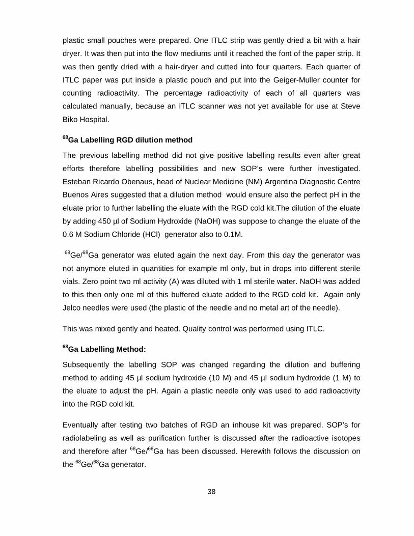
Table 3-1: Half-life in minutes: Time represents 1-68 min 43
Table 3-2: Three Monkeys Preclinical PET-CT scan info 59
Table 4-1: Purification 70
Table 4-2: Influence of Generator Eluate Pre-Purification on 68Ga-NOTA-RGD
radiolabeling 74
Table 4-3: Organ and Tissue Concentration of 68Ga-NOTA-RGD in Healthy
Vervet Monkeys 84
List of Figures
Figure 1-1: Lungs constitutes of different structures including lobes and epithelial
cells (Herbst, 2013). 2
Figure 1-2: Lymph nodes refer to the N in TNM staging of cancer (American
Cancer Society, 2013). 3
Figure 2-1: Schematic image of radiopharmaceutical basic labelling process. 8
Figure 2-2: Integrin expression cells (Weis and Cheresh, 2011). 11
Figure 2-3: Integrins influencing the tumour cell in various ways (Desgrosellier
and Cheresh, 2010). 12
Figure 2-4: Peptide receptor binding with a ligand (Jamous et al., 2013). 16
Figure 2-5: Different bifunctional chelators compared in 68Ga labelling (Berry et
al., 2011). 17

xii
Figure 2-6: The most common bifunctional chelators in radiolabeling involving
metals: A (DOTA), B (NOTA) and C (TETA), (Bartholomä, 2012). 17
Figure 2-7: Various 68Ga-RGD tracers compared (Knetsch et al., 2011). 18
Figure 2-8: 64Cu-RGD injected into tumour bearing mice (Galibert et al., 2010). 20
Figure 2-9: Patients with different injected radiotracers will have different
emissions and different cameras for example SPECT (left) and PET-
CT (right) therefore gamma emission and positron emission tracers
(Siemens, 2014). 23
Figure 2-10: Preclinical image of full bladder and kidney uptake. 24
Figure 2-11: Siemens training slides of bone scintigraphic. 25
Figure 2-12: Normal distribution in 68Ga-NOTA-RGD (Jeong, 2011). 26
Figure 2-13: Lung lesion 68Ga-NOTA-RGD (Jeong, 2011). 27
Figure 2-14: Different integrins and expression of 3 in prostate and pancreatic
tumours, (Desgrosellier and Cheresh, 2010). 29
Figure 2-15: MFBR particle (Lee, 2010). 32
Figure 3-1: Jeong, Seoul National University (SNU) shared with Prof. Sathekge
for labelling support. 35
Figure 3-2: Quality control done by Seoul National University (Joeng, 2011). 36
Figure 3-3: IDB Holland generator produced under cGMP conditions at iThemba
LABS in the Western Cape (Eckert & Ziegler, 2008). 39
Figure 3-4: iThemba LABS small and compact cGMP compliant generator behind
lead. 40
Figure 3-5: Laminar flow cabinet compulsory for cGMP conditions in which 68Ge/68Ga generator is hosted. 41

xiii
Figure 3-6: 0.6 M generator elution profile (IDB Holland user instructions). 42
Figure 3-7: Tumour bearing mice injected with 68Ga-NOTA-RGD including also
injecting cold RGDyk (Jeong et al., 2008). 44
Figure 3-8: Molecular Formula: C47H67N13014S.CF3COOH (Futurechem). 46
Figure 3-9: Radiolabeling of the inhouse prepared NOTA-RGD kit (SOP
Addendum B). 50
Figure 3-10: Purification SepPak. 52
Figure 3-11: Ethical approval from Animal use and care committee (AUCC),
Addendum D. 56
Figure 3-12: Volumes of interest(VOIs) drawn around the Monkey’s organs for
SUV calculation. 61
Figure 4-1: ITLC analytical results of radiolabelled 68Ga- NOTA-SCN-Bz-RGD
(Joeng, 2009). 65
Figure 4-2: HPLC results of 68Ga- NOTA-SCN-Bz-RGD (Joeng, 2009). 66
Figure 4-3: HPLC at iThemba labs testing the first inhouse prepared kits. 68
Figure 4-4: HPLC at iThemba labs testing the first inhouse prepared kits: specific
using the 20:80 percent ethanol saline to desorb the radiolabelled
product from the SepPak cartridge. 69
Figure 4-5: The final 2 ml of SepPak purification of the last ethanol/saline solution
showing no free 68Ga. 71
Figure 4-6: HPLC of the product before purification. 72
Figure 4-7: HPLC of product after purification. 73
Figure 4-8: NOTA RGD kit performance: K30 and K60. 75

xiv
Figure 4-9: Impact of the NOTA-RGD kit integrity on the percentage radiolabeling
efficiency. 76
Figure 4-10: Regeneration time from the parent decay. 77
Figure 4-13: Percentage Injected dose per gram in Xenografted mice. The first
(green) series is the tumour mice and the second (light blue) the
healthy mice. 79
Figure 4-14: Volume rendering monkey image of biodistribution of radiotracer
(Performed by Viola Satzinger from Siemens Erlangen). 79
Figure 4-15: Radiochemical purity and yield of three vervet monkeys. 80
Figure 4-16: Max SUV used for patient pathology and clinical example urine.[ VOIs
processing] 81
Figure 4-17: Pelvic volume rendering of one vervet monkey to visualize radiotracer
uptake in kidneys. 82
Figure 4-18: Volume rendering image of monkey of radiotracer in kidneys and
imaged of spine from CT. 83
Figure 4-19: Time/activity curve of 68Ga-NOTA-RGD in monkey’s blood. 85
Figure 4-20: Time/activity curve of 68Ga-NOTA-RGD in monkey’s urine. 85
Figure 4-21: Volume rendering of monkey from head to pelvis showing
endotracheal tube, kidneys and urine in bladder. 86

xv
List of Abbreviations
A Activity (radioactivity)
AIDS Acquired Immuno Deficiency Syndrome
ALARA As low a reasonably achievable
AUCC Animal use and care committee
cGMP current Good Manufacturing Practice
cGRPP Current Good Radiopharmacy Practice
CT Computed Tomography
CTI Competitive Technologies, Incorporated
18F Fluoor-18
FDG Fluorodeoxyglucose
68Ga Gallium-68
68Ge Germanium-68
GMP Good Manufacturing Practice
HIV Human Immunodeficiency Virus
HPL High pressure liquid chromatography
ITLC Instant thin layer chromatography
111In Indium-111
K30 and K60 Refers to 30 and 60 µg of peptide in cold kit
LE Labelling efficiency

xvi
MI Molecular Imaging
MICAD Molecular imaging and contrast agent database.
99Mo Molybdenum-99
MRI Magnetic Resonance Imaging
n sample size(number)
NOTA-cyclic RGDyK Cyclic Arg-Gly-Asp-D-Tyr-Lys-NOTA
NM Nuclear Medicine
PET-CT. Positron Emission Tomography- Computed
Tomography
pH Potential Hydrogen
RCP Radiochemical purity
SD Standard deviation
SEM Standard error of mean
SOP/SOP’s Standard operating procedure/procedures
SPECT Single Photon Emission Computed Tomography
SPN Solitary Pulmonary Nodule
SUV Standardized Uptake Value (of tumor Calculated
during PET-CT processing).
SUVs Standardized uptake values
TB Tuberculosis

xvii
TNM Tumour, Nodes, Metastasize
USA United States of America
VOI/VOIs Volume of interest/Volumes of interest

xviii
List of Symbols and Equations
°C Degrees in Celsius
%ID/g Percentage injected dose per gram
V 3 alpha-v beta-3
E=mc2 Theory of relativity
mCi milli Curie
ml milli litre
r Correlation
µ Micro sign
µg Microgram
µl micro Litre
Greater than or equal to
Less than or equal to
< Less than
> Greater than

1
Chapter 1: Introduction and problem statement
1.1 Introduction
Medical radio isotopes play a major role in new developments and patient management.
“There is something about the ‘radium isotopes’ that is so remarkable that for now we are telling only you...perhaps you can suggest some fantastic explanation”...Otto Hahn (1938) as cited in Bodanis (2000).
Ten years ago lung cancer was already known as the leading cause of death among
different cancers already almost ten years ago (Chen et al., 2005). Countries that are
economically developed still has cancer as the number one killer and in developing
countries it is the second reason for death according to global cancer statistics (Jemal et
al., 2011). It is therefore also geographically important to look at the statistics in Southern
Africa. According to global statistics lung cancer is increasing in some countries including
countries in Africa, ( Jemal et al., 2011). Lung cancer incidence in South Africa did not
decline even in a 9 year period (Bello et al., 2011). Furthermore cancer in itself has been
projected a seventy eight percent increase by 2030 in South Africa (Health24 press
release, 2013).
Even though cancer is treated worldwide, South Africa and Africa is unique in its current
epidemiologic pattern. South Africa has some of the highest Human Immunodeficiency
Virus (HIV) infection rates in the world (Sathekge and Buscombe, 2011) Acquired Immune
Deficiency Syndrome (AIDS) related malignancies are increasing. Advanced lung cancer
for example stages III or IV is now even diagnosed in younger patients with HIV. In
comparison to the general population, the incidence of lung cancer in HIV patients is 2 to 4
times higher (Pakkala and Ramalingam, 2010). About ten years ago studies by United
States of America (USA) researchers already showed lung cancer as one of the risk
cancers 4 to 27 months after onset of AIDS (Mbulaiteye et al., 2003).
Lung cancer in itself has some challenges in diagnosis especially when wanting to
diagnose a Solitary Pulmonary Nodule (SPN). It is important to view lung anatomy to

2
understand the complex structures where for example a SPN could be detected.
Anatomically lungs constitute of a few structures including different lung lobes (Fig 1-1) as
well as lung epithelial cells through which lung cancer usually can metastasize (Herbst,
2013). Today cancer can still metastasize aggressively according to the TNM (Tumour,
Nodes, and Metastases) staging criteria and therefore the availability of imaging modalities
to detect cancer as early as possible as well as very sensitively is essential. Its spread is
‘crablike’ as described by Hippocrates (Dos Santos, 1999) and this is relevant as it can be
imaged with specific Molecular Imaging (MI) scanners.
Figure 1-1: Lungs constitutes of different structures including lobes and epithelial cells (Herbst, 2013).

3
Figure.1-2 refers to the N of Tumour, Nodules, Metastases (TNM) staging therefore to
which lymph nodes the primary tumour could spread to (American Cancer Society, 2013).
PET-CT is the modality of choice for lymph node metastases detection.
Figure 1-2: Lymph nodes refer to the N in TNM staging of cancer (American Cancer Society, 2013).
It is important to note that different modalities and therefore also clinical studies are utilized
for detecting TNM status. It is in clinical studies that pharmaceuticals and
radiopharmaceuticals can meet technology in the form of various imaging modalities.
Several imaging modalities including gamma cameras also sometimes called single photon
emission computed tomography (SPECT) scanners as SPECT is a part of gamma camera
imaging, are available to investigate lung diseases as well as other non-imaging methods
for example lung fine needle biopsy and blood tests. It is important to note the other
possibilities as well as the route that a patient first go before visiting a Nuclear Medicine
department in which radiopharmaceuticals play a major role. Basic X-rays, for example a

4
chest X-ray is the first imaging modality of choice when imaging is needed for a patient’s
lung investigation. This is to view any anatomical changes in the lungs. More advanced
imaging that not only investigates anatomy but also physiology is nowadays available. The
different scanner capability of detecting disease as well as the specificity and sensitivity of
detection however differs a lot. It is therefore important for best patient management to
utilise the correct modality especially when more physiological information is needed which
can be detected with gamma cameras and other Molecular Imaging scanners. Hybrid
imaging scanners, for example Positron Emission Tomography-Computed Tomography
(PET-CT), plays a significant role in the management of the cancer patient. Since it is
possible, by using different radiopharmaceuticals, to image tumour processes, such as
angiogenesis, apoptosis and hypoxia with a PET-CT scanner. However, more clinical
studies are needed to show the value of PET-CT imaging in radiotherapy planning and
clinical studies are ongoing (IAEA, 2008). PET-CT could play a significant role in tumour
staging in lung cancer especially with referral to TNM (Tumour Nodules Metastases)
status.
Lung cancer and more specific a SPN lesion is an example of a tumour that needs very
sensitive and specific detection by the imaging modality. This should be possible using
molecular imaging modalities when a very specific in vitro tracer could be developed to
target these specific tumours. Such tumours are over expressing integrin 3 which still
needs a specific radiopharmaceutical that could help to detect these cells with a PET-CT
scanner. Several cancers have been investigated with various radiopharmaceuticals. In
lung disease, clinical studies on differentiation of benign from malignant SPN could not
conclude that the most currently available used radiopharmaceutical in South Africa in
PET-CT imaging, Fluorine-18-Fluorodeoxyglucose (18F-FDG) can distinguish between the
two types of nodules in Tuberculosis (TB) (Sathekge et al., 2010). Such nodules could be
followed up by PET-CT when a specific radiopharmaceutical could be developed for this
purpose because the Standard Uptake Value (SUV) of the tumour could then be quantified.
This is possible because the radiopharmaceutical tumour uptake could be measured by
post processing imaging software if the correct parameters was also available, for example
patient weight, injected dose and volume of interests drawn around applicable organs and
the tumour. The liver, because of its more uniform homogeneous tissue, is usually used as

5
a standard in quantification process. Acquisition time and injected time also plays a role,
however SUV could be calculated by using injected dose radioactivity concentration in a
specific organ at a specific time point or as reference to whole body uniformly distribution of
the radiotracer( but at the same time point and at a specific image frame,therefore also
requiring decay correction), ( Standardized uptake value, 2014).
SUV is usually of significance when helping with the patient follow up, for example during
or after radiotherapy or chemotherapy. Evaluation and measurements are possible with
PET-CT imaging and data processing. PET-CT scanners are usually utilized in conjunction
with radiotherapy planning because the different sets of images can give a lot of
information on both the anatomy as well as the metabolic status of a tumour. This is best
shown in imaging when patients could be injected with the best radiotracer that could
possibly be visualized by the PET detector of the PET-CT scanner. Radiotracers that are
used for PET-CT imaging as well as therapy could also play a role in Theranostics
(diagnosis and therapy work together). This could be a better way of patient management
when radiopharmaceuticals could be utilised more often in diagnosis as well as therapy, for
example when an imaging tracer could be administered in higher dosage for patient
therapy.
Therefore there is a need for more clinical studies to be done on various
radiopharmaceuticals, including on integrin expression 3 in angiogenesis (also page 10
&11).Molecular Imaging could play an enormous role in therapy and drug delivery systems
when angiogenesis could be detected. Tumours smaller than 2 cubic millimetres do not yet
have a blood supply, however blood supply formation directly could show tumour
progression and possibility to metastasize. Dr Folkman already showed from 1970’s a
strategy to stop cancer growth by investigating angiogenesis inhibitors (Angiogenesis in
cancer, 2014). Although there is continuous development of PET radiopharmaceuticals,
they are not yet available or developed everywhere in the world. Some
radiopharmaceuticals are not qualified yet for human administration and not yet
manufactured under current Good Manufacturing Practice (cGMP) conditions. The smaller
number of PET-CT scanners installed in Southern Africa is limited to availability of
cyclotron produced isotopes and only registered radiopharmaceuticals, for example 18F-
FDG is available. Cyclotrons are costly to set up if infrastructure is unavailable, especially

6
in Africa. Therefore research on a Germanium-68/Gallium-68 (68Ge/68Ga) generator for
investigating a variety of radiopharmaceuticals is a favourable alternative. Currently with
most studies being performed with Cyclotron produced isotopes, a generator is suggested
to be used supplementary to Cyclotrons, or in geographical areas which have no nearby
Cyclotron. A generator’s purchase is once a year in comparison to a Cyclotron which is
installed once and can be utilised at least 15 to 20 years. A Cyclotron set up could be at
least 20 million rand (Siemens Healthcare) in comparison to a generator with consumables
which would be low below 1 million rand depending on consumables used when
purchasing the iThemba Labs generator which should be replaced at least once or just less
than once a year.
The radiation dose per patient would be much cheaper when produced in a 68Ge/68Ga
generator than when cyclotron produced. The labelling of a radiopharmaceutical using the
generator is much easier and faster than the complex chemistry involved in cyclotron
produced isotopes and then synthesizing radiopharmaceuticals. Running costs of a
cyclotron, for example all the consumables needed, is also much more expensive.
Generators are also much more accessible than for example setting up cyclotrons. They
are much smaller therefore only laboratory space is needed. The cost in this case would
more be focused on a cGMP environment than the actual generator cost. Generators can
also be developed in a shorter time than setting up and installing a cyclotron. In the case of
the cyclotron set-up, the cGMP costs are also much higher due to the size of the
laboratories as well as the additional equipment needed. This also includes additional
cyclotron chemistry solutions.
1.2 Problem statement and aim of the study
Since the available radioisotopes for PET-CT imaging are expensive, it is also only
available close to cyclotrons and cannot distinguish between two types of nodules with
regards to TB nodules or Tumour nodule in a Single Pulmonary Nodule, the study’s
purpose was to develop a new, cheaper and geographically more available pharmaceutical
for the purpose of radiolabeling and PET-CT imaging (scanning) in lung cancer with the
advantage and probability of investigating other cancers in future with over expression of
3 integrin. Cold kits for radiolabeling in a cheaper way were donated but failed to show

7
consistent labelling with 68Ge/68Ga generator and therefore an in-house cold kit was
formulated.
1.3 Research objectives
In light of the poor labelling results achieved with the imported cold kits (see Chapter 2 for
more details) an in-house cold kit was developed, evaluated in the laboratory and
evaluated preclinically as preparation for future use in humans in order to meet the
purposes stated above.
The objectives of this study were to:
Create a SOP (Standard operating procedure) for kit formulation. This kit should be
able to label specific with the 0.6 M 68Ge/68Ga.
Create a SOP for 68Ga-RGD labelling and purification.
Conduct a preclinical study in xenografted mice for comparing the biodistribution of
healthy versus tumour bearing mice.
Conduct a preclinical imaging of 68Ga-RGD in healthy monkeys for the subsequent
molecular imaging of 3 integrin expression in patients.

8
Chapter 2: Background and literature review
2.1 Radiopharmaceuticals and cGMP in Molecular Imaging
2.1.1 Imported pharmaceutical kits and radio isotope production generator.
2.1.1.1 ’Cold kit’ pharmaceuticals and integrin expression cell detection.
Cold kits in Nuclear Medicine (NM) for example in the Radiopharmacy: It refers to
the non radio active ingredients/ raw materials in small amounts not resulting in
pharmaceutical or therapeutic effect when used for diagnosis only, therefore
when used in small amounts only as well as used with diagnostic radiation
dosage only. Radio isotopes are labelled to pharmaceutical ‘cold kits’ to form a
radiopharmaceutical as seen in Figure 2-1 below:
Figure 2-1: Schematic image of radiopharmaceutical basic labelling process.

9
‘Cold-kit’ pharmaceuticals and the development thereof play an enormous role in
disease management. It is in physiological preclinical and clinical imaging studies
that pathology could be best expressed when the actual in vitro diagnostic and or
therapeutic ingredients could be more specific for disease detection. This could
assist with overall disease management for example when a diagnostic
radiotracer could also become in larger therapeutic amounts (radio isotope) a
therapeutic probe for cancer treatment.
Cold kits of Arg-Gly-Asp (RGD) were originally kindly provided by Seoul
University for labelling with 68Ga with the purpose of imaging patients with
tumours over expressing integrin 3 for example lung cancer. Imaging of lung
cancer especially SPN is still a need and especially in South Africa where there
is limited PET radiopharmaceuticals available.68Ga labelled imported ‘cold kits’
have been investigated in this study. Jeong provided methodology (further
described in Chapter 3) in labelling the same kits with their different types of 68Ge/68Ga generator as well as send results via email. The cold kits donated that
were labelled in this study failed to show consistent reproducible results in
labelling with the .68Ge/68Ga generator currently used in South Africa.
Integrin 3 over expression on endothelial cells in tumours plays a major role in
tumour angiogenesis as in Figure 2-2 (Weiss and Cheresh, 2011). This can
include various different tumours. Angiogenesis and tumour growth is supported
by the expression of integrins on many cell types, playing a role in detecting
integrin physiology as well as playing a therapeutic role; the importance of these
cells are that integrin 3 is expressed on tumour cells and not on normal
endothelial cells, (Weiss and Cheresh, 2011). Figure 2-2 also contains an image
of fibroclasts: It is of significance to note that fibroclast growth factor or tumour
necrosis factor that leads to angiogenesis needs the function of integrin 3 ;
5 (also illustrated on the figure 2-2) is required when vascular endothelial
growth factor or transforming groth factor stimulates angiogenesis (Weiss and
Cheresh, 2011). Pericytes are also inllustrated in Figure 2-2. It is important to
note that pericytes are removed from blood vessels when new blood vessels

10
formation starts ( Danhier et al., 2012). For this study lung cancer will be the
focus as this was the clinical need of Steve Biko Hospital, although some other
cancers could also over express this integrin for example breast, prostate and
pancreatic cancer. Lung cancer was a concern specific in HIV patients more than
ten years ago (Poweles et al., 2003). Lung cancer was even then the mostly
diagnosed cancer in the world (Baum et al., 2004). Nowadays the total patient
management could change when more specific and sensitive diagnostic and
therapeutic radiotracers could be investigated because as shown before, lung
cancer is still a major problem. The specific need was for a radiotracer that could
be uptaken in e.g. lung cancer that has over expression of Integrin 3 cells and
be more specific in diagnosing a SPN lesion.
In the modernised world, lung cancer is still the leading cause of death among
different cancers in Europe and worldwide (Malvezzi et al., 2013).The need for
lung tracers therefore has been increased over many years. An opportunity to
investigate a new cold kit emerged.
Seoul University Korea kindly donated a batch of cold kits NOTA-RGD, buffered
with sodium carbonate to Pretoria Academic Hospital (Steve Biko Hospital) to
label for imaging over expression of integrin v 3 cells.

11
Figure 2-2: Integrin expression cells (Weis and Cheresh, 2011).
Integrin v 3 is an adhesion molecule involved in physiological and pathological
angiogenesis as well as tumour invasion and metastasis. Therefore, it is
considered an important target for molecular imaging and delivery of therapeutics
for cancer, and there is a strong interest in developing novel agents interacting
with this protein. Integrins also plays a much bigger role in tumor cells than just
tumour growth. Its purpose is quite invasive as seen in Figure 2-3 below that
shows how it also is involved in tumour progression including survival, migration
and invasion and proliferation (Desgrosellier and Cheresh, 2010).It is important
to know about metastases possible pathways in order to know how to image
such cancers for example that involves integrin v 3 over expression.

12
Figure 2-3: Integrins influencing the tumour cell in various ways (Desgrosellier and Cheresh, 2010).
PET-CT investigations in South Africa are currently dependant on the most
available PET-CT radiotracer 18F-FDG. Studies have already been done
internationally for labelling 18F with RGD (Arg-Gly-Asp) peptide (Lee et al., 2006;
Beer et al., 2007). However this is still 18F produced in a more expensive and
less available cyclotron facility. Clinically there is an essential need for more
novel radiopharmaceuticals. The PET principle is based on a PET scanner that
can detect positron emitters that were administered to a patient. Positron events
are registered after annihilation of electrons and positrons, meaning that
coincidence events could be detected. This means detection of 511 keV photons
moving in 180 ° opposite directions towards the PET detector that consists out of
very specific crystal technology in order to detect the events. A line of response
or source is therefore possible to locate (
http://en.wikipedia.org/wiki/Positron_emission_tomography, 2014).
The 68Ge/68Ga generator is a reliable source for 68Ga (IDB Holland bv operating
instructions/iThemba labs; Zhernosekov et al., 2007).It is a positron emitter and 68Ga as a metal can be labelled e.g. to various peptides. This positron emitter is

13
becoming more easily available. In the case of investigating integrin expression
in tumor, RGD (Arg-Gly-Asp) was investigated in this study. Gallium labelling with
proteins plays a major role in future of PET.CT tracer molecules (Wängler et al.,
2011). However for this study, only RGD was labelled with a radio isotope, 68Ga. 68Ga has radio physical properties making it favourable for PET.CT imaging with
high positron yield. It has a 68 min half-life(close to 18F-FDG that has a 109.8 min
half-life) and 89% positron emission (Blom et.al., 2012). It has a fast blood
clearance and is rapidly uptaken in the target areas. It has also showed superior
choice of preferred metal for labelling for example when compared with Indium-
111-labelled peptides (Antunes et al., 2006). A chelator is needed to the
macromolecule/ peptide on the one side to a metal(radio metal) ion on the other
side therefore a chelator forming the most stable complexes was investigated.
Chelator absence would lead to insufficient or no radiolabeling.
PET-CT using a new angiogenesis tracing radiopharmaceutical (68Ga-labelled)
was investigated in this study with NOTA as chelator.68Ga-NOTA-RGD may
differentiate tumours with angiogenesis and tumours without angiogenesis with
very high diagnostic accuracy. In addition, PET-CT scans using RGD-labelled
isotopes may show higher diagnostic value as it provides structural and
functional information in the same setting as compared to existing modalities for
detection of angiogenesis, therefore, likely to detect far more cases of
angiogenesis positive tumours.
RGD does not only have great affinity for integrin expression cells, but also have
been used for cancer therapy as RGD targeted nanoparticles (Weis & Cheresh,
2011). It has also been used as gold nanoparticles for tumor targeting, (Arosio et
al., 2011) and therefore has a variety of functionality and possibilities also for
future use. RGD has been described a non-invasive radiotracer for radiolabeling
purposes to image integrin 3 in preclinical and clinical studies. Therefore
radiotracers such as 68Ga-DOTA-RGD as well as the newer tracer 68Ga-
NODAGA-RGD could be compared and showed that 68Ga-NODAGA-RGD could
also be used as alternative to 18F-labelled RGD (Knetsch et al., 2011).This also

14
shows the variety and flexibility of RGD tracers. Therefore in this study labelling
with NOTA was an interesting alternative.NOTA has been proven in literature
also due to smaller than DOTA that its blood clearance is faster than DOTA and
that it showed more stable labelling complexes and low serum binding ( Joeng et
al., 2009). NOTA has also been proven superior as chelator to DOTA in 64Cu
labelling for PET-CT imaging ( Zhang et al., 2011).
2.1.1.2 Cold kits and 68Ga radiolabeling problem
’Cold kit’ pharmaceuticals and integrin expression cell detection:
Two batches of cold kits were tested; one kindly donated by Seoul National
University (SNU) and one purchased from SNU/Jeong till in house prepared kit
had been formulated.
Therefore an in house prepared kit, qualified specific for the use with this
generator has been utilized also for preclinical imaging. The near future purpose
is also clinical PET-CT imaging. RGD kit constitution differs from centres for
example a specific manufacture’s kit (Seoul National University) from an
international site contained 10 microgram RGD. Another according to a study
performed in Austria, 40 microgram (Knetsch et al., 2013) and currently in house
prepared RGD kit, at least 30 microgram is used for labelling efficiency. RGD is
the peptide of choice for this study. Knetsch also presented this publication in the
29th International Symposium, 2010 in Austria (Knetsch et al., 2010)
NOTA-SCN-RGD kits were kindly donated to Nuclear Medicine Department
Steve Biko Hospital South Africa. The cold kits were supplied by Seoul National
University (SNU), however did not show good labelling with 0.6 M HCl elution.
Five kits that showed labelling were not tested on HPLC and unfortunately kit
stability could not yet be tested. However In house prepared NOTA-RGD kit was
prepared for labelling with 0.6 M HCl eluted 68Ga from a SnO2 –based 68Ge/68Ga
generator.

15
RGD- NOTA kit labelled with 68Ga has been well described by Jeong as a 68Ga
labelled tracer for angiogenesis evaluation (Jeong et al., 2008).The same kit
formulated for labelling with 0.1 M 68Ge/68Ga generator therefore in this study
failed to show consistent labelling with 0.6 M 68Ge/68Ga generator. Jeong’s
method described a purification method of c (RGDyK)–SCN-Bz-NOTA (NOTA-
RGD) before labelling to 68Ga.Due to clinical and financial need for using more 68Ga labelled PET.CT tracers, the preparation on an inhouse kit was
investigated.
2.1.2 Peptide labelling with stable chelators and ‘lock and key’ structure
It was important to note that during a target like a peptide receptor labelling with a ligand, the ‘lock and key’ structure remains as in Figure 2-4 (Jamous et al., 2013) for the purpose of stable labelling which is essential for quality of imaging when injecting a radiopharmaceutical into a patient .In this study, we also found that NOTA labelled stable with 68Ga and the RGD peptide target, therefore other chelators were not further explored. “If, as in most reported cases (for instance DOTATATE), the NOTA or DOTA chelator is linked to the peptide via one of the acid groups (as is also reported herein) the conjugation ability of the chelator is reduced as compared to linking to the peptide via the carbon backbone. However studies have shown that the chelation is sufficient for in vivo use and stable for 24 h even if challenged with a 104 fold molar excess of DTPA “, ( de SA et al, 2010), ( Ferreirra et al., 2010), ( Kubicek et al., 2010).

16
Figure 2-4: Peptide receptor binding with a ligand (Jamous et al., 2013).
‘Lock-and-key’ in this regards means that on the surface of tumour cells are
receptors for example where there are over expression of the integrin v 3 cells.
The receptors have an area like an opening in which the peptide could fit into;
therefore like a key (peptide) that could fit into a keyhole (opening on receptor
cells).
It is emphasized that according to literature too 68Ga labelling for example Ga(III)
forms an exceedingly stable complex with the NOTA chelator (Brechbiel, 2008;
Guerin et al., 2010). It is essential to look at the requirements with regard to 68Ga-labelled peptides with purpose to do preclinical and clinical work. Nowadays
even a hydroxypiridinone is used as bifunctional chelator for 68Ga labelling,
therefore not alone NOTA anymore. This is compared below in Figure 2-5 (Berry
et al., 2011) with other chelators for example NOTA that has been used for the
purpose of this study.

17
Figure 2-5: Different bifunctional chelators compared in 68Ga labelling (Berry et al., 2011).
Chelators have also been described by Bartholomä (2012) including DOTA,
NOTA and TETA shown in Figure 2-6 below.
Figure 2-6: The most common bifunctional chelators in radiolabeling involving metals: A (DOTA), B (NOTA) and C (TETA), (Bartholomä, 2012).
It is important to note that the chelator should be chosen that is best to label with
the radiometal. Molecular structure shape is affected by the chemical structure
after labelling and therefore larger molecules for labelling purposes are preferred
for example peptides (Lee, 2010). In this study the RGD peptide was used.
Chelators are essential to secure radiolabeling which would without it not
happen, however different chelators would be when compared show more or less
stable labelled complexes.

18
Preclinically, a few 68Ga tracers using different chelators have been used in
comparison to evaluate uptake in melanoma tumour bearing mice. All tracers
showed positive uptake in M21 human melanoma (M21-L is the negative control
tumour) in Figure 2-7-4 below (Knetsch, 2012). It was only [68Ga] Oxo-DO3A-
RGD that was non-specific in tumour uptake. [68Ga]Oxo-DO3A-RGD as well as
[68Ga] NS3-RGD showed high uptake in liver and kidneys 60 minutes post
injection and therefore is not suitable for clinical imaging (Knetsch et al., 2011).
Figure 2-7: Various 68Ga-RGD tracers compared (Knetsch et al., 2011).
In above Figure 2-7, 68Ga-NOTA-RGD that was labelled in this study was not
compared; however it was important to view the comparison of other chelators
for future labelling opportunities.
68Ga has been labelled with RGD for mice feasibility studies (Jeong et al., 2008)
and then administered to humans in a study to evaluate tumor angiogenesis

19
(Baum et al., 2008). Other isotopes could be investigated further for example 64Cu.The production of this isotope yet is more complex on Cyclotron when other
targets for example Zink (Zn) Nickel (Ni) is used. The example below was
already done four years ago, however Copper-64 (Cu-64) has not been used
much for this purpose and is also a newer tracer regarding the fact that not many
known sites produce Cu-64 specific for RGD labelling. RGD was labelled to 68Ga
but also Cu-64 in another study (Dumont et al., 2011).
It has been used in RGD Labelling though with good results on mice xenografts
as shown in Figure 2-8 (Galibert et al., 2010) below. On top left image and
scanned again 3 days later as shown on the right image. K represents the
kidney, B the bladder and T the tumour. Image below, nr.12, represents the
radiopharmaceutical that the tumour-bearing mouse was injected with.

20
Figure 2-8: 64Cu-RGD injected into tumour bearing mice (Galibert et al., 2010).
In another study where only the radiometal 68Ga was labelled with RGD and
various chelators, therefore no other radiometals tested, it was suggested that
NOTA can label at room temperature (Blom et al., 2012). In this study this was
tested with a small sample only as we followed Jeong’s protocol that we knew
has been tested and succeeded therefore boiled the labelled product and did not
rely on room temperature only. A few years ago already various RGD multimeric
types have also been labelled with 68Ga-NOTA and all showed similar tumour-to-
background uptake for example 68Ga-NOTA-RGD, 68Ga-NOTA-RGD2 and 68Ga-
NOTA-RGD1, (Lee et al., 2006), therefore it does not have an effect whether one
chooses to use monomeric, dimeric or tetrameric. Another study investigated this
too and concluded with the same findings (Dijkgraaf et al., 2010). The inhouse
prepared kit might also be investigated in future for other pathology detection and
not only cancer for example myocardial perfusion. Eo et al., (2013) performed a
study where myocardial perfusion was investigated and if there is a need for this,
myocardial perfusion could be investigated with the in-house prepared cold kit.
2.2 68Ge/68Ga generator
The IDB Holland 68Ge/68Ga generator produced at iThemba LABS Cape Town
was used for the purpose of this study.

21
Four different 68Ge/68Ga generators are currently available (Ballinger & Solanki,
2011) .The iThemba LABS generator will be used for this study. This generator is
currently produced under GMP conditions. It is distributed by IDB Holland,
Netherlands.
68Ga has radio physical properties making it favourable for PET.CT imaging
(discussed in detail in Chapter 3) due to high positron yield. It has a fast blood
clearance and is rapidly uptaken in the target areas. It has also showed superior
choice of preferred metal for labelling for example when compared with Indium-
111 (111In)-labelled peptides in a study where 68Ga-DOTA was compared with 111In to see which metal has the best labelling efficiency in investigating
somatostatin receptor tumours (Antunes et. al., 2006).Therefore even a few
years ago 68Ga was already the radiometal of choice when compared to other
radiometals for example 111In. It is also suggested to investigate alternative
chelator systems for integrin expression.68Ga labelled RGD (Arg-Gly-Asp) is able
to directly trace in vivo biological processes of angiogenesis and integrin
expression as 68Ga-NOTA-RGD binds with high affinity to 3 integrin (Lee ,
2010). 18F-Galacto-RGD has been compared with 18F-FDG.18F-FDG was more
sensitive for tumor staging, but it was suggested that more studies were needed
to evaluate the role of 18F-galacto-RGD in targeted molecular therapies for
example with integrin v 3-targeted drugs (Beer A et al., 2007). Recently 68Ga-
NODAGA-RGD showed promising results when compared with 18F-labelled
peptides as a promising alternative as the two tracers compare well with clinical
purpose, however 18F-labelled peptides are complex and formulation as well as
more time consuming than its 68Ga compared tracer (Knetsch et al., 2011). It is
clear in all above comparison tracers that the formulation of an inhouse kit
specific for 68Ga labelling was an exciting and promising pharmaceutical.68Ge
breakthrough can be absorbed in SepPak cartridge. 68Ge distribution in rats was
tested and showed very fast excretion of the radiotracer. No uptake was evident
in any organ (Velikan, 2013) showing no harm to the patient when some
breakthrough detected.

22
2.3 Introduction: Radiopharmaceuticals in general/conservative Molecular Imaging
Siemens Medical solutions acquired the Nuclear Medicine division of Competitive
Technologies, Incorporated (CTI) in Chicago in 2005 to form Siemens Medical
Solutions Molecular Imaging. Concurrently PET started in Knoxville in Tennessee
in the United States of America (USA) during the 1980’s. The PET development
used Scintillation crystal technology that was further developed with the focus of
PET and SPECT camera crystals (Melcher, 2014).
(Nowadays it is possible to visit these crystal technology factories for PET in
Knoxville and for SPECT in Chicago USA).
The Nuclear Medicine division as it is known in most hospitals can investigate
physiological processes on molecular level for example bone scintigraphy in the
human body (or for example animal in preclinical pathology detection for example
stress fracture detection in race horses). The latter is possible by also doing
bone scintigraphic for example at Onderstepoort. Radiopharmaceuticals in
gamma camera work could be easily prepared on site in the hot laboratory.
Single photon emission isotopes can be labelled under cGMP(current Good
Manufacturing Practice) and cGRPP (current Good Radiopharmacy Practice)
conditions for example in laminar flow cabinet, sterile as well as only using cGMP
compliant raw materials for example cold kits, generator manufactured under
cGMP conditions, specific quality control equipment and quality control
performed to only mention a few examples.
Hospitals with Nuclear Medicine divisions mostly have gamma cameras for
SPECT (Single photon emission computed tomography) imaging of
radiopharmaceuticals. Nowadays there is a limited amount of PET-CT cameras
available throughout South Africa. Both types of cameras make use of
radiopharmaceuticals. These radiopharmaceuticals however differ in radio-
isotope needed for imaging as well as different indications for specific scans and
therefore pathology detection. However, both types of MI modalities, PET-CT

23
and SPECT(gamma cameras) as in Figure 2-9 below could investigate patient
physiology and therefore radiopharmaceutical biodistribution and
pharmacokinetics. The patients have different emissions of radio-isotopes due to
different radiotracers that have been injected. According to the Molecular imaging
and contrast agent database (MICAD), these modalities, both PET and SPECT
stand for 42% and 31 % with regards to contribution made in an environment
where there is still a need for more radiopharmaceuticals (Velikan, 2013).
Figure 2-9: Patients with different injected radiotracers will have different emissions and different cameras for example SPECT (left) and PET-CT (right) therefore gamma emission and positron emission tracers (Siemens, 2014).
It is always crucial though to have a very fast target uptake for example specific
tumour as well as blood clearance and fast excretion for example mainly through
urine. It is therefore normal to see high uptake of urine in the bladder during this
time as for example in Figure 2-10 below.

24
Figure 2-10: Preclinical image of full bladder and kidney uptake.
An example of a radiopharmaceutical is a cold kit labelled with a radio-isotope
and this will form a radiopharmaceutical. Phosphonate for example in a cold kit
such as Methylene diphosphonate (MDP) can be injected into a patient for bone
imaging. An example is below in Figure 2-11 that shows patient biodistribution at
least two hours after the bone injection. Only then could bone uptake throughout
the skeleton can be seen. The National Comprehensive Cancer Network (NCCN) proposed PET-CT scans since 2012 for patient management instead of their
previous approach of bone scintigraphy only (Xanthopoulos, 2013).This also

25
shows that there is a constant clinical need for more developments in
radiopharmaceuticals. Bone scintigraphy is not enough anymore to answer the
clinical need.
Figure 2-11: Siemens training slides of bone scintigraphic.
After labelling of 68Ga-NOTA-RGD, normal radiotracer uptake and biodistribution
in the whole body could be seen in Figure 2-12. Figure 2-13 shows abnormal 68Ga-NOTA-RGD distribution. Abnormal uptake is specifically seen in areas of
integrin over expression for example the lung lesion in the patient’s chest which
is clear by viewing the abnormal radiotracer uptake of the abnormal dark spots in
the patient’s chest area, (Jeong, 2011).

26
Figure 2-12: Normal distribution in 68Ga-NOTA-RGD (Jeong, 2011).
In MI imaging reporting, it is essential to understand the normal distribution in
clinical to better understand and report on abnormalities, because all radioactive
uptake in organs are not abnormal.68Ga-NOTA-RGD is excreted through kidneys
and then urine therefore bladder area. This explains a bit more increased uptake
that is seen in both kidneys.

27
Figure 2-13: Lung lesion 68Ga-NOTA-RGD (Jeong, 2011).
Nuclear Medicine departments all have hot laboratories where
radiopharmaceuticals could be prepared or kept if ordered from one major
Radiopharmacy. It is possible to prepare the radiopharmaceuticals at sites for
gamma camera work. The process is more complex and costly on the PET-CT
side and therefore positron emission radiopharmaceuticals are generally ordered
from a radiopharmacy. The cost effectiveness of PET-CT has also been
investigated. It was explained that despite infrastructure costs, PET-CT could
actually be cost effective in saving on additional unnecessary scans when one
reviews additional scans and operations that PET could prevent. Whole body
dosimetry has been performed by Kim et al., (2013) in using 68Ga-NOTA-RGD in
8 patients. Patients were scanned within 90 minutes post intravenous injection of

28
68Ga-NOTA-RGD on a PET-CT scanner. Ninety minutes is also within the
suggested clinical proposed time after biodistribution of three monkeys was
investigated.
In the clinical study above, the findings were for acceptable effective radiation
dose which is well in relation to this study’s findings also regarding more activity
shown in kidneys and bladder due to fast blood clearance and excretion through
kidneys and bladder in all three monkeys.
68Ga labelled RGD (Arg-Gly-Asp) is able to directly trace in vivo biological
processes of angiogenesis and integrin expression as 68Ga-NOTA-RGD binds
with high affinity to 3 integrin. 18F-Galacto-RGD has been compared with 18F-
FDG. 18F-FDG was more sensitive for tumor staging, but it was suggested that
more studies were needed to evaluate the role of 18F-galacto-RGD in targeted
molecular therapies (Beer et al, 2007). 18F was also labelled with RGD for Breast
cancer imaging (Kenny et al., 2008). Recently 68Ga-NODAGA-RGD showed
promising results when compared with 18F-labelled peptides as a promising
alternative (Knetsch et al., 2011). The need was for a lung SPN lesion targeting
tracer however it is good to know for future purposes that this tracer might be of
value in more cancers for example Figure 2-14 below shows the other types of
cancers that integrin 3 could also be over expressing on for example prostate
and pancreatic cancer.

29
Figure 2-14: Different integrins and expression of 3 in prostate and pancreatic tumours, (Desgrosellier and Cheresh, 2010).
2.3.1 PET going back to basics of ‘SPECT-type’ ‘Generator’ produced isotope labelled with ‘cold kit’
‘Cold kits’ are mostly available in a small, sterile and GMP compliant vial for the
preparation of a radiopharmaceutical. All different studies needs different ‘cold
kits’ even if labelled with the most general radio-isotope technetium 99m
produced by the Molybdenum/Technetium generator or PET generators as in
Table 2-1 (Saha., 2010).These are the most general generators available in MI.
The Molybdenum-99 (99Mo) generator produces 99mTechnetium.This is could for
labelling with cold kits to image patients (or unlabelled for example in thyroid
imaging). All ‘cold kits’ each have a very specific SOP for preparation. This does
not only ensure the right GMP procedures but also correct radiation dose
administered to patients for example administration amounts have to be
according to for example patient weight.

30
Table 2-1: Generators in medical use
99Mo Generator for SPECT and 68Ge for PET (Saha, 2010).

31
In PET.CT due to complexity, availability and cost of positron emission Cyclotron
produced isotopes that are then synthesized to form radiopharmaceuticals
emitting positron emission radiation for PET detection on the PET.CT scanner is
limited.
An easier as well as cheaper way of producing positron emitting
radiopharmaceuticals for specific cancer detection/evaluation/monitoring is by
using the basic principle of gamma emitting radiopharmaceutical production e.g.
Radioactive source e.g. Generator producing the needed radio isotope as well as
labelling the isotope to a GMP compliant cold kit under GMP conditions. In this
study the 68Ge/68Ga was used from IDB Holland and produced in the Western
Cape. It has a very small footprint.
Of course there has been studies done in PET.CT where Cyclotron produced
isotopes were labelled with ‘kit like’ cold kits (Lui et al., 2013).This process
however is different from suggested simplified ‘cold kit’ labelling when a
generator for radio isotope production could be available onsite. The onsite
generator would also result in cheaper radiopharmaceuticals for this purpose
resulting and larger patient numbers possible to investigate in a certain hospital
budget.
2.4 Other imaging modalities: MRI
Molecular Imaging is the only modality with the highest sensitivity for detecting
physiological changes in tumours and specific data regarding
radiopharmaceutical uptake when compared with Ultrasound, Computed
Tomography and Magnetic Resonance Imaging (Lee Y,2010). However another
scanning modalitie that did investigate 3 tumour uptake is MRI, however
MRI still is able to detect only from nanomolecular levels when compared with
Molecular Imaging that can detect lesions from pico to nano molecular levels.MRI
have been used to characterize 3 in lung cancers (Lee et al., 2006)

32
Eight years ago however then probe presentation differed in different tumours
and it was not concluded of method of choice .Currently molecular imaging is the
only imaging modality that is able to image direct molecular processes even in
sub-millimolar levels (Lee, 2010). MRI has been used in labelling magnetic-
fluorescent-bioluminescent-radioisotopic particle (MFBR particle) as in Figure
2-15 (Lee, 2010) therefore providing MRI as well as scintigraphic (Molecular
Imaging) information was obtained then a few years later. Jiang et al., (2009)
also demonstrated even three years later that MRI has indeed a place in clinical
when a special USPIO probe was designed for 1.5 Tesla MRI (not yet for 3
Tesla).The study’s purpose was to explore MRI in two tumour models with A549
lung tumours.
Figure 2-15: MFBR particle (Lee, 2010).
This means that multimodalities have been used in labelling the MRI particles as
it contains dyes that can provide MRI as well as molecular imaging scintigraphic
as well as optical signals (Lee, 2010).This could maybe rather be considered for
future purposes and the focus of this reference was more on molecular imaging
specific. The processes and methology is further discussed below. For future
purposes RGD cyclic peptides may be used for inhibition of integrin 3 receptor

33
and may play a major role in cancer suppression (Dechantsreiter et al., 1999).
MRI have been used to characterize 3 in lung cancers (Jiang et al., 2009)
however probe presentation differed in different tumours and currently molecular
imaging is the only imaging modality that is able to image direct molecular
processes even in sub millimolar levels (Lee, 2010) and play a therapeutic role

34
Chapter 3: Methodology
“I have not failed 1 000 times. I have successfully discovered 1000 ways NOT to make a light bulb”...Thomas Edison
3.1 Labelling methods and procedures including with reference to SOP’s
(Standard operating procedures):
Radiopharmaceutical procedures including radiolabeling and synthesis, purification,
quality control and radiopharmaceutical biodistribution procedures have been
investigated and documented. Several experiments have been performed on imported
RGD kits (Jeong, 2009) as well as in-house prepared RGD kits. The imported, kindly
donated, first batch of kits showed inconsistent radiolabeling results and were not
reproducible. Another batch was purchased and again failed to show consistent results.
Steve Biko Hospital however still had a clinical need that was not yet met. Formulation
therefore of an in-house prepared kit was conducted, especially because the new cold
kit had to label with 0.6 M 68Ge/68Ga generators’ eluted 68Ga.
3.1.1 Radiolabeling Imported Kits: Donated kits with inconsistent results:
Seoul National University (SNU) proposed the following labelling procedure in Figure
3-1 below:
35
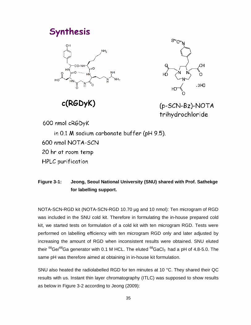
Figure 3-1: Jeong, Seoul National University (SNU) shared with Prof. Sathekge for labelling support.
NOTA-SCN-RGD kit (NOTA-SCN-RGD 10.70 µg and 10 nmol): Ten microgram of RGD
was included in the SNU cold kit. Therefore in formulating the in-house prepared cold
kit, we started tests on formulation of a cold kit with ten microgram RGD. Tests were
performed on labelling efficiency with ten microgram RGD only and later adjusted by
increasing the amount of RGD when inconsistent results were obtained. SNU eluted
their 68Ge/68Ga generator with 0.1 M HCL. The eluted 68GaCl3 had a pH of 4.8-5.0. The
same pH was therefore aimed at obtaining in in-house kit formulation.
SNU also heated the radiolabelled RGD for ten minutes at 10 °C. They shared their QC
results with us. Instant thin layer chromatography (ITLC) was supposed to show results
as below in Figure 3-2 according to Jeong (2009):

36
Figure 3-2: Quality control done by Seoul National University (Joeng, 2011).
It was suggested to place a drop of the 68Ga-NOTA-SCN-RGD on each plate of
chromatography paper. Sodium carbonate as well as 0.1 M Citric acid was used as flow
medium solutions. Results are described in Chapter 4.1; the Rr value was supposed to
be 1 in sodium carbonate and 0 in citric acid. HPLC was also performed and results
described later. The sodium carbonate ITLC was an indication of colloid formation. The
citric acid was a direct indication of labelling efficiency. Mostly in various cold kits
labelled, citric acid was available and used for ITLC in the in-house prepared study
performed.

37
At the beginning of this study when the donated cold kits were labelled, the focus was a
lot on perfecting the pH of the 0.6 M 68Ge/68Ga eluted 68GaCl3 that will be referred to 68Ga for the purpose of this study).
Shukla and Mittal (2013) described the pH influence on labelling efficiency. In this study
however, pH at a certain range for example 4-5 (using pH strips of up to 6 ranges) did
not affect labelling efficiency and for example still did not label well. It was noted later
when studies were done at iThemba labs, that adjusting the actual pH of the eluted 68Ga did not have an effect on labelling. When pH was not adjusted, there was a small
labelling yield but not enough labelling efficiency for preclinical or clinical studies. (It was
therefore then decided on an in-house kit formulation).
68Ga-NOTA-SCN-RGD was prepared in the following way after receiving the cold kits
from SNU:
The 68Ge/68Ga 0.6 M generator was eluted in fractions of 1 ml each into six sterile vials.
The Activity (A) in each vial was measured. Usually the highest activity was in vial
number 2.
A pipette was used and 1 ml eluate was pipetted from the second vial which always had
the highest activity. This was added to a clean sterile vial containing sodium hydroxide
and the solution was gently mixed. The sodium hydroxide volume depended on which of
the two methods further described shortly were used for example 68Ga labelling or 68Ga
labelling dilution method therefore either 45 µl sodium hydroxide (10 M) and 45 µl
sodium hydroxide (1 M) or only 450 µl of sodium hydroxide. A 0-6 pH paper strip was
used to test pH.
A metal needle for venting was used. A Jelco needle however was used for labelling in
order to prevent interference of the metal needle. One ml eluate was added to the RGD
cold kit vial. The needles were then removed and the vial transferred to the warmbath
for boiling. Boiling was always at least 5 minutes but up to 10 minutes at 90 degrees
Celsius.
Quality control was performed as suggested by Jeong, during correspondence, with
ITLC strips in sodium carbonate as well as citric acid flow mediums. Firstly a drop of the
radiolabelled 68Ga-NOTA-RGD was placed with either a 1 ml syringe or pipette onto
each ITLC paper strip approximately 8 mm from the origin of the paper strip. Four
38
plastic small pouches were prepared. One ITLC strip was gently dried a bit with a hair
dryer. It was then put into the flow mediums until it reached the font of the paper strip. It
was then gently dried with a hair-dryer and cutted into four quarters. Each quarter of
ITLC paper was put inside a plastic pouch and put into the Geiger-Muller counter for
counting radioactivity. The percentage radioactivity of each of all quarters was
calculated manually, because an ITLC scanner was not yet available for use at Steve
Biko Hospital.
68Ga Labelling RGD dilution method
The previous labelling method did not give positive labelling results even after great
efforts therefore labelling possibilities and new SOP’s were further investigated.
Esteban Ricardo Obenaus, head of Nuclear Medicine (NM) Argentina Diagnostic Centre
Buenos Aires suggested that a dilution method would ensure also the perfect pH in the
eluate prior to further labelling the eluate with the RGD cold kit.The dilution of the eluate
by adding 450 µl of Sodium Hydroxide (NaOH) was suppose to change the eluate of the
0.6 M Sodium Chloride (HCl) generator also to 0.1M.
68Ge/68Ga generator was eluted again the next day. From this day the generator was
not anymore eluted in quantities for example ml only, but in drops into different sterile
vials. Zero point two ml activity (A) was diluted with 1 ml sterile water. NaOH was added
to this then only one ml of this buffered eluate added to the RGD cold kit. Again only
Jelco needles were used (the plastic of the needle and no metal art of the needle).
This was mixed gently and heated. Quality control was performed using ITLC.
68Ga Labelling Method:
Subsequently the labelling SOP was changed regarding the dilution and buffering
method to adding 45 µl sodium hydroxide (10 M) and 45 µl sodium hydroxide (1 M) to
the eluate to adjust the pH. Again a plastic needle only was used to add radioactivity
into the RGD cold kit.
Eventually after testing two batches of RGD an inhouse kit was prepared. SOP’s for
radiolabeling as well as purification further is discussed after the radioactive isotopes
and therefore after 68Ge/68Ga has been discussed. Herewith follows the discussion on
the 68Ge/68Ga generator.

39
3.1.2 68Ge/68Ga Generator in detail
68Ge/68Ga Generator (Figure 3-3) was the generator of choice used in this study. This
generator is produced at iThemba LABS in the Western Cape, South Africa under
cGMP compliant conditions. The generator has a glass column that is made from tin-
dioxide with polyethylene tubing.
Figure 3-3: IDB Holland generator produced under cGMP conditions at iThemba LABS in the Western Cape (Eckert & Ziegler, 2008).
The 68Ge/68Ga generator consists of a mother isotope 68Ge which decays to its
daughter, 68Ga. 68Ge has a half-life of 271 days, which make it perfect to keep in a
hospital and nuclear medicine department for about one year. It is small and compact
and easily protected behind lead in compact space (Figure 3-4) and inside a laminar
flow cabinet Figure 3-5 and easy to transport. However, the daughter isotope, 68Ga that
is used for medical purposes, for example imaging, has a half-life of 68 min. This is
favourable with its positron emission for PET-CT scanning, however less practical to
transport due to short half-life. It is actually 68Ga (III) that is eluted from the generator.

40
Figure 3-4: iThemba LABS small and compact cGMP compliant generator behind lead.
The 68Ga has a specific elution profile (
Figure 3-6). This generator has no parts that are made from metal (except the outer
shielding). Metal can influence the labelling efficiency and therefore also no metal is
used in the radio labelling process. Needles for example are replaced with Jelco
needles where only the plastic part of the syringe was used to transfer the solution from
one glass vial to another. Sometimes when it was available, pipettes were utilised. This
was not always available at the Hospital site however plastic syringes were in order to
use as it did not interfere with labelling. It was important in this study to understand
more about the radio isotopes used for the correct handling of this for practicing the As
Low As Reasonably Achievable (ALARA) principle.68Ga is a positron emitter and should
be handled with the same care as any other radio-isotope. In the clinical environment
radiographers are more use to handling gamma emitters. However the same principles
adhere. A distance should be kept from a radio-isotope source as well as handling
should be as quick as possible.

41
Figure 3-5: Laminar flow cabinet compulsory for cGMP conditions in which 68Ge/68Ga generator is hosted.
The 68Ge/68Ga generator is suggested according to user instructions to be eluted in
fractions of 0.5 ml until a labelling procedure has been established for example to know
which volume of which ml fraction had to be used. Suprapure 0.6. M HCL (10 ml) was
used as solution to eluate the generator. Therefore if 1.5. ml was used for radiolabeling
the in-house prepared cold kit, then 8.5 ml eluted radio isotope was disregarded. It was
established in this study that the second 1.5 ml had the highest activity and was added
to the in-house prepared cold NOTA-RGD vial.

42
Figure 3-6: 0.6 M generator elution profile (IDB Holland user instructions).
It is not just important to understand fractionated elution but also the decay of the
mother isotope 68Ge as well as daughter isotope 68Ga.The Gallium decay chart was
extracted from another generator of 68Germanium/68Gallium’s instructions for use below
shown in Table 3-1 (Eckert & Ziegler, 2008). In all calculations for labelling efficiency the
Gallium decay factors were used to calculate decay corrected values.68Ga has 89%
decay by positron emission (Boschi et al., 2013) making it a favourable PET radiotracer.
The table below is for all time durations from one minute to 68 minutes. Radiolabeling
with this isotope will be further discussed in the following section 3.1.3. A comparison of
the Eckert & Ziegler pre-purification of 68Ga before radiolabeling as later in Chapter 4
compared with this study’s generator, the IDB Holland/iThemba LABS generator.
43
TABLE 3-1: Gallium-68 decay chart directly extracted from Eckert & Ziegler’s generator
User manual (Eckert & Ziegler, 2008).
Table 3-1: Half-life in minutes: Time represents 1-68 min
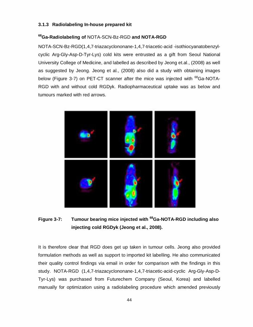
44
3.1.3 Radiolabeling In-house prepared kit
68Ga-Radiolabeling of NOTA-SCN-Bz-RGD and NOTA-RGD
NOTA-SCN-Bz-RGD(1,4,7-triazacyclononane-1,4,7-triacetic-acid -isothiocyanatobenzyl-
cyclic Arg-Gly-Asp-D-Tyr-Lys) cold kits were entrusted as a gift from Seoul National
University College of Medicine, and labelled as described by Jeong et.al., (2008) as well
as suggested by Jeong. Jeong et al., (2008) also did a study with obtaining images
below (Figure 3-7) on PET-CT scanner after the mice was injected with 68Ga-NOTA-
RGD with and without cold RGDyk. Radiopharmaceutical uptake was as below and
tumours marked with red arrows.
Figure 3-7: Tumour bearing mice injected with 68Ga-NOTA-RGD including also injecting cold RGDyk (Jeong et al., 2008).
It is therefore clear that RGD does get up taken in tumour cells. Jeong also provided
formulation methods as well as support to imported kit labelling. He also communicated
their quality control findings via email in order for comparison with the findings in this
study. NOTA-RGD (1,4,7-triazacyclononane-1,4,7-triacetic-acid-cyclic Arg-Gly-Asp-D-
Tyr-Lys) was purchased from Futurechem Company (Seoul, Korea) and labelled
manually for optimization using a radiolabeling procedure which amended previously

45
published knowledge by Rossouw and Breeman (2011) as well as Jeong’s formulation
and Dr Rossouw’s formulation for this cold kit. In order to allow non-metallic transfer of
the 68Ge/68Ga generator eluate into the reaction vial, the top of its septum was
punctured by a Jelco 22G x 1’’ polymer catheter (Smiths Medical, Croydon, South
Africa), sodium acetate trihydrate buffer salt was added to the 68Ga eluate to warrant pH
to 4-5 and 1.5 ml buffered 68Ga-eluate was used for labelling. Following the addition of
5.6 - 33.8 µM NOTA-RGD, stirring action was performed for 20-30 seconds. The
reaction vial was allowed to incubate in a temperature controlled water bath, routinely at
90 ºC for 10 min .It was then allowed to cool for at least 2 min. An aliquot was retained
for quality control purposes. This whole process is discussed in further detail below.
More detailed methods is discussed below and SOPs are available in Annexure A (cold
kit formulation) and Annexure B (radiolabeling and purification).
3.1.3.1 Certificate of analysis information
The Certificate of analysis (COA) was first obtained and explored in order to obtain
more info on product purity according to HPLC performed. The RGD was purchased
from Futurechem Company, Seoul, Korea. Important information below was obtained
on this staring material that was included in the in-house prepared cold kit.
Abbreviative name: NOTA-cyclic RGDyK
Product name: Cyclic Arg-Gly-Asp-D-Tyr-Lys-NOTA
Molecular Formula: C47H67N13014S.CF3COOH (Figure 3-8 below)

46
Figure 3-8: Molecular Formula: C47H67N13014S.CF3COOH (Futurechem).
Formula weight: 1184.18
Description: White solid
Purity(HPLC): >95%(at 240 nm)
Classification: Substance not fully tested.
It was important to know that all tests were not yet performed on this substance.This
included the kits that was donated originally.They were also from Korea and was
marked: ‘for experimental use only’. This was therefore not signed off for South Africa
as inhouse prepared cold kit.This also not yet passed cGMP compliant production
conditions.Most of the physical and chemical properties were not available according to
page four of the CoA.None of pH, density, bulk density, explosion limits, flamability and
47
autoignition temperature was available.The solubility was available and stated as
soluble in water.
In this study the solubility was tested. The substance quickly dissolved in water however
was not stable for more than about four weeks when frozen. The advice received from
Futurechem that aliquots should be prepared for kit vial preparation and immediately
freeze dried. This was therefore later done in this study. In the beginning of formulating
an in-house cold kit, the aim was rather to test if under the same conditions, the cold kit
would show reproducible radiolabeling with the eluted 68Ga from the 0.6 M 68Ge/68Ga
generator.
3.1.3.2 Peptide mass determination and shelf life
68Ga-NOTA-RGD buffered with sodium acetate trihydrate and containing 60 µg has
been tested up to 205 days post kit formulation date for shelf life integrity. Various
peptide mass were investigated especially after one RGD vial deteriorated in terms of
appearance for example most of the solid white powder disappeared and was sticky
onto the glass vials inside walls. It was decided to try and get all left over powder, even
though sticky, off the inside glass wall. This was used for cold kit formulation and tested
for further information regarding peptide mass and deterioration influence on labelling. It
was then suggested to rather use 60 µg instead of 30 µg of the RGD peptide.
Prior to this, at iThemba LABS in Cape Town, it was also agreed that different peptide
masses should be investigated as well as different buffer concentrations
Test A: Influence of pH at a smaller volume: (Experiment 1: pH 3.6)
Ninety eight mg of sodium acetate trihydrate was weighed in a glass vial. The generator
was milked/eluted with 0.6 M HCl as follows: Vial 1 with 1 ml, vial 2 with 2 ml, vial 3 with
6.5 ml HCl. One ml of vial 2 was added to 98 mg sodium acetate trihydrate and then 10
µl of the stock solution of NOTA-cyclic-RGDyk to obtain a 3.6 pH. The reaction volume
was 1010 µl and concentration 8.4 µM. The vial was sealed and heated for ten minutes
at 95 degrees Celsius. Then the vial was cooled for five minutes, uncapped and quality
control performed.
A few more tests were performed up to obtaining a Ph of 4.8-5 with 147 mg sodium
acetate trihydrate buffer. This was the final results for optimal labelling efficiency.

48
3.1.3.3 In-house prepared Cold kit raw materials, chemical structure and kit formulation.
Radiolabeling and in-house kit was prepared by using NOTA peptide from Futurechem
(Futurechem RGD structure). NOTA as chelating agent has been proven to form very
stable complexes with 68Ga (Brechbiel, 2008). A different buffer was added: Sodium
Acetate trihydrate.
The radiotracer was obtained by eluting a 68Ge/68Ga Generator (IDB Holland/iThemba
LABS) elution with ten ml 0, 6 M HCl. 68Ga-NOTA-RGD was purified by SepPak
purification process.
Materials and instruments were used according to SOP in Annexure A for kit formulation
and Annexure B for radiolabeling and purification as documented shortly below was
performed according to SOP steps below in Figure 3-9.
Equipment and materials: Instruments: All equipment was obtained and operated as
described in the SOP addendum A. The following equipment was therefore obtained:
Sterile vials (10 ml rubber seal) were prepared, crimper for cap sealing, balance for
weighing buffer, a small spatula for buffer weighing, filtration apparatus for working on
during buffer weighing, a vortex (it was tested to be working when the power was
switched on), various pipettes for example100-1000µl and 10-100µl, a labelling device
to print labels, a forceps to put rubber seals back onto the sterile vials, a freezer and a
freeze drier.
Chemicals and raw materials: The raw materials and chemicals that were obtained were
NOTA-RGD powder (from Futurechem) or aliquots of this peptide if this was already
previously prepared, suprapure water, sodium acetate trihydrate buffer and a 10-100µl
pipette and tips.
Step one was that sterile vials from NTP were set ready for adding buffer salts. Step two
was that the buffer (sodium acetate trihydrate) was weighed and transferred into all
sterile vials via small spatula. Step three was that the NOTA-RGD was prepared by
removing it from the fridge where according to the package insert of Futurechem it was
supposed to be kept 1 °C as maximum therefore it was kept at a cooler temperature of
minus 2-8 °C.

49
For the preparation of NOTA-RGD Kits (SOP Annexure A) 10 sterile vials on filtration
paper in laminar flow cabinet was prepared (unsealed rubber). A small spatula was
used and buffer weighed on the sensitive Balance for each vial (147 mg sodium acetate
trihydrate).
Buffer (Sodium Acetate Trihydrate). NB each time use forceps to put rubber seal cap
back on each vial. In order to prevent moist inside the vials and buffer, each time after
the buffer was weighed, a small forceps was used to put the rubber cap immediately
back onto a vial.
RGD-NOTA raw material was prepared by using 100 µl/ 100µg suprapure water to
dissolve the peptide in water. Therefore the 100-1 000µl pipette was used to transfer
1 000µl suprapure water to 1000 µg NOTA-RGD. It was then immediately vortexed.
Visually it was important to ensure that the solution was clear as water and no more
white powder visible. NOTA-RGD was transferred to each of the buffer containing sterile
vials. The 10-100µl pipette was used to transfer 60 µl (therefore 60 µg NOTA-RGD) to
each of the 10 prepared vials and then it was sealed only with rubber caps. It was noted
that a small volume of liquid was mixed with a larger volume of salt (buffer). It was still
vortexed leading to formation of a thicker liquid, which was vortex to be visible at the
bottom of the vial. The vials were stored in the freezer for about two hours in order for
the liquid to freeze before the vials were put into a freeze drier. The vials were freeze
dried overnight. All vials were inspected and all vials contained a white powder of freeze
dried NOTA-RGD. The vials were then sealed properly with rubber and metal caps. The
vials were then stored at about minus 20 °C. Transportation was only carried out when
the vials were packed with an ice pack.
In-house prepared kit radiolabeling and purification NOTA-RGD (Addendum B): Sterile
cGMP kit vials were produced at Radiochemistry; The South African Nuclear Energy
Corporation (Necsa, Pelindaba) using 5 ml certified sterile pyrogen-free sealed
borosilicate glass vials provided by NTP Radioisotopes. After peptide dissolution in
Millipore water aliquots of 60 µl of the 1 mg/ml NOTA-RGD stock solution were
transferred into glass vials (K60 kits 33.8µM). Alternatively, kits with reduced NOTA-
RGD content were used (K30 16.8 µM); all kits were supplemented with 147 mg of
sodium acetate trihydrate buffered salts as described above. Steps for radiolabeling and
purification were provided from the SOP (Annexure B) below in Figure 3-9.

50
Figure 3-9: Radiolabeling of the inhouse prepared NOTA-RGD kit (SOP Addendum B).
Radiolabeling was performed according to step one. The 68Ge/68Ga generator was
eluted using 0.6 M HCl (10 ml). The first 1.5 ml of eluted solution was disregarded. The
second 1.5 ml elution was added to the NOTA-RGD kit vial by using the plastic part of
the Jelco needle, 22 Gauge. The kit vial was vortexed for 30 seconds resulting in a clear
liquid. The kit vial was boiled for ten min at 90 °C in a boiler or when not in use by other
staff of Steve Biko Hospital, heated in a heating block for ten min also at 90 °C. The kit
vial was left to cool for 5 min. The pH was tested and documented. The radioactivity
was measured, therefore counted in the Geiger-Muller counter.
Purification was then done in step 2 to 4 (Figure 3-9). Purification materials was
prepared before the radiolabeling and included SepPak Alumina N (500 cc), ETOH,
Suprapure water, 2 sterile vials, decrimper, pipette, 2 x 2 ml syringes,1x 5ml syringe
(use for step 2 saline rinsing), 10 ml EtOH: Saline :10:90 (10 ml ensures enough for a
second labelling), forceps ( medium size) for vial transfer when activity needed to be

51
measured, 50 ml syringe with plastic connecting tube (same as nebulizer connecting
tube), two needles:25 Gauge for connecting to connecting tube and 50 ml syringe, 23
gauge for connecting to SepPak, saline 68Ge/68Ga generator (iThemba labs: IDB
Holland).
The SepPak was connected to the needle inserted into the one sterile empty vial.
The 50 ml syringe was attached to the 25 gauge needle and also connected to the
same vial. The syringe plunger was used by pulling it backwards to withdraw air and
precondition the SepPak with 4 ml ethanol (EtOH) and 2 ml water. The ethanol and
water was disregarded of and the vial kept for the following step (step 3 in Figure 3-9)
The reaction mixture (1.5 ml) was transferred into the SepPak with a pipette or syringe
(without the needle). The empty vial was rinsed with 1 ml saline and also transferred
into the SepPak. It was left to run through the column again by pulling plunger on 50 ml
syringe while drops and not a stream of liquid was visible, therefore e speed of
withdrawing air was also important. Disconnecting the syringe from the tube at any time
resulted in the stop of flow of liquid therefore ensuring while more liquid added to the
SepPak that the column did not run dry as yet. Another 4 ml of saline was used to rinse
the solution from the SepPak column and the column was still kept wet.
Step 4 (Figure 3-9) was the important step in which a new sterile vial was used and
connected to the SepPak, connection tube and as well as 50 ml syringe to this vial.
Three ml of the 10 % ethanol: saline (EtOH: NaCl) solution was transferred to the
SepPak until the column ran dry. The liquid collected in this last collection vial was the
product therefore 68Ga-NOTA-RGD. ITCL was performed as previously described by
using Citric Acid as well as Sodium Carbonate as flow mediums.
3.1.4 Radiolabeling and Purification: additional info
68Ge/68Ga-generator elution and pre-purification (done by overseas for additional data and publication with this study’s in-house prepared kits). 68Gallium(III)chloride (68Ga) was routinely eluted from a SnO2-based 68Ge/68Ga
generator (1.85 GBq, IDB Holland, The Netherlands) by fractionated elution with
moderate acidic condition (0.6 N HCl) assembling the 68Ga activity in 2-3 ml eluate
(reference method). For a set of experimental radiolabeling (N = 4) the 68Ga activity was
eluted from a TiO2-based 68Ge/68Ga generator (1.1 GBq, Eckert & Ziegler, Germany)

52
using lower acidic condition (0.1 N HCl); yielding 7.5 ml eluate which was subsequently
mixed with 30% HCL to yield a molarities > 5.5 M. A strong anionic Dowex resin (100-
200mesh, Fluka Analytics) was used to purify the activity solution from potentially co-
eluted metals and 68Ge. The resin-bound 68Ga was desorbed with ultrapure water
(Fluka Analytics) and assembled in 0.8 ml acidic solution (+SAX method). Alternatively,
a fractionated elution similar to the reference method was performed on the TiO2-based 68Ge/68Ga generator (-SAX method).
3.1.5 Purification of the Radiolabelled Product: Solid phase extraction/SPE:
In this study purification solutions were prepared prior to radiolabeling of 68Ga-NOTA-
RGD; Ethanol (EtOH), Suprapure water and EtOH: Saline in 20:80 and 10:90 was
prepared. SepPak connected to a sterile vial and a connecting tube was also prepared
(Figure 3-10).
Figure 3-10: Purification SepPak.
68Ga-NOTA-RGD was purified from unreacted 68Ga species by reversed-phase
chromatography. The C18 cartridge (500 mg 3 cc vac) was be pre-conditioned with 4ml

53
ethanol and equilibrated with 2ml sterile pyrogen free water (suprapure, deionised
Millipore 18.2M ). A sterile 2 ml syringe was used and sometimes a pipette to transfer
the reaction mixture onto the C18 Sep-Pak cartridge. It was essential to use non-metal
in this step for example no metal needles were used in this process due to metal
influence in the labelling process when radioactive metals used in labelling. The outflow
was collected in a waste vial one. A sterile syringe was used and sometimes a pipette
rather when available to rinse the reaction vial with 1 ml saline. This then was collected
in waste vial number two. This was rinsed with another 4 ml saline. Then experiments
was done with rinsing concentrations of 3 ml of ethanol/saline (10:90) and 2ml
ethanol/saline (20:80) to wash out the product from the C18 Sep-Pak cartridge into a
sterile vial. The activity of the product, empty vial and cartridge was measured.
Originally the last step was rinsing with 2 ml ethanol/saline (20:80) due to great purity
however this changed when a clinical dose concentration and correct pH was needed
for the preclinical imaging of three vervet monkeys. It was required for preclinical
imaging that the 68Ga-NOTA-RGD solutions were sterile-filtered through a 0.22 µm
membrane using a Millex, low protein-binding filter (Millipore).Preclinical
Vervet monkeys to be scanned were treated as clinical patients for example everything
regarding sterility, pH, minimum radioactive dose per weight injected and injecting a
pure product was applied. Therefore the correct clinical conditions were mimicked.
3.1.6 Quality Control
Instant thin layer chromatography (ITLC) analysis
Labelling efficiency was determined as described by Jeong (2008) and Breeman et al.,
(2005). Briefly, ITLC-SG paper was used as stationary phase and spiked at the bottom
with 3-4 µl of the crude or pure 68Ga-peptide solution, followed by 4-5 min exposure to
the mobile phase (0.1 M citrate, pH 4.5). For testing colloid forming a mixture of 1 M
ammonium acetate/methanol 1:1 (v/v) was used. The activity distribution was analyzed
on an ITLC scanner (VSC-201, Veenstra Ind., Oldenzaal, Netherlands) using a gamma
radiation detector (Scionix 25B25/1.5-E2, Bunnik, Netherlands) by obtaining
chromatograms. If peak identification was possible, an “area under the curve” analysis
allowed subsequent calculation of percentage peak recovery (Genie2000 software,
Veenstra Ind., Oldenzaal, Netherlands) to calculate the percentage labelling
efficiency.
54
High performance liquid chromatography (HPLC) analysis
Radiochemical purity was determined by high performance liquid chromatography
(HPLC) analysis using a Agilent 1200 series instrument coupled to 6100 Quadruple
mass spectrometry detector, (Agilent Technologies Inc., Wilmington DE, USA), diode
array detector (DAD) and radioactive detector (Gina Star, Raytest, Straubenhardt,
Germany). A Phenomenex Luna C18 column was used (250 x 4.6 mm, 5 µm) for
studies at iThemba LABS, Cape Town and Agilent Zorbax SB column (250x 4.6 mm,5
µm) at the rest of the studies performed at Necsa, Pretoria. The mobile phase consists
of (A) 0.1% trifluoroacetic acid (TFA) in water and (B) 0.1% TFA in acetonitrile; gradient
elution 0-2min 5%B and 2-32min 65%B and 32-35 min 5 %(this was added to
equilibrate the column faster for the next injection). Column temperature and flow rate
were set on 40 C and 1.0 ml/min respectively. Unbound 68Ga was eluted at 2.3-4 min; 68Ga-NOTA-RGD was eluted 11.4-12.5 min, respectively. The retention times of the
crude and SPE-purified compounds were corresponding to each other.
Quality control was performed by High pressure liquid chromatography instant thin layer
chromatography (ITLC) was also done in comparison to HPLC when possible.
Resource wise, Steve Biko Hospital would need to do quality control in the clinical
environment. .Even though HPLC is the preferred method due to its sensitivity in
detecting radiochemical purity as well as impure peaks. It was also interesting to
compare the HPLC results with the results that National University (SNU) obtained by
labelling also a RGD kit with their 0.1 M 68Ge68Ga Generator.
3.1.7 Kit shelf-life
The kit shelf life as well as labelled product’s shelf life is of significance for patient
management and planning in a Nuclear Medicine department. The half-life of the radio-
isotope plays a role in the labelling shelf life. It is suggested that the labelled shelf life
should be of for example Technetium-99m (SPECT compound) compounds should be
between 0.5 and 18 hour (Saha, 2010). When looking at positron emission labelled
radiopharmaceuticals, one would anyway want the quickest possible patient
administration due to very short half-lives and therefore such calculations would not
necessarily play a major role.
The kit stability and shelf life integrity was investigated for good radiopharmacy practice
purpose and optimum labelling efficiency as well as purity needed for clinical purposes.

55
In this study, the NOTA-RGD kit integrity and labelling efficiency was investigated over
a period of 205 days post kit manufacturing. Kits containing 60 µg of RGD was
investigated. According to IAEA documentation specific SPECT, Technetium-99m cold
kits have a shelf life from months to years, depending on the compound (IAEA, 2008)
3.2 Preclinical
3.2.1 Ethical approval
Xenografted study; Ethical approval was obtained from the North-West University
Ethical Committee for Mice Xenografts with approval project number: NWU-00042-12-
A5. This included inoculation of a 3 cell line A549 in nude mice as well as
comparing uptake of the radiotracer in healthy versus tumour bearing nude mice.
Vervet monkey study: The study was approved by the Animal use and care committee
University of Pretoria (Annexure C): S4560/09 as in Figure 3-11: Ethical approval
from Animal use and care committee (AUCC), Addendum D.below. The initial
application included requesting approval for 68Ga as well as Copper-64 (64Cu) labelled
with UBI which was changed to requesting labelling with RGD due to UBI labelling
giving problems at the time. Three primates were imaged on the PET-CT scanner at
Steve Biko Hospital. The approval request was specific due to lack of small animal PET-
CT scanners (so called micro-PET) in South Africa. The reason that monkeys were the
preferred animals for this study is because they have the size of small children. Small
children are scanned on the Human PET-CT scanner and a small amount of radiotracer
is needed for the advanced PET crystals to detect this. The monkeys that were scanned
for this study, ranged from 5, 8 -7, 6 kg (Mean weight is 6, 8 kg) and mean injected dose
of radiotracer only 2, 56 mCi. This is only 0, 37 mCi per kg of the patient/monkey.

56
Figure 3-11: Ethical approval from Animal use and care committee (AUCC),
Addendum D.
3.2.2 Xenografts: Mice
The experiments with xenografts were conducted according to the South African code
of practice for the care and use of animals and carried out by the Preclinical Drug
development Platform (PCDDP) of the North West University. Mice ex vivo data were
obtained. Two mice were tumour bearing after implantation of the A549 cell line.RGD
has been proven to be uptaken in 3 integrin expression cells as discussed
previously; therefore internalisation studies were not needed. It was therefore decided
to do very limited mice xenografts study to indicate the in house prepared 68Ga-NOTA-
RGD performs as expected.
The organs of two healthy and two tumour mice organs were harvested 2 hours after
intravenous injection in mouse tail with 68Ga-NOTA-RGD.Several organs as
documented below were harvested. Only two mice data have been used for
comparison.
The organ containing tubes were counted in the Geiger-Muller counter. All tubes were
also weighed according to ‘empty’ and ‘full’ when organs were included after harvested.

57
Harvested organs / tissues
Brain
Thyroid
Heart
Lung
Stomach
Intestines: large and small separate
Liver
Kidneys Right and left together
Bladder/Urine
Muscle
Femur
Tail
Blood
Corpse
Testis
Pancreas
Tumour reference

58
3.2.3 Scanning on Clinical PET.CT scanner and SUV
3.2.3.1 Patient background: Vervet Monkeys
Biodistribution in healthy vervet monkeys
The present study was undertaken with NOTA-RGD to validate its potential PET/CT.
The purpose is to evaluate the normal distribution of 68Ga-NOTA-RGD in primates. 68Ga-NOTA-RGD has showed in studies that it has a very fast blood clearance.
From former experience we know that the animals handle the administration
radiopharmaceuticals and subsequent scanning well (Ebenhan et al., 2012). The half-
life of the radionuclides to be used is short which limits the contamination risk by
excretion. During these experiments the biodistribution needs to be recorded in larger
animals to have sufficient resolution on a human PET-CT scanner. These experiments
will be non-terminal vervet monkey trials.
Healthy vervet monkeys were scanned after 68Ga-NOTA-RGD administration to reveal
generic tracer biodistribution and to obtain pharmacokinetic parameters in vivo. High
uptake levels in urinary bladder and kidneys dominated the 68Ga-NOTA-RGD
biodistribution, however, it’s acceptable for a PET agent, because it can be easily kept
under a hazardous radiation level and will seldom disturb image analysis.
The obtained tracer activity was sufficient for administration of 14 MBq /kg in large
vervet monkeys up to 7.50 kg to evaluate the in vivo biodistribution and also achieve
prior information about the in vivo stability. Results showed rapid blood clearance within
60 min post-injection and gradual soft tissue clearance via renal excretion and transient
liver uptake; no activity was found in potential target organs such as lungs, brain or
abdomen.
Animals, chemicals, and material
The experiments with primates were conducted according to the South African code of
practice for the care and use of animals approved carried out by the Biomedical
Research Centre of the Pretoria University. The animals were obtained from its colony
at Onderstepoort and checked for general health to void occult infection.
Pharmaceutical grade chemicals including 30% hydrochloric acid (HCl) Suprapure
grade were purchased from Merck. Cartridges for solid-phase extraction (SPE) were

59
obtained from Varian, Phenomenex and Waters Corporation. Instant thin-layer
chromatography-silica gel (ITLC-SG) was purchased from PALL Life Science. Sterile
Millex GV filters were obtained from Millipore. High-performance liquid chromatography
grade water was produced in-house using a Simplicity 185 Millipore system.
3.2.3.2 Scintigraphy and pharmacokinetics
The monkeys were treated like human patients and handled with respect and professional ethics at all times.
Three vervet monkeys (Cercopethicus aetiops) according to ethical application and
approval was investigated for normal biodistribution of the radiopharmaceutical 68Ga-
NOTA-RGD.The monkeys travelled from Onderstepoort to Steve Biko Hospital,
assisted, supported and handled by veterinary staff including a veterinarian. They were
housed according to ethical approval in accordance with South African National
Standards (SANS) and at all times handled by the veterinarian as well as scanned in his
presence (behind the lead glass at the PET-CT scanner).The animals were sedated.
They were weighed and during scanning protocol, urine and blood samples taken. The
animals were scanned as below on different dates to be able to give full attention and
support to one monkey only. The anaesthetic was administered by the veterinarian only
through an infusion pump. The endotracheal tube inserted into the trachea. An ECG
was connected and the animal at all times monitored. Some of the tubes are visualised
on the scintigraphic images obtained on the scanner and above explains the foreign
objects on the images. The Jelco needle that was placed in the femoral artery’s purpose
was to connect this to a device monitoring the monkey’s arterial blood pressure. The
purpose was secondly to obtain blood samples through the arterial catheter.
The monkey’s radiopharmaceutical as well as weight information is as below:
Table 3-2: Three Monkeys Preclinical PET-CT scan info
Scanned patient Monkey one Monkey two Monkey three Date of PET/CT
Scan
24 August 2012 27 August 2012 31 October 2012
Patient
Weight
7,6 kg 7 kg 5,8 kg

60
Injected
Dose
4,100 mCi 1,498 mCi 2,100 mCi
Scan 30 minutes
post injection
_ _ Scanned
Scan 60 minutes
post injection
Scanned Scanned Scanned
Scan 90 minutes
post injection
Scanned Scanned Scanned
Mean injected dose of monkeys is 2, 56 mCi. Mean weight of three monkeys is 6, 8 kg.
A clinical PET-CT camera (Siemens Biograph True Point, 40 slice CT) was used for
image acquisition. Images were acquired in three-dimensional mode and reconstructed
with and without attenuation correction (CT-based) using ordered subset expectation
maximization to yield axial, sagittal, and coronal slices (CT parameters: 120 kV, 100
mAs, slice thickness 5 mm, 0.8 mm pitch, matrix size 512 x 512) to yield axial, sagittal
and coronal slices. All images were first evaluated qualitatively followed by semi-
quantification analysis using Siemens e. Soft software on MI Processing workstation
(version 2.0.21; ABX GmbH). For the biodistribution experiments, three-dimensional
volume of interests (VOIs) were drawn manually over urinary bladder, brain, bone
(shoulder), muscles (triceps/quadriceps), kidneys, heart, liver and spleen. The final
values were expressed as organ standardized uptake value (SUV = [activity per ml
tissue] / [injected activity per body weight], g/ml)). Additional images for illustration were
processed with courtesy of Siemens International (Erlangen, Germany) on a
Multimodality Work Place using Software version VE52A with Fused Vision 3D. For the
patient case study, if abnormal uptake was present, a region of interest was manually
drawn around any lung lesions or any organ of interest (the size was selected to
correspond to the pathology on CT) and expressed as SUV. We used the maximum
SUV (SUVmax) in this study, which represents the pixel with the highest tracer uptake in
the lesion. All VOI’s done independently. Volume rendering techniques were done by
Siemens International. VOIS and SUV is further explained below.

61
3.2.3.3 Data processing: scintigraphy, blood and urine samples and SUV
VOIS: organs / tissues to obtain SUV
A volume of interest (VOI) has been drawn as listed below and as example in Figure
3-12, around the monkey’s organs to obtain SUV values as discussed in this previous
section. Volumes of interest were drawn over the liver, lung, heart, small intestine, large
intestine, spleen, kidneys and a background area to determine the uptake in each
organ. These values together with the information gained from the blood and urine
samples, have been used to draw time-activity curves to determine the biodistribution of
the radiopharmaceutical.
Figure 3-12: Volumes of interest(VOIs) drawn around the Monkey’s organs for SUV calculation.
VOIs have been drawn over the following organs:
Brain

62
Heart
Left hind
Kidney Right
Kidney Left
Shoulder Left
Liver
Triceps Right
Bladder
Spleen
SUV as explained previously as automatically calibrated in a PET-CT scanner
nowadays regarding the system’s quality control with its suggested radioactive
calibration sources and calibrations are performed to ensure perfect system operation
as well as other info that different PET-CT scanners need for example patient weight
and the amount of injected radiopharmaceutical dose is available. VOIs are drawn
around the patient organs and SUVs are then calculated automatically by the system.
SUVs (Standardized uptake values) are as explained by Kinahan and Fletcher (2010)
as related to kBq/ml therefore concentration and calculated as below:
SUV=r(a /w) [1]
In which r refers to radioactivity and a refers to the radiopharmaceutical amount. As
stated before, patient weight needs to be available in order for software of the PET-CT
to calculate SUV. The w in this formula refers to patient weight. The above authors
further explain that it does not matter how much activity is injected, the distribution
throughout the body would still be 1 g/ml.SUV significance comes in handy when
oncology patients need evaluation of Tumours for example before, during or after
chemotherapy. This will depend on when a patient could be scanned for example when
funds would be available. In South Africa therefore the scans are not always performed
in the above chronologic order.SUV is used for reporting purposes by Nuclear Medicine

63
Physicians or Radiologists in a PET-CT environment. As discussed above in this study
SUVmax have been used for monkey organ comparison in this study. This is in relation
with Kinahan and Fletcher’s findings (2010) for example that SUVmean does not have the
same reproducibility as SUVmax. All results are discussed in the next chapter.

64
Chapter 4: Results and discussion
“Negative results are just what I want. They’re just as valuable to me as positive results. I can never find the thing that does the job best until I find the ones that don’t.”...Thomas Edison
4.1 Imported cold kits background results
68Ga-Radiollabeling of NOTA-SCN-Bz-RGD and NOTA-RGD (inhouse prepared):
At Seoul National University analysis of the prepared results by ITLC using sodium
carbonate as well as citric acid as flow mediums (Figure 4-1) showed ITLC reaction
mixtures citric acid and sodium carbonate. Citric acid refers to labelling efficiency and
sodium carbonate to the radiochemical purity with regards to colloid formation. Figure
4.1b shows the HPLC results which refer to good product radiochemical purity and a
very small peak of possibly free 68Ga. while showed good product radiochemical purity.
A very small peak of free 68Ga was observed. However during the formulation of the
inhouse prepared kit in this study, below two percent of free 68Ga was also observed on
HPLC. It was possible though to get rid of free 68Ga that was observed on the HPLC
results by conducting a purification method.

65
Figure 4-1: ITLC analytical results of radiolabelled 68Ga- NOTA-SCN-Bz-RGD (Joeng, 2009).

66
Figure 4-2: HPLC results of 68Ga- NOTA-SCN-Bz-RGD (Joeng, 2009).
4.1.1 ITLC and results SNU imported cold kits
It was extremely difficult to get any labelling with the first batch using the imported kit.
Therefore we focused on obtaining the optimum pH by varying the pH to the 68Ga
eluate. This also did not improve labelling. Since no stability tests as well as stability test
on travelling outside a fridge, for the imported kits were obtained and no information
regarding the shelf-life was available it could be that the kits expired and therefore the
unsuccessful results. All imported kits were marked ‘for experimental use only’.
The second batch also only gave intermittent results; labelling was achieved before the
purification step but the SepPak purification step retained the biggest percentage of the
product which suggested that this was not suitable for preclinical imaging or for human
scintigraphic.
The imported tests that we tested gave the following results: Based on ITLC analysis
the NOTA-SCN-Bz-RGD kits from SNU showed of a 55 ± 10% labelling efficiency (N =
11), however, during SPE purification a complete adsorption of the activity to the

67
cartridge matrix (SepPak Alumina N, Waters) occurred. HPLC analysis showed multiple
peaks indicating decomposition. An overall high colloid forming tendency was observed,
amounting to 24 ±13%. Colloid formation was tested specifically with ITLC using the
sodium carbonate reaction mixture as discussed in chapter 3. Varying the labelling
parameters (molarity or type of buffering solution, molarity or volume of 68Ga-solution,
pH, incubation duration or temperature) did not positively affect the purification step.
The mean pH was 5.4 after radiolabeling. The HCl molarity was always 0.6 M. The
elution buffer was also changed to investigate if labelling efficiency would increase. At
the start of the study sodium hydroxide was used which was later changed to sodium
acetate (NaOAc). This however had no impact on the imported kit labelling efficiency
but was key to the success of the in- house prepared kits.
4.1.2 Purification:
4.1.2.1 HPLC at iThemba LABS
HPLC results of the first in-house prepared kits formulated at iThemba LABS and dried
in a desiccator is hereby documented. The rest of the kits were formulated at
Radiochemistry NECSA. The first inhouse kits prepared from RGD from Futurechem
were formulated and dried in a desiccator and showed great labelling efficiency of
almost 90% as well as purity of almost 90% were obtained. Examples are below in
Figure 4-2and Figure 4-3 of HPLC done on the same methods as discussed in Chapter
3, however different column as discussed was used than the column used at Necsa.
Initially the results were obtained after SepPak and desorbing radiolabelled product with
20% ethanol and 80% saline solution. As discussed this was then changed for clinical
purposes and lower volume for preclinical scanning. (Initial purity was higher with 20%
ethanol but the yield resulted in an amount of activity too low for example never more
than 50% for scanning purposes.)

68
Figure 4-3: HPLC at iThemba labs testing the first inhouse prepared kits.
Figure 4-3 contains a small peak at 3,316 min which could most likely be an
impurity/free 68Ga of 0, 87 % radiochemical purity. The two peaks though at 12,096 min
and 14,193 min are both RGD product peaks contributing to 7,10 and 87,50 %
radiochemical purity respectively.

69
Figure 4-4: HPLC at iThemba labs testing the first inhouse prepared kits:
specific using the 20:80 percent ethanol saline to desorb the radiolabelled product from the SepPak cartridge.
In Figure 4-4, the peak at 12,745 minutes has a 7,31 % radiochemical purity while the
peak at 14,271 minutes has 89, 47 % radiochemical purity. These two peaks are the
product peaks which sometimes differed in intensity for example two conformant peaks.
The labelling efficiency was 99, 50%. All product peaks were always visible between
10.5 and 14 min.
Purification (solid phase extraction)
K30 refers to 30 µg of RGD in the cold kit and K60 to 60 µg of RGD in the cold kit.Post
radiolabeling, SPE matrices were evaluated for optimal purification of 68Ga-NOTA-RGD
from free radiogallium and 68Ga-colloids (Table 4-1). Both SPE types show high total
desorption levels with an alcoholic saline solution, amounting to a total percentage of
adsorbed activity of 89 ± 3% and 76 ± 5% for Sep-Pak C18 500 mg 3cc vac and SPE
cartridge Sep-Pak light C18 100 mg, respectively. Significant higher amounts of activity
was desorbed with 10% alcoholic saline solution (P= 0.049), hence, a significant lower

70
SPE bound activity (P= 0.038) occurred for Sep-Pak C18 500 mg 3cc vac over the
online C18 light cartridge. For preclinical application the SPE elution with ethanol/saline
20/80 (v/v) protocol was avoided to achieve the minimum solvent levels for ethanol in
the final solution to be injected. The purified 68Ga-NOTA-RGD was found to be stable in
the injection solution up to the 60 min tested. The product radioactivity achieved was
204 ± 44 MBq (n = 10); (even with this small scale production it would be sufficient to
inject a patient with 185 MBq within 15-20 min after radiolabeling, full scale production
would ideally amount to 812 ± 84 MBq (calculated from N = 3 experiments). The
radiochemical purity (RCP) of 68Ga-NOTA-RGD determined by HPLC was 99.2 ± 0.54%
(n = 11), samples showed traces of 68Ge ( 0.0004%) and cationic metal impurities of
0.5 to 9 ppm, only. The calculated percentage of ethanol was 2.0-2.5% in the final
solution of the radiopharmaceutical product. K60 ( 60 µg RGD kit) 68Ga-NOTA-RGD
kits labelled with an average specific activity of 6.1 GBq/µmol (Range: 2.8-7.9
GBq/µmol), the full-scale specific activity based on the herein reported kit performance
is calculated as 18.1 GBq/µmol.
Table 4-1: Purification
Performance of SPE Purification on K60 68Ga-NOTA-RGD Kit Radiolabeling
SPE Sep Pak Type C18 500mg 3cc vac column #
C18 light (100mg) online cartridge #
Adsorbed Activity (AA) (MBq) 250 ± 64 119 ± 10
Total SPE Elution (% AA) 89 ± 3 76 ± 5
Elution Fraction: Ethanol /
Saline
% Ethanol / Saline (v/v)
10 / 90 (v/v) 77 ± 4* 63 ± 5
20 / 80 (v/v) 12 ± 1 13 ± 1
Retained Activity (% Range AA) 4-13* 15-37
#) Analysis of K60 kits with 90 days shelf lives are summarized. Values are expressed as mean (± SEM; N = 4). * = statistically significant difference (Student’s t-test P 0.05).

71
Figure 4-5 is an example of the final 2 ml of SepPak purification of the last
ethanol/saline solution desorbing the radiolabelled product from the cartridge and it is
clear that no free 68Ga is present. K30 refers to 30 µg RGD kit and K60 to 60 µg RGD
kit. Figure 4-5 below is an example of K60.
Figure 4-5: The final 2 ml of SepPak purification of the last ethanol/saline solution showing no free 68Ga.
SNU results discussed previously (section 4) showed a small percentage of free 68Ga
(retention time at 3.0 min) on the HPLC of the purified product although it showed a very
little of free of free 68Gallium using the suggested purification method of this study, it
was possible to remove the 2% free 68Gallium from the raw product (Figure 4-6) so that
the purified product contained no free gallium (Figure 4-7)

72
The peak of the purified product appears between 11 and 14 min as a multipeak due to
conformers that are formed by 68Ga-NOTA-RGD which varied between different
batches.
Figure 4-6: HPLC of the product before purification.

73
Figure 4-7: HPLC of product after purification.
4.1.3 68Ga eluate purification and NOTA-RGD radiolabeling
NOTA-RGD was successfully labelled with 68Ga eluted from SnO2- and a TiO2-based
generator (Table 4-2). Both generators provided a similar yield of radiogallium and the
complete labelling procedure including quality control was achieved in 34 and 38 min
(reference and –SAX , fractionated elution applied) or 43 min with implementing anion
exchange pre-purification (+SAX) of the 68Ga-eluate solution. Results indicate both a
substantial reduction of colloid formation and a slight increase in the percentage
labelling efficiency (%LE) for 68Ga-NOTA-RGD utilizing pre-purified generator eluate.
4.1.3.1 Pre purification additional info (performed by co-worker at the University of in Leuven, Belgium

74
Table 4-2: Influence of Generator Eluate Pre-Purification on 68Ga-NOTA-RGD radiolabeling
68Ga-NOTA-RGD Radiolabeling
Elution method* FE (Reference) FE, + SAX FE , - SAX
Generator type iThemba 50 mCi Eckert & Ziegler 30 mCi
Solid matrix material SnO2 TiO2
Mobile phase 0.6 M HCl 0.1 M HCl
Generator shelf life span (d) 96 - 140 124 - 134 101 - 108
Yield 68Ga (%) 94 ± 2 (n=7) 81 ± 4 (n = 11)
90 ± 1 (n = 7)
68Ga-elution discharge (%) bound - 8.6 ± 4.0 -
liquid 5.9 ± 1.9 8.5 ± 2.9 10.2 ± 1.3
ITLC
Crude labelling (%LE) 71 ± 6 81 ± 6 74 ± 6
RCP (%) 99.0 ± 0.50 99.2 ± 0.33 99.4 ± 0.25
Colloids (%) loss 8.0 ± 2.0 4.5 ± 2.2 8.5 ± 3.8
Sep Pak (%) loss 13 ± 2 12 ± 2 13 ± 3
Duration (min) 38 ± 2 43 ± 2 34 ± 4
*) Elution method: fractionated elution (FE) combined with (+SAX) and without (-SAX) pre-purification followed by standard labelling procedure were applied. Values are expressed as mean (± SD; N 3)
4.1.3.2 Mass determination of NOTA-RGD for radiolabeling
Manual radiolabeling was done prior to the kit formulation. At first about 10 µg as
NOTA-RGD was the same amount as in the SNU donated cold kit was giving a 31± 3 %
LE (N = 4). For improvement, kits containing 30 µg (K30; 16.8 µM) or 60 µg (K60; 33.8
µM) were compared with manual radiolabeling procedures adding 30 µg (NK30; 16.5
µM) or 60 µg (NK60; 33.1 µM) to the buffered generator eluate solution. Levels of

75
colloid formation were less for both the kits K30 (P = 0.073) and K60 (P = 0.081),
compared to manual 68Ga-NOTA-RGD labelling (NK30 and NK60; Figure 4-8 A).
Significant higher radiolabeling (% LE) was achieved using the K30 (P= 0.01) or K60
(P= 0.00017) kits over manual radiolabeling (NK30 and NK60; Figure 4-8 B). Neither
the amount of colloid forming (P = 0.225) nor the %LE (P = 0.139) differed between K30
(N = 4) and K60 (N = 7) kits labelling.
Figure 4-8: NOTA RGD kit performance: K30 and K60.
4.1.4 Kit shelf life integrity and K60 performance
A linear decrease was observed when kit integrity was analysed, comparing
radiolabeling efficiency (%LE) with kit stability over days. 68Ga-labeling of NOTA-RGD
kits (K60) with the shelf span up to 205 days post manufacturing (y = -0.3286x + 100; (r2
= 0,892) N= 19). Results indicated that > 75% LE and > 50% LE radiolabeling can be
achieved within the first 76 d and 152 d, respectively (Figure 4-8: NOTA RGD kit
performance: K30 and K60.Figure 4-8). A kit vial labelled 205 days post manufacturing
still show 19.6% labelling efficiency, however HPLC/ITLC analysis detected >52%
compound losses (SepPak and colloid forming) and radiolabelled by-products
presuming kit decomposition. The limited data on K30 kits show reduced integrity (93%
at 31 d and 66% at 90 d) compared to K60 kits. Figure 4-8 shows the impact of the

76
NOTA-RGD kit integrity on the percentage radiolabeling efficiency. Samples were
radiolabelled and analyzed with HPLC and/or ITLC. Results from N=19 individual
experiments are expressed as percentage of added 68Ga-activity.
4.1.5 68Ge/68Ga generator data
Figure 4-9 represents a generator that was 150 days old when this graph was created.
By the time of this graph created the eluted 68Ga was documented over 7.5 months.
This generator was eluted by fractions of 0.5 ml. This is as proposed by IDB
Holland/iThemba LABS user manual as documented earlier. This generator was eluted
by using 0.6 M HCl. Dat comparison is made with N = 3 elutions within one to two
weeks around the stated manufacturing time. This graph shows the gallium-68 peak
performance obtained in a specific volume that assisted us in knowing which ml to focus
on using and prevent us from measuring all the 0, 5 ml fractions in a clinical
environment.
Figure 4-9: Impact of the NOTA-RGD kit integrity on the percentage
radiolabeling efficiency.

77
It is also essential in clinical application to know the best time to milk (elute) the
generator for 68Ga use in order to obtain the best activity and labelling efficiency. A
rapid activity increase has been obtained with the iThemba LABS generator (Figure
4-10) indicating that the best time to milk the generator is 4-5 hours after the previous
elution and this could be determined by fractionated elutions of 0.5 ml.
Figure 4-10: Regeneration time from the parent decay.
4.2 Xenografts: Mice
Figure 4-11 represents the percentage of injected dose per gram of organ of the mice
xenografts. In this limited (confirmation) study only two xenografted mice and two
healthy mice were used. Both tumours of the two tumour mice did not have good

78
condition tumours i.e. the tumours were too big and blood supply was limited However
one tumour bearing mouse (inoculated with the A549 cell line) gave a 6.2:1 tumour to
muscle ratio and a 5.1:1 tumour to lung ratio. In Figure 4-11 the average of 2 animals
was reported As far as we are aware this was the first xenografted study performed with 68Ga-NOTA-RGD produced from a 0.6 M generator.
Unfortunately due to very low counts (due to the rapid decay of Ga-68), very small
organs (uncertainty in weight) and only 2 animals, the standard deviation (SD) in this
case is very high. Especially high SD for bladder and urine was recorded due to very
small weight of the organs. Nonetheless an average tumour to muscle ratio of 3.2:1 was
recorded while the tumour to lung ratio was 2.6:1. A tumour to healthy tissue ratio of 3:1
is essential to proof positive uptake of a tracer by the tumour.
It was clear that increased activity through excretion organs for example kidney and
urine/bladder was seen. It is also evident that there is no target organ in which the 68Ga-
NOTA-RGD shows increased uptake. The tumour however does take up the 68Ga-
NOTA-RGD which was expected.
0.00
5.00
10.00
15.00
20.00
25.00
30.00
urine
bladd
erTe
stis
large
intes
splee
nsto
mach
small
intes
Kidn
eys
liver
Tumo
ur re
fhe
art
lung
femu
rmu
scle tail
panc
reas
whole
bloo
d
%ID
/g
Organs

79
Figure 4-11: Percentage Injected dose per gram in Xenografted mice. The first (green) series is the tumour mice and the second (light blue) the
healthy mice.
4.3 Preclinical Imaging: Pharmacokinetics, biodistribution, and Image gallery
Preclinical imaging has been performed and volume rendering techniques applied to
show detailed preclinical images as the one below in Figure 4-12.
Figure 4-12: Volume rendering monkey image of biodistribution of radiotracer (Performed by Viola Satzinger from Siemens Erlangen).

80
Figure 4-13: Radiochemical purity and yield of three vervet monkeys.
The radiochemical purity (RCP) and labelling efficiency of the three different
radiopharmaceuticals that was prepared for scanning the vervet monkeys were
compared. The labelling efficiency mean was 51.21%.The RCP mean however was
almost 100% ,99.50% which was crucial for preparing protocol and SOP’s that would
ensure this purity before this study goes clinical. The labelling procedure for the last
monkey gave the highest labelling efficiency of 64%.This monkey was scanned during
the first stages of a new generator.
SUVs were obtained as on the example of Figure 4-14 below and as previously
discussed, specific SUVmax was used. (SUV = [activity per ml tissue] / [injected activity
per body weight], g/ml).

81
Figure 4-14: Max SUV used for patient pathology and clinical example urine.[
VOIs processing]
Volume rendering processing shows excretion through kidneys more clear below in
Figure 4-15.

82
Figure 4-15: Pelvic volume rendering of one vervet monkey to visualize
radiotracer uptake in kidneys.
Figure 4-16 shows no uptake in bones: Bone images and spinal detail are seen from the
CT scan. Again the two colourful blue, orange and yellow images are the two kidneys
through which the 68Ga-NOTA-RGD is excreted.

83
Figure 4-16: Volume rendering image of monkey of radiotracer in kidneys and
imaged of spine from CT.
All organ or tissues applicable to this tracer uptake (except kidneys) showed peak tracer
concentration at 30 min declining more or less gradually until 120 min. The present
study included the monitoring of the activity levels in blood and urine samples and
subsequent calculation of a rapid release represented by a biological half-life of 5 min
for 68Ga-NOTA-RGD, however, 54% of the injected activity dose was recovered in total
urine, 120 min post injection. Based on our findings we have reason to believe that
uptake, distribution and excretion of NOTA-RGD is very favourable to monitor lung
cancer. It is noted, that the biodistribution showed no hepatobiliar excretion and may
therefore postulated to may detect tumours located in pancreas, abdomen and even
prostate (as previously described).

84
Table 4-3: Organ and Tissue Concentration of 68Ga-NOTA-RGD in Healthy Vervet Monkeys;
Values expressed as mean SUV (±SEM) if applicable.
*) N = 3 animals; $) N = 2 animals, #) N = single animal
Figure 4-17 and d shows Concentration of 68Ga-NOTA-RGD in blood and urine samples
from healthy vervet monkeys. Values in blood (Figure 4-17) assembled as a time-
activity curve following an exponential decline (y=0.751e-0.151x; R2= 0.971) after
Savitzky-Golay data smoothing algorithm applied. Measurements in urine (Figure 4-18)
show a linear incline (y=0.125x; R2=0.967). Results are expressed as mean ± SD of N
3 animals and expressed as percentage of the injected 68Ga-activity per bodyweight
(%ID/g).
Organ/ Tissue SUV
30 min p.i.# 60 min p.i.* 90 min p.i.# 120 min p.i.$
Heart 1.01 0.89 ± 0.32 0.62 0.35 ± 0.12
Liver 1.24 1.71 ± 0.70 1.91 0.49 ± 0.08
Spleen 1.22 1.60 ± 0.49 1.39 1.10 ± 0.60
Bladder, urinary tract 24.7 31.2 ± 14.5 24.1 23.5 ± 3.4
Kidney 3.53 3.19 ± 0.88 2.78 1.94 ± 0.84
Quadriceps muscle 0.19 0.24 ± 0.04 0.22 0.22 ± 0.04
Triceps muscle 0.18 0.23 ± 0.05 0.22 0.22 ± 0.03
Bone (shoulder) 0.36 0.43 ± 0.12 0.38 0.39 ± 0.09
Brain 0.04 0.04 ± 0.02 0.05 0.07 ± 0.01

85
Figure 4-17: Time/activity curve of 68Ga-NOTA-RGD in monkey’s blood.
Figure 4-18: Time/activity curve of 68Ga-NOTA-RGD in monkey’s urine.
0.00
0.25
0.50
0.75
1.00
1.25
0 10 20 30 40 50 60 70
BLO
OD
-R
GD
%ID
/g
Time (min postinjection)
0
4
8
12
16
20
24
0 20 40 60 80 100 120 140
UR
INE
-RG
D %
ID/g
Time (min postinjection)

86
Figure 4-19: Volume rendering of monkey from head to pelvis showing
endotracheal tube, kidneys and urine in bladder.

87
4.4 68Ga-NOTA-RGD Concentrations in Blood and Urine of healthy vervet Monkeys
The time dependent concentration of 68Ga-NOTA-RGD measured from individually
taken blood samples (N = 10) was further optimized by mathematical data smoothing
using Savitzky-Golay algorithm (Origin Pro 8, OriginLab Corporation, Northampton).
The results of the fluid analysis of 68Ga-NOTA-RGD confirmed that the peak blood
activity concentration was obtained 1 min after injection, amounting to 0.75 ± 0.07 %
ID/g (Figure4.1.1.1.c). The blood-bound activity rapidly declined (y=0.751e-0.151x R2 =
0.971) as represented by a pharmacological half-life of only 5 min and an elimination
rate of 9 %ID/h. Urine samples showed steadily accumulating activity levels of 68Ga-
NOTA-RGD following a linear incline (y=0.125x; R2=0.967; N = 6) peaking at 16 ±
5%ID/g at 120 min (Figure4.1.1.1.d). The accumulation rate was calculated 7.5 % ID/h
with a total urine recovery reaching 54 ± 4 %ID (N = 3) after 120 min.
The obtained tracer activity was sufficient for administration of 14 MBq /kg in large
vervet monkeys up to 7.5 kg to evaluate the in vivo biodistribution and also achieve prior
information about the in vivo stability. Results showed rapid blood clearance within
60 min post-injection and gradual soft tissue clearance via renal excretion and transient
liver uptake; no activity was found in potential target organs such as lungs, brain or
abdomen in is in relation to what is needed in a clinical radiopharmaceutical.
Additionally, all organ or tissues (except kidneys) showed peak tracer concentration at
30 min declining more or less gradually until 120 min. The present study included the
monitoring of the activity levels in blood and urine samples and subsequent calculation
of a rapid release represented by a biological half-life of 5 min for 68Ga-NOTA-RGD,
however, 54% of the injected activity dose was recovered in total urine, 120 min post
injection. Based on our findings we have reason to believe that uptake, distribution and
excretion of NOTA-RGD is very favourable to monitor lung cancer. It is noted, that the
biodistribution showed no hepatobiliar excretion and may therefore be postulated to
may detect tumours located in pancreas, abdomen and even prostate (as earlier
discussed).

88
Chapter 5: Conclusions, future work and recommendations
Cyclotron produced radioisotopes are currently used for imaging lung cancer, although
it cannot yet differentiate, for example in a single pulmonary lesion, between a tumour
and tuberculosis. Consequently, imaging can only be done close to cyclotrons, limiting
the availability of imaging as well as the availability of various cyclotron radiotracers.
Therefore there was a need for the formulation of an in-house prepared kit for labelling
NOTA-RGD with 68Ga eluted from a 68Ge/68Ga generator for imaging integrin 3
expression in lung cancer.
We set out to formulate an in-house prepared cold kit after the imported NOTA-SCN-Bz-
RGD cold kits failed to show consistent radiolabeling with 68Ga eluted from the 68Ge/68Ga iThemba LABS generator, distributed by IDB Holland bv. We have completed
formulation of cold kits, radiolabeling and purification standard operating procedures
(SOPs) successfully in productions of 30 and 60 µg batches. SOPs for kit formulation
and radiolabeling with purification can be found in the attached annexures A and B
respectively.
The synthesis involved raw peptide material from Futurechem, Korea. Seoul National
University’s radiolabeling steps with regards to radiolabeling, heating of the product as
well as purification were followed, although different kit formulation and purification
methods were implemented. HPLC results were compared with ITLC done at Necsa in
order for correct ITLC results to be known when working in a clinical environment where
only ITLC is available.
HPLC results were compared with ITLC results done at Necsa in order for correct
procedure and results to be found for implementation at Steve Biko Hospital.
This study succeeded in meeting the initial needs as stated below:
A new NOTA-RGD cold kit was formulated for labelling with gallium-68 eluted from a
0.6 M generator and a SOP was written for kit formulation (Annexure A). A radiolabeling
and purification method was formulated to label and then clean all free gallium-68 and
other impurities from the radiolabelled tracer and a SOP was written (Annexure B). Mice
xenografts were performed and the biodistribution between healthy and tumour bearing
mice compared. The 68Ga-NOTA-RGD that was labelled with the in-house prepared

89
cold kit showed uptake in the tumour of the tumour bearing mice that was inoculated
with A 459 cells. The radiotracer also showed a higher uptake in the tumour than in the
muscle and showed fast blood clearance and excretion through the kidneys and then
the bladder. Three vervet monkeys were scanned and the biodistribution of the 68Ga-
labelled radiotracer examined. Normal radiotracer uptake and also fast blood clearance
and excretion through the kidneys and bladder were obtained with no adverse
reactions, which make this a promising radiopharmaceutical. The advantage is that
cheaper, faster than cyclotron produced radiotracers, can be produced, which are more
readily available as a positron emitter for imaging lung cancer and integrin 3
expression tumours.
Therefore this new in-house cold kit for labelling with a 0.6 M 68Ge/68Ga generator has
been tested from molecule to mouse to monkey and in the near future will be tested in
man.
This study may in future have a major impact on guidelines and management of TB
infected patients, with regard to targeted imaging and therapy. Clinical trials are still
needed for this purpose. Furthermore it may facilitate the dissemination of the 68Ga-
labelled radiotracers approach worldwide especially in developing countries as an
alternative to cyclotron produced PET tracers.
The future might include testing tracers like this on both PET-CT and PET-MRI which
could have an advantage of combining high MRI spatial resolution and PET’s high soft
tissue contrast to obtain physiological information. Due to lack of resources, it would be
an option to contact the raw product supplier, Futurechem, regarding future possibilities
of qualifying a cGMP kit. Integrin expression comparison with PET.CT and MR or
scanning with PET.MR may be utilized in the future.
“The modern 68Ge/68Ga generators have proved to be a milestone for non-invasive
state-of-the –art PET/CT imaging. After that there was no looking back for Ga-68
imaging”, (Shukla and Mittal, 2013).

90
References
American Cancer Society., 2013:
http://www.cancer.org/acs/groups/cid/documents/webcontent/003115-pdf.pdf Date of
access: 13 March 2014.
Angiogenesis in cancer.,http://angioworld.com/cancer.htm Date of access: 16 July
2014.
Antunes P,Ginj M,Zhang H,Waser B,Baum R.P, Reubi J.C, Maecke H,Are radiogallium-
labelled DOTA-conjugated somatostatin analogues superior to those labeled with other
radiometals?Eur J Nucl Med Mol Imaging,2007,34:982-993.
Arosio D,Manzoni L,Araldi E.M.V,Scolastico§ C, Cyclic RGD Functionalized Gold
Nanoparticles for Tumor Targeting. Bioconjugate Chem., 2011, 22(4):664-672.
Arosio D,Manzonit L, Araldi E.M.V,Scolasticos C,Cyclic RGD Functionalized Gold
CHECK: 28.Weis S.M,Cheresh D.A, Integrins in Angiogenesis and Cancer.UCSD
Nanoparticles for Tumor Targeting.Bioconjugate Chem,2011,22(4):664-672.
Ballinger J.R,Solanki K.K,What will be required to bring 68Ga-labelled peptides into
routine clinical use? Nuclear Medicine Communications, 2011, 32:1109-1112.
Bartholomä M.D, Recent developments in the design of bifunctional chelators for metal-
based radiopharmaceuticals used in Positron Emission Tomography.Anorganica
Chimica Acta, Volume 389, 2012,389:36 – 51.
Baum R.P, Hellwig D, Mezzetti M, Position of nuclear medicine modalities in the
diagnostic workup of cancer patients: lung cancer. Quarterly Journal of Nuclear
Medicine and Molecular Imaging, 2004, 48(2).
Baum R.P, Prasad V, Jeong J.M, Ga-68 NOTA-RGD PET/CT For The Evaluation of
Tumor Angiogenesis-First Clinical Results.EANM, 2008: Poster.
Beer A.J, Grosu A,Carlsen J, Kolk A,Sarbia M, Stangier I, Watzlowik P, Wester
H,Haubner R,Schwaiger M, [18 F]Galacto-RGD Positron Emission Tomography for
Imaging of 3 Expression on the Neovasculature in Patients with Squamous Cell
Carcinoma of the Head and Neck.Clin Cancer Res,2007,13(22):6610-6616.

91
Beer A.J,Lorenzen S,Metz S,Herrmann K,Watzlowik P,Wester H,Peschel C,Lordick
F,Schwaiger M,Comparison of Integrin 3 Expression and Glucose Metabolism in
Metastatic Lesions in Cancer Patients:A PET study Using 18F-Galacto-RGD and 18F-
FDG.The Journal of Nulcear Medicine,2008,49(1):22-29.
Bello B, Fadahun O, Kielkowski D, Nelson G, 2011.Trend in lung cancer mortaility in
South Africa: 1995-2006. BMC Public Health. http://www.biomedcentral.com/1471-
2458/11/209 [Date of access:13 March 2014].
Berry D.J,Yonmin M,Ballinger J.R, Tavaré, R,Koers A, Sunassee K, Zhou T, Nawaz S,
Mullen G.E.D., Hider R.C, Blower P.J, Efficient bifunctional gallium-68 chelators for
positron emission tomography: tris(hydroxypyridinone) ligands.Chemical
Communications, 2011:47:7068-7070.
Blom E,Velikyan I,Estrada S,Hall H,Muhammad T,Ding C,Nair M,Langström B. 68Ga-
Labeling of RGD peptides and biodistribution., Int J Clin Exp Med, 2012:5(2):165-172.
Bodanis D.2000.E=mc2.America:Walker Books.
Boschi S,Malizia C,Lodi F,Overview and Perspectives on Automation Strategies,(in 68GaRadiopharmaceutical Preparations in Baum R.P & Rösch F,Theranostics,Gallium-
68,and other radionuclides.A Pathway to personalized Diagnosis and
Treatment).2013:17-31.
Breeman WA, de Jong M, de Blois E, Bernard BF, Konijnenberg M, and Krenning EP.
Radiolabeling DOTA-peptides with 68Ga. Eur J Nucl Med Mol Imaging 2005;32:478-85.
Briechbiel M,Bifunctional Chelates for Metal Nuclides.Q J Nucl Med Mol
Imaging,2008,2008,52(2):166-173.
Chen X, Sievers E, Hou Y, Park Y, Tohme M, Bart R, Bremner R, Bading J.R, Conti
P.S,Integrin 3 –Targeted Imaging of Lung Cancer. Neoplasia, 2005, 7(3):271-279.
Danhier F, Le Breton A, Préat V, RGD-based strategies to target alpha(v) beta (3)
integrin in cancer therapy and diagnosis. Molecular Pharmaceutics, 2012 5;9(11):2961-
73.

92
Dechantsreiter M.A,Planker E,Matha B,Lohof E,Holzemann G,Jonczyk A, Goodman
S.L, Kessler H,J. Med.Chem,1999,42.
Decristoforo C,Gonzalez I.H,Carlsen J,Rupprich M,Huisman M,Virgolini I,Wester
H,Haubner R,68Ga-and 111In-labelled DOTA-RGD peptides for imaging of integrin 3
expression.Eur J Nucl Mol Imaging,2007.
de Sa A, Matias AA, Prata MI, Geraldes CF, Ferreira PM, and Andre JP. Gallium labeled NOTA-based conjugates for peptide receptor-mediated medical imaging. Bioorganic & medicinal chemistry letters 2010;20:7345-8.
Desgrosellier J.S, Cheresh D.A, Integrins in cancer: biological implications and
therapeutic opportunities. CANCER (Nature reviews), 2010, 10:9-22
Dijkgraaf I, Yim C, Fransen G.M, Schuit R.C,Luurtsema G,Liu S,Oyen W.J.G,Boerman
O.C, PET imaging of 3 expression in tumors with 68Ga-labelled mono-,di-and
tetrameric RGD peptides.Eur J Nucl Med Mol Imaging,2011,38:128-137.
Dos Santos I.1999.Cancer Epidemiology:Principles and Methods.Geneva:IARC Library
Cataloguing Data.
Dumont R.A,friederike H,Haubner R,Maecke H.R, Weber W.A,Fani M,Novel 64Cu and 68 Ga-labelled RGD Conjugates Show Improved PET Imaging of 3 Integrin
Expression and Facile Radiosynthesis.J Nucl Med, 2011,52:1276-1284.
Ebenhan T, Govender T, Kruger G, Pulker T, Zeevaart JR, and Sathekge M. Synthesis
of 68Ga-NOTA-UBI30-41 and in vivo biodistribution in vervet monkeys towards potential
PET/CT imaging of infection. Journal of nuclear medicine : official publication, Society of
Nuclear Medicine 2012;53:1520.
Eckert & Ziegler IGG 100 Instructions for use Gallium-68 Decay Chart:2008.
Eo JS, Paeng JC, Lee S, Lee YS, Jeong JM, Kang KW, Chung JK, Lee DS,
Angiogenesis imaging in myocardial infarction using 68Ga-NOTA-RGD PET:
characterization and application to therapeutic efficacy monitoring in rats. Coron Artery
Dis.,2013,24(4):303-311.

93
Ferreira CL, Lamsa E, Woods M, Duan Y, Fernando P, Bensimon C, et al. Evaluation of bifunctional chelates for the development of gallium-based radiopharmaceuticals. Bioconjugate chemistry 2010;21:531-6.
Futurechem RGD structure: NOTA-cyclic RGDyK: South Korea.
Galibert M,Z Jin,Furukawa T,Fukumura T,Saga T,Fujibayashi Y,Dumy O,Boturyn
D,RGD-cyclam conjugate:Sythesis and potential application for positron emission
tomography.Bioorganic & Medicinal Chemistry Letters,2010,20:5422-5425.
Guerin B,Ait-Mohand S,Tremblay M,Dumulon-Perreault V,Fournier P,Benard F, Total
Solid-Phase Synthesis of NOTA-Functionalized Peptides for PET
Imaging.Org.Lett.,2010,12(2):280-283.
Herbst M.C,CANSA Fact sheet on lung cancer,2013:1-17.
Health24 press release., 2013:Data access: 14 March
2014.http://www.health24.com/Medical/Cancer/Facts-and-figures/South-Africa-78-
increase-in-cancer-by-2030-20120721
I.D.B.Holland, Operating Instructions GMP Produced 68a Generator iThemba LABS
IAEA-TECDOC-466.Technetium-99m RadiopharmaceuticalsManufactureof Kits.2008
IAEA-TECDOC-1603. The role of PET/CT in Radiation Treatment Planning for Cancer
Patient Treatment. 2008.
Jamous M, Haberkorn U, and Mier W. Synthesis of peptide radiopharmaceuticals for the
therapy and diagnosis of tumor diseases. Molecules 2013;18:3379-409.
Jemal A, Bray F, Center M, Ferlay J, Ward E, Forman D, Global Cancer Statistics.CA
CANCER J Clin 2011, 61:69-90.
Jeong J.M, Formulation and Preparation of 68Ga-NOTA-RGD for Angiogenesis
Imaging. 2008. [correspondence].2011.
Jeong J.M,HPLC 68Ga-NOTA-RGD.[email]. 9 November 2011.
Jeong J.M, Hong M. K,Chang Y.S,Lee Y,Young J.K,Cheon G.J,Lee D.S,Chung
J,Lee,M.L, Preparation of a promising Angiogenesis PET Imaging Agent:68Ga-Labeled

94
c(RGDyK)-Isothiocyanabenzyl-1,4,7-Triazaclononane-1,4,7-Triatetic Acid and
Feasibility Studies in Mice. The Journal of Nuclear Medicine,2008,49(5):830-836..
Jeong J.M, Kim Y.J, Lee D.S ,Chung J.K, Lee M.C, Radiolabeling of NOTA and DOTA
with Positron Emitting 68Ga and Investigation of In Vitro Properties. Nucl Med Mol
Imaging, 2009,Aug; 43 (4) :330-336.
Jiang T, Zhang C,Zheng X,Xu X,Xie X,Liu H,Liu S,Noninvasively characterizing the
different alphavbeta3 expression patters in lung cancers RGD-USPIO using a clinical
3.0 T MR scanner.Int J Nanomedicine,2009,4:241-9.
Joshi S .C,Pant I, Hamzah F,Kumar G, Shukla AN, Integrated positron emission
tomography/computed tomography fusion imaging: an emerging gold standard in lung
cancer.Indian J Cancer,2008 45(4):137-141
Kenny L.M,Coombes R.C,Oulie I,Contractor K.B, Miller M,Spinks T.J, McParland B,
Cohen P.S, Hui A, Palmieri C, Osman S, Glaser M, Turton D,Al-Nahhas A, Aboagye
E.O,Phase I Trial of the Positron-Emirring Arg-Gly-Asp(RGD) Peptide Radioligand 18F-
AH111585 in Breast Cancer Patients.J Nucl Med,2008,49:879-886.
Kim J.H.K,Lee J.S,Keon W.K,Lee H,Han S,Kim T,Lee Y,Jeong J.M,Le D.S, Whole-Body
Distribution and Radiation Dosimetry of 68Ga-NOTA-RGD, a Positron Emission
Tomography Agent for Angiogenesis Imaging. Cancer Biotherapy &
Radiopharmaceuticals.,2012,27(1):65-71
Kinahan P.E,Fletcher J.W.PET-CT Standardized Uptake Values(SUVs) in Clinical
Practice and Assessing Response to Therapy.Semin.Ultrasound CT
MR.2010:31(6):496-505.
Knetsch P.A,Milos P,Christoph M,Griessinger M,Rangger C,Fani M,Kesenheimer C,von
Guggenberg E,Pichler B.J,Virgolini I,Decristoforo C,Haubner R, [68Ga]NODAGA-RGD
for imaging integrin 3 expression.Eur J Nucl Med Mol Imaging,2011,38:1303-1312.
Knetsch P.A,Petrik M,Rangger C,Pietzsch H.J,Virgolini I,Decristoforo C,Haubner R.N
Synthesis and evaluation of [68Ga]NS3-RGD-a new tracer for imagaing integrin 3
expression.Nucl Med Biol.2013,40(1):65-72

95
Knetsch P.A,Petrik M,Rangger C,Seidel G,Pietzsch H,Virgolini I,Decristoforo C,Haubner
R, [68Ga]NS3-RGD and [68Ga] Oxo-DO3A-RGD for imaging 3 integrin expression.
Nuclear Medicine and Biology 2012:1-8.
Knetsch P.A,Seidel G,Rangger C,Decristoforo C ,Pietzsch H,Pietzsch J,Haubner
R,Synthesis and evaluation of [68Ga]NS3-RGD-a new tracer for imagaing integrin 3
expression.Radioactive Isotopes in Clinical Medicine and Research 29th International
Symposium.2010.
Kubicek V, Havlickova J, Kotek J, Tircso G, Hermann P, Toth E, et al. Gallium(III) complexes of DOTA and DOTA-monoamide: kinetic and thermodynamic studies. Inorganic chemistry 2010;49:10960-9.
Lee Y,Jeong J.M, Kim H.W,Chang Y.S,Kim Y.J, Hong M.K, Rai G.B, Chi D.Y,Kang W.J,
Kang J.H, Lee D.S, Chung J,Lee M.C,Suh Y,An improved method of 18F peptide
labeling:hydrazone formation with HYNIC-conjugated c(RGDyK).Nuclear Medicine and
Biology,2006,33:677-683.
Lee Y,Radiopharmaceuticals for Molecular Imaging.The Open Nuclear Medicine
Journal,2010,2:178-185.
Lui S,Park R, Conti P,Li Z, “Kit-like” 18F labeling method for synthesis of RGD peptide-
based PET probes.American Journal of Nuclear Medicine and Molecular
Imaging,2013,3(1):97-101
Malvezzi M, Bertuccio P, Levi F, La Vecchia C, and Negri E. European cancer mortality
predictions for the year 2013. Annals of oncology : official journal of the European
Society for Medical Oncology / ESMO 2013;24:792-800.
Melcher C,PET-CT history: http://www.engr.utk.edu/smrc/history.html ; Date of
Access:17 April 2014.
Mbulaiteye S.M, Parkin D.M, Rabkin C.S, Epidemiology of AIDS-related malignancies
An international perspective, Hematology/Oncology Clinics of North America, 2003, 17;
673-696.
Pakkala S, Ramalingam S.S, Lung Cancer in HIV-positive patients. Journal of Thoracic
Oncology, 2010, 5(11):1864-71.

96
Positron emission tomography:
http://en.wikipedia.org/wiki/Positron_emission_tomography ; Date of Access: 21 July
2014.
Powles T, Nelson M, Bower M, HIV-related lung cancer-a growing concerning J STD
AIDS,2004,15(3):211-2.
Rossouw D.D,Breeman W.A.P, Scaled-up radiolabeling of DOTATAE with 68Ga eluted
from a SnO2- Based 68Ge/68Ga generator.Applied Radiation and Isotopes,2012,70(171-
175).
Saha G.B,2010.Fundamentals of Nuclear Pharmacy.6th edition.Cleveland
USA,:Springer.
Sathekge M, Buscombe J.R, Can positron emission tomography work in the African
tuberculosis epidemic? Nuclear Medicine Communications, 2011,32:241-244.
Sathekge M.M, Maes A,Pottel H,Stolz A,van de Wiele C,Dual time –point FDG PET/CT
for differentiating benign from malignant solitary pulmonary nodules in a TB endemic
area.SAMJ,2010,100(9):598-601.
Shukla J,Mittal B,R,68Ga-A versatile PET imaging radionuclide.J Postgrad Med Edu
Res,2013,47(1):74-76.
Siemens, Molecular Imaging Training. 2014
South Africa: 78% increase in cancer by 2030. http:// www CDC .GOV/CANCER
/LUNG/ Date of access:13 March 2014
Standardized uptake value. http://en.wikipedia.org/wiki/Standardized Uptake Value;
Date of access: 21 July 2014.
Tan M,Lu Z,Integrin Targeted MR Imaging.Theranostics,2011:1:83-101
Velikyan I, Prospective of 68Ga-Radiopharmaceutical
Development.Theranostics,2014,4(1):47-80.

97
Wängler C, Björn, Lehner S, Elsner A, Todica A, Bartenstein P, Hacke M, Schirrmacher
R, A universally Applicable 68Ga- Labeling Technique for Proteins.J Nuc
Med,2011,52(4):586-91.
Weis S.M,Cheresh D.A, Integrins in Angiogenesis and Cancer.UCSD Cancer
centre & Department of Patholgy,2011.
Xanthopoulos EP, Corradetti MN, Mitra N, Fernandes AT, Kim M, Grover S, et al.
Impact of PET staging in limited-stage small-cell lung cancer. Journal of thoracic
oncology : official publication of the International Association for the Study of Lung
Cancer 2013;8:899-905.
Zhang Y, Hong H, Engle JW, Bean J, Leigh BR, Barnhart TE, Cai W. Positron Emission
Tomography Imaging of CD105 Expression with a 64Cu-labeled Monoclonal
Antibody:NOTA is superior to DOTA. PLoS ONE 6(12): e28005, 2011.
Zhernosekov K.P, Filosofov D.V, Baum R.P,Aschof P, Heiner B, Anatoli A.R,Jahn
M,Jennewin M, Rösch F,Processing of Generator-Produced 68Ga for Medical
Application.Society of Nuclear Medicne,2007,48(10):1741-1748.

Applied Chemistry
This document is the property of NECSA and shall not be used, reproduced, transmitted or disclosed without prior written permission
The electronic version of this document within the DocMan System is regarded as the official document Page 1 of 7
CONFIDENTIAL
Annexure A
Manufacturing work instruction for NOTA-cyclic RGDyK Kit production
DOC NO: RC-NOTA RGD-WIN-11002
Revision: 0
Date: January 2014
Document Approval: Name Signature Date Prepared By: J Wagener/(I Schoeman) Checked By: Approved By: J R Zeevaart
Distribution 1. QA Records 2. JR Zeevaart
3. J Wagener 4.
5. 6.
7. 8.
9. 10.
11. 12. The revision history of the document is available in the DocMan System

Applied Chemistry
RC-TRAPT195-WIN-11001 Rev: 1 Page 2 of 7
CONFIDENTIAL
Manufacturing work instruction for NOTA-cyclic RGDyK Kit production
DOC NO: RC-NOTA RGD-WIN-11002
Revision: 0
Date: 2014-01-14
TABLE OF CONTENTS
1 Purpose 3 2 Scope 3
3 Abbreviations and definitions 3
4 Reference 3 5 Responsibilities 3
6 Safety 4
6.1 Prerequisites............................................................................................................................ 4 6.2 PPE......................................................................................................................................... 4 6.3 Precautions ............................................................................................................................. 4
7 Method 4 7.1 Flow chart for kit production of RGD-NOTA:According to SOP step numbers: ....................... 4 Equipment and materials ....................................................................................................................... 5 7.2 Preparation of NOTA-RGD Kits ............................................................................................. 6
8 Records 7

Applied Chemistry
RC-TRAPT195-WIN-11001 Rev: 1 Page 3 of 7
CONFIDENTIAL
1 Purpose This document defines and specifies the manufacturing procedures applicable to the
pharmaceutical product NOTA-cyclic-RGDyk(Futurechem).
2 Scope This procedure describes the synthesis of NOTA-cyclic-RGDyk(Futurechem)
manufactured kits by Radiochemistry, Necsa.
3 Abbreviations and definitions NOTA-RGD NOTA-cyclic-RGDyk
RPO Radiation Protection Officer
PPE Personal Protective Equipment
RSP Responsible Pharmacist
4 Reference The following documents had a direct influence in the process and activities defined in this document: 1.COA Futurechem manufacturers of NOTA-RGD 2. Jeong J.M, Hong M. K,Chang Y.S,Lee Y,Young J.K,Cheon G.J,Lee D.S,Chung J,Lee,M.L,
Preparation of a promising Angiogenesis PET Imaging Agent:68Ga-Labeled c(RGDyK)-
Isothiocyanabenzyl-1,4,7-Triazaclononane-1,4,7-Triatetic Acid and Feasibility Studies in Mice.
The Journal of Nuclear Medicine,2008,49(5):830-836.
3. Jeong J.M et al, Formulation and Preparation of 68Ga-NOTA-RGD for Angiogenesis Imaging.
Seoul National University:(powerpoint ).
5 Responsibilities 1. Responsible Scientist is responsible for:
a) carrying out the synthesis as detailed in this instruction and for completion of
the Batch record (RC-NOTA RGD-WIN-11002) b) overall performance of the QC procedures, including the label checking.
2. Responsible Pharmacist is responsible for:
a) checking of QC results;
b) final release of product.
3. QC staff are responsible for:
a) QC testing of the product according to the appropriate work instructions.

Applied Chemistry
RC-TRAPT195-WIN-11001 Rev: 1 Page 4 of 7
CONFIDENTIAL
6 Safety
6.1 Prerequisites i) Each scientist assigned to manufacture this product must be suitably trained to
perform the synthesis and be informed of relevant regulations.
6.2 PPE i) Wear a clean room suit, powder free gloves, and overshoes during synthesis.
6.3 Precautions i) New materials entering the clean room must be cleaned and then wiped with 70%
alcohol before and after pass-through.
7 Method
7.1 Flow chart for kit production of RGD-NOTA:According to SOP step numbers:
1.Prepare Sterile Vials
2. Weigh Buffer and transfer to vials
3.Prepare NOTA-RGD
4. Transfer with pipette
NOTA-RGD to each of vials in step 2.Seal/Vortex

Applied Chemistry
RC-TRAPT195-WIN-11001 Rev: 1 Page 5 of 7
CONFIDENTIAL
Equipment and materials Instruments: Operate all equipment as described in manuals or SOP
a) Sterile vials.(10 ml rubber seal)
b) Crimper for cap sealing.
c) Balance(for weighing buffer)
d) Spatula:small(for use while weighing buffer)
e) Filtration apparatus ( filter paper to work on during buffer measurements)
f) Vortex
g) 100-1 000µl pipette and tips
h) 10-100µl pipette and tips
i) Labeling device to print etiquettes
j) Forceps(to put rubber seal caps back on sterile vials)
k) Freezer
l) Freeze drier
Chemicals/Raw materials: m) NOTA-RGD Powder/Aliquots(if previously prepared)
n) Suprapure water
o) Buffer:Sodium acetate tryhidrate
5.Put all vials in freezer for at least
two hours till frozen.
6.Freeze dry overnight
7.Check vials and seal with metal cap
and Crimper.
8&9: Store in freezer:
Transport with ice packs.

Applied Chemistry
RC-TRAPT195-WIN-11001 Rev: 1 Page 6 of 7
CONFIDENTIAL
p) 10-100µl pipette and tips
7.2 Preparation of NOTA-RGD Kits 1 Prepare e.g. 10 sterile vials on filtration paper in laminar flow cabinet.(Unsealed
rubber)
2 Use the small spatula and weigh on the sensitive Balance for each vial 147 mg
Buffer(Sodium Acetate Tryhidrate).NB each time use forceps to put rubber seal
cap back on each vial.
3 Prepare the RGD-NOTA:Use 100 µl/100µg suprapure water/NOTA-RGD:
Therefore use the 100-1 000µl pipette to transfer 1 000µl suprapure water
to 1000µg NOTA-RGD and vortex(500 µl to the current 500 µl aliquots).
NB: Visually: make sure clear transparent like water and no more white powder
visible. Therefore CLEAR.
4 Transfer NOTA-RGD to each of the buffer containing sterile vials:
Use the 10-100µl pipette and transfer 60 µl(therefore 60 µg) to each of the
10 prepared vials and seal only with rubber cap.
NB: Little liquid with a lot of salt but VORTEX till this forms a thicker’ liquid and
the salt has a clearer dissolved appearance if not fully dissolved. Make sure all is
on bottom of the vial still.
5 Freezer: Put vials in freezer for about two hours. Check that solvent is solid and
frozen before putting in the freeze drier.
6 Freeze dry overnight (rubber caps positioned also for freeze dryer so not yet fully
sealed with metal cap).
7 Check that all vials freeze dried and then seal with Crimper and metal cap.
8 Store in freezer.
9 Transport with ice pack.
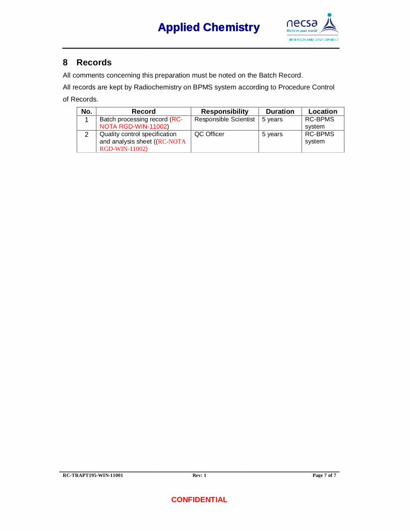
Applied Chemistry
RC-TRAPT195-WIN-11001 Rev: 1 Page 7 of 7
CONFIDENTIAL
8 Records All comments concerning this preparation must be noted on the Batch Record.
All records are kept by Radiochemistry on BPMS system according to Procedure Control
of Records.
No. Record Responsibility Duration Location 1 Batch processing record (RC-
NOTA RGD-WIN-11002) Responsible Scientist 5 years RC-BPMS
system 2 Quality control specification
and analysis sheet ((RC-NOTA RGD-WIN-11002)
QC Officer 5 years RC-BPMS system

Applied Chemistry
This document is the property of NECSA and shall not be used, reproduced, transmitted or disclosed without prior written permission
The electronic version of this document within the DocMan System is regarded as the official document Page 1 of 8
CONFIDENTIAL
Annexure B
Manufacturing work instruction for NOTA-cyclic RGDyK Kit labelling
with Gallium-68 eluted from 68Ge/68Ga generator
and purification
DOC NO: RC-NOTA RGD-WIN-11002
Revision: 0
Date: January 2014
Document Approval: Name Signature Date Prepared By: J Wagener/(I Schoeman) Checked By: Approved By: J R Zeevaart
Distribution 1. QA Records 2. JR Zeevaart
3. J Wagener 4.
5. 6.
7. 8.
9. 10.
11. 12. The revision history of the document is available in the DocMan System

Applied Chemistry
RC-TRAPT195-WIN-11001 Rev: 1 Page 2 of 8
CONFIDENTIAL
Manufacturing work instruction for NOTA-cyclic RGDyK Kit labeling
with Gallium-68 eluted from 68Ge/68Ga generator
and purification
DOC NO: RC-NOTA RGD-WIN-11002
Revision: 0
Date: 2014-01-14
TABLE OF CONTENTS
2 Purpose 4 3 Scope 4
4 Abbreviations and definitions 4 5 Reference 4
6 Responsibilities 4
7 Safety 5 7.1 Prerequisites............................................................................................................................ 5 7.2 PPE......................................................................................................................................... 5 7.3 Precautions ............................................................................................................................. 5 7.4 Housekeeping ......................................................................................................................... 5
8 Method 6
8.1 Flow chart for NOTA-RGD kit with Gallium-68 and Purification: ........................................... 6

Applied Chemistry
RC-TRAPT195-WIN-11001 Rev: 1 Page 3 of 8
CONFIDENTIAL
Equipment and materials ....................................................................................................................... 7 8.2 Preparation of 68Ga-NOTA-RGD Kits and purification ........................................................... 7
9 Records 8

Applied Chemistry
RC-TRAPT195-WIN-11001 Rev: 1 Page 4 of 8
CONFIDENTIAL
1 Purpose This document defines and specifies the labeling and purification procedures applicable
to the pharmaceutical product NOTA-cyclic-RGDyk(Futurechem) for labeling with
Gallium 68 eluted from the 68Ge/68Ga generator.
2 Scope This procedure describes the labeling of NOTA-cyclic-RGDyk(Futurechem)
manufactured kits by Radiochemistry, Necsa and Gallium-68 therefore
68Ga-NOTA-RGD as well as the purification method.
3 Abbreviations and definitions NOTA-RGD NOTA-cyclic-RGDyk
RPO Radiation Protection Officer
PPE Personal Protective Equipment
RSP Responsible Pharmacist 68Ga Gallium-68
4 Reference The following documents had a direct influence in the process and activities defined in this document: 1.COA Futurechem manufacturers of NOTA-RGD 2. Jeong J.M, Hong M. K,Chang Y.S,Lee Y,Young J.K,Cheon G.J,Lee D.S,Chung J,Lee,M.L,
Preparation of a promising Angiogenesis PET Imaging Agent:68Ga-Labeled c(RGDyK)-
Isothiocyanabenzyl-1,4,7-Triazaclononane-1,4,7-Triatetic Acid and Feasibility Studies in Mice.
The Journal of Nuclear Medicine,2008,49(5):830-836.
3. Jeong J.M et al, Formulation and Preparation of 68Ga-NOTA-RGD for Angiogenesis Imaging.
Seoul National University:(powerpoint ).
2.? As jy nog references benodig van tesis
5 Responsibilities 1. Responsible Scientist is responsible for:
a) carrying out the synthesis as detailed in this instruction and for completion of
the Batch record (RC-NOTA RGD-WIN-11002) b) overall performance of the QC procedures, including the label checking.
2. Responsible Pharmacist is responsible for:

Applied Chemistry
RC-TRAPT195-WIN-11001 Rev: 1 Page 5 of 8
CONFIDENTIAL
a) checking of QC results;
b) final release of product.
3. QC staff are responsible for:
a) QC testing of the product according to the appropriate work instructions.
6 Safety
6.1 Prerequisites i) Each scientist assigned to manufacture this product must be suitably trained to
perform the synthesis and be informed of relevant regulations.
ii) Request for irradiation and transport records must be checked to ensure all the
required information has been supplied.
iii) Reagents used in this synthesis should be prepared beforehand and stored
appropriately.
iv) The Labeling laboratory used in the production process must be clean and free of
any items associated with any previous work procedures.
v) All precautions and safety instructions have been read and understood.
6.2 PPE i) Wear a clean room suit, powder free gloves, and overshoes during synthesis.
ii) Use electronic personal dosimeter to monitor possible radiation exposure after
production.
6.3 Precautions i) New materials entering the clean room must be cleaned and then wiped with 70%
alcohol before and after pass-through.
ii) All production activities must be performed behind lead shielding.
iii) RPO shall monitor radioactive starting material before entering the facility and
product or any items before exiting the facility for radioactive contamination.
6.4 Housekeeping i) Remove all items used during synthesis after RPO monitoring.

Applied Chemistry
RC-TRAPT195-WIN-11001 Rev: 1 Page 6 of 8
CONFIDENTIAL
7 Method
7.1 Flow chart for NOTA-RGD kit with Gallium-68 and Purification: Labeling 68Ga with NOTA-RGD cold kit

Applied Chemistry
RC-TRAPT195-WIN-11001 Rev: 1 Page 7 of 8
CONFIDENTIAL
Equipment and materials Instruments and raw materials: Operate all equipment as described in manuals or SOP
Step 1: Radiolabelling: a)86Ge/68Ga generator elute. b)Disregard the first 1.5 ml. c) Add second second 1.5 ml to the NOTA-RGK Kit vial by using the plastic of the jelco needle 22 G. d)Vortex 30 seconds.Liquid should be clear. e)Boil at 90 Degrees Celsius for 10 minutes. f)Test pH.Measure radio activity. g)Do Purification step 2-4.
Purification Step 2-4 a) SepPak Alumina N (500 cc)
b) ETOH
c) Suprapure water
d) 2 sterile vials
e) Decrimper
f) Pippet/2 x 2 ml syringes
g)1x 5ml syringe(use for step 2 saline rinsing)
h)10 ml EtOH:Saline :10:90(10 ml ensures enough for a second labeling)
i) Forceps medium for vial transfer when need to measure
j) 50 ml syringe with plastic connecting tube(same as nebulizer connecting tube)
k)Two needles:25 Gauge for connecting to connecting tune and 50 ml syringe
l) 23 gauge for connecting to sepPak
m) Saline
n)68Ge/68Ga generator(iThemba labs:IDB Holland)
7.2 Preparation of 68Ga-NOTA-RGD Kits and purification
Applied Chemistry
RC-TRAPT195-WIN-11001 Rev: 1 Page 8 of 8
CONFIDENTIAL
Step 2: Precondition the sepPak that is connected to needle inserted in the one sterile emty vial.
Attach the 50 ml syringe to the 25 gauge needle and also connect to the same vial.
Pull syringe plunger to withdraw air and precondition the sepPak with 4 ml EtOH and 2
ml water.
Discard the water and use the same vial and kepe connected to sepPak and 50 ml
syringe.
Step 3: Now transfer the reaction mixture(1.5 ml) onto the sepPak with a pippet or
syringe(without the needle).
Then rinse the empy vial with 1 ml saline and also transfer onto sepPak and let it run
through the column again by pulling plunger on 50 ml syringe.
Stop transfer if need by disconnecting syringe from tube.This should not yet run dry!
Now also rinse another 4 ml saline and still keep the column wet.
Step 4: NB!Now use a new sterile empty vial(the second vial) and connect the sepPak as well as
50 ml syringe to this vial.
Transfer 3 ml of the 10 % EtoH in EtOH:Saline solution and let this run dry.
Do ITLC by using Citric Acid as well as Sodium Carbonanate as reaction mixtures.
8 Records All comments concerning this preparation must be noted on the Batch Record.
All records are kept by Radiochemistry on BPMS system according to Procedure Control
of Records.
No. Record Responsibility Duration Location 1 Batch processing record (RC-
NOTA RGD-WIN-11002) Responsible Scientist 5 years RC-BPMS
system 2 Quality control specification
and analysis sheet ((RC-NOTA RGD-WIN-11002)
QC Officer 5 years RC-BPMS system


![Review The Search for an Alternative to [ Ga]Ga-DOTA-TATE ...thno.org/v09p1336.pdf · [68Ga]Ga-DOTA-TATE, [68Ga]Ga-DOTA-TOC, and [68Ga]Ga-DOTA-NOC allows for NET staging with high](https://static.fdocuments.in/doc/165x107/5e2a1b5b2104573c786ad22c/review-the-search-for-an-alternative-to-gaga-dota-tate-thnoorg-68gaga-dota-tate.jpg)


![[68Ga]PSMA-HBED-CC Uptake in Osteolytic, Osteoblastic, and ... · Conclusions: [68Ga]PSMA-HBED-CC uptake is higher in osteolytic and bone marrow metastases compared to osteoblastic](https://static.fdocuments.in/doc/165x107/607572caf32e2d79681dbd86/68gapsma-hbed-cc-uptake-in-osteolytic-osteoblastic-and-conclusions-68gapsma-hbed-cc.jpg)


![[68Ga]NOTA-Galactosyl Human Serum Albumin: a Tracer for ... · 68Ga]NOTA-GSA showed slower uptake in comparison with 68Ga-DTPA-GSA (123 ± 10 vs. 89 ± 3 s, p G 0.01). Conclusions:](https://static.fdocuments.in/doc/165x107/5f6d9cae33a64152a27bd022/68ganota-galactosyl-human-serum-albumin-a-tracer-for-68ganota-gsa-showed.jpg)


![68Ga] peptide high-output production on commercially · [68Ga] peptide high-output production on commercially available MiniAIO® synthesizer Objectives Optmization parameters Material](https://static.fdocuments.in/doc/165x107/5f95b4c0eecbac70717355d8/68ga-peptide-high-output-production-on-68ga-peptide-high-output-production-on.jpg)

