SAMPLE NOTES, FULL EDITION IS ORDERED AND … · Lecture: Role of the Kidneys in Long-Term Blood...
12
SAMPLE NOTES, FULL EDITION IS ORDERED AND COMPLETE. Topics: 1. Role of the Kidneys in Long-Term Blood Pressure Regulation 2. Sex differences in Cardiovascular Disease 3. Endothelial control of blood flow: novel mechanisms 4. Brain and Stroke 5. Kidney Disease 6. Steroid Hormones and Cardiovascular Disease 7. Early Origins of CVD 8. Heart Failure 1&2 9. Early origins of CVD -Stress and pregnancy (Sympathetic overactivity) 10. Metabolic Syndrome 11. Steroid Hormones and Cardiovascular Disease 12. Steroid Hormones and signalling mechanisms (alternate explanation)
Transcript of SAMPLE NOTES, FULL EDITION IS ORDERED AND … · Lecture: Role of the Kidneys in Long-Term Blood...

SAMPLE NOTES, FULL EDITION IS ORDERED AND COMPLETE.
Topics: 1. Role of the Kidneys in Long-Term Blood Pressure Regulation
2. Sex differences in Cardiovascular Disease
3. Endothelial control of blood flow: novel mechanisms
4. Brain and Stroke
5. Kidney Disease
6. Steroid Hormones and Cardiovascular Disease
7. Early Origins of CVD
8. Heart Failure 1&2
9. Early origins of CVD -Stress and pregnancy (Sympathetic overactivity)
10. Metabolic Syndrome
11. Steroid Hormones and Cardiovascular Disease
12. Steroid Hormones and signalling mechanisms (alternate explanation)

Lecture: Role of the Kidneys in Long-Term Blood Pressure Regulation
Role of the kidneys in the long-term regulation of blood pressure (mates I recommend to
go through the lecture while studying notes for this lecture, Roger kinda jumped from one
place to another)
From Previous lectures:
● Hypertension is predominantly caused by a shift in pressure natriuresis/diuresis
relationship, so in order for kidney to excrete the daily intake of salt and water a
higher blood pressure is required. So a higher blood pressure will be developed
because there is a slowly developing increase in extracellular fluid volume, and so a
slowly developing increase in blood volume, and therefore a slowly developing
increase in arterial pressure.
● It has been found that in people with high blood pressure, TPR is elevated instead of
CO.
● Autoregulation is the ability of a tissue to maintain the blood flow within the tissue
at a relatively stable level in the face of change in perfusion pressure.
● Local blood flow = arterial pressure / organ vascular resistance
The basic components of cardiovascular
control:
● Arterial pressure is the product of CO
and TPR, and not only the autonomic
nerve and hormones influence CO and
TPR, but also the level of arterial
pressure influences the activity of
those neural and hormonal
mechanisms (baroreceptor reflex).
Cardiac output is controlled by the
frank-staling law of the heart, and
cardiac filling is mostly controlled by
the level of blood volume in the longer
term. Blood volume is largely a product of ECF volume. ECF volume is determined by
the balance between salt and water input and output. Therefore, the kidney is the
central factor, because it controls the excretion of salt and water, also the level of
arterial pressure influences the amount of excretion (the pressure natriuresis).
The Concept of Salt-Resistance, and ‘Salt-Sensitive and Non-Salt-Sensitive Hypertension
● The pressure natriuresis relationship is normally very steep. So consequently, an
increase in salt intake has a small effect on the blood pressure.

● In all cases of high blood pressure the pressure natriuresis/diuresis relationship is
shifted to the right.
● Some people develop salt sensitive hypertension. In these people the pressure
nantruresis relationship is not really steep, which means that change in salt intake
causes dramatically changes their arterial pressure.
● Not only the kidney response to changes in blood pressure, but it also responds to
change in the neural and hormonal control mechanisms.
● Experimentally reducing the ability of the kidney to excrete salt and water, it is
possible to induce salts sensitive hypertension.
The kidney-body fluid mechanism operates in long-term blood pressure control
● The baroreceptors reflex is one of the first mechanisms that can modulate the blood
pressure for seconds to minutes, however, longer than that the baroreceptors will
start to reset.
● The renal-blood volume pressure
control, can bring back the blood
pressure to normal, and it doesn't
reset like the baroreceptors.
In head-out water immersion, the increase
in pressure on lower parts of the body,
pushes the veins and decreases their
volume. Therefore, venous blood will be
pushed up to the chest, which increases
the central blood volume. Therefore, it
increase the venous return, which
increases the cardiac output.
Increasing the arterial pressure
causes the execution of salt and
water to increase by the kidney.
Not only the renal blood flow
(RBF) is autoregulated so as the
glomerular filtration rate (GFR).
Therefore the increase in salt and
water excretion as arterial
pressure increases, is related to
inhibition of sodium
reabsorption. As the arterial
pressure increases (within the

physiological range), the kidney reabsorbs less sodium in the tubules (the sodium is not
filtered more). So pressure natriuresis/diuresis is mediated by altered tubular function.
During the head out water immersion there is an increase in cardiac output and arterial
pressure, also there is also activation of baroreceptors. Therefore, the sympathetic drive will
be inhibited. This means that for considering the effect of head out water immersion on
kidney function, it is important to consider the effect of blood pressure and change in neural
and hormonal status.
SAMPLE NOTES, FULL EDITION IS ORDERED AND COMPLETE.
Lecture: Sex difference in cardiovascular disease:
· All forms of CVD disease effect women more greatly than men.
Sex Bias in Research:
· Historically women were excluded from clinical trials because of different reasons:
o If women were pregnant or they had the chance of being pregnant, the
researchers did not want to treat them with drugs that can have potential
side effects on the baby.
o There is the idea that hormonal cycle in women makes the data more
variable, therefore more patients is needed for a significant effect, which
increases the costs.
· In phase I drugs safety, tolerability and other factors are checked. Phase II trials are
designed for testing dosing and efficacy. These two phases do not require the use of
women. Therefore, there are more adverse drug effects in women than in men, because
the drugs were mostly tested in men.
o One of the problems in drug development is that, the drugs pass phase I
and II trials, however they will be stopped in phase III trials, because side
effects become apparent in women and then the drug is discontinued.
Therefore, some of the drugs that work well in men are being
discontinued.
Representation of females in clinical trials:
· Evidence-based medicine is much more applicable to the men than women:
o Low number of female participants, and the data are not being analysed
relative to sex.
o Differences are apparent in CVD, including: Women have atypical
symptoms that can lead to delays in treatment, diagnostic test that are
developed in men don’t work in women, and biomarkers.
o Pregnancy is also a major limiting factor.

Sex differences in treatment (based on studies):
· Cumulative probability of death or myocardial infarction in patients with confirmed
coronary disease and stable angina according to gender. Greater risk of death following
myocardial infarction in women than men.
· Post myocardial infarction women were not getting standard treatments. Women
were undergoing less invasive procedures to correct the problems. These all resulted in
greater death rate.
· Women are less likely to receive standard therapy or best evidence-based therapy
following acute coronary syndrome.
Sex differences in treatments is because of sex differences in CVD pathophysiology:
· Typical symptoms in women are different to men, which results in slower
recognition of cardiac events.
· Heart attract symptoms:
o Men can suffer from the atypical symptoms, but women mostly suffer
from the atypical symptoms. Therefore, the doctors cannot recognise the
symptoms fast enough, which ultimately results in greater death rate.
· Yentl Syndrome: If the women represent the symptoms similar to men, they were
taken seriously.
o The symptoms of typical angina are based on definitions in men.
o Women report more angina than men. In men angina is because of
obstructive coronary artery disease. However, women have less coronary
artery disease of the large arteries. Women get narrowing of the
microvasculature, and small vessel obstruction is much harder to detect
with the current techniques.
Physiological differences in cardiovascular system:
Blood pressure is age and sex-dependant:
· It is well recognised that females during the reproductive years have lower blood
pressure than men, and during those years they are protected from cardiovascular
diseases.

· Estrogen is known to keep the blood pressure down, however testosterone
elevates the blood pressure. This is the reason that blood pressure in men increases at
puberty.
Sex differences in BP regulation:
· Women have higher heart rate than men, sleep, rest or during exercise.
· QT interval is longer in women than men, which means that the way that heart
beats is different in men than women.
· Atrial fibrillation is higher in men who are heavy drinkers, women do not
experience an increase in atrial fibrillation.
· The contractility of the heart is greater in women than men. Contractility in
women starts at a lower baseline at rest, therefore they can increase contractility more
than men.
· In response to any cardiovascular stress, women will tend to have an increase in
heart rate, whereas men will increase total peripheral resistance. This means that
sympathetic nervous system is responding differently in men and women.
Sex differences in vascular function:
· Arterial stiffness: Women up to age of 50 have lower arterial stiffness (more
compliant vessels), however after 50 the arterial stiffness accelerates and becomes
same as men.
· Endothelial function:
o Flow mediated vasodilation may be higher in women than men up to the
age of 50. Endothelial function is the ability of the vessel to whether it can
dilate to Nitric Oxide (NO). After constricting a major blood vessel for a
moment, the recovery of blood flow in part is due to release of NO.
Women have a greater ability to produce NO.
o Estrogen is regulating the enhanced NO producing ability in females.
· Atherosclerosis:
o Women Produce atherosclerosis different to men. It starts to develop at
much younger age in men than women. However, the processes are
similar.
o The development of atherosclerosis is related to loss of reproductive
hormones in women.
Potential causes of sex-differences in CVD:
· Differences in lifestyle and behaviour, environmental effects, and healthcare
approaches.
· It has been found that independent of hormonal changes the genes themselves
can be effective. Y-chromosome might carry genes that can be detrimental to
cardiovascular disease, and X-chromosome might carry genes that can be beneficial.
· Sex steroids are also effective.

· Estrogen is thought to act as cardiovascular protection. Most of the effects of
estrogen are beneficial.
· Testosterone increases the CVD risk
o It effects the vasodilator and vasoconstrictor balance. Men have much
more vasoconstrictor side of the system.
o Men have alteration in lipid profile and vascular growth. Men have higher
levels of LDL compare to women.
o Women have left shifted pressure diuresis/natriuresis curve, meaning that
they can excrete the same load of sodium at a lower pressure.
o There are significant differences between RAS between men and women.
Excitation-contraction coupling in cardiac muscle cells:
· Men and women have different L-type channels.
Sex Hormones and Ca2+ handling in the heart:
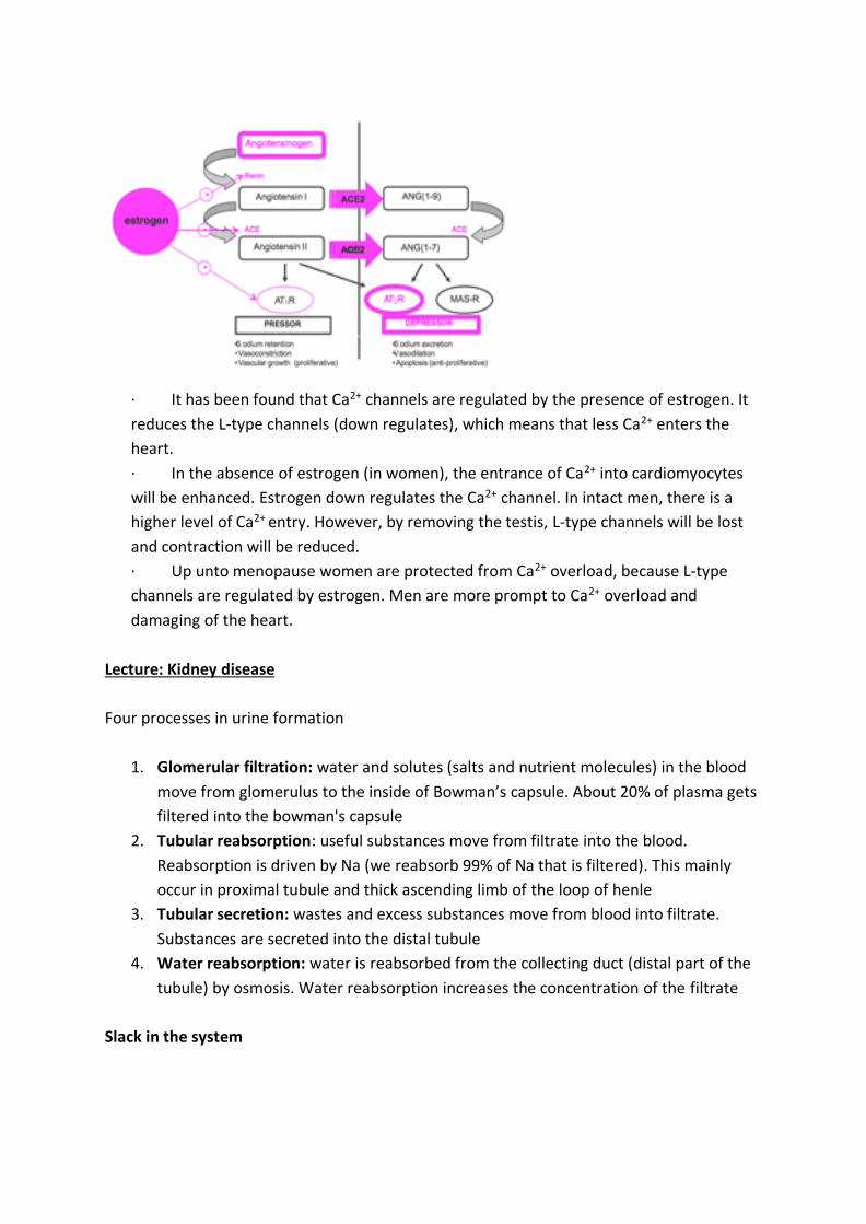
· It has been found that Ca2+ channels are regulated by the presence of estrogen. It
reduces the L-type channels (down regulates), which means that less Ca2+ enters the
heart.
· In the absence of estrogen (in women), the entrance of Ca2+ into cardiomyocytes
will be enhanced. Estrogen down regulates the Ca2+ channel. In intact men, there is a
higher level of Ca2+ entry. However, by removing the testis, L-type channels will be lost
and contraction will be reduced.
· Up unto menopause women are protected from Ca2+ overload, because L-type
channels are regulated by estrogen. Men are more prompt to Ca2+ overload and
damaging of the heart.
Lecture: Kidney disease
Four processes in urine formation
1. Glomerular filtration: water and solutes (salts and nutrient molecules) in the blood
move from glomerulus to the inside of Bowman’s capsule. About 20% of plasma gets
filtered into the bowman's capsule
2. Tubular reabsorption: useful substances move from filtrate into the blood.
Reabsorption is driven by Na (we reabsorb 99% of Na that is filtered). This mainly
occur in proximal tubule and thick ascending limb of the loop of henle
3. Tubular secretion: wastes and excess substances move from blood into filtrate.
Substances are secreted into the distal tubule
4. Water reabsorption: water is reabsorbed from the collecting duct (distal part of the
tubule) by osmosis. Water reabsorption increases the concentration of the filtrate
Slack in the system

● You can lose 50% or more of your functional nephrons without experiencing a
reduction in glomerular filtration rate (GFR). This is facilitated by glomerular and
tubular hypertrophy. Glomerular and tubular hypertrophy may exacerbate
progression of chronic kidney disease.
● GFR is a major way to measure kidney function
How do we measure ‘kidney function’?
Mostly by measuring or estimating GFR
● The ideal substance to measure GFR would …
● Be freely filtered at the glomeruli
● Not be bound to plasma proteins (substance wont be freely filtered as plasma
proteins can’t get through glomerular membrane)
● Not be metabolized
● Be non‐toxic
● Be excreted only by the kidneys
● Be neither reabsorbed nor secreted by the renal tubules
● Be stable in blood and urine
● Be easily measured
Clearance of Inulin The ‘gold standard’ we use in the laboratory
Inulin (polymer of fructose)
● Freely filtered into the Bowman’s capsule
● Not reabsorbed, secreted or metabolized by the nephron
● Non‐endogenous, has to be infused intravenously
● This method is not practical for routine clinical use since it involves infusion of inulin
at steady state through a catheter. This means stay at medical facility is longer which
translates to greater costs. This however is the gold standard method of estimating
kidney function.
Clearance of creatinine The ‘not so gold standard’
SAMPLE NOTES, FULL EDITION IS ORDERED AND COMPLETE.

Chronic kidney disease: slowly developing reduction in glomerular filtration rate (years –
decades)
● CKD → exist in patients where their GFR has reduced over years to decades
● CKD can be staged
● More progressive forms of CKD are characterised by greater and greater reduction in
GFR
● End stage renal disease (ESRD) is characterised by having a 10% of the normal GFR
● Acute kidney injury (AKI): rapidly developing reduction in glomerular filtration rate
and or urine output (hours – days)
● AKI occurs in people where their GFR falls over hours to days (very rapidly occurring
reduction in GFR) …..
SAMPLE NOTES, FULL EDITION IS ORDERED AND COMPLETE.
Lecture: Stress and Pregnancy
Stress- causes release of cortisol (glucocorticoid)
– Stress is good in some circumstances
• Triggers flight or fight responses
• Cortisol can cross the placenta
➢ However the placenta can inactivate cortisol to inactive cortisone
➢ enzyme that does this: 11 beta hydroxysteroid dehydrogenase type 2 (remember 11-
beta-HSD2)
Fetus requires glucocorticoids however only in small concentrations
Reduced 11-B-HSD2 in pre-term infants. Lower activity of 11-B-HSD2 results in more cortisol
getting to the fetus likely affecting the growth of the fetus.
Glucocorticoid Treatment

2 days of cortisone treatment results in reduced nephron number and elevation of BP and
increased AT2R.
Offspring from maternal dexamethasone treated dams also have • an increase in protein
expression of bumetanide sensitive sodium-potassium-chloride (NKCC2) co-transporter •
Increase in protein expression of sodium-hydrogen exchanger type-3 (NHE3) • Thiazide
sensitive Na-Cl cotransporter (NCC)
Kidney and programming of hypertension-summary
• Perturbations during development (eg., maternal dietary protein restriction, elevations in
maternal stress hormones, maternal alcohol consumption)
– Result in low nephron number
– increase in renal renin angiotensin system
– Increase in expression of sodium transporters
• These phenotypes may alter function of the kidney and result in greater sodium retention,
extracellular fluid volume expansion and eventually elevate blood pressure
Why is the kidney vulnerable?
Kidneys grown in DEX (dexamethasone a glucocorticoid) had less tips and branches and less
glomeruli
Prenatal Nicotine Exposure
– Maternal Smoking
• One of the most widespread intrauterine insults in world
• Rates of maternal smoking are still high despite links with IUGR & SIDS
• Nicotine major component of cigarette smoke
– Readily crosses placenta
– Plasma levels in fetus > mum
Same BP however, it results in increased responsiveness to Angiotensin II. (nicotine
exposure)
Increased vasoconstriction in response to noradrenaline and Ang II (nicotine exposure)
Prenatal cocaine exposure
Blood Pressure: • Basal BP normal • ‐ Response to Noradrenaline
Vascular Reactivity: • ‐ Contraction to Noradrenaline • ¯ Vasodilatation to acetylcholine
•¯NOS activity & Nitric Oxide production
Prenatal Hypoxia
High salt increased vascular stiffness in both hypoxia exposed and control offspring (left-
ward shift in stressstrain relationship) • The shift was exaggerated in hypoxia exposed
offspring indicating greater vascular stiffness
SAMPLE NOTES, FULL EDITION IS ORDERED AND COMPLETE.

Week 11 Steroid Hormones and signalling mechanisms (alternate explanation)
Goal: to understand the role of steroid hormone signalling in general, and as a signalling
mechanism in cardiovascular disease
Lecture Outline
1.What is cardiovascular disease?
2.What are steroid hormones and how do they act?
3.How do steroid hormones regulate cardiac function?
4.Mineralocorticoid receptors (MR) and cardiovascular disease.
5.What is the clinical problem we are trying to address?
6.Cell selective actions of the MR.
7.Where to next?
The mineralocorticoid receptor (MR)
Mineralocorticoids = sodium retaining hormones (retain sodium in kidney and distal
convoluting collecting ducts)
Sites of MR expression
MR function in Cardiomyocytes
● MR signalling in cardiomyocytes influences force
generation, rate of contraction and hypertrophy
○ Binding of aldosterone to MR leads to increased heart
rate with greater force of contraction → this is the normal
physiological response
○ When someone is bleeding → blood volume decreases
→ RAAS is activated and aldosterone is released →
aldosterone act on myocytes to increase cardiac output
● Aldosterone can regulate cardiomyocyte primary gene expression
● Over-expression of the MR in cardiomyocytes promotes arrhythmia
SAMPLE NOTES, FULL EDITION IS ORDERED AND COMPLETE.