SAGES guidelines for laparoscopic ventral hernia repair · 2016-08-12 · GUIDELINES SAGES...
21
GUIDELINES SAGES guidelines for laparoscopic ventral hernia repair David Earle 1 • J. Scott Roth 1 • Alan Saber 1 • Steve Haggerty 1 • Joel F. Bradley III 1 • Robert Fanelli 1 • Raymond Price 1 • William S. Richardson 1 • Dimitrios Stefanidis 1 • SAGES Guidelines Committee Received: 2 March 2016 / Accepted: 21 June 2016 / Published online: 12 July 2016 Ó Society of American Gastrointestinal and Endoscopic Surgeons (SAGES) 2016 Preamble The goals of ventral hernia repair are relief of patient symptoms and/or cure of the hernia with minimization of recurrence rates. While laparoscopic ventral hernia repair (LVHR) has gained popularity in recent years, there is still significant controversy about the optimal approach to ventral hernia repair. This document has been written to assist surgeons utilizing a laparoscopic approach to ventral hernia repair in terms of patient selection, operative tech- nique, and postoperative care. It is not intended to debate the merits of prosthetic use and specific types of prosthetics. Disclaimer Guidelines for clinical practice are intended to indicate preferable approaches to medical problems as established by experts in the field. These recommendations have been made based on existing data or a consensus of expert opinion when little or no data are available. Guidelines are applicable to all physicians, without regard to specialty training or interests, who address these clinical prob- lem(s) and are intended to indicate the preferable, but not necessarily the only, acceptable approaches. Guidelines are intended to be flexible. Given the wide range of specifics in any healthcare problem, the surgeon must always choose the course best suited to the individual patient and the variables in existence at the moment of decision. Guidelines are developed under the auspices of the Society of American Gastrointestinal Endoscopic Surgeons (SAGES) and its various committees and approved by the Board of Governors. Each clinical practice guideline has been systematically researched, reviewed, and revised by the guidelines committee and reviewed by an appropriate multidisciplinary team. This guideline was available for public comment for a period of 1 month prior to its final- ization. The recommendations are therefore considered valid at the time of their production, based on the data available. Each guideline is scheduled for periodic review to allow incorporation of pertinent new developments in medical research, knowledge, and practice. Methodology A systematic literature search was performed on MEDLINE in March 2009 and was updated in October 2012. The search strategy included the following terms: Hernia, Ventral/su [Surgery] ? Laparoscopy ? English ? Human. The abstracts were reviewed by the assigned working group members of the SAGES guidelines committee. Duplicates, letters to the editor, and articles not involving ventral, incisional, or Spigelian hernias were not generally included unless there was a direct correlation with the specific subject. Studies that compared different types of mesh were beyond the scope of this guideline and were not included. The reviewers graded the level of evidence using the GRADE guidelines (Table 1) and searched the bibliogra- phy of each article for additional articles that may have been missed during the original search. Additional relevant & William S. Richardson [email protected] 1 Ochsner Clinic, 1514 Jefferson Highway, New Orleans, LA 70121, USA 123 Surg Endosc (2016) 30:3163–3183 DOI 10.1007/s00464-016-5072-x and Other Interventional Techniques
Transcript of SAGES guidelines for laparoscopic ventral hernia repair · 2016-08-12 · GUIDELINES SAGES...

GUIDELINES
SAGES guidelines for laparoscopic ventral hernia repair
David Earle1 • J. Scott Roth1 • Alan Saber1 • Steve Haggerty1 • Joel F. Bradley III1 •
Robert Fanelli1 • Raymond Price1 • William S. Richardson1 • Dimitrios Stefanidis1 •
SAGES Guidelines Committee
Received: 2 March 2016 / Accepted: 21 June 2016 / Published online: 12 July 2016
� Society of American Gastrointestinal and Endoscopic Surgeons (SAGES) 2016
Preamble
The goals of ventral hernia repair are relief of patient
symptoms and/or cure of the hernia with minimization of
recurrence rates. While laparoscopic ventral hernia repair
(LVHR) has gained popularity in recent years, there is still
significant controversy about the optimal approach to
ventral hernia repair. This document has been written to
assist surgeons utilizing a laparoscopic approach to ventral
hernia repair in terms of patient selection, operative tech-
nique, and postoperative care. It is not intended to debate
the merits of prosthetic use and specific types of
prosthetics.
Disclaimer
Guidelines for clinical practice are intended to indicate
preferable approaches to medical problems as established
by experts in the field. These recommendations have been
made based on existing data or a consensus of expert
opinion when little or no data are available. Guidelines are
applicable to all physicians, without regard to specialty
training or interests, who address these clinical prob-
lem(s) and are intended to indicate the preferable, but not
necessarily the only, acceptable approaches. Guidelines are
intended to be flexible. Given the wide range of specifics in
any healthcare problem, the surgeon must always choose
the course best suited to the individual patient and the
variables in existence at the moment of decision.
Guidelines are developed under the auspices of the
Society of American Gastrointestinal Endoscopic Surgeons
(SAGES) and its various committees and approved by the
Board of Governors. Each clinical practice guideline has
been systematically researched, reviewed, and revised by
the guidelines committee and reviewed by an appropriate
multidisciplinary team. This guideline was available for
public comment for a period of 1 month prior to its final-
ization. The recommendations are therefore considered
valid at the time of their production, based on the data
available. Each guideline is scheduled for periodic review
to allow incorporation of pertinent new developments in
medical research, knowledge, and practice.
Methodology
A systematic literature search was performed on MEDLINE
inMarch 2009 and was updated in October 2012. The search
strategy included the following terms: Hernia, Ventral/su
[Surgery] ? Laparoscopy ? English ? Human.
The abstracts were reviewed by the assigned working
group members of the SAGES guidelines committee.
Duplicates, letters to the editor, and articles not involving
ventral, incisional, or Spigelian hernias were not generally
included unless there was a direct correlation with the
specific subject. Studies that compared different types of
mesh were beyond the scope of this guideline and were not
included.
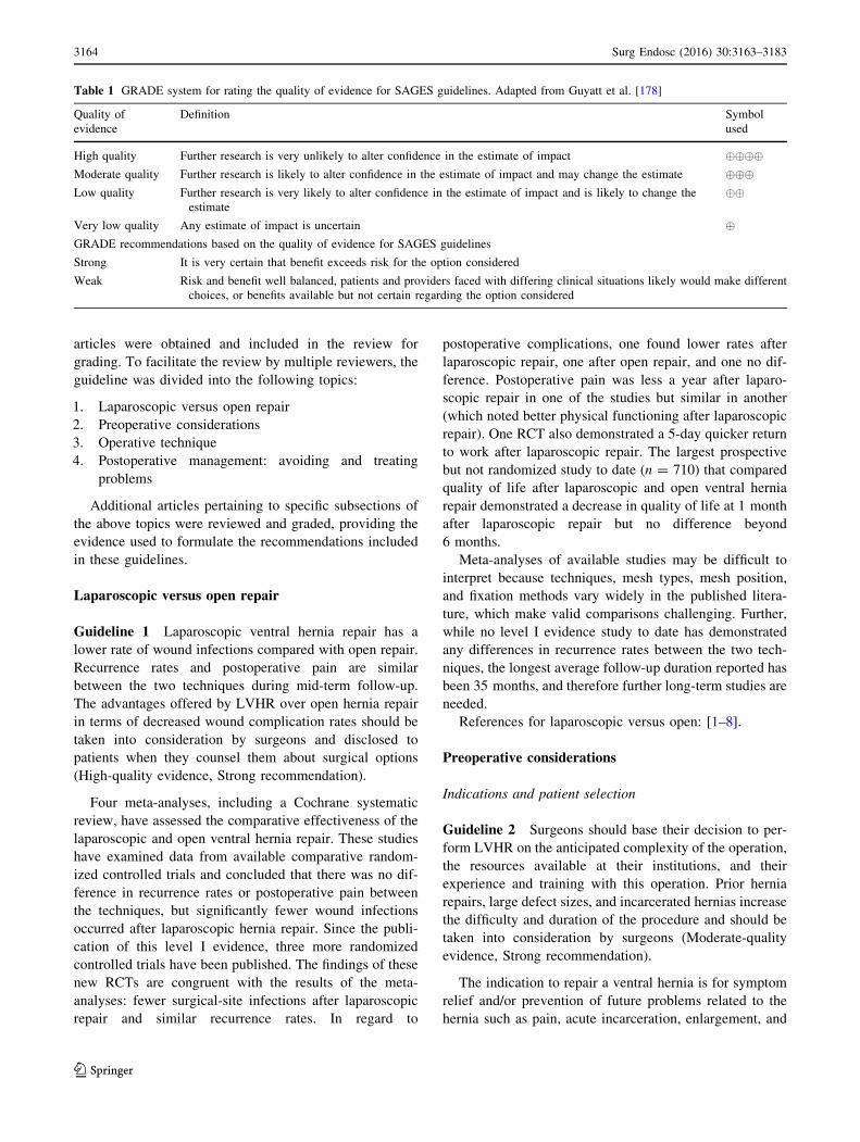
The reviewers graded the level of evidence using the
GRADE guidelines (Table 1) and searched the bibliogra-
phy of each article for additional articles that may have
been missed during the original search. Additional relevant
& William S. Richardson
1 Ochsner Clinic, 1514 Jefferson Highway, New Orleans,
LA 70121, USA
123
Surg Endosc (2016) 30:3163–3183
DOI 10.1007/s00464-016-5072-x
and Other Interventional Techniques
articles were obtained and included in the review for
grading. To facilitate the review by multiple reviewers, the
guideline was divided into the following topics:
1. Laparoscopic versus open repair
2. Preoperative considerations
3. Operative technique
4. Postoperative management: avoiding and treating
problems
Additional articles pertaining to specific subsections of
the above topics were reviewed and graded, providing the
evidence used to formulate the recommendations included
in these guidelines.
Laparoscopic versus open repair
Guideline 1 Laparoscopic ventral hernia repair has a
lower rate of wound infections compared with open repair.
Recurrence rates and postoperative pain are similar
between the two techniques during mid-term follow-up.
The advantages offered by LVHR over open hernia repair
in terms of decreased wound complication rates should be
taken into consideration by surgeons and disclosed to
patients when they counsel them about surgical options
(High-quality evidence, Strong recommendation).
Four meta-analyses, including a Cochrane systematic
review, have assessed the comparative effectiveness of the
laparoscopic and open ventral hernia repair. These studies
have examined data from available comparative random-
ized controlled trials and concluded that there was no dif-
ference in recurrence rates or postoperative pain between
the techniques, but significantly fewer wound infections
occurred after laparoscopic hernia repair. Since the publi-
cation of this level I evidence, three more randomized
controlled trials have been published. The findings of these
new RCTs are congruent with the results of the meta-
analyses: fewer surgical-site infections after laparoscopic
repair and similar recurrence rates. In regard to
postoperative complications, one found lower rates after
laparoscopic repair, one after open repair, and one no dif-
ference. Postoperative pain was less a year after laparo-
scopic repair in one of the studies but similar in another
(which noted better physical functioning after laparoscopic
repair). One RCT also demonstrated a 5-day quicker return
to work after laparoscopic repair. The largest prospective
but not randomized study to date (n = 710) that compared
quality of life after laparoscopic and open ventral hernia
repair demonstrated a decrease in quality of life at 1 month
after laparoscopic repair but no difference beyond
6 months.
Meta-analyses of available studies may be difficult to
interpret because techniques, mesh types, mesh position,
and fixation methods vary widely in the published litera-
ture, which make valid comparisons challenging. Further,
while no level I evidence study to date has demonstrated
any differences in recurrence rates between the two tech-
niques, the longest average follow-up duration reported has
been 35 months, and therefore further long-term studies are
needed.
References for laparoscopic versus open: [1–8].
Preoperative considerations
Indications and patient selection
Guideline 2 Surgeons should base their decision to per-
form LVHR on the anticipated complexity of the operation,
the resources available at their institutions, and their
experience and training with this operation. Prior hernia
repairs, large defect sizes, and incarcerated hernias increase
the difficulty and duration of the procedure and should be
taken into consideration by surgeons (Moderate-quality
evidence, Strong recommendation).
The indication to repair a ventral hernia is for symptom
relief and/or prevention of future problems related to the
hernia such as pain, acute incarceration, enlargement, and
Table 1 GRADE system for rating the quality of evidence for SAGES guidelines. Adapted from Guyatt et al. [178]
Quality of
evidence
Definition Symbol
used
High quality Further research is very unlikely to alter confidence in the estimate of impact ����Moderate quality Further research is likely to alter confidence in the estimate of impact and may change the estimate ���Low quality Further research is very likely to alter confidence in the estimate of impact and is likely to change the
estimate
��
Very low quality Any estimate of impact is uncertain �GRADE recommendations based on the quality of evidence for SAGES guidelines
Strong It is very certain that benefit exceeds risk for the option considered
Weak Risk and benefit well balanced, patients and providers faced with differing clinical situations likely would make different
choices, or benefits available but not certain regarding the option considered
3164 Surg Endosc (2016) 30:3163–3183
123
skin problems. For all hernia repairs, it is important to
define the goals of the operation preoperatively and align
those goals between the patient and the surgeon.
It is important to consider the size of the hernia defect
when contemplating a laparoscopic approach, as larger
defects generally increase the difficulty of the procedure. A
recently published guideline by an Italian Consensus
Conference recommended caution for defects greater than
10 cm but did not consider such defects as absolute con-
traindication. On the other hand, the same group recom-
mended that hernias with a defect size\3 cm should not be
approached laparoscopically. This recommendation was
based on expert opinion and a survey showing that less
than 10 % of surgeons used prosthetics in defects less than
3 cm; it was therefore deemed as ‘‘an indirect indication of
a minimum size limit for laparoscopy.’’ The present
MEDLINE search of the literature did not reveal any evi-
dence in support of this recommendation. In addition, the
best available evidence today suggests that prosthetics
should be used even for small (umbilical) hernia defects, as
the recurrence rates are minimized compared with primary
repair. Further, the recent guidelines published by the
International Endohernia Society recommend the use of
mesh for all abdominal wall hernias independent of the
defect size. Expert opinion suggests that the laparoscopic
approach to ventral hernia repair may be beneficial in the
morbidly obese patient and in recurrent cases compared
with the open approach. Therefore, additional evidence is
needed before a minimum size for laparoscopic repair can
be defined. Reported conversion rates in the literature range
between 0 and 14 % in series with over 50 patients. Pos-
sible reasons for a higher conversion rate may include poor
patient selection, severe adhesions, incarcerated hernia
content impossible to reduce, and/or inadequate training
and expertise.
Given the variation of technical ability and institutional
capability, along with the gradual acquisition of experi-
ence, surgeons must use their judgment when determining
whether to perform a laparoscopic or open ventral hernia
repair. When considering a laparoscopic approach to a
ventral hernia, the surgeon should consider his or her own
experience when selecting patients. There is limited evi-
dence on how expertise with laparoscopic ventral hernia
repair is developed; however, it appears prudent to rec-
ommend that less experienced surgeons should start with
less complex cases. A study analyzing 180 cases of a
prospectively collected database found a number of clinical
factors that significantly increased operative time (which
was used as a surrogate of laparoscopic repair complexity).
The most significant factors influencing operative time
were adhesiolysis and prior ventral hernia repair(s), both of
which clearly increased the complexity of the laparoscopic
repair. Other factors reported in the literature that increase
the complexity of laparoscopic ventral hernia repair
include large defects ([10 cm diameter), hernias in unu-
sual locations (subxiphoid, suprapubic, flank, etc.), incar-
cerated hernia, hernias with small defect size but large
hernia sac, obesity, bowel distention, pregnancy, and
presence of ascites.
The aforementioned factors, which are known to
increase the technical difficulty of the operation, should
help guide the surgeon in selecting the appropriate patients
for LVHR. The decision of whether or not to perform a
LVHR encompasses the surgeon’s training and experience,
as well as the institution’s capability to provide the proper
equipment and supplies. As training and experience is
gained, gradually more complex laparoscopic ventral her-
nia repairs may be appropriately undertaken.
References for indications and patient selection: [9–30].
Special considerations
Guideline 3 Special situations such as loss of domain,
presence of abdominal skin grafts or of an active entero-
cutaneous fistula, the need to remove previously placed
prosthetic mesh, or large abdominal wall defects may
represent contraindications to laparoscopic repair (Moder-
ate-quality evidence, Strong recommendation).
In the following situations, the laparoscopic approach to
ventral hernia repair may be problematic and associated
with higher conversion rates and potentially suboptimal
outcomes.
1. Loss of domain
Loss of domain is a term describing a situation of a
massive hernia (hernia sac) where reduction in the hernia
content would cause major abdominal hypertension leading
to the potential complication of abdominal compartment
syndrome. The term is used in patients with a massive
hernia sac containing greater than 30 % of the abdominal
contents and possibly some of the solid organs.
2. Abdominal skin grafts
An abdominal skin graft overlying the hernia defect
makes a proper adhesiolysis between the viscera and the
posterior aspect of the graft difficult, with the consequence
of graft ischemia and necrosis, thus exposing the pros-
thetic, which would risk infection and the need for
removal. Further, adhesiolysis can lead to visceral injuries
that are missed, increasing the risk of fistula formation. An
open approach is therefore preferable.
3. Need to remove large prosthetic mesh
If removal of a previously placed prosthetic is deemed
necessary and this will result in a large enough wound to
Surg Endosc (2016) 30:3163–3183 3165
123

perform the hernia repair in an open fashion, an open
approach should be undertaken.
4. Small defect but large hernia sac
This situation represents a scenario that may be difficult
to address laparoscopically, especially if there are adhe-
sions between the viscera and the hernia sac. The adhesions
may be difficult to visualize or reach with laparoscopic
instruments due to the narrow defect size or angulation
required to visualize the hernia sac contents through the
hernia defect. Pulling on the viscera with great force can
also result in inadvertent visceral injury. On the other hand,
external pressure on the abdominal wall in the area of the
hernia may help reduce the sac contents intraabdominally
or make them easier visible for safe dissection and should
be employed when this situation is encountered. A
laparoscopic approach may be associated with a higher
probability of conversion to an open approach under these
circumstances.
5. Incarcerated hernia
This scenario is often present with the small defect and
large sac scenario as described above. Trying to reduce
acutely or chronically incarcerated viscera can result in
inadvertent visceral injury. Experts might be able to reduce
an incarcerated hernia laparoscopically by incising the
neck of the hernia with a laparoscopic scissor without
diathermy (expert opinion). Further, external pressure may
prove very valuable. A laparoscopic approach may be
attempted but will have a higher probability of being
converted to an open or laparoscopic-assisted approach.
Good surgical judgment is required to minimize the risk of
visceral injury.
6. Active enterocutaneous fistula
Active enterocutaneous fistulas are generally considered
a contraindication for laparoscopic hernia repair, as the
current laparoscopic techniques include prosthetic mesh
placement and are therefore reserved for clean and clean-
contaminated cases.
7. Abdominal wall defect
Hernias from previous gunshot wounds to the abdomen
with a missing abdominal muscle or fascia or areas on the
abdominal wall with previously elevated flaps (TRAM)
might have defects too large to bridge and impossible to
approximate with laparoscopic techniques and should
therefore be considered for an open component separation
technique.
References for special considerations: [9–11, 16, 18, 20,
27, 31–40].
Diagnosis
Guideline 4 While most ventral hernias are easily diag-
nosed based on clinical examination, a preoperative
abdominal CT scan or ultrasound may be considered for
selected patients with suspected ventral hernias to confirm
the diagnosis or to aid the surgeon with preoperative
planning (Moderate quality, Strong recommendation).
Diagnosis of a ventral hernia is typically made during
the history and physical examination. Imaging studies
including ultrasound, provocative ultrasound, computed
tomography (CT) with and/or without Valsalva, and mag-
netic resonance imaging (MRI) can also be used for diag-
nosis. Imaging studies may be helpful to assess the
anatomic details of a ventral hernia, augmenting the
physical examination, especially when a hernia cannot be
reduced, and therefore the defect cannot be palpated and
the size not estimated. These situations commonly arise
with small defects, obese patients, or incarceration (acute
or chronic). CT has been found to be useful in diagnosing
occult hernias, multiple defects, abscess, and hematoma, as
well as in differentiating incarcerated hernias from
abdominal wall neoplasms. In a retrospective review of 146
consecutive LVHRs, 48 % of patients had occult defects
not detected on preoperative physical examination.
In one study, diagnosis of hernia recurrence after mesh
repair was correct 88 % of the time on physical exam,
while CT was correct in 98 % of cases. Another study that
evaluated the comparative effectiveness of dynamic
abdominal sonography versus CT for the diagnosis of
ventral hernias demonstrated a sensitivity of 98 % and
specificity of 88 %, with PPV 91 % and NPV 97 % for the
dynamic sonography, which identified hernias in a few
more patients than CT imaging.
Preoperative CT is also helpful in defining the abdom-
inal wall anatomy in non-midline hernias such as those on
the flanks, suprapubic or subxiphoid regions, and identi-
fying posterior abdominal wall defects. CT scans accu-
rately assess the relationship of the hernia to structures
such as the bladder, pubic symphysis, anterior superior
iliac spine, and the ribs, in addition to defining the integrity
and nature of the muscles of the abdominal wall and the
size of the defect. This information is useful for the sur-
geon when deciding on the safest point of access and where
or how to anchor the mesh. Imaging data may also allow
the surgeon to determine that an open or laparoscopic-as-
sisted approach may be more appropriate. CT scanning is
not useful in determining the presence and character of
intraabdominal adhesions, a known factor that increases
complexity of LVHR. Finally, CT cannot determine com-
pliance of the abdominal wall or the feasibility of closing
3166 Surg Endosc (2016) 30:3163–3183
123
the defect and replacing all the viscera within the peri-
toneal cavity.
References for diagnosis: [41–45].
Bowel preparation
Guideline 5 Mechanical bowel preparation prior to
LVHR may be useful in select cases, but additional evi-
dence on its risks/benefits is needed before a recommen-
dation can be provided (Low-quality evidence).
Prior to LVHR some surgeons prefer bowel preparation
either routinely or selectively. Reported advantages of
bowel preparation include dealing with a clean bowel in
case there is an enterotomy, decompressing the GI tract to
avoid bowel distension (especially if colon was contained
in the hernia sac), improving the safety of intraoperative
bowel handling and adhesiolysis, and avoiding a full colon
in the event of a postoperative ileus (which has been
reported to range between 0.8 and 20 %). Most authors
report using mechanical preoperative bowel preparation
without antibiotics. While the available evidence on the
benefits and risks of bowel prep before LVHR is lacking, a
recent study on pigs by Vlot et al. demonstrated increased
working space after bowel preparation.
References for bowel preparation: [9, 11, 16, 20, 26, 29,
31, 32, 46–52].
Patient position
Guideline 6 Patient positioning should use all appropriate
precautions to prevent patient injurywhile enabling access to
the needed abdominal wall to allow for adequate size mesh
placement and fixation. Supine positionwith the arms tucked
will offer the most versatile position when performing
LVHR. Hernias requiring lateral or posterior access should
be performed with the patient in a full or partial lateral
position (Low quality, Strong recommendation).
The patient should be placed during LVHR in a safe and
stable position to assure access to the hernia through
operative exposure and an ergonomic working position for
the surgeon and operating team. A supine position with the
arms tucked at the patient’s sides is the standard position
for patients with midline hernias, while hernias of the
flanks or posterior abdominal wall require a lateral decu-
bitus or modified lateral decubitus position. Careful patient
positioning ensures adequate working space to perform
adhesiolysis and handling and fixation of the mesh.
Frequent tilting of the OR bed, Trendelenburg, and/or
reverse Trendelenburg position may be needed to optimize
operative exposure by passively retracting the viscera out
of the way, especially for large hernias or hernias located
off the midline or in the subxiphoid/suprapubic location.
The need for frequent position changes during LVHR
makes the secure attachment of the patient to the OR bed
with appropriate padding of pressure points imperative.
References for patient position: [9, 11, 16, 20, 26, 29,
31, 32, 46–51].
Urinary bladder catheter
Guideline 7 Placement of a urinary bladder catheter
during LVHR should be determined based on the antici-
pated duration of the procedure and the location of the
hernia. For LVHR near the symphysis that requires dis-
section and prosthetic fixation to the pubic bone, the
placement of a three-way catheter should be considered to
allow drainage and easy instillation of sterile saline solu-
tion to distend the bladder, which may help in recognizing
and avoiding bladder injuries (Low quality, Weak
recommendation).
Placement of a urinary bladder catheter during LVHR
should be determined based on the anticipated duration of
the procedure and the location of the hernia. Incisional
hernias from previous lower midline incisions, particularly
if the defect is near the symphysis, generally require fixa-
tion to the inferior pubic ramus. In order to properly fix the
prosthetic to these structures, extraperitoneal dissection and
mobilization of the bladder (similar to exposure in
laparoscopic inguinal hernia repair) is necessary. Place-
ment of a urinary bladder catheter before prepping and
draping the patient allows for continuous drainage and
monitoring of urine output. An empty bladder also gives
additional space in the abdominal cavity, which might be
essential in reducing larger hernias. If a three-way catheter
is used, the bladder can be filled with sterile saline solution
while clamping the outflow temporarily, which may facil-
itate easier identification of the urinary bladder during
dissection and may help prevent injury to the bladder. In
the event of a urinary bladder injury, a distended bladder
may further aid in the detection and repair of the injury.
References for urinary bladder catheter: [11, 53, 54].
Prophylactic antibiotics
Guideline 8 A single-dose first-generation cephalosporin
(cefazolin) should be given preoperatively for LVHR.
Vancomycin should be added in patients colonized with
MRSA. Vancomycin or clindamycin should be given to
patients allergic to cephalosporins (Moderate quality,
Strong recommendation).
The guidelines recently published jointly by the Amer-
ican Society of Health-System Pharmacists (ASHP), the
Infectious Diseases Society of America (IDSA), the Sur-
gical Infection Society (SIS), and the Society for
Surg Endosc (2016) 30:3163–3183 3167
123

Healthcare Epidemiology of America (SHEA) recommend
that a single dose of a first-generation cephalosporin (ce-
fazolin) be administered within 60 min prior to incisional
hernia repair with a prosthetic. For patients known to be
colonized with MRSA, a single preoperative dose of van-
comycin should be added and administered within 120 min
prior to surgery. For b-lactam allergic patients, clin-
damycin or vancomycin should be given. Even though
these guidelines were developed from the evidence inclu-
ded in the inguinal hernia literature, they also apply to
incisional hernias, where the risk of infection seems to be
higher than in inguinal hernias.
The most common microorganisms isolated from sur-
gical-site infections after hernia repair are aerobic gram-
positive organisms and include aerobic streptococci, Sta-
phylococcus species, and Enterococcus species. MRSA is
commonly found in prosthetic mesh infections.
References for prophylactic antibiotics: [1, 9–11, 15, 16,
20, 22, 27, 28, 31, 34, 36–39, 44, 46–49, 54–86].
Plastic adhesive drape
Guideline 9 Antimicrobial-impregnated plastic adhesive
drapes are often used during LVHR, but the current liter-
ature does not support their use, as no evidence exists that
they decrease surgical-site infections (Low quality, Weak
recommendation).
Plastic adhesive drapes are used commonly during
LVHR. The only study looking at this practice related to
ventral hernia repair was a retrospective review of 506
laparoscopic and open ventral hernia repairs at a single
institution that reported plastic adhesive drapes were used
in 59.1 % of the cases, mostly by the highest volume
laparoscopic surgeons. Proposed benefits of utilizing these
types of drapes are (1) reducing contact between the
prosthetic and skin and (2) securing the drapes to the
patient to avoid breaks in the sterile field. Because the
drapes are placed so laterally, unless they are adherent to
the skin, they are at risk for lifting off the skin and
exposing unprepped portions of the abdominal wall or the
operating table itself. While this study noted a perceived
benefit among surgeons in terms of improving surgical
draping, it failed to demonstrate a benefit in terms of
infection reduction.
The use of an iodophor-impregnated plastic incise drape
in abdominal surgery has been shown to lower the inci-
dence of wound contamination but not surgical-site infec-
tion (SSI) rates, and a meta-analysis in 2007 of a variety of
studies and case types revealed there was no evidence that
plastic adhesive drapes reduced SSI rates and some evi-
dence that they may actually increase SSI rates if the
drapes were non-iodinated.
During LVHR, some surgeons place the prosthetic on
the abdominal wall to assess prosthetic and suture anchor
location. The prosthetic therefore will potentially be
exposed to skin flora, the most common bacteria associated
with prosthetic infection, and this practice has largely
driven the concept of utilizing a plastic, adhesive drape for
LVHR. Although prosthetic infection is typically due to
Staphylococcus species, review of the literature reveals that
prosthetic contamination and/or infection in the early
postoperative period (within 30 days) after LVHR is
almost always due to missed or delayed enterotomy.
Prosthetic infection not related to enterotomy is likely due
to factors such as the infectious disease-related medical
history of the patient (particularly history of wound or
prosthetic infection), existing infectious disease-related
issues (e.g., occult infection), wound classification (based
on concomitant GI procedures, skin infection, etc.), use of
prophylactic antibiotics, abdominal wall preparation,
prosthetic choice, and prosthetic handling. Further com-
plicating the issue is the fact that most reports do not
include microbiological data related to prosthetic infection,
and many prosthetic infections occur after 30 days, often
up to 1 year postoperatively.
In summary, while plastic adhesive drapes impregnated
with iodine reduce wound bacterial inoculum, they have
not been proven to reduce clinical infection rates. Plastic
adhesive drapes may reduce breaks in sterile technique due
to the wide draping requirements of LVHR. Besides the
small additional cost, potential side effects of the adhesive
drape include mild skin irritation and adverse wound
healing which are rare.
References for plastic adhesive drape: [13, 39, 41, 47,
58, 67, 73, 84, 87–92].
Operative technique
Abdominal access and trocar placement
Guideline 10 The location of initial abdominal access
(primary port placement) for LVHR should be as far from
the hernia defect and prior laparotomy incisions as possi-
ble. The ideal location for this port may be the left or right
upper quadrant, but location should be modified according
to the patient’s surgical history and anatomy (Moderate
quality, Strong recommendation).
Guideline 11 A Veress needle, open Hasson technique,
or optical trocar entry may all safely be used for primary
port placement during LVHR. The specific technique used
should be primarily based on the surgeon’s experience and
outcomes with the technique and take into consideration
the patient’s surgical history and anatomy (Moderate
quality, Strong recommendation).
3168 Surg Endosc (2016) 30:3163–3183
123

Guideline 12 Secondary port placement should be per-
formed under direct vision and placed as lateral from the
hernia defect as possible to allow the surgeon to work in an
ergonomically favorable position for adhesiolysis and
placement/fixation of the prosthetic (Moderate quality,
Strong recommendation).
The principles of safe abdominal access for laparoscopic
surgery apply to LVHR, and technical details about
establishment of pneumoperitoneum can be found in the
SAGES Fundamentals of Laparoscopic Surgery (FLS)
program. There have been no comparative data regarding
techniques for establishing pneumoperitoneum specifically
for LVHR, although a variety of techniques have been
described in the published literature, all with low rates of
complications and successful establishment of pneu-
moperitoneum. Current options for initial peritoneal access
for LVHR include direct trocar insertion with an optical
trocar (with or without a pneumoperitoneum with the use
of the Veress needle), or an open Hasson technique. Mul-
tiple meta-analyses and randomized controlled trials with a
variety of general surgical and gynecological laparoscopic
procedures reveal no difference in major complication rates
with direct trocar insertion without pneumoperitoneum
compared with establishment of pneumoperitoneum with
Veress needle prior to initial trocar insertion. Regardless of
the technique, the surgeon should have adequate training
and/or experience with it in similar clinical situations.
Additionally, since many LVHRs are performed for mid-
line hernias, it is recommended to access the abdomen off
the midline, to avoid areas with potential adhesions of
bowel. The techniques reported in the literature for LVHR
are listed in Table 2.
Table 2 Methods for establishing pneumoperitoneum and port placement
First author Year n Access Technique Location of initial entry site Total number and location of ports
Bageacu [58] 2002 159 Veress needle ‘‘…as far as possible from the hernia
(typically on the left anterior axillary
line in the case of a midline hernia.’’
3–4; location not mentioned
Ben-Haim [10] 2002 100 Open technique ‘‘…as laterally from the hernia site as
possible.’’
No number mentioned. Location
‘‘according to hernia site’’
Berger [60] 2002 150 Open technique ‘‘as far from the hernia as possible and
mainly in the subcostal area.’’
5–7; location ‘‘laterally on both sides’’
Chowbey [46] 2000 200 Veress, open
technique
‘‘…unless contraindicated, the Veress
needle is inserted in the left subcostal
region…’’ If Veress insertion
unsuccessful, open technique used.
3; left abdominal wall laterally
Franklin [32] 2004 384 Veress, ‘‘very
rarely’’ open
technique
‘‘…usually from a non-midline
location…’’
3–4; ‘‘lateral to rectus muscles’’
Frantzides [69] 2004 208 Optical viewing
trocar
‘‘…as far as possible from the hernia
defect…’’
Number ‘‘depended on the difficulty of
the subsequent adhesiolysis.’’
Location not mentioned
Gillian [48] 2002 100 Optical viewing
trocar
Left subcostal 3–4; left abdomen, right side as needed
Heniford [16] 2003 850 Open Hassan 650,
Veress 200
‘‘usually just inferior to the tip of the
eleventh rib.’’
Number ‘‘as needed’’; location lateral
abdominal wall for midline defects,
dependent of defect location for non-
midline defects
LeBlanc [34] 2000 100 Open Hassan,
Veress, optical
viewing
Not mentioned No number; Location ‘‘…as far laterally
as possible…’’; scope port should be
on same side as surgeon.
Mizrahi [98] 2003 231 Veress Left subcostal 3; :location ‘‘along an imaginary
semilunar line connecting the
epigastrium with the left lower
quadrant’’
Morales-Conde [36] 2004 140 Subcostal Veress,
optical viewing
Left subcostal Number and location of secondary ports
not mentioned
Moreno Egea [76] 2004 90 Veress Not mentioned ‘‘The position of the trocars depended
on the size, location, and number of
abdominal wall defects, usually three
in a line along the left flank.’’
Surg Endosc (2016) 30:3163–3183 3169
123

Regarding placement location, it is desirable to have the
ports as far lateral as possible to expose midline hernias
and to be able to place a large piece of mesh without
interference. The operation is usually accomplished using
3–5 ports. A larger port (10–12 mm) is typically utilized
for the insertion of the prosthetic mesh. There are usually
three ports placed in the left lateral abdominal wall and 1–2
ports placed on the right. This pattern is often reversed in
patients who have had previous left-sided abdominal
operations such as left colectomy or open splenectomy.
Many authors report their entry techniques; however, none
directly compare the techniques. One of the largest retro-
spective series described placement of a Veress needle at
least 10 cm away from the prior scar, preferentially 2 cm
below the left costal margin in the mid clavicular line
(Palmer’s point). The left upper quadrant (LUQ) is the
most commonly reported initial entry site with all
techniques.
Secondary ports should be placed far enough away from
the hernia defect to allow for adhesiolysis and prosthetic
placement and fixation. Longer, bariatric-length instru-
ments may be necessary in obese patients. There may be
interference with the instrument handles on the patient’s
arms or legs; therefore, optimal port placement is critical.
References for abdominal access and trocar placement:
[5–7, 10, 11, 16, 20, 26, 29, 32, 34, 36, 39, 46, 48,
58, 60, 69, 76, 82, 93–101].
Adhesiolysis
Guideline 13 Adhesiolysis should be performed care-
fully with sharp and/or blunt dissection and sparing use of
energy for hemostasis to avoid inadvertent delayed
enterotomy. Use of sutures and hemostatic agents is
preferable to energy application to achieve hemostasis near
the bowel (Low quality, Strong recommendation).
Table 2 continued
First author Year n Access Technique Location of initial entry site Total number and location of ports
Palanivelu [20] 2007 721 Not mentioned Not mentioned 3; epigastric port for scope, two lateral
ports for working instruments;
occasionally 3 ports on all one side
laterally
Perrone [11] 2005 116 Veress 88 %, Open
technique 12 %
(only when
Veress needle
failed)
‘‘…well away from the hernia, typically
in either subcostal area or the lateral
abdomen lateral to the rectus sheath.’’
No number mentioned; Additional ports
placed ‘‘…as far from the hernia
defect and as lateral as possible.’’
Rosen [38] 2003 100 Veress, open
technique, or
optical trocar
‘‘…far from the defect…’’ 3–5; ‘‘…as far laterally as possible.’’
Saber [179] 2008 174 Open technique Right or left upper quadrant and ‘‘away
from the hernia’’
2–3; ‘‘…away from the hernia defects
to allow adequate surgical
manipulations.’’
Sharma [82] 2011 1242 Veress ‘‘…at least 10 cm away from the hernia/
previous scar. The most preferred site
… was Palmer’s point—a point 2 cm
below the left costal margin in the
midclavicular line.’’
3, more for larger hernias; ‘‘Ports were
placed in the form of an arc around
the hernial defect…’’
Toy [39] 1998 144 Veress, open ‘‘…away from the hernia defect and any
abdominal incisions…’’
‘‘…number and position are
individualized.’’ All placed ‘‘…as far
laterally as possible.’’
Ujiki [26] 2004 100 Veress—primary
hernia
Open—previous
abdominal
surgery
Lateral, ‘‘on the side of the abdomen
farthest from the hernia defect’’
No number mentioned; Lateral, ‘‘on the
side of the abdomen farthest from the
hernia defect’’
Yavuz [29] 2005 150 Veress—primary
and trocar site
hernias
Open—all other
incisional hernias
Left hypochondrium 3, with more ‘‘as necessary’’; ‘‘…as
laterally as possible…,’’ usually all on
the left side
3170 Surg Endosc (2016) 30:3163–3183
123
Guideline 14 The adhesiolysis should include the entire
old incision. Dependent on the hernia location, the falci-
form and umbilical ligaments should be taken down and
the space of Retzius dissected to identify occult hernia
defects and allow adequate exposure of the abdominal wall
for placement of an appropriately sized prosthetic (Low
quality, Strong recommendation).
Guideline 15 The surgeon should inspect the bowel after
adhesions are taken down as the adhesiolysis progresses,
and/or at the conclusion of the entire adhesiolysis to rule
out any inadvertent enterotomies (Low quality, Strong
recommendation).
Safe adhesiolysis is the most challenging step of LVHR.
Although preoperative adhesion detection with ultrasound
and cine MRI has been shown to be accurate in 76–92 % of
cases, it is not used clinically with significant frequency in
the USA for the purpose of estimating the quantity and/or
quality of adhesions preoperatively. Review of the opera-
tive records can give a reasonable sense of the difficulty
level of previous abdominal operations related to adhe-
sions. Adhesions between bowel or omentum and the
abdominal wall should be taken down to allow complete
visualization of the defect and the abdominal wall, as well
as provide an adequate area for placement of the appro-
priate-sized prosthetic. This should also include exposure
of the entire old incision, even if the symptomatic or pal-
pable defect is only a small portion of the old incision. This
might expose occult fascial defects, which occur in almost
half of cases, thus allowing adequate mesh coverage of all
defects and the entire old incision.
Enterotomy during LVHR has been reported between 1
and 6 % and usually occurs during adhesiolysis. Several
maneuvers facilitate safe adhesiolysis, including:
• Traction/counter traction technique.
• Use of angled or flexible laparoscope.
• Moving the scope among ports.
• Improved exposure utilizing outside pressure on the
abdominal wall, particularly for adhesions within a
hernia sac.
• Meticulous sharp dissection under direct vision close to
the anterior abdominal wall.
• Limited use of an energy source, particularly near the
hollow viscera.
• Working in a good ergonomic position.
• Repositioning/adding ports as needed to maintain
appropriate ergonomic position and access to the
operative field.
• Use of instruments with appropriate length (may need
longer instruments to maintain the fulcrum near the
middle of the instrument shaft).
• Avoiding too much torque on access ports during
critical aspects of the adhesiolysis.
• Maintaining a clear camera image.
• Maintaining a conscious vigilance for the mucosa of the
GI tract, as an enterotomy may only be visible for a
fleeting moment.
• Repeat inspection of the bowel after adhesiolysis to
look for enterotomies.
Use of an energy source for hemostasis should be kept to
a minimum to avoid missed or delayed bowel injury. It is
important to recognize that as the adhesiolysis progresses,
vigilance regarding the proximity of the hollow viscera,
particularly in the GI tract, must be in the forefront of the
surgeon’s mind. In fact, many experts will consciously look
for the mucosa of the GI tract during a difficult adhesiolysis
involving bowel, as an enterotomy may only be visible for
a fleeting moment, and lack of alertness to this could lead
to a missed enterotomy. In general, bleeding near the bowel
should be controlled with sutures, clips, or a topical
hemostatic agent rather than an energy source.
At the conclusion of the adhesiolysis, it is prudent to
inspect the areas of the hollow viscera involved for evi-
dence of partial thickness, full thickness, or thermal injury.
This can be accomplished for each area separately as the
adhesiolysis progresses, and/or at the conclusion of the
adhesiolysis.
‘‘Anatomic adhesions’’ such as the falciform and
umbilical ligaments may also need to be taken down to
adequately expose the abdominal wall for abdominal wall
exploration and/or prosthetic placement. Mobilization of
the urinary bladder may also be necessary to expose the
symphysis pubis and ramus pubis for mesh fixation, par-
ticularly for lower midline defects. This may be done with
more judicious use of an energy source, compared with
adhesions between the abdominal wall and the GI tract, but
care must be taken to avoid injury to the urinary bladder.
Mobilization of the colon may be necessary for hernia
defects in the lateral abdominal wall. The reader is also
referred to the Fundamentals Use of Safe Energy (FUSE)
program developed by SAGES for additional recommen-
dations on safe energy use during laparoscopy.
References for adhesiolysis: [2, 9–11, 16, 17, 20, 28,
29, 31, 32, 35, 36, 44, 48, 57, 58, 73, 83, 102–106].
Measuring the hernia defect
Guideline 16 Surgeons should measure and document
the size of the hernia defect they are repairing. The total
area encompassing all the defects should be measured, and
surgeons should be familiar with internal and external
measurement techniques for all hernia locations, as well as
Surg Endosc (2016) 30:3163–3183 3171
123

how to avoid common measurement errors (Moderate
quality, Strong recommendation).
Accurate measurement of the defect is a key to ensuring
adequate mesh coverage and minimizing recurrence. There
are two main approaches to measuring the defect—exter-
nally on the abdominal wall and internally within the
peritoneal cavity. This will in turn allow for selection of the
most appropriately sized prosthetic, which should reduce
the chance for a hernia recurrence. If there are multiple
defects, it is important to measure the total area between
the rectus muscles (for midline defects) that contain the
multiple defects to determine which sized prosthetic is
appropriate. The craniocaudal extent of the measurement
will be the distance between the most superior defect and
the most inferior defect. Measurement of each defect is not
necessary and may lead to underestimating the defect and
prosthetic size, thus leading to increased risk of recurrence.
External measurements This is typically accomplished
using a (spinal) needle placed through the skin, hernia sac,
and/or abdominal wall near the edges of the defect and
observing the needle laparoscopically to make sure the
needle tip is at or adjacent to the edge of the defect. The
borders of the defect are then marked on the abdominal
wall using a sterile marking pen. A sterile ruler is then used
to measure the dimensions on the abdominal wall. Because
the circumference of the abdominal wall is larger on the
outside compared with the inside, this technique will typ-
ically overestimate the size of the defect. It is important to
note that the thicker the abdominal wall, the larger the
difference will be between measured and actual size of
defect. Factors increasing this discrepancy include a fully
insufflated abdomen, obesity, and a large hernia sac. In the
setting of obesity and a large sac, the discrepancy will be
greatest. Pitfalls of performing the external measurements
include (1) angling the needle (either toward or away from
the defect) through the abdominal wall rather than
removing and re-inserting the needle when identifying the
borders of the defect; and (2) performing the measurements
with a fully insufflated abdomen, particularly if there is a
large hernia sac. It is important to note that reducing the
insufflation pressure may reduce but will not eliminate the
overestimation of defect size.
Internal measurements This is accomplished laparo-
scopically. A variety of techniques have been employed,
but the most common are utilizing an umbilical tape or
sterile ruler. The measurement tool is placed on opposite
sides of the defect in two dimensions, and the size is
recorded. Regardless of what is used to measure the dis-
tance, it is important to measure the distance at the proper
angles to accurately determine defect size. To minimize the
problem of obtaining the proper angles for measurement,
two spinal needles (or spinal needle trocars) can be used by
placing them through the skin, hernia sac, and/or abdomi-
nal wall, similar to the external measurement technique but
placing them on opposite sides of the defect and viewing
the tips at the opposite edges of the defect laparoscopically.
The measurement tool can then be placed adjacent to the
needle tip, thus ensuring the defect is being measured at a
proper angle.
Alternatively, the instrument tip (with or without the
jaws open) can be useful for measuring the defect, espe-
cially when the defects are small, since the stainless steel
portion of the jaws of the instrument tip usually has a
known length and width. For defects larger than 4–5 cm in
diameter, however, this technique may be inaccurate.
Additionally, because there is a fixed fulcrum, and the
instrument is often approaching the defect from an angle,
this method is more error prone, especially as the defect
becomes larger and more elliptical in shape.
Overestimating the defect size will result in the choice
of a larger prosthetic size, which may be more difficult to
handle and may have more laxity, allowing it to bulge into
the defect more than if it was placed taut. The difficulty in
prosthetic handling may also lead to errors in fixation, and
the prosthetic can more easily sway between fixation
points, due to the larger dimensions. On the other hand, a
larger prosthetic is generally associated with a lower
chance for hernia recurrence. Underestimating the defect
size may lead to choosing a prosthetic that is too small,
thus increasing the risk of hernia recurrence. While none of
the current techniques are highly precise, knowledge of the
inherent flaws in the measurement processes will allow
surgeons to choose as appropriately sized prosthetic as
possible to minimize their recurrence rate and maximize
the ease of operation.
References for measuring the hernia defect: [2, 11,
13, 17, 26, 32, 44, 48, 49, 63, 70, 82, 83, 103, 107–109].
Closing the hernia defect
Guideline 17 Closure of hernia defect should be under-
taken at the surgeon’s discretion, as theoretical advantages
exist but have not been proven definitively by good-quality
comparative studies. Further evidence is needed (Weak
quality, Weak recommendation).
Reasons to close the defect during LVHR prior to mesh
insertion include the possibility of reduced seroma rate,
reduced recurrence rate, improved abdominal wall ‘‘func-
tion,’’ and improved abdominal wall contour postopera-
tively. Indeed, the International Endohernia Society
guidelines for laparoscopic hernia repair recommend pri-
mary closure of the fascial defect with mesh overlay for
3172 Surg Endosc (2016) 30:3163–3183
123
defects of limited size. In our opinion, none of these out-
comes have been rigorously studied, and there is no general
agreement on the definition of the term ‘‘abdominal wall
function.’’
Techniques of defect closure are highly variable and
include a variety of suture passer techniques through a
series of mini incisions through the hernia sac and/or old
scar, laparoscopic techniques utilizing both intra- and
extracorporeal knot tying techniques, the use of barbed
suture material, and the use of endoscopic ‘‘component
separation’’ to assist defect closure. When LVHR is com-
bined with component separation, care should be taken
with regard to lateral port placement after component
separation, as there is an increased risk of port site hernia if
ports are placed through only two layers of muscle (internal
oblique and transversus abdominis).
Palanivelu et al. reported laparoscopic-sutured closure
of midline defect with mesh reinforcement of incisional
hernias in 721 midline incisional hernias. Mean defect size
was 96 cm2. Recurrence rate was 0.55 % with a mean
follow-up of 4.2 years. Several techniques of fascial clo-
sure have been reported including laparoscopic, open, and
transfacial. Agarwal et al. reported on 29 patients with
primary fascial closure using an overlapping repair with
transfacial vertical mattress sutures. Orenstein et al.
reported a ‘‘shoelacing’’ technique of sequential transfacial
figure of eight sutures.
In summary, favorable outcomes have been reported
utilizing a variety of closure techniques; however,
prospective and comparative data are lacking.
References for closing the hernia defect: [20, 33, 38,
51, 58, 95, 110–118].
Prosthetic choice, overlap, and fixation
Guideline 18 The prosthetic used during LVHR should
be designed to bridge a defect in the abdominal wall and
sized with appropriate overlap for the size and location of
the defect, considering clinical factors such as previous
recurrences and obesity (Moderate quality, Strong
recommendation).
Guideline 19 Fixation type and amount should be
appropriate for the size, shape, and location of the defect.
Increased fixation strength is required as the defect
becomes larger and the prosthetic/defect ratio decreases
(Moderate quality, Strong recommendation).
Guideline 20 Fixation to the bony/ligamentous portions
of the pelvis should be used for defects near the symphysis
(Moderate quality, Strong recommendation).
Guideline 21 Fixation into the rectus muscles and lat-
eral/posterior abdominal wall should be used with caution
to avoid injury to the epigastric vessels, peripheral nerves,
ureters, and retroperitoneal vascular structures (Low qual-
ity, Strong recommendation).
Guideline 22 Fixation above the costal margin should be
used with caution to prevent cardiac and lung injuries
(Moderate quality, Strong recommendation).
Modern laparoscopic ventral hernia repair is always
performed by placing a prosthetic in an intraperitoneal
position. Therefore, the prosthetic will contact the
abdominal viscera on the one side and abdominal wall on
the other side. This concept has launched a large amount of
ongoing research by clinicians and industry to develop of
variety of absorbable and non-absorbable prosthetics for
use with LVHR. It is beyond the scope of this guideline to
catalog the available prosthetic choices along with all of
their associated features, raw materials, and design char-
acteristics. Rather, this guideline will focus on some gen-
eral prosthetic characteristics, as well as sizing and overlap
issues.
Traditionally, the LVHR technique did not include
defect closure, and the prosthetic bridged the gap. Even in
cases where the defect is closed, as suggested by recent
publications, the rate of it reopening is poorly studied, and
the prosthetic may be bridging a gap at some point after
repair. In almost all instances, bridging a gap with a hernia
prosthetic will have the best results in terms of hernia
recurrence with a permanent prosthetic. Therefore, for
LVHR, a permanent prosthetic should generally be used.
There may be unique circumstances such as contaminated
cases that bring the surgeon and patient to the decision to
utilize an absorbable (biologic or synthetic) prosthetic to
bridge a gap. Currently, there are no commercially avail-
able biologic meshes on the market for the laparoscopic
approach.
The optimal amount of prosthetic overlap over the
defect has been poorly studied and is not known. This is
recognized by the Italian Laparoscopic Ventral Incisional
Hernia Guidelines, but they do recommend a minimum of
3-cm overlap but note a trend to extend to at least 5-cm
overlap, especially in larger defects. Generally, the larger
the defect is, the more stress will be placed on the fixation
points of the prosthetic. The more points of fixation there
are, the more the tension will be distributed among them,
hence the less the tension on each individual fixation point.
Additionally, as the size of the prosthetic increases, the
prosthetic area/defect area ratio will increase, and the
tension on the prosthetic fixation sites will decrease. Since
all defects are of different sizes and shapes and in a variety
of locations, it is important that surgeons consider the total
area encompassing all defects in a patient, rather than
basing their prosthetic calculation on the largest or domi-
nant defect. In patients with an incisional hernia from a
Surg Endosc (2016) 30:3163–3183 3173
123
previous midline incision with multiple hernia defects, it is
more useful for choosing the right-sized prosthetic to refer
to the entire area as a single defect, encompassing the
entire gap between the rectus muscles.
Table 3 summarizes factors that are associated with
choice of prosthetic size when considering recurrence as
the primary outcome measure. Since there are few data
available directly comparing the long-term outcomes of
different prosthetics in humans, no recommendation can be
made about a specific prosthetic. Selection of the prosthetic
is typically based on surgeon’s experience, intraoperative
handling characteristics, and the purported features asso-
ciated with the prosthetic. Post-market, continuous evalu-
ation in terms of patient-centered outcomes of all
prosthetics is needed. It is technically easier to achieve the
parameters listed in Table 3 to reduce recurrence rates for
smaller defects, and it is relatively less important to
achieve these for smaller defects. As the defect becomes
larger in size, these parameters will be more important in
determining recurrence rates.
Fixation of the mesh Fixation of a hernia prosthetic to the
abdominal wall is required as part of LVHR. Controversy
exists regarding the amount, strength, and type (absorbable
or permanent) of fixation required.
Currently, there are two main categories of fixation
methods available for use in the operating room—tacks and
sutures, both of which are available in absorbable or per-
manent varieties. Sutures are commonly anchored to the
mesh with conventional instruments in combination with a
suture-passing device. Tacks are usually deployed via a
mechanical device typically referred to as a ‘‘tacker’’ (de-
ploys a variety of anchoring devices collectively known as
‘‘tacks’’). There are human and laboratory reports utilizing
fibrin-based sealant for fixation during LVHR, but the
available evidence is limited. Proponents of tacks-only
fixation have cited the shorter operating time, fewer skin
incisions, improved cosmesis, and less acute and chronic
pain as the main advantages of this approach. Proponents
of suture-only fixation cite as advantages the lower cost
and stronger attachment of the prosthetic to the abdominal
wall, which may minimize recurrences. Proponents of a
combination of tack and suture fixation argue that the
combination method affords the advantage of maximum
fixation strength and reduced operative time compared with
suture or tack fixation only. In a recent prospective, ran-
domized study that compared tack versus suture fixation of
prosthetic during LVHR, suture fixation was found to be
more cost-effective with less early postoperative pain and
quicker return to activity than tacker fixation in patients
with small- to medium-sized defects. This study demon-
strated that the two procedures were equally effective
regarding recurrence rates, complications, hospital stay,
chronic pain, quality of life, and patient satisfaction. In a
collective literature review of 1211 patients who underwent
laparoscopic ventral hernia with mesh fixation utilizing
tacks with no transfascial sutures, hernia recurrence
occurred in 32 patients (2.6 %). The follow-up ranged from
6 to 72 months. Pain was experienced in all patients but
was well controlled by medications except in five patients
(0.4 %) with moderate to severe pain. Non-absorbable
transfascial sutures were used as the sole method for mesh
fixation in 1095 patients with an overall recurrence rate of
0.5 %. The follow-up was short ranged, from 2 to
40 months. Pain was experienced by all patients, and two
patients (0.18 %) experienced severe pain that necessitated
suture removal. Another collective review of 49 articles
reported the outcomes of 12,384 patients and revealed that
using sutures in combination with tacks for mesh fixation
led to a recurrence rate of 2.5 %. The follow-up ranged
from 1 month to 7.5 years. Most cases did not close the
hernia defect nor excise the hernia sac.
The location of fixation also determines the strength of
attachment of the prosthetic to the abdominal wall inde-
pendently of the fixation method used. Fixation into bony
and ligamentous structures such as the symphysis pubis,
Cooper’s ligaments, ribs, and the iliac crest are generally
considered to be stronger than fixation to the muscular
abdominal wall anteriorly, which in turn is stronger than
fixation to the musculature of the posterior abdominal wall.
Further, taking down the preperitoneal fat and/or falciform
ligament allows the prosthetic to oppose directly to the
fascia and may provide stronger attachment as compared to
fixating it to undissected preperitoneal fat or the falciform
ligament.
Regardless of the method of fixation to the anterior
abdominal wall, and considering midline defects only,
fixing the prosthetic lateral to the rectus muscles may result
in a better mechanical advantage compared with fixation in
the middle of the rectus muscle. Another advantage of
fixation lateral to the rectus muscles is reducing the risk of
epigastric vessel injury that can result in hemorrhage and/
or hematoma, both a potential etiology for re-operation,
postoperative pain, and hernia recurrence due to prosthetic
displacement.
Table 3 Factors associated with higher versus lower recurrence rates
Recurrence rate higher Recurrence rate lower
Low prosthetic/defect size ratio Large prosthetic/defect size
ratio
Prosthetic does not reach lateral to
rectus muscles
Prosthetic reaches lateral to
rectus muscles
Overlap from defect edges\3 cm Overlap from defect edges
[5 cm
Prosthetic/defect size ratio refers to a continuum dependent on the
size of the hernia
3174 Surg Endosc (2016) 30:3163–3183
123
Challenges of anatomic location of the hernia Fixation
above the costal margins should not be accomplished with
sutures placed between the ribs. Further, while fixation
with tacks may be feasible, it should generally be avoided
to prevent lung or cardiac injury or injuries to the neu-
rovascular bundles running along the inferior surface of
each rib. There are multiple known cases of cardiac
tamponade after ventral and hiatal hernia repair, mostly
from tacking devices, but also from sutures near the
pericardium. The majority of these cases resulted in
mortality. If fixation is deemed necessary near the peri-
cardium, the diaphragm should be grasped and tented
away from the pericardium, and a superficial suture,
rather than a tack should be carefully placed. An alter-
native and safer option to prosthetic fixation above the
costal margin during LVHR is to allow the prosthetic to
drape over the diaphragm superiorly without fixation, and
add full-thickness fixation to the edge of the costal margin
and xiphoid process away from the edge of the prosthetic.
There is almost always a small rim of either abdominal
wall or scar tissue that will accommodate sutures. Even in
the absence of this rim of tissue, full-thickness fixation
could be placed at the closest area near the defect, and
circumferentially around the defect, in addition to
increasing the prosthetic/defect size ratio.
Defects near the symphysis pubis should be fixed to the
pubic bone, pubic ramus, and Cooper’s ligaments. Whether
or not this is performed with permanent or absorbable
fixation will depend on how close the defect is to the pubic
symphysis, the size of the defect, and the body mass index
(BMI). Increased fixation strength is required for larger
BMI, larger defects, and defects closer to the symphysis.
Permanent fixation can be placed with a tacking device,
bone anchor, or suture material utilizing intra- or extra-
corporeal knotting techniques with anticipated equal
effectiveness, although there are no data comparing these
methods.
Fixation to the lateral and posterior abdominal wall
should avoid injury to retroperitoneal structures such as
peripheral nerves, major vascular structures, and the
ureters. As with inguinal hernia repair, avoiding fixation
inferior to the iliopubic tract will help avoid injury to the
lateral femoral cutaneous nerve, genitofemoral nerve, and
the external iliac vessels. Likewise, placing deep fixation
superior to the iliopubic tract puts the ilioinguinal and
iliohypogastric nerves at risk of injury. Proper exposure of
these areas often requires lateral patient position that
requires proper planning during the prepping and draping
process.
Increasing fixation strength and amount and prosthetic
size (particularly as it relates to the defect size) will likely
lower recurrence rates. In summary, method of fixation
depends on the size, shape, and location of the hernia
defect, as well as patient-related factors such as previous
known response to a particular prosthetic, collagen disor-
ders, and body habitus to name a few. Increased fixation
strength occurs with increasing the prosthetic/defect size
ratio, depth of fixation, and fixation into bony/ligamentous
structures. Increased fixation strength is generally required
for larger defects.
References for prosthetic choice, overlap, and fixation:
[1, 2, 9, 11, 14, 15, 17, 20, 27, 30, 32, 34–37, 41–44, 46,
47, 49, 51, 53, 57–59, 61–67, 69, 70, 74, 76, 78, 88, 89,
91, 94, 97, 99, 102, 107–110, 112, 119–144].
Postoperative management: avoiding and treating
problems
Pain management
Guideline 23 Persistent pain following laparoscopic
ventral hernia repair should be treated with analgesics,
anti-inflammatory medications, steroids, trigger point
injection, or nerve block (Low quality, Strong
recommendation).
There is a high degree of patient variability regarding
postoperative pain, and clinical experience has shown that
acute postoperative pain should be resolved by 4–6 weeks.
The incidence of protracted pain ranges from 1.6 to 28 %.
Nonsteroidal anti-inflammatory medications have been
utilized with success in the management of persistent
postoperative pain. Elastomeric pumps continuously
delivering bupivacaine in the hernia sac above the mesh
have not been shown to influence acute postoperative pain.
Topical anesthetic patches have been shown to reduce pain
scores after LVHR, but the clinical significance of this is
uncertain. Successful relief of protracted pain has also been
demonstrated with injections of either a local anesthetic or
a combination of local anesthetic and steroid. Intercostal
nerve blocks have also been successfully employed in the
treatment of chronic postoperative pain. Excision of sutures
or tacks has been reported to result in pain resolution in
some circumstances.
Seroma management
Guideline 24 Postoperative seroma following laparo-
scopic ventral hernia repair is common and should be
anticipated. Asymptomatic seromas should be observed.
Persistent symptomatic seromas may be aspirated under
sterile conditions with a low risk of complications.
Recurrent seromas after aspiration that are symptomatic
should be treated with surgical drainage and excision of the
seroma lining if possible (Low quality, Strong
recommendation).
Surg Endosc (2016) 30:3163–3183 3175
123

Guideline 25 Cauterization of the hernia sac, the use of
pressure dressings (such as abdominal binders), or suture
closure of the hernia defect may be utilized to reduce the
incidence of postoperative seroma (Low quality, Weak
recommendation).
Seroma formation following laparoscopic hernia repair
should be considered an expected outcome, rather than a
complication. Seromas that are persistent for prolonged
periods of time or those that are symptomatic may require
treatment. Clinically detectable seromas in the early post-
operative period have been reported to occur in about 35 %
of patients. Two prospective studies have evaluated the
incidence of postoperative seroma formation following
laparoscopic ventral hernia repair. The incidence of early
postoperative seroma detected utilizing ultrasound or CT
scan in these studies ranged from 95 to 100 % in the early
postoperative period, while the rate of seroma persisting
beyond 90 days decreased to 0–20 %, and the incidence of
seroma formation not resolving spontaneously was repor-
ted to occur in 3–4 % of patients. Aspiration of the seroma
fluid has been shown to reduce patient symptoms, although
recurrence following aspiration is common. Spontaneous
seroma resolution may be anticipated in the majority of
cases, and persistent seromas may be drained percuta-
neously or surgically with success.
Aspiration of a postoperative seroma carries a potential
risk of bacterial inoculation. However, retrospective stud-
ies have demonstrated the safety of seroma aspiration.
Similarly, conservative seroma management is not associ-
ated with an increased risk of infectious complications.
Chronic seromas that fail to respond to aspiration and/or
drainage have been reported to be successfully treated with
either mesh excision or laparoscopic excision of the
mesothelial layer surrounding the seroma.
Techniques for prevention of seromas may be employed
to minimize the likelihood of developing this persistent
problem. A prospective study of 51 patients undergoing
laparoscopic ventral hernia demonstrated a reduction in
seroma formation following cauterization of the hernia sac.
The use of abdominal pressure dressings and abdominal
binders may reduce the incidence of postoperative seroma.
Retrospective studies have demonstrated a reduction in
postoperative seroma formation in patients treated with
these measures. Laparoscopic defect closure at the time of
laparoscopic ventral hernia repair has been performed to
help restore the contour of the abdominal wall, reduce
abdominal bulging as well as reduce seroma formation.
Reports of this technique have demonstrated a beneficial
impact upon seroma formation with a low or absent inci-
dence of clinically significant seromas. In a retrospective
study comparing laparoscopic hernia repair without defect
closure to repair with defect closure with either continuous
or interrupted sutures, the incidence of seroma formation
beyond 8 weeks was decreased. A larger series of 176
hernia repairs demonstrated a reduction in the incidence of
seroma formation from 28 to 6 % in patients in which the
hernia defect was closed percutaneously.
References for seroma management: [53, 58, 95, 115,
145, 146].
Postoperative ileus
Guideline 26 Laparoscopic ventral hernia repair is
associated with a low incidence of postoperative ileus.
Patients developing a postoperative ileus should be initially
treated non-operatively with fluid administration, bowel
rest, and/or gastric decompression (Low quality, Weak
recommendation).
There is no uniformly accepted definition of prolonged
ileus following laparoscopic ventral hernia repair, but no
return of bowel activity for more than 5 days is infrequent.
A prospective trial of 144 patients undergoing laparo-
scopic ventral hernia repair reported a mean time of
1.8 days to return of bowel function with a range of
0–8 days. A retrospective study demonstrated a 1.3 %
incidence of postoperative ileus with a duration greater
than 7 days. Other authors who defined prolonged ileus as
ones occurring longer than 24 h have reported rates of
20 %. A study of 819 laparoscopic hernia repairs reported a
3 % incidence of prolonged ileus, although the duration of
the ileus was not defined. In each of these studies, post-
operative ileus resolved uneventfully.
Among others, common factors that may prolong return
of bowel activity after LVHR include extensive adhesiol-
ysis, especially when bowel is involved, size of prosthetic
used, and amount of postoperative opioid analgesics used.
Minimal manipulation of the intestine during the procedure
may reduce the rate of postoperative ileus. Patients
developing a postoperative ileus should be treated non-
operatively with fluid administration, bowel rest, and/or
gastric decompression.
Management of enterotomies
1. Recognized intraoperative enterotomy
Guideline 27 The laparoscopic repair of a hernia with a
permanent synthetic mesh immediately following an
enterotomy should be carefully considered in light of a
paucity of evidence and the potential for infectious com-
plications. A tailored approach may include open or
laparoscopic techniques and should be based upon opera-
tive findings, degree of contamination, surgeon experience,
and patient interests. The possibility of enterotomy and
3176 Surg Endosc (2016) 30:3163–3183
123
management options should be discussed with the patient
preoperatively (Low quality, weak recommendation).
Adhesiolysis involving the GI tract is frequently
required during LVHR. Although enterotomies occur
infrequently, immediate recognition and treatment is
essential to successful outcomes. The incidence of
enterotomy in laparoscopic ventral hernia repair is reported
to occur in up to 6 % of patients, with injuries to the small
intestine occurring much more commonly than injuries to
the colon. Increasing surgical experience has been associ-
ated with a reduced risk of enterotomy as well as a reduced
incidence of postoperative death. Recognized intestinal
injuries may be approached by any of several methods
under two main categories: (1) enterotomy repair with
hernia repair (lap or open; with or without permanent
mesh) and (2) enterotomy repair with delayed hernia repair
(lap or open; short or long delay; permanent mesh). When
delayed hernia repair is chosen, it is usually accomplished
either before or after the proliferative wound healing phase,
i.e., within the first week, or after 6–8 weeks postopera-
tively, respectively.
Successful outcomes have been reported following
enterotomy repair and concomitant implantation of a per-
manent synthetic mesh. This practice is further corrobo-
rated by animal and human studies documenting the safety
and efficacy of placing permanent synthetic mesh at the
time of other clean-contaminated procedures such as
colectomy, creation of a stoma, and gastric bypass. Many
of these studies were prospective randomized trials that
resulted in lower hernia rates without mesh-related com-
plications. Successful outcomes have also been reported
following laparoscopic or open repair of the visceral injury
followed by primary closure of the hernia defect. This
approach will obviously eliminate the risk of mesh-related
complications but carries a high risk of hernia recurrence.
There have been some studies attempting to compare
cases of various wound classifications where ventral hernia
repair with or without mesh was performed at the same time
bowel resection was performed. These have all been retro-
spective and included mostly open hernia repairs. Addi-
tionally, the hernia repairs in these studies, both with and
without mesh, were performed on a heterogeneous group of
patients and utilized a variety of undefined techniques aswell
as a variety of prosthetics. Additionally, these studies
included both elective and emergency cases. None of these
studies, however, specifically addressed small enterotomy
during LVHR, which represented the minority of cases in
these reviews. The largest of these studies reviewed
administrative data of 33,832 patients and recommended
avoidance of the use of mesh at any level of contamination;
this study documented a wound complication rate of 21.2 %
when mesh was used versus 17.5 % (p\ 0.001) without
mesh in clean-contaminated cases, but no difference in
wound complications was found in contaminated cases (31.2
vs. 27.3 %, respectively; p = 0.875). This study did not
report the rate ofmesh infection or need formesh removal. In
another series of 1071 open and laparoscopic ventral hernia
repairs with concomitant intraabdominal procedures using a
variety of prosthetics and techniques, factors associated with
the need for mesh explantation were analyzed. The authors
found that 4.6 % (4/88) of LVHR cases with PTFE pros-
thetics had to be removed a median of 1 year postopera-
tively, two of which were for infection, and two of which
were not defined. They concluded permanent prosthetic
mesh should be used with caution when incisional hernia
repair is performed with concomitant intraabdominal pro-
cedures. In a retrospective study of 1124 elective incisional
hernia repairs utilizing a variety of techniques and pros-
thetics, unplanned enterotomy or bowel resection due to
incarceration or other factors during ventral hernia repair (for
LVHR it was 7.9 %) was found to increase postoperative
complications, return to the operating room, risk of entero-
cutaneous fistula, length of hospitalization, and operative
time. This study, however, did not analyze the impact of
prosthetic usage in this setting.
One of the primary concerns during LVHR is what to do
when a small enterotomy occurs during adhesiolysis with
limited contamination of the operating field. There has
been no study to date that has directly addressed this
concern. There is a moderate amount of anecdotal and
retrospective evidence and clinical experience with a
variety of management strategies, all of which can be safe
and effective.
References for recognized intraoperative enterotomy:
[34, 47, 71, 87, 100, 116, 130, 137, 147–173].
2. Delayed enterotomy
Guideline 28 Patients with a delayed intestinal injury
following laparoscopic hernia repair should be returned to
the operating room for bowel repair, resection, and/or GI
tract diversion. Consideration should be given to mesh
removal at the time of re-operation (Low quality, Weak
recommendation).
A delayed enterotomy is considered an enterotomy that
manifests clinically during the postoperative period and
was either unrecognized or not present during the opera-
tion. This group would include intraoperative entero-
tomies that were unrecognized, thermal injuries that were
not full thickness at the time of occurrence, and entero-
tomies that occurred in the postoperative period due to
fixation devices such as mechanical tacks. Intestinal
injuries occurring following laparoscopic ventral hernia
repair represent one of the most serious complications
Surg Endosc (2016) 30:3163–3183 3177
123

reported. Numerous reports include descriptions of the
management of these delayed injures. The majority of
these reports describe management to include exploration
with intestinal repair and mesh removal. One report
includes a description of two cases in which intestinal
injuries were repaired without the removal of the mesh,
although the long-term outcomes associated with the
mesh were not reported. No prospective trials are avail-
able to guide management following this complication.
Management should be guided by sound surgical judg-
ment. Decisions to remove mesh should be guided by the
patient’s physiologic status, extent of contamination, and
type of mesh.
3. Surgical-site infections
Guideline 29 Laparoscopic ventral hernia repair leads to
fewer surgical-site infections compared with open repair
and should therefore be considered in patients with higher
risk of infection (High level, Strong recommendation).
Compared with open ventral hernia repair, LVHR has
been reported to lead to fewer superficial and deep surgi-
cal-site infections in several randomized clinical trials. In a
recent multicenter randomized trial of laparoscopic versus
open ventral incisional hernia repair in 162 patients, sur-
gical-site infections were significantly fewer in the
laparoscopic group (2.8 vs 21.9 %; OR 10.5; 95 % CI
2.3–48.2; p = 0.003). A meta-analysis of eight randomized
trials comparing laparoscopic and open incisional or ven-
tral hernia repair with mesh revealed that laparoscopic
hernia repair was associated with decreased SSI rates
(relative risk, 0.22; 95 % CI 0.09–0.54) and a trend toward
fewer infections requiring mesh removal
4. Cellulitis
Guideline 30 Postoperative cellulitis following laparo-
scopic ventral hernia repair may be treated with a short
course of antibiotics (Low quality, Weak recommendation).
Cellulitis following laparoscopic ventral hernia repair
occurs infrequently, in 2–4 % of patients. The etiology of
cellulitis overlying the surgical mesh has been postulated to
occur as a result of either infection or an inflammatory
response related to the prosthetic mesh.
Successful resolution of cellulitis with administration of
antibiotics has been demonstrated in retrospective studies.
Fever of unknown origin has been reported following
laparoscopic ventral hernia in 5–60 % of patients, all of
whom responded to antibiotics without sequelae. The eti-
ology of these postoperative febrile events is not clear.
Prophylactic antibiotics are administered prior to
laparoscopic hernia repair to reduce the risk of wound and
mesh infections. A single retrospective study demonstrated
a reduction in abdominal wall cellulitis overlying the
postoperative seroma in patients who were administered
postoperative antibiotics for 7 days following laparoscopic
hernia repair.
There was no difference in mesh infections between
those patients receiving preoperative antibiotics and those
receiving a longer duration of antibiotics.
5. Mesh infection
Guideline 31 Infected prosthetic mesh salvage may be
successful with a combination of antibiotics, percutaneous
drainage, and/or wound debridement with negative pres-
sure wound therapy placement. When this approach fails
(or in septic patients), mesh excision should be undertaken
(Low quality, Strong recommendation).
Prosthetic mesh infections following laparoscopic hernia
repair represent a significant source of morbidity. Numerous
strategies for dealing with mesh infection have been repor-
ted. Successful management strategies include antibiotics,
local wound care with mesh salvage, and mesh removal with
or without concomitant hernia repair. Successful laparo-
scopic removal of the infected mesh and placement of a
biologic mesh has been described, but long-term outcomes
were not reported. This strategy minimizes the risk of bowel
incarceration into the hernia defect and allows for a tem-
porary hernia repair. Numerous retrospective case series
have described the successful treatment of mesh infections
after laparoscopic hernia repair with open mesh excision.
In the largest series of laparoscopic hernia repairs
included, the incidence of mesh infection was less than
1 %. One patient was successfully treated with mesh sal-
vage utilizing a strategy including antibiotics and percu-
taneous drainage. This patient remained free of hernia
recurrence and infection 2 years following treatment.
There are no prospective trials specifically evaluating
the management of patients with infected mesh following
laparoscopic incisional hernia repair. The decision to
attempt mesh salvage should be based on the incorporation
of the mesh and the patient’s clinical condition. Expert
opinion suggests that meshes that incorporate with encap-
sulation are less likely to be successfully treated with
conservative measures and typically require removal.
Further, septic patients require immediate removal of the
infected prosthetic. The single-stage management of
patients with infected mesh has been described utilizing
techniques of component separation with the adjunctive
use of biologic mesh materials. Further, mesh salvage has
been successful in a small case series with wound
debridement and the use of a negative pressure wound
therapy (VAC).
3178 Surg Endosc (2016) 30:3163–3183
123
References for surgical-site infections, cellulitis, and
mesh infections: [11, 16, 23, 26, 29, 34, 36, 38–40, 46,
48, 51, 58, 63, 68, 69, 75, 77, 109, 174–177].
Compliance with ethical standards
Disclosures Dr. Roth receives PI research, consulting/speaking fees
for Bard. He also receives PI research, speaking fees for WL Gore and
LifeCell. He receives PI research, consulting stock for Miromatrix.
He also receives PI research, speaking fees for MTF. Dr. Earle
receives honoraria/speaking fees for Atrium Medical. He also
receives consulting fees for Bard, DSM Biosurgical, and RTI Bio-
logics. He has an ownership stake and is on the advisory board for
EndoSphere. He receives a grant for Foundation for Surgical Fel-
lowships. He has an ownership stake and receives consulting fees for
Surgiquest. He also has an ownership stake and is on the advisory
board on Via Surgical. Dr. Fanelli receives royalties from Cook Inc.
for inventions unrelated to current work. He is a consultant and
advisor for EndoGastric Solutions, Inc. He has minority ownership
interest in Allurion Technologies, Mozaic Medical, and Respiratory
Motion, Inc. Dr. Haggerty is a consultant and speaker for Covidien/
Medtronic (Renal Division), as well as receiving honoraria from Bard.
Dr. Stefanidis receives honoraria for teaching from WL Gore and
Davol. He receives research support for Ethicon. Drs. Bradley, Saber,
Price, and Richardson have no conflicts of interest or financial ties to
disclose.
References
1. Forbes SS, Eskicioglu C, McLeod RS, Okrainec A (2009) Meta-
analysis of randomized controlled trials comparing open and
laparoscopic ventral and incisional hernia repair with mesh. Br J
Surg 96:851–858
2. Itani KM, Hur K, Kim LT, Anthony T, Berger DH, Reda D,
Neumayer L (2010) Comparison of laparoscopic and open repair
with mesh for the treatment of ventral incisional hernia: a ran-
domized trial. Arch Surg 145:322–328 (discussion 328)3. Rogmark P, Petersson U, Bringman S, Eklund A, Ezra E,
Sevonius D, Smedberg S, Osterberg J, Montgomery A (2013)
Short-term outcomes for open and laparoscopic midline inci-
sional hernia repair: a randomized multicenter controlled trial:
the ProLOVE (prospective randomized trial on open versus
laparoscopic operation of ventral eventrations) trial. Ann Surg
258:37–45
4. Eker HH, Hansson BM, Buunen M, Janssen IM, Pierik RE, Hop
WC, Bonjer HJ, Jeekel J, Lange JF (2013) Laparoscopic vs.
open incisional hernia repair: a randomized clinical trial. JAMA
Surg 148:259–263
5. Sajid MS, Bokhari SA, Mallick AS, Cheek E, Baig MK (2009)
Laparoscopic versus open repair of incisional/ventral hernia: a
meta-analysis. Am J Surg 197:64–72
6. Kapischke M, Schulz T, Schipper T, Tensfeldt J, Caliebe A
(2008) Open versus laparoscopic incisional hernia repair:
something different from a meta-analysis. Surg Endosc
22:2251–2260
7. Sauerland S, Walgenbach M, Habermalz B, Seiler CM, Miserez
M (2011) Laparoscopic versus open surgical techniques for
ventral or incisional hernia repair. Cochrane Database Syst Rev
(3):CD007781. doi:10.1002/14651858.CD007781.pub2
8. Colavita PD, Tsirline VB, Belyansky I, Walters AL, Lincourt
AE, Sing RF, Heniford BT (2012) Prospective, long-term
comparison of quality of life in laparoscopic versus open ventral
hernia repair. Ann Surg 256:714–722 (discussion 722-713)
9. Aura T, Habib E, Mekkaoui M, Brassier D, Elhadad A (2002)
Laparoscopic tension-free repair of anterior abdominal wall
incisional and ventral hernias with an intraperitoneal Gore-Tex
mesh: prospective study and review of the literature. J La-
paroendosc Adv Surg Tech A 12:263–267
10. Ben-Haim M, Kuriansky J, Tal R, Zmora O, Mintz Y, Rosin D,
Ayalon A, Shabtai M (2002) Pitfalls and complications with
laparoscopic intraperitoneal expanded polytetrafluoroethylene
patch repair of postoperative ventral hernia. Surg Endosc
16:785–788
11. Perrone JM, Soper NJ, Eagon JC, Klingensmith ME, Aft RL,
Frisella MM, Brunt LM (2005) Perioperative outcomes and
complications of laparoscopic ventral hernia repair. Surgery
138:708–715 (discussion 715–706)12. Ching SS, Sarela AI, Dexter SP, Hayden JD, McMahon MJ
(2008) Comparison of early outcomes for laparoscopic ventral
hernia repair between nonobese and morbidly obese patient
populations. Surg Endosc 22:2244–2250
13. Costanza MJ, Heniford BT, Arca MJ, Mayes JT, Gagner M
(1998) Laparoscopic repair of recurrent ventral hernias. Am
Surg 64:1121–1125 (discussion 1126–1127)14. Earle DB, Mark LA (2008) Prosthetic material in inguinal hernia
repair: how do I choose? Surg Clin North Am 88:179–201, x
15. Franklin ME, Dorman JP, Glass JL, Balli JE, Gonzalez JJ (1998)
Laparoscopic ventral and incisional hernia repair. Surg Laparosc
Endosc 8:294–299
16. Heniford BT, Park A, Ramshaw BJ, Voeller G (2003) Laparo-
scopic repair of ventral hernias: nine years’ experience with 850
consecutive hernias. Ann Surg 238:391–399 (discussion399–400)
17. Jenkins ED, Yom VH, Melman L, Pierce RA, Schuessler RB,
Frisella MM, Christopher Eagon J, Michael Brunt L, Matthews
BD (2010) Clinical predictors of operative complexity in
laparoscopic ventral hernia repair: a prospective study. Surg
Endosc 24:1872–1877
18. Jenkins ED, Yom V, Melman L, Brunt LM, Eagon JC, Frisella
MM, Matthews BD (2010) Prospective evaluation of adhesion
characteristics to intraperitoneal mesh and adhesiolysis-related
complications during laparoscopic re-exploration after prior
ventral hernia repair. Surg Endosc 24:3002–3007
19. Melvin WS, Renton D (2011) Laparoscopic ventral hernia
repair. World J Surg 35:1496–1499
20. Palanivelu C, Jani KV, Senthilnathan P, Parthasarathi R, Mad-
hankumar MV, Malladi VK (2007) Laparoscopic sutured clo-
sure with mesh reinforcement of incisional hernias. Hernia
11:223–228
21. Raftopoulos I, Courcoulas AP (2007) Outcome of laparoscopic
ventral hernia repair in morbidly obese patients with a body
mass index exceeding 35 kg/m2. Surg Endosc 21:2293–2297
22. Robbins SB, Pofahl WE, Gonzalez RP (2001) Laparoscopic
ventral hernia repair reduces wound complications. Am Surg
67:896–900
23. Sanchez LJ, Bencini L, Moretti R (2004) Recurrences after
laparoscopic ventral hernia repair: results and critical review.
Hernia 8:138–143
24. Tagaya N, Mikami H, Aoki H, Kubota K (2004) Long-term
complications of laparoscopic ventral and incisional hernia
repair. Surg Laparosc Endosc Percutan Tech 14:5–8
25. Tsereteli Z, Pryor BA, Heniford BT, Park A, Voeller G, Ram-
shaw BJ (2008) Laparoscopic ventral hernia repair (LVHR) in
morbidly obese patients. Hernia 12:233–238
26. Ujiki MB, Weinberger J, Varghese TK, Murayama KM, Joehl
RJ (2004) One hundred consecutive laparoscopic ventral hernia
repairs. Am J Surg 188:593–597
27. Varnell B, Bachman S, Quick J, Vitamvas M, Ramshaw B,
Oleynikov D (2008) Morbidity associated with laparoscopic
Surg Endosc (2016) 30:3163–3183 3179
123
repair of suprapubic hernias. Am J Surg 196:983–987 (discus-sion 987–988)
28. Wright BE, Niskanen BD, Peterson DJ, Ney AL, Odland MD,
VanCamp J, Zera RT, Rodriguez JL (2002) Laparoscopic ventral
hernia repair: are there comparative advantages over traditional
methods of repair? Am Surg 68:291–295 (discussion 295–296)29. Yavuz N, Ipek T, As A, Kapan M, Eyuboglu E, Erguney S
(2005) Laparoscopic repair of ventral and incisional hernias: our
experience in 150 patients. J Laparoendosc Adv Surg Tech A
15:601–605
30. Cuccurullo D, Piccoli M, Agresta F, Magnone S, Corcione F,
Stancanelli V, Melotti G (2013) Laparoscopic ventral incisional
hernia repair: evidence-based guidelines of the first Italian
Consensus Conference. Hernia 17(5):557–566
31. Chelala E, Gaede F, Douillez V, Dessily M, Alle JL (2003) The
suturing concept for laparoscopic mesh fixation in ventral and
incisional hernias: preliminary results. Hernia 7:191–196
32. Franklin ME Jr, Gonzalez JJ Jr, Glass JL, Manjarrez A (2004)
Laparoscopic ventral and incisional hernia repair: an 11-year
experience. Hernia 8:23–27
33. Heniford BT, Park A, Ramshaw BJ, Voeller G (2000) Laparo-
scopic ventral and incisional hernia repair in 407 patients. J Am
Coll Surg 190:645–650
34. LeBlanc KA, Booth WV, Whitaker JM, Bellanger DE (2000)
Laparoscopic incisional and ventral herniorrhaphy in 100
patients. Am J Surg 180:193–197
35. LeBlanc KA, Elieson MJ, Corder JM 3rd (2007) Enterotomy
and mortality rates of laparoscopic incisional and ventral hernia
repair: a review of the literature. JSLS 11:408–414
36. Morales-Conde S (2004) Laparoscopic ventral hernia repair:
advances and limitations. Semin Laparosc Surg 11:191–200
37. Reitter DR, Paulsen JK, Debord JR, Estes NC (2000) Five-year
experience with the ‘‘four-before’’ laparoscopic ventral hernia
repair. Am Surg 66:465–468 (discussion 468–469)38. Rosen M, Brody F, Ponsky J, Walsh RM, Rosenblatt S, Duperier
F, Fanning A, Siperstein A (2003) Recurrence after laparoscopic
ventral hernia repair. Surg Endosc 17:123–128
39. Toy FK, Bailey RW, Carey S, Chappuis CW, Gagner M,
Josephs LG, Mangiante EC, Park AE, Pomp A, Smoot RT Jr,
Uddo JF Jr, Voeller GR (1998) Prospective, multicenter study of
laparoscopic ventral hernioplasty. Preliminary results. Surg
Endosc 12:955–959
40. Tsimoyiannis EC, Tsimogiannis KE, Pappas-Gogos G, Nikas K,
Karfis E, Sioziou H (2008) Seroma and recurrence in laparo-
scopic ventral hernioplasty. JSLS 12:51–57
41. Gutierrez de la Pena C, Vargas Romero J, Dieguez Garcia JA
(2001) The value of CT diagnosis of hernia recurrence after
prosthetic repair of ventral incisional hernias. Eur Radiol
11:1161–1164
42. Ianora AA, Midiri M, Vinci R, Rotondo A, Angelelli G (2000)
Abdominal wall hernias: imaging with spiral CT. Eur Radiol
10:914–919
43. Lee GH, Cohen AJ (1993) CT imaging of abdominal hernias.
Am J Roentgenol 161:1209–1213
44. Saber AA, Rao AJ, Itawi EA, Elgamal MH, Martinez RL (2008)
Occult ventral hernia defects: a common finding during
laparoscopic ventral hernia repair. Am J Surg 195:471–473
45. Beck WC, Holzman MD, Sharp KW, Nealon WH, Dupont WD,
Poulose BK (2013) Comparative effectiveness of dynamic
abdominal sonography for hernia vs computed tomography in
the diagnosis of incisional hernia. J Am Coll Surg 216:447–453
(quiz 510-441)46. Chowbey PK, Sharma A, Khullar R, Mann V, Baijal M,
Vashistha A (2000) Laparoscopic ventral hernia repair. J La-
paroendosc Adv Surg Tech A 10:79–84
47. Eid GM, Prince JM, Mattar SG, Hamad G, Ikrammudin S,
Schauer PR (2003) Medium-term follow-up confirms the safety
and durability of laparoscopic ventral hernia repair with PTFE.
Surgery 134:599–603 (discussion 603-594)48. Gillian GK, Geis WP, Grover G (2002) Laparoscopic incisional
and ventral hernia repair (LIVH): an evolving outpatient tech-
nique. JSLS 6:315–322
49. Gonzalez R, Mason E, Duncan T, Wilson R, Ramshaw BJ
(2003) Laparoscopic versus open umbilical hernia repair. JSLS
7:323–328
50. LeBlanc KA, Booth WV (1993) Laparoscopic repair of inci-
sional abdominal hernias using expanded polytetrafluo-
roethylene: preliminary findings. Surg Laparosc Endosc
3:39–41
51. Rosen MJ, Jin J, McGee MF, Williams C, Marks J, Ponsky JL
(2007) Laparoscopic component separation in the single-stage
treatment of infected abdominal wall prosthetic removal. Hernia
11:435–440
52. Vlot J, Slieker JC, Wijnen R, Lange JF, Bax KN (2013) Opti-
mizing working-space in laparoscopy: measuring the effect of
mechanical bowel preparation in a porcine model. Surg Endosc
27:1980–1985
53. Baccari P, Nifosi J, Ghirardelli L, Staudacher C (2009)
Laparoscopic incisional and ventral hernia repair without
sutures: a single-center experience with 200 cases. J Laparoen-
dosc Adv Surg Tech A 19:175–179
54. Carbajo MA, Martin del Olmo JC, Blanco JI, de la Cuesta C,
Toledano M, Martin F, Vaquero C, Inglada L (1999) Laparo-
scopic treatment vs open surgery in the solution of major inci-
sional and abdominal wall hernias with mesh. Surg Endosc
13:250–252
55. Abramov D, Jeroukhimov I, Yinnon AM, Abramov Y, Avissar
E, Jerasy Z, Lernau O (1996) Antibiotic prophylaxis in umbil-
ical and incisional hernia repair: a prospective randomised
study. Eur J Surg 162:945–948 (discussion 949)56. Aguilar B, Chapital AB, Madura JA 2nd, Harold KL (2010)
Conservative management of mesh-site infection in hernia
repair. J Laparoendosc Adv Surg Tech A 20:249–252
57. Aufenacker TJ, Koelemay MJ, Gouma DJ, Simons MP (2006)
Systematic review and meta-analysis of the effectiveness of
antibiotic prophylaxis in prevention of wound infection after
mesh repair of abdominal wall hernia. Br J Surg 93:5–10
58. Bageacu S, Blanc P, Breton C, Gonzales M, Porcheron J,
Chabert M, Balique JG (2002) Laparoscopic repair of incisional
hernia: a retrospective study of 159 patients. Surg Endosc
16:345–348
59. Ballem N, Parikh R, Berber E, Siperstein A (2008) Laparoscopic
versus open ventral hernia repairs: 5 year recurrence rates. Surg
Endosc 22:1935–1940
60. Berger D, Bientzle M, Muller A (2002) Postoperative compli-
cations after laparoscopic incisional hernia repair. Incidence and
treatment. Surg Endosc 16:1720–1723
61. Bingener J, Kazantsev GB, Chopra S, Schwesinger WH (2004)
Adhesion formation after laparoscopic ventral incisional hernia
repair with polypropylene mesh: a study using abdominal
ultrasound. JSLS 8:127–131
62. Bower CE, Reade CC, Kirby LW, Roth JS (2004) Complica-
tions of laparoscopic incisional-ventral hernia repair: the expe-
rience of a single institution. Surg Endosc 18:672–675
63. Carbonell AM, Kercher KW, Matthews BD, Sing RF, Cobb WS,
Heniford BT (2005) The laparoscopic repair of suprapubic
ventral hernias. Surg Endosc 19:174–177
64. Chari R, Chari V, Eisenstat M, Chung R (2000) A case con-
trolled study of laparoscopic incisional hernia repair. Surg
Endosc 14:117–119
3180 Surg Endosc (2016) 30:3163–3183
123
65. Cobb WS, Carbonell AM, Kalbaugh CL, Jones Y, Lokey JS
(2009) Infection risk of open placement of intraperitoneal
composite mesh. Am Surg 75:762–767 (discussion 767–768)66. DeMaria EJ, Moss JM, Sugerman HJ (2000) Laparoscopic
intraperitoneal polytetrafluoroethylene (PTFE) prosthetic patch
repair of ventral hernia. Prospective comparison to open pre-
fascial polypropylene mesh repair. Surg Endosc 14:326–329
67. Edwards C, Angstadt J, Whipple O, Grau R (2005) Laparoscopic
ventral hernia repair: postoperative antibiotics decrease inci-
dence of seroma-related cellulitis. Am Surg 71:931–935 (dis-cussion 935–936)
68. Finan KR, Vick CC, Kiefe CI, Neumayer L, Hawn MT (2005)
Predictors of wound infection in ventral hernia repair. Am J
Surg 190:676–681
69. Frantzides CT, Carlson MA, Zografakis JG, Madan AK, Moore
RE (2004) Minimally invasive incisional herniorrhaphy: a
review of 208 cases. Surg Endosc 18:1488–1491
70. Golash V (2006) Laparoscopic geometrical repair of ventral
hernia. Surgeon 4(33–38):62
71. Hawn MT, Gray SH, Snyder CW, Graham LA, Finan KR, Vick
CC (2011) Predictors of mesh explantation after incisional
hernia repair. Am J Surg 202:28–33
72. Holzman MD, Purut CM, Reintgen K, Eubanks S, Pappas TN
(1997) Laparoscopic ventral and incisional hernioplasty. Surg
Endosc 11:32–35
73. Koehler RH, Voeller G (1999) Recurrences in laparoscopic
incisional hernia repairs: a personal series and review of the
literature. JSLS 3:293–304
74. Kyzer S, Alis M, Aloni Y, Charuzi I (1999) Laparoscopic repair
of postoperation ventral hernia. Early postoperation results. Surg
Endosc 13:928–931
75. McGreevy JM, Goodney PP, Birkmeyer CM, Finlayson SR,
Laycock WS, Birkmeyer JD (2003) A prospective study com-
paring the complication rates between laparoscopic and open
ventral hernia repairs. Surg Endosc 17:1778–1780
76. Moreno-Egea A, Carrasco L, Girela E, Martin JG, Aguayo JL,
Canteras M (2002) Open vs laparoscopic repair of spigelian her-
nia: a prospective randomized trial. Arch Surg 137:1266–1268
77. Park A, Birch DW, Lovrics P (1998) Laparoscopic and open
incisional hernia repair: a comparison study. Surgery
124:816–821 (discussion 821–812)78. Pierce RA, Spitler JA, Frisella MM, Matthews BD, Brunt LM
(2007) Pooled data analysis of laparoscopic vs. open ventral
hernia repair: 14 years of patient data accrual. Surg Endosc
21:378–386
79. Raftopoulos I, Vanuno D, Khorsand J, Kouraklis G, Lasky P
(2003) Comparison of open and laparoscopic prosthetic repair of
large ventral hernias. JSLS 7:227–232
80. Salameh JR, Sweeney JF, Graviss EA, Essien FA, Williams
MD, Awad S, Itani KM, Fisher WE (2002) Laparoscopic ventral
hernia repair during the learning curve. Hernia 6:182–187
81. Sanabria A, Dominguez LC, Valdivieso E, Gomez G (2007)
Prophylactic antibiotics for mesh inguinal hernioplasty: a meta-
analysis. Ann Surg 245:392–396
82. Sharma A, Mehrotra M, Khullar R, Soni V, Baijal M, Chowbey
PK (2011) Laparoscopic ventral/incisional hernia repair: a single
centre experience of 1,242 patients over a period of 13 years.
Hernia 15:131–139
83. Stickel M, Rentsch M, Clevert DA, Hernandez-Richter T, Jauch
KW, Lohe F, Angele MK (2007) Laparoscopic mesh repair of
incisional hernia: an alternative to the conventional open repair?
Hernia 11:217–222
84. Swenson BR, Camp TR, Mulloy DP, Sawyer RG (2008)
Antimicrobial-impregnated surgical incise drapes in the pre-
vention of mesh infection after ventral hernia repair. Surg Infect
(Larchmt) 9:23–32
85. Wantz GE (1999) Laparoscopic hernioplasty without staples.
J Am Coll Surg 188:531
86. White TJ, Santos MC, Thompson JS (1998) Factors affecting
wound complications in repair of ventral hernias. Am Surg
64:276–280
87. Bellows CF, Berger DH (2006) Infiltration of suture sites with
local anesthesia for management of pain following laparoscopic
ventral hernia repairs: a prospective randomized trial. JSLS
10:345–350
88. Birgisson G, Park AE, Mastrangelo MJ Jr, Witzke DB, Chu UB
(2001) Obesity and laparoscopic repair of ventral hernias. Surg
Endosc 15:1419–1422
89. Dewan PA, Van Rij AM, Robinson RG, Skeggs GB, Fergus M
(1987) The use of an iodophor-impregnated plastic incise drape
in abdominal surgery—a controlled clinical trial. Aust N Z J
Surg 57:859–863
90. Kaafarani HM, Kaufman D, Reda D, Itani KM (2010) Predictors
of surgical site infection in laparoscopic and open ventral inci-
sional herniorrhaphy. J Surg Res 163:229–234
91. Pring CM, Tran V, O’Rourke N, Martin IJ (2008) Laparoscopic
versus open ventral hernia repair: a randomized controlled trial.
ANZ J Surg 78:903–906
92. Webster J, Alghamdi AA (2007) Use of plastic adhesive drapes
during surgery for preventing surgical site infection. Cochrane
Database Syst Rev (4):CD006353. doi:10.1002/14651858.
CD006353.pub2
93. Compeau C, McLeod NT, Ternamian A (2011) Laparoscopic
entry: a review of Canadian general surgical practice. Can J Surg
54:315–320
94. Jiang X, Anderson C, Schnatz PF (2012) The safety of direct
trocar versus Veress needle for laparoscopic entry: a meta-
analysis of randomized clinical trials. J Laparoendosc Adv Surg
Tech A 22:362–370
95. Clapp ML, Hicks SC, Awad SS, Liang MK (2013) Trans-cuta-
neous Closure of Central Defects (TCCD) in laparoscopic ven-
tral hernia repairs (LVHR). World J Surg 37:42–51
96. Deffieux X, Ballester M, Collinet P, Fauconnier A, Pierre F
(2011) Risks associated with laparoscopic entry: guidelines for
clinical practice from the French College of Gynaecologists and
Obstetricians. Eur J Obstet Gynecol Reprod Biol 158:159–166
97. Gagne JP, Poulin EC, Seely A (2005) Direct trocar insertion vs
Veress needle in nonobese patients undergoing laparoscopic
procedures: a randomized prospective single-center study. Surg
Endosc 19:1667
98. Mizrahi S, Lantsberg L, Kirshtein B, Bayme M, Avinoah E(2003) The experience with a modified technique for laparo-
scopic ventral hernia repair. J Laparoendosc Adv Surg Tech A
13:305–307
99. Palmer R (1974) Safety in laparoscopy. J Reprod Med 13:1–5
100. Saber AA, Elgamal MH, Rao AJ, Itawi EA, Martinez RL (2009)
Early experience with lidocaine patch for postoperative pain
control after laparoscopic ventral hernia repair. Int J Surg 7:36–38
101. SAGES/ACS (2013) Fundamentals of laparoscopic surgery
102. Bishoff JT, Allaf ME, Kirkels W, Moore RG, Kavoussi LR,
Schroder F (1999) Laparoscopic bowel injury: incidence and
clinical presentation. J Urol 161:887–890
103. Frantzides CT, Welle SN (2012) Cardiac tamponade as a life-
threatening complication in hernia repair. Surgery 152:133–135
104. Zinther NB, Fedder J, Friis-Andersen H (2010) Noninvasive
detection and mapping of intraabdominal adhesions: a review of
the current literature. Surg Endosc 24:2681–2686
105. Zinther NB, Zeuten A, Marinovskij E, Haislund M, Friis-An-
dersen H (2010) Functional cine MRI and transabdominal
ultrasonography for the assessment of adhesions to implanted
synthetic mesh 5–7 years after laparoscopic ventral hernia
repair. Hernia 14:499–504
Surg Endosc (2016) 30:3163–3183 3181
123

106. Feldman LS, Fuchshuber PR, Jones DB (2012) The SAGES
manual on the fundamental use of surgical energy (FUSE).
Springer, New York
107. Barbaros U, Asoglu O, Seven R, Erbil Y, Dinccag A, Deveci U,
Ozarmagan S, Mercan S (2007) The comparison of laparoscopic
and open ventral hernia repairs: a prospective randomized study.
Hernia 11:51–56
108. LeBlanc KA (2001) The critical technical aspects of laparo-
scopic repair of ventral and incisional hernias. Am Surg
67:809–812
109. Misiakos EP, Machairas A, Patapis P, Liakakos T (2008)
Laparoscopic ventral hernia repair: pros and cons compared
with open hernia repair. JSLS 12:117–125
110. Albright E, Diaz D, Davenport D, Roth JS (2011) The compo-
nent separation technique for hernia repair: a comparison of
open and endoscopic techniques. Am Surg 77:839–843
111. Giurgius M, Bendure L, Davenport DL, Roth JS (2012) The
endoscopic component separation technique for hernia repair
results in reduced morbidity compared to the open component
separation technique. Hernia 16:47–51
112. Lowe JB, Garza JR, Bowman JL, Rohrich RJ, Strodel WE
(2000) Endoscopically assisted ‘‘components separation’’ for
closure of abdominal wall defects. Plast Reconstr Surg
105:720–729 (quiz 730)113. Millbourn D, Cengiz Y, Israelsson LA (2009) Effect of stitch
length on wound complications after closure of midline inci-
sions: a randomized controlled trial. Arch Surg 144:1056–1059
114. Novitsky YW, Elliott HL, Orenstein SB, Rosen MJ (2012)
Transversus abdominis muscle release: a novel approach to
posterior component separation during complex abdominal wall
reconstruction. Am J Surg 204:709–716
115. Orenstein SB, Dumeer JL, Monteagudo J, Poi MJ, Novitsky YW
(2011) Outcomes of laparoscopic ventral hernia repair with
routine defect closure using ‘‘shoelacing’’ technique. Surg
Endosc 25:1452–1457
116. Rea R, Falco P, Izzo D, Leongito M, Amato B (2012) Laparo-
copic ventral hernia repair with primary transparietal closure of
the hernial defect. BMC Surg 12(Suppl 1):S33
117. Bittner R, Bingener-Casey J, Dietz U, Fabian M, Ferzli GS, For-
telny RH, Kockerling F, Kukleta J, Leblanc K, Lomanto D, Misra
MC, Bansal VK,Morales-Conde S, RamshawB, ReinpoldW, Rim
S, RohrM, Schrittwieser R, Simon T, Smietanski M, Stechemesser
B, Timoney M, Chowbey P (2014) Guidelines for laparoscopic
treatment of ventral and incisional abdominal wall hernias (Inter-
national Endohernia Society (IEHS)-Part 1. Surg Endosc 28:2–29
118. Agarwal BB, Agarwal S, Mahajan KC (2009) Laparoscopic
ventral hernia repair: innovative anatomical closure, mesh
insertion without 10-mm transmyofascial port, and atraumatic
mesh fixation: a preliminary experience of a new technique.
Surg Endosc 23:900–905
119. Bansal VK, Misra MC, Babu D, Singhal P, Rao K, Sagar R,
Kumar S, Rajeshwari S, Rewari V (2012) Comparison of long-
term outcome and quality of life after laparoscopic repair of
incisional and ventral hernias with suture fixation with and
without tacks: a prospective, randomized, controlled study. Surg
Endosc 26:3476–3485
120. Barzana D, Johnson K, Clancy TV, Hope WW (2012) Hernia
recurrence through a composite mesh secondary to transfascial
suture holes. Hernia 16:219–221
121. Beldi G, Wagner M, Bruegger LE, Kurmann A, Candinas D
(2011) Mesh shrinkage and pain in laparoscopic ventral hernia
repair: a randomized clinical trial comparing suture versus tack
mesh fixation. Surg Endosc 25:749–755
122. Bickel A, Eitan A (1999) A simplified laparoscopic technique
for mesh placement in ventral hernia repair. Surg Endosc
13:532–534
123. Binnebosel M, Rosch R, Junge K, Flanagan TC, Schwab R,
Schumpelick V, Klinge U (2007) Biomechanical analyses of
overlap and mesh dislocation in an incisional hernia model
in vitro. Surgery 142:365–371
124. Brill JB, Turner PL (2011) Long-term outcomes with transfas-
cial sutures versus tacks in laparoscopic ventral hernia repair: a
review. Am Surg 77:458–465
125. Carlson MA, Frantzides CT, Shostrom VK, Laguna LE (2008)
Minimally invasive ventral herniorrhaphy: an analysis of 6,266
published cases. Hernia 12:9–22
126. Ahmad G, O’Flynn H, Duffy JM, Phillips K, Watson A (2012)
Laparoscopic entry techniques. Cochrane Database Syst Rev
2:Cd006583
127. Conze J, Rosch R, Klinge U, Weiss C, Anurov M, Titkowa S,
Oettinger A, Schumpelick V (2004) Polypropylene in the intra-
abdominal position: influence of pore size and surface area.
Hernia 8:365–372
128. Khandelwal RG, BibyanM, Reddy PK (2010) Transfascial suture
hernia: a rare form of recurrence after laparoscopic ventral hernia
repair. J Laparoendosc Adv Surg Tech A 20:753–755
129. Knook MT, van Rosmalen AC, Yoder BE, Kleinrensink GJ,
Snijders CJ, Looman CW, van Steensel CJ (2001) Optimal mesh
size for endoscopic inguinal hernia repair: a study in a porcine
model. Surg Endosc 15:1471–1477
130. Lederman AB, Ramshaw BJ (2005) A short-term delayed
approach to laparoscopic ventral hernia when injury is sus-
pected. Surg Innov 12:31–35
131. Liang MK, Clapp ML, Garcia A, Subramanian A, Awad SS
(2012) Mesh shift following laparoscopic ventral hernia repair.
J Surg Res 177:e7–13
132. Malik K, Bowers SP, Smith CD, Asbun H, Preissler S (2009) A
case series of laparoscopic components separation and rectus
medialization with laparoscopic ventral hernia repair. J La-
paroendosc Adv Surg Tech A 19:607–610
133. Ioban 2 Antimicrobial Incise Drapes 6035, 6048, 6050, 6051,
6635, 6640, 6648, 6650 and 6651 St. Paul, MN: 3M (2012)
134. Motson RW, Engledow AH, Medhurst C, Adib R, Warren SJ
(2006) Laparoscopic incisional hernia repair with a self-centring
suture. Br J Surg 93:1549–1553
135. Muysoms FE, Cathenis KK, Claeys DA (2007) ‘‘Suture her-
nia’’: identification of a new type of hernia presenting as a
recurrence after laparoscopic ventral hernia repair. Hernia
11:199–201
136. Nguyen SQ, Divino CM, Buch KE, Schnur J, Weber KJ, Katz
LB, Reiner MA, Aldoroty RA, Herron DM (2008) Postoperative
pain after laparoscopic ventral hernia repair: a prospective
comparison of sutures versus tacks. JSLS 12:113–116
137. Ozlem N, Erdogan B, Gultekin S, Dedeoglu S, Aydin A (1999)
Repairing great duodenal defects in rabbits by ePTFE patch.
Acta Chir Belg 99:17–21
138. Paz YE, Vazquez J, Bessler M (2011) Cardiac tamponade as a
complication of laparoscopic hiatal hernia repair: case report
and literature review. Catheter Cardiovasc Interv 78:819–821
139. Ramshaw BJ, Esartia P, Schwab J, Mason EM, Wilson RA,
Duncan TD, Miller J, Lucas GW, Promes J (1999) Comparison
of laparoscopic and open ventral herniorrhaphy. Am Surg
65:827–831 (discussion 831–822)140. Rios A, Rodriguez JM, Munitiz V, Alcaraz P, Perez Flores D,
Parrilla P (2001) Antibiotic prophylaxis in incisional hernia
repair using a prosthesis. Hernia 5:148–152
141. Sajid MS, Parampalli U, McFall MR (2013) A meta-analysis
comparing tacker mesh fixation with suture mesh fixation in
laparoscopic incisional and ventral hernia repair. Hernia
17:159–166
142. Topart P, Ferrand L, Vandenbroucke F, Lozac’h P (2005)
Laparoscopic ventral hernia repair with the Goretex Dualmesh:
3182 Surg Endosc (2016) 30:3163–3183
123
long-term results and review of the literature. Hernia 9:
348–352
143. Yee JA, Harold KL, Cobb WS, Carbonell AM (2008) Bone
anchor mesh fixation for complex laparoscopic ventral hernia
repair. Surg Innov 15:292–296
144. Zanghi A, Di Vita M, Lomenzo E, De Luca A, Cappellani A
(2000) Laparoscopic repair vs open surgery for incisional hernias:
a comparison study. Ann Ital Chir 71:663–667 (discussion 668)145. Liang MK, Subramanian A, Awad SS (2012) Laparoscopic
transcutaneous closure of central defects in laparoscopic inci-
sional hernia repair. Surg Laparosc Endosc Percutan Tech
22:e66–e70
146. Sharma D, Jindal V, Pathania OP, Thomas S (2010) Novel
technique for closure of defect in laparoscopic ventral hernia
repair. J Minim Access Surg 6:86–88
147. Alder AC, Alder SC, Livingston EH, Bellows CF (2007) Current
opinions about laparoscopic incisional hernia repair: a survey of
practicing surgeons. Am J Surg 194:659–662
148. Astarcioglu H, Kocdor MA, Sokmen S, Karademir S, Ozer E,
Bora S (2001) Comparison of different surgical repairs in the
treatment of experimental duodenal injuries. Am J Surg
181:309–312
149. Carbonell AM, Harold KL,Mahmutovic AJ, Hassan R,Matthews
BD, Kercher KW, Sing RF, Heniford BT (2003) Local injection
for the treatment of suture site pain after laparoscopic ventral
hernia repair. Am Surg 69:688–691 (discussion 691-682)150. Choi JJ, Palaniappa NC, Dallas KB, Rudich TB, Colon MJ,
Divino CM (2012) Use of mesh during ventral hernia repair in
clean-contaminated and contaminated cases: outcomes of
33,832 cases. Ann Surg 255:176–180
151. Datta T, Eid G, Nahmias N, Dallal RM (2008) Management of
ventral hernias during laparoscopic gastric bypass. Surg Obes
Relat Dis 4:754–757
152. Gomez NA, Zapatier JA, Vargas PE (2003) Repair of full-
thickness defects in alimentary tract wall with patches of
expanded polytetrafluoroethylene. Ann Surg 238:301 (authorreply 301–302)
153. Geisler DJ, Reilly JC, Vaughan SG, Glennon EJ, Kondylis PD
(2003) Safety and outcome of use of nonabsorbable mesh for
repair of fascial defects in the presence of open bowel. Dis
Colon Rectum 46:1118–1123
154. Guzman-Valdivia G, Guerrero TS, Laurrabaquio HV (2008)
Parastomal hernia-repair using mesh and an open technique.
World J Surg 32:465–470
155. Gray SH, Vick CC, Graham LA, Finan KR, Neumayer LA,
Hawn MT (2008) Risk of complications from enterotomy or
unplanned bowel resection during elective hernia repair. Arch
Surg 143:582–586
156. Hansson BM, de Hingh IH, Bleichrodt RP (2007) Laparoscopic
parastomal hernia repair is feasible and safe: early results of a
prospective clinical study including 55 consecutive patients.
Surg Endosc 21:989–993
157. Helgstrand F, Gogenur I, Rosenberg J (2008) Prevention of
parastomal hernia by the placement of a mesh at the primary
operation. Hernia 12:577–582
158. Herbert GS, Tausch TJ, Carter PL (2009) Prophylactic mesh to
prevent incisional hernia: a note of caution. Am J Surg
197:595–598 (discussion 598)159. Janes A, Cengiz Y, Israelsson LA (2009) Preventing parastomal
hernia with a prosthetic mesh: a 5-year follow-up of a ran-
domized study. World J Surg 33:118–121 (discussion 122-113)160. Kelly ME, Behrman SW (2002) The safety and efficacy of
prosthetic hernia repair in clean-contaminated and contaminated
wounds. Am Surg 68:524–528 (discussion 528–529)161. Lauscher JC, Yafaei K, Buhr HJ, Ritz JP (2008) Laparoscopic
and open inguinal hernia repair with alloplastic material: do the
subjective and objective parameters differ in the long-term
course? Surg Laparosc Endosc Percutan Tech 18:457–463
162. Nikeghbalian S, Atefi S, Kazemi K, Jalaeian H, Roshan N,
Naderi N, Hajizadeh R, Tanideh N (2008) Repairing large
duodenal injuries in dogs by expanded polytetrafluoroethylene
patch. J Surg Res 144:17–21
163. Oh DS, Manning MM, Emmanuel J, Broyles SE, Stone HH
(2002) Repair of full-thickness defects in alimentary tract wall
with patches of expanded polytetrafluoroethylene. Ann Surg
235:708–711 (discussion 711-702)164. Rosen MJ, Duperier T, Marks J, Onders R, Hardacre J, Ponsky J,
Ermlich B, Laughinghouse M (2009) Prospective randomized
double-blind placebo-controlled trial of postoperative elas-
tomeric pain pump devices used after laparoscopic ventral her-
nia repair. Surg Endosc 23:2637–2643
165. Saygun O, Topaloglu S, Avsar FM, Ozel H, Hucumenoglu S,
Sahin M, Hengirmen S (2006) Reinforcement of the suture line
with an ePTFE graft attached with histoacryl glue in duodenal
trauma. Can J Surg 49:107–112
166. Schuster R, Curet MJ, Alami RS, Morton JM, Wren SM, Safadi
BY (2006) Concurrent gastric bypass and repair of anterior
abdominal wall hernias. Obes Surg 16:1205–1208
167. Shah RH, Sharma A, Khullar R, Soni V, Baijal M, Chowbey PK
(2008) Laparoscopic repair of incarcerated ventral abdominal
wall hernias. Hernia 12:457–463
168. Steele SR, Lee P, Martin MJ, Mullenix PS, Sullivan ES (2003)
Is parastomal hernia repair with polypropylene mesh safe? Am J
Surg 185:436–440
169. Strzelczyk J, Czupryniak L, Loba J, Wasiak J (2002) The use of
polypropylene mesh in midline incision closure following gas-
tric by-pass surgery reduces the risk of postoperative hernia.
Langenbecks Arch Surg 387:294–297
170. Strzelczyk JM, Szymanski D, Nowicki ME, Wilczynski W,
Gaszynski T, Czupryniak L (2006) Randomized clinical trial of
postoperative hernia prophylaxis in open bariatric surgery. Br J
Surg 93:1347–1350
171. Sugerman HJ, Kellum JM Jr, Reines HD, DeMaria EJ, Newsome
HH, Lowry JW (1996) Greater risk of incisional hernia with
morbidly obese than steroid-dependent patients and low recur-
rence with prefascial polypropylene mesh. Am J Surg 171:80–84
172. Vijayasekar C, Marimuthu K, Jadhav V, Mathew G (2008)
Parastomal hernia: is prevention better than cure? Use of
preperitoneal polypropylene mesh at the time of stoma forma-
tion. Tech Coloproctol 12:309–313
173. Xourafas D, Lipsitz SR, Negro P, Ashley SW, Tavakkolizadeh
A (2010) Impact of mesh use on morbidity following ventral
hernia repair with a simultaneous bowel resection. Arch Surg
145:739–744
174. Egea DA, Martinez JA, Cuenca GM, Miquel JD, Lorenzo JG,
Albasini JL, Jordana MC (2004) Mortality following laparo-
scopic ventral hernia repair: lessons from 90 consecutive cases
and bibliographical analysis. Hernia 8:208–212
175. Motson RW (1994) Direct puncture technique for laparoscopy.
Ann R Coll Surg Engl 76:346–347
176. Parker HH III, Nottingham JM, Bynoe RP, Yost MJ (2002)
Laparoscopic repair of large incisional hernias. Am Surg
68:530–533 (discussion 533–534)177. Susmallian S, Gewurtz G, Ezri T, Charuzi I (2001) Seroma after
laparoscopic repair of hernia with PTFE patch: is it really a
complication? Hernia 5:139–141
178. Guyatt GH, Oxman AD, Kunz R, Falck-Ytter Y, Vist GE,
Liberati A, Schunemann HJ (2008) Going from evidence to
recommendations. BMJ 336:1049–1051
179. Saber AA, Elgamal MH, Rao AJ, Itawi EA, Mancl TB (2008) A
simplified laparoscopic ventral hernia repair: the scroll tech-
nique. Surg Endosc 22:2527–2531
Surg Endosc (2016) 30:3163–3183 3183
123