![Laparoscopic ventral hernia repair using a novel ... · Laparoscopic ventral hernia repair (LVHR) requires a prosthesis specifically designed for intraperitoneal place-ment [4].](https://static.fdocuments.in/doc/165x107/5fb20b3abaf58c091e741d43/laparoscopic-ventral-hernia-repair-using-a-novel-laparoscopic-ventral-hernia.jpg)
Ventral Hernia After Hand-Assisted Laparoscopic...
5
Ventral Hernia After Hand-Assisted Laparoscopic Nephrectomy David Radvinsky, MD, Paul Chung, MD, Michael Kennedy, MD, Koby Herman, BS, Gainosuke Sugiyama, MD, FACS Department of Surgery, SUNY Downstate University Medical Center, Brooklyn, New York, USA (Drs Radvinsky, Chung, Kennedy, and Sugiyama). SUNY Downstate College of Medicine, Brooklyn, NY (Herman). ABSTRACT Introduction: The incidence of incisional hernias after hand-assisted laparoscopic surgery (HALS) ranges from 3 to 10%. Robotic-assisted ventral hernia repair is technically feasible and gaining popularity as an acceptable alternative to open repair. Case Report: We report a case of a robot-assisted repair for an incisional hernia from a hand-assist port site in a 50-year-old man after a hand-assisted laparoscopic nephrectomy (HALN). Conclusion: We present a novel approach for recreating the anterior abdominal wall using the robotic platform. Key Words: Hernia, Robotic, Ventral, Pre-peritoneal. INTRODUCTION Violation of the fascial integrity of the anterior abdominal wall with hand-assisted laparoscopic surgery increases the risk of hernia formation. The incidence of incisional her- nia after hand-assisted laparoscopic surgery (HALS) has been quoted as ranging from 3 to 10%, falling between standard laparoscopic surgery and midline laparotomy. 1,2 Primary repair of this form of hernia is particularly difficult as a result of the attenuated fascia created by the hernia- tion. Nonmidline HALS incisional hernias are a surgical problem of great complexity, but to date, little information on repair of this problem is available. Although technically challenging, repair of this condition is feasible with a laparoscopic approach and has been described with in- traperitoneal and preperitoneal mesh placement. The robotic platform allows surgeons to operate with a 3-dimensional view, perform complex wristed move- ments with improved ergonomics, and accomplish com- plicated procedures with more precision, flexibility, and control, compared with more conventional techniques. Robot-assisted laparoscopic ventral hernia repair allows surgeons to safely perform a lysis of adhesions, repair hernia defects primarily, create peritoneal flaps for pre- peritoneal mesh placement, and suture in place an intra- peritoneal mesh without having to use transmuscular and transfacial sutures. We report a case of a robotic-assisted repair of a hand-assist site incisional hernia. CASE REPORT A 50-year-old man, body mass index (BMI) of 35, with a history of HALN for complications related to recurrent nephrolithiasis developed discomfort at the hand assist site in the right lower quadrant of his abdomen. The patient was referred to our clinic for evaluation of a ventral hernia and possible operative intervention. On physical examination, a hernia defect was palpable on the Citation Radvinsky D, Chung P, Kennedy M, Herman K, Sugiyama G. Ventral hernia after hand-assisted laparoscopic nephrectomy. CRSLS e2016.00089. DOI: 10.4293/CRSLS.2016.00089. Copyright © 2016 by SLS, Society of Laparoendoscopic Surgeons. This is an open-access article distributed under the terms of the Creative Commons Attribution-Noncommercial-ShareAlike 3.0 Unported license, which permits unrestricted noncommercial use, distribution, and reproduction in any medium, provided the original author and source are credited. Disclosures: none reported. Address correspondence to: David Radvinsky, SUNY Downstate University Medical Center, Department of Surgery, 450 Clarkson Avenue, Brooklyn, NY 11203, USA. Telephone: 201– 873-0500, E-mail: [email protected] 1 e2016.00089 CRSLS MIS Case Reports from SLS.org CASE REPORT
Transcript of Ventral Hernia After Hand-Assisted Laparoscopic...

Ventral Hernia After Hand-AssistedLaparoscopic Nephrectomy
David Radvinsky, MD, Paul Chung, MD, Michael Kennedy, MD, Koby Herman, BS,Gainosuke Sugiyama, MD, FACS
Department of Surgery, SUNY Downstate University Medical Center, Brooklyn, New York, USA (Drs Radvinsky, Chung,Kennedy, and Sugiyama).
SUNY Downstate College of Medicine, Brooklyn, NY (Herman).
ABSTRACT
Introduction: The incidence of incisional hernias after hand-assisted laparoscopic surgery (HALS) ranges from 3 to 10%.Robotic-assisted ventral hernia repair is technically feasible and gaining popularity as an acceptable alternative to openrepair.
Case Report: We report a case of a robot-assisted repair for an incisional hernia from a hand-assist port site in a50-year-old man after a hand-assisted laparoscopic nephrectomy (HALN).
Conclusion: We present a novel approach for recreating the anterior abdominal wall using the robotic platform.
Key Words: Hernia, Robotic, Ventral, Pre-peritoneal.
INTRODUCTION
Violation of the fascial integrity of the anterior abdominalwall with hand-assisted laparoscopic surgery increases therisk of hernia formation. The incidence of incisional her-nia after hand-assisted laparoscopic surgery (HALS) hasbeen quoted as ranging from 3 to 10%, falling betweenstandard laparoscopic surgery and midline laparotomy.1,2
Primary repair of this form of hernia is particularly difficultas a result of the attenuated fascia created by the hernia-tion. Nonmidline HALS incisional hernias are a surgicalproblem of great complexity, but to date, little informationon repair of this problem is available. Although technicallychallenging, repair of this condition is feasible with alaparoscopic approach and has been described with in-traperitoneal and preperitoneal mesh placement.
The robotic platform allows surgeons to operate with a3-dimensional view, perform complex wristed move-ments with improved ergonomics, and accomplish com-
plicated procedures with more precision, flexibility, andcontrol, compared with more conventional techniques.Robot-assisted laparoscopic ventral hernia repair allowssurgeons to safely perform a lysis of adhesions, repairhernia defects primarily, create peritoneal flaps for pre-peritoneal mesh placement, and suture in place an intra-peritoneal mesh without having to use transmuscular andtransfacial sutures. We report a case of a robotic-assistedrepair of a hand-assist site incisional hernia.
CASE REPORT
A 50-year-old man, body mass index (BMI) of 35, with ahistory of HALN for complications related to recurrentnephrolithiasis developed discomfort at the hand assistsite in the right lower quadrant of his abdomen. Thepatient was referred to our clinic for evaluation of aventral hernia and possible operative intervention. Onphysical examination, a hernia defect was palpable on the
Citation Radvinsky D, Chung P, Kennedy M, Herman K, Sugiyama G. Ventral hernia after hand-assisted laparoscopic nephrectomy. CRSLS e2016.00089. DOI:10.4293/CRSLS.2016.00089.
Copyright © 2016 by SLS, Society of Laparoendoscopic Surgeons. This is an open-access article distributed under the terms of the Creative CommonsAttribution-Noncommercial-ShareAlike 3.0 Unported license, which permits unrestricted noncommercial use, distribution, and reproduction in any medium,provided the original author and source are credited.
Disclosures: none reported.
Address correspondence to: David Radvinsky, SUNY Downstate University Medical Center, Department of Surgery, 450 Clarkson Avenue, Brooklyn, NY 11203, USA.Telephone: �201–873-0500, E-mail: [email protected]
1e2016.00089 CRSLS MIS Case Reports from SLS.org
CASE REPORT

lateral aspect of his right lower quadrant incision. A com-puted tomographic scan of the abdomen and pelvis re-vealed omentum and small bowel contents within thehernia sac, without evidence of obstruction or strangula-tion (Figure 1).
The patient was brought to the operating room for electiverobot-assisted laparoscopic ventral hernia repair. He wasplaced supine on the operating room table with both armstucked and right side up at �30°. The abdomen wasentered in the left upper quadrant with an Optiview tech-nique (Optiview, Jacksonville, Florida, USA). Under direct
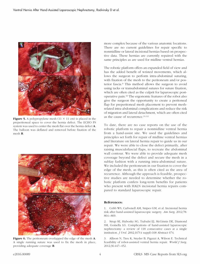
vision, 2 additional 10-mm ports were placed laterally inthe left mid abdomen and left lower quadrant (Figure 2).Inspection of the abdomen showed extensive adhesionssurrounding the hernia defect, which included multipleloops of small bowel tethered to the anterior abdominalwall (Figure 3). After extensive adhesiolysis, the contentsof the hernia were reduced, revealing an �6 � 6-cmdefect. The edges of the external and internal obliquemuscles were dissected, and the hernia defect was closedprimarily with a running absorbable barbed sutures (Fig-ure 4). Peritoneal flaps were created by dissection of thetransversalis fascia from the internal oblique to allow apreperitoneal mesh placement. An 11 � 11-cm compositemesh composed of polypropylene and an absorbable hy-drogel barrier was placed into the preperitoneal space(Figure 5). The mesh was secured with a circumferentialabsorbable running suture, resulting in 3 cm of perito-neum overlap from the edge of the mesh circumferentially(Figure 6). The fascia was closed at all port sites with a 0Vicryl suture, and the skin was closed with running 4-0Monocryl at all port sites. Estimated blood loss was min-imal. Total operative time was 190 min, time on the ro-botic console was 146 minutes, and docking time was 8minutes. An assistant was at the patient’s side during theentire course of the procedure.
The postoperative clinical course was uneventful. Thepatient was discharged home the following morning. Hehas been seen for routine follow-up visits in the office andshowed no adverse events 30 days after surgery.
DISCUSSION
Ventral incisional hernia is a common complication afterabdominal surgery. Although the increase in the use of lapa-
Figure 1. CT scan findings suggestive of hernia at the priorhand-assist port site, with small bowel contents, but withoutevidence of obstruction or strangulation. R, rectus muscle; EO,external oblique muscle; IO internal oblique muscle; TA trans-verse abdominis muscle.
Figure 2. Port placement in relation to the hand-assist incisionsite.
Figure 3. Intra-abdominal inspection of the hand-assist site hernia,once the robot was docked. The small-bowel contents were re-duced with initial insufflation. Adhesions of the small bowel to theperimeter of the hernia were reduced with adhesiolysis.
Ventral Hernia After Hand-Assisted Laparoscopic Nephrectomy, Radvinsky D et al.
2e2016.00089 CRSLS MIS Case Reports from SLS.org

roscopic surgery has permitted a marked decrease in the rateof incisional hernia, this approach does not completely pre-vent the complication. Extraction sites and hand-assist sitesrequire a larger incision compared to standard laparoscopicincisions. The overall incidence of incisional hernia afterhand-assisted laparoscopic surgery is 3 to 10% and varies bylocation in relation to the midline.1,2
In a retrospective analysis, Troxel and Das4 reviewed 50patients who underwent HALN and reported on the post-operative incidence of incisional hernias. Of the 50 pa-tients, incision hernias developed in 6% (3 patients). Theyconcluded that such hernias have a multifactorial etiology,including patient’s BMI, comorbidities, smoking status,and functional status, but that obese patients are at higherrisk. They also stated that interrupted closure of the inci-sion was superior to a running polydioxanone suture(PDS) and furthermore, with the adoption of this tech-nique, they had not experienced any additional hernias.They did not offer suggestions for repair, however. Arecent retrospective analysis of the treatment of flank andlateral abdominal wall hernias advocates creating muscu-lofascial flaps and performing a primary nonbridged mesh
repair to recreate anatomical congruity.5 Another prospec-tive review of kidney transplant recipients with flank her-nias described a large posterior component separationwith transverse abdominis muscle release with meshplacement in the retromuscular plane in a sublay fashion.6
Both groups reported decreased recurrence rates 3.4 and9%, respectively, compared to the 11.4% reported by Sau-erland et al. 7 in their comparison of open to laparoscopicrepair of primary ventral hernias.
SAGES provides evidence-driven guidelines for laparo-scopic repair of ventral hernias.8 Multiple sources havesupported a tension-free primary closure of the herniadefect with mesh reinforcement, citing a reduced seromarate, reduced recurrence rate, and improved abdominalwall integrity and contour. A minimum of 3-cm and max-imum of 5-cm overlap of mesh is recommended to reducetension at the fixation points and provide adequate cov-erage. The use of suture fixation over tacking has beenshown to be more cost effective, with less postoperativepain and a quicker return to activity with a comparablerecurrence rate. Nonmidline ventral hernias are rarer thanmidline ventral hernias, and their surgical management is
Figure 4. After adhesiolysis, the external and internal oblique muscles were dissected from the peritoneum, and the hernia defect wasrepaired primarily before mesh placement.
3e2016.00089 CRSLS MIS Case Reports from SLS.org
more complex because of the various anatomic locations.There are no current guidelines for repair specific tononmidline or lateral incisional hernias based on prospec-tive data. These hernias are currently repaired with thesame principles as are used for midline ventral hernias.
The robotic platform offers an expanded field of view andhas the added benefit of wristed movements, which al-lows the surgeon to perform intra-abdominal suturing,with fixation of the mesh to the peritoneum and/or pos-terior fascia.9 This method allows the surgeon to avoidusing tacks or transabdominal sutures for suture fixation,which are often cited as the culprit for laparoscopic post-operative pain.10 The ergonomic features of the robot alsogive the surgeon the opportunity to create a peritonealflap for preperitoneal mesh placement to prevent mesh-related intra-abdominal complications and reduce the riskof migration and lateral detachment, which are often citedas the cause of recurrence.11,12
To date, there are no case reports on the use of therobotic platform to repair a nonmidline ventral herniafrom a hand-assist site. We used the guidelines andprinciples set forth for repair of midline ventral herniasand literature on lateral hernia repair to guide us in ourrepair. We were able to close the defect primarily, afterraising musculofascial flaps, to recreate the abdominalwall contour. We were able to provide adequate meshcoverage beyond the defect and secure the mesh in asublay fashion with a running intra-abdominal suture.We included the peritoneum in our fixation to cover theedge of the mesh, as this is often cited as the area ofrecurrence. Although the approach is feasible, prospec-tive studies are needed to determine whether the ro-botic platform confers long-term benefits for patientswho present with HALN incisional hernia repairs com-pared to standard laparoscopic repair.
References:
1. Cobb WS, Carbonell AM, Snipes GM, et al. Incisional herniarisk after hand-assisted laparoscopic surgery. Am Surg. 2012;78:864–869.
2. Strup SE, Hubosky SG, Trabulsi EJ, McGinnis DE, DiamondSM, Gomella LG. Complications of hand-assisted laparoscopicnephrectomy: a review of 118 consecutive cases at a singleinstitution. J Urol. 2002;167(4 suppl):168 Abstract 674.
3. Allison N, Tieu K, Snyder B, Pigazzi A, Wilson E. Technicalfeasibility of robot-assisted ventral hernia repair. World J Surg.2012;36:447–452.
Figure 6. The peritoneum overlapped the edge of the mesh A.A single running suture was used to fix the mesh in place,providing adequate coverage B.
Figure 5. A polypropylene mesh (11 � 11 cm) is placed in thepreperitoneal space to cover the hernia defect. The ECHO PSsystem was used to center the mesh flat over the hernia defect A.The balloon was deflated and removed before fixation of themesh B.
Ventral Hernia After Hand-Assisted Laparoscopic Nephrectomy, Radvinsky D et al.
4e2016.00089 CRSLS MIS Case Reports from SLS.org

4. Troxel SA, Das S. Incisional hernia following hand-assistedlaparoscopic surgery for renal cell cancer. JSLS. 2005;9:196–198.
5. Pezeshk RA, Pulikkottil BJ, Bailey SH, et al. An evidence-based model for the successful treatment of flank and lateralabdominal wall hernias. Plast Reconstr Surg. 2015;136:377–385.
6. Petro CC, Orenstein SB, Criss CN, et al. Transversus abdomi-nis muscle release for repair of complex incisional hernias inkidney transplant recipients. Am J Surg. 2015;210:334–339.
7. Sauerland S, Walgenbach M, Habermalz B, Seiler C, MiserezM. Laparoscopic versus open surgical techniques for ventral orincisional hernia repair. Cochrane Database of Systematic Re-views. March 16, 2011.
8. Earle D, Roth S, Saber A, et al. Guidelines for LaparoscopicVentral Hernia Repair. SAGES Guidelines Committee, July 2016.
9. Ballantyne, GH, Hourmont K, Wasielewski A. Teleroboticlaparoscopic repair of incisional ventral hernias using intraperi-toneal prosthetic mesh. JSLS. 2003;7:7–14.
10. Wassenaar E, Schoenmaeckers E, Raymakers J, Van derPalen J, Rakic S. Mesh-fixation method and pain and quality oflife after laparoscopic ventral or incisional hernia repair: a ran-domized trial of three fixation techniques. Surg Endosc. 2010;24:1296–1302.
11. Prasad P, Tantia O, Patle NM, Khanna S, Sen B. Laparoscopicventral hernia repair: a comparative study of transabdominalpre-peritoneal versus intraperitoneal onlay mesh repair. J Lapa-roendosc Adv Surg Tech A. 2011;21:477–483.
12. Awad ZT, Puri V, LeBlanc K, et al. Mechanisms of ventralhernia recurrence after mesh repair and a new proposed classi-fication. J Am Coll Surg. 2005;201:132–140.
5e2016.00089 CRSLS MIS Case Reports from SLS.org