RS1 protein dependent and independent short and long … mismanagement leads to chronic disorders...
68
RS1 protein dependent and independent short and long term regulation of sodium dependent glucose transporter -1 RS1 Protein abhängige und unabhängige Kurz- und Langzeitregulation des Natrium-abhängigen Glukosetransporter -1 Doctoral thesis Graduate School of Life Sciences, Julius-Maximilians-Universität Würzburg, Section Life Sciences Submitted by Aruna Srinivasan Würzburg, 2013
Transcript of RS1 protein dependent and independent short and long … mismanagement leads to chronic disorders...
RS1 protein dependent and independent short and long term regulation of
sodium dependent glucose transporter -1
RS1 Protein abhängige und unabhängige Kurz- und Langzeitregulation des
Natrium-abhängigen Glukosetransporter -1
Doctoral thesis
Graduate School of Life Sciences,
Julius-Maximilians-Universität Würzburg,
Section Life Sciences
Submitted by
Aruna Srinivasan
Würzburg, 2013

Submitted on:
…………………………………………………………..……………
Office stamp
Members of the Promotionskomitee:
Chairperson: Prof. Dr. Thomas Müller
Primary Supervisor: Prof. Dr. Hermann Koepsell
Supervisor (Second): Prof. Dr. Rainer Hedrich
Supervisor (Third): Prof. Dr. med. Manfred Gessler
Date of Public Defence: ……………………………………………..
Date of receipt of Certificates: ……………………………………...

Table of Contents
Table of Contents:
1. Introduction: ................................................................................................................................. 1
1.1 Sodium Dependent Glucose Co-transporter 1 (SGLT1): ......................................................... 1
1.1.1 Regulation of SGLT1: ....................................................................................................... 2
1.1.1.1 The RS1 protein and its regulation on SGLT1:............................................................. 6
1.1.1.2 PKC dependent regulation of SGLT1: .......................................................................... 8
2. Aim of the study: ......................................................................................................................... 10
3. Materials & Methods .................................................................................................................. 11
3.1 Materials ............................................................................................................................... 11
3.1.1 Chemicals: ..................................................................................................................... 11
3.1.2 Animals: ......................................................................................................................... 11
3.1.3 Diets: ............................................................................................................................. 11
3.1.4 Inhibitors and Activators: .............................................................................................. 12
3.1.5 Synthetic tripeptides: .................................................................................................... 12
3.1.6 Radioactive substrate: .................................................................................................. 12
3.1.7 Buffers and solutions: ................................................................................................... 13
3.1.8 Software: ....................................................................................................................... 13
3.2 Methods ................................................................................................................................ 14
3.2.1 Small intestinal brush border membrane vesicle (BBMV) preparation: ....................... 14
3.2.2 AMG affinity uptake in BBMVs: .................................................................................... 15
3.2.3 Protein estimation in BBMVs: ....................................................................................... 16
3.2.4 AMG uptake in small intestinal everted segments: ...................................................... 16
3.2.5 Calculation and Statistics .............................................................................................. 17
3.2.6 Experimental design setup: ........................................................................................... 18
4. Results .......................................................................................................................................... 20
4.1 Effect of glucose and RS1 on SGLT1 activity: ........................................................................ 20
4.2 Effect of glucose and RS1 on SGLT1 affinity: ........................................................................ 21
4.3 Effect of regulatory tripeptides (QSP and QEP) on glucose dependent short term regulation
of SGLT1: ........................................................................................................................................... 22
4.4 The role of PKC in RS1 dependent short term regulation of SGLT1: .................................... 26
4.5 Effect of PKC on SGLT1 affinity: ............................................................................................ 29
4.6 Effect of RS1 on SGLT1 activity during diet adaptation: ....................................................... 30
5. Discussion..................................................................................................................................... 33
6. Appendix ...................................................................................................................................... 37

Table of Contents
Appendix I: Tissue pre-incubation alters SGLT1 dependent substrate transport:............................ 37
Appendix II: Tissue pre-incubation does not alter SGLT1 affinity: .................................................... 38
Appendix III: Segmental difference occurs within jejunum region of mice small intestine: ............ 39
Appendix IV: Brefeldin A activity is dependent on glucose and RS1: ............................................... 41
Appendix V: QEP down regulates SGLT1 activity by exocytotic inhibition of SGLT carrying vesicles:
.......................................................................................................................................................... 43
Appendix VI: PKC down regulates SGLT1 activity by aiding to exocytotic inhibition of SGLT1
vesicles: ............................................................................................................................................. 44
Appendix VII: SGLT1 affinity for substrate transport during diet adaptation might be RS1
dependent: ........................................................................................................................................ 45
7. Summary: .................................................................................................................................... 47
8. Zusammenfassung: ..................................................................................................................... 48
9. Abbreviations: ............................................................................................................................. 50
10. References: ................................................................................................................................... 53
Acknowledgements: ............................................................................................................................ 60
Curriculum Vitae: ............................................................................................................................... 62
Publications ......................................................................................................................................... 63
Participation in scientific international conferences ........................................................................ 63
Affidavit ............................................................................................................................................... 64
Eidesstattliche Erklärung ................................................................................................................... 64

Introduction
1
1. Introduction:
Carbohydrate mismanagement leads to chronic disorders such as obesity and type 2 diabetes.
Sedentary lifestyles and high caloric diet intake have been noted as causes for increase in
carbohydrate mismanagement disorders. Therapeutic drug development against these
disorders relies on understanding the mechanism of carbohydrate metabolism. Carbohydrates
forms one of major nutrients in diet and are a major source of energy for mammals. They are
hydrolysed during digestion to monosaccharides by enzymes in the gastrointestinal tract.
Monosaccharides cannot infuse the selectively permeable plasma membrane surface of
intestinal epithelial cells (enterocytes). This entry is mediated by transporters present on the
apical surface of the enterocytes. Later the monosaccharides exit into the blood stream via the
transporters located in the enterocyte baso-lateral surface (Hediger, Coady et al. 1987, Wood
and Trayhurn 2003). The sodium dependent glucose co-transporter (SGLT) of solute carrier
family 5A (SLC5A) (Wright, Loo et al. 1994, Wright, Loo et al. 2004, Krimi, Letteron et al.
2009, Wong, Debnam et al. 2009) and the facilitative glucose transporter (GLUT) of the
solute carrier family 2 (SLC2) family (Uldry and Thorens 2004) are involved in the
absorption of glucose and some structurally related substances in the small intestine. These
transporters are also involved in reabsorption of glucose from the glomerular filtrate, uptake
of glucose across the blood-brain barrier, and in uptake and release of glucose from all cells
in the body.
1.1 Sodium Dependent Glucose Co-transporter 1 (SGLT1):
SGLT1 is identified as a high affinity low capacity transporter (Hediger, Coady et al. 1987,
Corpe and Burant 1996, Tavakkolizadeh, Berger et al. 2001, Wright, Martin et al. 2003,
Houghton, Zarroug et al. 2006). Various organs (intestine, trachea, kidneys, heart, brain,
testis and prostate glands) in humans and rats contain SGLT1 at distinct locations (Balen,
Ljubojevic et al. 2008, Wright, Loo et al. 2011). The importance of SGLT1 in intestinal
glucose transport was recognised when it was observed that the absence of functional SGLT1
in human intestine led to glucose-galactose mal-absorption (Shirazi-Beechey, Gribble et al.
1994, Wright 1998). SGLT1 is most strongly expressed in the apical membrane of
enterocytes where it mediates the first step in small intestinal glucose absorption (Hediger,
Coady et al. 1987, Kellett and Brot-Laroche 2005, Gorboulev, Schurmann et al. 2012).
SGLT1 mediates glucose transport by symport of sodium ions and glucose with a
stoichiometric ratio of 2:1. This symport occurs via a secondary active transport where the

Introduction
2
sodium gradient is generated by the Na+/K
+ ATPase (Figure 1) (Wright, Loo et al. 1994,
Wright, Martin et al. 2003, Wright, Hirayama et al. 2007).
Figure 1: Model for intestinal secondary active glucose (and galactose) transport across
the small intestine: The sketch depicts the mechanism of glucose transport occurring within
a mature enterocyte at the upper villus of the small intestine. Glucose (or galactose) is co-
transported with Na+ across the brush border membrane by SGLT1. Glucose then exists
across the basolateral membrane by GLUT2 or by exocytosis. Na+ is actively transported
across the basolateral membrane by Na+/K
+ ATPase. The energy provided by Na
+ gradient is
used for the transport of glucose. This figure is based on a scheme provided by Wright et al.,
2004.
1.1.1 Regulation of SGLT1:
The functional capacity of SGLT1 is regulated to meet up with physiological demands of the
species (Ferraris 2001).
Depending on the mechanism, the regulation of SGLT1 can be classified into post-
transcriptional short term regulation and transcriptional and post-transcriptional long term
regulation (Karasov and Debnam 1987, Wright, Hirsch et al. 1997).
Post-transcriptional short term regulation occurs in the brush border membrane of enterocytes
layered with an intact mucosa layer (Sharp, Debnam et al. 1996). This regulation enables
adaptation to rapid changes of luminal glucose concentration (Wright, Hirsch et al. 1997,
Kellett and Brot-Laroche 2005). They include modifications of SGLT1 that may alter either
the affinity, turn over number, vesicular trafficking or degradation bringing change in the
number of transporter molecules within the plasma membrane (Hirsch, Loo et al. 1996,
Vayro and Silverman 1999).

Introduction
3
Several factors mediate the short term post-transcriptional regulation of SGLT1. Protein RS1
identifies SGLT1 as a physiological target for regulation (Osswald, Baumgarten et al. 2005).
It mediates post-transcriptional short term regulation of SGLT1 by modulating the
recruitment of vesicles containing SGLT1 from the intracellular pool at Trans Golgi network
(TGN) to enterocyte apical surface (Veyhl, Keller et al. 2006). This regulation is dependent
on intracellular glucose concentration and Protein kinase C (PKC) activation (Veyhl, Wagner
et al. 2003). Peptides such as Proadrenomedullin N-terminal 20 peptide (PAMP) and
adrenomedullin (AM) can also post-transcriptionally modulate the exocytosis of intracellular
vesicles containing SGLT1 for short term regulation. This regulation is suggested to depend
on cyclic adenosine monophosphate (cAMP) - Protein kinase A (PKA) pathway (Fernandez
de Arcaya, Lostao et al. 2005). Hormones which can mediate short term regulation of SGLT1
include glucagon like peptide-2 (GLP-2) (Shirazi-Beechey, Moran et al. 2011), epinephrine
(Ishikawa, Eguchi et al. 1997), portal insulin (Stumpel, Kucera et al. 1996), glucagon-37
(Stumpel, Scholtka et al. 1997), leptin (Ducroc, Guilmeau et al. 2005), cholecystokinin
(CCK) (Hirsh and Cheeseman 1998) and prostaglandin E (PGE2) (Scholtka, Stumpel et al.
1999). These hormones and peptides may involve activation of secondary messengers such as
G-protein (α-gustiducin) (Margolskee, Dyer et al. 2007) and cAMP (Ishikawa, Eguchi et al.
1997, Luz Sdos, de Campos et al. 1997, Stumpel, Scholtka et al. 1997, Lee, Loflin et al.
2000, Fernandez de Arcaya, Lostao et al. 2005, Shirazi-Beechey, Moran et al. 2011). Protein
kinases such as PKC (Hirsch, Loo et al. 1996, Wright, Hirsch et al. 1997, Vayro and
Silverman 1999, Veyhl, Wagner et al. 2003, Barrenetxe, Sainz et al. 2004, Kim, Lee et al.
2004, Castaneda-Sceppa, Subramanian et al. 2010), PKA (Ishikawa, Eguchi et al. 1997,
Fernandez de Arcaya, Lostao et al. 2005, Subramanian, Glitz et al. 2009), Serum and
glucocorticoid inducible kinase-1 (SGK-1) (Shojaiefard, Strutz-Seebohm et al. 2007) and
glycogen synthase kinase 3 beta (GSK-3β) (Rexhepaj, Dermaku-Sopjani et al. 2010) can be
involved in post-transcriptional regulation of SGLT1.
Transcriptional long term regulation of SGLT1 occurs in the cells present in lower villus and
crypts which later migrate to upper villus and manifest the regulated SGLT1 function
(Ferraris, Villenas et al. 1992). Although long term regulation of SGLT1 mainly involves
transcriptional mechanism, it can also be regulated by post-transcriptional mechanism. Long
term regulation enables adaptation to different diet conditions (Shirazi-Beechey, Hirayama et
al. 1991), ontogenetic development (Shirazi-Beechey, Smith et al. 1991) differentiation of
enterocyte for maturation (Freeman, Heavens et al. 1992) and also involves changes during

Introduction
4
chronic metabolic disorders like diabetes (Fedorak, Gershon et al. 1989, Dyer, Garner et al.
1997, Dyer, Wood et al. 2002), hypo- or hyperinulinaemia (Philpott, Butzner et al. 1992),
carcinoma (Alesutan, Sopjani et al. 2012, Hanabata, Nakajima et al. 2012, Lai, Xiao et al.
2012), obesity (Krimi, Letteron et al. 2009, Sopjani, Bhavsar et al. 2010) and inflammatory
bowel diseases (Thomson and Wild 1997, Thomson and Wild 1997). These regulations of
SGLT1 also control its diurnal rhythmic expression depending on the feeding pattern of the
species (Furuya and Yugari 1974, Fisher and Gardner 1976, Corpe and Burant 1996, Rhoads,
Rosenbaum et al. 1998, Tavakkolizadeh, Berger et al. 2001, Pan, Terada et al. 2004,
Houghton, Zarroug et al. 2006). The long term SGLT1 regulation involves modulations in
mRNA level or change in membrane integrity. Modulation in mRNA level could include
changes in promoter activity (Dyer, Vayro et al. 2003), mRNA expression (Miyamoto, Hase
et al. 1993) or mRNA stability (Loflin and Lever 2001, Pedder, Ford et al. 2008).
Modulations at membrane integrity could include changes in height of microvilli (Smith,
Peacock et al. 1991), membrane physio-chemical composition (Schwarz, Hostetler et al.
1985, Schwarz, Bostwick et al. 1989, Vazquez, Rovira et al. 1997), membrane actin
polymerization (Chung, Wong et al. 1999) or change in the fraction of transporting to non-
transporting cells (Fedorak, Gershon et al. 1989). These modulations at mRNA level and
membrane integrity may lead to changes in density of transporter molecules (Ferraris and
Diamond 1992, Ferraris and Diamond 1993), change in surface area for absorption (Smith,
Peacock et al. 1991), change in rate of transport (Diamond, Karasov et al. 1984), change in
the cells intended for transporter activity (Fedorak, Gershon et al. 1989) or increase
translocation of other transporters to apical membrane surface (Krimi, Letteron et al. 2009,
Sopjani, Bhavsar et al. 2010).
Prevalence of diet dependent SGLT1 regulation has been observed with changes in sodium
(Donowitz, De La Horra et al. 1998), fibre (Reimer, Field et al. 1997) and carbohydrate
content. High consumption of carbohydrate in diet leads to increase in rate of intestinal
glucose transport by changing SGLT1 mRNA and protein abundance (Shirazi-Beechey,
Gribble et al. 1994, Dyer, Hosie et al. 1997, Ferraris 2001). Carbohydrate based SGLT1 diet
regulation is reported among vertebrates like mammals (rats, mice, cows, sheep and humans),
amphibians (tadpoles) and fishes (tilapia and catfish) but not in birds (Ferraris 2001). Several
factors are involved in the diet dependent regulation of SGLT1. Protein RS1 regulates
SGLT1 transcriptionally for adaptation to diet having reduced glucose-galactose content
(Filatova, Leyerer et al. 2009). High nutrient diet shows transcriptional upregulation of

Introduction
5
SGLT1 activity by elevation of cAMP levels. Elevated levels of cAMP lead to PKA
dependent increase in SGLT1 promoter activity (Dyer, Vayro et al. 2003).
Regulation of SGLT1 with regards to animal development depends on the gestation period,
age and feeding (Ferraris 2001). These regulations include maintenance of high intestinal
SGLT1 activity in foetal and neonatal stage (Buddington and Diamond 1989, Vazquez,
Rovira et al. 1997, Buddington, Malo et al. 2000), gradual decrease in SGLT1 mRNA levels
from duodenum to ileum of adults (Kojima, Nishimura et al. 1999, Yoshikawa, Inoue et al.
2011), accumulation of SGLT1 mRNA and insertion of SGLT1 transporters at the apical
membrane during enterocyte maturation (Delezay, Baghdiguian et al. 1995), maintenance of
SGLT1 diurnal rhythm (Rhoads, Rosenbaum et al. 1998, Martin, Wang et al. 2000, Vayro,
Wood et al. 2001, Balakrishnan, Stearns et al. 2008, Kekuda, Saha et al. 2008) and abnormal
SGLT1 activity during chronic occurrence of various metabolic disorders (Fedorak, Gershon
et al. 1989). Several factors are involved for regulation of SGLT1 for animal development
and cellular differentiation. Protein RS1 mediates transcriptional regulation of SGLT1 in
LLCPK1 cells (porcine kidney epithelial cell line) dependent on cell confluence (Korn,
Kuhlkamp et al. 2001, Filatova, Leyerer et al. 2009). Other proteins like a 38kDa nucleo-
cytoplasmic protein (Lee, Loflin et al. 2000) and a RNA binding protein (HuR) (Loflin and
Lever 2001) are involved in SGLT1 regulation during cell differentiation. Some factors
which are involved in regulation of SGLT1 during chronic disorders or diseases such as
carcinoma, diabetes, obesity, short bowel syndrome and intestinal inflammation include
protein complexes like Nuclear factor kappa-light-chain-enhancer of activated B cells
(NFκB) (Lee, Heo et al. 2007), hormones like angiotensin II (Wong, Debnam et al. 2009) and
resistin like molecule β (RELMβ) (Krimi, Letteron et al. 2009, Sopjani, Bhavsar et al. 2010),
growth factors like epidermal growth factor (EGF) (Chung, Wong et al. 1999) protein kinases
like 5' Adenosine Mono-phosphate activated protein kinase (AMPK) (Krimi, Letteron et al.
2009, Sopjani, Bhavsar et al. 2010) and Tau tubulin kinase 2 (TTBK2) (Alesutan, Sopjani et
al. 2012) and transcriptional factors like hepatocyte nuclear factors 1 (HNF1) and specificity
protein 1 (Sp1) (Rhoads, Rosenbaum et al. 1998, Martin, Wang et al. 2000, Vayro, Wood et
al. 2001, Balakrishnan, Stearns et al. 2008, Kekuda, Saha et al. 2008). Vagus nerve
contributes in diurnal rhythm regulation of SGLT1 (Stearns, Balakrishnan et al. 2008,
Stearns, Balakrishnan et al. 2012). Few of the different secondary messengers and protein
kinases involved in the long term regulation of SGLT1 include cAMP (Peng and Lever 1995,
Loflin and Lever 2001), PKA (Dyer, Vayro et al. 2003) and PKC (Castaneda-Sceppa,

Introduction
6
Subramanian et al. 2010). PKC is involved in transcriptional regulation of SGLT1 by
involving several intracellular signalling pathways such as p38/MAPK (Mitogen activated
protein kinase), Extracellular signal regulated kinase (ERK)/MAPK, c-Jun N-terminal kinase
(JNK)/MAPK and Phosphatidylinositide 3-kinases (PI3K)/Protein kinase B (Akt)/
mammalian target of rapamycin (mTOR) (Castaneda-Sceppa, Subramanian et al. 2010).
1.1.1.1 The RS1 protein and its regulation on SGLT1:
The Mammalian specific, intronless single copy gene RSC1A1 encodes a 67- 68 KDa protein
named RS1. Depending on the mammalian species, RSC1A1 gene expression shows a wide
tissue distribution. RS1 is expressed in kidneys, small intestine, liver, spleen, brain and lungs
(Veyhl, Spangenberg et al. 1993, Lambotte, Veyhl et al. 1996, Poppe, Karbach et al. 1997,
Reinhardt, Veyhl et al. 1999, Osswald, Baumgarten et al. 2005). Within the cell RS1 protein
is distributed below the apical plasma membrane, around the Trans Golgi network (TGN) and
within the cell nucleus (Valentin, Kuhlkamp et al. 2000, Osswald, Baumgarten et al. 2005,
Kroiss, Leyerer et al. 2006).
RS1 regulates the activity of different transporters from different families such as SGLT1
(Veyhl, Spangenberg et al. 1993), SGLT1 homologous Na+ myo-inositol co-transporter
(SMIT) (Lambotte, Veyhl et al. 1996), organic cation transporters (OCT1 and 2) and
concentrative nucleoside transporters (CNT1, 2 and 3) (Reinhardt, Veyhl et al. 1999, Veyhl,
Wagner et al. 2003, Errasti-Murugarren, Fernandez-Calotti et al. 2012). Regulation of SGLT1
has been studied extensively due to its physiological importance. The deletion of RSC1A1
gene in mice (RS1-/-) lead to increase in SGLT1 activity. Hence SGLT1 is identified as a
physiologically important target for RS1 (Osswald, Baumgarten et al. 2005).
RS1 is involved the transcriptional and post-transcriptional regulation of SGLT1 (Korn,
Kuhlkamp et al. 2001, Veyhl, Keller et al. 2006). The transcriptional regulation of SGLT1 by
RS1 was studied in the porcine kidney epithelial cell line - LLCPK1. In LLCPK1 a
confluence dependent inverse relationship occurs between the nuclear distribution of RS1 and
the expression of SGLT1. When RS1 expression is down-regulated by antisense strategy or
overexpressed artificially it resulted in opposite effects on SGLT1 mRNA expression and
activity. RS1 thereby reveals a transcriptional regulation dependent on cell confluence (Korn,
Kuhlkamp et al. 2001). In mice fed with glucose-galactose reduced diet (GGRD) RS1
transcriptionally regulates SGLT1 to mediate higher SGLT1 mRNA and protein expression
in wild type mice as compared to RS1-/- mice (Filatova, Leyerer et al. 2009). The post-

Introduction
7
transcriptional regulation by RS1 was studied in Xenopus laevis oocytes system where co-
expression of human RS1 (hRS1) and human SGLT1 (hSGLT1) cRNA decreased SGLT1
activity (Veyhl, Spangenberg et al. 1993, Lambotte, Veyhl et al. 1996, Veyhl, Wagner et al.
2003). Down regulation of SGLT1 activity also occurred on injecting hRS1 protein in
hSGLT1 expressing Xenopus laevis oocytes (Veyhl, Keller et al. 2006). This indicated RS1
as a post-transcriptional regulator of SGLT1. This regulation occurs by inhibiting dynamin
dependent exocytotic release of hSGLT1 containing vesicles from TGN (Veyhl, Wagner et
al. 2003, Kroiss, Leyerer et al. 2006, Veyhl, Keller et al. 2006).The regulation is enhanced by
PKC activation and is dependent on intracellular glucose concentration (Veyhl, Wagner et al.
2003, Veyhl, Keller et al. 2006).
Functional characterization of RS1 protein identified an N-terminal domain (TGN-Reg)
consisting of about 90 residues from 16-98 and 15-92 in human and mouse RS1 (hRS1 and
mRS1) respectively responsible for post-transcriptional regulation of SGLT1. Two peptide
sequences - Gln-Ser-Pro (QSP) (Vernaleken, Veyhl et al. 2007) and Ser-Asp-Ser-Asp-Arg-
Ile-Glu-Pro (SDSDRIEP) (M. Vehyl-Wichmann, et al data unpublished) in the TGN-Reg are
identified capable to mediate this regulation of SGLT1. This domain also comprises of a 21
residue long nuclear shuttling domain (RNS) to mediate the cell confluence dependent
nuclear localization of RS1 protein and another domain for controlling the transcriptional
regulation of SGLT1 (Filatova, Leyerer et al. 2009). The C-terminal sequence of RS1
contains a tripeptide Gln-Cys-Pro (QCP) which can also mediate post-transcriptional
regulation of hSGLT1 (Vernaleken, Veyhl et al. 2007) and an ubiquitin associated domain
(UBA) which has not been characterized (Valentin, Kuhlkamp et al. 2000). QCP and QSP are
also substrates of H-peptide co-transporter PepT1 and are effectively absorbed when applied
in the intestine lumen (Vernaleken, Veyhl et al. 2007). The TGN-Reg domain also consists
of several consensus sequences for phosphorylation by a variety of protein kinases. The
putative phosphorylation sites in this domain of hRS1 and mRS1 were screened and their
comparative alignment is depicted in Figure 2.

Introduction
8
Figure 2: Comparative alignment between TGN-Reg Domains of hRS1 with mRS1: Both hRS1 and mRS1 have several serine and threonine putative phosphorylation sites. The
sites common in both have been marked in blue. Two QSP motifs are present in hRS1
whereas only one QSP motif is present in mRS1. SDSDRIEP motif is specifically present
only in hRS1 sequence. SDSDRIEP is a putative site for PKC α phosphorylation while QSP
is a putative site for CamK2 phosphorylation.
1.1.1.2 PKC dependent regulation of SGLT1:
Several factors mediate the regulation of SGLT1 by involving activation of secondary
messengers and protein kinases. Involvement of PKC in the regulation of SGLT1 is shown
during sepsis caused by bacterial lipopolysaccharide (LPS) (Amador, Garcia-Herrera et al.
2007), during hormonal stimulus like epinephrine (Kim, Lee et al. 2004), Angiotensin II
(Han, Park et al. 2004), leptin (Ducroc, Guilmeau et al. 2005) and in RS1 protein dependent
post-transcriptional regulation of SGLT1 (Veyhl, Wagner et al. 2003, Veyhl, Keller et al.
2006). PKC regulates SGLT1 by involving modulation in level of cAMP (Hirsch, Loo et al.
1996). PKC regulates SGLT1 both transcriptionally and post-transcriptionally. This
regulation might modulate SGLT1 protein level in the apical surface or affect the rate of
SGLT1 dependent transport. Post transcriptional regulations that can modulate SGLT1
number in apical surface include changes in trafficking of SGLT1 containing vesicles,
SGLT1 degradation rate or SGLT1 mRNA stability. The post-transcriptional regulations that
can alter SGLT1 affinity or turn over number can modulate the rate of SGLT1 mediated
transport. PKC dependent diverse post-transcriptional modulations have been observed in
different systems. PKC decreases rabbit SGLT1 turn over number in monkey kidney derived
fibroblast like cell line (COS-7) (Vayro and Silverman 1999). In Chinese hamster ovary cells
expressing rabbit SGLT1 (CHO-G6D3), PKC activation lead to increase in transcription of
SGLT1 mRNA (Castaneda-Sceppa, Subramanian et al. 2010). In LLCPK1 cells, PKC
Introduction
9
demonstrates a decrease in SGLT1 mRNA due to mRNA degradation (Shioda, Ohta et al.
1994). In SGLT1 expressing Xenopus laevis oocytes (Hirsch, Loo et al. 1996, Wright, Hirsch
et al. 1997, Veyhl, Keller et al. 2006), in rat intestinal brush border membrane vesicles
(Ishikawa, Eguchi et al. 1997), in differentiating human cell clone HT-29-D4 (Delezay,
Baghdiguian et al. 1995), CHO-G6D3 cell line (Castaneda-Sceppa, Subramanian et al. 2010)
and LLCPK1 cells (Shioda, Ohta et al. 1994, Peng and Lever 1995) PKC mediates SGLT1
regulation by modulating exo – and endocytosis of SGLT1 carrying vesicles. The different
systems not only show different forms of post-transcriptional modulations but also show
diversity in respect to the effect (increase or decrease) on SGLT1 activity, protein or mRNA
caused due to PKC activation.
The different effects caused by PKC on SGLT1 regulation are dependent not only on the
expression system but also on the species homolog of SGLT1 sequence. In Xenopus laevis
oocytes expressing rabbit and rat SGLT1, PKC activation mediates a negative regulation
decreasing the rate of substrate transport whereas when human SGLT1 is expressed it
mediates a positive regulation increasing substrate transport. The SGLT1 sequence in rat and
human has 5 consensus PKC sites while rabbit contains 4 sites (Wright, Hirsch et al. 1997).
The amino acid residues between positions 550-636 of SGLT1 cytoplasmic loop sequence are
considered to be the most likely domain involved in PKC dependent sequence specific
regulation of SGLT1. (Hirsch, Loo et al. 1996). Serine 418 residue in hSGLT1 sequence is
speculated for SGLT1 regulation by phosphorylation/ de-phosphorylation (Kumar, Tyagi et
al. 2007). These complementary mechanisms for PKC dependent SGLT1 regulation may also
depend on the choice of path followed. The activation of MAPK and PI3K/Akt/mTOR
signalling pathway by PKC lead to increase in SGLT1 dependent uptake. The activation of
p38/MAPK signalling pathway by PKC exerts the inhibitory action on SGLT1 uptake
(Castaneda-Sceppa, Subramanian et al. 2010).
The further investigation of PKC in the transcriptional and posttranscriptional regulatory
mechanism of RS1 may bring new insights in understanding SGLT1 regulation and thereby
might impart knowledge for the better management of carbohydrate metabolism.
Aim of the study
10
2. Aim of the study:
The aim of this study was to further elucidate the role of the regulatory protein RS1 in the
short term post-translational and long term transcriptional regulation of SGLT1by employing
tools which were available in the laboratory of Professor H. Koepsell. These tools were mice
in which RS1 was removed genetically (RS1-/- mice) and tripeptides derived from the
regulatory domain of RS1 which mediates post-translational regulation of SGLT1. Since it is
known that post-translational regulation of SGLT1 is modulated by PKC (Hirsch, Loo et al.
1996, Wright, Hirsch et al. 1997) and that PKC also influences the post-translational
regulation mediated by RS1 (Veyhl, Wagner et al. 2003) the effect of PKC in wild type mice
versus RS1-/- mice was compared. Concerning investigation of the role of RS1 in
transcriptional regulation the present study was focussed on the investigation of effects of
diets on the expression of SGLT1 in small intestine. Due to restriction of time and technical
difficulties arising during the study the investigation was focussed on functional analyses.

Materials & Methods
11
3. Materials & Methods
3.1 Materials
3.1.1 Chemicals:
All laboratory chemicals used were of pro analysi (p.a.) grade and were purchased form one
of the firms namely Sigma-Aldrich (Taufkirchen, Germany), Merck (Darmstadt, Germany),
Perkin Elmer (Darmstadt, Germany) or AppliChem (Darmstadt, Germany).
3.1.2 Animals:
Institutional guidelines and German laws were met in order to handle mice as the model
system. Wild type mice and RS1-/- mice of 129/OLA/C57BL/6 or C57BL/6 background
were used for comparative studies on SGLT1 activity. Animals of age 2 – 3 months were
chosen for study. All animals were housed in a temperature – controlled environment with a
12h – light/12h – dark cycle with free access to specific diet and tap water.
3.1.3 Diets:
For the study of diet effects on SGLT1 regulation, weaning mice of 10-14 days old were
segregated to different groups. Each group were fed with free access to specific diet and tap
water for 2 months. Standard control or normal diet (ND) (Ssniff V1534-000R/M-H, 10mm),
High fat high glucose diet (HFHGD) (Ssniff® EF R/M by D12492 mod.) and Glucose-
galactose reduced diet (GGRD) (Ssniff® EF R/M Glucose-arm) were purchased from
Spezialdiäten GmbH, (Soest, Germany). The specific composition of each diet is mentioned
in (table 1) and their total metabolizable energy is mentioned in (table 2)
Table – 1: Diet composition
Diet
Components
Standard
control diet
(%)
High fat high
glucose diet
(%)
Glucose-
galactose
reduced diet (%)
Starch 36.4 2.2 5.8
Protein 19 24.1 29.7
Fibre 4.9 6 28.8
Mono and Di
saccharides
4.7 22.4 0.5
Fat, Minerals, Vitamins 3.3 34 20.5
Materials & Methods
12
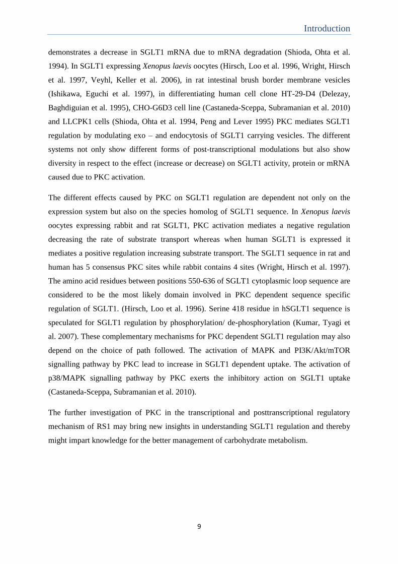
Table -2: Total metabolizable energy
Standard control
diet (MJ/kg)
High fat high
glucose diet
(MJ/kg)
Glucose-galactose
reduced diet
(MJ/kg)
Total
metabolizable
energy
12.8 25.2 22.9
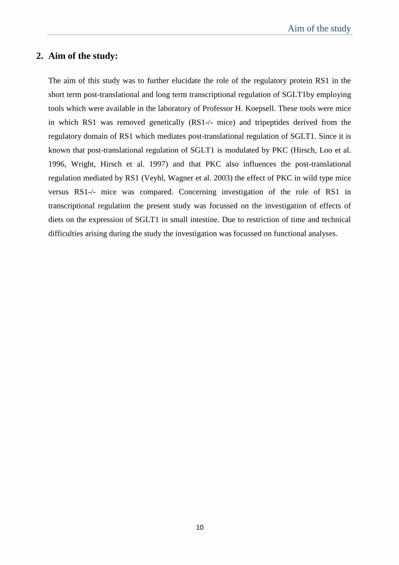
3.1.4 Inhibitors and Activators:
Protease inhibitors used for the study were obtained from Roche Diagnostics Deutschland
GmbH (Mannheim, Germany). The working concentrations of the same are specified in the
following table.
Table -3: Working stock concentration of protease inhibitor:
Protease Inhibitor Working concentration (µM)
Aprotinin 0.8
Benzamidin 1000
Leupeptin 20
Phenylmethylsulfonyl fluoride (PMSF) 100
Pefabloc SC (AEBSF) 2000
Phorbol 12 - myristate 13 - acetate (PMA) which is an activator of PKC was obtained from
the firm Sigma-Aldrich (Taufkirchen, Germany). The working concentration of the PMA
used was 5 µM.
3.1.5 Synthetic tripeptides:
Two synthetic tripeptides QSP (Gln-Ser-Pro) and QEP (Gln-Glu-Pro) were obtained from the
lab of Dr. Rüdiger Pipkorn, German Cancer Research Center (Heidelberg, Germany). The
working concentration of the peptides used was 15 mM.
3.1.6 Radioactive substrate:
ARC0131 C14
radiolabelled Methyl–α-D-Glucopyranoside (AMG), [glucose-14
C (U)]; (11.1
Gbq/mMol) was obtained from American Radiolabeled Chemicals, Inc. (St. Louis, MO,
USA). Radioactive [14
C] AMG was added in tracer amounts to a required concentration of
AMG for performing SGLT1 uptake and affinity studies. The final AMG working

Materials & Methods
13
concentration for uptake studies in everted segment method was 10 µM while for affinity
measurements in brush border membrane vesicles (BBMV), a concentration range from 0.02
– 5 mM was used.
3.1.7 Buffers and solutions:
Deionised water was used to prepare all aqueous buffer solutions. Compositions of the
different buffer solutions are mentioned in the respective method sections.
3.1.8 Software:
Putative phosphorylation motifs in TGN-Reg domain of human and mice RS1 sequence were
searched using MnM 3.0 (Minimotif Miner, version 3.0)(http://minimotifminer.org) and GPS
2.1 (Group-based Prediction System, version 2.1) (http://gps.biocuckoo.org) software.
Statistical analyses were performed using PRISM (GraphPad software, Inc., San Diego,
Calif.) software.

Materials & Methods
14
3.2 Methods
3.2.1 Small intestinal brush border membrane vesicle (BBMV) preparation:
Isolation of BBMVs was performed using a modified magnesium precipitation -
centrifugation procedure (Koepsell, Fritzsch et al. 1990).
Each set of BBMV preparation required 3 mice. The mice were starved for 24 hours prior to
use for BBMV preparation. Small intestine from these mice were isolated, everted and
washed either directly with 20 ml ice cold homogenization buffer or after incubation for 1
hour at 37°C in a shaking water bath under different study conditions. After washing, the
small intestines were mechanically homogenized using the Power Gen 125 homogenizer
(Fischer Scientific, Waltham, Massachusetts, USA). The homogenization was performed in
35 ml ice cold homogenization buffer with protease inhibitors. The homogenate obtained was
precipitated after adding magnesium chloride (MgCl2) to a final concentration of 10 mM. For
precipitation the homogenate was incubated with MgCl2 for 20 min in ice with intermittent
shaking at every 5 min. The precipitated homogenate was centrifuged at 3000 relative
centrifugal force (rcf), 4°C for 15 min in a super speed centrifuge (Sorvall® Evolution RC
centrifuge, Thermo Fisher Scientific, Langenselbold, Germany) using rotor SS-34. Pellet
containing mitochondria and nuclei was discarded and the remaining supernatant was
centrifuged for 30 min at 27000 rcf, 4°C. 35 ml of ice cold vesicle buffer containing protease
inhibitor was added to the resulting pellet and homogenized using glass Potter Elvehjem
homogenizer (Sartorius, Gottingen, Germany). The homogenate was centrifuged for 30 min
at 27000 rcf, 4°C and the pellet was collected. To this pellet 300 µl of ice cold vesicle buffer
with protease inhibitors was added and it was aspirated using syringe with needle having pore
size 26G x 3/8”. The BBMVs of each preparation were divided in 3-4 portions and stored in
liquid nitrogen until use.
Homogenization buffer (100 mM mannitol, 2 mM Hepes) pH 7.1
adjusted using 1 M Tris
Vesicle buffer (100 mM mannitol, 20 mM Hepes) pH 7.4
adjusted using 1 M Tris

Materials & Methods
15
3.2.2 AMG affinity uptake in BBMVs:
Substrate uptake in BBMVs was performed using rapid filtration technique (Hopfer, Nelson
et al. 1973).
Aliquots of 2-3 different BBMV preparations for the same condition were thawed at 37°C in
a water bath and pooled to overcome differences between preparations. Hence each
preparation used for uptake measurement was freeze-thawed only once. AMG, an SGLT
specific non-metabolizing substrate, was used at concentrations 0.02 – 5 mM, to measure the
substrate dependence of SGLT1 mediated glucose transport. Different AMG concentrations
were prepared with tracer amount of [14
C] AMG in uptake buffer containing either 100 mM
sodium thiocyanate or 100 mM potassium thiocyanate. Sodium dependent uptake
measurements were performed by incubating 20 µl BBMV for 2 seconds in 50 µl of sodium
or potassium containing uptake buffer with different concentrations of AMG. Uptake in the
presence of sodium was blocked with 1 ml ice cold stop buffer containing 100 mM sodium
chloride plus 0.2 mM phlorizin. Uptake measured in the absence of sodium was stopped
using 1 ml ice cold stop buffer containing 100 mM potassium chloride. The vesicles were
washed on nitrocellulose membrane filters using respective ice cold blocking buffer. The
filters were transferred into 5 ml scintillation vials. 1ml of scintillation fluid - Lumasafe
scintillation cocktail (Lumac LSC (Groningen, Netherlands) was added and the vials were
placed in a shaker for 1 hour for mixing. The radioactivity on the filters was measured using
Beckman Coulter TM LS 6500 Multi-purpose scintillation counter (Beckman Coulter, Inc.,
Fullerton, CA, USA).
Na+ containing uptake buffer 100 mM mannitol, 20 mM Hepes, 100 mM
sodium thiocynate pH 7.4 adjusted with 1 M
Tris
K+ containing uptake buffer 100 mM mannitol, 20 mM Hepes, 100 mM
potassium thiocynate pH 7.4 adjusted with 1
M Tris
Na+ containing stop buffer 100 mM mannitol, 20 mM Hepes, 100 mM
sodium hydroxide pH 7.4 adjusted with 5 M
sodium hydroxide
K+ containing stop buffer 100 mM mannitol, 20 mM Hepes, 100 mM
potassium hydroxide pH 7.4 adjusted with 5
M potassium hydroxide
Materials & Methods
16
3.2.3 Protein estimation in BBMVs:
To measure the amount of protein in BBMV preparations, Lowry’s method for protein
estimation was used (Lowry, Rosebrough et al. 1951).
Bovine serum albumin (BSA) at concentrations 5 µg, 10 µg, 20 µg and 40 µg were used as
controls. BBMV samples to be analysed were diluted 17.5 times in 0.5 % sodium dodecyl
sulphate (SDS) and 100 µl was taken for analysis. 1 ml Lowry solution was added to both
samples and controls and incubated for 10 min. Later 100 µl of 1 X Folic and Ciocalteu’s
phenol reagent was added and vortexed. The controls and samples were incubated for 30
minutes at room temperature and measured for optical density at absorbance of 760 nm
wavelength using tungsten light in Pharmacia LKB Ultraspec III UV/visible scanning
spectrophotometer (Pharmacia Biotech, Uppsala, Sweden). A graph was plotted using the
values of optical density measured against control concentrations and the slope, y = m(x) + c,
(where y = optical density values, x = unknown concentration and m and c are linear equation
constants) was obtained. Using this slope, sample concentration was determined in µg/µl
after correcting to dilution factor and sample volume.
3.2.4 AMG uptake in small intestinal everted segments:
Mice were starved for 24 hours and AMG uptake measurements were performed in small
intestinal everted segments at 10 am.
To determine the distribution of SGLT1 activity along the small intestine segments from
duodenum (2-6 cm distal to pylorus), jejunum (8-12 cm distal to pylorus) and ileum (4-8 cm
proximal to ileocecal valve) were used. Short term regulation of SGLT1 mediated transport
was studied using jejunal segments derived from 6 cm – 14 cm distal to pylorus. The small
intestine isolated from the mice was first washed with Krebs ringer solution (20 ml) kept at
room temperature. The small intestine from mice was then everted using a steel rod and 1 cm
long segments of the above defined regions were dissected. The Krebs ringer buffer used for
AMG uptake was bubbled for few seconds with 95% O2 5% CO2 gas. AMG uptake into the
mucosa of the segments was either measured directly or after the segments had been pre-
incubated under different study conditions for 1 hour at 37°C in a shaking water bath.
Lowry solution Lowry’s reagent (2 % sodium carbonate, 0.4
% sodium hydroxide) + 0.02 % potassium
sodium tartarate + 0.01 % copper sulphate
Materials & Methods
17
Phlorizin inhibitable uptake of AMG was measured after 2 min incubation at 37°C in shaking
water bath in Krebs ringer buffer containing 10 µM [14
C] AMG without or with 1 mM
phlorizin Uptake was stopped by incubating the segments for 5 min in ice cold Krebs ringer
buffer containing 1 mM phlorizin. The segments were washed on a nitrocellulose filter with
ice-cold Krebs ringer buffer. The accurate length of the individual segments was determined
under a light microscope using a 1/10 mm scale. The segments were dissolved in 500 µl of
tissue solubilizer - Soluene® 350 (PerkinElmer, Massachusetts, USA) by heating them in a
water bath at 60°C for 2 hours. 200 µl from each dissolved sample was taken in duplicates
into 5 ml scintillation vials. 1 ml of scintillation fluid - Lumasafe scintillation cocktail
(Lumac LSC, Groningen, Netherlands) was added and the vials were placed in a shaker for 1
hour for mixing. The radioactivity of the dissolved samples was measured was measured
using Beckman Coulter TM LS 6500 Multi-purpose scintillation counter (Beckman Coulter,
Inc., Fullerton, CA, USA).
Krebs Ringer solution (10 mM Hepes, 103 mM sodium chloride,
4.8 mM potassium chloride, 1.2 mM
magnesium sulphate, 1.2 mM potassium di-
hydrogen phosphate, 1.2 mM calcium
chloride) pH 7.35 adjusted with 5 M sodium
hydroxide
Krebs Ringer solution for pre-incubation (10 mM Hepes, 103 mM sodium chloride,
4.8 mM potassium chloride, 1.2 mM
magnesium sulphate, 1.2 mM potassium di-
hydrogen phosphate, 1.2 mM calcium
chloride, 5 mM MES) pH 6.5 adjusted with 5
M sodium hydroxide
3.2.5 Calculation and Statistics
Uptake measurements in everted segments of small intestine were performed at least in 3
mice for every condition. The radioactive measurements were calculated in Picomoles/cm/2
min. The uptake measurements were normalized twice in order to overcome two types of
variations. One variation was the technical difference in controls measured on different days.
The other variation was the segmental difference within a particular region of small intestine
(Appendix III). The measurement obtained for the control segment in the different mice used

Materials & Methods
18
per day was averaged. This average value was used to normalize the other conditions
measured in the different segments of these mice. These values were then re-normalized
according to the regional segmental difference. The mean value ± standard error obtained for
the twice normalized values were compared between wild type or RS1-/- control to
understand the effect of a particular study condition.
Aliquots from different BBMV preparations of the same conditions were thawed and pooled
for affinity measurements. This was done to avoid the variation between individual BBMV
preparations. At least two different BBMV preparations were pooled for every affinity
uptake. Sodium dependent SGLT1 affinity uptake was assayed at least 3 times. The
radioactive counts measured for every affinity assay were determined in Picomoles/mg/2
seconds. These values of each assay were fitted in non-linear regression to obtain Km value
using the Michaelis-Menten enzyme kinetic equation (Y=Vmax*X/(Km+X); where X
symbolizes the different AMG concentrations and Y is the relative transport velocity
obtained). Sodium-dependent uptake rates of individual experiments were normalized to the
respective Vmax values. The mean uptake rates ± standard error values obtained from
independent experiments was represented in non-linear Michaelis-Menten curve and the Km.
value was obtained. To find the change in SGLT1 affinity, the Km values obtained from
independent experiments of different conditions were compared.
Significance of difference between standard error of mean (SEM) of three or more groups
were tested using one way ANOVA with post hoc Tukey comparison, marked as asterisk (*).
Significance of difference between SEM values of two groups was calculated using one sided
student’s unpaired or paired t-test. One sided student’s t test was used for comparing the
effect of difference in respect to only control condition. Unpaired t-test was used when
different mice sample SEM values were compared and significant differences were marked
with exclamation (!). Paired t-test was done to compare differences between segment SEM
values of the same mice subjected to different conditions and significant differences were
marked as section sign (§).
3.2.6 Experimental design setup:
The mechanism underlying RS1 dependent and independent short term and long term
regulation of SGLT1 in mouse system was investigated in the present study.
SGLT1 activity and affinity for transport were assessed in wild type and RS1-/- mice. For
estimating SGLT1 activity, phlorizin inhibited uptake of 10 µM AMG, labelled with tracer

Materials & Methods
19
amount of [14
C] AMG was performed in everted segments of mice small intestine. Affinity of
SGLT1 was determined by measuring Na+ specific uptake at different concentrations of
AMG, labelled with tracer amount of [14
C] AMG, in brush border membrane vesicles of mice
small intestine.
Short term regulation of SGLT1 was studied after 1 hour pre-incubation of mice small
intestine in presence and absence of glucose. For understanding diet adaptations of SGLT1
direct measurements were performed in mice subjected to diets varied in their glucose
content. RS1 dependent regulation for these changes was studied by measuring the difference
in SGLT1 mediated AMG uptake and kinetics in small intestine of wild type and RS1-/-
mice. Further study was done to characterize RS1 in the role of short term SGLT1 regulation
by using its functional tripeptide QSP (Vernaleken, Veyhl et al. 2007) and its
phosphorylation mimicking mutant QEP. Since RS1 has many putative phosphorylation sites
(Figure 2) and its regulation of SGLT1 is known to be PKC dependent (Veyhl, Wagner et al.
2003), the role of PKC in RS1 dependent SGLT1 short term regulation was also investigated.
QSP has a CamKII phosphorylation putative site (Figure 2) hence the addition of QSP
phosphorylation with PKC activation was also studied to infer whether they follow a
common pathway for mediating SGLT1 short term regulation. Since PKC is also known for
its role as a post-transcriptional regulator of SGLT1 (Wright, Hirsch et al. 1997) the effect of
PKC on SGLT1 affinity for transport was checked to understand its mechanism of regulation.

Results
20
4. Results
In this study attempt was made to improve the understanding of glucose in short term and
long term regulation of SGLT1. In particular, effort was made to understand better, the
dependence of the regulator protein RS1 in these regulations. For this, functional studies were
performed in wild type and RS1-/- mice in combination with application of the RS1 derived
regulatory tripeptide QSP. In addition, experiments were also made to elucidate the role of
PKC in RS1 dependent short term regulation of SGLT1.
4.1 Effect of glucose and RS1 on SGLT1 activity:
SGLT1 plays a pivotal role in small intestine for regulation of glucose absorption. After
application of D-glucose in mice by gavage in vivo and analyses of glucose transport in
isolated brush-border membrane vesicles (BBMV) it has been shown that glucose bolus
increases the amount and transport activity of SGLT1 in wild type mice (Gorboulev,
Schurmann et al. 2012). On the other hand mice lacking RSC1A1 gene which shows
enhanced glucose absorption in small intestine in brush border membrane (BBM) and
increased amount of SGLT1 protein in plasma membrane enriched (PME) fractions
(Osswald, Baumgarten et al. 2005), did not show any further increase in SGLT1 amount and
activity in the BBM after gavage with glucose. The upregulation of SGLT1 in wild type mice
after a glucose bolus equals SGLT1 activity of RS1-/- mice (H. Kipp and H. Koepsell,
unpublished data). RS1 is involved in the post-transcriptional short term regulation of SGLT1
in a glucose dependent manner (Veyhl, Keller et al. 2006).
In my study the effects of RS1 removal or incubation with D-Glucose on SGLT1 mediated
transport activity was investigated by performing uptake measurement in everted small
intestinal segments. These measurements allowed more direct control of the experimental
conditions such as luminal glucose concentrations compared to in vivo gavage with D-
glucose. However this experimental setup had the drawback that pre-incubation of the
everted small intestinal segments led to decrease in SGLT1 activity (Appendix I). In figure 3
the phlorizin inhibited uptake of 10 µM AMG, labelled with tracer amount of [14
C] AMG
was measured in everted jejunal segments of wild type and RS1-/- mice after pre-incubating
them in the presence or absence of 5 mM D-glucose. In the absence of glucose, AMG uptake
was slightly higher in RS1-/- mice, compared to wild type mice. In the presence of glucose,
AMG uptake was increased in wild type and RS1-/- mice. This increase reached the same
level of transport activity in both cases. The data are consistent with the uptake measurements

Results
21
in isolated BBMV obtained after glucose gavage, with the exception that the activity of
SGLT1 in the absence of glucose in RS1-/- mice was smaller, compared to activity in the
presence of glucose observed in wild type mice.
Figure 3: Glucose alters SGLT1 activity: Wild type ( ) and RS1-/- mice ( ) were starved
for 24 hours prior to measuring SGLT1 dependent uptake. Phlorizin inhibited uptake of 10
µM AMG, labelled with tracer amount of [14
C] AMG was measured for 2 minutes in everted
segments of mice jejunum after pre-incubating the segment in buffer or 5 mM glucose for 1
hour. Radioactivity in the segments was measured and was calculated in terms of
picomoles/cm/2 min. RS1-/- mice showed elevated AMG uptake in comparison to wild type
mice (P=0.0294) by 1 sided, un-paired t test, marked by exclamation (!). Glucose effect in
wild type mice was highly significant (p<0.001) by ANOVA test, indicated by asterisk (*).
Glucose effect in RS1-/- mice was slightly significant (P = 0.0116). The SGLT1 activity
observed in RS1-/- mice in absence of glucose was smaller compared to activity in wild type
mice in presence of glucose (P = 0.0047).
4.2 Effect of glucose and RS1 on SGLT1 affinity:
To determine whether the up-regulation of SGLT1 activity observed after removal of RS1
and/or after pre-incubation with glucose include changes of SGLT1 affinity, the substrate
dependence of SGLT1 mediated AMG uptake in isolated BBMVs was measured. Pre-
incubation of everted small intestine segments for 1 hour led to decreased SGLT1 activity but
did not change the Michaelis Menten (Km) values for AMG uptake into BBMV (Appendix I
and II). Hence Km values for AMG uptake were measured using BBMVs prepared from small
intestines of mice which had been either pre-incubated for 1 hour with 5 mM D-glucose or
were not pre-incubated. AMG uptake measurements for 2 seconds were performed at AMG
concentrations (0.02 – 7.14 mM) containing tracer amount of [14
C] AMG in the presence and
) )

Results
22
absence of 100 mM sodium. Based on the obtained sodium dependent uptake rates, the Km
values were calculated. The data presented in Figure 4A and 4B indicate similar Km values
for AMG uptake in RS1-/- mice compared to wild type mice. They showed that the Km values
in RS1-/- and wild type mice were significantly increased when the small intestines had been
pre-incubated with D-glucose. This indicated that the glucose dependent up-regulation of
SGLT1 in BBM is correlated with a decrease in affinity which appears to be independent of
RS1. Thus my data suggested a RS1 independent strong glucose induced increase of SGLT1
in the BBM.
Figure 4: Glucose alters SGLT1 affinity independent of RS1: Wild type and RS1-/- mice
were starved for 24 hours prior to BBMV preparation. Sodium dependent uptake
measurements were performed for 2 sec in BBMVs incubated in Na+ containing buffer and
K+ containing buffer at different radioactive AMG concentrations (0.02 – 7.14 mM).
Radioactivity measured for uptake was calculated in picomoles/mg/2 seconds. The values
obtained were analysed using Michael-Menten equation to determine the Km value. Figure 4A
represents the sodium-dependent uptake rates of individual experiments that were normalized
to the respective Vmax values. Mean ± standard error values from independent experiments
are shown. Figure 4B represents the comparison between Km values for sodium-dependent
AMG uptake into BBMVs. Significant difference was observed between Km values after pre-
incubation with glucose and no pre-incubation in wild type (P=0.0485) and RS1-/- mice
(P=0.0004) by 1 sided, un-paired t test (!). The SGLT1 affinity in wild type and RS1-/- mice
did not differ (P=0.1202) by 1 sided, un-paired t test.
4.3 Effect of regulatory tripeptides (QSP and QEP) on glucose dependent short term
regulation of SGLT1:
In small intestine, RS1 down-regulates the amount of SGLT1 in the BBM if the glucose
concentration in small intestine lumen is low. In the presence of high glucose concentration
in the intestine lumen, the RS1 dependent down-regulation is reduced, resulting in a higher

Results
23
expression of SGLT1 (H.Kipp and H. Koepsell, unpublished data) . The TGN-Reg domain of
human RS1 which mediates post-transcriptional down-regulation contains two copies of the
active motif - QSP (figure 2). The data of Prashanth R Reddy and Alexandra Friedrich
suggest that QSP and its phosphorylation mimicking variant QEP do not down-regulate
SGLT1 in small intestine of wild type mice when the glucose concentration in the lumen is
low, because the endogenous RS1 is effective under this circumstance (Prashanth Reddy,
Alexandra Friedrich, Hermann Koepsell, unpublished data). Experiments of Maike Veyhl-
Wichmann indicate that the affinity of QEP to down-regulate human SGLT1 expressed in
oocytes of Xenopus laevis is higher, compared to QSP (M. Vehyl-Wichmann, Prashanth R.
Reddy, A. Friedrich and H. Koepsell, unpublished data). In figure 5 I investigated whether
the SGLT1 mediated glucose transport in small intestine of wild type and RS1-/- mice was
down-regulated after pre-incubation without and with 5 mM D-glucose without or with 15
mM QSP. Phlorizin inhibited uptake of 10 µM AMG was measured in everted segments of
mice small intestine. The measurements were normalized to the uptake after pre-incubation
of small intestine everted segments of wild type mice or RS1-/- mice in buffer (Figure 5A and
5B) or D-glucose (Figure 5C and 5D). The mean ± standard error values in picomoles/cm/2
min used for normalization were: wild type mice pre-incubation without glucose 291.9 ±
15.67, RS1-/- mice pre-incubation without glucose 296.2 ± 13.47, wild type mice pre-
incubation with glucose 324.5 ± 12.02, RS1-/- mice pre-incubation with glucose 354.2 ±
36.69. The results indicated a border line significance for glucose dependent up-regulation in
wild type (P = 0.1843) and RS1-/- mice (P = 0.0728) (data not shown) which suggested the
glucose dependent up-regulation of SGLT1 to be independent of RS1. The data represented
in Figure 5 indicated significant down-regulation of SGLT1 dependent AMG uptake in wild
type and RS1-/- mice after pre-incubation with 15 mM QSP in the absence as well as in the
presence of 5 mM D-glucose. The effect observed in wild type mice in the absence of glucose
was unexpected (see discussion).
Results
24
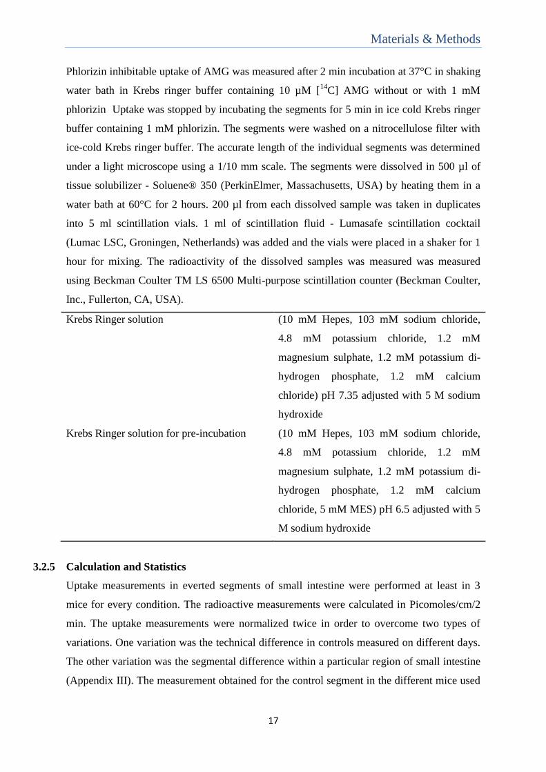
Figure 5: QSP at 15 mM concentration mediated down regulation of SGLT1 activity
independent of glucose: Wild type and RS1-/- mice were starved for 24 hours prior to
measuring SGLT1 dependent uptake. Phlorizin inhibited uptake of 10 µM AMG, labelled
with tracer amount of [14
C] AMG was measured for 2 minutes in everted jejunum segments
of wild type mice (Figure5 A and C) and RS1-/- mice (Figure5 B and D). The segments were
pre-incubated in buffer (Figure5 A and B) or in glucose (Figure5 C and D) with or without 15
mM QSP for 1 hour. Radioactivity was calculated in terms of Picomoles/cm/2 min and
normalized to wild type or RS1-/- mice in buffer or glucose control respectively. QSP
significantly down-regulated AMG uptake in wild type and RS1-/- mice independent of
glucose (P < 0.01 by 1 sided paired t test, marked by section sign (§)).
Since QEP has a higher affinity than QSP to post-transcriptionally down-regulate human
SGLT1 expressed in oocytes of Xenopus laevis, I investigated whether the down-regulation
of SGLT1 in small intestine of wild type mice after pre-incubation in 5 mM D-glucose with
15 mM QEP was more pronounced compared to 15 mM QSP (Figure 6). Only wild type mice

Results
25
were considered for this measurement as QSP down regulated SGLT1 in wild type and RS1-
/- mice in the presence of 5 mM D-glucose (Figure 5 C and 5 D). The phlorizin inhibited 10
µM AMG uptake measurements were normalized to pre-incubation in glucose condition of
wild type mice in the absence of tripeptides (Figure 6). The mean ± standard error values in
picomoles/cm/2 min used for normalization of wild type mice segments pre-incubated with
glucose was 293.9 ± 11.36. The results showed that 15 mM QSP and QEP significantly
down-regulated SGLT1 dependent AMG uptake in wild type mice. No significant difference
was observed between QSP and QEP mediated down regulation in wild type mice (P
=0.3426, 1 sided un-paired t test). This indicated that QSP and QEP were equally effective at
the employed concentration and experimental conditions.
Figure 6: QSP and QEP at 15 mM concentration are equipotent in mediating down
regulation of SGLT1: Wild type mice were starved for 24 hours prior to performing SGLT1
dependent AMG uptake. Phlorizin inhibited uptake of 10 µM AMG, labelled with tracer
amount of [14
C] AMG was measured for 2 minutes in everted jejunum segments of wild type
mice after pre-incubating the segments in glucose with or without 15 mM concentration of
tripeptide for 1 hour. Radioactivity was calculated in terms of Picomoles/cm/2 min and
normalized to mice glucose control. QSP and QEP down regulate AMG uptake with a
significance of (P = 0.0212) and (P = 0.0155) respectively by 1 sided un-paired t test marked
by exclamation mark (!).

Results
26
4.4 The role of PKC in RS1 dependent short term regulation of SGLT1:
PKC activation is involved in the post-transcriptional short term regulation of SGLT1
(Wright, Hirsch et al. 1997, Vayro and Silverman 1999, Castaneda-Sceppa, Subramanian et
al. 2010). Previously, data were obtained showing that the activation of PKC influences the
regulation of total RS1 protein on the human SGLT1 expressed in Xenopus laevis oocytes
(Veyhl, Wagner et al. 2003, Veyhl, Keller et al. 2006). Since the TGN-Reg domain of mouse
RS1 contains many putative phosphorylation sites for protein kinases including PKC (Figure
2), the previously observed effect of PKC activation could be due to activation of the TGN-
Reg by phosphorylation. To investigate whether the short term down-regulation by RS1 on
SGLT1 is PKC dependent in mouse small intestine, the effect of PKC activation on SGLT1
dependent AMG uptake was performed in wild type and RS1-/- mice. Everted segments of
wild type and RS1-/- mice small intestine were pre-incubated for 1 hour in buffer or 5 mM
D-glucose with or without 5 µM Phorbol 12-myristate 13-acetate (PMA), an activator of
PKC (Castagna, Takai et al. 1982)). The phlorizin inhibited AMG uptake was measured in
these everted segments. The measurements were normalized to pre-incubation in buffer
(Figure 7A and B) or glucose (Figure 7C and D) condition of wild type (Figure 7A and C)
and RS1-/- (Figure 7B and D) mice respectively. The mean ± standard error values in
picomoles/cm/2 min used for normalization were: wild type mice pre-incubation without
glucose 206.1 ± 26.52, RS1-/- mice pre-incubation without glucose 276.7 ± 27.39, wild type
mice pre-incubation with glucose 295.0 ± 12.29, RS1-/- mice pre-incubation with glucose
296.6 ± 12.75. The data (Figure 7) showed a 20-30% down-regulation of SGLT1 dependent
AMG uptake in wild type and RS1-/- mice after pre-incubation with 5 µM PMA which was
independent of pre-incubation with 5 mM D-glucose. This indicated a PKC dependent short
term down-regulation of SGLT1 which appeared to be independent of RS1 as well as
glucose.
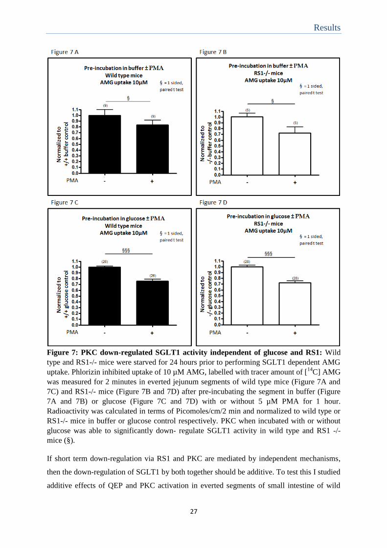
Results
27
Figure 7: PKC down-regulated SGLT1 activity independent of glucose and RS1: Wild
type and RS1-/- mice were starved for 24 hours prior to performing SGLT1 dependent AMG
uptake. Phlorizin inhibited uptake of 10 µM AMG, labelled with tracer amount of [14
C] AMG
was measured for 2 minutes in everted jejunum segments of wild type mice (Figure 7A and
7C) and RS1-/- mice (Figure 7B and 7D) after pre-incubating the segment in buffer (Figure
7A and 7B) or glucose (Figure 7C and 7D) with or without 5 µM PMA for 1 hour.
Radioactivity was calculated in terms of Picomoles/cm/2 min and normalized to wild type or
RS1-/- mice in buffer or glucose control respectively. PKC when incubated with or without
glucose was able to significantly down- regulate SGLT1 activity in wild type and RS1 -/-
mice (§).
If short term down-regulation via RS1 and PKC are mediated by independent mechanisms,
then the down-regulation of SGLT1 by both together should be additive. To test this I studied
additive effects of QEP and PKC activation in everted segments of small intestine of wild

Results
28
type and RS1-/- mice. Employing only the TGN-Reg modified motif QEP, the effect of PKC
on the entire TGN-Reg domain was excluded. The measurements were performed in the
presence of 5 mM D-glucose, as the endogenous RS1 in wild type mice is expected to be less
active under this condition. Everted segments of mice jejunum were pre-incubated in 5 mM
D-glucose either in the presence of 15 mM QEP or 5 µM PMA alone or QEP with PMA for 1
hour. The experiments were performed in wild type mice (Figure 8 A) and RS1-/- mice
(Figure 8 B). Phlorizin inhibited uptake of 10 µM AMG, labelled with tracer amount of [14
C]
AMG was measured. The data were normalized to control AMG uptake after pre-incubation
with D-glucose without QEP and PMA in wild type and RS1-/- mice respectively. The mean
± standard error values in picomoles/cm/2 min used for normalization were: wild type mice
pre-incubation with glucose 293.9 ± 11.36 and RS1-/- mice pre-incubation with glucose
289.5 ± 10.45. The data showed no additive inhibition of SGLT1 activity mediated by PMA
with QEP over QEP or PMA alone in wild type and RS1-/- mice. Since PMA inhibition was
independent of RS1 (figure 7), the short term down-regulation of SGLT1 by RS1 and PKC
suggested independent phenomenon for the same pathway of inhibition.
Figure 8: PKC has no additive effect on QEP dependent short term regulation of
SGLT1: Wild type and RS1-/- mice were starved for 24 hours prior to performing SGLT1
dependent AMG uptake. Phlorizin inhibited uptake of 10 µM AMG, labelled with tracer
amount of [14
C] AMG was measured for 2 minutes in everted jejunum segments of wild type
mice (Figure 8 A) and RS1-/- mice (Figure 8 B) after pre-incubating the segment in glucose
with or without QEP, PMA and QEP + PMA for 1 hour. Radioactivity was calculated in
terms of Picomoles/cm/2 min and normalized to wild type or RS1-/- mice in glucose control
respectively. QEP with PMA showed no additive inhibition to QEP or PMA alone in wild
type and RS1-/- mice.

Results
29
4.5 Effect of PKC on SGLT1 affinity:
Trying to elucidate how PKC activation down-regulates SGLT1 mediated AMG uptake in
small intestinal brush border membranes (BBM), we wondered whether the Michaelis
Menten (Km) value for SGLT1 dependent AMG uptake was altered. The Km value of sodium
dependent AMG uptake was measured in brush border membrane vesicles (BBMVs) which
were isolated from small intestine of RS1-/- mice pre-incubated without or with 5 mM D-
glucose. BBMVs isolated from small intestine of RS1-/- mice were used because PKC effect
appeared to be independent of RS1 (Figure 7). In these experimental series the pre-incubation
of small intestine in the absence of glucose was performed in the presence of 9 mM sodium
pyruvate for trying to avoid intracellular ATP depletion. Control experiments had suggested
that sodium pyruvate exhibited some protection against SGLT1 inactivation during the pre-
incubation period (Appendix I) without altering the Km value for SGLT1 dependent AMG
uptake (Appendix II). Small intestines were pre-incubated for 1 hour with 9 mM sodium
pyruvate or 5 mM D-glucose with or without 5 µM PMA and BBMVs were isolated. Sodium
dependent AMG uptake measurements were performed at AMG concentrations (0.02 – 5
mM) containing tracer amount of [14
C] AMG using an incubation time of 2 seconds. Uptake
rates per mg protein were calculated (picomoles/mg/2 seconds).
Km values were determined by fitting the Michael-Menten equation to the uptake rates
obtained (Figure 9). The data indicated that the Km value for SGLT1 mediated AMG uptake
was decreased when PKC was active. In my experiment this effect was significant in the
presence of glucose. Noteworthy the decrease of Km after stimulation of PKC resulted in
decrease of 10 µM AMG uptake mediated by SGLT1. Thus the data suggested that PKC
stimulation might affect either the turnover number or amount of functional active SGLT1 at
the BBM.

Results
30
Figure 9: PKC altered SGLT1 affinity: Wild type and RS1-/- mice were starved for 24
hours prior to BBMV preparation. BBMVs were prepared after pre-incubation in 5 mM
glucose or 9 mM sodium pyruvate with or without 5µM PMA for 1 hour. Sodium dependent
uptake measurements were performed for 2 sec in BBMVs incubated in Na+ containing
buffer and K+ containing buffer at different radioactive AMG concentrations (0.02 – 5 mM).
Figure 9A represents the sodium-dependent uptake rates of individual experiments that were
normalized to the respective Vmax values. Mean ± standard error values from independent
experiments are shown. Figure 9B represents the comparison between Km values for sodium-
dependent AMG uptake into BBMVs. Border line significance was observed for the
difference between Km values of BBMVs prepared with pre-incubation with sodium pyruvate
with or without PMA, P = 0.1356, 1 sided un-paired t test. Significant difference was
observed between Km values of BBMVs prepared with pre-incubation with glucose in
presence or absence of PMA, P = 0.0019, 1 sided un-paired t test (!).
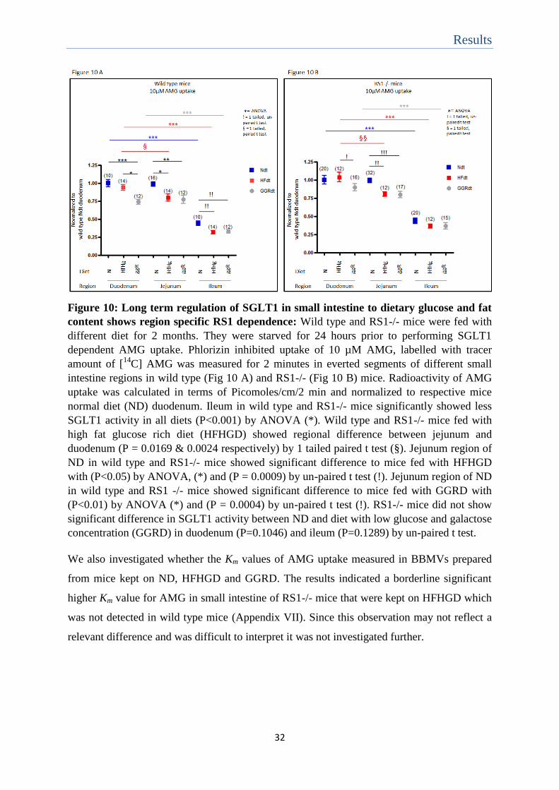
4.6 Effect of RS1 on SGLT1 activity during diet adaptation:
Diet dependent long term up-regulation of SGLT1 in small intestine increases the capacity
for glucose absorption during high carbohydrate content in food (Shirazi-Beechey, Hirayama
et al. 1991, Moran, Al-Rammahi et al. 2010). This glucose dependent regulation is influenced
by types of ingested carbohydrate and is influenced by extrinsic factors (Luz Sdos, de
Campos et al. 1997). Because RS1 is involved in the transcriptional regulation of SGLT1 and
preliminary data suggest RS1 to influence SGLT1 regulation in mouse small intestine in
response to hyper-caloric, fat rich but glucose-galactose reduced diet (GGRD, see Table 1)
(Filatova, Leyerer et al. 2009), we compared the transport activity in different small intestinal
segments of wild type and RS1-/- mice that were kept for 2 months on normal diet (ND),
GGRD and high fat glucose rich diet (HFHGD). ND is normo-caloric and possesses high
polysaccharide and low fat content while HFHGD is hyper-caloric and contains high
monosaccharide and high fat content. The compositions and calorie contents of the three diets
are shown in Table 1 and 2. After diet application, the mice were starved for 24 hours and the
Results
31
phlorizin inhibited uptake of 10 µM AMG, labelled with tracer amount of [14
C] AMG was
measured in everted small intestinal segments taken from different parts of small intestine.
These measurements were performed without pre-incubation of the everted small intestinal
segments. The data were normalized to duodenal SGLT1 activity for AMG uptake in wild
type and RS1-/- mice respectively. The mean ± standard error values in picomoles/cm/2 min
used for normalization were: duodenum uptake in wild type mice fed with ND 370.7 ± 18.25
and duodenum uptake in RS1-/- mice fed with ND 320 ± 19.36.
The results are shown in Figure 10. Independent of diet in wild type and RS1-/- mice
significant low activity was observed in ileum compared to duodenum and jejunum. In
jejunum of wild type and RS1-/- mice, SGLT1 mediated uptake of 10 µM AMG in animals
kept on hyper-caloric, fat rich diet (HFHGD and GGRD) were significantly reduced
compared to animals kept on normo-caloric, low fat diet (ND). Since this effect was observed
independent of the glucose or galactose content in diet, the effect might depend on the fat or
total calorie content in diet. This fat dependent down-regulation was apparently also
independent of RS1 as it was also observed in RS1-/- mice. In duodenum of wild type mice
significant lower transport of SGLT1 was observed when mice were fed glucose-galactose
reduced, fat rich diet (GGRD) compared to ND. This effect was absent in RS1-/- mice with
different diet. Thus a glucose dependent long term regulation in duodenum was seen which
was dependent on RS1.
Results
32
Figure 10: Long term regulation of SGLT1 in small intestine to dietary glucose and fat
content shows region specific RS1 dependence: Wild type and RS1-/- mice were fed with
different diet for 2 months. They were starved for 24 hours prior to performing SGLT1
dependent AMG uptake. Phlorizin inhibited uptake of 10 µM AMG, labelled with tracer
amount of [14
C] AMG was measured for 2 minutes in everted segments of different small
intestine regions in wild type (Fig 10 A) and RS1-/- (Fig 10 B) mice. Radioactivity of AMG
uptake was calculated in terms of Picomoles/cm/2 min and normalized to respective mice
normal diet (ND) duodenum. Ileum in wild type and RS1-/- mice significantly showed less
SGLT1 activity in all diets (P<0.001) by ANOVA (*). Wild type and RS1-/- mice fed with
high fat glucose rich diet (HFHGD) showed regional difference between jejunum and
duodenum (P = 0.0169 & 0.0024 respectively) by 1 tailed paired t test (§). Jejunum region of
ND in wild type and RS1-/- mice showed significant difference to mice fed with HFHGD
with (P<0.05) by ANOVA, (*) and (P = 0.0009) by un-paired t test (!). Jejunum region of ND
in wild type and RS1 -/- mice showed significant difference to mice fed with GGRD with
(P<0.01) by ANOVA (*) and (P = 0.0004) by un-paired t test (!). RS1-/- mice did not show
significant difference in SGLT1 activity between ND and diet with low glucose and galactose
concentration (GGRD) in duodenum (P=0.1046) and ileum (P=0.1289) by un-paired t test.
We also investigated whether the Km values of AMG uptake measured in BBMVs prepared
from mice kept on ND, HFHGD and GGRD. The results indicated a borderline significant
higher Km value for AMG in small intestine of RS1-/- mice that were kept on HFHGD which
was not detected in wild type mice (Appendix VII). Since this observation may not reflect a
relevant difference and was difficult to interpret it was not investigated further.

Discussion
33
5. Discussion
In this study, data was presented for a PKC dependent short term down-regulation of SGLT1
function in the small intestine of mice which is independent of RS1. It is suggested that this
effect may be due to PKC dependent phosphorylation of SGLT1. A second finding of this
study is the observation that SGLT1 transport activity in jejunum is down-regulated
independently of RS1 for long term adaptation to high fat containing diet. Thirdly it is
observed that substrate transport by SGLT1 in duodenum is down-regulated in an RS1
dependent manner after reduction in glucose and galactose content from the diet.
I also obtained some data concerning the role of RS1 in SGLT1 regulation where absence of
RS1 led to enhanced activity of SGLT1 (Figure 3). However this data showed relatively less
enhanced effect in comparison to results previously demonstrated by other colleagues.
Furthermore I also obtained some data concerning the role of RS1 in glucose dependent short
term regulation of SGLT1 which contradicted the results obtained by colleagues in the
laboratory, who used a different experimental approach. Dr. Helmut Kipp measured phlorizin
inhibited AMG uptake in brush border membrane vesicles isolated from wild type mice small
intestine after application of a 2.2 M glucose gavage in vivo or after incubation for 2 minutes
of the small intestine with 20 mM AMG. He observed increased SGLT1 protein in the BBM
and 2-4 times increase in SGLT1 mediated AMG uptake. In his experiments the RS1-/- mice
was shown to already have an enhanced SGLT1 expression and no further increase in SGLT1
activity or protein was observed with increased glucose luminal concentration. Performing
AMG uptake measurements in everted small intestinal segments of small intestine after pre-
incubation in the absence or presence of 5 mM D-glucose I observed a relative small glucose
dependent increase of AMG uptake in wild type mice as well as in RS1-/- suggesting an RS1
independent short term up-regulation by glucose. These differences in results may be
explained on the basis of the different methods employed for SGLT1 dependent uptake
measurements. Since Dr. Kipp detected that glucose dependent up-regulation in small
intestine occurs very rapidly and could be observed after 2 minutes incubation period, it is
probable that the up-regulation by 5 mM D-glucose in my experiments were reversed during
the uptake measurements in the everted segments which were performed using an incubation
period of 2 minutes. A second difficultly was that the transport activity of SGLT1 in small
intestine was decreased during 1 hour pre-incubation period, which was employed to study
the short term effects on SGLT1 regulation (Appendix I). Thus these different degrees of

Discussion
34
inactivation during preincubation with and without glucose may have been determined in my
experiments rather than the glucose dependent regulation. The inactivation by pre-incubation
could have also altered the enhancement in SGLT1 activity of RS1-/- mice leading to the
resultant small enhancement observed (Figure 3). Another reason for the differences in
results could also be the difference in concentration of glucose applied for understanding its
effect in short term regulation of SGLT1. Dr. Kipp employed a concentration of 20 mM and
about 2 M to study the glucose dependent regulation of SGLT1. In my experiments I
performed the study with only 5 mM D-glucose. Hence the decreased effect in the level of
rise of SGLT1 activity for glucose dependent short term regulation may be dependent on the
concentration of glucose along with the effect of inactivation during pre-incubation. Due to
these limitations the further study to elucidate the effect of RS1 in short term regulation of
SGLT1 were performed by employing the active motif QSP and its phosphorylation
mimicking mutant QEP. QSP belongs to the N-terminal TGN-Reg domain of RS1 and
mediate post-transcriptional down-regulation of SGLT1 (Vernaleken, Veyhl et al. 2007).
Although QSP did mediate short term down-regulation of SGLT1, this down-regulation was
independent of glucose and endogenous RS1 (Figure 5). This glucose independent down-
regulation of SGLT1 by QSP could be explained in terms of concentration. 15 mM
concentration of QSP might be sufficient to inhibit the up-regulation of SGLT1 by 5 mM
glucose. However the down-regulation of QSP to be effective in absence of glucose, which is
a condition when endogenous RS1 is active, can be explained by different reasons. It could
be stated that the SGLT1 activity is not completely down-regulated by endogenous RS1 and
additive 15 mM QSP aids in further inhibition. Another possible explanation could be the
experimental conditions. Mice were starved for 24 hours prior to uptake measurements.
Starvation has been shown to lower affinity of SGLT1 and increase its uptake activity
(Marciani, Lindi et al. 1987, Gupta and Waheed 1992, Gal-Garber, Mabjeesh et al. 2000).
Hence the starvation of mice might have elevated SGLT1 activity that could have allowed
QSP to mediate down-regulation on AMG transport even under a condition when endogenous
RS1 is expected to be in an active state.
The glucose dependent short term regulation of SGLT1 also involves increasing Km value for
SGLT1 dependent AMG uptake (Figure 4). However RS1 did not affect the affinity for
SGLT1 dependent AMG uptake (Figure 4). Hence it can be said that the glucose effect on
SGLT1 affinity is independent of RS1. The SGLT1 affinity was also altered on PKC
activation (Figure 9), where the Km value for SGLT1 mediated AMG transport is decreased
Discussion
35
about 2 fold. In addition the PKC dependent short term regulation of SGLT1 occurred in an
RS1 independent manner (Figure 7). But the observation of no additive down-regulation of
SGLT1 when QEP was used with PKC activation (Figure 8) indicated that PKC and RS1
might act independently within the same pathway to down-regulate SGLT1. RS1 is known to
mediate short term regulation of SGLT1 post-transcriptionally by inhibiting the trafficking of
vesicles containing SGLT1 to BBM (Veyhl, Keller et al. 2006). PKA and PKC are involved
in regulating SGLT1 by altering exo-endocytosis of SGLT1 carrying vesicles to BBM
(Wright, Hirsch et al. 1997). Protein kinases like PKA has also been reported to mediate short
term regulation of SGLT1 by causing SGLT1 phosphorylation to change its affinity for
substrate transport (Subramanian, Glitz et al. 2009). The SGLT1 sequence also has several
putative phosphorylation sites for PKC (Hirsch, Loo et al. 1996). Hence the possible
mechanism for PKC to mediate an increase in SGLT1 affinity and down-regulate SGLT1
activity for glucose transport might involve the transporter phosphorylation that would affect
post-transcriptionally SGLT1 turn over number and/or amount in the BBM.
Based on the preliminary data which stated that RS1 is involved in the transcriptional
regulation of SGLT1 for adaptation to glucose-galactose reduced diet (Filatova, Leyerer et al.
2009) further data was obtained in this study to understand the dependence of RS1 in long
term regulation of SGLT1 in small intestine for adaptation to diets different in glucose and fat
content. The data obtained (Figure 10) showed the glucose dependent down-regulation of
SGLT1 transport in duodenum of wild type mice for adaptation to glucose-galactose reduced
diet (GGRD). The possible explanation for SGLT1 down regulation with low carbohydrate
diet could be explained in terms of biosynthetic cost. In GGRD the cost of producing
transporter might exceed the benefits of its activity. While on the other hand with high
carbohydrate diet, energy synthesis would increase with increased absorption hence allowing
increase in SGLT1 transporter activity (Ferraris and Diamond 1997). In support to this theory
of biosynthetic cost, it was observed that the SGLT1 activity in wild type mice duodenum did
not differ between polysaccharide rich (ND) or monosaccharide rich (HFHGD) diets.
However this glucose dependent down-regulation of SGLT1 transport in duodenum was
absent in RS1-/- mice. Hence the RS1 dependent long term down-regulation of SGLT1 for
diet glucose adaptation was shown to be localized to duodenum region of small intestine.
It was also observed that in the jejunum the SGLT1 activity was decreased in wild type and
RS1-/- mice fed with fat rich diets (GGRD and HFHGD) when compared to the jejunual
SGLT1 activity in mice fed with normal diet (ND) (Figure 10). This indicated a fat dependent

Discussion
36
but RS1 independent long term down-regulation of SGLT1. Studies have reported increased
SGLT1 gene expression in jejunum of mice fed with high carbohydrate/fat ratio when
compared to low carbohydrate/fat diet (Honma, Mochizuki et al. 2009, Inoue, Mochizuki et
al. 2011). Likewise this fat dependent down-regulation of SGLT1 activity could also be
dependent on carbohydrate concentration. The HFHGD is a diet rich in simple sugars like
monosaccharides and disaccharides. As complexity in carbohydrate availability is known to
shift SGLT1 expression from jejunum to ileum due to slow digestion (Shimada, Mochizuki et
al. 2009) these simple monosaccharides could have been easily absorbed in duodenum
thereby leading to decreased SGLT1 function at jejunum. This effect would also account for
the RS1 independent regional difference of SGLT1 activity between duodenum and jejunum
region in mice fed with HFHGD. Likewise the decrease in jejunual SGLT1 activity for mice
with GGRD could be due to the reduced amount of glucose and galactose in diet. Hence the
RS1 independent down-regulation of SGLT1 in mice fed with HFHGD and GGRD could be
a fat dependent and a glucose dependent effect.
An RS1 independent and diet independent down-regulation of SGLT1 activity was observed
in ileum. This could suggest that the age related long term regulation for prevalence of
gradient distribution in SGLT1 transport from proximal to distal region in the small intestine
(Toloza and Diamond 1990) is in-differed by dietary composition and RS1.
In conclusion it can be stated that the RS1 dependence for SGLT1 regulation differs with
respect to time scale of glucose exposure and is also specific to region in the small intestine.
PKC activation was involved in the short term down-regulation of SGLT1 independent of
RS1, but may follow the same path of RS1 for causing SGLT1 down-regulation. RS1
mediated the long term down-regulation of SGLT1 specifically in duodenum region of small
intestine in a manner dependent on glucose-galactose diet content. RS1 and diet independent
long-term regulation of SGLT1 was observed to maintain posterior to distal gradient in
SGLT1 function. An RS1 independent but dietary fat and/or glucose content dependent
down-regulation of SGLT1 is present in mice jejunum region.

Appendix
37
6. Appendix
Appendix I: Tissue pre-incubation alters SGLT1 dependent substrate transport:
In my study SGLT1 regulation was investigated by performing uptake measurement in
everted small intestinal segments. These measurements allowed more direct control of the
experimental conditions such as luminal glucose concentrations compared to in vivo gavage
with D-glucose. The study of short term regulation of SGLT1 required pre-incubating the
everted segments under required study condition for 1 hour. Pre-incubation of tissue might
deprive tissue of energy source leading to decrease in SGLT1 efficiency for transport. In
order to understand the effect of pre-incubation on SGLT1 activity phlorizin inhibited uptake
of 10 µM AMG, labelled with tracer amount of [14
C] AMG was measured in everted
segments of wild type mice small intestine were compared without or with pre-incubation
under 3 conditions – buffer (no energy resource), sodium pyruvate (energy resource) and
glucose (substrate for SGLT1 and an energy resource). The result (Figure 11) showed no
significant difference between buffer and sodium pyruvate pre-incubation. Pre-incubation
with buffer and sodium pyruvate significantly decreased SGLT1 activity. However glucose
showed significantly higher activity than pre-incubation with buffer or sodium pyruvate.
Figure 11: Pre-incubation alters SGLT1 activity: Wild type mice were starved for 24hrs
prior to perform SGLT1 dependent AMG uptake. Phlorizin inhibited uptake of 10 µM AMG,
labelled with tracer amount of [14
C] AMG was measured for 2 minutes in everted jejunum
segments of wild type mice either without pre-incubation or after pre-incubating the segment
in buffer, sodium pyruvate or glucose for 1hr. Radioactivity of AMG uptake was calculated
in terms of Picomoles/cm/2min. Pre-incubation decreases SGLT1 activity in buffer (P<0.001)
and sodium pyruvate (P<0.05), by ANOVA (*).

Appendix
38
It can be discussed that though pre-incubation itself alters SGLT1 activity, the significant
difference observed between pre-incubation condition of sodium pyruvate and glucose
indicated substrate specific upregulation of SGLT1. Hence this method of pre-incubation was
considered for the study of glucose dependent SGLT1 regulation despite the limitation in
alteration of SGLT1 activity by pre-incubation. Furthermore the presence of the energy
source sodium pyruvate failed to completely remove the pre-incubation effect on SGLT1
activity. Hence pre-incubation in sodium pyruvate or buffer either could be used as control
condition for glucose effect.
Appendix II: Tissue pre-incubation does not alter SGLT1 affinity:
Pre-incubation was shown to alter SGLT1 activity (Figure 11). Hence it was of interest
whether pre-incubation altered SGLT1 affinity for transport. For this the Km values for AMG
uptake were measured using BBMVs prepared from small intestines of RS1-/- mice without
and with pre-incubation in sodium pyruvate for 1hr. Radioactive uptake measurements of 2
seconds were made for different AMG concentrations (0.02 – 5 mM) in the presence and
absence of 100 mM sodium and the Km values for sodium dependent uptake rates were
calculated. The data represented in Figure 12 showed that pre-incubation does not alter
SGLT1 affinity (P = 0.2036).
Figure 12: Pre-incubation did not alter SGLT1 affinity: RS1-/- mice were starved for 24
hours prior to BBMV preparation. BBMVs were prepared either with or without pre-
incubation in 9 mM sodium pyruvate for 1 hour. Sodium dependent affinity uptake
measurements were performed for 2 sec in BBMVs incubated in Na+ containing buffer and
K+ containing buffer at different radioactive AMG concentrations (0.02 mM – 5 mM). Figure
12 represents the sodium-dependent uptake rates of individual experiments that were
normalized to the respective Vmax values. Mean ± standard error values from independent
experiments are shown. No significant difference was observed between Km values of
BBMVs prepared without or with pre-incubation in sodium pyruvate, P = 0.2036, 1 sided un-
paired t test.
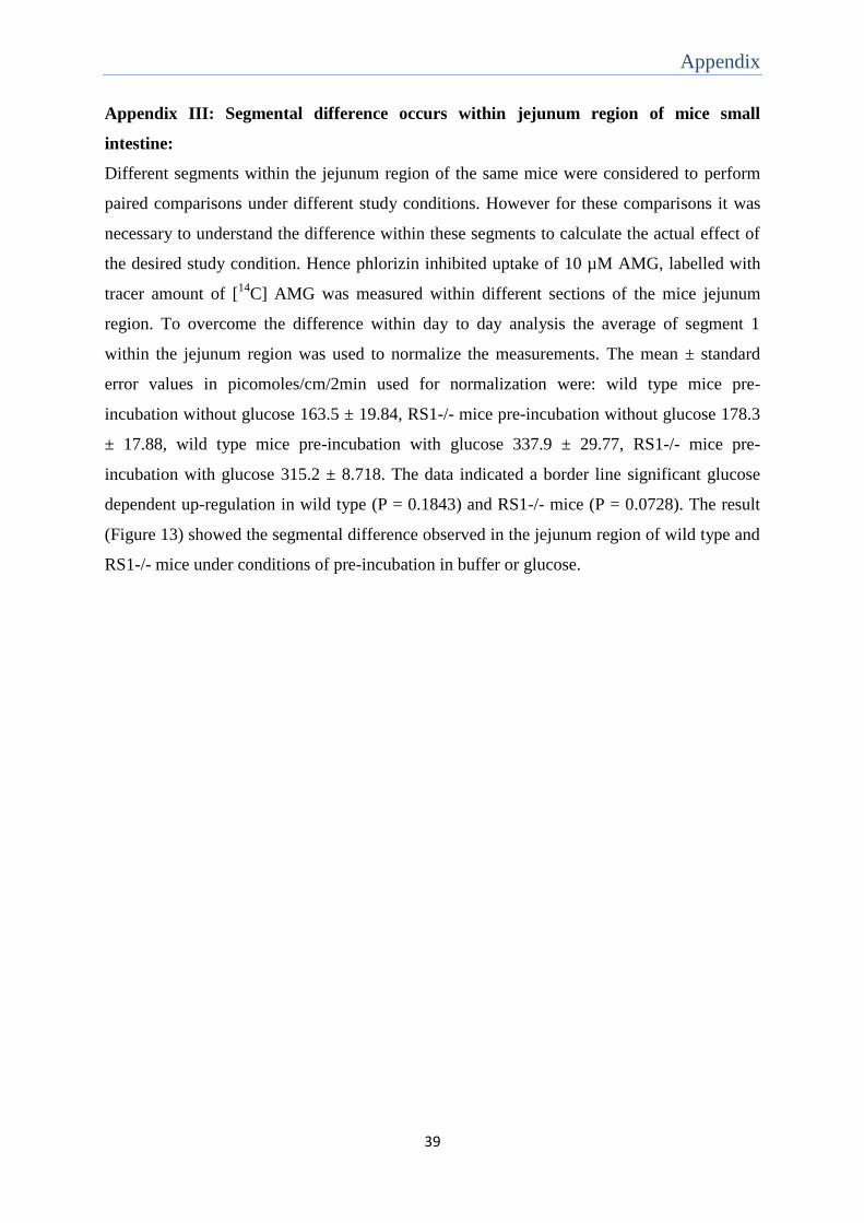
Appendix
39
Appendix III: Segmental difference occurs within jejunum region of mice small
intestine:
Different segments within the jejunum region of the same mice were considered to perform
paired comparisons under different study conditions. However for these comparisons it was
necessary to understand the difference within these segments to calculate the actual effect of
the desired study condition. Hence phlorizin inhibited uptake of 10 µM AMG, labelled with
tracer amount of [14
C] AMG was measured within different sections of the mice jejunum
region. To overcome the difference within day to day analysis the average of segment 1
within the jejunum region was used to normalize the measurements. The mean ± standard
error values in picomoles/cm/2min used for normalization were: wild type mice pre-
incubation without glucose 163.5 ± 19.84, RS1-/- mice pre-incubation without glucose 178.3
± 17.88, wild type mice pre-incubation with glucose 337.9 ± 29.77, RS1-/- mice pre-
incubation with glucose 315.2 ± 8.718. The data indicated a border line significant glucose
dependent up-regulation in wild type (P = 0.1843) and RS1-/- mice (P = 0.0728). The result
(Figure 13) showed the segmental difference observed in the jejunum region of wild type and
RS1-/- mice under conditions of pre-incubation in buffer or glucose.

Appendix
40
Figure 13: Jejunal region of small intestine shows segmental difference in AMG uptake
activity: Wild type and RS1-/- mice were starved for 24hrs prior to perform SGLT1
dependent AMG uptake. Phlorizin inhibited uptake of 10 µM AMG, labelled with tracer
amount of [14
C] AMG was measured for 2 minutes in everted jejunum segments of wild type
(Figure 13 A and B) and RS1-/- (Figure 13 C and D) mice after pre-incubation in buffer
(Figure 13 A and C) or glucose (Figure 13 B and D). Radioactivity of AMG uptake was
calculated in terms of Picomoles/cm/2min. The values were normalized to the first segment
of wild type or RS1-/- mice jejunum in buffer or glucose condition respectively. Differences
were analysed by ANOVA (*) or 1 sided paired t test (§) between 4 different segments of
jejunum region within the mice small intestine. Significant segmental difference was
observed in the region of jejunum in mice small intestine.
The values obtained in the above results for the different segments of jejunum region were
used to normalize the respective segment in any study performed.

Appendix
41
Appendix IV: Brefeldin A activity is dependent on glucose and RS1:
Brefeldin A is an exocytotic inhibitor. Since post-transcriptional short term regulation of
SGLT1 can be mediated by vesicular trafficking of SGLT1 carrying vesicles, the role of
Brefeldin A was investigated for short term regulation of SGLT1. For this, phlorizin inhibited
uptake of 10 µM AMG, labelled with tracer amount of [14
C] AMG was measured in wild type
and RS1-/- mice small intestine everted segments after pre-incubation in buffer or glucose
with or without Brefeldin A (12µM). The measurements were normalized to uptake after pre-
incubation in buffer (Figure 14A and 14B) or glucose (Figure 14C and 14D) condition of
wild type (Figure 14A and 14C) and RS1-/- (Figure 14B and 14D) mice respectively. The
measurements were normalized to uptake after pre-incubation of small intestine everted
segments of wild type mice or RS1-/- mice in buffer (Figure 5A and 5B) or D-glucose
(Figure 5C and 5D). The mean ± standard error values in picomoles/cm/2min used for
normalization were: wild type mice pre-incubation without glucose 120.6 ± 11.17, RS1-/-
mice pre-incubation without glucose 144.3 ± 21.42, wild type mice pre-incubation with
glucose 258.7 ± 17.32, RS1-/- mice pre-incubation with glucose 227.2 ± 13.89. The results
showed a borderline significant downregulation of SGLT1 dependent AMG uptake in wild
type and RS1-/- mice on pre-incubation with Brefeldin A in buffer. However in glucose, pre-
incubation with Brefeldin A mediated only a borderline significant down regulation in wild
type mice.

Appendix
42
Figure 14: Brefeldin A inhibits exocytotic inhibition of SGLT1 vesicular trafficking in a
complex manner: Wild type and RS1-/- mice were starved for 24hrs prior to perform
SGLT1 dependent AMG uptake. Phlorizin inhibited uptake of 10 µM AMG, labelled with
tracer amount of [14
C] AMG was measured for 2 minutes in everted jejunum segments of
wild type mice (Fig 14A and C) and RS1-/- mice (Fig 14B and D) after pre-incubating the
segment in buffer (Figure 14A and B) or glucose (Figure 14C and D) with or without 7-
12µM Brefeldin A for 1hr. Radioactivity of AMG uptake was calculated in terms of
Picomoles/cm/2min and normalized to respective mice buffer or glucose control. Buffer pre-
incubated with Brefeldin A inhibited SGLT1activity in wild type mice (P = 0.0472) and in
RS1 -/- mice (P = 0.0440) tested by 1sided paired test (§). However Brefeldin A inhibited
SGLT1 activity only in wild type mice when incubated in glucose (P = 0.0440) (§).

Appendix
43
The difference in the inhibition by Brefeldin A between wild type and RS1-/- under glucose
condition indicated a complex mechanism. SGLT1 regulation mediated by Brefeldin A might
be dependent on intracellular glucose concentration and endogenous RS1.
Appendix V: QEP down regulates SGLT1 activity by exocytotic inhibition of SGLT
carrying vesicles:
QCP a functional tripeptide within the RS1 sequence down-regulates SGLT1 without adding
to the exocytotic inhibition of SGLT1 vesicular trafficking by Brefeldin A in the Xenopus
laevis oocyte system (Vernaleken, Veyhl et al. 2007). QSP is another functional tripeptide of
RS1 sequence. For understanding the mechanism of SGLT1 regulation by QSP, its
phosphorylation mimicking mutant QEP effect on SGLT1 activity was compared to
inhibitory effect of Brefeldin A. As Brefeldin A did not mediate inhibition in RS1-/- mice
when glucose was present (Figure 15 D), phlorizin inhibited uptake of 10 µM AMG, labelled
with tracer amount of [14
C] AMG was measured in everted segments of wild type mice alone
after pre-incubation in glucose with Brefeldin A (12 µM), QEP (15 mM) or Brefeldin A with
QEP. The uptake measurements were normalized to the mean ± standard error value 293.9 ±
11.36 in picomoles/cm/2 min obtained for the everted segments of wild type mice small
intestine after pre-incubation with glucose. The data in figure 15 showed no additive down-
regulation of SGLT1 activity by QEP to the exocytotic inhibition of SGLT1 vesicles by
Brefeldin A. This possibly indicated that QEP mediated down-regulation of SGLT1 by
inhibiting the trafficking of SGLT1 carrying vesicles to the apical surface.
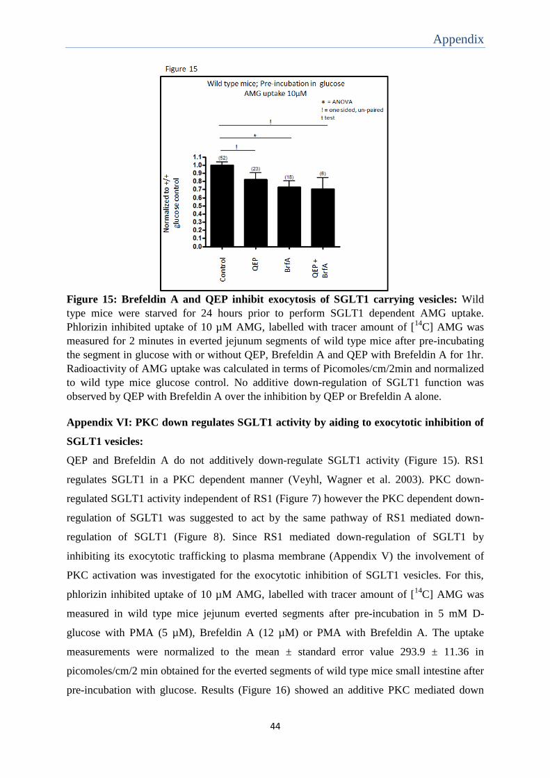
Appendix
44
Figure 15: Brefeldin A and QEP inhibit exocytosis of SGLT1 carrying vesicles: Wild
type mice were starved for 24 hours prior to perform SGLT1 dependent AMG uptake.
Phlorizin inhibited uptake of 10 µM AMG, labelled with tracer amount of [14
C] AMG was
measured for 2 minutes in everted jejunum segments of wild type mice after pre-incubating
the segment in glucose with or without QEP, Brefeldin A and QEP with Brefeldin A for 1hr.
Radioactivity of AMG uptake was calculated in terms of Picomoles/cm/2min and normalized
to wild type mice glucose control. No additive down-regulation of SGLT1 function was
observed by QEP with Brefeldin A over the inhibition by QEP or Brefeldin A alone.
Appendix VI: PKC down regulates SGLT1 activity by aiding to exocytotic inhibition of
SGLT1 vesicles:
QEP and Brefeldin A do not additively down-regulate SGLT1 activity (Figure 15). RS1
regulates SGLT1 in a PKC dependent manner (Veyhl, Wagner et al. 2003). PKC down-
regulated SGLT1 activity independent of RS1 (Figure 7) however the PKC dependent down-
regulation of SGLT1 was suggested to act by the same pathway of RS1 mediated down-
regulation of SGLT1 (Figure 8). Since RS1 mediated down-regulation of SGLT1 by
inhibiting its exocytotic trafficking to plasma membrane (Appendix V) the involvement of
PKC activation was investigated for the exocytotic inhibition of SGLT1 vesicles. For this,
phlorizin inhibited uptake of 10 µM AMG, labelled with tracer amount of [14
C] AMG was
measured in wild type mice jejunum everted segments after pre-incubation in 5 mM D-
glucose with PMA (5 µM), Brefeldin A (12 µM) or PMA with Brefeldin A. The uptake
measurements were normalized to the mean ± standard error value 293.9 ± 11.36 in
picomoles/cm/2 min obtained for the everted segments of wild type mice small intestine after
pre-incubation with glucose. Results (Figure 16) showed an additive PKC mediated down

Appendix
45
regulation on SGLT1 activity to Brefeldin A inhibition. This data suggested that PKC and
Brefeldin A alone might mediate only partial exocytotic inhibition of SGLT1 carrying
vesicles and when together complete the down-regulation of SGLT1 or PKC and Brefeldin A
inhibited SGLT1 by different pathways.
Figure 16: Brefeldin A and PKC inhibit SGLT1 additively: Wild type mice were starved
for 24hrs prior to perform SGLT1 dependent AMG uptake. Phlorizin inhibited uptake of 10
µM AMG, labelled with tracer amount of [14
C] AMG was measured for 2 minutes in everted
jejunum segments of wild type mice after pre-incubating the segment in glucose with or
without PMA, Brefeldin A and PMA with Brefeldin A for 1hr. Radioactivity of AMG uptake
was calculated in terms of Picomoles/cm/2min and normalized to wild type mice glucose
control. PMA with Brefeldin A inhibition were additive to PMA (P<0.05) and Brefeldin A
(P=0.015) inhibition alone by one sided un-paired, t test (!).
Appendix VII: SGLT1 affinity for substrate transport during diet adaptation might be
RS1 dependent:
To understand whether adaptation to different diet modulates SGLT1 substrate affinity and to
explore the involvement of RS1 in this regulation, Km values for sodium dependent AMG
uptake rates were calculated. The radioactive uptake measurements of 2 seconds were made
for different AMG concentrations (0.02 – 5 mM) in the presence and absence of 100 mM
sodium in BBMVs isolated from small intestine of wild type and RS1-/- mice kept for 2
months with different diets. The radioactivity measured was calculated in Picomoles/mg/2
seconds and the values were plotted using Michaelis-Menten equation in a nonlinear
regression curve to obtain Km values. Comparing the Km values the results (Figure 17)

Appendix
46
showed that diet does not alter affinity in wild type mice. However RS1-/- mice fed with
HFHGD (Km value 2.232 ± 0.471 mM) showed a borderline significance for difference in Km
value when compared with mice fed with ND (Km value 0.8717 ± 0.197) and GGRD (Km
value 0.8290 ± 0.0315 mM).
Figure 17: RS1 might alter SGLT1 substrate affinity for diet adaptation: Mice were fed
with different diets for 2 months and starved for 24 hours prior to preparing BBMVs from
mice small intestine. Sodium dependent uptake measurements were performed for 2 seconds
in BBMVs incubated in Na+ containing buffer and K
+ containing buffer at different
radioactive AMG concentrations (0.02 mM – 5 mM). Radioactivity was measured in terms of
Picomoles/mg/2 seconds and plotted to fit in non-linear regression curve using Michaelis –
Menten equation to determine Km value. Significant difference was observed between Km
value of RS1-/- mice fed with HFHGD to mice fed with ND (P=0.0395) and GGRD (P =
0.0339) by1 sided un-paired t test (!).

Summary
47
7. Summary:
The Na+-D-glucose cotransporter in small intestine is regulated in response to food
composition. Short term regulation of SGLT1 occurs post-transcriptionally in response to
changes in luminal glucose. Adaptation to dietary carbohydrate involves long term regulation
at the transcriptional level. The intracellular protein RS1 (gene RSC1A1) is involved in
transcriptional and post-transcriptional regulation of SGLT1. RS1 contains an N-terminal
domain with many putative phosphorylation sites. By Expressing SGLT1 in oocytes of
Xenopus laevis it was previously demonstrated that the post-transcriptional down-regulation
of SGLT1 by RS1 was dependent on the intracellular glucose concentration and activated by
protein kinase C (PKC). The role of RS1 for short term regulation of SGLT1 in mouse small
intestine in response to glucose and PKC was investigated comparing effects in RS1-/- mice
and wildtype mice. Effects on SGLT1 activity were determined by measuring phlorizin
inhibited uptake of α-methylglucoside (AMG). The involvement of RS1 in glucose dependent
short term regulation could not be elucidated for technical reasons. However, evidence for
RS1 independent short-term downregulation of SGLT1 after stimulation of PKC could be
provided. It was shown that this downregulation includes decrease in the amount and/or in
turnover of SGLT1 in the brush-border membrane as well as an increase of substrate affinity
for AMG transport. Trying to elucidate the role of RS1 in long term regulation of SGLT1 in
small intestine in response to glucose and fat content of the diet, wildtype and RS1-/- mice
were kept for 2 months on a normo-caloric standard diet with high glucose and low fat
content (ND), on a hyper-caloric glucose-galactose reduced diet with high fat content
(GGRD) or on a hyper-caloric diet with a high fat and high glucose content (HFHGD).
Thereafter the animals were starved overnight and SGLT1 mediated AMG uptake was
measured. Independent of diet AMG uptake in ileum was smaller compared to duodenum and
jejunum. In jejunum of wildtype and RS1-/- mice kept on the fat rich diets (GGRD and
HFHGH) transport activity of SGLT1 was lower compared to mice kept on ND with low fat
content. This result suggests an RS1 independent downregulation due to fat content of diet.
Different to RS1-/- mice, the duodenum of wildtype mice showed transport activity of
SGLT1 smaller in mice kept on glucose galactose reduced diet (GGRD) compared to the
glucose galactose rich diets (ND and HFHGG). These data indicate that RS1 is involved in
glucose dependent long term regulation in duodenum.

Zusammenfassung
48
8. Zusammenfassung:
Der Na+-Glukose-Cotransporter SGLT1 im Dünndarm wird in Abhängigkeit zur
Nahrungszusammensetzung reguliert. Kurzzeitregulation von SGLT1 tritt posttranskritionell
als Antwort zu sich ändernden Glukosekonzentrationen im Darmlumen auf. Anpassung an
Nahrungskohlenhydrate beinhaltet die Langzeitregulation auf transkripionellem Level. Das
intrazelluläre Protein RS1 (Gen RSC1A1) ist an der transkriptionellen und post-
transkriptionellen Regulation von SGLT1 beteiligt. Es enthält eine N-terminale Domäne mit
vielen putativen Phosphorylierungsstellen. Bei der Expression von SGLT1 im Xenopus leavis
Oocytensystem wurde gezeigt, dass die posttranskriptionelle Herunterregulation von SGLT1
durch RS1 von der intrazelluläre Glukosekonzentration abhängt und durch Proteinkinase C
(PKC) aktiviert wird. Die Rolle von RS1 in der Kurzzeitregulation von SGLT1 im Dünndarm
der Maus als Antwort auf Glukose und PKC wurde durch vergleichende Studien zwischen
RS1- knockout (RS1-/-)- Mäusen und Wildtyp-Mäusen untersucht. Effekte auf die SGLT1-
Aktivität wurden durch Messung der durch Phlorizin inhibierbaren Aufnahme des SGLT1-
spezifischen Substrats α-Methyl-Glycopyranosid (AMG) bestimmt. Der Einfluss von RS1 in
der Glukose-abhängigen Kurzzeitregulation konnte aus technischen Gründen nicht untersucht
werden, jedoch gab es Anzeichen für eine von RS1 unabhängige Kurzzeitregulation von
SGLT1 durch PKC. Es wurde gezeigt, dass diese Herunterregulation sowohl eine Abnahme
der Menge und/oder der Umsatzrate von SGLT1 in der Bürstensaummembran wie auch eine
Zunahme der Substrat-Affinität für den AMG-Transport beinhaltet. Um die Rolle von RS1
auf die Langzeitregulation von SGLT1 in Dünndarm als Antwort auf den Glukose- und
Fettgehalt der Nahrung zu untersuchen, wurden Wildtyp- und RS1-/- Mäuse für 2 Monate
entweder auf einer normalenergetischen Standarddiät mit hohem Glukose- und niedrigem
Fettgehalt (ND), auf einer hochenergetischen Diät mit reduziertem Glukose und Galaktose-
Gehalt (GGRD) oder auf einer hochenergetischen Diät mit hohem Fett- und Glukosegehalt
(HFHGD) gehalten. Danach wurden die Tiere über Nacht gefastet und die durch SGLT1
vermittelte AMG –Aufnahme gemessen. Unabhängig der Diät war die AMG-Aufnahme im
Ileum geringer als in Duodenum und Jejunum. Im Jejunum von Wildtyp- und RS1-/- Mäusen
die auf einer fettreichen Diät (GGRD und HFHGD) gehalten wurden war die
Transportaktivität von SGLT1 geringer verglichen mit der Aktivität von Mäusen auf ND.
Dieses Ergebnis lässt eine RS1-unabhängige Herunterregulation die durch den Fettgehalt
hervorgerufen wird vermuten. Anders als in RS1-/- Mäusen war die Transportaktivität von
SGLT1 im Duodenum von Wildtypmäusen bei der Glukose-Galaktose- reduzierten Diät

Zusammenfassung
49
niedriger verglichen mit den Glukose-Galaktose-reichen Diäten (ND und HFHGD). Diese
Daten legend die Vermutung nahen, das RS1 an der Glukose-abhängigen Langzeitregulation
im Duodenum beteiligt ist.

Abbreviations
50
9. Abbreviations:
AM Adrenomedullin
AMG Alpha Methyl D-Glucopyranoside
AMPK 5’ – Adenosine Mono phosphate activated protein kinase
BBMV Brush border membrane vesicles
cRNA Complementary Ribonucleic acid
cAMP cyclic Adenosine Mono phosphate
cPLA2 cytosolic Phospholipase A2
CCK Cholecystokinin
ERK Extracellular signal regulated kinase
EGF Epidermal growth factor
GGRD Glucose-galactose reduced diet
GLP-2 Glucagon like peptide-2
GLUT Facilitated glucose transporters
GSK3β Glycogen Synthase kinase 3 beta
HFHGD High fat high glucose diet
HNF1 α Hepatocyte nuclear factor 1 alpha
HNF1 β Hepatocyte nuclear factor 1 beta
IL-6 Interleukin 6
JNK Jun – N – Terminal kinase
LLCPK1 Porcine kidney epithelial cell line
mTOR Mammalian target of rapamycin
MAPK Mitogen activated protein kinase
ND Normal diet / high polysaccharide diet /Standard control diet
NF-κB Nuclear factor kappa light chain enhancer of activated B cells
p38 MAPK p38 Mitogen activated protein kinase
PAMP Proadrenomedullin N – terminal 20 peptide

Abbreviations
51
PI3K Phosphotidylinositol 3 Kinase
PIP5K3 Phosphotidylinositol 3 phosphate 5 kinase
PI3,5P2 Phosphotidylinositol 3,5 biphophate
PKA Protein Kinase A
PKB Protein Kinase B/ Akt
PKC Protein Kinase C
PMA Phorbol – 12 – Myristate – 13 – Acetate
PME n Plasma membrane enriched
PTCs Renal proximal tubule cells
QEP Gln-Glu-Pro
QSP Gln-Ser-Pro
RAS renin – angiotensin system
RNS 21 residue long nuclear shuttling domain
RELMβ Resistin like molecule β
SMIT SGLT1 homologous Na+ myoinositol co-transporter
SEM Standard Error of Mean ± standard error
SLC5A Solute carrier family
SGK-1 Serum and Gluco-corticoid inducible kinase
SGLT1 Sodium dependent glucose transporter – 1
TGN Trans Golgi Network
TGN-Reg Post-transcriptional regulatory domain of RS1
TTK2 Tau tubulin kinase 2
OCT1 Organic Cation transporter 1
OCT2 Organic Cation transporter 2
UBA Ubiquitin associated domain
URE Uridine rich sequence
UTR 3’ Un-translated Region

Abbreviations
52
Vmax Maximum velocity of transport
References
53
10. References:
Alesutan, I., M. Sopjani, M. Dermaku-Sopjani, C. Munoz, J. Voelkl and F. Lang (2012). "Upregulation of Na-coupled glucose transporter SGLT1 by Tau tubulin kinase 2." Cell Physiol Biochem 30(2): 458-465. Amador, P., J. Garcia-Herrera, M. C. Marca, J. de la Osada, S. Acin, M. A. Navarro, M. T. Salvador, M. P. Lostao and M. J. Rodriguez-Yoldi (2007). "Intestinal D-galactose transport in an endotoxemia model in the rabbit." J Membr Biol 215(2-3): 125-133. Balakrishnan, A., A. T. Stearns, D. B. Rhoads, S. W. Ashley and A. Tavakkolizadeh (2008). "Defining the transcriptional regulation of the intestinal sodium-glucose cotransporter using RNA-interference mediated gene silencing." Surgery 144(2): 168-173. Balen, D., M. Ljubojevic, D. Breljak, H. Brzica, V. Zlender, H. Koepsell and I. Sabolic (2008). "Revised immunolocalization of the Na+-D-glucose cotransporter SGLT1 in rat organs with an improved antibody." Am J Physiol Cell Physiol 295(2): C475-489. Barrenetxe, J., N. Sainz, A. Barber and M. P. Lostao (2004). "Involvement of PKC and PKA in the inhibitory effect of leptin on intestinal galactose absorption." Biochem Biophys Res Commun 317(3): 717-721. Buddington, R. K. and J. M. Diamond (1989). "Ontogenetic development of intestinal nutrient transporters." Annu Rev Physiol 51: 601-619. Buddington, R. K., C. Malo, P. T. Sangild and J. Elnif (2000). "Intestinal transport of monosaccharides and amino acids during postnatal development of mink." Am J Physiol Regul Integr Comp Physiol 279(6): R2287-2296. Castagna, M., Y. Takai, K. Kaibuchi, K. Sano, U. Kikkawa and Y. Nishizuka (1982). "Direct activation of calcium-activated, phospholipid-dependent protein kinase by tumor-promoting phorbol esters." J Biol Chem 257(13): 7847-7851. Castaneda-Sceppa, C., S. Subramanian and F. Castaneda (2010). "Protein kinase C mediated intracellular signaling pathways are involved in the regulation of sodium-dependent glucose co-transporter SGLT1 activity." J Cell Biochem 109(6): 1109-1117. Chung, B. M., J. K. Wong, J. A. Hardin and D. G. Gall (1999). "Role of actin in EGF-induced alterations in enterocyte SGLT1 expression." Am J Physiol 276(2 Pt 1): G463-469. Corpe, C. P. and C. F. Burant (1996). "Hexose transporter expression in rat small intestine: effect of diet on diurnal variations." Am J Physiol 271(1 Pt 1): G211-216. Delezay, O., S. Baghdiguian and J. Fantini (1995). "The development of Na(+)-dependent glucose transport during differentiation of an intestinal epithelial cell clone is regulated by protein kinase C." J Biol Chem 270(21): 12536-12541. Diamond, J. M., W. H. Karasov, C. Cary, D. Enders and R. Yung (1984). "Effect of dietary carbohydrate on monosaccharide uptake by mouse small intestine in vitro." J Physiol 349: 419-440. Donowitz, M., C. De La Horra, M. L. Calonge, I. S. Wood, J. Dyer, S. M. Gribble, F. S. De Medina, C. M. Tse, S. P. Shirazi-Beechey and A. A. Ilundain (1998). "In birds, NHE2 is major brush-border Na+/H+ exchanger in colon and is increased by a low-NaCl diet." Am J Physiol 274(6 Pt 2): R1659-1669. Ducroc, R., S. Guilmeau, K. Akasbi, H. Devaud, M. Buyse and A. Bado (2005). "Luminal leptin induces rapid inhibition of active intestinal absorption of glucose mediated by sodium-glucose cotransporter 1." Diabetes 54(2): 348-354. Dyer, J., A. Garner, I. S. Wood, A. K. Sharma, I. Chandranath and S. P. Shirazi-Beechey (1997). "Changes in the levels of intestinal Na+/glucose co-transporter (SGLT1) in experimental diabetes." Biochem Soc Trans 25(3): 479S. Dyer, J., K. B. Hosie and S. P. Shirazi-Beechey (1997). "Nutrient regulation of human intestinal sugar transporter (SGLT1) expression." Gut 41(1): 56-59. Dyer, J., S. Vayro, T. P. King and S. P. Shirazi-Beechey (2003). "Glucose sensing in the intestinal epithelium." Eur J Biochem 270(16): 3377-3388.

References
54
Dyer, J., I. S. Wood, A. Palejwala, A. Ellis and S. P. Shirazi-Beechey (2002). "Expression of monosaccharide transporters in intestine of diabetic humans." Am J Physiol Gastrointest Liver Physiol 282(2): G241-248. Errasti-Murugarren, E., P. Fernandez-Calotti, M. Veyhl-Wichmann, M. Diepold, I. Pinilla-Macua, S. Perez-Torras, H. Kipp, H. Koepsell and M. Pastor-Anglada (2012). "Role of the transporter regulator protein (RS1) in the modulation of concentrative nucleoside transporters (CNTs) in epithelia." Mol Pharmacol 82(1): 59-67. Fedorak, R. N., M. D. Gershon and M. Field (1989). "Induction of intestinal glucose carriers in streptozocin-treated chronically diabetic rats." Gastroenterology 96(1): 37-44. Fernandez de Arcaya, I., M. P. Lostao, A. Martinez, A. Berjon and A. Barber (2005). "Effect of adrenomedullin and proadrenomedullin N-terminal 20 peptide on sugar transport in the rat intestine." Regul Pept 129(1-3): 147-154. Ferraris, R. P. (2001). "Dietary and developmental regulation of intestinal sugar transport." Biochem J 360(Pt 2): 265-276. Ferraris, R. P. and J. Diamond (1992). "Crypt-villus site of glucose transporter induction by dietary carbohydrate in mouse intestine." Am J Physiol 262(6 Pt 1): G1069-1073. Ferraris, R. P. and J. Diamond (1997). "Regulation of intestinal sugar transport." Physiol Rev 77(1): 257-302. Ferraris, R. P. and J. M. Diamond (1993). "Crypt/villus site of substrate-dependent regulation of mouse intestinal glucose transporters." Proc Natl Acad Sci U S A 90(12): 5868-5872. Ferraris, R. P., S. A. Villenas and J. Diamond (1992). "Regulation of brush-border enzyme activities and enterocyte migration rates in mouse small intestine." Am J Physiol 262(6 Pt 1): G1047-1059. Filatova, A., M. Leyerer, V. Gorboulev, C. Chintalapati, Y. Reinders, T. D. Muller, A. Srinivasan, S. Hubner and H. Koepsell (2009). "Novel shuttling domain in a regulator (RSC1A1) of transporter SGLT1 steers cell cycle-dependent nuclear location." Traffic 10(11): 1599-1618. Fisher, R. B. and M. L. Gardner (1976). "A diurnal rhythm in the absorption of glucose and water by isolated rat small intestine." J Physiol 254(3): 821-825. Freeman, T. C., R. P. Heavens, J. Dyer, D. J. Sirinathsinghji and S. P. Shirazi-Beechey (1992). "The expression of the Na+/glucose cotransporter in the lamb small intestine." Biochem Soc Trans 20(2): 186S. Furuya, S. and Y. Yugari (1974). "Daily rhythmic change of L-histidine and glucose absorptions in rat small intestine in vivo." Biochim Biophys Acta 343(3): 558-564. Gal-Garber, O., S. J. Mabjeesh, D. Sklan and Z. Uni (2000). "Partial sequence and expression of the gene for and activity of the sodium glucose transporter in the small intestine of fed, starved and refed chickens." J Nutr 130(9): 2174-2179. Gorboulev, V., A. Schurmann, V. Vallon, H. Kipp, A. Jaschke, D. Klessen, A. Friedrich, S. Scherneck, T. Rieg, R. Cunard, M. Veyhl-Wichmann, A. Srinivasan, D. Balen, D. Breljak, R. Rexhepaj, H. E. Parker, F. M. Gribble, F. Reimann, F. Lang, S. Wiese, I. Sabolic, M. Sendtner and H. Koepsell (2012). "Na(+)-D-glucose cotransporter SGLT1 is pivotal for intestinal glucose absorption and glucose-dependent incretin secretion." Diabetes 61(1): 187-196. Gupta, P. D. and A. A. Waheed (1992). "Effect of starvation on glucose transport and membrane fluidity in rat intestinal epithelial cells." FEBS Lett 300(3): 263-267. Han, H. J., S. H. Park and Y. J. Lee (2004). "Signaling cascade of ANG II-induced inhibition of alpha-MG uptake in renal proximal tubule cells." Am J Physiol Renal Physiol 286(4): F634-642. Hanabata, Y., Y. Nakajima, K. Morita, K. Kayamori and K. Omura (2012). "Coexpression of SGLT1 and EGFR is associated with tumor differentiation in oral squamous cell carcinoma." Odontology 100(2): 156-163. Hediger, M. A., M. J. Coady, T. S. Ikeda and E. M. Wright (1987). "Expression cloning and cDNA sequencing of the Na+/glucose co-transporter." Nature 330(6146): 379-381. Hirsch, J. R., D. D. Loo and E. M. Wright (1996). "Regulation of Na+/glucose cotransporter expression by protein kinases in Xenopus laevis oocytes." J Biol Chem 271(25): 14740-14746.

References
55
Hirsh, A. J. and C. I. Cheeseman (1998). "Cholecystokinin decreases intestinal hexose absorption by a parallel reduction in SGLT1 abundance in the brush-border membrane." J Biol Chem 273(23): 14545-14549. Honma, K., K. Mochizuki and T. Goda (2009). "Inductions of histone H3 acetylation at lysine 9 on SGLT1 gene and its expression by feeding mice a high carbohydrate/fat ratio diet." Nutrition 25(1): 40-44. Hopfer, U., K. Nelson, J. Perrotto and K. J. Isselbacher (1973). "Glucose transport in isolated brush border membrane from rat small intestine." J Biol Chem 248(1): 25-32. Houghton, S. G., A. E. Zarroug, J. A. Duenes, M. E. Fernandez-Zapico and M. G. Sarr (2006). "The diurnal periodicity of hexose transporter mRNA and protein levels in the rat jejunum: role of vagal innervation." Surgery 139(4): 542-549. Inoue, S., K. Mochizuki and T. Goda (2011). "Jejunal induction of SI and SGLT1 genes in rats by high-starch/low-fat diet is associated with histone acetylation and binding of GCN5 on the genes." J Nutr Sci Vitaminol (Tokyo) 57(2): 162-169. Ishikawa, Y., T. Eguchi and H. Ishida (1997). "Mechanism of beta-adrenergic agonist-induced transmural transport of glucose in rat small intestine. Regulation of phosphorylation of SGLT1 controls the function." Biochim Biophys Acta 1357(3): 306-318. Karasov, W. H. and E. S. Debnam (1987). "Rapid adaptation of intestinal glucose transport: a brush-border or basolateral phenomenon?" Am J Physiol 253(1 Pt 1): G54-61. Kekuda, R., P. Saha and U. Sundaram (2008). "Role of Sp1 and HNF1 transcription factors in SGLT1 regulation during chronic intestinal inflammation." Am J Physiol Gastrointest Liver Physiol 294(6): G1354-1361. Kellett, G. L. and E. Brot-Laroche (2005). "Apical GLUT2: a major pathway of intestinal sugar absorption." Diabetes 54(10): 3056-3062. Kim, E. J., Y. J. Lee, J. H. Lee and H. J. Han (2004). "Effect of epinephrine on alpha-methyl-D-glucopyranoside uptake in renal proximal tubule cells." Cell Physiol Biochem 14(4-6): 395-406. Koepsell, H., G. Fritzsch, K. Korn and A. Madrala (1990). "Two substrate sites in the renal Na(+)-D-glucose cotransporter studied by model analysis of phlorizin binding and D-glucose transport measurements." J Membr Biol 114(2): 113-132. Kojima, T., M. Nishimura, T. Yajima, T. Kuwata, Y. Suzuki, T. Goda, S. Takase and E. Harada (1999). "Developmental changes in the regional Na+/glucose transporter mRNA along the small intestine of suckling rats." Comp Biochem Physiol B Biochem Mol Biol 122(1): 89-95. Korn, T., T. Kuhlkamp, C. Track, I. Schatz, K. Baumgarten, V. Gorboulev and H. Koepsell (2001). "The plasma membrane-associated protein RS1 decreases transcription of the transporter SGLT1 in confluent LLC-PK1 cells." J Biol Chem 276(48): 45330-45340. Krimi, R. B., P. Letteron, P. Chedid, C. Nazaret, R. Ducroc and J. C. Marie (2009). "Resistin-like molecule-beta inhibits SGLT-1 activity and enhances GLUT2-dependent jejunal glucose transport." Diabetes 58(9): 2032-2038. Kroiss, M., M. Leyerer, V. Gorboulev, T. Kuhlkamp, H. Kipp and H. Koepsell (2006). "Transporter regulator RS1 (RSC1A1) coats the trans-Golgi network and migrates into the nucleus." Am J Physiol Renal Physiol 291(6): F1201-1212. Kumar, A., N. K. Tyagi, E. Arevalo, K. W. Miller and R. K. Kinne (2007). "A proteomic study of sodium/D-glucose cotransporter 1 (SGLT1): topology of loop 13 and coverage of other functionally important domains." Biochim Biophys Acta 1774(8): 968-974. Lai, B., Y. Xiao, H. Pu, Q. Cao, H. Jing and X. Liu (2012). "Overexpression of SGLT1 is correlated with tumor development and poor prognosis of ovarian carcinoma." Arch Gynecol Obstet 285(5): 1455-1461. Lambotte, S., M. Veyhl, M. Kohler, A. I. Morrison-Shetlar, R. K. Kinne, M. Schmid and H. Koepsell (1996). "The human gene of a protein that modifies Na(+)-D-glucose co-transport." DNA Cell Biol 15(9): 769-777.
References
56
Lee, W. Y., P. Loflin, C. J. Clancey, H. Peng and J. E. Lever (2000). "Cyclic nucleotide regulation of Na+/glucose cotransporter (SGLT1) mRNA stability. Interaction of a nucleocytoplasmic protein with a regulatory domain in the 3'-untranslated region critical for stabilization." J Biol Chem 275(43): 33998-34008. Loflin, P. and J. E. Lever (2001). "HuR binds a cyclic nucleotide-dependent, stabilizing domain in the 3' untranslated region of Na(+)/glucose cotransporter (SGLT1) mRNA." FEBS Lett 509(2): 267-271. Lowry, O. H., N. J. Rosebrough, A. L. Farr and R. J. Randall (1951). "Protein measurement with the Folin phenol reagent." J Biol Chem 193(1): 265-275. Luz Sdos, S., P. L. de Campos, S. M. Ribeiro and J. Tirapegui (1997). "[Current concepts of digestion and absorption of carbohydrates]." Arq Gastroenterol 34(3): 175-185. Marciani, P., C. Lindi, A. Faelli and G. Esposito (1987). "Effects of semistarvation on transintestinal D-glucose transport and D-glucose uptake in brush border and basolateral membranes of rat enterocytes." Pflugers Arch 408(3): 220-223. Margolskee, R. F., J. Dyer, Z. Kokrashvili, K. S. Salmon, E. Ilegems, K. Daly, E. L. Maillet, Y. Ninomiya, B. Mosinger and S. P. Shirazi-Beechey (2007). "T1R3 and gustducin in gut sense sugars to regulate expression of Na+-glucose cotransporter 1." Proc Natl Acad Sci U S A 104(38): 15075-15080. Martin, M. G., J. Wang, R. S. Solorzano-Vargas, J. T. Lam, E. Turk and E. M. Wright (2000). "Regulation of the human Na(+)-glucose cotransporter gene, SGLT1, by HNF-1 and Sp1." Am J Physiol Gastrointest Liver Physiol 278(4): G591-603. Miyamoto, K., K. Hase, T. Takagi, T. Fujii, Y. Taketani, H. Minami, T. Oka and Y. Nakabou (1993). "Differential responses of intestinal glucose transporter mRNA transcripts to levels of dietary sugars." Biochem J 295 ( Pt 1): 211-215. Moran, A. W., M. A. Al-Rammahi, D. K. Arora, D. J. Batchelor, E. A. Coulter, C. Ionescu, D. Bravo and S. P. Shirazi-Beechey (2010). "Expression of Na+/glucose co-transporter 1 (SGLT1) in the intestine of piglets weaned to different concentrations of dietary carbohydrate." Br J Nutr 104(5): 647-655. Osswald, C., K. Baumgarten, F. Stumpel, V. Gorboulev, M. Akimjanova, K. P. Knobeloch, I. Horak, R. Kluge, H. G. Joost and H. Koepsell (2005). "Mice without the regulator gene Rsc1A1 exhibit increased Na+-D-glucose cotransport in small intestine and develop obesity." Mol Cell Biol 25(1): 78-87. Pan, X., T. Terada, M. Okuda and K. Inui (2004). "The diurnal rhythm of the intestinal transporters SGLT1 and PEPT1 is regulated by the feeding conditions in rats." J Nutr 134(9): 2211-2215. Pedder, C. M., D. Ford and J. E. Hesketh (2008). "Targeting of transcripts encoding membrane proteins in polarized epithelia: RNA-protein binding studies of the SGLT1 3'-UTR." Biochem Soc Trans 36(Pt 3): 525-527. Peng, H. and J. E. Lever (1995). "Post-transcriptional regulation of Na+/glucose cotransporter (SGTL1) gene expression in LLC-PK1 cells. Increased message stability after cyclic AMP elevation or differentiation inducer treatment." J Biol Chem 270(35): 20536-20542. Philpott, D. J., J. D. Butzner and J. B. Meddings (1992). "Regulation of intestinal glucose transport." Can J Physiol Pharmacol 70(9): 1201-1207. Poppe, R., U. Karbach, S. Gambaryan, H. Wiesinger, M. Lutzenburg, M. Kraemer, O. W. Witte and H. Koepsell (1997). "Expression of the Na+-D-glucose cotransporter SGLT1 in neurons." J Neurochem 69(1): 84-94. Reimer, R. A., C. J. Field and M. I. McBurney (1997). "Ontogenic changes in proglucagon mRNA in BB diabetes prone and normal rats weaned onto a chow diet." Diabetologia 40(8): 871-878. Reinhardt, J., M. Veyhl, K. Wagner, S. Gambaryan, C. Dekel, A. Akhoundova, T. Korn and H. Koepsell (1999). "Cloning and characterization of the transport modifier RS1 from rabbit which was previously assumed to be specific for Na+-D-glucose cotransport." Biochim Biophys Acta 1417(1): 131-143. Rexhepaj, R., M. Dermaku-Sopjani, E. M. Gehring, M. Sopjani, D. S. Kempe, M. Foller and F. Lang (2010). "Stimulation of electrogenic glucose transport by glycogen synthase kinase 3." Cell Physiol Biochem 26(4-5): 641-646.
References
57
Rhoads, D. B., D. H. Rosenbaum, H. Unsal, K. J. Isselbacher and L. L. Levitsky (1998). "Circadian periodicity of intestinal Na+/glucose cotransporter 1 mRNA levels is transcriptionally regulated." J Biol Chem 273(16): 9510-9516. Scholtka, B., F. Stumpel and K. Jungermann (1999). "Acute increase, stimulated by prostaglandin E2, in glucose absorption via the sodium dependent glucose transporter-1 in rat intestine." Gut 44(4): 490-496. Schwarz, S. M., H. E. Bostwick, M. D. Danziger, L. J. Newman and M. S. Medow (1989). "Ontogeny of basolateral membrane lipid composition and fluidity in small intestine." Am J Physiol 257(1 Pt 1): G138-144. Schwarz, S. M., B. Hostetler, S. Ling, M. Mone and J. B. Watkins (1985). "Intestinal membrane lipid composition and fluidity during development in the rat." Am J Physiol 248(2 Pt 1): G200-207. Sharp, P. A., E. S. Debnam and S. K. Srai (1996). "Rapid enhancement of brush border glucose uptake after exposure of rat jejunal mucosa to glucose." Gut 39(4): 545-550. Shimada, M., K. Mochizuki and T. Goda (2009). "Feeding rats dietary resistant starch shifts the peak of SGLT1 gene expression and histone H3 acetylation on the gene from the upper jejunum toward the ileum." J Agric Food Chem 57(17): 8049-8055. Shioda, T., T. Ohta, K. J. Isselbacher and D. B. Rhoads (1994). "Differentiation-dependent expression of the Na+/glucose cotransporter (SGLT1) in LLC-PK1 cells: role of protein kinase C activation and ongoing transcription." Proc Natl Acad Sci U S A 91(25): 11919-11923. Shirazi-Beechey, S. P., S. M. Gribble, I. S. Wood, P. S. Tarpey, R. B. Beechey, J. Dyer, D. Scott and P. J. Barker (1994). "Dietary regulation of the intestinal sodium-dependent glucose cotransporter (SGLT1)." Biochem Soc Trans 22(3): 655-658. Shirazi-Beechey, S. P., B. A. Hirayama, Y. Wang, D. Scott, M. W. Smith and E. M. Wright (1991). "Ontogenic development of lamb intestinal sodium-glucose co-transporter is regulated by diet." J Physiol 437: 699-708. Shirazi-Beechey, S. P., A. W. Moran, D. J. Batchelor, K. Daly and M. Al-Rammahi (2011). "Glucose sensing and signalling; regulation of intestinal glucose transport." Proc Nutr Soc 70(2): 185-193. Shirazi-Beechey, S. P., M. W. Smith, Y. Wang and P. S. James (1991). "Postnatal development of lamb intestinal digestive enzymes is not regulated by diet." J Physiol 437: 691-698. Shojaiefard, M., N. Strutz-Seebohm, J. M. Tavare, G. Seebohm and F. Lang (2007). "Regulation of the Na(+), glucose cotransporter by PIKfyve and the serum and glucocorticoid inducible kinase SGK1." Biochem Biophys Res Commun 359(4): 843-847. Smith, M. W., M. A. Peacock and P. S. James (1991). "Galactose increases microvillus development in mouse jejunal enterocytes." Comp Biochem Physiol A Comp Physiol 100(2): 489-493. Sopjani, M., S. K. Bhavsar, S. Fraser, B. E. Kemp, M. Foller and F. Lang (2010). "Regulation of Na+-coupled glucose carrier SGLT1 by AMP-activated protein kinase." Mol Membr Biol 27(2-3): 137-144. Stearns, A. T., A. Balakrishnan, A. Radmanesh, S. W. Ashley, D. B. Rhoads and A. Tavakkolizadeh (2012). "Relative contributions of afferent vagal fibers to resistance to diet-induced obesity." Dig Dis Sci 57(5): 1281-1290. Stearns, A. T., A. Balakrishnan, J. Rounds, D. B. Rhoads, S. W. Ashley and A. Tavakkolizadeh (2008). "Capsaicin-sensitive vagal afferents modulate posttranscriptional regulation of the rat Na+/glucose cotransporter SGLT1." Am J Physiol Gastrointest Liver Physiol 294(4): G1078-1083. Stumpel, F., T. Kucera, A. Gardemann and K. Jungermann (1996). "Acute increase by portal insulin in intestinal glucose absorption via hepatoenteral nerves in the rat." Gastroenterology 110(6): 1863-1869. Stumpel, F., B. Scholtka and K. Jungermann (1997). "A new role for enteric glucagon-37: acute stimulation of glucose absorption in rat small intestine." FEBS Lett 410(2-3): 515-519. Subramanian, S., P. Glitz, H. Kipp, R. K. Kinne and F. Castaneda (2009). "Protein kinase-A affects sorting and conformation of the sodium-dependent glucose co-transporter SGLT1." J Cell Biochem 106(3): 444-452.

References
58
Tavakkolizadeh, A., U. V. Berger, K. R. Shen, L. L. Levitsky, M. J. Zinner, M. A. Hediger, S. W. Ashley, E. E. Whang and D. B. Rhoads (2001). "Diurnal rhythmicity in intestinal SGLT-1 function, V(max), and mRNA expression topography." Am J Physiol Gastrointest Liver Physiol 280(2): G209-215. Thomson, A. B. and G. Wild (1997). "Adaptation of intestinal nutrient transport in health and disease. Part I." Dig Dis Sci 42(3): 453-469. Thomson, A. B. and G. Wild (1997). "Adaptation of intestinal nutrient transport in health and disease. Part II." Dig Dis Sci 42(3): 470-488. Toloza, E. M. and J. M. Diamond (1990). "Ontogenetic development of transporter regulation in bullfrog intestine." Am J Physiol 258(5 Pt 1): G770-773. Uldry, M. and B. Thorens (2004). "The SLC2 family of facilitated hexose and polyol transporters." Pflugers Arch 447(5): 480-489. Valentin, M., T. Kuhlkamp, K. Wagner, G. Krohne, P. Arndt, K. Baumgarten, W. Weber, A. Segal, M. Veyhl and H. Koepsell (2000). "The transport modifier RS1 is localized at the inner side of the plasma membrane and changes membrane capacitance." Biochim Biophys Acta 1468(1-2): 367-380. Vayro, S. and M. Silverman (1999). "PKC regulates turnover rate of rabbit intestinal Na+-glucose transporter expressed in COS-7 cells." Am J Physiol 276(5 Pt 1): C1053-1060. Vayro, S., I. S. Wood, J. Dyer and S. P. Shirazi-Beechey (2001). "Transcriptional regulation of the ovine intestinal Na+/glucose cotransporter SGLT1 gene. Role of HNF-1 in glucose activation of promoter function." Eur J Biochem 268(20): 5460-5470. Vazquez, C. M., N. Rovira, V. Ruiz-Gutierrez and J. M. Planas (1997). "Developmental changes in glucose transport, lipid composition, and fluidity of jejunal BBM." Am J Physiol 273(3 Pt 2): R1086-1093. Vernaleken, A., M. Veyhl, V. Gorboulev, G. Kottra, D. Palm, B. C. Burckhardt, G. Burckhardt, R. Pipkorn, N. Beier, C. van Amsterdam and H. Koepsell (2007). "Tripeptides of RS1 (RSC1A1) inhibit a monosaccharide-dependent exocytotic pathway of Na+-D-glucose cotransporter SGLT1 with high affinity." J Biol Chem 282(39): 28501-28513. Veyhl, M., T. Keller, V. Gorboulev, A. Vernaleken and H. Koepsell (2006). "RS1 (RSC1A1) regulates the exocytotic pathway of Na+-D-glucose cotransporter SGLT1." Am J Physiol Renal Physiol 291(6): F1213-1223. Veyhl, M., J. Spangenberg, B. Puschel, R. Poppe, C. Dekel, G. Fritzsch, W. Haase and H. Koepsell (1993). "Cloning of a membrane-associated protein which modifies activity and properties of the Na(+)-D-glucose cotransporter." J Biol Chem 268(33): 25041-25053. Veyhl, M., C. A. Wagner, V. Gorboulev, B. M. Schmitt, F. Lang and H. Koepsell (2003). "Downregulation of the Na(+)- D-glucose cotransporter SGLT1 by protein RS1 (RSC1A1) is dependent on dynamin and protein kinase C." J Membr Biol 196(1): 71-81. Wong, T. P., E. S. Debnam and P. S. Leung (2009). "Diabetes mellitus and expression of the enterocyte renin-angiotensin system: implications for control of glucose transport across the brush border membrane." Am J Physiol Cell Physiol 297(3): C601-610. Wood, I. S. and P. Trayhurn (2003). "Glucose transporters (GLUT and SGLT): expanded families of sugar transport proteins." Br J Nutr 89(1): 3-9. Wright, E. M. (1998). "I. Glucose galactose malabsorption." Am J Physiol 275(5 Pt 1): G879-882. Wright, E. M., B. A. Hirayama and D. F. Loo (2007). "Active sugar transport in health and disease." J Intern Med 261(1): 32-43. Wright, E. M., J. R. Hirsch, D. D. Loo and G. A. Zampighi (1997). "Regulation of Na+/glucose cotransporters." J Exp Biol 200(Pt 2): 287-293. Wright, E. M., D. D. Loo and B. A. Hirayama (2011). "Biology of human sodium glucose transporters." Physiol Rev 91(2): 733-794. Wright, E. M., D. D. Loo, B. A. Hirayama and E. Turk (2004). "Surprising versatility of Na+-glucose cotransporters: SLC5." Physiology (Bethesda) 19: 370-376. Wright, E. M., D. D. Loo, M. Panayotova-Heiermann, M. P. Lostao, B. H. Hirayama, B. Mackenzie, K. Boorer and G. Zampighi (1994). "'Active' sugar transport in eukaryotes." J Exp Biol 196: 197-212.

References
59
Wright, E. M., M. G. Martin and E. Turk (2003). "Intestinal absorption in health and disease--sugars." Best Pract Res Clin Gastroenterol 17(6): 943-956. Yoshikawa, T., R. Inoue, M. Matsumoto, T. Yajima, K. Ushida and T. Iwanaga (2011). "Comparative expression of hexose transporters (SGLT1, GLUT1, GLUT2 and GLUT5) throughout the mouse gastrointestinal tract." Histochem Cell Biol 135(2): 183-194.

Acknowledgements
60
Acknowledgements:
The work presented here was carried out at the Institute of Anatomy and Cell Biology in the
laboratory of Prof. Dr. med. Hermann Koepsell at the University of Würzburg. I share my
gratitude for his guidance, support and concern during the course of my study. His interest
and belief in my work was shown by his eagerness to allot time for discussions at any period
of my study. I immensely thank him for the valuable suggestions that had been important in
the advancement of my work.
I would also share my acknowledgments towards my co-supervisors - Prof. Dr. Rainer
Hedrich and Prof. Dr. med. Manfred Gessler for their valuable suggestions and scientific
insights during the annual reviewing conducted on behalf of the Graduate School of Life
Sciences (GSLS)
I am grateful to GSLS for their curriculum activities. My special thanks to the Dr.Stephan
Schröder-Köhne, Dr.Gabriele Blum-Oehler, Dr. Rose Liebert and Mrs. Jennifer Braysher for
their consult, help and support in regards to matters of GSLS activities and procedures.
I express thanks to the funding organization Deutsche Forschungsgemeinschaft (DFG) and
DAAD- Stipendien und Betreuungsprogram (STIBET) for providing monetary support
during my PhD thesis.
I would like to take this opportunity and express my sincere thankfulness also to:
Dr.Helmut Kipp for teaching me the work with brush border membrane vesicles;
Alexandra Friedrich and Prashant Reddy Rikkala for transferring and optimizing method for
AMG uptake in everted segment of mice small intestine;
Dr. Thorsten Keller and Alexandra Friedrich for reviewing my thesis manuscript;
Alexandra Friedrich and Ursula Roth for their help with mice handling;
Mr. M Christof, Mr. J Guenther, Mr. W Axt, Mr. H Eichelmann and Mrs.R.Grabandt for their
help concerned to technical, mechanical or administrative matters.

Acknowledgements
61
I would like to thank my former and current colleagues at the Institute of Anatomy and Cell
Biology who made a pleasant work environment. It has been a pleasure to have worked with
Alina, Chakri, Brigitte, Saba, Neha, Prashant, Alex and Michael in the lab. It has also been a
pleasure to share the space and experience with Dr. Keller, Dr. Kipp, Dr. Veyhl-Wichmann,
Dr. Gorboulev and Dr. Volk.
I am thankful to all my friends in Germany and India who made me feel less far from home.
My special thanks to Vandana, Sonam, Lipi, Bhawna and Divya for their constant love and
encouragement. I thank Visu for his valuable suggestions during my thesis writing. I would
also extend my thanks to Sweena, Priya, Shuchi, Mohinder, Manish, Deepika, Bhuvaneish,
Hari, Nathiya, Rasmi and Sreelaxshmi for their wonderful company during my stay.
I thank the team of Sant‘Egidio and the people at AWO old age home for their warm regards
and well wishes.
Above all I am highly indebted to all the members of my family – my parents, my in-laws,
my cousins and my husband for their everlasting love, blessings and constant words for
encouragement.There are not enough words to express all my gratitude and feelings for my
late grandmother for her everlasting love and concern.
Deeply within I thank God for his everlasting blessing.
Lastly I pray peace for all the mice sacrificed during the study.

Curriculum Vitae
62
Curriculum Vitae:
Name: Aruna Srinivasan
Citizenship: Indian
Date of Birth: January 04, 1984
Place of Birth: Chennai, India
Education:
Sept. 1999-June 2004
Amity Institute of Biotechnology,
Uttar Pradesh, India
Bachelor of Science (Honours) in
Biotechnology
Aug 2004 - Jun 2006
Vellore Institute of Technology,
Vellore, India
Master of Science in Biomedical Genetics
Dec 2005 – May 2006
Institute of Genomics and Integrative
Biology
Council of Scientific and Industrial Research
(CSIR),
New Delhi, India.
Primary supervisor:
Project:
Masters Dissertation student
Dr. D. Bharadwaj
Polymorphism studies in a long pentraxin in
North Indian population
Aug 2006 – Mar 2007
Department of Biochemistry
Indian Institute of Science
Bangalore, India
Primary supervisor:
Project:
Project Assistant
Dr. R. Manjunath
DNA Topoisomerase I from mycobacteria:
Studies on in vivo role and development of
peptide inhibitors
Apr 2007 – Jan 2009
Bio-analytical Research & Development
Biocon Ltd.
Bangalore, India.
Principal Investigator:
Scientist
Dr.Aparna Kasinath and Dr. M. N. Dixit
Since Mar 2009
Graduate School of Life Sciences
Institute of Anatomy and Cell Biology,
University of Wuerzburg,
Wuerzburg, Germany
Primary supervisor:
Project:
PhD student, scientific co-worker
Prof. Dr. Hermann Kopsell
RS1 dependent and independent short term
and long term regulation of Sodium
dependent glucose transporter - 1

Publications & Presentations
63
Publications
Filatova A, Leyerer M, Gorboulev V, Chintalapati C, Reinders Y, Müller TD, Srinivasan A,
Hübner S, Koepsell H. Novel shuttling domain in a regulator (RSC1A1) of transporter
SGLT1 steers cell cycle-dependent nuclear location. Traffic. 2009 Nov;10(11):1599-618.
doi: 10.1111/j.1600-0854.2009.00982.x. Epub 2009 Sep 2. PubMed PMID: 19765263.
Gorboulev V, Schürmann A, Vallon V, Kipp H, Jaschke A, Klessen D, Friedrich
A,Scherneck S, Rieg T, Cunard R, Veyhl-Wichmann M, Srinivasan A, Balen D,Breljak D,
Rexhepaj R, Parker HE, Gribble FM, Reimann F, Lang F, Wiese S,Sabolic I,Sendtner M,
Koepsell H. Na(+)-D-glucose cotransporter SGLT1 is pivotal for intestinal glucose
absorption and glucose-dependent incretin secretion. Diabetes.2012 Jan;61(1):187-96.
doi: 10.2337/db11-1029. Epub 2011 Nov 28. PubMed PMID: 22124465; PubMed Central
PMCID: PMC3237647.
Participation in scientific international conferences
Srinivasan A., Koepsell H. Involvement of regulator protein RS1 and Sodium Dependent
glucose Co-transporter 1 (SGLT1) in short term regulation of glucose absorption in small
intestine.
Presented at 5th International Symposium – “Chiasma”, Oct 2010
Graduate school of Life sciences, Wuerzburg, Germany.
Reddy R P., Srinivasan A., Veyhl-Wichmann M., Koepsell H. Km Determination of mSGLT1
using oocyte and mice model systems.
Presented at International Symposium – “Molecular Pharmacology of Receptors, Channels
and Transporters”, SFB-487, Jul 2011
Rudolf Virchow centre, University Clinic, Wuerzburg, Germany.
Srinivasan A., Reddy R P., Veyhl-Wichmann M., Friedrich A., Kipp H., Koepsell H. Effects
on the Regulation of Sodium Dependent Glucose Transporter 1 (SGLT1) on Removal of RS1
Presented at 6th International Symposium – “Bio Bang”, Oct 2011
Graduate School of Life Sciences, Wuerzburg, Germany.

64
Affidavit
I hereby confirm that my thesis entitled “RS1 protein dependent and independent short
term and long term regulation of sodium dependent glucose transporter 1” is the result
of my own work. I did not receive any help or support from commercial consultants. All
sources and / or materials applied are listed and specified in the thesis.
Furthermore, I confirm that this thesis has not yet been submitted as part of another
examination process neither in identical nor in similar form.
Place, Date Signature
Eidesstattliche Erklärung
Hiermit erkläre ich an Eides statt, die Dissertation RS1 Protein abhängige und
unabhängige Kurz- und Langzeitregulation des Natrium-abhängigen
glukosetransporter 1 eigenständig, d.h. insbesondere selbständig und ohne Hilfe eines
kommerziellen Promotionsberaters, angefertigt und keine anderen als die von mir
angegebenen Quellen und Hilfsmittel verwendet zu haben.
Ich erkläre außerdem, dass die Dissertation weder in gleicher noch in ähnlicher Form bereits
in einem anderen Prüfungsverfahren vorgelegen hat.
Ort, Datum Unterschrift