Role of iNOS in Bystander Signaling Between Macrophages and Lymphoma Cells
8
BIOLOGY CONTRIBUTION ROLE OF iNOS IN BYSTANDER SIGNALING BETWEEN MACROPHAGES AND LYMPHOMA CELLS SOMNATH GHOSH, M.SC.,* DHARMENDRA KUMAR MAURYA,PH.D.,* AND MALINI KRISHNA,PH.D.* * Radiation Biology and Health Science Division, Bhabha Atomic Research Centre, Mumbai, India Purpose: The present report describes the bystander effects of radiation between similar and dissimilar cells and the role of iNOS in such communication. Materials and Methods: EL-4 and RAW 264.7 cells were exposed to 5 Gy g-irradiation. The medium from irra- diated cells was transferred to unirradiated cells. Results: Irradiated EL-4 cells as well as those cultured in the presence of medium from g-irradiated EL-4 cells showed an upregulation of NF-kB, iNOS, p53, and p21/waf1 genes. The directly irradiated and the bystander EL-4 cells showed an increase in DNA damage, apoptosis, and NO production. Bystander signaling was also found to exist between RAW 264.7 (macrophage) and EL-4 (lymphoma) cells. Unstimulated or irradiated RAW 264.7 cells did not induce bystander effect in unirradiated EL-4 cells, but LPS stimulated and irradiated RAW 264.7 cells induced an upregulation of NF-kB and iNOS genes and increased the DNA damage in bystander EL-4 cells. Treat- ment of EL-4 or RAW 264.7 cells with L-NAME significantly reduced the induction of gene expression and DNA damage in the bystander EL-4 cells, whereas treatment with cPTIO only partially reduced the induction of gene expression and DNA damage in the bystander EL-4 cells. Conclusions: It was concluded that active iNOS in the irradiated cells was essential for bystander response. Ó 2008 Elsevier Inc. Radiation, Bystander signaling, Nuclear factor-kB, iNOS, p53 and p21. INTRODUCTION It is now apparent that the target for biologic effects of ioniz- ing radiation is not solely the irradiated cell, but also the sur- rounding cells and tissues. Preliminary evidence indicates that similar signal transduction pathways are activated in di- rectly irradiated cells and the bystander cells and contribute to the induction of bystander DNA damage (1). Many genes have been shown to be activated in bystander cells (2), but evidence for genes that are involved in the signaling path- ways is lacking. Several factors involving soluble growth factors (3, 4), ox- idative metabolites (5), and those that pass through gap junc- tion (5–7) have been implicated. Among the various compounds that could transmit the signal from irradiated cells to bystander cells, nitric oxide (NO), by virtue of its dif- fusible nature and long life in vivo is a promising candidate (8–10). The accumulation of inducible nitric oxide synthase (iNOS) and the increased activity of constitutive NOS have been known to be an early signaling event induced by radia- tion. Gamma irradiation–induced DNA damage is known to enhance NO production in lipopolysaccharide (LPS)-stimu- lated macrophages (11). It is likely that stimulated and unsti- mulated macrophage behave differently where bystander response is concerned. Although abscopal effects have been deemed responsible for retarding the growth of a similar tumors located at a distant site in the mice (12), their contribution in transferring signals between dissimilar cells have not been extensively examined. To validate the presence of abscopal effects in vitro, we have used two dissimilar cell lines viz. EL-4 cells, a lymphoma cell line and RAW 264.7 cells, a macrophage cell line. Because there is extensive intercellular communications between dis- similar cells during immune responses, the leukocytes and lymphocytes were deemed suitable candidates to study bystander effects. The present report looks at the mechanism of bystander re- sponse in similar cells (EL-4 Vs EL-4 cells) and whether the state of activation of RAW 264.7 cells affects the bystander response in dissimilar cells (EL-4 Vs RAW 264.7). METHODS AND MATERIALS Cell culture, reagents, and irradiation schedule Mouse lymphoma cell line, EL-4 and mouse macrophage RAW 264.7 cells were grown in RPMI 1640 (Sigma, St. Louis, Mo), Reprint requests to: Dr. Malini Krishna, Ph.D., Bhabha Atomic Research Centre, Radiation Biology and Health Science Division, Trombay, Mumbai, Mumbai, Maharashtra 400085, India. Tel: (+91) 22-25593868; Fax: (+91) 22-2550 5151; E-mail: malini@ barc.gov.in Conflict of interest: none Received April 25, 2008, and in revised form July 30, 2008. Accepted for publication Aug 2, 2008. 1567 Int. J. Radiation Oncology Biol. Phys., Vol. 72, No. 5, pp. 1567–1574, 2008 Copyright Ó 2008 Elsevier Inc. Printed in the USA. All rights reserved 0360-3016/08/$–see front matter doi:10.1016/j.ijrobp.2008.08.006
-
Upload
somnath-ghosh -
Category
Documents
-
view
213 -
download
0
Transcript of Role of iNOS in Bystander Signaling Between Macrophages and Lymphoma Cells

BIOLOGY CONTRIBUTION
ROLE OF iNOS IN BYSTANDER SIGNALING BETWEEN MACROPHAGES ANDLYMPHOMA CELLS
SOMNATH GHOSH, M.SC.,* DHARMENDRA KUMAR MAURYA, PH.D.,* AND MALINI KRISHNA, PH.D.*
*Radiation Biology and Health Science Division, Bhabha Atomic Research Centre, Mumbai, India
Purpose: The present report describes the bystander effects of radiation between similar and dissimilar cells andthe role of iNOS in such communication.Materials and Methods: EL-4 and RAW 264.7 cells were exposed to 5 Gy g-irradiation. The medium from irra-diated cells was transferred to unirradiated cells.Results: Irradiated EL-4 cells as well as those cultured in the presence of medium from g-irradiated EL-4 cellsshowed an upregulation of NF-kB, iNOS, p53, and p21/waf1 genes. The directly irradiated and the bystanderEL-4 cells showed an increase in DNA damage, apoptosis, and NO production. Bystander signaling was also foundto exist between RAW 264.7 (macrophage) and EL-4 (lymphoma) cells. Unstimulated or irradiated RAW 264.7cells did not induce bystander effect in unirradiated EL-4 cells, but LPS stimulated and irradiated RAW 264.7 cellsinduced an upregulation of NF-kB and iNOS genes and increased the DNA damage in bystander EL-4 cells. Treat-ment of EL-4 or RAW 264.7 cells with L-NAME significantly reduced the induction of gene expression and DNAdamage in the bystander EL-4 cells, whereas treatment with cPTIO only partially reduced the induction of geneexpression and DNA damage in the bystander EL-4 cells.Conclusions: It was concluded that active iNOS in the irradiated cells was essential for bystander response.� 2008 Elsevier Inc.
Radiation, Bystander signaling, Nuclear factor-kB, iNOS, p53 and p21.
Int. J. Radiation Oncology Biol. Phys., Vol. 72, No. 5, pp. 1567–1574, 2008Copyright � 2008 Elsevier Inc.
Printed in the USA. All rights reserved0360-3016/08/$–see front matter
doi:10.1016/j.ijrobp.2008.08.006
INTRODUCTION
It is now apparent that the target for biologic effects of ioniz-
ing radiation is not solely the irradiated cell, but also the sur-
rounding cells and tissues. Preliminary evidence indicates
that similar signal transduction pathways are activated in di-
rectly irradiated cells and the bystander cells and contribute to
the induction of bystander DNA damage (1). Many genes
have been shown to be activated in bystander cells (2), but
evidence for genes that are involved in the signaling path-
ways is lacking.
Several factors involving soluble growth factors (3, 4), ox-
idative metabolites (5), and those that pass through gap junc-
tion (5–7) have been implicated. Among the various
compounds that could transmit the signal from irradiated
cells to bystander cells, nitric oxide (NO), by virtue of its dif-
fusible nature and long life in vivo is a promising candidate
(8–10). The accumulation of inducible nitric oxide synthase
(iNOS) and the increased activity of constitutive NOS have
been known to be an early signaling event induced by radia-
tion. Gamma irradiation–induced DNA damage is known to
enhance NO production in lipopolysaccharide (LPS)-stimu-
lated macrophages (11). It is likely that stimulated and unsti-
15
mulated macrophage behave differently where bystander
response is concerned.
Although abscopal effects have been deemed responsible
for retarding the growth of a similar tumors located at a distant
site in the mice (12), their contribution in transferring signals
between dissimilar cells have not been extensively examined.
To validate the presence of abscopal effects in vitro, we have
used two dissimilar cell lines viz. EL-4 cells, a lymphoma cell
line and RAW 264.7 cells, a macrophage cell line. Because
there is extensive intercellular communications between dis-
similar cells during immune responses, the leukocytes and
lymphocytes were deemed suitable candidates to study
bystander effects.
The present report looks at the mechanism of bystander re-
sponse in similar cells (EL-4 Vs EL-4 cells) and whether the
state of activation of RAW 264.7 cells affects the bystander
response in dissimilar cells (EL-4 Vs RAW 264.7).
METHODS AND MATERIALS
Cell culture, reagents, and irradiation scheduleMouse lymphoma cell line, EL-4 and mouse macrophage RAW
264.7 cells were grown in RPMI 1640 (Sigma, St. Louis, Mo),
Reprint requests to: Dr. Malini Krishna, Ph.D., Bhabha AtomicResearch Centre, Radiation Biology and Health Science Division,Trombay, Mumbai, Mumbai, Maharashtra 400085, India. Tel:(+91) 22-25593868; Fax: (+91) 22-2550 5151; E-mail: [email protected]
6
Conflict of interest: noneReceived April 25, 2008, and in revised form July 30, 2008.
Accepted for publication Aug 2, 2008.
7
1568 I. J. Radiation Oncology d Biology d Physics Volume 72, Number 5, 2008
supplemented with 10% fetal calf serum (Sigma, St. Louis, Mo).
Cells were maintained at 37�C in humidified atmosphere with 5%
CO2.
EL-4 and RAW 264.7 cells were resuspended in RPMI medium at
a density of 1 � 106 cells/mL and exposed to 5 Gy g irradiation us-
ing Gamma Cell 220 irradiator (Atomic Energy of Canada Ltd), at
a dose rate of 4.74 Gy/min. Medium from irradiated cells was trans-
ferred to unirradiated cells (1 h postirradiation). Some group of
RAW 264.7 cells were stimulated with 500 ng/mL of bacterial
lipopolysaccharide (LPS, Sigma, St. Louis, Mo) for 3 h and then
the medium was replaced before irradiation.
Before designing the experiment, there were two aspects that had
to be taken care of. One was the likelihood that the replacement of
the medium itself caused transient expression of certain signaling
factors and second that the components of the medium, which in-
clude proteins such as growth factors, which are unavoidably ex-
posed to radiation, contribute to the changes in the bystander cell.
The experimental design, therefore, consisted of the following
sets: (a) control unirradiated cells, (b) g-irradiated cells, (c) EL-4
cells receiving medium from unirradiated EL-4 cells or RAW
264.7 cells or LPS-stimulated RAW 264.7 cells, (d) EL-4 cells re-
ceiving medium from g-irradiated EL-4 cells or irradiated RAW
264.7 cells or LPS-stimulated and irradiated RAW 264.7 cells,
and (e) EL-4 cells receiving g-irradiated medium. The cells (EL-4
and RAW 264.7) were also divided into two groups; one was treated
with 20 mM cPTIO (NO scavenger; Molecular Probe, Invitrogen,
Carlsbad, CA) and the other with 1 mmol L-NAME (NOS inhibitor;
Sigma, St. Louis, MO). L-NAME was washed off after 30 min so
that it is not present in the bystander cells during incubation.
Measurement of DNA damageDNA damage in all the groups of EL-4 cells after exposure to dif-
ferent conditioned media was measured after 2 h by alkaline single
cell gel electrophoresis (comet assay) (13).
Semiquantitative reverse transcriptional polymerase chainreaction
Total RNA from all the groups of EL-4 cells (1 � 106 cells) was
extracted 2 h after exposure to different conditioned media and eluted
in 30 mL RNAase free water using RNA tissue kit (Roche, Mannheim,
Germany). Equal amount of RNA in each group was reverse tran-
scribed using cMaster RT kit (Eppendorf, Hamburg, Germany).
Equal amount (2 mL) of cDNA in each group was used for specific
amplification of b-actin, NF-kB (p65), iNOS, p21, or p53 gene using
gene specific primers (Table 1). The polymerase chain reaction (PCR)
condition were 94�C, 5 min initial denaturation followed by 30 cycles
of 94�C 45 s, 55–62�C 45 s (depending on the annealing temperature
of specific primers given in Table 1), 72�C 45 s, and final extension at
72�C for 10 min. Equal amount of each PCR product (10 mL) was run
on 1.5% agarose gel containing ethidium bromide in tris borate
EDTA buffer at 60 V. The bands in the gel were visualized under
an ultraviolet lamp and relative intensities were quantified using
Gel doc software (Syngene, Nuffied Road, Cambridge, UK).
NO production in EL-4 cellsAll the groups of EL-4 cells after exposure to different condi-
tioned media were cultured for 24 h. The production of NO in the
culture supernatants was measured by assaying NO2– using colori-
metric Griess reaction (14).
Apoptosis in EL-4 cellsApoptosis in control unirradiated cells, cells exposed to radiation,
and the cells exposed to different conditioned media as mentioned
previously was measured by DNA fragmentation assay (15).
The characterization and quantification of apoptosis was further
confirmed by confocal microscopy (Carl Zeiss, LSM 510 META,
Carl Zeiss Micro Imaging GMBH, Jena, Germany) using an annexin
V/PI-staining kit (Roche, Mannheim, Germany) according to the
manufacturer’s instructions. Five fields of cells for each group
(�1,000 cells) were counted to generate apoptotic index. An-
nexin-V binds to phosphatidylserine is a marker of early apoptosis
and binding of PI indicates necrosis.
Statistical analysisThe data were imported to Excel work sheets, and graphs were
made using Origin version 5.0. One-way analysis of variance with
Tukey-Kramer multiple comparisons as posttest for p < 0.05 used
to study the significant level. Data were insignificant at p > 0.05.
Each point represented as mean � standard error of the mean.
RESULTS
Bystander effect in similar cells (EL-4 Vs EL-4)The expression of NF-kB (p65), iNOS, p53, and p21 genes
were significantly upregulated in irradiated as well as in by-
stander cells (Fig. 1 A–D, Lanes 2, 3). However, in the cells
cultured in the presence of only irradiated medium, there was
a marginal upregulation of p53 but no upregulation of p21
(data not shown). The expression of p53 gene in these cells
was significantly lower than irradiated cells as well as by-
stander cells.
Because the expressions of iNOS gene was significantly
upregulated in irradiated and bystander cells (Fig. 1B, Lanes
2, 3), it was of interest to look for NO production in these
cells and expectedly NO production was also found to be sig-
nificantly enhanced in irradiated and bystander cells (Fig. 2,
Lanes 2, 3).
Damage to cellular DNA, expressed as tail length
(Fig. 3A), percent DNA in tail (Fig. 3B), Olive tail moment
(Fig. 3C) and tail moment (Fig. 3D) was significantly higher
in irradiated EL-4 cells and bystander cells as compared with
the unirradiated control cells (Fig. 3A–D, Lanes 2, 3).
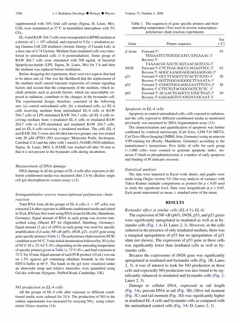
Table 1. The sequences of gene specific primers and theirannealing temperature (Tm) used in reverse transcriptase-
polymerase chain reaction experiments
Gene Primer sequenceTm(�C)
b-Actin Forward 50-TGGAATCCTGTGGCATCCATGAAAC-30
55
Reverse 50-TAAAACGCAGCTCAGTAACAGTCCG-30
iNOS Forward 50-CTCTGACAGCCCAGAGTTCC-30 62Reverse 50-AGGCAAAGGAGGAGAAGGAG-30
p21 Forward 50-GCCTTAGCCCTCACTCTGTG-30 58Reverse 50-GGTTGGGAGGGGCTTAAATA-30
p53 Forward 50-CGGGTGGAAGGAAATTTGTA-30 58Reverse 50-CTTCTGTACGGCGGTCTCTC -30
p65 Forward 50-ACAACTGAGCCCATGCTGAT-30 50Reverse 50-GAGAGGTCCATGTCCGCAAT-30

Bystander effect: role of iNOS d S. GHOSH et al. 1569
There was a clear DNA ladder formation (a hallmark of ap-
optosis) in irradiated and bystander EL-4 cells. Apoptosis
was found to be higher in irradiated cells than bystander cells
(Fig. 4, Lanes 2, 3). Apoptosis in irradiated and bystander
EL-4 cells was further confirmed by annexin V/PI assay
(Fig. 5).
EL-4 cells that had received only irradiated medium or
medium from unirradiated control cells did not show any
upregulation of NF-kB and iNOS or NO production or
DNA damage or apoptosis (data not shown).
Bystander effect in dissimilar cells (EL-4 Vs RAW 264.7)Having confirmed the existence of bystander signaling be-
tween similar cells (EL-4 Vs EL-4), the work was extended to
determine if bystander effects can be demonstrated between
different cell type (RAW 264.7 and EL-4) and whether the
Fig. 1. Gene expression of inducible nitric oxide synthase (iNOS),p21, p53, and NF-kB (p65) in EL-4 cells cultured for 2 h in presenceof different conditioned media. Total RNA from EL-4 cells was iso-lated and reverse transcribed. Reverse transcriptase-polymerasechain reaction (PCR) analysis of iNOS, p21, p53, and NF-kB(p65) genes was carried out as described in materials and methods.PCR products were resolved on 1.5% agarose gels containing ethid-ium bromide. b-Actin gene expression in each group was used as aninternal control. Ratio of intensities of (A) NF-kB (p65), (B) iNOS,(C) p53, and (D) p21band to that of respective b-actin band as quan-tified from gel pictures are shown above each gel picture. Key: Lane1, control unirradiated EL-4 cells; Lane 2, g irradiated EL-4 cells;Lane 3, unirradiated EL-4 cells receiving medium from g-irradiatedEL-4 cells. Data represent means � SE of three independent exper-iments; significantly different from unirradiated controls: *p < 0.05,**p < 0.01.
state of stimulation of macrophages (RAW 264.7) affects by-
stander response. The expression of NF-kB was not altered in
EL-4 cells that had received medium from either unstimu-
lated RAW 264.7 cells or irradiated RAW 264.7 cells, but
the expression of iNOS was downregulated (Fig. 6, Lanes
3, 5). In EL-4 cells that had received medium from LPS stim-
ulated RAW 264.7 cells there was an upregulation of iNOS
gene (Fig. 6, Lane 4). The expression of both the genes
(NF-kB and iNOS) was significantly upregulated in EL-4
cells that had received medium from LPS stimulated and
irradiated RAW 264.7 cells (Fig. 6, Lane 2).
The EL-4 cells that had received medium from unstimu-
lated RAW 264.7 cells or LPS-stimulated RAW 264.7 cells
or irradiated RAW 264.7 cells did not show any increase in
DNA damage (Fig. 7, Lanes 3–5). But there was significantly
higher DNA damage in EL-4 cells that had received medium
from LPS stimulated and irradiated RAW 264.7 cells
(p < 0.01) (Fig. 7, Lane, 2).
Effect of L-NAME and cPTIO on Bystander responseBecause the expressions of NF-kB and iNOS gene were
significantly upregulated in irradiated and bystander cells
(Fig. 8, Lanes 2, 3), it was of interest to determine what
Fig. 2. Nitric oxide (NO) production from EL-4 cells cultured for 24h in the presence of different conditioned media. The culture super-natant from each group of EL-4 cells was used for determination ofNO2
– with Griess reagent. Key: Lane 1, control unirradiated EL-4cells; Lane 2, g irradiated EL-4 cells; Lane 3, unirradiated EL-4 cellsreceiving medium from g irradiated EL-4 cells. Data representmeans � SE of three independent experiments; significantly differ-ent from unirradiated controls: *p < 0.05, **p < 0.01.

1570 I. J. Radiation Oncology d Biology d Physics Volume 72, Number 5, 2008
transmits the signal to the bystanding cells. The response was
looked at in the presence of cPTIO or L-NAME. L-NAME
was washed out before irradiation so that the bystander cells
are not exposed to L-NAME, and iNOS is inhibited only in
the irradiated cells. The addition of cPTIO, which scavenges
the NO released in the medium, did not affect the expression
of iNOS gene if the bystanding cell was of same origin, al-
though NF-kB was somewhat inhibited (Fig. 8, compare
Lanes 5 and 3). The addition of L-NAME, which inhibits
the iNOS in the irradiated cells, completely inhibited the by-
stander response in the EL-4 cells (Fig. 8, compare Lanes 4
and 3).
However, both L-NAME and cPTIO inhibited the expres-
sion of NF-kB and iNOS in the bystander EL-4 cells if the
medium was from LPS stimulated and irradiated RAW
264.7 cells (Fig. 8, Lanes 6, 7) (i.e., the cell type was differ-
ent), indicating that the bystander response was dependent on
the cells type and the state of stimulation of the irradiated
cells.
DNA damage was significantly higher in irradiated and
bystander cells (Fig. 9, Lanes 2, 3). However, the EL-4 cells
that had received medium either from L-NAME–treated and
irradiated EL-4 cells or from LPS-stimulated, L-NAME-
Fig. 3. DNA damage in EL-4 cells cultured for 2 h in presence ofdifferent conditioned media was measured using single-cell gel elec-trophoresis (‘‘comet assay’’). (A) Tail length, (B) % DNA in tail, (C)olive tail moment, and (D) tail moment. Key: Lane 1, control unirra-diated EL-4 cells; Lane 2, g-irradiated EL-4 cells; Lane 3, unirradi-ated EL-4 cells receiving medium from g-irradiated EL-4 cells. Datarepresent means � SE of three independent experiments; signifi-cantly different from unirradiated controls: *p < 0.05, **p < 0.01.
treated and irradiated RAW 264.7 cells did not show any in-
crease in DNA damage (Fig. 9, Lanes 4, 6). Similarly, the EL-
4 cells that had received medium either from cPTIO-treated
and irradiated EL-4 cells or from LPS-stimulated, cPTIO-
treated and irradiated RAW 264.7 cells did not show any in-
crease in DNA damage (Fig. 9, Lanes 5, 7). Neither L-NAME
nor cPTIO itself had any effect on DNA damage in EL-4 cells
(Fig. 9, Lanes 8, 9).
DISCUSSION
In an attempt to understand bystander signaling, several
studies have been made between the same cell lines (1, 2,
16). However, it is not known whether bystander effect is
extendable to dissimilar cells, particularly those cells that
communicate with each other under physiologic situations
like the macrophages and lymphocytes. In immunology,
there is an immense cross-talk between the lymphocytes
and the macrophages. This could make them more prone to
bystander signaling.
Fig. 4. DNA fragmentation assay of EL-4 cells. EL-4 cells culturedfor 24 h in the presence of different conditioned media. DNAs ofeach group of cells were isolated as described in Methods and Ma-terials section and electrophoresed on 1.8% agarose gel containingethidium bromide. Key: Lane M, DNA molecular weight marker;Lane 1, control unirradiated EL-4 cells; Lane 2, g irradiated EL-4cells; Lane 3, unirradiated EL-4 cells receiving medium from g-irradiated EL-4 cells. Data shown from representative experimentof three independent experiments.

Bystander effect: role of iNOS d S. GHOSH et al. 1571
Fig. 5. Apoptosis assay of control, irradiated, and bystander EL-4 cells by Annexin-PI staining. EL-4 cells cultured for 18 hin the presence of different conditioned media. The EL-4 cells were stained using an annexin V/PI-staining kit. Character-ization and quantification of apoptosis was done by confocal microscopy. Key: (A) Annexin-V; (B) PI; (C) DAPI; (D)Merge image of annexin-v, PI, and DAPI. Data represent means � SE of three independent experiments; significantlydifferent from unirradiated controls: **p < 0.01.
There are numerous reports on the activation of signaling
factors in the bystanding cells (1,2). However, not much is
known about the genes that are activated in the bystanding
cells in response to these signals. In our earlier work, we
had shown that NF-kB and p21 levels were elevated in
K562 cells (16). The present study was carried out to inves-
tigate which genes are activated in the bystanding cell and
whether the state of activation of the irradiated cell alters
the bystander response. The study was restricted only to the
genes because, from our experience, even the change of irra-
diated medium leads to an activation of NF-kB (16). Addi-
tionally, minor stresses are known to alter the state of
phosphorylation of signaling proteins. Our present results re-
vealed a significant increase in DNA damage in the bystander
cells, but only in those cells that had received medium from
irradiated cells (Fig. 3). Because the cells treated only with
irradiated medium did not show any DNA damage or NO
production (results not shown), the signals that caused the
damage in the bystander cells must be coming from the irra-
diated cells themselves. The expression of cell cycle–related
genes (p53 and p21) was significantly upregulated in by-
stander similar cells (Fig. 1C, D, Lane 3). Our results are in
agreement with the work on bystander signaling (1,2). The
first report describing the induction of sister chromatid ex-
changes was examined in Chinese hamster ovary cells. Be-
cause Chinese hamster ovary cells contain mutant p53, p53
could not be involved in the bystander induction of sister
chromatid exchanges (17). If the p53 of the bystander cell
is mutated, there should be less DNA repair and in fact
more bystander effect can be seen when assessed in terms
of DNA damage.
A clear cause and effect relationship was established be-
tween DNA damage, gene expression, and cell survival and
was evident in the form of increase apoptosis in bystander
cells (Fig. 4, Lane 3, Fig. 5).
An increased expression of NF-kB gene (p65) could lead
to the activation of iNOS gene because the promoter of the
iNOS gene contains binding sites for NF-kB. NF-kB is
known to be activated by several oxidative stresses including
ionization radiation (18–23). As a consequence of iNOS gene
1572 I. J. Radiation Oncology d Biology d Physics Volume 72, Number 5, 2008
upregulation and increased production of NO, many forms of
reactive nitrogen species would be formed leading to cova-
lent modification of proteins and the expression of various
other genes (24).
Having confirmed the existence of bystander effect be-
tween similar cells (EL-4 Vs EL-4), the work was extended
to look for the existence of cross-bystander effect between
EL-4 and RAW 264.7 cells (i.e., lymphocytes and macro-
phages). There was a significant upregulation of NF-kB and
iNOS genes in EL-4 cells that had received medium from
LPS stimulated and irradiated RAW 264.7 cells (Fig. 6,
Lane 2) and expectedly the DNA damage was also signifi-
cantly higher in these cells (Fig. 7, Lane 2). A noteworthy
finding of this study is that the unirradiated EL-4 cells that
had received medium from LPS-stimulated RAW 264.7 cells
also showed upregulation of iNOS gene but not NF-kB gene
(Fig. 6, Lane 4). The LPS treatment of macrophage upregu-
lates many genes in the macrophage hence their response to
irradiation will be different from unstimulated macrophage.
Another noteworthy finding of this study is that the addition
of medium from nonirradiated RAW 264.7 cells resulted in
significantly decrease in iNOS gene expression in EL-4 cells
Fig. 6. Gene expression of NF-kB (p65) and iNOS gene in EL-4cells that had received medium from irradiated RAW 264.7 cells.EL-4 cells cultured for 2 h in presence of different conditioned me-dia. b-Actin gene expression in each group was used as an internalcontrol. Ratio of intensities of NF-kB (p65) and iNOS band to that ofrespective b-actin band as quantified from gel pictures are shownabove each gel picture. Key: Lane 1, control EL-4 cells; Lane 2,EL-4 cells receiving medium from lipopolysaccharide (LPS)-stimu-lated (500 ng/mL) and g-irradiated RAW 264.7 cells; Lane 3, EL-4cells receiving medium from g-irradiated RAW 264.7 cells withoutLPS stimulation; Lane 4, EL-4 cells receiving medium from LPS-stimulated RAW 264.7 cells; Lane 5, EL-4 cells receiving mediumfrom unirradiated RAW 264.7 cells. Data represent means � SE ofthree independent experiments; significantly different from unirradi-ated controls: *p < 0.05, **p < 0.01.
relative to the control (Fig. 6, Lane 5). Nonirradiated macro-
phages may release some factor that is responsible for down
regulation of iNOS in EL-4 cells. Further studies in this direc-
tion will reveal the nature of the factor. It is clear from these
results that effective cross-bystander effects exists between
these two cell lines only when the RAW 264.7 cells are
both stimulated and irradiated. Other studies have looked at
only the clonogenic survival of dissimilar bystander cells (25).
Numerous studies have implicated extracellular secreted
signaling proteins in mediating the bystander effect (5), but
the strongest contenders have been reactive oxygen and nitro-
gen species (26). To investigate what factors are involved in
radiation induced bystander effect, the cells were treated with
either L-NAME (NOS inhibitor) or cPTIO (NO scavenger).
The effect of cPTIO and L-NAME in bystander response
between similar cell (EL-4 Vs EL-4) was different in that
treatment with L-NAME reduces the bystander induction of
NF-kB as well as iNOS gene expression in EL-4 cells receiv-
ing medium from irradiated EL-4 cells (Fig. 8, Lane 4), but
the treatment with cPTIO reduces the bystander induction
of only NF-kB gene expression and not iNOS gene expres-
sion (Fig. 8, Lane 5). These results demonstrate that the acti-
vated NOS in the irradiated cell is essential for gene
expression in the bystanding cell.
The addition of either L-NAME or cPTIO reduces the
bystander induction of NF-kB as well as iNOS gene expres-
sion in EL-4 cells receiving medium from LPS stimulated
and irradiated RAW 264.7 cells (Fig. 8, Lanes 6, 7), indicat-
ing that both activation of iNOS in the irradiated cell and NO
production play a role in cross-bystander effect.
Fig. 7. DNA damage in EL-4 cells cultured for 2 h in presence ofdifferent conditioned media was measured using single-cell gel elec-trophoresis (‘‘comet assay’’) and expressed as tail length. Key: Lane1, control EL-4 cells; Lane 2, EL-4 cells receiving medium fromLPS-stimulated (500 ng/mL) and g-irradiated RAW 264.7 cells;Lane 3, EL-4 cells receiving medium from g irradiated RAW264.7 cells without lipopolysaccharide stimulation; Lane 4, EL-4cells receiving medium from LPS-stimulated RAW 264.7 cells;Lane 5, EL-4 cells receiving medium from unirradiated RAW264.7 cells. Data represent means� SE of three independent; signif-icantly different from unirradiated controls: *p < 0.01.

Bystander effect: role of iNOS d S. GHOSH et al. 1573
The treatment with L-NAME or cPTIO also reduces the
bystander induction of DNA damage in EL-4 cells receiving
medium from irradiated EL-4 cells or LPS-stimulated and
LPS-irradiated RAW 264.7 cells (Fig. 9, Lanes 4–7), demon-
strating that NO contributes to the DNA damage in bystander
EL-4 cells. NO is generated endogenously from L-arginine
by inducible NO synthases (27, 28) that can be stimulated
by radiation in mammalian cells (8). It is believed that NO it-
self does not induce DNA strand breaks, although one of its
oxidation product, peroxynitrite, is toxic and can cause DNA
Fig. 8. Gene expression of NF-kB (p65) and iNOS gene in EL-4cells that had received medium from irradiated cells. EL-4 cells cul-tured for 2 h in presence of different conditioned media. b-Actingene expression in each group was used as an internal control. Ratioof intensities of NF-kB (p65) and inducible nitric oxide synthaseband to that of respective b-actin band as quantified from gel pic-tures are shown above each gel picture. Key: Lane 1, control EL-4 cells; Lane 2, g-irradiated EL-4 cells; Lane 3, EL-4 cells receivingmedium from g-irradiated EL-4 cells; Lane 4, EL-4 cells receivingmedium from L-NAME treated and g-irradiated EL-4 cells; Lane 5,EL-4 cells receiving medium from cPTIO-treated and g-irradiatedEL-4 cells; Lane 6, EL-4 cells receiving medium from LPS-stimu-lated (500 ng/mL), L-NAME treated and g-irradiated RAW 264.7cells; Lane 7, EL-4 cells receiving medium from lipopolysaccha-ride-stimulated (500 ng/mL), cPTIO-treated and g-irradiatedRAW 264.7 cells. Data represent means � SE of three independentexperiments; significantly different from unirradiated controls:*p < 0.05, **p < 0.01.
damage by both attacking deoxyribose and direct oxidation
of purine and pyrimidine (29). Radiation-induced NO has
the possibility of attacking nearby cells within their diffusion
distance of less than 0.5 mm (30, 31). However, because of
their having very short half-lives, they may not be direct con-
tributors to the cellular damage in the bystander population.
This is confirmed by the fact that inclusion of cPTIO in the
medium only marginally inhibits the bystander response in
similar cells (Fig. 8, Lane 5). Contrarily, it has also been re-
ported that NO is involved in the bystander responses caused
by the conditioned medium harvested from irradiated cells
(32). Therefore some long-lived bioactive factors down-
stream of NO are most likely involved in this bystander
response.
Fig. 9. DNA damage in EL-4 cells cultured for 2 h in presence ofdifferent conditioned media was measured using single-cell gel elec-trophoresis (‘‘comet assay’’) and expressed as tail length. Key: Lane1, control EL-4 cells; Lane 2, g-irradiated EL-4 cells; Lane 3, EL-4cells receiving medium from g-irradiated EL-4 cells; Lane 4, EL-4cells receiving medium from L-NAME treated and g-irradiated EL-4 cells; Lane 5, EL-4 cells receiving medium from cPTIO treatedand g-irradiated EL-4 cells; Lane 6, EL-4 cells receiving mediumfrom lipopolysaccharide (LPS)-stimulated (500 ng/mL), L-NAME-treated, and g-irradiated RAW 264.7 cells; Lane 7, EL-4cells receiving medium from LPS-stimulated (500 ng/mL),cPTIO-treated, and g-irradiated RAW 264.7 cells; Lane 8, EL-4cells treated with L-NAME; Lane 9, EL-4 cells treated with cPTIO.Data represent means � SE of three independent experiments;significantly different from unirradiated controls: *p < 0.05,**p < 0.01.
REFERENCES
1. Azzam EI, De Toledo SM, Spitz DR, et al. Oxidative metabo-lism modulates signal transduction and micronucleus formationin bystander cells from alphaparticle- irradiated normal humanfibroblast cultures. Cancer Res 2002;62:5436–5442.
2. Azzam EI, De Toledo SM, Gooding T, et al. Intercellular com-munication is involved in the bystander regulation of gene ex-pression in human cells exposed to very low fluences ofa particles. Radiat Res 1998;150:497–504.
3. Mothersill C, Seymour C. Radiation-induced bystander effects,carcinogenesis and models. Oncogene 2003;22:7028–7033.
4. Mothersill C, Seymour C. Radiation-induced bystander effects:Are they good, bad or both? Med Confl Surviv 2005;21:101–110.
5. Azzam EI, De Toledo SM, Little JB. Oxidative metabolism, gapjunctions and the ionizing radiation-induced bystander effect.Oncogene 2003;22:7050–7057.
1574 I. J. Radiation Oncology d Biology d Physics Volume 72, Number 5, 2008
6. Hall EJ, Hei TK. Genomic instability and bystander effects in-duced by high-LET radiation. Oncogene 2003;22:7034–7042.
7. Prise KM, Folkard M, Michael BD. Bystander responses in-duced by low LET radiation. Oncogene 2003;22:7043–7049.
8. Matsumoto H, Hayashi S, Hatashita M, et al. Induction of radio-resistance to accelerated carbon-ion beams in recipient cells bynitric oxide excreted from irradiated donor cells of human glio-blastoma. Int J Radiat Biol 2000;76:1649–1657.
9. Matsumoto H, Hayashi S, Hatashita M, et al. Induction of radio-resistance by a nitric oxide-mediated bystander effect. RadiatRes 2001;155:387–396.
10. Shao C, Furusawa Y, Aoki M, et al. Nitric oxide-mediated by-stander effect induced by heavy-ions in human salivary glandtumour cells. Int J Radiat Biol 2002;78:837–844.
11. Iluki Y, Mizuno S, Goto R. g-irradiation-induced DNA damageenhance NO production via NF-kB activation in RAW 264.7cells. Biochim Biophys Acta 2003;1593:159–167.
12. Camphausen K, Moses MA, Menard C, et al. Radiation absco-pal antitumor effect is mediated through p53. Cancer Res 2003;63:1990–1993.
13. Maurya DK, Adhikari S, Nair CK, et al. DNA protective prop-erties of vanillin against gamma-radiation under different condi-tions: possible mechanisms. Mutat Res 2007;634:69–80.
14. Green LC, Wagner DA, Glogowski J, et al. Analysis of nitrate,nitrite and [15N] nitrate in biological fluids. Anal Biochem1982;126:131–138.
15. Jeon SH, Kang MG, Kim YH, et al. A new mouse gene, SRG3,related to the SWI3 of Saccharomyces cerevisiae, is required forapoptosis induced by glucocorticoids in a thymoma cell line.J Exp Med 1997;185:1827–1836.
16. Mitra AK, Krishna M. Radiation-induced bystander effect: ac-tivation of signaling molecules in K562 erythroleukemia cells.J Cell Biochem 2007;100:991–997.
17. Nagasawa H, Little JB. Induction of sister chromatid exchangesby extremely low doses of a-particles. Cancer Res 1992;52:6394–6396.
18. Brach MA, Hass R, Sherman ML, et al. Ionizing radiation in-duces expression and binding activity of the nuclear factorkB. J Clin Invest 1991;88:691–695.
19. Ginn-Pease ME, Whisler RL. Redox signals and NF-kB activa-tion in T cells. Free Radic Biol Med 1998;25:346–361.
20. Zhou H, Ivanov VN, Lien YC, et al. Mitochondrial function andnuclear factor-kappa-mediated signaling in radiation inducedbystander effects. Cancer Res 2008;68:2233–2240.
21. Li N, Karin M. Ionizing radiation and short wavelength UV ac-tivate NF-kB through two distinct mechanisms. Proc Natl AcadSci U S A 1998;95:13012–13017.
22. Lowenstein CJ, Alley EW, Raval P, et al. Macrophage nitric ox-ide synthase gene: two upstream regions mediate induction byinterferon g and lipopolysaccharide. Proc Natl Acad Sci U S A1993;90:9730–9734.
23. Xie QW, Whisnant R, Nathan C. Promoter of the mouse geneencoding calcium-independent nitric oxide synthase confers in-ducibility by interferon g and bacterial lipopolysaccharide.J Exp Med 1993;177:1779–1784.
24. Martinez-Ruiz A, Lamas S. Signalling by NO-induced proteinS-nitrosylation and S-glutathionylation: Convergences and di-vergences. Cardiovasc Res 2007;75:220–228.
25. Mothersill C, Seymour C. Medium from irradiated human epi-thelial cells but not human fibroblasts reduces the clonogenicsurvival of unirradiated cells. Int J Radiat Biol 1997;71:421–427.
26. Kadhim MA, Moore SR, Goodwin EH. Interrelationshipsamongst radiation-induced genomic instability, bystander ef-fects, and the adaptive response. Mutat Res 2004;568:21–32.
27. Maragos CM, Morley D, Wink DA, et al. Complexes of NOwith nucleophiles as agents for the controlled biological releaseof nitric oxide: Vasorelaxant effects. J Med Chem 1991;34:3242–3247.
28. Nathan C. Nitric oxide as a secretory product of mammaliancells. FASEB J 1992;6:3051–3064.
29. Tamir S, Tannenbaum SR. The role of nitric oxide (NO) in thecarcinogenic process. Biochim Biophys Acta 1996;1288:F31–F36.
30. Lancaster JR Jr. Diffusion of free nitric oxide. Methods Enzymol1996;268:31–50.
31. Saran M, Bors W. Signaling by O2- and NO: How far can eitherradical, or any specific reaction product, transmit a message un-der in vivo conditions? Chem Biol Interact 1994;90:35–45.
32. Shao C, Stewart V, Folkard M, et al. Nitric oxide-mediated sig-naling in the bystander response of individually targeted gliomacells. Cancer Res 2003;63:8437–8442.