Robust expression of EZH2 in endocervical neoplastic lesions
10
ORIGINAL ARTICLE Robust expression of EZH2 in endocervical neoplastic lesions Evelin Makk 1 & Levente Bálint 1 & János Cifra 2 & Tamás Tornóczky 1 & Angéla Oszter 1 & Arnold Tóth 3 & Endre Kálmán 1 & Krisztina Kovács 1 Received: 24 October 2018 /Revised: 22 February 2019 /Accepted: 25 February 2019 /Published online: 22 March 2019 # The Author(s) 2019 Abstract The aim of this study was to evaluate the nuclear expression of histone methyltransferase enhancer of zeste homolog 2 (EZH2) in endocervical neoplastic lesions such as invasive endocervical adenocarcinoma (ECA) and cervical in situ adenocarcinoma (AIS) in comparison with normal endocervix and non-neoplastic counterparts. A total of 54 consecutive neoplastic cases (37 ECA, 17 AIS) and 32 non-neoplastic endocervical lesions (15 reactive atypia, 9 microglandular hyperplasia, 3 tuboendometrioid metaplasia, 3 tunnel cluster, 2 endometriosis) were included in the study with adjacent normal endocervix if present. EZH2 immunoreactivity was evaluated semiquantitatively by three independent experts in lesions and adjacent normal glandular epithelium as well. EZH2 expression was defined robust if at least two of the three experts rated partial or diffuse positivity. Robust EZH2 expression was statistically compared among the neoplastic, non-neoplastic, and normal glandular epithelium samples. Diagnostic test capability of robust EZH2 expression was calculated. Fifty-three out of the 54 neoplastic cases (98%) showed robust EZH2 expression. Robust EZH2 expression was significantly less often (4 out of 32 cases, 12.5%) found in the non-neoplastic endocervical lesions (p < 0.0001) and never (0 out of 66 samples) in the adjacent normal glandular epithelium. Robust EZH2 overexpression had a sensitivity and specificity of over 95% in detecting neoplastic lesions versus non-neoplastic lesions or normal glandular epithelium. EZH2 may play a role in the pathogenesis of endocervical neoplasia, and the detection of robust expression of EZH2 might be a useful differential diagnostic tool in problematic endocervical lesions in histology and cytology as well. Keywords Cervical cancer . EZH2 . Endocervical adenocarcinoma . AIS Introduction Endocervical adenocarcinoma (ECA) is the second most com- mon histological type of cervical cancer; it comprises approx- imately 20 to 25% of cervical malignancies [1] and has a poorer prognosis than squamous cell carcinoma [2]. Cervical adenocarcinomas and their precursor lesions are heteroge- neous and have several different subtypes, most of them close- ly related to HR-HPVs [3]. It is well established that the pRB pathway is involved in the pathogenesis of cervical cancer due to the interaction with HR-HPV E7 oncoproteins leading to genomic instability [4]. It is also known that viral E6/E7 oncoproteins may interact with different types of epigenetic enzymes, such as p300, CBP, and pCAF, which can be involved in the oncogenesis [5]. Enhancer of zeste homolog 2 (EZH2), a member of the polycomb group of genes, is a methyltransferase that methyl- ates histone H3 on gene promoters and plays a critical role in epigenetic gene silencing and chromatin remodeling. EZH2 inhibits cell differentiation and targets gene expression. In conjunction with the p53 protein, it induces tumor cell prolif- eration, metastasis, and immortalization [6]. Recent studies focused on the role of EZH2 in the patho- genesis of various adenocarcinomas as well as malignant tu- mors of the breast [7], lung [8], stomach [9], colon [10], pancreatobiliary tract [11], liver [12], thyroid gland [13], pros- tate [14], bladder [15], uterus [16], and ovary [17]. High ex- pression of EZH2 was shown to be associated with tumor aggressiveness and was suggested as a potential differential * Evelin Makk [email protected] 1 Department of Pathology, University of Pécs Medical School, Szigeti út 12, Pécs 7624, Hungary 2 Department of Pathology, County Hospital Tolna, János Balassa Hospital, Béri Balogh Ádám u. 5-7, Szekszárd 7100, Hungary 3 Department of Radiology, University of Pécs Medical School, Ifjúság út 13, Pécs 7624, Hungary Virchows Archiv (2019) 475:95–104 https://doi.org/10.1007/s00428-019-02550-8
Transcript of Robust expression of EZH2 in endocervical neoplastic lesions

ORIGINAL ARTICLE
Robust expression of EZH2 in endocervical neoplastic lesions
Evelin Makk1 & Levente Bálint1 & János Cifra2 & Tamás Tornóczky1 & Angéla Oszter1 & Arnold Tóth3& Endre Kálmán1
&
Krisztina Kovács1
Received: 24 October 2018 /Revised: 22 February 2019 /Accepted: 25 February 2019 /Published online: 22 March 2019# The Author(s) 2019
AbstractThe aim of this study was to evaluate the nuclear expression of histone methyltransferase enhancer of zeste homolog 2 (EZH2) inendocervical neoplastic lesions such as invasive endocervical adenocarcinoma (ECA) and cervical in situ adenocarcinoma (AIS) incomparison with normal endocervix and non-neoplastic counterparts. A total of 54 consecutive neoplastic cases (37 ECA, 17 AIS)and 32 non-neoplastic endocervical lesions (15 reactive atypia, 9 microglandular hyperplasia, 3 tuboendometrioid metaplasia, 3tunnel cluster, 2 endometriosis) were included in the study with adjacent normal endocervix if present. EZH2 immunoreactivity wasevaluated semiquantitatively by three independent experts in lesions and adjacent normal glandular epithelium as well. EZH2expression was defined robust if at least two of the three experts rated partial or diffuse positivity. Robust EZH2 expression wasstatistically compared among the neoplastic, non-neoplastic, and normal glandular epithelium samples. Diagnostic test capability ofrobust EZH2 expression was calculated. Fifty-three out of the 54 neoplastic cases (98%) showed robust EZH2 expression. RobustEZH2 expression was significantly less often (4 out of 32 cases, 12.5%) found in the non-neoplastic endocervical lesions(p < 0.0001) and never (0 out of 66 samples) in the adjacent normal glandular epithelium. Robust EZH2 overexpression had asensitivity and specificity of over 95% in detecting neoplastic lesions versus non-neoplastic lesions or normal glandular epithelium.EZH2 may play a role in the pathogenesis of endocervical neoplasia, and the detection of robust expression of EZH2 might be auseful differential diagnostic tool in problematic endocervical lesions in histology and cytology as well.
Keywords Cervical cancer . EZH2 . Endocervical adenocarcinoma . AIS
Introduction
Endocervical adenocarcinoma (ECA) is the secondmost com-mon histological type of cervical cancer; it comprises approx-imately 20 to 25% of cervical malignancies [1] and has apoorer prognosis than squamous cell carcinoma [2]. Cervicaladenocarcinomas and their precursor lesions are heteroge-neous and have several different subtypes, most of them close-ly related to HR-HPVs [3].
It is well established that the pRB pathway is involved inthe pathogenesis of cervical cancer due to the interaction withHR-HPV E7 oncoproteins leading to genomic instability [4].It is also known that viral E6/E7 oncoproteins may interactwith different types of epigenetic enzymes, such as p300,CBP, and pCAF, which can be involved in the oncogenesis[5].
Enhancer of zeste homolog 2 (EZH2), a member of thepolycomb group of genes, is a methyltransferase that methyl-ates histone H3 on gene promoters and plays a critical role inepigenetic gene silencing and chromatin remodeling. EZH2inhibits cell differentiation and targets gene expression. Inconjunction with the p53 protein, it induces tumor cell prolif-eration, metastasis, and immortalization [6].
Recent studies focused on the role of EZH2 in the patho-genesis of various adenocarcinomas as well as malignant tu-mors of the breast [7], lung [8], stomach [9], colon [10],pancreatobiliary tract [11], liver [12], thyroid gland [13], pros-tate [14], bladder [15], uterus [16], and ovary [17]. High ex-pression of EZH2 was shown to be associated with tumoraggressiveness and was suggested as a potential differential
* Evelin [email protected]
1 Department of Pathology, University of PécsMedical School, Szigetiút 12, Pécs 7624, Hungary
2 Department of Pathology, County Hospital Tolna, János BalassaHospital, Béri Balogh Ádám u. 5-7, Szekszárd 7100, Hungary
3 Department of Radiology, University of Pécs Medical School,Ifjúság út 13, Pécs 7624, Hungary
Virchows Archiv (2019) 475:95–104https://doi.org/10.1007/s00428-019-02550-8

diagnostic marker [8, 10–17]. In the cervix, one study reportedoverexpression and the possible prognostic significance ofEZH2 in squamous cell carcinoma [18].
Expression of EZH2 in endocervical neoplastic lesions isyet unknown. In this study, we examined EZH2 expression inECA and AIS, compared with non-neoplastic cervical lesionsand normal glandular epithelium.
Materials and methods
Patients and specimen collection
Consecutive patients from 2007 to 2017 with a diagnosis ofendocervical neoplastic lesions (ECA and/or AIS with orwithout cervical intraepithelial neoplasia) and patients withbenign findings as a control group were collected from thearchives of the Department of Pathology, University of Pécs,Hungary, and Department of Pathology, County HospitalTolna, János Balassa Hospital, Szekszárd, Hungary.
Formalin-fixed and paraffin-embedded tissue samplesfrom biopsy, cone, or hysterectomy specimens were availablealong with the HE slides in each case. Slides were re-evaluated to select the most feasible specimens for immuno-histochemistry for each patient. We classified endocervicaladenocarcinomas based on the International EndocervicalAdenocarcinoma Criteria and Classification (IECC) [19].
This work has been approved by the local ethical commit-tee (number of permission: PTE/57682/2017).
Immunohistochemistry
Prior to immunohistochemistry, formalin-fixed paraffin-em-bedded tissue specimens were cut into 4-μm-thick sectionsand dried for 20 min at 60 °C.
Immunostaining was performed using Leica Bond Maxautostainer (Leica Biosystems, Bannockburn, IL) and LeicaBond Polymer Refine Detection Kit (Leica Biosystems,Newcastle Upon Tyne, UK). The mouse monoclonal EZH2antibody (clone 6A10) was obtained from Leica Biosystems(Newcastle Upon Tyne, UK) and used at a dilution of 1:200.The immunostaining protocol included deparaffinization andpH 9 epitope retrieval for 20 min, peroxidase blocking for5 min, primary antibody incubation for 15 min, post-primaryrabbit anti-mouse IgG for 8 min, polymer anti-rabbit Poly-HRP-IgG for 8 min, diaminobenzidine chromogen for10 min, and hematoxylin counterstain for 5 min. Positiveand negative controls were included in all reactions.
Evaluation of immunoreactivity
Immunoreactivity evaluation included not only the neoplasticlesions (ECA, AIS) or non-neoplastic lesions in control
patients but the adjacent normal glandular epithelium as well,if present. The presence of concurrent cervical intraepithelialneoplasia was noted; however, these lesions were not includedin the immunoreactivity analysis. The histological patternswere detected in the original, routine HE stained slides.
Immunoreactivity was evaluated semiquantitatively bythree independent board-certified pathologists with over15 years of professional experience (expert 1: E.K., expert 2:K.K., expert 3: A.O.). Cases were regarded as positive if theywere obviously positive by × 40 magnification and furtherclassified according to the percentage of cells with nuclearstaining: < 10% as focally positive B+^, 10–50% as partlypositive B++^, and > 50% as diffusely positive B+++^ [20].
The inter-expert agreement was determined usingIntraclass Correlation Coefficient (ICC) [21] for both the neo-plastic and the non-neoplastic lesions ratings. Two-way mod-el, absolute agreement type was applied; both single and av-erage measurement reliability was calculated. The analysiswas run in MedCalc statistical software (version 13.0.0.0,MedCalc Software bvba, Ostend, Belgium) [22].
For statistical analyses, the individual ratings per lesionsand normal glandular epithelium if present were transformedinto a binary overall score. Immunoreactivity of a lesion ornormal glandular epithelium was regarded Brobust^ if at leasttwo of the three experts rated either B++^ or B+++^.Immunoreactivity was regarded as Bnegative/focally positive^if at least two of the three experts rated either B−^ or B+^.Adjacent normal glandular epithelium was included in analy-ses if it was detected by at least two experts.
Neoplastic (ECA and AIS) and non-neoplastic lesion im-munoreactivity overall scores were statistically comparedusing Fisher’s exact test (MedCalc). P value was consideredstatistically significant if under 0.05.
Diagnostic test capability (sensitivity, specificity, and pos-itive and negative predictive value) of EZH2 overexpressionin distinguishing (a) neoplastic lesions from non-neoplastic,(b) neoplastic lesions from normal endocervical epithelium,and (c) neoplastic lesions from non-neoplastic lesions andnormal endocervical epithelium combined was evaluated alsousing MedCalc.
Results
A total of 54 cases of endocervical neoplastic lesions (37ECA, 17 AIS) were retrieved from the archive. In 12 out ofthese cases, concurrent HSILs were present. Concurrent LSILwas present in one case. The median patient age was 44.5(range 29–84). The most common IECC diagnoses were hu-man papillomavirus–associated adenocarcinoma (HPVA)type (92% of the cohort). Between subcategories, usual typeadenocarcinoma was the most common HPVA type (88% ofthe cohort), followed by villoglandular, mucinous not
96 Virchows Arch (2019) 475:95–104
otherwise specified (NOS), and mucinous including intestinaland invasive stratified mucin-producing carcinoma (iSMILE)categories (3%) (Table 1). There were only three patients withnonhuman papillomavirus–associated adenocarcinoma(NHPVA) (8%). Between subcategories, there were two cases
with serous type and one with the endometrioid type ofNHPVA.
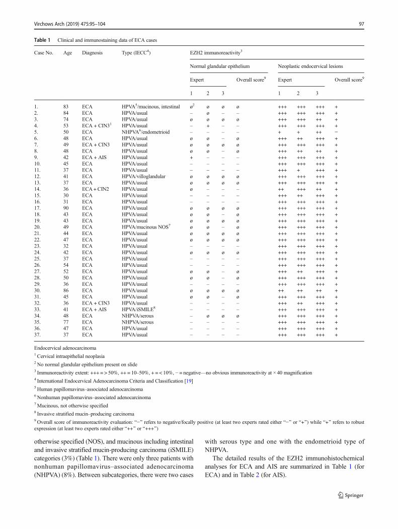
The detailed results of the EZH2 immunohistochemicalanalyses for ECA and AIS are summarized in Table 1 (forECA) and in Table 2 (for AIS).
Table 1 Clinical and immunostaining data of ECA cases
Case No. Age Diagnosis Type (IECC4) EZH2 immunoreactivity3
Normal glandular epithelium Neoplastic endocervical lesions
Expert Overall score9 Expert Overall score9
1 2 3 1 2 3
1. 83 ECA HPVA5/mucinous, intestinal ø2 ø ø ø +++ +++ +++ +2. 84 ECA HPVA/usual – ø – – +++ +++ +++ +3. 74 ECA HPVA/usual ø ø ø ø +++ +++ ++ +4. 53 ECA + CIN31 HPVA/usual – + – – +++ +++ +++ +5. 50 ECA NHPVA6/endometrioid – – – – + + ++ −6. 48 ECA HPVA/usual ø ø – ø +++ ++ +++ +7. 49 ECA + CIN3 HPVA/usual ø ø ø ø +++ +++ +++ +8. 48 ECA HPVA/usual ø ø – ø +++ ++ ++ +9. 42 ECA + AIS HPVA/usual + – – – +++ +++ +++ +10. 45 ECA HPVA/usual – – – – +++ +++ +++ +11. 37 ECA HPVA/usual – – – – +++ + +++ +12. 41 ECA HPVA/villoglandular ø ø ø ø +++ +++ +++ +13. 37 ECA HPVA/usual ø ø ø ø +++ +++ +++ +14. 36 ECA+CIN2 HPVA/usual ø – – – ++ +++ ++ +15. 30 ECA HPVA/usual – – – – +++ ++ +++ +16. 31 ECA HPVA/usual – – – – +++ +++ +++ +17. 90 ECA HPVA/usual ø ø ø ø +++ +++ +++ +18. 43 ECA HPVA/usual ø ø – ø +++ +++ +++ +19. 43 ECA HPVA/usual ø ø ø ø +++ +++ +++ +20. 49 ECA HPVA/mucinous NOS7 ø ø – ø +++ +++ +++ +21. 44 ECA HPVA/usual ø ø ø ø +++ +++ +++ +22. 47 ECA HPVA/usual ø ø ø ø +++ +++ +++ +23. 32 ECA HPVA/usual – – – – +++ +++ +++ +24. 42 ECA HPVA/usual ø ø ø ø +++ +++ +++ +25. 37 ECA HPVA/usual – – – – +++ +++ +++ +26. 54 ECA HPVA/usual – – – – +++ +++ +++ +27. 52 ECA HPVA/usual ø ø – ø +++ ++ +++ +28. 50 ECA HPVA/usual ø ø – ø +++ +++ +++ +29. 36 ECA HPVA/usual – – – – +++ +++ +++ +30. 86 ECA HPVA/usual ø ø ø ø ++ ++ ++ +31. 45 ECA HPVA/usual ø ø – ø +++ +++ +++ +32. 36 ECA + CIN3 HPVA/usual – – – – +++ ++ +++ +33. 41 ECA + AIS HPVA/iSMILE8 – – – – +++ +++ +++ +34. 48 ECA NHPVA/serous – ø ø ø +++ +++ +++ +35. 77 ECA NHPVA/serous – – – – +++ +++ +++ +36. 47 ECA HPVA/usual – – – – +++ +++ +++ +37. 37 ECA HPVA/usual – – – – +++ +++ +++ +
Endocervical adenocarcinoma1 Cervical intraepithelial neoplasia2 No normal glandular epithelium present on slide3 Immunoreactivity extent: +++ = > 50%, ++ = 10–50%, + = < 10%, − = negative—no obvious immunoreactivity at × 40 magnification4 International Endocervical Adenocarcinoma Criteria and Classification [19]5 Human papillomavirus–associated adenocarcinoma6Nonhuman papillomavirus–associated adenocarcinoma7Mucinous, not otherwise specified8 Invasive stratified mucin–producing carcinoma9Overall score of immunoreactivity evaluation: B−^ refers to negative/focally positive (at least two experts rated either B−^ or B+^) while B+^ refers to robustexpression (at least two experts rated either B++^ or B+++^)
Virchows Arch (2019) 475:95–104 97
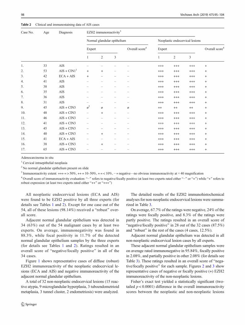
All neoplastic endocervical lesions (ECA and AIS)were found to be EZH2 positive by all three experts (fordetails see Tables 1 and 2). Except for one case out of the54, all of these lesions (98.14%) received a Brobust^ over-all score.
Adjacent normal glandular epithelium was detected in34 (63%) out of the 54 malignant cases by at least twoexperts. On average, immunonegativity was found in88.3%, while focal positivity in 11.7% of the detectednormal glandular epithelium samples by the three experts(for details see Tables 1 and 2). Ratings resulted in anoverall score of Bnegative/focally positive^ in all of the34 cases.
Figure 1 shows representative cases of diffuse (robust)EZH2 immunoreactivity of the neoplastic endocervical le-sions (ECA and AIS) and negative immunoreactivity of theadjacent normal glandular epithelium.
A total of 32 non-neoplastic endocervical lesions (15 reac-tive atypia, 9 microglandular hyperplasia, 3 tuboendometrioidmetaplasia, 3 tunnel cluster, 2 endometriosis) were analyzed.
The detailed results of the EZH2 immunohistochemicalanalyses for non-neoplastic endocervical lesions were summa-rized in Table 3.
On average, 67.7% of the ratings were negative, 24% of theratings were focally positive, and 8.3% of the ratings werepartly positive. The ratings resulted in an overall score ofBnegative/focally positive^ in 28 out of the 32 cases (87.5%)and Brobust^ in the rest of the cases (4 cases, 12.5%).
Adjacent normal glandular epithelium was detected in allnon-neoplastic endocervical lesion cases by all experts.
These adjacent normal glandular epithelium samples wereon average rated immunonegative in 95.84%, focally positivein 2.08%, and partially positive in other 2.08% (for details seeTable 3). These ratings resulted in an overall score of Bnega-tive/focally positive^ for each sample. Figures 2 and 3 showrepresentative cases of negative or focally positive (+) EZH2immunoreactivity of the non-neoplastic lesions.
Fisher’s exact test yielded a statistically significant (two-tailed p < 0.0001) difference in the overall immunoreactivityscores between the neoplastic and non-neoplastic lesions
Table 2 Clinical and immunostaining data of AIS cases
Case No. Age Diagnosis EZH2 immunoreactivity3
Normal glandular epithelium Neoplastic endocervical lesions
Expert Overall score4 Expert Overall score4
1 2 3 1 2 3
1. 33 AIS – – – – +++ +++ +++ +
2. 53 AIS + CIN11 + + – – +++ +++ +++ +
3. 42 ECA + AIS + – – – +++ +++ +++ +
4. 41 AIS – – – – +++ +++ +++ +
5. 38 AIS – – – – +++ +++ +++ +
6. 35 AIS – – – – +++ +++ +++ +
7. 36 AIS – – – – +++ +++ +++ +
8. 31 AIS – – – – +++ +++ +++ +
9. 45 AIS + CIN3 ø2 ø – ø ++ ++ ++ +
10. 48 AIS + CIN3 – + – – +++ +++ +++ +
11. 46 AIS + CIN3 – – – – +++ +++ +++ +
12. 41 AIS + CIN3 – – – – +++ +++ +++ +
13. 45 AIS + CIN3 – – – – +++ +++ +++ +
14. 40 AIS + CIN3 – + – – +++ +++ +++ +
15. 41 ECA + AIS – – – – +++ +++ +++ +
16. 38 AIS + CIN3 – + – – +++ +++ +++ +
17. 65 AIS + CIN3 – – – – +++ +++ +++ +
Adenocarcinoma in situ1 Cervical intraepithelial neoplasia2 No normal glandular epithelium present on slide3 Immunoreactivity extent: +++ = > 50%, ++ = 10–50%, + = < 10%, − = negative—no obvious immunoreactivity at × 40 magnification4Overall score of immunoreactivity evaluation: B−^ refers to negative/focally positive (at least two experts rated either B−^ or B+^) while B+^ refers torobust expression (at least two experts rated either B++^ or B+++^)
98 Virchows Arch (2019) 475:95–104

(Brobust^ score was found in 53 out of the 54 neoplastic le-sions vs. in 4 out of the 32 non-neoplastic lesions).
Robust EZH2 expression was found to have a sensitiv-ity of 98.15% (95% CI = 90.11 to 99.95%) and a specific-i t y o f 87 . 5% (95% CI = 71 .01 t o 96 . 49%) i ndistinguishing neoplastic lesions from non-neoplastic le-sions, with a positive predictive value of 92.98% (95%CI = 83 to 98.05%) and a negative predictive value of96.55% (95% CI = 82.24 to 99.91%). A sensitivity of98.15% (95% CI = 90.11 to 99.95%) and a specificity of100% (95% CI = 94.4 to 100%) were found indistinguishing neoplastic lesions from all normal glandu-lar epithelium samples (n = 66), with a positive predictivevalue of 100% (95% CI = 93.28 to 100%) and a negativepredictive value of 98.46% (95% CI = 91.72 to 99.96%).A sensitivity of 98.15% (95% CI = 90.11 to 99.95%) anda specificity of 95.88% (95% CI = 89.78% to 98.87%)were found in distinguishing neoplastic from non-neoplastic lesions and normal endocervical epitheliumsamples combined (n = 98), with a positive predictive val-ue of 92.98% (95% CI = 83 to 98.05%) and a negativepredictive value of 98.46% (95% CI = 94.17 to 99.97%).
For the neoplastic endocervical lesion (ECA and AIS) im-munoreactivity ratings, inter-expert ICCs were 0.53 for single
measures (95% confidence interval = 0.37–0.67) and 0.77 foraverage measures (95% confidence interval = 0.64–0.86).
For the non-neoplastic endocervical lesion immunoreactiv-ity ratings, inter-expert ICCs were 0.8 for single measures(95% confidence interval = 0.68–0.89) and 0.92 for averagemeasures (95% confidence interval = 0.86–0.96).
Discussion
The aim of this study was to investigate the EZH2 expressionstatus of neoplastic endocervical lesions such as ECA and AIScompared with normal glandular epithelium and non-neoplastic endocervical lesions.
All endocervical neoplastic lesions in this study were foundto be EZH2 positive by all experts. Moreover, immunoreac-tivity was found to be very extensive. Except for one case, all(98.14%) neoplastic lesions showed a robust EZH2expression.
In contrast, robust EZH2 expression was significantly lessoften (4 out of 32 cases, 12.5%) found in the non-neoplasticglandular lesions (two-tailed p < 0.0001) and never (0 out of66 samples) in the adjacent normal glandular epithelium.
Fig. 1 EZH2 nuclear expression in CIN, AIS, and ECA. a Diffusepositive (robust) expression of EZH2 in CIN3 and AIS, negativestaining of EZH2 in normal endocervical glandules (Table 2; Case No.11; immunohistochemistry; × 100 magnification). b Diffuse positive
(robust) expression of EZH2 in AIS (Table 2; Case No. 17;immunohistochemistry; ×300 magnification). c–d Diffuse positive(robust) nuclear expression of EZH2 in ECA (Table 1; Case No. 22.;immunohistochemistry; × 200 and × 400 magnification)
Virchows Arch (2019) 475:95–104 99

Occasionally, false positivity was caused by squamous meta-plasia or reserve cell hyperplasia (e.g., in Fig. 2 case b).
Robust EZH2 expression appeared to have an excellentdiagnostic test capability in differentiating neoplastic lesionsfrom non-neoplastic lesions and normal endocervix. A sensi-tivity of 98.15% and a specificity of 95.88% were found indistinguishing neoplastic from non-neoplastic lesions and
normal endocervical epithelium samples combined (n = 98),with a positive predictive value of 92.98% and a negativepredictive value of 98.46%.
Inter-observer agreement for average measurements couldbe interpreted as excellent [23].
Our presented data suggest that EZH2 plays a role in thepathogenesis of not only malignancies of the breast [7], lung
Table 3 Clinical and immunostaining data of non-neoplastic cases
Case No. Age Diagnosis EZH2 immunoreactivity2
Normal glandular epithelium Non-neoplastic endocervical lesions
Expert Overall score3 Expert Overall score3
1 2 3 1 2 3
1. 33 Reactive atypia – – – – – – – –
2. 50 – – – – + + – –
3. 39 – – – – – – – –
4. 54 – – – – + + + –
5. 40 – – – – + + – –
6. 40 – – – – – – – –
7. 42 – – – – – – – –
8. 39 ++ – – – ++ ++ + +
9. 49 ++ – – – ++ ++ + +
10. 43 – – + – + + + –
11. 32 – – – – – – – –
12. 46 – – – – + – + –
13. 61 – – – – – – – –
14. 48 – – – – – – – –
15. 48 – – – – – – – –
16. 51 Microglandular hyperplasia – – + – – – – –
17. 50 – – – – – – – –
18. 37 – – – – – – – –
19. 69 – – – – + + + –
20. 37 – – – – – – – –
21. 54 – – – – + + + –
22. 37 – – – – – – – –
23. 48 – – – – – – + –
24. 51 – – – – – – – –
25. 35 Endometriosis – – – – – – – –
26. 33 – – – – – – – –
27. 36 TEM1 – – – – – – – –
28. 41 – – – – ++ ++ + +
29. 46 – – – – – – – –
30. 61 Tunnel cluster – – – – – – – –
31. 53 – – – – ++ ++ + +
32. 67 – – – – – – – –
1 Tuboendometrioid metaplasia2 Immunoreactivity extent: ++ = 10–50%, + = < 10%, − = negative—no obvious immunoreactivity at × 40 magnification3Overall score of immunoreactivity evaluation: B−^ refers to negative/focally positive (at least two experts rated either B−^ or B+^) while B+^ refers torobust expression (at least two experts rated either B++^ or B+++^)
100 Virchows Arch (2019) 475:95–104

[8], stomach [9], colon [10], pancreatobiliary tract [11], liver[12], thyroid gland [13], prostate [14], bladder [15], endome-trium [16], and ovary [17] but also in endocervical neoplasiaas well. Since EZH2 expression was found in all investigatedcases including the non-human papillomavirus–related ones,authors speculate that EZH2 is a substantial and independentfactor in endocervical carcinogenesis.
Yuting Gu et al. [24] studied the expression of EZH2 inendometrial carcinomas. The expression rate of EZH2 in en-dometrial carcinoma tissue (68.27%) was significantly higherthan that in adjacent tissue (24.03%). Nan Jia et al. [25] dem-onstrated that EZH2 was overexpressed (medium to strongreactivity) in complex hyperplasia, atypical hyperplasia, and
endometrial cancer, but not in simple hyperplasia and normalendometrium (with negative to weak expression). In the studyby Jin et al., aberrant overexpression of EZH2 was frequentlyobserved in cervical squamous cell carcinoma as comparedwith adjacent normal tissues (P = 0.0005). Although thesestudies investigated immunoreactivity intensity, unlike immu-noreactivity extent as in the present study, the results appear tobe still comparable. EZH2 immunoreactivity differences be-tween neoplastic and non-neoplastic and/or normal tissuesappear to be at least as appreciable in the endocervix as inthe endometrium or cervical squamous epithelium. This raisesthat EZH2 staining might be applied as a differential diagnos-tic tool in endocervical lesions. At present, panels including
Fig. 2 Reactive atypia, microglandular hyperplasia, and endometriosiswith HE. a1, b1 (× 400 magnification), c1 (× 200 magnification). a2EZH2 focal positivity in reactive atypia (Table 3; Case No. 10;immunohistochemistry; × 400 magnification). b2 No expression of
EZH2 in microglandular hyperplasia, EZH2 focal positivity insquamous metaplasia (Table 3; Case No. 24; immunohistochemistry; ×400 magnification). c2 Negative staining of EZH2 in endometriosis(Table 3; Case No. 25; immunohistochemistry; × 200 magnification)
Virchows Arch (2019) 475:95–104 101

combinations of various markers are suggested forendocervical differential diagnosis. Sandra Lee et al. [26]showed that p16, p16/Ki67 dual stain, ProExC, CEA, ESA,HIK1083, Claudin 18, and ER losses in perilesional stromalcells were useful with high (≥ 0.75) sensitivity and specificityestimates in ≥ 1 malignant versus benign comparisons. Ourdata indicate that robust expression of EZH2 alone has aneven higher diagnostic reliability, with a sensitivity and spec-ificity of over 95%.
As a conclusion, EZH2may play a role in the pathogenesisof endocervical neoplasia, and the detection of robust expres-sion of EZH2 might be a useful differential diagnostic tool in
problematic endocervical lesions in histology and probably incytology as well.
Acknowledgments The authors gratefully acknowledge theImmunopathology Laboratory, Department of Pathology, University ofPécs and for their technical assistance. The present scientific certificationis dedicated to the 650th anniversary of the foundation of the Universityof Pécs, Hungary.
Author contributions Evelin Makk played a major role in data collectionand overview, interpreted the data, edited and revised the manuscript forintellectual content.
Levente Bálint played a major role in literary research, interpreted thedata and revised the manuscript for intellectual content.
Fig. 3 Tuboendometrioid metaplasia (TEM) and tunnel cluster with HE.a1, b1 (a1, × 400 magnification; b1 × 300 magnification). a2 EZH2negative staining in TEM (Table 3; Case No. 27; × 400 magnification).a3 Partly positive (robust) expression (++) of EZH2 in another sample
with TEM (Table 3; Case No. 28; × 400 magnification). b2 EZH2negativity in tunnel cluster (Table 3; Case No. 30; × 300 magnification).b3 Partly positive (robust) expression (++) of EZH2 in another case oftunnel cluster (Table 3; Case No. 31; × 300 magnification)
102 Virchows Arch (2019) 475:95–104
János Cifra played a major role in data collection, interpreted the dataand revised the manuscript for intellectual content.
Tamás Tornóczky played a major role in the design and conception ofthe work, interpreted the data and revised the manuscript for intellectualcontent.
Angéla Oszter played a major role in data analysis, interpreted thedata, and revised the manuscript for intellectual content.
Arnold Tóth played a major role in statistical data analysis, interpretedthe data, edited and revised the manuscript for intellectual content.
Endre Kálmán played a major role in the conception of the work;analyzed the data, interpreted the data, edited and revised the manuscriptfor intellectual content.
Krisztina Kovács played a major role in the conception of the work,analyzed the data, interpreted the data, edited and revised the manuscriptfor intellectual content.
All authors approved the submitted manuscript and agreed to be ac-countable for all aspects of the work in ensuring that questions related tothe accuracy or integrity of any part of the work are appropriately inves-tigated and resolved.
Funding Information Open access funding provided by University ofPécs (PTE).
Compliance with ethical standards
The use of patient data for the present study has been approved by thelocal ethical committee (number of permission: PTE/57682/2017).
Conflict of interest The authors declare that they have no conflict ofinterest.
Open Access This article is distributed under the terms of the CreativeCommons At t r ibut ion 4 .0 In te rna t ional License (h t tp : / /creativecommons.org/licenses/by/4.0/), which permits unrestricted use,distribution, and reproduction in any medium, provided you give appro-priate credit to the original author(s) and the source, provide a link to theCreative Commons license, and indicate if changes were made.
References
1. Karamurzin YS, Kiyokawa T, Parkash V, Jotwani AR, Patel P, PikeMC, Soslow RA, Park KJ (2015) Gastric-type endocervical adeno-carcinoma: an aggressive tumor with unusual metastatic patternsand poor prognosis. Am J Surg Pathol 39(11):1449–1457. https://doi.org/10.1097/PAS.0000000000000532
2. Yokoi E, Mabuchi S, Takahashi R, Matsumoto Y, Kuroda H,Kozasa K, Kimura T (2017) Impact of histological subtype onsurvival in patients with locally advanced cervical cancer that weretreated with definitive radiotherapy: adenocarcinoma/adenosquamous carcinoma versus squamous cell carcinoma. JGynecol Oncol 28(2):e19. https://doi.org/10.3802/jgo.2017.28.e19
3. Ronnett BM (2016) Endocervical adenocarcinoma: selected diag-nostic challenges. Mod Pathol 29(Suppl 1):S12–S28. https://doi.org/10.1038/modpathol.2015.131
4. Guo CP, Liu KW, Luo HB, Chen HB, Zheng Y, Sun SN, Zhang Q,Huang L (2011) Potent anti-tumor effect generated by a novel hu-man papillomavirus (HPV) antagonist peptide reactivating the pRb/E2F pathway. PLoS One 6(3):e17734. https://doi.org/10.1371/journal.pone.0017734
5. Huang SM, McCance DJ (2002) Down regulation of theinterleukin-8 promoter by human papillomavirus type 16 E6 and
E7 through effects on CREB binding protein/p300 and P/CAF. JVirol 76(17):8710–8721
6. Zhang HM, Chen SQ, Yao SZ (2016) Expression and clinical im-plications of enhancer of Zeste homolog 2 and p53 protein in squa-mous cell carcinoma and precancerous lesions in the cervix. GenetMol Res 15(2). https://doi.org/10.4238/gmr.15027408
7. Pourakbar S, Pluard TJ, Accurso AD, Farassati F (2017) Ezh2, anovel target in detection and therapy of breast cancer. Onco TargetsTher 10:2685–2687. https://doi.org/10.2147/OTT.S138777
8. Findeis-Hosey JJ, Huang J, Li F, Yang Q, McMahon LA, Xu H(2011) High-grade neuroendocrine carcinomas of the lung highlyexpress enhancer of zeste homolog 2, but carcinoids do not. HumPathol 42(6):867–872. https://doi.org/10.1016/j.humpath.2010.09.019
9. Choi JH, Song YS, Yoon JS, Song KW, Lee YY (2010) Enhancerof zeste homolog 2 expression is associated with tumor cell prolif-eration and metastasis in gastric cancer. APMIS 118(3):196–202.https://doi.org/10.1111/j.1600-0463.2009.02579.x
10. Fluge O, Gravdal K, Carlsen E, Vonen B, Kjellevold K, Refsum S,Lilleng R, Eide TJ, Halvorsen TB, Tveit KM, Otte AP, Akslen LA,Dahl O, NorwegianGastrointestinal Cancer G (2009) Expression ofEZH2 and Ki-67 in colorectal cancer and associations with treat-ment response and prognosis. Br J Cancer 101(8):1282–1289.https://doi.org/10.1038/sj.bjc.6605333
11. Toll AD, Dasgupta A, Potoczek M, Yeo CJ, Kleer CG, Brody JR,Witkiewicz AK (2010) Implications of enhancer of zeste homo-logue 2 expression in pancreatic ductal adenocarcinoma. HumPathol 41(9):1205–1209. https://doi.org/10.1016/j.humpath.2010.03.004
12. Zhai R, Tang F, Gong J, Zhang J, Lei B, Li B,Wei Y, Liang X, TangB, He S (2016) The relationship between the expression of USP22,BMI1, and EZH2 in hepatocellular carcinoma and their impacts onprognosis. Onco Targets Ther 9:6987–6998. https://doi.org/10.2147/OTT.S110985
13. Borbone E, Troncone G, Ferraro A, Jasencakova Z, Stojic L,Esposito F, Hornig N, Fusco A, Orlando V (2011) Enhancer ofzeste homolog 2 overexpression has a role in the development ofanaplastic thyroid carcinomas. J Clin Endocrinol Metab 96(4):1029–1038. https://doi.org/10.1210/jc.2010-1784
14. Abdelrahman AE, Arafa SA, Ahmed RA (2017) Prognostic valueof Twist-1, E-cadherin and EZH2 in prostate cancer: an immuno-histochemical study. Turk Patoloji Derg. https://doi.org/10.5146/tjpath.2017.01392
15. Raman JD, Mongan NP, Tickoo SK, Boorjian SA, Scherr DS,Gudas LJ (2005) Increased expression of the polycomb group gene,EZH2, in transitional cell carcinoma of the bladder. Clin CancerRes 11(24 Pt 1):8570–8576. https://doi.org/10.1158/1078-0432.CCR-05-1047
16. Zhou J, Roh JW, Bandyopadhyay S, Chen Z, Munkarah AR,Hussein Y, Alosh B, Jazaerly T, Hayek K, Semaan A, Sood AK,Ali-Fehmi R (2013) Overexpression of enhancer of zeste homolog2 (EZH2) and focal adhesion kinase (FAK) in high grade endome-trial carcinoma. Gynecol Oncol 128(2):344–348. https://doi.org/10.1016/j.ygyno.2012.07.128
17. Xu Y, Li X, Wang H, Xie P, Yan X, Bai Y, Zhang T (2016)Hypermethylation of CDH13, DKK3 and FOXL2 promoters andthe expression of EZH2 in ovary granulosa cell tumors. Mol MedRep 14(3):2739–2745. https://doi.org/10.3892/mmr.2016.5521
18. Jin M, Yang Z, Ye W, Yu X, Hua X (2015) Prognostic significanceof histone methyltransferase enhancer of zeste homolog 2 in pa-tients with cervical squamous cell carcinoma. Oncol Lett 10(2):857–862. https://doi.org/10.3892/ol.2015.3319
19. Stolnicu S, Barsan I, Hoang L, Patel P, Terinte C, Pesci A, Aviel-Ronen S, Kiyokawa T, Alvarado-Cabrero I, PikeMC, Oliva E, ParkKJ, SoslowRA (2018) International Endocervical AdenocarcinomaCriteria and Classification (IECC): a new pathogenetic
Virchows Arch (2019) 475:95–104 103
classification for invasive adenocarcinomas of the endocervix. AmJ Surg Pathol 42(2):214–226. https://doi.org/10.1097/PAS.0000000000000986
20. Bosari S, Lee AK, Viale G, Heatley GJ, Coggi G (1992) Abnormalp53 immunoreactivity and prognosis in node-negative breast carci-nomas with long-term follow-up. Virchows Arch A Pathol AnatHistopathol 421(4):291–295
21. Shrout PE, Fleiss JL (1979) Intraclass correlations: uses in assessingrater reliability. Psychol Bull 86(2):420–428
22. Schoonjans F, Zalata A, Depuydt CE, Comhaire FH (1995)MedCalc: a new computer program for medical statistics. ComputMethods Prog Biomed 48(3):257–262
23. Koo TK, Li MY (2016) A guideline of selecting and reportingintraclass correlation coefficients for reliability research. J ChiroprMed 15(2):155–163. https://doi.org/10.1016/j.jcm.2016.02.012
24. Gu Y, Zhang J, Guan H (2017) Expression of EZH2 in endometrialcarcinoma and its effects on proliferation and invasion of
endometrial carcinoma cells. Oncol Lett 14(6):7191–7196. https://doi.org/10.3892/ol.2017.7171
25. Jia N, Li Q, Tao X, Wang J, Hua K, Feng W (2014) Enhancer ofzeste homolog 2 is involved in the proliferation of endometrialcarcinoma. Oncol Lett 8(5):2049–2054. https://doi.org/10.3892/ol.2014.2437
26. Lee S, Rose MS, Sahasrabuddhe VV, Zhao R, Duggan MA (2017)Tissue-based immunohistochemical biomarker accuracy in the di-agnosis of malignant glandular lesions of the uterine cervix: a sys-tematic review of the literature and meta-analysis. Int J GynecolPathol 36(4) :310–322. ht tps : / /doi .o rg/10.1097/PGP.0000000000000345
Publisher’s note Springer Nature remains neutral with regard to jurisdic-tional claims in published maps and institutional affiliations.
104 Virchows Arch (2019) 475:95–104