Endometrioid Carcinoma of the Ovary and Uterus - JCDR - Journal
Buell-Gutbrod et al. Diagnostic Pathology 2013, 8:128http://www.diagnosticpathology.org/content/8/1/128
CASE REPORT Open Access
Endometrioid adenocarcinoma with simultaneousendocervical and intestinal-type mucinousdifferentiation: report of a rare phenomenonand the immunohistochemical profileRebecca Buell-Gutbrod, C James Sung, W Dwayne Lawrence and M Ruhul Quddus*
Abstract
Intestinal differentiation in the endometrium is rare with only case reports in the international literature. Wedescribe a case of simultaneous endocervical and intestinal-type mucinous differentiation with goblet cells arisingin a FIGO grade 1 endometrioid adenocarcinoma. The patient had no involvement of the myometrium, cervix, orextra-uterine sites. There were no intestinal metaplastic changes of the endocervical canal. The etiology of thischange is unknown, although recent reports suggest an association with hyperestrogenism.Virtual slides: The virtual slides for this article can be found here: http://www.diagnosticpathology.diagnomx.eu/vs/1209512176931698
Keywords: Endometrium, Endometrioid adenocarcinoma, Intestinal differentiation, Goblet cells
IntroductionEndocervical-type mucinous differentiation in endome-trioid adenocarcinoma is not uncommon. However, in-testinal differentiation in the endometrium is rare withonly case reports in the international literature. Theseincluded two cases associated with endometrial carcinomawhere the intestinal differentiation was uncovered uponapplication of special stains [1] and two cases associatedwith an endometrial polyp and endometrial hyperplasia[2]. Reports of rare carcinoma of the endometrium,their immunohistocheamical staining patterns andtissue reaction to endometrial carcinoma are availablein the literature [3-5]. We present a case of simultaneousintestinal differentiation with goblet cells and endocervical-type mucinous differentiation in a FIGO grade 1 endo-metrioid adenocarcinoma.
Case presentationThe patient is a 55 year old gravida 2, para 2 with a chiefcomplaint of postmenopausal bleeding. Her past medical
* Correspondence: [email protected] of Pathology and Laboratory Medicine, Women & InfantsHospital and the Warren Alpert Medical School of Brown University, 101Dudley Street, Providence, RI 02905, USA
© 2013 Buell-Gutbrod et al.; licensee BioMed CCreative Commons Attribution License (http:/distribution, and reproduction in any medium
history is significant for morbid obesity (BMI 40), type IIdiabetes, hypertension, and asthma. CT scan showed het-erogeneity of the uterus and bilateral hydrosalpinx, withno evidence of metastatic disease. The patient underwentan endometrial biopsy and a subsequent robotic assistedtotal laparoscopic hysterectomy and bilateral salpingo-oophorectomy. The post-surgical course was unremark-able and the patient was discharged on post-op day 3.
Pathological findingsEndometrial biopsy revealed a well differentiated endome-trioid adenocarcinoma, FIGO grade 1, arising in a back-ground of complex atypical hyperplasia. On hysterectomy,the uterus measured 6.0 × 5.5 × 4.5 cm uterus with asmooth glistening serosal surface and an attached cervix.The specimen was opened to reveal a 1.5 × 1.5 cm intra-uterine mass occupying the posterior aspect of the uterinefundus. The remainder of the 3.9 × 3.8 cm endometriumwas pink-tan. The myometrium was grossly uninvolved bytumor. The endocervical canal was unremarkable. Bilateraladnexae consisted of white bosselated ovaries and previ-ously ligated fallopian tube with fimbriated ends. Theendometrium was submitted in its entirety.
entral Ltd. This is an Open Access article distributed under the terms of the/creativecommons.org/licenses/by/2.0), which permits unrestricted use,, provided the original work is properly cited.
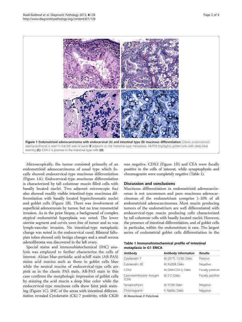
Figure 1 Endometrioid adenocarcinoma with endocervical (A) and intestinal type (B) mucinous differentiation. Classic endometrioidadenocarcinoma is seen in the left side of panel B adjacent to the intestinal-type metaplasia. AB-PAS highlights goblet cells with deep bluestaining (C). CDX-2 is positive in the intestinal type cells (D).
Table 1 Immunohistochemical profile of intestinalmetaplasia in G1 EMCA
Antibody Antibody information Results
Cytokeratin 7 M; OV-TL 12/30; Dako Positive
Cytokeratin 20 M; Ks20.8; Dako Negative
CDX2 M; DAK-CDX-2; Dako Focally positive
Carcinoembryonic Antigen(CEA)
M; II-7; Dako Focally positive
Synaptophysin M; SY38; Dako Negative
Chromogranin P; Rabbit; Dako Negative
M: Monoclonal, P: Polyclonal.
Buell-Gutbrod et al. Diagnostic Pathology 2013, 8:128 Page 2 of 4http://www.diagnosticpathology.org/content/8/1/128
Microscopically, the tumor consisted primarily of anendometrioid adenocarcinoma of usual type which fo-cally showed endocervical-type mucinous differentiation(Figure 1A). Endocervical-type mucinous differentiationis characterized by tall columnar mucin filled cells withbasally located nuclei. Two adjacent microscopic focialso showed readily visible intestinal-type mucinous dif-ferentiation with basally located hyperchromatic nucleiand goblet cells (Figure 1B). There was involvement ofsuperficial adenomyosis by tumor, but no true myometrialinvasion. As in the prior biopsy, a background of complexatypical endometrial hyperplasia was noted. The loweruterine segment and cervix were free of tumor and so waslymph-vascular invasion. No intestinal-type metaplasticchange was noted in the endocervical canal. Bilateral fallo-pian tubes showed only benign changes and a small serousadenofibroma was discovered in the left ovary.Special stains and immunohistochemical (IHC) ana-
lysis was employed to further characterize the cells ofinterest. Alcian blue-perioidic acid-schiff stain (AB-PAS)stains acid mucins such as those in goblet cells bluewhile the neutral mucins of endocervical-type cells arepink as in the classic PAS stain. AB-PAS stain in thiscase confirms the morphologic impression of goblet cellsby staining the acid mucin a deep blue color while theendocervical-type mucinous cells show faint pink stain-ing (Figure 1C). IHC of the areas with intestinal differen-tiation revealed Cytokeratin (CK) 7 positivity, while CK20
was negative. CDX2 (Figure 1D) and CEA were focallypositive in the cells of interest, while synaptophysin andchromogranin were completely negative (Table 1).
Discussion and conclusionsMucinous differentiation in endometrioid adenocarcin-omas is not uncommon and pure mucinous adenocar-cinomas of the endometrium comprise 1-10% of allendometrioid adenocarcinomas. Most mucin producingtumors of the endometrium are well differentiated withendocervical-type mucin producing cells characterizedby tall columnar cells with basally located nuclei. However,the presence of intestinal differentiation, and of goblet cellsin particular, within the endometrium is rare. The largestseries of endometrial goblet cells differentiation in the
Buell-Gutbrod et al. Diagnostic Pathology 2013, 8:128 Page 3 of 4http://www.diagnosticpathology.org/content/8/1/128
world literature reports only two cases, which were iden-tified with the assistance of special stains [1]. Two casesof intestinal metaplasia, one in an endometrial polyp andone in association with complex hyperplasia withoutatypia, have recently been reported [2]. Neither was asso-ciated with endometrial carcinoma, in contrasts to ours.One was associated with concurrent intestinal metaplasiaof the endocervical canal. Two cases of endometrialadenocarcinomas with signet ring cells have been reportedin the literature [6] and an additional case of mucinousadenocarcinoma of the endometrium was found to have agastric phenotype on immunohistochemistry mimicking aminimal deviation adenocarcinoma of the cervix [7]. As inthis case, all were associated with more typical appearingendometrioid adenocarcinoma. Enteric-type mucin hasbeen identified in endometrial adenocarcinomas, a findingwhich verifies the ability of the Mullerian epithelium toundergo metaplasia along an intestinal lineage [8]. Nocases in that series, however, showed morphologic intes-tinal differentiation and there was no correlation withtumor grade.The presence of goblet cell within the endometrium
raises the legitimate question for metastasis. Bland well-differentiated intestinal-type mucinous epithelium withgoblet cell in the endometrium, endocervix, and fallo-pian tubes, has been shown to be metastatic lesions fromgastrointestinal primaries [9]. Clinical evaluation foran occult primary gastrointestinal lesion as well as carefulgross and histologic exam is necessary. Cytokeratins areuseful in this setting as metaplastic Mullerian epitheliumretains CK7 positivity, as in our case. In this current case,the patient had no clinical gastrointestinal lesion. Thegross exam was that of a classic non-invasive carcin-oma arising from endometrial. No involvement of theadnexa, serosa, or the myometrium by the tumor wasnoted and the cells with intestinal differentiation wereintimately associated and readily visible with the endo-metrioid carcinoma.Intestinal-type metaplasia is more common within the
endocervix, with reports of cases in non-neoplastic andendocervical adenocarcinoma [1,10]. Intestinal metapla-sia in the endocervix is associated with both adenocarcin-oma in-situ and endocervical adenocarcinoma and thepresence of intestinal-type cells on cervical biopsy war-rants further workup.The etiology of intestinal-type differentiation within
the endometrium is unclear. The two recently reportedcases associated with non-neoplastic lesions were as-sociated with probable hyperestrogenism [2], a find-ing replicated in this patient who was obese and hada background of endometrial hyperplasia. This is incontrast to the proposed mechanism of endocervical-typemucinous metaplasia which has been linked to use of tam-oxifen and exogenous progestin [11,12].
With the addition of our case, the number of cases ofintestinal-type differentiation with goblet cells in theendometrium is increased to five, with two cases beingbenign and two cases associated with adenocarcinomaonly detected with special stains [1,2]. This entity is ex-tremely rare, this being the first case encountered in oursub-specialty practice and consult service.The clinical implication of finding intestinal-type dif-
ferentiation in endometrial carcinoma, as in our casereport, remains in the fact that the pathologists and cli-nicians needed to be aware that such differentiation mayoccur in primary endometrial carcinoma and that thefinding of intestinal-type mucinous differentiation inendometrial carcinoma should also warrant careful clinico-pathologic work-up to exclude a metastatic carcinomafrom the gastro-intestinal tract.
ConsentThe patient has given written consent for the use of theimages and case presentation for educational and scien-tific purposes provided the unique personal identifica-tion is not revealed.
Competing interestsThe authors declare no competing financial interest. All the authors haveactively participated in the diagnosis process or manuscript writing.
Authors’ contributionsRBG is the Stuart Lauchlan Fellow in Gynecologic and Breast Pathology andhas written up the case report and MRQ is the attending Pathologist on thecase. CJS and WDL were involved in interpreting the special stains. Allauthors read and approved the final manuscript.
Received: 28 February 2013 Accepted: 21 June 2013Published: 2 August 2013
References1. Nieuwenhuizen L, Khalil MK, Naik VR, Othman NH: Prevalence of goblet
cell metaplasia in endocervical and endometrial adenocarcinoma: ahistochemical study. Malays J Med Sci 2007, 14:56–61.
2. Nicolae A, Goyenaga P, McCluggage WG, Preda O, Nogales FF: Endometrialintestinal metaplasia: a report of two cases, including one associatedwith cervical intestinal and pyloric metaplasia. Int J Gynecol Pathol 2011,30:492–496.
3. Yasuda M, Katoh T, Hori S, et al: Endometrial intraepithelial carcinoma inassociation with polyp: review of eight cases. Diagn Pathol 2013, 8:25.
4. Ribeiro-Silva A: Immunohistohemical features of a papillary squamouscell carcinoma of the endomertrium with transitional cell differentiation.Diagn Pathol 2007, 2:26.
5. Uehara K, Yasuda M, Ichimura T, et al: Peritoneal granuloma associatedwith endometrial adenocarcinoma of the uterine corpus. Diagn Pathol2011, 6:106.
6. Boyd C, Cameron I, McCluggage WG: Endometrial adenocarcinoma withsignet ring cells: report of two cases of an extremely rare phenomenon.Int J Gynecol Pathol 2010, 29:579–582.
7. Abiko K, Baba T, Ogawa M, Mikami Y, Koyama T, Mandai M, Konishi I: Minimaldeviation mucinous adenocarcinoma (‘adenoma malignum’) of the uterinecorpus. Pathol Int 2010, 60:42–47.
8. McCluggage WG, Roberts N, Bharucha H: Enteric differentiation inendometrial adenocarcinomas: a mucin histochemical study. Int J GynecolPathol 1995, 14:250–254.
9. Moore WF, Bentley RC, Kim KR, Olatidoye B, Gray SR, Robboy SJ: Goblet-cellmucinous epithelium lining the endometrium and endocervix: evidence
Buell-Gutbrod et al. Diagnostic Pathology 2013, 8:128 Page 4 of 4http://www.diagnosticpathology.org/content/8/1/128
of metastasis from an appendiceal primary tumor through the use ofcytokeratin-7 and −20 immunostains. Int J Gynecol Pathol 1998, 17:363–367.
10. Park KJ, Bramlage MP, Ellenson LH, Pirog EC: Immunoprofile ofadenocarcinomas of the endometrium, endocervix, and ovary withmucinous differentiation. Appl Immunohistochem Mol Morphol 2009, 17:8–11.
11. Clement PB, Young RH: Endometrioid carcinoma of the uterine corpus: areview of its pathology with emphasis on recent advances andproblematic aspects. Adv Anat Pathol 2002, 9:145–184.
12. Dallenbach-Hellweg G, Hahn U: Mucinous and clear cell adenocarcinomasof the endometrium in patients receiving antiestrogens (tamoxifen) andgestagens. Am J Pathol 1995, 14:7–15.
doi:10.1186/1746-1596-8-128Cite this article as: Buell-Gutbrod et al.: Endometrioid adenocarcinomawith simultaneous endocervical and intestinal-type mucinousdifferentiation: report of a rare phenomenon and theimmunohistochemical profile. Diagnostic Pathology 2013 8:128.
Submit your next manuscript to BioMed Centraland take full advantage of:
• Convenient online submission
• Thorough peer review
• No space constraints or color figure charges
• Immediate publication on acceptance
• Inclusion in PubMed, CAS, Scopus and Google Scholar
• Research which is freely available for redistribution
Submit your manuscript at www.biomedcentral.com/submit