Robert C Basner, MD1 Antoinette S Gomes, MD5 Eric Hoffman ...
19
Pulmonary Hyperinflation and Left Ventricular Mass Benjamin M Smith, MD MS 1,2 , Steven M. Kawut, MD MS 3 , David A Bluemke, MD PhD 4 , Robert C Basner, MD 1 , Antoinette S Gomes, MD 5 , Eric Hoffman, PhD 6 , Ravi Kalhan, MD MS 7 , João AC Lima, MD 8 , Chia-Ying Liu, PhD 9 , Erin D Michos, MD MHS 8 , Martin R Prince, MD PhD 10 , LeRoy Rabbani, MD 1 , Daniel Rabinowitz, PhD 11 , Daichi Shimbo, MD 1 , Steven Shea, MD MS 1,12 , and R Graham Barr DrPH, MD 1,12 1 Department of Medicine, College of Physicians and Surgeons, Columbia University, New York, NY 2 Department of Medicine, McGill University Health Center, Montreal, Canada 3 Department of Medicine, Perelman School of Medicine, University of Pennsylvania, Philadelphia, PA 4 Radiology and Imaging Sciences, National Institutes of Health, Bethesda, MD 5 David Geffen UCLA School of Medicine, Los Angeles, CA 6 Department of Radiology, University of Iowa Carver College of Medicine, Iowa City, IA 7 Asthma and COPD Program, Division of Pulmonary and Critical Care, Northwestern University Feinberg School of Medicine, Johns Hopkins University, Baltimore, MD 8 Department of Medicine, Johns Hopkins University, Baltimore, MD 9 Department of Radiology, Johns Hopkins University, Baltimore, MD 10 Department of Radiology, College of Physicians and Surgeons, Columbia University, New York, NY 11 Department of Statistics, Columbia University, New York, NY 12 Department of Epidemiology, Mailman School of Public Health, Columbia University, New York, NY Correspondence: R Graham Barr, MD DrPH Columbia University Medical Center, Presbyterian Hospital 9 East Room 105 630 West 168 th St, New York, NY, 10032 Phone: 212.305.4895 Fax: 212.305.9349 [email protected]. Conflict of Interest Disclosures: RCB has received research grants from the ALS Association, Muscular Dystrophy Association, NIH/NHLBI, Will Rogers Respiratory Institute, and honoraria from UpToDate. EH has received research grants from the Alpha-1 Foundation, American Lung Association, Roche Pharmaceuticals, NIH/NHLBI, and has ownership interest in VIDA Diagnostics as a founder and shareholder. RK has received research grants from Boehringer Ingelheim, speakers’ bureau compensation from Forest laboratories, and consultation compensation from Forest laboratories, Boehringer Ingelheim and Elevation pharmaceuticals.SS has received research grants from the American Cancer Society, Health Resources and Services Administration, National Center for Minority Health and Health Disparities, and NIH/ NHLBI. RGB has received research grants from the Alpha-1 Foundation, NIH/NHLBI, US-EPA, and research support from Cenestra Health. The other authors report no disclosures. Publisher's Disclaimer: This is a PDF file of an unedited manuscript that has been accepted for publication. As a service to our customers we are providing this early version of the manuscript. The manuscript will undergo copyediting, typesetting, and review of the resulting proof before it is published in its final citable form. Please note that during the production process errors may be discovered which could affect the content, and all legal disclaimers that apply to the journal pertain. NIH Public Access Author Manuscript Circulation. Author manuscript; available in PMC 2014 May 12. Published in final edited form as: Circulation. 2013 April 9; 127(14): 1503–1511e6. doi:10.1161/CIRCULATIONAHA.113.001653. NIH-PA Author Manuscript NIH-PA Author Manuscript NIH-PA Author Manuscript
Transcript of Robert C Basner, MD1 Antoinette S Gomes, MD5 Eric Hoffman ...

Pulmonary Hyperinflation and Left Ventricular Mass
Benjamin M Smith, MD MS1,2, Steven M. Kawut, MD MS3, David A Bluemke, MD PhD4,Robert C Basner, MD1, Antoinette S Gomes, MD5, Eric Hoffman, PhD6, Ravi Kalhan, MDMS7, João AC Lima, MD8, Chia-Ying Liu, PhD9, Erin D Michos, MD MHS8, Martin R Prince,MD PhD10, LeRoy Rabbani, MD1, Daniel Rabinowitz, PhD11, Daichi Shimbo, MD1, StevenShea, MD MS1,12, and R Graham Barr DrPH, MD1,12
1Department of Medicine, College of Physicians and Surgeons, Columbia University, New York,NY
2Department of Medicine, McGill University Health Center, Montreal, Canada
3Department of Medicine, Perelman School of Medicine, University of Pennsylvania, Philadelphia,PA
4Radiology and Imaging Sciences, National Institutes of Health, Bethesda, MD
5 David Geffen UCLA School of Medicine, Los Angeles, CA
6Department of Radiology, University of Iowa Carver College of Medicine, Iowa City, IA
7Asthma and COPD Program, Division of Pulmonary and Critical Care, Northwestern UniversityFeinberg School of Medicine, Johns Hopkins University, Baltimore, MD
8Department of Medicine, Johns Hopkins University, Baltimore, MD
9Department of Radiology, Johns Hopkins University, Baltimore, MD
10Department of Radiology, College of Physicians and Surgeons, Columbia University, New York,NY
11Department of Statistics, Columbia University, New York, NY
12Department of Epidemiology, Mailman School of Public Health, Columbia University, New York,NY
Correspondence: R Graham Barr, MD DrPH Columbia University Medical Center, Presbyterian Hospital 9 East Room 105 630 West168th St, New York, NY, 10032 Phone: 212.305.4895 Fax: 212.305.9349 [email protected].
Conflict of Interest Disclosures:RCB has received research grants from the ALS Association, Muscular Dystrophy Association, NIH/NHLBI, Will Rogers RespiratoryInstitute, and honoraria from UpToDate. EH has received research grants from the Alpha-1 Foundation, American Lung Association,Roche Pharmaceuticals, NIH/NHLBI, and has ownership interest in VIDA Diagnostics as a founder and shareholder. RK has receivedresearch grants from Boehringer Ingelheim, speakers’ bureau compensation from Forest laboratories, and consultation compensationfrom Forest laboratories, Boehringer Ingelheim and Elevation pharmaceuticals.SS has received research grants from the AmericanCancer Society, Health Resources and Services Administration, National Center for Minority Health and Health Disparities, and NIH/NHLBI. RGB has received research grants from the Alpha-1 Foundation, NIH/NHLBI, US-EPA, and research support from CenestraHealth. The other authors report no disclosures.
Publisher's Disclaimer: This is a PDF file of an unedited manuscript that has been accepted for publication. As a service to ourcustomers we are providing this early version of the manuscript. The manuscript will undergo copyediting, typesetting, and review ofthe resulting proof before it is published in its final citable form. Please note that during the production process errors may bediscovered which could affect the content, and all legal disclaimers that apply to the journal pertain.
NIH Public AccessAuthor ManuscriptCirculation. Author manuscript; available in PMC 2014 May 12.
Published in final edited form as:Circulation. 2013 April 9; 127(14): 1503–1511e6. doi:10.1161/CIRCULATIONAHA.113.001653.
NIH
-PA
Author M
anuscriptN
IH-P
A A
uthor Manuscript
NIH
-PA
Author M
anuscript

Abstract
Background—Left ventricular (LV) mass is an important predictor of heart failure and
cardiovascular mortality, yet determinants of LV mass are incompletely understood. Pulmonary
hyperinflation in chronic obstructive pulmonary disease (COPD) may contribute to changes in
intrathoracic pressure that increase LV wall stress. We therefore hypothesized that residual lung
volume in COPD would be associated with greater LV mass.
Methods and results—The Multi-Ethnic Study of Atherosclerosis (MESA) COPD Study
recruited smokers aged 50–79 years who were free of clinical cardiovascular disease. LV mass
was measured by cardiac magnetic resonance. Pulmonary function testing was performed
according to guidelines. Regression models were used to adjust for age, sex, body size, blood
pressure and other cardiac risk factors.
Among 119 MESA COPD Study participants, mean age was 69±6 years, 55% were male and 65%
had COPD, mostly of mild or moderate severity. Mean LV mass was 128±34 grams. Residual
lung volume was independently associated with greater LV mass (7.2 grams per standard
deviation increase in residual volume; 95% CI 2.2 to 12; P=0.004), and was similar in magnitude
to that of systolic blood pressure (7.6 grams per standard deviation increase in systolic blood
pressure, 95% CI 4.3 to 11 grams; p<0.001). Similar results were observed for LV mass to end-
diastolic volume ratio (p=0.02) and with hyperinflation measured as residual volume to total lung
capacity ratio (P=0.009).
Conclusions—Pulmonary hyperinflation, as measured by residual lung volume or residual lung
volume to total lung capacity ratio, is associated with greater LV mass.
Keywords
Left ventricular mass; hyperinflation; chronic obstructive pulmonary disease
INTRODUCTION
Heart disease and chronic obstructive pulmonary disease (COPD) are leading causes of
mortality in the United States.1 These two common diseases often co-exist: for example,
approximately 35% of patients hospitalized for heart failure have COPD when tested
systematically, and clinical or subclinical cardiovascular disease is increased in COPD
independent of shared risk factors.2-4 The physiologic mechanisms underlying this
association remain incompletely understood.
Left ventricular (LV) mass predicts incident cardiovascular events, including heart failure,
sudden death and cardiovascular mortality,5-7 and regression of LV mass from afterload
reducing therapies is associated with improved cardiovascular outcomes.8-10 Early autopsy
and ventriculography studies reported increased LV mass and wall thickness in the presence
of obstructive lung disease,11-13 subsequent studies, however, produced conflicting results
and none assessed the role of pulmonary hyperinflation.14-18
COPD is a heterogeneous disorder defined by persistent airflow limitation that arises from
increased airways resistance (e.g., airway narrowing) and loss of lung elastic recoil (e.g.,
Smith et al. Page 2
Circulation. Author manuscript; available in PMC 2014 May 12.
NIH
-PA
Author M
anuscriptN
IH-P
A A
uthor Manuscript
NIH
-PA
Author M
anuscript

emphysema).19-20 Pulmonary hyperinflation occurs in COPD and other obstructive lung
diseases due in part to impaired expiratory airflow.19, 21 Breathing at the resultant increased
lung volume requires more negative inspiratory pleural pressure, the magnitude of which
can be large (e.g., 4-10 mmHg at rest and 13-16 mmHg on exercise).22-26 On expiration,
airway pressures in obstructive lung disease increase, but to a lesser extent when compared
to inspiration (e.g., 1.6 to 3.2 mmHg).23-24 Indeed, a study of reversible airways obstruction
demonstrated mean pleural pressure over the entire respiratory cycle to be more negative
during exacerbation (−16 mmHg in exacerbation versus −5 mmHg in controls).26 Inspiratory
maneuvers generating negative pleural pressure have been shown to alter juxtacardiac
pressure resulting in increased LV transmural pressure load which augments LV wall
stress.27-31 Although it is well-known that chronic exposure to increased LV wall stress
results in LV hypertrophy,32 studies assessing the relationship of pulmonary hyperinflation
to LV mass are lacking.
We hypothesized that residual lung volume, a standard clinical measure of hyperinflation on
body plethysmography,19 would be independently associated with greater LV mass on
cardiac magnetic resonance (MR) in COPD.
METHODS
Study participants
The multicenter Multi-Ethnic Study of Atherosclerosis (MESA) COPD Study recruited
unmatched cases of COPD and controls from MESA, a population-based prospective cohort
study of subclinical atherosclerosis,33 and the Emphysema and Cancer Action Project
(EMCAP),34 a separate, non-overlapping lung cancer screening study, in addition to a small
number from the local outpatient community. Included participants were 50–79 years of age
with ≥10 pack-year smoking history. Exclusion criteria were clinical cardiovascular disease
(physician diagnosis of myocardial infarction, angina, heart failure, valve disease, atrial
fibrillation or stroke), stage IIIb-V chronic kidney disease, asthma prior to age 45 years,
prior lung resection, cancer, allergy to gadolinium, claustrophobia, metal in the body and
pregnancy. The current report describes the 119 participants recruited from EMCAP and the
outpatient community at one site, Columbia University Medical Center, for whom body
plethysmography was performed. Ninety of these participants were recruited from EMCAP
and 29 from the local outpatient community (Figure 1), the latter via flyers and local
physicians.
Pulmonary Function Testing
Body plethysmography and post-bronchodilator spirometry were assessed using a V6200
Series Autobox (Sensormedics, Yorba Linda, CA) and an OMI rolling barrel spirometer
following American Thoracic Society/European Respiratory Society (ATS/ERS)
recommendations35-36 and as previously described.37 Functional residual capacity was
measured while panting at a frequency of 0.5–1.0 Hz. At least 2 technically satisfactory
maneuvers were performed, followed by a linked inspiratory capacity maneuver, and slow
vital capacity maneuver. Functional residual capacity was reported as the mean of the
satisfactory measurements. Plethysmographic total lung capacity was calculated as the sum
Smith et al. Page 3
Circulation. Author manuscript; available in PMC 2014 May 12.
NIH
-PA
Author M
anuscriptN
IH-P
A A
uthor Manuscript
NIH
-PA
Author M
anuscript

of functional residual capacity and inspiratory capacity, and residual volume as the
difference between total lung capacity and slow vital capacity, and reported in liters at body
temperature and pressure saturated. Predicted spirometry values were calculated using
Hankinson reference equations,37 and Garcia-Rio equations for lung volumes for
participants 65 years and older and Crapo equations for lung volumes for participants under
65 years.6-7 COPD status and severity were defined as per ATS/ERS COPD criteria.38
Cardiac magnetic resonance (MR) and LV mass analysis
LV mass was assessed by cardiac MR following the MESA Exam 5 protocol. Images were
obtained using a 1.5 Tesla whole-body MR system (Signa LX, GE Healthcare). Ventricular
structure and function was measured with images in short-axis orientation with 12 or more
slices using a retrospectively gated steady state free precession sequence. Imaging
parameters were: TR/TE: 5.6/1.7ms, slice thickness: 8mm, gap: 2mm, FOV: 360×360mm,
matrix: 256×192. Cine images were reconstructed at 20–35-msec intervals over the cardiac
cycle with 40 phases. Semi-automated contouring was used to determine LV mass, volumes
and ejection fraction (Cardiac Image Modeller, NZ),39 in addition to right ventricular
parameters using QMass (v7.2, Medis, The Netherlands).40 LV wall thickness was measured
from the inferoseptal and lateral walls of mid-ventricular short-axis cine images at the end-
diastolic phase.
Chest computed tomography (CT) and assessment of emphysema
Participants underwent full-lung thoracic CT on a GE 64-slice helical scanner (120 kVp,
200mAs at 0.5 seconds) with 0.75 mm slice thickness. Images were obtained at suspended
full inspiration. Image attenuation was assessed at a single reading center by trained readers
without knowledge of other participant information (VIDA Diagnostics, Coralville IA).
Percent of emphysema-like lung (also known as percent low attenuation area and hereafter
referred to as percent emphysema) was defined as the percentage of total voxels within the
lung field which fell below −950 Hounsfield units (HU).41
Anthropometry, blood pressure and other co-variates
Height and weight were measured following the MESA protocol,42 as was resting seated
blood pressure, which was measured 3 times with Dinamap model Pro 100 automated
oscillometric sphygmomanometer (Critikon, GE Healthcare, Waukesha, Wisconsin). Age,
gender and race or ethnic group were self-reported. Smoking history was assessed using
standard questionnaire items and was confirmed with plasma cotinine levels (Immulite 2000
Nicotine Metabolite Assay; Diagnostic Products Corp., Los Angeles, CA, USA).
Information on medication use was obtained by medication inventory.43 Glucose and
cholesterol were measured from blood samples after a twelve-hour fast. Hypertension was
defined according to the seventh report of the Joint National Committee on the Detection,
Evaluation, and Treatment of High Blood Pressure.44
Smith et al. Page 4
Circulation. Author manuscript; available in PMC 2014 May 12.
NIH
-PA
Author M
anuscriptN
IH-P
A A
uthor Manuscript
NIH
-PA
Author M
anuscript

Study Oversight
Study procedures were approved by the institutional review boards of the participating
institutions and by the National Heart, Lung, and Blood Institute. Written informed consent
was obtained from all participants in the MESA COPD Study.
Statistical Analysis
The cohort was stratified by percent predicted residual lung volume for descriptive purposes.
Dichotomous variables are presented as proportions and continuous variables as means with
standard deviation unless otherwise indicated. Bivariate comparisons were tested by Chi-
square, Fisher's exact or Student t-tests where appropriate. Simple linear regression was
performed for crude comparisons between LV mass and measures of pulmonary
hyperinflation.
The primary analysis of the relationship between LV mass and residual volume was
performed using multiple linear regression with adjustment for the following potential
confounders: age, gender, height, body size indexing term and race-ethnicity. The body size
indexing term is similar to body surface area but specific to LV mass and was included as a
co-variate.45 A second model adjusted for additional potential confounders including current
smoking status, systolic blood pressure, hypertension, diabetes, total cholesterol level and
lipid lowering medication use. Percent emphysema and the forced expiratory volume in the
first second (FEV1) were also included in the second model, given that emphysema on CT
scan has associated with reduced LV mass,46 and the contribution of reduced lung elastic
recoil to impaired airflow,20 respectively. To minimize the possibility of confounding by
body size, we repeated analyses for a second measure of hyperinflation, residual volume to
total lung capacity ratio. A generalized additive model with locally weighted smoothing
function was used to test for non-linearity of the relationship between residual volume and
LV mass.
As recruitment in this largely nested case-control study was based on COPD status
(presence/absence), ignoring the sampling strategy would yield non-conservative standard
errors for the association of two continuous measures, hyperinflation and LV mass.47 In an
effort to obtain unbiased, population-based effect estimates, participants were weighted on
the inverse ratio of probability of selection. Weights were computed as the ratio of case or
control prevalence in the source study population to that in the MESA COPD Study. In
primary analyses, participants recruited from the local community were assigned the same
weights as those recruited from EMCAP. Sensitivity analyses were performed without any
sample weighting, restricting the sample to participants from the EMCAP cohort only, using
an additional term for participant recruitment source and with alternate weighting for those
recruited from the local community based on the National Health and Nutrition Examination
Survey III.48 We chose a conservative analysis method in which standard errors were
computed by first summing empirical estimates of the covariance matrices of COPD-stratum
specific contributions to the gradient of the weighted sum of squares, and pre- and post-
multiplying by the matrices of mixed partial derivatives of the gradient weighted sum of
squares evaluated at the weighted least squares solutions (analogous to generalized
estimated equation estimator).47 Analyses were stratified by hypertension and COPD status,
Smith et al. Page 5
Circulation. Author manuscript; available in PMC 2014 May 12.
NIH
-PA
Author M
anuscriptN
IH-P
A A
uthor Manuscript
NIH
-PA
Author M
anuscript

were restricted to participants without physician diagnosed obstructive sleep apnea, diabetes,
bronchodilator use, systolic ejection fraction ≤50 percent or current smoking status, were
performed without terms for percent emphysema and FEV1 in the model. Additional
sensitivity analyses included terms for educational attainment, fasting plasma glucose, right
ventricular mass, right ventricular enddiastolic volume, oxygen saturation, and using
alternate metrics of body size, smoking intensity, percent emphysema and lipid profile.
RESULTS
Pulmonary Hyperinflation and LV Mass
Potential participants from EMCAP and the community who were screened and enrolled are
shown in the Figure 1. EMCAP participants who were not enrolled into the MESA COPD
Study were more obese, had greater number of pack-years of smoking and differed by race-
ethnicity compared to those in the analysis (Supplementary Appendix Table S1).
Participants enrolled in the MESA COPD Study who did not complete cardiac MR or
plethysmography were more obese, with higher blood pressure, lower lung function, and
fewer years of formal education (Supplementary Appendix Table S2).
Of the 119 participants who completed plethysmography and cardiac MR, the mean age was
69±6 years, 55% were male and the mean LV mass was 128±34 grams. Table 1 summarizes
the characteristics of the study participants stratified by quartile of percent predicted residual
lung volume. Seventy-seven (65%) of participants had COPD, mostly of mild or moderate
severity. Mean age, percent male and body size were approximately equal across quartiles
but the prevalence of current smoking, hypertension and diabetes increased. Severity of
COPD, alternate measures of hyperinflation, and percent emphysema also increased with
residual lung volume.
A significant association was observed between pulmonary hyperinflation, as measured by
residual lung volume, and LV mass (Table 2). In the fully adjusted model, a one standard
deviation increase in residual volume (0.71 L) was associated with 7.2 gram increase in LV
mass (95% CI 2.2 to 12 grams; p=0.004). By comparison, a one standard deviation increase
in systolic blood pressure (16 mmHg) was associated with 7.6 gram increase in LV mass
(95%CI 4.3 to 11 grams; p<0.001) in the same model. Figure 2 shows the fully adjusted
relationship of residual lung volume to LV mass from a smoothed regression model. There
was no evidence for non-linearity (i.e., a threshold effect) in this relationship (p=0.32). The
increase in LV mass associated with residual volume was approximately symmetric: residual
volume was associated with increases in inferoseptal and lateral wall thickness in fully
adjusted models of 0.5 and 0.4 mm, respectively, per SD unit (95% CI 0.1 to 0.8 mm and 0.0
to 0.7 mm; p=0.01 and p=0.03). In addition, residual volume was associated with greater LV
mass to end-diastolic volume ratio (Table 2) and there was no evidence of effect
modification by gender (p-interaction=0.15).
Residual volume was not associated with right ventricular mass or end-diastolic volume in
fully adjusted models (−0.2 gram change in right ventricular mass per SD increase in
residual lung volume; 95% CI −1.3 to 0.9 grams; p=0.74; 2.3 mL change in right ventricular
end-diastolic volume per SD increase in residual volume; 95% CI −5.0 to 9.6 mL; p=0.54).
Smith et al. Page 6
Circulation. Author manuscript; available in PMC 2014 May 12.
NIH
-PA
Author M
anuscriptN
IH-P
A A
uthor Manuscript
NIH
-PA
Author M
anuscript

Further, the association between residual volume and LV mass remained significant with
additional adjustment for right ventricular mass (p=0.004) or end-diastolic volume
(p=0.009).
Similar significant associations were observed in fully adjusted models for residual volume
to total lung capacity ratio with LV mass and LV mass to end-diastolic volume ratio (Table
2). There were no statistically significant relationships of functional residual capacity or
total lung capacity to these cardiac parameters (Supplementary Table S3).
Sensitivity Analyses
Among participants without hypertension a one standard deviation increase in residual
volume was associated with 7.2 gram greater LV mass (95%CI 1.9 to 12; p=0.02). The
association was also significant in unweighted analyses (8.0 gram increase in LV mass per
standard deviation increase in residual volume [95%CI 3.2 to 13 grams; p=0.001]) and
among former smokers (10 gram increase in LV mass per standard deviation increase in
residual volume [95%CI 5.3 to 15 grams; p<0.001]). Similar results were obtained in
sensitivity analyses that used alternate approaches to population weighting, alternate
adjustments for body size, smoking intensity, cardiac risk factors, percent emphysema, lung
function and resting oxygen saturation, restriction for medication use, obstructive sleep
apnea, diabetes and LV ejection fraction (Figure 3). In stratified analyses, the association
between pulmonary hyperinflation and LV mass was greater among those with COPD and
attenuated among those without COPD (Figure 3), although there was no evidence for effect
modification by COPD status (p-interaction=0.84).
DISCUSSION
Pulmonary hyperinflation, as measured by residual lung volume or residual volume to total
lung capacity ratio, was associated with greater LV mass independent of blood pressure and
other traditional cardiac risk factors among older smokers with predominantly mild-to-
moderate COPD. The magnitude of the association of residual volume to LV mass was
similar to that of systolic blood pressure.
The current report is the first, to our knowledge, to consider the relationship between
pulmonary hyperinflation and LV mass. Our findings, based on pulmonary function testing
and cardiac MR, are consistent with early autopsy studies that described LV hypertrophy in
patients with chronic obstructive lung disease.12-13 Similarly, Baum and colleagues
demonstrated LV dysfunction and increased LV wall thickness by ventriculography among
fifteen patients with very severe obstructive lung disease.11 Subsequent studies failing to
show an association between COPD and LV mass did not account for several known
determinants of LV mass or omitted morphologic (i.e. emphysema) and physiologic (i.e.
hyperinflation) derangements that often accompany COPD.14-18
LV mass predicts incident cardiovascular events.5, 7 In the present study of predominantly
mild-to-moderate COPD, hyperinflation was associated with ~5% increase in LV mass per
standard deviation increase in residual volume. In prior longitudinal studies, an increase in
LV mass of this magnitude was associated with 6% increase in all-cause mortality, 7%
Smith et al. Page 7
Circulation. Author manuscript; available in PMC 2014 May 12.
NIH
-PA
Author M
anuscriptN
IH-P
A A
uthor Manuscript
NIH
-PA
Author M
anuscript

increase in risk of cardiovascular death and 20% increase in risk of incident heart failure.5, 7
COPD increases risk of sudden death and heart failure.49-50 Increased LV mass in COPD
might contribute in part to this risk, although this hypothesis was not tested directly in the
present study. Measures of hyperinflation were also associated with LV mass to end-
diastolic volume ratio. This metric of concentric LV remodelling also predicts incident
cardiovascular events and is associated with heart failure with preserved ejection fraction,51
a condition frequently associated with COPD.2
Changes in pleural pressure influence juxtacardiac pressure.30 Further, it has been shown
that more negative pleural pressures increase LV transmural pressure, effectively
augmenting LV wall stress.27-29 The resulting increase in stroke work may represent a
mechanism by which hyperinflation is associated with increased LV mass. In support of this
proposed mechanism, prior studies have demonstrated that patients with COPD and
hyperinflation generate more negative pleural pressure on inspiration during tidal volume
breathing (e.g., 4-10 mmHg at rest and 13-16 mmHg on exercise).22-24 Applied over time
and greater on exertion, these pressure changes may explain the greater LV mass observed
with hyperinflation.
Advanced COPD is associated with right ventricular dysfunction which may have impacted
on LV structure or function via mechanisms of cardiac chamber interdependence.52 In the
present study, however, measures of hyperinflation were not associated with right
ventricular mass or end-diastolic volume. Further, additional adjustment for right ventricular
dimensions did not alter the association between hyperinflation and LV mass. Conceivably,
a mechanism by which hyperinflation increases atrial dimensions could also lead to greater
LV chamber diameter and thus, LV wall stress. While the atria were not fully imaged in this
study, prior work by Watz and colleagues has shown hyperinflation to be associated with
reduced atrial and ventricular chamber dimensions.53 We therefore believe that mechanisms
involving cardiac chamber interdependence are less likely to explain the observed
association between hyperinflation and LV mass.
Hyperinflation can be defined various ways, but generally refers to an increase in residual
volume, functional residual capacity, total lung capacity, or ratios thereof.19 In COPD,
hyperinflation can result from increases in airways resistance (e.g., airway narrowing), lung
compliance (e.g., emphysema), or both.19 In our study, residual volume had the strongest
association with LV mass and residual volume to total lung capacity ratio was also
associated with LV mass. Functional residual capacity and total lung capacity are thought to
be more sensitive to changes in lung elastic recoil from emphysema.19, 54-55 In the context
of our proposed mechanism of greater negative inspiratory pressures increasing LV
afterload, residual volume may be a better metric of hyperinflation with preserved lung
elastic recoil. Coexistent emphysema may attenuate the magnitude of these inspiratory
pressure swings by increasing lung compliance.19, 54-55 Consistent with this framework,
emphysema has been shown to be associated with reduced LV mass.46 Also, residual
volume is the first lung volume to increase in COPD.56-57 In our study of predominantly
mild-to-moderate COPD, the range of hyperinflation as measured by functional residual
capacity or total lung capacity may have been insufficient to demonstrate an association with
LV mass.
Smith et al. Page 8
Circulation. Author manuscript; available in PMC 2014 May 12.
NIH
-PA
Author M
anuscriptN
IH-P
A A
uthor Manuscript
NIH
-PA
Author M
anuscript

The present study has several limitations. First, residual confounding by hypertension or
other confounders may have contributed to the observed association. However, restricting
our analysis to participants with or without hypertension while adjusting for blood pressure
yielded similar results, as did similar analyses for diabetes and smoking status. Second, the
proposed mechanism relating hyperinflation to LV mass remains speculative, as we did not
directly measure LV transmural pressure or wall stress. The measures to demonstrate such a
mechanism are too invasive to be applied to a population-based study of participants free of
clinical cardiovascular disease with predominantly mild-to-moderate COPD. Third, we did
not formally assess patients for obstructive sleep apnea, which can also generate negative
pleural pressure and is associated with increased LV mass.58-59 However, obstructive sleep
apnea is associated with reduced static lung volumes and would therefore be expected to
weaken the association observed in this study.60 Inclusion of self-reported physician
diagnosis of obstructive sleep apnea did not alter our observation. Fourth, selection bias can
be of concern in MR studies; however, weighting based on the source population COPD
prevalence, as well as various other weighting schemes and analyses nested only within
EMCAP yielded consistent results. Fifth, the cross-sectional design prevents inference on
the direction of the association. On physiological grounds we believe the effect of
hyperinflation on LV mass is more likely than the reverse. Sixth, participants that were not
enrolled or with incomplete data differed with respect to anthropometrics, smoking history,
blood pressure and spirometry when compared to those included in analyses. This may limit
the generalizability of our findings despite efforts to obtain population-based effect
estimates. Finally, we did not measure dynamic hyperinflation. We suspect, however, that
greater intrathoracic pressure changes with dynamic hyperinflation would strengthen the
association with LV mass.
In summary, residual lung volume and residual lung volume to total lung capacity ratio were
associated with greater LV mass. Pulmonary hyperinflation in obstructive lung disease may
represent a novel and modifiable risk factor for cardiovascular disease.
Supplementary Material
Refer to Web version on PubMed Central for supplementary material.
Acknowledgments
Funding Sources This study was supported by NIH/NHLBI R01-HL093081, R01-HL077612, R01-HL075476,N01-HC95159-HC95169, K24-HL103844, Fonds de la recherche en santé Québec.
References
1. Minino AM, Murphy SL. Deaths in the United States. NCHS Data Brief. 2012:1–8. [PubMed:23050606]
2. Iversen KK, Kjaergaard J, Akkan D, Kober L, Torp-Pedersen C, Hassager C, Vestbo J, Kjoller E.Chronic obstructive pulmonary disease in patients admitted with heart failure. J Intern Med. 2008;264:361–9. [PubMed: 18537871]
3. Rutten FH, Cramer MJ, Grobbee DE, Sachs AP, Kirkels JH, Lammers JW, Hoes AW.Unrecognized heart failure in elderly patients with stable chronic obstructive pulmonary disease.Eur Heart J. 2005; 26:1887–94. [PubMed: 15860516]
Smith et al. Page 9
Circulation. Author manuscript; available in PMC 2014 May 12.
NIH
-PA
Author M
anuscriptN
IH-P
A A
uthor Manuscript
NIH
-PA
Author M
anuscript

4. Curkendall SM, DeLuise C, Jones JK, Lanes S, Stang MR, Goehring E Jr. She D. Cardiovasculardisease in patients with chronic obstructive pulmonary disease, Saskatchewan Canadacardiovascular disease in COPD patients. Ann Epidemiol. 2006; 16:63–70. [PubMed: 16039877]
5. Levy D, Garrison RJ, Savage DD, Kannel WB, Castelli WP. Prognostic implications ofechocardiographically determined left ventricular mass in the Framingham Heart Study. N Engl JMed. 1990; 322:1561–6. [PubMed: 2139921]
6. Haider AW, Larson MG, Benjamin EJ, Levy D. Increased left ventricular mass and hypertrophy areassociated with increased risk for sudden death. J Am Coll Cardiol. 1998; 32:1454–9. [PubMed:9809962]
7. Bluemke DA, Kronmal RA, Lima JA, Liu K, Olson J, Burke GL, Folsom AR. The relationship ofleft ventricular mass and geometry to incident cardiovascular events: the MESA (Multi-EthnicStudy of Atherosclerosis) study. J Am Coll Cardiol. 2008; 52:2148–55. [PubMed: 19095132]
8. Okin PM, Devereux RB, Jern S, Kjeldsen SE, Julius S, Nieminen MS, Snapinn S, Harris KE, AurupP, Edelman JM, Wedel H, Lindholm LH, Dahlof B. Regression of electrocardiographic leftventricular hypertrophy during antihypertensive treatment and the prediction of majorcardiovascular events. JAMA. 2004; 292:2343–9. [PubMed: 15547161]
9. Okin PM, Devereux RB, Harris KE, Jern S, Kjeldsen SE, Julius S, Edelman JM, Dahlof B.Regression of electrocardiographic left ventricular hypertrophy is associated with lesshospitalization for heart failure in hypertensive patients. Ann Intern Med. 2007; 147:311–9.[PubMed: 17785486]
10. Devereux RB, Wachtell K, Gerdts E, Boman K, Nieminen MS, Papademetriou V, Rokkedal J,Harris K, Aurup P, Dahlof B. Prognostic significance of left ventricular mass change duringtreatment of hypertension. JAMA. 2004; 292:2350–6. [PubMed: 15547162]
11. Baum GL, Schwartz A, Llamas R, Castillo C. Left ventricular function in chronic obstructive lungdisease. N Engl J Med. 1971; 285:361–5. [PubMed: 4326623]
12. Kohama A, Tanouchi J, Hori M, Kitabatake A, Kamada T. Pathologic involvement of the leftventricle in chronic cor pulmonale. Chest. 1990; 98:794–800. [PubMed: 2145135]
13. Fluck DC, Chandrasekar RG, Gardner FV. Left ventricular hypertrophy in chronic bronchitis. BrHeart J. 1966; 28:92–7. [PubMed: 4221900]
14. Scott KW. A clinicopathological study of fatal chronic airways obstruction. Thorax. 1976; 31:693–701. [PubMed: 138210]
15. Vonk-Noordegraaf A, Marcus JT, Holverda S, Roseboom B, Postmus PE. Early changes of cardiacstructure and function in COPD patients with mild hypoxemia. Chest. 2005; 127:1898–903.[PubMed: 15947300]
16. Jorgensen K, Muller MF, Nel J, Upton RN, Houltz E, Ricksten SE. Reduced intrathoracic bloodvolume and left and right ventricular dimensions in patients with severe emphysema: an MRIstudy. Chest. 2007; 131:1050–7. [PubMed: 17426209]
17. Funk GC, Lang I, Schenk P, Valipour A, Hartl S, Burghuber OC. Left ventricular diastolicdysfunction in patients with COPD in the presence and absence of elevated pulmonary arterialpressure. Chest. 2008; 133:1354–9. [PubMed: 18339780]
18. Anderson WJ, Lipworth BJ, Rekhraj S, Struthers AD, George J. Left Ventricular Hypertrophy inChronic Obstructive Pulmonary Disease without Hypoxaemia: The Elephant in the Room? Chest.Jul.2012 Epub. DOI: doi: 10.1378/chest.12-0775.
19. Leith DE, Brown R. Human lung volumes and the mechanisms that set them. Eur Respir J. 1999;13:468–72. [PubMed: 10065702]
20. Timmins SC, Diba C, Farrow CE, Schoeffel RE, Berend N, Salome CM, King GG. TheRelationship between Airflow Obstruction, Emphysema Extent and Small Airways Function inCOPD. Chest. 2012; 142:312–9. [PubMed: 22345381]
21. Sciurba FC. Physiologic similarities and differences between COPD and asthma. Chest. 2004;126:117S–24S. discussion 59S-61S. [PubMed: 15302772]
22. Potter WA, Olafsson S, Hyatt RE. Ventilatory mechanics and expiratory flow limitation duringexercise in patients with obstructive lung disease. J Clin Invest. 1971; 50:910–9. [PubMed:5547281]
Smith et al. Page 10
Circulation. Author manuscript; available in PMC 2014 May 12.
NIH
-PA
Author M
anuscriptN
IH-P
A A
uthor Manuscript
NIH
-PA
Author M
anuscript

23. O'Connell JM, Campbell AH. Respiratory mechanics in airways obstruction associated withinspiratory dyspnoea. Thorax. 1976; 31:669–77. [PubMed: 1013938]
24. Montes de Oca M, Rassulo J, Celli BR. Respiratory muscle and cardiopulmonary function duringexercise in very severe COPD. Am J Respir Crit Care Med. 1996; 154:1284–9. [PubMed:8912737]
25. Laveneziana P, Webb KA, Ora J, Wadell K, O'Donnell DE. Evolution of dyspnea during exercisein chronic obstructive pulmonary disease: impact of critical volume constraints. Am J Respir CritCare Med. 2011; 184:1367–73. [PubMed: 21885624]
26. Stalcup SA, Mellins RB. Mechanical forces producing pulmonary edema in acute asthma. N Engl JMed. 1977; 297:592–6. [PubMed: 887117]
27. Robotham JL, Lixfeld W, Holland L, MacGregor D, Bryan AC, Rabson J. Effects of respiration oncardiac performance. J Appl Physiol. 1978; 44:703–9. [PubMed: 649472]
28. Scharf SM, Brown R, Saunders N, Green LH. Effects of normal and loaded spontaneousinspiration on cardiovascular function. J Appl Physiol. 1979; 47:582–90. [PubMed: 533753]
29. Buda AJ, Pinsky MR, Ingels NB Jr. Daughters GT 2nd, Stinson EB, Alderman EL. Effect ofintrathoracic pressure on left ventricular performance. N Engl J Med. 1979; 301:453–9. [PubMed:460363]
30. Takata M, Mitzner W, Robotham JL. Influence of the pericardium on ventricular loading duringrespiration. J Appl Physiol. 1990; 68:1640–50. [PubMed: 2347803]
31. Fessler, HE.; Permutt, S. The thorax.. In: Roussos, C., editor. Lung biology in health and disease.2nd ed.. M. Dekker; New York: 1995. p. 1621-40.
32. Oparil S. Pathogenesis of ventricular hypertrophy. J Am Coll Cardiol. 1985; 5:57B–65B.
33. Bild DE, Bluemke DA, Burke GL, Detrano R, Diez Roux AV, Folsom AR, Greenland P, Jacob DRJr. Kronmal R, Liu K, Nelson JC, O'Leary D, Saad MF, Shea S, Szklo M, Tracy RP. Multi-ethnicstudy of atherosclerosis: objectives and design. Am J Epidemiol. 2002; 156:871–81. [PubMed:12397006]
34. Mesia-Vela S, Yeh CC, Austin JH, Dounel M, Powell CA, Reeves A, Santella RM, Stevenson L,Yankelevitz D, Barr RG. Plasma carbonyls do not correlate with lung function or computedtomography measures of lung density in older smokers. Biomarkers. 2008; 13:422–34. [PubMed:18484356]
35. Wanger J, Clausen JL, Coates A, Pedersen OF, Brusasco V, Burgos F, Casaburi R, Crapo R,Enright P, van der Grinten CP, Gustafsson P, Hankinson J, Jensen R, Johnson D, Macintyre N,McKay R, Miller MR, Navajas D, Pellegrino R, Viegi G. Standardisation of the measurement oflung volumes. Eur Respir J. 2005; 26:511–22. [PubMed: 16135736]
36. Miller MR, Hankinson J, Brusasco V, Burgos F, Casaburi R, Coates A, Crapo R, Enright P, vander Grinten CP, Gustafsson P, Jensen R, Johnson DC, MacIntyre N, McKay R, Navajas D,Pedersen OF, Pellegrino R, Viegi G, Wanger J. Standardisation of spirometry. Eur Respir J. 2005;26:319–38. [PubMed: 16055882]
37. Hankinson JL, Kawut SM, Shahar E, Smith LJ, Stukovsky KH, Barr RG. Performance ofAmerican Thoracic Society-recommended spirometry reference values in a multiethnic sample ofadults: the multi-ethnic study of atherosclerosis (MESA) lung study. Chest. 2010; 137:138–45.[PubMed: 19741060]
38. Celli BR, MacNee W. Standards for the diagnosis and treatment of patients with COPD: asummary of the ATS/ERS position paper. Eur Respir J. 2004; 23:932–46. [PubMed: 15219010]
39. Young AA, Cowan BR, Thrupp SF, Hedley WJ, Dell'Italia LJ. Left ventricular mass and volume:fast calculation with guide-point modeling on MR images. Radiology. 2000; 216:597–602.[PubMed: 10924592]
40. Kawut SM, Barr RG, Lima JA, Praestgaard A, Johnson WC, Chahal H, Ogunyankin KO, BristowMR, Kizer JR, Tandri H, Bluemke DA. Right Ventricular Structure Is Associated With the Risk ofHeart Failure and Cardiovascular Death: The Multi-Ethnic Study of Atherosclerosis (MESA)-Right Ventricle Study. Circulation. 2012; 126:1681–8. [PubMed: 22932258]
41. Gevenois PA, de Maertelaer V, De Vuyst P, Zanen J, Yernault JC. Comparison of computeddensity and macroscopic morphometry in pulmonary emphysema. Am J Respir Crit Care Med.1995; 152:653–7. [PubMed: 7633722]
Smith et al. Page 11
Circulation. Author manuscript; available in PMC 2014 May 12.
NIH
-PA
Author M
anuscriptN
IH-P
A A
uthor Manuscript
NIH
-PA
Author M
anuscript

42. [April 26, 2012] Exam 5 Field Center Procedures Manual of Operations. MESA - NHLBI. 2010.(2012, at http://www.mesa nhlbi.org/publicdocs/2011/mesae5_mopjanuary2011.pdf.)
43. Psaty BM, Lee M, Savage PJ, Rutan GH, German PS, Lyles M. Assessing the use of medicationsin the elderly: methods and initial experience in the Cardiovascular Health Study. TheCardiovascular Health Study Collaborative Research Group. J Clin Epidemiol. 1992; 45:683–92.[PubMed: 1607909]
44. Chobanian AV, Bakris GL, Black HR, Cushman WC, Green LA, Izzo JL Jr. Jones DW, MatersonBJ, Oparil S, Wright JT Jr. Roccella EJ. The Seventh Report of the Joint National Committee onPrevention, Detection, Evaluation, and Treatment of High Blood Pressure: the JNC 7 report.JAMA. 2003; 289:2560–72. [PubMed: 12748199]
45. Brumback LC, Kronmal R, Heckbert SR, Ni H, Hundley WG, Lima JA, Bluemke DA. Body sizeadjustments for left ventricular mass by cardiovascular magnetic resonance and their impact onleft ventricular hypertrophy classification. Int J Cardiovasc Imaging. 2010; 26:459–68. [PubMed:20107905]
46. Barr RG, Bluemke DA, Ahmed FS, Carr JJ, Enright PL, Hoffman EA, Jiang R, Kawut SM,Kronmal RA, Lima JA, Shahar E, Smith LJ, Watson KE. Percent emphysema, airflow obstruction,and impaired left ventricular filling. N Engl J Med. 2010; 362:217–27. [PubMed: 20089972]
47. Zeger SL, Liang KY, Albert PS. Models for longitudinal data: a generalized estimating equationapproach. Biometrics. 1988; 44:1049–60. [PubMed: 3233245]
48. National Center for Health Statistics (U.S.). National Health and Nutrition Examination Survey III,1988-94.. In: Hyattsville, Md, editor. SETS 1.22a; rev. Oct. 1997. U.S. Dept. of Health andHuman Services, Centers for Disease Control and Prevention, National Center for HealthStatistics; 1997. 1 computer laser optical disc.
49. Bilchick KC, Stukenborg GJ, Kamath S, Cheng A. Prediction of mortality in clinical practice formedicare patients undergoing defibrillator implantation for primary prevention of sudden cardiacdeath. J Am Coll Cardiol. 2012; 60:1647–55. [PubMed: 23021331]
50. Sidney S, Sorel M, Quesenberry CP Jr. DeLuise C, Lanes S, Eisner MD. COPD and incidentcardiovascular disease hospitalizations and mortality: Kaiser Permanente Medical Care Program.Chest. 2005; 128:2068–75. [PubMed: 16236856]
51. Zile MR, Gottdiener JS, Hetzel SJ, McMurray JJ, Komajda M, McKelvie R, Baicu CF, MassieBM, Carson PE. Prevalence and significance of alterations in cardiac structure and function inpatients with heart failure and a preserved ejection fraction. Circulation. 2011; 124:2491–501.[PubMed: 22064591]
52. MacNee W. Pathophysiology of cor pulmonale in chronic obstructive pulmonary disease. Part two.Am J Respir Crit Care Med. 1994; 150:1158–68. [PubMed: 7921453]
53. Watz H, Waschki B, Meyer T, Kretschmar G, Kirsten A, Claussen M, Magnussen H. Decreasingcardiac chamber sizes and associated heart dysfunction in COPD: role of hyperinflation. Chest.2010; 138:32–8. [PubMed: 20190002]
54. Petty TL, Silvers GW, Stanford RE. Mild emphysema is associated with reduced elastic recoil andincreased lung size but not with air-flow limitation. Am Rev Respir Dis. 1987; 136:867–71.[PubMed: 3662240]
55. Yip CK, Epstein H, Goldring RM. Relationship of functional residual capacity to static pulmonarymechanics in chronic obstructive pulmonary disease. Am J Med Sci. 1984; 287:3–6. [PubMed:6731476]
56. O'Donnell DE, Laveneziana P. The clinical importance of dynamic lung hyperinflation in COPD.COPD. 2006; 3:219–32. [PubMed: 17361503]
57. Dykstra BJ, Scanlon PD, Kester MM, Beck KC, Enright PL. Lung volumes in 4,774 patients withobstructive lung disease. Chest. 1999; 115:68–74. [PubMed: 9925064]
58. Martin RJ, Pennock BE, Orr WC, Sanders MH, Rogers RM. Respiratory mechanics and timingduring sleep in occlusive sleep apnea. J Appl Physiol. 1980; 48:432–7. [PubMed: 7372513]
59. Hedner J, Ejnell H, Caidahl K. Left ventricular hypertrophy independent of hypertension inpatients with obstructive sleep apnoea. J Hypertens. 1990; 8:941–6. [PubMed: 2174947]
60. Onal E, Leech JA, Lopata M. Relationship between pulmonary function and sleep-inducedrespiratory abnormalities. Chest. 1985; 87:437–41. [PubMed: 3979130]
Smith et al. Page 12
Circulation. Author manuscript; available in PMC 2014 May 12.
NIH
-PA
Author M
anuscriptN
IH-P
A A
uthor Manuscript
NIH
-PA
Author M
anuscript
Clinical Summary
Heart disease and chronic obstructive pulmonary disease (COPD) are leading causes of
mortality in the United States that often co-exist. Left ventricular (LV) mass predicts
cardiovascular events including heart failure and mortality, yet determinants of LV mass
are incompletely understood. Early physiological studies suggest pulmonary
hyperinflation in obstructive lung disease may contribute to changes in intrathoracic
pressure that alter juxtacardiac pressure and effectively increase LV afterload on
inspiration. The relationship of pulmonary hyperinflation to LV mass has never been
assessed.
The current study provides evidence, for the first time, that pulmonary hyperinflation is
strongly associated with greater LV mass in a population with predominantly mild-to-
moderate COPD, independent of blood pressure and other traditional cardiac risk factors.
The magnitude of this association was similar to that of systolic blood pressure.
Pulmonary hyperinflation was also associated with greater LV mass to end-diastolic
volume ratio, suggesting a pattern of concentric remodelling.
In summary, this study identifies a novel relationship between two common and deadly
diseases. Further investigation is required to determine if pulmonary hyperinflation in
obstructive lung disease may represent a novel and modifiable risk factor for
cardiovascular disease.
Smith et al. Page 13
Circulation. Author manuscript; available in PMC 2014 May 12.
NIH
-PA
Author M
anuscriptN
IH-P
A A
uthor Manuscript
NIH
-PA
Author M
anuscript

Figure 1.Flowchart of Study Participants in the LV Mass and Hyperinflation Analysis. Flowchart of
screening and enrollment of participants included in the analysis with body
plethysmography and cardiac MR. Abbreviations: LV denotes left ventricle, MR magnetic
resonance and CT computed tomography.
Smith et al. Page 14
Circulation. Author manuscript; available in PMC 2014 May 12.
NIH
-PA
Author M
anuscriptN
IH-P
A A
uthor Manuscript
NIH
-PA
Author M
anuscript
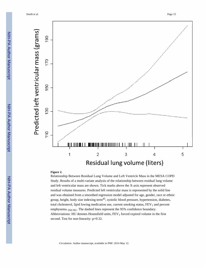
Figure 2.Relationship Between Residual Lung Volume and Left Ventricle Mass in the MESA COPD
Study. Results of a multi-variate analysis of the relationship between residual lung volume
and left ventricular mass are shown. Tick marks above the X-axis represent observed
residual volume measures. Predicted left ventricular mass is represented by the solid line
and was obtained from a smoothed regression model adjusted for age, gender, race or ethnic
group, height, body size indexing term45, systolic blood pressure, hypertension, diabetes,
total cholesterol, lipid lowing medication use, current smoking status, FEV1 and percent
emphysema−950 HU. The dashed lines represent the 95% confidence boundary.
Abbreviations: HU denotes Hounsfield units, FEV1 forced expired volume in the first
second. Test for non-linearity: p=0.32.
Smith et al. Page 15
Circulation. Author manuscript; available in PMC 2014 May 12.
NIH
-PA
Author M
anuscriptN
IH-P
A A
uthor Manuscript
NIH
-PA
Author M
anuscript

Figure 3.Mean Increment in Left Ventricle Mass by Residual Lung Volume in the MESA COPD
Study. Original model was adjusted for: age, gender, race or ethnic group, height, body size
indexing term, systolic blood pressure, hypertension, fasting plasma glucose level, diabetes,
total cholesterol, lipid lowing medication use, current smoking status, FEV1 and percent
emphysema−950 HU. Abbreviations: HU denotes Hounsfield units, FEV1 forced expired
volume in the first second, EMCAP Emphysema and Cancer Action Project, NHANES
National Health and Nutrition Examination Survey, COPD chronic obstructive pulmonary
disease, LV left ventricle and CI confidence interval.
Smith et al. Page 16
Circulation. Author manuscript; available in PMC 2014 May 12.
NIH
-PA
Author M
anuscriptN
IH-P
A A
uthor Manuscript
NIH
-PA
Author M
anuscript

NIH
-PA
Author M
anuscriptN
IH-P
A A
uthor Manuscript
NIH
-PA
Author M
anuscript
Smith et al. Page 17
Table 1
Baseline Characteristics of MESA COPD Study Participants Stratified by Quartiles of Percent Predicted
Residual Lung Volume.
Characteristic Quartiles of Percent Predicted Residual Volume
No. 1.33 L N=29 1.73 L N=30 2.09 L N=30 2.89 L N=30
Age - years 69±5 69±6 69±8 68±6
Male sex - no. (%) 17 (59) 16 (53) 15 (50) 18 (60)
Race or ethnic group - no. (%)†
Caucasian 19 (66) 23 (77) 24 (80) 23 (77)
African American 5 (17) 4 (13) 5 (17) 7 (23)
Other 5 (17) 3 (10) 1 (3) 0 (0)
Height - cm 169±11 168±9 167±10 169±9
Weight - kg 77±16 79±17 75±18 76±19
Body-surface area - m2 1.9±0.2 1.9±0.2 1.9±0.3 1.9±0.3
Cigarette smoking status - no. (%)
Former smoker 21 (72) 18 (60) 20 (67) 15 (50)
Current smoker 8 (28) 12 (40) 10 (33) 15 (50)
Pack-years of smoking - no.†
Median (IQR) 36 (27) 32 (26) 40 (30) 50 (34)
Blood pressure - mm Hg
Systolic 119±15 119±16 126±18 123±13
Diastolic 67±9 70±12 72±10 75±9
Hypertension - no. (%) 7 (24) 12 (40) 17 (57) 16 (53)
Fasting plasma glucose - mg/dl
Median (IQR) 96 (19) 104 (22) 102 (20) 98 (17)
Diabetes mellitus - no. (%) 2 (7) 4 (13) 6 (20) 6 (20)
Total Cholesterol - mg/dl 183±36 197±45 187±41 178±37
Lipid lowering med use - no. (%) 15 (52) 8 (27) 15 (50) 11 (37)
COPD - no. (%) 13 (45) 16 (53) 21 (70) 27 (90)
GOLD - COPD severity - no. (%)
Mild 9 (31) 7 (23) 11 (37) 2 (7)
Moderate 4 (14) 9 (30) 8 (27) 13 (43)
Severe / very severe 0 (0) 0 (0) 2 (7) 12 (40)
Plethysmography:
Residual volume - L 1.33±0.30 1.73±0.31 2.09±0.35 2.89±0.64
Circulation. Author manuscript; available in PMC 2014 May 12.

NIH
-PA
Author M
anuscriptN
IH-P
A A
uthor Manuscript
NIH
-PA
Author M
anuscript
Smith et al. Page 18
Characteristic Quartiles of Percent Predicted Residual Volume
FRC - L 2.94±0.75 2.99±0.67 3.41±0.71 4.04±0.80
TLC - L 5.28±1.27 5.48±1.18 5.69±1.21 6.14±1.14
Residual volume to TLC ratio 0.26±0.04 0.32±0.04 0.37±0.04 0.47±0.07
Post-bronchodilator spirometry:
FEV1 - L 2.54±0.68 2.46±0.64 2.15±0.69 1.54±0.55
FVC - L 3.63±0.97 3.61±0.92 3.47±0.95 3.14±0.86
FEV1 to FVC ratio 0.70±0.07 0.69±0.09 0.62±0.12 0.50±0.12
Percent emphysema−950 HU - median (IQR) 1.2 (2.4) 1.4 (2.4) 1.7 (3.9) 7.7 (16)
Left ventricle measures:
Mass - grams 122±29 131±39 129±34 131±35
End-diastolic volume - ml 121±26 116±26 114±23 112±36
End-systolic volume - ml 49±14 44±15 46±14 46±21
Stroke volume - ml 71±16 72±16 68±14 66±18
Ejection fraction - % 59±6 62±7 60±7 60±8
Mass to end-diastolic volume ratio 1.03±0.18 1.13±0.26 1.13±0.17 1.20±0.22
Inferoseptal wall thickness - mm 9.6±1.7 10±2.1 10±1.7 11±1.9
Lateral wall thickness - mm 7.9±1.4 8.8±1.9 8.9±1.4 8.8±1.5
Plus-minus values are means ± standard deviation.
Abbreviations: COPD denotes chronic obstructive pulmonary disease, GOLD Global Initiative for Chronic Obstructive Lung Disease, FRCfunctional residual capacity, TLC total lung capacity, FEV1 forced expired volume in the first second, FVC forced vital capacity and HUHounsfield units.
†Race or ethnic group, smoking status and pack-year history were self-reported.
Circulation. Author manuscript; available in PMC 2014 May 12.

NIH
-PA
Author M
anuscriptN
IH-P
A A
uthor Manuscript
NIH
-PA
Author M
anuscript
Smith et al. Page 19
Table 2
Mean Increment in Left Ventricle Mass According to Measures of Hyperinflation in the MESA COPD Study.
Left Ventricular Mass Quartiles of residual lung volume Mean increment in grams of LV mass per standarddeviation increase in residual lung volume (95% CI)
P Value
Mean residual volume - L 1.24 1.71 2.15 2.99
LV mass - grams 109 121 135 148 13 (7.8 to 19) <0.001
Predicted LV mass - grams
Model 1 125 128 130 134 4.2 (0.2 to 8.2) 0.035
Model 2 119 123 127 135 7.2 (2.2 to 12) 0.004
Left Ventricular Mass to End-Diastolic VolumeRatio
Quartiles of residual lung volume Mean increment in LV mass to EDVratio per standard deviation increase
in residual lung volume (95% CI)
P Value
Mean residual volume - L 1.24 1.71 2.15 2.99
LV mass to end-diastolic volume ratio 1.00 1.09 1.19 1.21 0.06 (0.02 to 0.10) 0.001
Predicted LV mass to end-diastolic volume ratio
Model 1 1.06 1.10 1.13 1.20 0.06 (0.01 to 0.11) 0.016
Model 2 1.03 1.07 1.11 1.18 0.07 (0.01 to 0.13) 0.021
Left Ventricular Mass Quartiles of residual volume to total lung capacityratio
Mean increment in gramsof LV mass per standard
deviation increase inresidual volume to total
lung capacity ratio (95%CI)
P Value
Mean residual volume to total lungcapacity ratio
0.25 0.32 0.38 0.48
LV mass - grams 124 126 136 127 2.4 (-3.4 to 8.3) 0.42
Predicted LV mass - grams
Model 1 125 128 130 133 3.1 (-0.5 to 6.7) 0.087
Model 2 119 124 128 134 5.9 (1.4 to 10) 0.009
Left Ventricular Mass to End-Diastolic Volume Ratio
Quartiles of residual volume to total lung capacityratio
Mean increment in LV massto EDV ratio per standard
deviation increase inresidual volume to total lung
capacity ratio (95% CI)
P Value
Mean residual volume to total lungcapacity ratio
0.25 0.32 0.38 0.48
LV mass to end-diastolic volumeratio
1.03 1.13 1.15 1.17 0.05 (0.02 to 0.09) 0.005
Predicted LV mass to end-diastolic volume ratio
Model 1 1.06 1.10 1.14 1.20 0.05 (0.02 to 0.09) 0.004
Model 2 1.03 1.08 1.12 1.19 0.06 (0.01 to 0.12) 0.013
Model 1 adjusted for: age, gender, race-ethnicity, height and body size indexing term45.
Model 2 additionally adjusted for: systolic blood pressure, hypertension, diabetes, total cholesterol level, lipid lowering medication use, currentsmoking status, FEV1 and percent emphysema−950 HU.
Abbreviations: LV denotes left ventricle, CI confidence interval, EDV end-diastolic volume, HU Hounsfield units, and FEV1 forced expired
volume in the first second.
Circulation. Author manuscript; available in PMC 2014 May 12.