RNA Complexity in Developing Sea Urchin Oocytes G. ERNST ... · RNA Complexity in Developing Sea...
12
DEVELOPMENTAL BIOLOGY 69.258-269 (1979) RNA Complexity in Developing Sea Urchin Oocytes BARBARA R. HOUGH-EVANS, SUSAN G. ERNST, ROY J. BRITTEN,’ AND ERIC H. DAVIDSON Division of Biology, California Institute of Technology, Pasadena, California 91125, and Kerckhoff Marine Laboratory of the Division of Biology, California Znstitute of Technology, Corona de1 Mar, California 92625 Received August 23, 1978; accepted in revised form October 6, 1978 Nuclear and cytoplasmic RNAs extracted from previtellogenic and vitellogenic oocytes of the sea urchin Strongylocentrotus purpuratus were characterized by hybridization reactions with radioactively labeled single-copy sea urchin DNA. The complexity of nuclear RNA from previtel- logenic oocytes was 1.6 x 10’ nucleotides. The previtellogenic nuclear RNA sequence set is included in the hnRNA of gastrula stage embryos. The nulcear RNA of vitellogenic oocytes may also contain a class of more prevalent transcripts. A single-copy C3H]DNA tracer enriched for the sequences of mature egg RNA was reacted with cytoplasmic RNA of previtellogenic oocytes. This experiment showed that less than half of the mature egg RNA sequence set is accumulated before the onset of vitellogenesis. Therefore, a large fraction of the maternal message sequences appears in the egg during the last several weeks of oocyte development. INTRODUCTION The mature sea urchin egg contains RNA molecules stored for use during early de- velopment (reviewed by Davidson, 1976). In Strongylocentrotuspurpuratus eggs, the complexity of the maternal messages is about 37 x lo6 nucleotides, and there are aboue 1600 copies of each mRNA sequence (Hough-Evans et al., 1977). Very little is known about the accumulation of the ma- ternal mRNA sequence set during oogene- sis. Galau et al. (1976) reported the com- plexity of polysomal RNA from whole im- mature ovaries to be 20 x lo6 nucleotides, or about half the complexity of the mature egg RNA. The total cytoplasmic RNA of such ovaries contains about three-quarters of the maternal RNA sequence set (Hough- Evans et al., 1977). Leahy et al. (1978; un- published observations) found from oocyte counts after ovarian dissociation that ova- ries of gravid S. purpuratus which contain l-2 X lo7 mature oocytes also contain about 0.5-2 x lo6 vitellogenic oocytes and about 10’ previtellogenic oocytes and oogonia. Vi- tehogenic oocytes are present in the ovaries ’ Also staff member, Carnegie Institution of Wash- ington. from a few weeks before ripe eggs first appear until just prior to the end of the gravid period. A gravid female may produce a full clutch of eggs at least four times in one season, and on each occasion she may shed over lo-fold more eggs than the steady-state content of vitellogenic oocytes in her ovaries. It follows that the vitello- genie stage is relatively short, and may last only 2-4 weeks. During the rest of the year, the ovary contains a population of around 10’ oogonia, primary oocytes, and previtel- logenic oocytes. Holland and Giese (1965) reported that premeiotic DNA synthesis was observed late in the gravid season of intertidal females. However, they were un- able to demonstrate the appearance in ma- ture oocytes of DNA labeled during pre- meiotic replication. Although this can prob- ably be attributed to difficulties in main- taining animals for a sufficiently long period of time, or to other technical problems, it cannot be excluded that primary oocytes may remain in the ovary for more than a year before advancing into vitellogenesis and maturity. Cytological examination has shown that the ovaries are covered by a peritoneal ep- 258 OOIZ-X06/79/030258-12$02.00/O Copyright 0 1979 by Academic Press, Inc. All rights of reproduction in any form reserved.
-
Upload
nguyendiep -
Category
Documents
-
view
218 -
download
0
Transcript of RNA Complexity in Developing Sea Urchin Oocytes G. ERNST ... · RNA Complexity in Developing Sea...

DEVELOPMENTAL BIOLOGY 69.258-269 (1979)
RNA Complexity in Developing Sea Urchin Oocytes
BARBARA R. HOUGH-EVANS, SUSAN G. ERNST, ROY J. BRITTEN,’ AND ERIC H. DAVIDSON
Division of Biology, California Institute of Technology, Pasadena, California 91125, and Kerckhoff Marine Laboratory of the Division of Biology, California Znstitute of Technology, Corona de1 Mar, California 92625
Received August 23, 1978; accepted in revised form October 6, 1978
Nuclear and cytoplasmic RNAs extracted from previtellogenic and vitellogenic oocytes of the sea urchin Strongylocentrotus purpuratus were characterized by hybridization reactions with radioactively labeled single-copy sea urchin DNA. The complexity of nuclear RNA from previtel- logenic oocytes was 1.6 x 10’ nucleotides. The previtellogenic nuclear RNA sequence set is included in the hnRNA of gastrula stage embryos. The nulcear RNA of vitellogenic oocytes may also contain a class of more prevalent transcripts. A single-copy C3H]DNA tracer enriched for the sequences of mature egg RNA was reacted with cytoplasmic RNA of previtellogenic oocytes. This experiment showed that less than half of the mature egg RNA sequence set is accumulated before the onset of vitellogenesis. Therefore, a large fraction of the maternal message sequences appears in the egg during the last several weeks of oocyte development.
INTRODUCTION
The mature sea urchin egg contains RNA molecules stored for use during early de- velopment (reviewed by Davidson, 1976). In Strongylocentrotuspurpuratus eggs, the complexity of the maternal messages is about 37 x lo6 nucleotides, and there are aboue 1600 copies of each mRNA sequence (Hough-Evans et al., 1977). Very little is known about the accumulation of the ma- ternal mRNA sequence set during oogene- sis. Galau et al. (1976) reported the com- plexity of polysomal RNA from whole im- mature ovaries to be 20 x lo6 nucleotides, or about half the complexity of the mature egg RNA. The total cytoplasmic RNA of such ovaries contains about three-quarters of the maternal RNA sequence set (Hough- Evans et al., 1977). Leahy et al. (1978; un- published observations) found from oocyte counts after ovarian dissociation that ova- ries of gravid S. purpuratus which contain l-2 X lo7 mature oocytes also contain about 0.5-2 x lo6 vitellogenic oocytes and about 10’ previtellogenic oocytes and oogonia. Vi- tehogenic oocytes are present in the ovaries
’ Also staff member, Carnegie Institution of Wash- ington.
from a few weeks before ripe eggs first appear until just prior to the end of the gravid period. A gravid female may produce a full clutch of eggs at least four times in one season, and on each occasion she may shed over lo-fold more eggs than the steady-state content of vitellogenic oocytes in her ovaries. It follows that the vitello- genie stage is relatively short, and may last only 2-4 weeks. During the rest of the year, the ovary contains a population of around 10’ oogonia, primary oocytes, and previtel- logenic oocytes. Holland and Giese (1965) reported that premeiotic DNA synthesis was observed late in the gravid season of intertidal females. However, they were un- able to demonstrate the appearance in ma- ture oocytes of DNA labeled during pre- meiotic replication. Although this can prob- ably be attributed to difficulties in main- taining animals for a sufficiently long period of time, or to other technical problems, it cannot be excluded that primary oocytes may remain in the ovary for more than a year before advancing into vitellogenesis and maturity.
Cytological examination has shown that the ovaries are covered by a peritoneal ep-
258
OOIZ-X06/79/030258-12$02.00/O Copyright 0 1979 by Academic Press, Inc. All rights of reproduction in any form reserved.

HOUGH-EVANS ET AL. RNA Complexit?/ in Sea C:rchin 0oc.vte.q 259
ithelium and a wall of connective tissue and smooth muscle. The inner epithelium in- cludes accessory cells (or “nutritive phago- cytes”) as well as germinal cells (Tennent and Ito, 1941; Gross et al., 1965; Holland and Giese, 1965; Anderson, 1968; Chat- lynne, 1969). Although some nongerminal cells are thus present in disaggregated prep- arations of ovaries, the majority of cells is small previtellogenic oocytes. In the first section of this paper, we report the com- plexity of the nuclear RNA of these small oocytes. We then compare their nuclear RNA sequences with the sequences present in gastrula-stage embryo nuclear RNA. Measurements are also described which show that the cytoplasmic RNA sequence set of mature eggs accumulates largely dur- ing the final period of oogenesis, and is mostly lacking in the cytoplasm of previtel- logenic oocytes.
MATERIALS AND METHODS
Previtellogenic and vitellogenic oocytes. Mature female sea urchins (Strongylocen- trotus purpuratus) were maintained in our laboratory holding system (as described by Leahy et aE., 1978). Their oocytes are clas- sified mainly by size. Transparent previtel- logenic oocytes are less than 50 pm in di- ameter; vitellogenic oocytes are 50-80 pm in diameter, and their cytoplasm is opaque due to the increasing amounts of yolk. Ma- ture oocytes are 80-100 pm in diameter (see Giudice et al., 1972; Giudice, 1973; Leahy et al., 1978). Ovaries from nonreproductive (i.e., “out of season”) animals contained only primary and small previtellogenic oo- cytes. The ovaries of animals about to enter the reproductive state contain, in addition, vitellogenic oocytes. Ovaries were sheared briefly in a Waring Blendor to disrupt the tissue and the cells, and to release nuclei. To some small extent, the nuclear RNA preparations were probably contaminated with cytoplasmic RNA from previtellogenic oocytes. Diphenylamine and phloroglucinol determinations of DNA and RNA content were carried out on sample nuclear pellets
(Burton, 1956; Dishe and Borenfreund, 1957).
Previtellogenic oocyte nuclear RNA. RNA was extracted essentially as described by Scheller et al. (1978). Ovaries from one or two sea urchins were examined by phase microscopy; only ovaries in which all of the oocytes were less than 50 pm in diameter were used. The ovaries were minced and washed in Ca”-Mg”‘-free seawater brought to pH 3 with citric acid. The minced ovaries were then stirred at room temperature in Ca”+-Mg”‘-free seawater containing 1 mg Pronase/ml for 1 hr, and the suspension of oocytes was filtered through gauze to remove ovarian mem- branes. Fifty to one hundred milliliters of homogenization buffer (2 rnJ4 MgCl, 10 rnJ4 Pipes [piperazine-N,N’-bis(2-ethane- sulfonic acid)], pH 6.5, 10 pg/ml PVS (pol- yvinyl sulfate), 1 M glucose) was added. The ovaries were sheared in an ice-cold Waring Blendor for about 1 min, or until few whole cells could be seen under the phase microscope. Nuclei were pelleted at 5000 rpm, and resuspended and lysed in 7 M urea, 50 m&f sodium acetate (pH 5.1), 10 n-&f EDTA, 15 m&f EGTA, 2% SDS (so- dium dodecyl sulfate), 10 pg/ml PVS. The solution was deproteinized at room temper- ature with an equal volume of a 1: 1 mixture of phenol:m-cresol:8-hydroxyquinoline (Kirby, 1965)-chloroform:isoamyl alcohol (24:l). After removal of the aqueous phase, to which about 2 mg/ml Bentonite was added, the interface was suspended in the same 7 M urea buffer, containing, in addi- tion, 1 M sodium perchlorate, and reex- tracted with the phenol-chloroform mix- ture. The aqueous phases were combined and extracted once with the phenol- chloroform mixture and 2x with chloro- form:isoamyl alcohol (24:1), and then pre- cipitated at -20°C with 2 vol 100% ethanol. The precipitate was dissolved in 10 mM Pipes (pH 6.5), 5 n-&f MgCli. This opales- cent solution was centrifuged at 20°C for 30-45 min at 50,000 rpm and the pellet was discarded. DNase I (Worthington) was pu-

260 DEVELOPMENTAL BIOLOGY VOLUME 69,1979
rifled by passage over a uridine-substituted agarose affinity column to remove ribonu- clease activity (Maxwell et al., 1977). The DNase was added to the supernatant to 100 pg/ml. After incubation for 2 hr at room temperature, the solution was brought to 0.1 M Tris (pH 8.0), 0.2% SDS, 50 m&f EDTA, and incubated with 50 pg proteinase K (E. Merck)/ml for 1 hr at 37°C. The solution was extracted with the phenol- chloroform mixture and with chloroform: isoamyl alcohol, and precipitated with ethanol. The RNA precipitate was dis- solved in 0.3 M sodium acetate (pH 6.5) and chromatographed on Sephadex G-100 in the same buffer. The RNA in the excluded volume of the column was precipitated with ethanol and stored at -20°C in 3 m&f so- dium acetate.
Previtellogenic oocyte cytoplasmic RNA. Ovaries containing only previtellogenic oo- cytes were removed from four sea urchins as described above for previtellogenic oo- cyte nuclear RNA. The same procedures were used to wash and shear the ovaries; then the nuclei were pelleted at 10,000 rpm for 10 min, and discarded. The supernatant was added to 5 ~017 M urea buffer. Extrac- tion and purification of supernatant RNA were carried out as just described.
Vitellogenic oocyte nuclear RNA. Fe- male sea urchins were injected with l-2 ml 0.5 M KCl, and allowed to shed mature eggs for about 30 min. The ovaries were then dissected out and monitored for the pres- ence of vitellogenic (50- to 80-pm-diameter) oocytes. These ovaries were minced and washed in Ca2+-Mg’+-free seawater, pH 3. The ovarian tissue was stirred with a mag- netic stirrer for about 1 hr, at room tem- perature, in Ca*+-Mg2+-free seawater, pH 3, causing the oocytes to be released from ovarian tissue. The oocyte suspension was poured through gauze into an ice-cold beaker and examined microscopically. Ali- quots of 5-10 ml were placed in 50-ml Nal- gene tubes and underlayered with about 10 ml 1 M sucrose. The tubes were centrifuged at 3000 rpm in a DuPont (Sorvall) HB-4
rotor for 2 min, and the solutions separated into approximately equal supernatant, in- termediate, and sucrose layers. The vitel- logenic oocytes were concentrated in the intermediate layer, while previtellogenic oocytes generally pelleted. However, the separation from previtellogenic oocytes was not complete. The oocytes in the pooled intermediate layers were sheared in the Waring Blendor, and nuclear RNA was ex- tracted as described above for previtello- genie oocytes.
Gastrula nuclear RNA. Sea urchin em- bryos were cultured for 36 hr to gastrula stage (Smith et al., 1974). Nuclei were pre- pared and RNA was extracted as described in detail by Scheller et al. (1978).
Single-copy pH]DNA. Unlabeled single- copy sea urchin DNA was prepared by har- vesting single-stranded material after two successive incubations of total DNA to Cot 200 (Graham et al., 1974; Galau et al., 1976). It was reassociated at a very high Cot to promote hyperpolymer formation, and la- beled in vitro by the gap translation method using Escherichia coli polymerase I. Labeled DNA was purified of unincor- porated precursor, and of self-complemen- tary “foldback” sequences generated during the labeling, by appropriate hydroxyapatite chromatography (Galau et al., 1976; Hough-Evans et al., 1977). The single-copy tracers produced had a fragment size of about 200-250 nucleotides and a specific activity of lo7 cpm/pg under our (40% effi- cient) counting conditions.
pH]DNA recovered from nuclear RNA hybrids. Single-copy [3H]DNA was incu- bated with excess nuclear RNA of previtel- logenic oocytes to an RNA Cot greater than 70,000. The hybridization mixture was treated with 10 pg ribonuclease A/ml in 0.24 M phosphate buffer for 1 hr at 4°C and extracted with chloroform: isoamyl al- cohol (24:1), before chromatography on hy- droxyapatite under standard conditions. The double-stranded material was eluted from the column with 0.5 M phosphate buffer, diluted to 0.05 M phosphate buffer,

HOUGH-EVANS ET AL. RNA Complex-it? in Sea Urchin Oocytes 261
and digested with 10 pg ribonuclease A/ml at 37°C for 16 hr. Under these conditions, hybridized RNA is hydrolyzed, and the [.‘H]DNA previously hybridized is left as single-stranded DNA fragments in solution. In a second passage over hydroxyapatite, this [“H]DNA is not bound. The solution containing single-stranded [“HIDNA was treated with proteinase K to remove ribo- nuclease activity, extracted with chloro- form:isoamyl alcohol (24:1), and concen- trated by precipitation with calf thymus DNA carrier.
oDNA. Preparation of oDNA, a single- copy DNA tracer enriched for oocyte RNA sequences, is described in detail by Hough- Evans et al. (1977). Single-copy [3H]DNA was incubated with excess total egg RNA (extracted from unfertilized eggs by the same procedures as previtellogenic oocyte nuclear RNA) to an equivalent Cot greater than 50,000. The hybridized sequences were further enriched by a second reaction with egg RNA, and harvested.
The oDNA used in these experiments reacted 93% at a high Cot with excess whole sea urchin DNA, and reassociated less than 7R at Cot values of lo-35 M-sec. The resid- ual zero-time binding measured 4%. The extent of reaction with egg RNA was 63%. A pseudo-first-order function with a rate constant of 10e4 Me1 set-’ fit the data from this reaction with a root mean square error of 3.6%. This rate is close to that measured for the reaction of other single-copy [“HI- DNA tracers with egg RNA (Galau et al., 1976; Hough-Evans et al., 1977). The yield of oDNA in this preparation was 10% (see Hough-Evans et al., 1977, for a detailed discussion of oDNA preparation and yield).
Hybridization of [“H]DNAs with RNA and analysis of hybrid content. Single-copy [“H]DNA and oDNA were incubated with excess unlabeled RNA in 0.4-0.5 M phos- phate buffer, 0.1% SDS, at 60°C after de- naturation for l-2 min at 98°C. RNA mass excess was 210’ for single-copy [3H]DNA reactions. In oDNA reactions, the mass ex- cess was at least lo”. RNA C,,t values (M-
set) were calculated in terms of the total RNA mass. All RNA and DNA C,,t values referred to in this paper are equizlalent Gt values; that is, they have been corrected for acceleration in reaction rate relative to the rate in 0.12 M phosphate buffer at 60°C due to higher Na’ concentrations (Britten et al., 1974).
Reaction mixtures containing RNA and total single-copy [“HIDNA were analyzed by procedures described earlier, with minor modifications (Hough et al., 1975; Hough- Evans et al., 1977; Galau et al., 1974, 1976). Reaction mixtures containing RNA and oDNA were analyzed in most cases by di- luting the sample to 0.12 M phosphate buffer, 0.06%’ SDS, and placing it directly over a hydroxyapatite column in the same buffer. Zero-time binding of 4% was sub- tracted from the total for each determina- tion. Data were reduced by nonlinear least- squares analysis assuming pseudo-first-or- der kinetics, using the computer program described by Pearson et al. (1977,.
RESULTS
Complexity of Previtellogenic Oocyte Nu- clear RNA
RNA was extracted from nuclei of ovaries which contained no oocytes >50 pm in di- ameter. This RNA was reacted in excess with a single-copy [3H]DNA tracer, and tracer hybridization was analyzed by hy- droxyapatite chromatography. The kinetics of these reactions are shown in Fig. 1. The kinetics are severalfold slower than usually observed with sea urchin nuclear RNAs, due apparently to the low fraction of the total RNA in the preparation which is het- erogeneous in sequence. The reaction has not quite terminated even at the highest RNA Cot attainable (1.2 x 10;‘). If a single pseudo-first-order kinetic component is ar- bitrarily assumed, the data of Fig. 1 show that about 13.5% of the single-copy tracer is represented in the nuclear RNA. How- ever, it is clear that this could be an under- estimate, since the kinetics could be more complex.

DEVELOPMENTAL BIOLOGY VOLUME 69,1979
20 40 60 SO 100
RNA Cot x lO-3
FIG. 1. Hybridization of single-copy r3H]DNA with excess nuclear RNA of previtellogenic oocytes. The pseudo-first-order function used to fit the data is D/DO = exp [-k Cot], where D/DO is the fraction of r3H]DNA remaining single stranded at time t, CO is the RNA concentration, and k is the pseudo-first-order rate constant. The terminal value obtained from this analysis is 13.5 + 0.9% of the r3H]DNA hybridized, with a rate k = 3.2 + 0.6 X 1O-5 Mm’ set-‘. The root mean square error (RMS) was 0.9%. The value for [3H]DNA bound was corrected in each case for reactivity with whole sea urchin DNA of the particular single-copy r3H]DNA preparation hybridized. Reactivity of these tracers varied from 80 to 95%. Three preparations of previtellogenic oocyte nuclear RNA were hybridized in separate experiments: 1 (O), 2 (A), and 3 (0). The RNA of preparation 2 was extracted from nuclei of isolated previtellogenic oocytes, and RNAs 1 and 3 from nuclei of whole ovaries containing only previtellogenic oocytes.
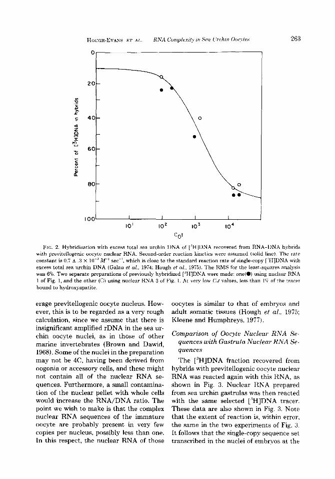
We next prepared a [3H]DNA tracer con- sisting primarily of the single-copy se- quences represented in previtellogenic oo- cyte nuclear RNA. This was accomplished hy annealing single-copy [3H]DNA with the ~-11. xlear RNA and isolating the hybridizing sbquences, as described in Materials and Methods. Figure 2 shows reactions of this r3H]DNA fraction with excess total sea ur- chin DNA. The kinetics of this reaction are as expected for a single-copy tracer, except that a few percent of the hybridized DNA could consist of repetitive sequences. How- ever, at least 85% of the reactable r3H]DNA obtained from the nuclear RNA hybrids consists of nonrepetitive sequences. There- fore, the extent of reaction in the experi- ment of Fig. 1 is a measure of the complex- ity of the previtellogenic oocyte nuclear RNA. Assuming asymmetric transcription of nuclear RNA (Hough et al., 1975), and a single-copy complexity of the Strongylo-
centrotus purpuratus genome equal to 6.1 x lo8 nucleotide pairs (Graham et al., 1974), the complexity of the nuclear RNA is at least 1.6 x lOa nucleotides. From the rate of the reaction in Fig. 1, the concentration of most of the reacting sequences can be calculated (see, e.g., Galau et al, 1974) to be about 0.5% of the total RNA in the preparation. To estimate the number of transcripts of each such sequence present per nucleus, the DNA and RNA contents of previtellogenic oocyte nuclear pellets were measured in two different prepara- tions by the diphenylamine and phloroglu- cinol procedures. These data provide an estimate of 2.2 and 7.7 pg RNA/4C nuclear amount DNA [i.e., per 3.56 pg DNA; the haploid genome contains 0.89 pg DNA (Hinegardner, 1968)]. If only 0.5% of the RNA consists of the complex reacting se- quences, there would be about 0.2-0.8 copy of each complex hnRNA sequence per av-
HOUGH-EVANS ET AL. RNA Complexity in Sea I.‘rchin 0ocyte.s 263
100~ I I I I
IO’ IO2 IO3 IO4
cot
FIG. 2. Hybridization with excess total sea urchin DNA of [“HIDNA recovered from RNA-DNA hybrids with previtellogenic oocyte nuclear RNA. Second-order reaction kinetics were assumed (solid line). The rate constant is 0.7 + .3 X 10.” W’ set-I, which is close to the standard reaction rate of single-copy [ ‘H]DNA with excess total sea urchin DNA (Galau et al., 1974; Hough et al., 1975). The RMS for the least-squares analysis was 6%. Two separate preparations of previously hybridized [‘H]DNA were made: one(O) using nuclear RNA 1 of Fig. 1, and the other (0) using nuclear RNA 3 of Fig. 1. At very low C,,t values, less than 1C; of the tracer bound to hydroxyapatite.
erage previtellogenic oocyte nucleus. How- ever, this is to be regarded as a very rough calculation, since we assume that there is insignificant amplified rDNA in the sea ur- chin oocyte nuclei, as in those of other marine invertebrates (Brown and Dawid, 1968). Some of the nuclei in the preparation may not be 4C, having been derived from oogonia or accessory cells, and these might not contain all of the nuclear RNA se- quences. Furthermore, a small contamina- tion of the nuclear pellet with whole cells would increase the RNA/DNA ratio. The point we wish to make is that the complex nuclear RNA sequences of the immature oocyte are probably present in very few copies per nucleus, possibly less than one. In this respect, the nuclear RNA of those
oocytes is similar to that of embryos and adult somatic tissues (Hough et al., 1975; Kleene and Humphreys, 1977).
Comparison of Oocyte Nuclear RNA Se- quences with Gastrula Nuclear RNA Se- quences
The [3H]DNA fraction recovered from hybrids with previtellogenic oocyte nuclear RNA was reacted again with this RNA, as shown in Fig. 3. Nuclear RNA prepared from sea urchin gastrulas was then reacted with the same selected [“HIDNA tracer. These data are also shown in Fig. 3. Note that the extent of reaction is, within error, the same in the two experiments of Fig. 3. It follows that the single-copy sequence set transcribed in the nuclei of embryos at the

264 DEVELOPMENTAL BIOLOGY VOLUME 69.1979
n 60 - E 0 0
.E
20 40 60 60
RNA Co’ x lO-3
FIG. 3. Hybridization with gastrula and previtellogenic oocyte nuclear RNAs of single-copy r3H]DNA recovered from duplexes with previtellogenic oocyte nuclear RNA. A pseudo-first-order function was used to fit data from the reaction of this [3H]DNA with previtellogenic oocyte nuclear RNA (O), and with gastrula nuclear RNA (A), with the assumptions that there is a single kinetic component and that the ordinate intercept is zero. The termination value of 53 f 2% and rate constant of 7 f 1.4 x lo-” W’ set-’ were obtained by least-squares analysis.
gastrula stage includes essentially all of the single-copy sequences transcribed in the nuclei of previtellogenic oocytes. This con- clusion, of course, does not refer to oocyte nuclear RNA sequences which are so rare that their hybridization would be detected only at an RNA Cot beyond lo5 M-sec.
Nuclear RNA of Vitellogenic Oocytes
One to two months before the appear- ance of mature eggs, vitellogenic oocytes are found in the ovaries, both in wild pop- ulations and in animals maintained over long periods in our laboratory culture sys- tem (Chatlynne, 1969; Gonor, 1973; Leahy et al., 1978). Ovaries containing vitellogenic oocytes were dissociated, and the sus- pended oocytes were centrifuged over a cushion of 1 M sucrose, as described in Materials and Methods. Most of the previ- tellogenic oocytes pelleted through the su- crose, while the larger vitellogenic oocytes
remained in the middle layer between the sucrose and the seawater. However, it was
not possible to exclude all previtellogenic oocytes. The ratio of vitellogenic oocytes to previtellogenic oocytes was increased about lo- to 20-fold from their starting ratio in the ovary, which is about 0.5-l x 10P2.
Hybridization of single-copy [3H]DNA with the nuclear RNA of the enriched vi- tellogenic oocyte preparation is shown in Fig. 4. The hybridization curve rises toward about 13% hybridization. However, the complex nuclear RNA sequence class re- sponsible for this portion of the reaction could have derived from the previtellogenic oocytes also present (Fig. 1). Thus, the total complexity of the vitellogenic oocyte nu- clear RNA per se is not established. An interesting feature of this RNA prepara- tion, however, is that it may contain a set of prevalent transcripts. This is suggested by the form of the low RNA Cd data (solid symbols) and by experiments (not shown) in which the tracer reacting at a low RNA Cot was isolated and reacted again with the enriched vitellogenic oocyte nuclear RNA preparation.

HOUGH-EVANS ET AL. RNA Complexity in Sea L’rchin Oocytes 265
t I 1 / 1 I 1 I I 1- .i 20 40 60 80
RNA Cot x lO-3
FIG. 4. Hybridization of total single-copy [“HIDNA with nuclear RNA from preparations enriched for vitellogenic oocytes. RNA of preparations A (0) and C (7) was extracted from nuclei of oocytes selected and enriched for vitellogenic oocytes as described in the text. Preparation B (A) RNA was extracted from nuclei of a total ovary homogenate, made from ovaries with a high vitellogenic oocyte content. Data from hybridizations of single-copy [“H]DNA with the three RNA preparations were analyzed assuming two pseudo-first-order kinetic components. Terminal extent of reaction was fixed at 14% (from Fig. 1). The kinetic components were 2.0% of the [3H]DNA, reacting with a rate constant set at 3.5 x 10 ’ M ’ set-‘: and 10.6% of the [‘H]DNA, with a rate constant set at 2.3 X lo-” K’ set -‘.
Accumulation of Single-Copy DNA Se- quence Transcripts in Oocytes
Hough-Evans et al. (1977) showed that most of the single-copy transcripts stored in the cytoplasm of mature sea urchin eggs are maternal messages, since the same se- quences appear on embryo polysomes at the beginning of development. In that study, embryo RNAs were hybridized with an “oDNA” (oocyte DNA) tracer highly enriched for the maternal RNA single-copy sequences set. A similar tracer was pre- pared and reacted with cytoplasmic RNA extracted from ovaries which had been ex- amined carefully and found to contain no oocytes larger than 50 pm in diameter. These ovaries were washed, minced, and sheared in a Waring Blendor. The nuclei were centrifuged out of solution at 1.5 times the force and for twice the time as in the preparation of nuclear RNA, to minimize contamination of the cytoplasmic superna- tant with nuclei or whole cells. The cyto- plasmic RNA was extracted from the post- nuclear supernatant. The oDNA tracer used in these experiments was enriched
about 25-fold for the 3% of total single-copy DNA which hybridizes with egg RNA. The tracer fraction was selected as those single- copy sequences bound to hydroxyapatite through two cycles of hybridization with egg RNA, as described in Hough-Evans et al. (1977). Reactions of the oDNA tracer with cytoplasmic RNA prepared from pre- vitellogenic oocytes are shown in Fig. 5. Less than half of the mature egg RNA sequence set can be found in the previtel- logenic oocyte cytoplasm. The oDNA does not hybridize with possible contaminating nuclear RNAs, since the reaction termi- nates at a level far lower than when oDNA is reacted with nuclear RNA (Fig. 6). Nor is there evidence of a gradual increase in hybridization at high RNA Cot values. The upper curve in Fig. 5 shows the reaction of oDNA with mature oocyte RNA (normal- ized from 59% terminal reaction to 100% ). The rate of reaction of oDNA with previ- tellogenic oocyte cytoplasmic RNA appears to be similar to that of the mature oocyte RNA. The implication is that mat,ernal mRNA sequences are present at about the

266 DEVELOPMENTAL BIOLOGY VOLUME 69,1979
100
<‘co 5
20
0
/
l 0
z’
,r __________ ~ ____ ---___- _---- ----------~---
I/’
RNA Cot x b3
FIG. 5. Reactions of oDNA with egg and oocyte RNAs. Upper curve: oDNA hybridization with total mature egg RNA. The rate constant is lo-” M-’ set-‘. Middl e 1 ine: Reaction of oDNA with total RNA of immature ovaries (from Hough-Evans et al., 1977). Lower curve: Hybridization of oDNA with cytoplasmic RNA of previtellogenic oocytes, assuming an ordinate intercept of zero. The fraction hybridized is 44 f 5% of that reacting with egg RNA; the reaction rate constant was 4 X 1O-4 M-’ set-‘. This oDNA preparation reacted 59% with egg RNA and 26% with cytoplasmic RNA of previtellogenic oocytes, after the 4% zero-time binding was subtracted. The normalized data are shown as percentages of the total reaction of oDNA with egg RNA.
IO
5 20 2 2
.E 30 a
B 6 40 s .- t
E 50
60
RNA co’
FIG. 6. Hybridization of oDNA with nuclear RNA of vitellogenic oocytes. A pseudo-first-order function was used to fit the data. The analysis showed 60 + 6% of the oDNA reacting with a rate constant of 6 x 10m5 M-’ set-‘. The oDNA reacted 59% with egg RNA.

HOUGH-EVANS ET AL. RNA Complexity in Seu Llrchzn 0ocyte.s 267
same concentrations with respect to the total stored RNA at early stages as in the mature egg. The experiment shows that most of the single-copy sequences charac- teristic of the mature egg RNA are accu- mulated later in oogenesis, during the vi- tellogenic stages of oocyte development. The level of oDNA hybridization with total RNA of immature ovaries is also repro- duced in Fig. 5, from Hough-Evans et al. (1977). This reaction amounted to 72% of the oDNA hybridization with mature egg RNA. The ovaries used in the latter exper- iment contained oocytes which were more mature than those of the present study. Apparently the storage of complex se- quences of maternal RNA in oocyte cyto- plasm is sequential, with most of the tran- scripts and most of the sequence diversity being added during vitellogenesis.
Since new maternal mRNA sequences are accumulated during vitellogenesis, it is conceivable that they are a major product of transcription during this period and are present at higher concentrations in vitello- genie oocyte nuclear RNA than are other single-copy sequence transcripts. To test this, the oDNA was reacted with the same vitellogenic oocyte-enriched nuclear RNA as used for the experiments in the preceding section. Figure 6 shows these reactions (0). The oDNA sequence set is completely represented in the nuclear RNA prepara- tion, that is, at least 60% of it reacts, which is comparable to its reaction with mature oocyte RNA. This reaction could be due in part to a low concentration of contaminat- ing cytoplasmic RNAs. Whatever the source of the RNAs reacting with the oDNA tracer, however, the kinetics of this reaction show that the concentration of transcripts complementary to oDNA (ma- ternal message sequences) is similar to that of single-copy transcripts, in general, in this nuclear RNA.
DISCUSSION
Little information has been available re- garding the transcriptional activities of sea
urchin oocytes during the long previtello- genie stages, other than autoradiographic evidence that some RNA synthesis occurs. Sconzo et al. (1972) showed that ribosomal RNA synthesis can be detected in vitello- genie oocytes of Paracentrotus licidus, and Gross et al. (1965) obtained evidence that heterogeneous repetitive sequence tran- scripts as well as ribosomal RNAs are la- beled in near-terminal (i.e., vitellogenic) oo- cytes. On the other hand, the characteris- tics of the RNA stored in the mature sea urchin egg are relatively well known (see review in Davidson, 1976). This egg con- tains a complex set of maternal messenger RNAs which has been shown to include an enormous variety of single-copy structural gene transcripts, as well as large quantities of histone mRNA. Recently, Costantini et al. (1978) found that sea urchin egg RNA also contains a specific and heterogeneous population of transcripts complementary to interspersed repetitive sequences, which is different from that of the other sea urchin tissues studied. Almost certainly, the RNAs stored in the mature egg are of major sig- nificance in the processes of early develop- ment.
As a first step in examining the transcrip- tional activities of immature oocyte nuclei, we measured the complexity of their nu- clear RNA. Figure 1 shows that the com- plexity of the previtellogenic oocyte nuclear RNA is at least 1.6 x 10” nucleotides, close to that of gastrula-stage embryo nuclear RNA (Hough et al., 1975). The experiment of Fig. 3 demonstrates that the previtello- genie oocyte nuclear RNA sequence set is totally included in the gastrula nuclear RNA sequence set. While it is clearly pos- sible that we have underestimated the com- plexity of the previtellogenic oocyte nuclear RNA (see Results), our calculations suggest that any transcripts not included in these measurements must be present at far less than one copy per immature oocyte nu- cleus. The complete inclusion of the meas- ured previtellogenic oocyte germinal vesicle RNA sequence set in the gastrula nuclear

268 DEVELOPMENTAL BIOLOGY VOLUME 69,1979
RNA sequence set must be considered in light of the fact that the cytoplasmic RNA (presumably message) sequence set of the previtellogenic oocyte and that of gastrula message are of similar complexity and over- lap significantly (Galau et al., 1976). On the other hand, it may be that nuclear RNAs of sea urchin cells in general contain largely overlapping single-copy sequence sets, re- gardless of differences in their mRNA pop- ulations (Kleene and Humphreys, 1977; Wold et al., 1978).
The mature sea urchin oocyte contains about 50-100 pg maternal messenger RNA (reviewed in Davidson, 1976). About 1600 copies of each mRNA sequence transcribed from single-copy genes are present per egg (Hough-Evans et al., 1977). The maternal mRNAs of the mature oocyte could derive from synthesis earlier in oogenesis, as in amphibians (Rosbash and Ford, 1974), al- though Davidson (1976) calculated that most of the maternal mRNA species could be synthesized within only a few hours. Our results, together with those presented ear- lier, show that this complex set of mRNAs is accumulated sequentially during oogen- esis. Less than half the mature egg sequence set is represented in previtellogenic oocyte cytoplasmic RNA. The complexity of the latter RNA is about 1.6 X lo7 nucleotides (44% of 3.7 x lo7 nucleotides, the complex- ity of mature egg RNA; from Fig. 5). We estimate that there are about 600 copies of these mature egg RNA sequences present in previtellogenic oocyte cytoplasm. Of course, it is not known whether these are stored while new copies are slowly added, or whether there is a steady-state popula- tion of polysomal messages which continues to turn over until the polysomes finally disaggregate at the end of oogenesis. In either case, the remaining 56% of the ma- ternal RNA sequence set is not loaded into the egg cytoplasm until after vitellogenesis is underway. This must be true even if all of the mature egg sequences are being tran- scribed as hnRNA in previtellogenic oocyte germinal vesicles. This seems likely from
the hybridization of oDNA with nuclear RNA of vitellogenic and previtellogenic oo- cytes, shown in Fig. 6, and from the results of Wold et al. (1978). The latter authors showed that the complete blastula mRNA sequences set is represented in adult intes- tine nuclear RNA, although 85% of it is absent from intestine cell polysomes.
The small number of copies of each com- plex maternal mRNA sequence in the egg argues against the idea that synthesis and storage of maternal message are quantita- tively a major aspect of transcription in vitellogenic oocytes. However, it appeared worthwhile to test whether the prevalent nuclear RNA in oocyte preparations en- riched for vitellogenic oocytes includes the maternal mRNA sequences. The data in Fig. 6 show that this is not the case. It follows that the maternal mRNAs are prob- ably transcribed as typical rare nuclear RNA sequences present at steady state in only a few copies per nucleus. Were this so, a rough calculation suggests that it could require as long as 20 days, the approximate minimum length of the vitellogenic period, to accumulate the 1600 copies of each sin- gle-copy sequence transcript which are present on the average in the mature egg.
This research was supported by a grant from the NIH (Child Health and Human Development). The sea urchin maintenance system was partially equipped with funds supplied by NIH Biomedical Grant RR07003, and this culture system is maintained by Grant RR00986 from the Division of Research Re- sources. S.G.E. was supported by a NIH postdoctoral fellowship.
REFERENCES
ANDERSON, E. (1968). Oocyte differentiation in the sea urchin Arbacia pun&data, with particular refer- ence to the origin of cortical granules and their participation in the cortical reaction. J. Cell Biol. 37, 514-539.
BRITTEN, R. J., GRAHAM, D. E., AND NEUFELD, B. R. (1974). Analysis of repeating DNA sequences by reassociation. In “Methods in Enzymology” (L. Grossman and K. Moldave, eds.), 29E, pp. 363-418. Academic Press, New York.
BROWN, D. D., AND DAWID, I. B. (1968). Specific gene amplification in oocytes. Science 160, 272-280.
HOLXH-EVANS ET AL. RNA Complexity in Sea L’rchin Oocytes 269
BURTON, K. (1956). A study of the conditions and mechanism of the diphenylamine reaction for the calorimetric estimation of deoxyribonucleic acid. Proc. Natl. Acad. Sci. USA 62, 315-323.
CHATLYNNE, L. G. (1969). A histochemical study of oogenesis in the sea urchin Strongylocentrotus pur- puratus. Biol. Bull 136, 167-184.
COSTANTINI, F. D., SCHELLER, R. H., BRITTEN, R. J., AND DAVIDSON, E. H. (1978). Repetitive sequence transcripts in the mature sea urchin oocyte. Cell 15, 173-187.
DAVIDSON, E. H. (1976). “Gene Activity in Early De- velopment,” 2nd ed. Academic Press, New York.
DISHE, Z., AND BORENFREUND, E. (1957). A new color reaction for the determination of aldopentose in the presence of other saccharides. Biochim. Biophys. Acta 23,639-642.
GALAU, G. A., BRITTEN, R. J., AND DAVIDSON, E. H. (1974). A measurement of the sequence complexity of polysomal messenger RNA in sea urchin embryos. Cell 2, 9-21.
GALAU, G. A., KLEIN, W. H., DAVIS, M. M., WOLD, B. J., BRITTEN, R. J., AND DAVIDSON, E. H. (1976). Structural gene sets active in embryos and adult tissues of the sea urchin. Cell 7, 487-505.
GIUDICE, G. (1973). “Developmental Biology of the Sea Urchin Embryo.” Academic Press, New York.
GIUDICE, G., SCONZO, G., BONO, A., AND ALBANESE, I. (1972). Studies on sea urchin oocytes. I. Purifica- tion and cell fractionation. Exp. Cell Res. 72,90-94.
GONOR, J. J. (1973). Reproductive cycles in Oregon populations of the Echinoid, Strongylocentrotus purpuratus (Stimpson). II. Seasonal changes in oo- cyte growth and in abundance of gametogenic stages in the ovary. J. Exp. Mar. Biol. Ecol. 12, 65-78.
GRAHAM, D. E., NEUFELD, B. R., DAVIDSON, E. H., AND BRITTEN, R. J. (1974). Interspersion of repeti- tive and nonrepetitive DNA sequences in the sea urchin genome. Cell 1, 127-137.
GROSS, P. R., MALKIN, L. I., AND HUBBARD, M. (1965). Synthesis of RNA during oogenesis in the sea ur- chin. J. Mol. Biol. 13, 463-481.
HINEGARDNER, R. T. (1968). Evolution of cellular DNA content in teleost fishes. Amer. Natur. 102, 51-f-523.
HOLLAND, N. D., AND GIESE, A. C. (1965). An auto- radiographic investigation of the gonads of the pur- ple sea urchin (Strongylocentrotus purpuratus). Biol. Bull. 128, 241-258.
HOUGH, B. R., SMITH, M. .J., BRITTEN, R. J., 4~1) DAVIDSON, E. H. 11975). Sequence complexity 01 heterogeneous nuclear RNA in sea urchin embryos. Cell 5, 291-299.
HOUGH-EVANS, B. R., WOLD, B. J., EKNST, S. G., BRITTEN, R. J., API’D DAVIDSON, E. H. (1977). Xp- pearance and persistence of maternal RNA se- quences in sea urchin development. Der,elop. Biol. 60, 258-277.
KIRBY, K. S. (1965). Isolation and characterization of ribosomal ribonucleic acid. Biochem. J. 96,266-269.
KLEENE, K. C., AND HUMPHREYS, T. (1977). Similarity of hnRNA sequences in blastula and pluteus stage sea urchin embryos. Cell 12, 143-155.
LEAHY, P. S., TUTSCHULTE, T. C., BRITTEN. K. .J., AKI) DAVIDSON, E. H. (1978). A large-scale laborator? maintenance system for gravid purple sea urchins (Strongylocentrotuspurpuratus). J. Exp. Zool. 204, 369-380.
MAXWELL, I. H., MAXWELL, F., AND HAHX. W. E. (1977). Removal of RNase activity from L)Nase b> affinity chromatography on agarose-coupled ami- nophenylphosphoryl-uridine 2’(3’) phosphate. .Vlc~ cleic Acids Res. 4, 241-246.
PEARSON, W. R., DAVIDSON, E. H., AND BRITTEN, R. J. (1977). A program for least squares analysis of reassociation and hybridization data. ,Vucleic .4ci& Res. 4, 1727-1737.
ROSBASH, M., AND FORD, P. J. (1974). Polgadenylic acid-containing RNA in Xenopus lael% oocytes. d Mol. Biol. 85, 87-101.
SCHELLER, R. H., COSTANTINI, F. D., KOXOWSKI, M. R., BRITTEN, R. J., AND DAVIDSOX, El. H. (1978). Specific representation of cloned repetitive DNA sequences in sea urchin RNAs. Cell 15, 189-20:~.
SCONZO, G., BONO, A., ALBANESE, I., AND GICLXX, G. (1972). Studies on sea urchin oocytes. II. Synthesis of RNA during oogenesis. Exp. Cell Res. 72, 95-100.
SMITH, M. J., HOLXH, B. R., CHAMBERLIN. M. E.. ,i~n DAVIDSON, E. H. (1974). Repetitive and nonrepeti- tive sequences in sea urchin hnRNA. -1. ,210l. Rio/. 85, 103-126.
TENNENT, D. H., AND ITO, T. (1941). A study of the oogenesis of Mespilia globulus (Linne). .J. &forpho/. 69, 347-404.
WOLD, B. J., KLEIN, W. H., HO~GH-EVANS. B. H., BRITTEN, R. J.. AND DAVIDSON. E. H. (1978). Sea urchin embryo mRNA sequences expressed in the nuclear RNA of adult tissues. Cell 14,94 I -950.