Revista Colombiana de Anestesiología para craneotomía en el ... Electrophysiological mapping and...
7
rev colomb anestesiol. 2015; 43(S1) :22–28 Revista Colombiana de Anestesiología Colombian Journal of Anesthesiology www.revcolanest.com.co Essay Anesthesia for awake craniotomy: An update Jason Chui ∗ Assistant Professor, Department of Anesthesia and Perioperative Medicine, University of Western Ontario, Ontario, Canada article info Article history: Received 26 May 2014 Accepted 8 July 2014 Available online 3 September 2014 Keywords: Craniotomy Epilepsy Brain neoplasms Anesthesia Perioperative period abstract Awake craniotomy has become a common procedure and its application has been continually evolving. Anesthesia for awake craniotomy poses a unique challenge to anesthe- siologists. The aims of this article are to review under a critical perspective of the author, the current evidence and application of awake craniotomy and to briefly describe the principles of anesthetic management during this procedure. © 2014 Sociedad Colombiana de Anestesiología y Reanimación. Published by Elsevier España, S.L.U. All rights reserved. Anestesia para craneotomía en el paciente despierto: una actualización Palabras clave: Craneontomía Epilepsia Neoplasias encefálicas Anesthesia Periodo perioperatorio resumen La craneotomía en el paciente despierto se ha generalizado y su aplicación ha evolucionado continuamente. La anestesia para este procedimiento plantea un reto singular para los anestesiólogos. Son revisar, bajo una perspectiva crítica del autor, la evidencia actual y la aplicación de la craneotomía en el paciente despierto, y describir brevemente los principios del manejo anestésico durante el procedimiento. © 2014 Sociedad Colombiana de Anestesiología y Reanimación. Publicado por Elsevier España, S.L.U. Todos los derechos reservados. Please cite this article as: Chui J. Anestesia para craneotomía en el paciente despierto: una actualización. Rev Colomb Anestesiol. 2015;43:22–28. ∗ Correspondence to: Department of Anesthesia and Perioperative Medicine, University of Western Ontario, 339 Windermere Road, London, ON, Canada N6A 5A5. E-mail address: [email protected] 2256-2087/© 2014 Sociedad Colombiana de Anestesiología y Reanimación. Published by Elsevier España, S.L.U. All rights reserved.
-
Upload
trinhtuyen -
Category
Documents
-
view
215 -
download
1
Transcript of Revista Colombiana de Anestesiología para craneotomía en el ... Electrophysiological mapping and...

r e v c o l o m b a n e s t e s i o l . 2 0 1 5;43(S1):22–28
Revista Colombiana de AnestesiologíaColombian Journal of Anesthesiology
www.revcolanest .com.co
Essay
Anesthesia for awake craniotomy: An update�
Jason Chui ∗
Assistant Professor, Department of Anesthesia and Perioperative Medicine, University of Western Ontario, Ontario, Canada
a r t i c l e i n f o
Article history:
Received 26 May 2014
Accepted 8 July 2014
Available online 3 September 2014
Keywords:
Craniotomy
Epilepsy
Brain neoplasms
Anesthesia
Perioperative period
a b s t r a c t
Awake craniotomy has become a common procedure and its application has been
continually evolving. Anesthesia for awake craniotomy poses a unique challenge to anesthe-
siologists. The aims of this article are to review under a critical perspective of the author, the
current evidence and application of awake craniotomy and to briefly describe the principles
of anesthetic management during this procedure.
© 2014 Sociedad Colombiana de Anestesiología y Reanimación. Published by Elsevier
España, S.L.U. All rights reserved.
Anestesia para craneotomía en el paciente despierto: una actualización
Palabras clave:
Craneontomía
Epilepsia
Neoplasias encefálicas
Anesthesia
Periodo perioperatorio
r e s u m e n
La craneotomía en el paciente despierto se ha generalizado y su aplicación ha evolucionado
continuamente. La anestesia para este procedimiento plantea un reto singular para los
anestesiólogos. Son revisar, bajo una perspectiva crítica del autor, la evidencia actual y la
aplicación de la craneotomía en el paciente despierto, y describir brevemente los principios
del manejo anestésico durante el procedimiento.
© 2014 Sociedad Colombiana de Anestesiología y Reanimación. Publicado por Elsevier
España, S.L.U. Todos los derechos reservados.
� Please cite this article as: Chui J. Anestesia para craneotomía en el paciente despierto: una actualización. Rev Colomb Anestesiol.2015;43:22–28.
∗ Correspondence to: Department of Anesthesia and Perioperative Medicine, University of Western Ontario, 339 Windermere Road, London,ON, Canada N6A 5A5.
E-mail address: [email protected]/© 2014 Sociedad Colombiana de Anestesiología y Reanimación. Published by Elsevier España, S.L.U. All rights reserved.
r e v c o l o m b a n e s t e s i o l . 2 0 1 5;43(S1):22–28 23
Table 1 – Current indications for awake craniotomy.
Indication Rationale Examples
Awake functional cortical mapping An awake and cooperative state isrequired for performing the requiredtasks
Lesions located in close proximity to theeloquent area (e.g., tumor resection andlesionectomy)Vascular lesions that supply the eloquentarea (e.g., AVM and aneurysm excision)
Electrophysiological mapping and recording Cortical or subcortical signals are easilyabolished by anesthetic agents
Intraoperative electrocorticographyDeep brain stimulation
Improving perioperative outcomes Non-functional purposes aiming atearlier discharges, reduced lengths ofstay, and reduced ICU admissions
Stereotactic brain biopsy, ventriculostomyand resection of small brain lesions
Source: Author.
Introduction
Awake craniotomy poses a unique challenge to anesthesiolo-gists, and its success is highly dependent on careful patientselection and the experience of the surgical and anesthesiateam. The modern era of awake craniotomies began morethan 50 years ago when Penfield and Andre Pasquet startedto perform awake craniotomies for epileptic foci excision.1
Historically, anesthesia for awake craniotomy was regardedas a high-risk procedure and was only performed when itwas absolutely indicated. With the improved understandingof cerebral localization and the availability of new anestheticagents, the application of awake craniotomy has becomemuch broader and safer than before. The aims of this articleare to review the current evidence and application of awakecraniotomy and to briefly describe the principles of anestheticmanagement during this procedure.
Indication for awake craniotomy
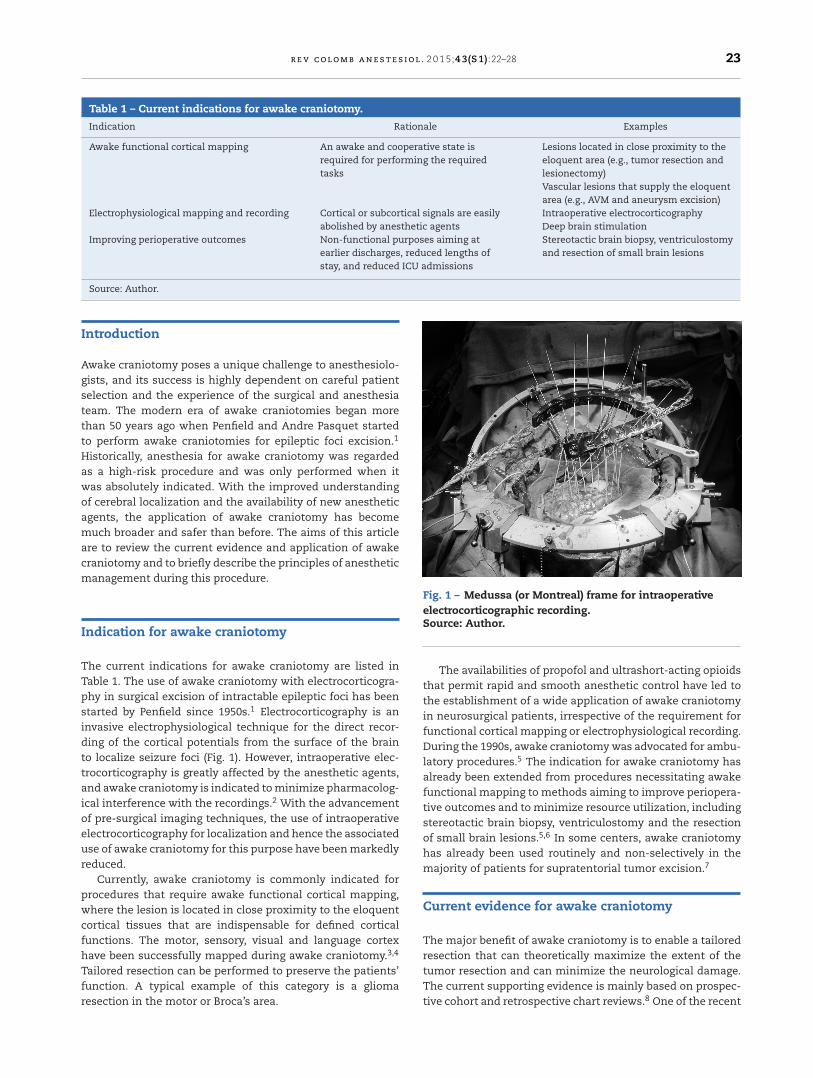
The current indications for awake craniotomy are listed inTable 1. The use of awake craniotomy with electrocorticogra-phy in surgical excision of intractable epileptic foci has beenstarted by Penfield since 1950s.1 Electrocorticography is aninvasive electrophysiological technique for the direct recor-ding of the cortical potentials from the surface of the brainto localize seizure foci (Fig. 1). However, intraoperative elec-trocorticography is greatly affected by the anesthetic agents,and awake craniotomy is indicated to minimize pharmacolog-ical interference with the recordings.2 With the advancementof pre-surgical imaging techniques, the use of intraoperativeelectrocorticography for localization and hence the associateduse of awake craniotomy for this purpose have been markedlyreduced.
Currently, awake craniotomy is commonly indicated forprocedures that require awake functional cortical mapping,where the lesion is located in close proximity to the eloquentcortical tissues that are indispensable for defined corticalfunctions. The motor, sensory, visual and language cortexhave been successfully mapped during awake craniotomy.3,4
Tailored resection can be performed to preserve the patients’function. A typical example of this category is a gliomaresection in the motor or Broca’s area.
Fig. 1 – Medussa (or Montreal) frame for intraoperativeelectrocorticographic recording.Source: Author.
The availabilities of propofol and ultrashort-acting opioidsthat permit rapid and smooth anesthetic control have led tothe establishment of a wide application of awake craniotomyin neurosurgical patients, irrespective of the requirement forfunctional cortical mapping or electrophysiological recording.During the 1990s, awake craniotomy was advocated for ambu-latory procedures.5 The indication for awake craniotomy hasalready been extended from procedures necessitating awakefunctional mapping to methods aiming to improve periopera-tive outcomes and to minimize resource utilization, includingstereotactic brain biopsy, ventriculostomy and the resectionof small brain lesions.5,6 In some centers, awake craniotomyhas already been used routinely and non-selectively in themajority of patients for supratentorial tumor excision.7
Current evidence for awake craniotomy
The major benefit of awake craniotomy is to enable a tailoredresection that can theoretically maximize the extent of thetumor resection and can minimize the neurological damage.The current supporting evidence is mainly based on prospec-tive cohort and retrospective chart reviews.8 One of the recent
24 r e v c o l o m b a n e s t e s i o l . 2 0 1 5;43(S1):22–28
large population cohort studies, involving 575 patients, hascompared the gross total resection rate and postoperativeneurological deficits between awake craniotomy and generalanesthesia patients.9 Awake craniotomy was associated witha higher gross total resection rate in the eloquent area (37% vs14%, p < 0.05), fewer permanent neurological deficit (4.6% vs16%, p < 0.001) and fewer new-onset postoperative neurologi-cal deficits (3.3% vs 58% of patients, p < 0.001). However, thesebenefits were not consistently demonstrated by other studies,particularly for the prevention of neurological deficits.8 Inter-estingly, the only available randomized controlled study onthis topic revealed that general anesthesia yielded fewer andnon-significant neurological deficits.10
The evidence supporting awake craniotomy in earlier hos-pital discharges are more consistent, and nearly all theprevious studies have demonstrated shorter hospital stays forawake craniotomy patients. The additional benefits includeshorter ICU stays, a shorter total length of stay, less resourceutilization and high patient satisfaction.7,8
Perioperative management
Preoperative preparation
Careful patient selection is one of the major components ofsuccessful awake craniotomy. The selection should be individ-ualized and based on the airway assessment, risks of sedationfailure, patient’s cooperation and the risks of intraoperativesurgical complications such as bleeding. A history of alco-hol or drug abuse, chronic pain disorders, low tolerance topain and anxiety or psychiatric disorders are known risk fac-tors for sedation failure.11 The selection of patients presentingwith seizures or mixed dysphasia, having a history of usingmultiple anti-convulsants or undergoing perioperative load-ing of phenytoin is associated with the loss of intraoperativecooperation and results in failed awake craniotomies.12 Well-motivated and mature patients are the best candidates forawake craniotomy. There was also a recent pilot study of usingobjective psychological and psychophysiological test to selecttheir patients.13 Although there are reports of the safe useof awake craniotomy in pediatric patients, the case selectionshould be more rigorous and based on the patients’ maturityand the individual risk-benefit assessment.14
Preoperative psychological preparation and rapport build-ing are the important components of preoperative prepara-tion. The preoperative discussion should include a realisticdescription of the entire procedure, the expected discom-forts, the level of cooperation desired, the tasks that mustbe performed and the possibility of adverse events. A well-conducted preoperative consultation usually can alleviate thepatient’s anxiety and improve his or her cooperation duringthe awake craniotomy.
Intraoperative management
There are three major anesthetic challenges for awakecraniotomy: (i) provision of a rapid and smooth transitionof the anesthetic depth according to the different surgicalstages, (ii) maintenance of stable cerebral hemodynamic and
cardiopulmonary function, and (iii) crisis management foran awake patient with an open cranium. The requirementregarding the depth of anesthesia varies markedly at thedifferent stages of surgery. Over-sedation may lead to apnea,hypoxemia, hypercapnia and cerebral swelling, whereasunder-sedation may result in agitation, arterial hypertensionand tachycardia. The common anesthetic goals for all neuro-surgical patients, such as the avoidance of hypercapnia andhypoxemia, adequate cerebral perfusion pressure and brainrelaxation, may not be easily achieved in awake craniotomypatients with uncontrolled airways.
Adequate local anesthesia can usually be achieved byeither using a scalp block or a regional field block. Arterialline is commonly used; but other invasive monitoring, suchas central venous lines, is not routinely required. Comfort-able positioning is mandatory because extremely limitedmovements will be possible after the head-pinning. Thedraping should always allow easy access to the patient’sface and airway. A microphone may be used to facilitatecommunication with the staff.
Choice of anesthetic techniques
Various institutions and anesthesiologists have their ownfavored techniques for awake craniotomies, including localanesthesia, conscious sedation, asleep-awake-asleep tech-niques and asleep-awake techniques. None of these tech-niques has any demonstrated superiority. In the asleep-awake-asleep techniques, the patient is under generalanesthesia, with the use of a laryngeal mask airway or endo-tracheal intubation during the positioning, head-pinning andcraniotomy. The general anesthesia is discontinued for theperiod of functional cortical mapping and intraoperative elec-trocorticography. After the complete resection, the patient isput back under general anesthesia for the skin closure.15 In theasleep-awake technique, the patient is kept awake through-out the rest of the procedure, with the flexibility of permittingadditional mapping if symptoms develop.16 By contrast, forconscious sedation, the patient is maintained with sponta-neous breathing throughout the entire procedure, and thesedatives and analgesics are titrated based on the surgicalstages.
Choice of anesthetic agents
The choice of anesthetic agents for awake craniotomy is highlydependent upon the requirement for functional cortical map-ping and intraoperative electrocorticography. Intraoperativeelectrocorticography recordings are highly affected by differ-ent anesthetics, and the choice of anesthetic agents shouldhave minimal effects on the suppression and over-activation.Complete cessation of all anesthetic agents for 20–30 min priorto the electrocorticography has been recommended; however,this recommendation may not be feasible in all cases of awakecraniotomy, and general anesthesia may even be necessary insome patients. A detailed discussion of the effects of anesthet-ics on electrocorticography is beyond the scope of this article,and readers can refer to other papers for references.4
The anesthetic goal for awake cortical mapping is to main-tain an awake and cooperative patient for performing the
r e v c o l o m b a n e s t e s i o l . 2 0 1 5;43(S1):22–28 25
Table 2 – Commonly used anesthetic agents anddosages for awake craniotomy.
Drugs Dosages
Propofol19,27 Manual infusion: 50–150 mcg/kg/minTCI effect site (sedation): 2.4–4.8 mcg/mlTCI effect site (mapping): 0.6–1.2 mcg/ml
Remifentanil19,27 Manual infusion: 0.03–0.05 mcg/kg/minTCI effect site (sedation): 2–2.8 ng/mlTCI effect site (mapping): 1.6–2 ng/ml
Fentanyl17 Bolus: 0.75 mcg/kgInfusion: 0.01 mcg/kg/min
Alfentanil17 Bolus: 0.075 mcg/kgInfusion: 0.0015 mcg/kg/min
Sulfentanil17 Bolus: 7.5 mcg/kgInfusion: 0.5 mcg/kg/min
Dexmedetomidine20,21 Bolus: 0–0.5 mcg/kgInfusion: 0.2–0.7 mcg/kg/min
Source: Author.
required tasks. The purpose of the mapping procedure isto reliably identify the cortical areas and subcortical path-ways involved in the motor, sensory, language, and cognitivefunctions. Inaccurate mapping could create a false sense ofsecurity with incorrect resection margins. Observation of thepatient’s reaction to the trans-cortical stimulation is used todefine the function of the testing area. Clonic movement signi-fies primary motor area stimulation, whereas tonic movementis more related to the premotor area. For the mapping oflanguage and cognitive functions, a fully awake and compli-ant patient is crucial for performing naming tasks and basicmental gymnastics, such as thinking of items within a cat-egory. Induced-inhibition of speech, language and cognitionshould be distinguished from non-compliance, over-sedationand focal seizures.2 During the mapping, the patient mightfeel uncomfortable and anxious due to experiencing involun-tary movements or the inhibition of voluntary movements orlanguage. The level of anesthetic depth should strike a bal-ance between the patient’s comfort and the accuracy of themapping result.
The most commonly used agents and dosages for awakecraniotomy are listed in Table 2. Propofol infusion with asupplementary opioid is the most commonly reported choicefor awake craniotomy. Propofol-only anesthesia with spon-taneously breathing patients has been described as safe.17
The addition of an opioid is a common practice to improvethe analgesic quality and reduce the hypnotic requirement. Acomparison of different types of opioids, including fentanyl,alfentanil, or sulfentanil, found no significant difference inthe operative condition, electrocorticography and stimulationtesting.18 In a comparison of continuous remifentanil infu-sion or intermittent fentanyl, both narcotics yielded similarpatient satisfaction, recall, and intraoperative complications,although fewer patients experienced reversible respiratorydepression with the use of remifentanil.19 Therefore, all nar-cotics are regarded as equally good for use during awakecraniotomy. Notably, the time of emergence from sedation forintraoperative testing with the use of remifentanil-propofol isapproximately 9 min.20
The �2 agonist dexmedetomidine has gained popularity foruse in awake craniotomy owing to its unique ano-analgesic
property, less dis-inhibition and minimal respiratory depres-sant effects. There is an increasing trend of using thecombination of propofol and dexmedetomidine to minimizedis-inhibition and to ensure speedy awakening. Dexmedeto-midine has also been demonstrated to have minimal effectson the electrocorticography and has been successfully usedin awake craniotomy for epileptic surgery.21,22 However, therehave been reports of more intense efforts being requiredto rouse the patient from dexmedetomidine sedation (i.e.,via a physical stimulus such as sternal rubbing and callingof the patient’s name). However, once roused and engaged,the patient remains able to cooperate with the cognitivetesting.23 A direct comparison between dexmedetomidineand the propofol-remifentanil combination, particularly forperforming awake cortical mapping, has yet to be evaluated.
Complications and the safe performance of awakecraniotomy
Major intraoperative complications include seizures, respi-ratory depression, air embolisms, cerebral swelling and thetrigeminocardiac reflex. The common complications, pre-ventive measures and management techniques are listed inTable 3. The overall complication rate is reported to be approx-imately 16.5%,5 with 6.4% patients failing to complete themapping procedure.12 The main causes of failure are the onsetof seizures and the loss of the patient’s cooperation due tosevere somnolence, restlessness or the development of mixeddysphasia. Failed awake craniotomies are associated with alower incidence of gross-total resections, increased postop-erative speech deterioration and a longer length of stay.12
Because intraoperative complications are not uncommon,certain preventive measures and monitoring are mandatoryfor the safe performance of awake craniotomy. In additionto all routine monitoring, monitoring of breathing both clin-ically and via end-tidal CO2 are mandatory for all patients.Optimizing the positioning and access are essential for easilycontrolling the airway, dealing with emergencies and com-municating with patients during the mapping procedure.Intubation equipment, such as laryngeal mask, endotra-cheal tube, laryngoscopy, and medications should be preparedfor emergency use. Fiber-optic bronchoscopy should also beeasily accessible in the operation theater complex. Addition-ally, intraoperative seizure is a known complication duringawake craniotomy. The majority of intraoperative seizures aremostly focal seizures related to cortical stimulation, and theseseizures usually resolve spontaneously after the cessation ofstimulation. However, ice-cold saline applied by the surgeonto the brain surface and small doses of anti-convulsants (e.g.1 mg/kg of propofol) may required in some cases.4
Protocols of outpatient awake craniotomy
Ambulatory awake craniotomy has been performed andstudied extensively in Toronto since the early 1990s. The ini-tial successful same-day discharge rate was reported to be89.1%,24 and the data of the subsequent two cohort stud-ies from the same center were maintained as high as 92.4and 94%, respectively.25,26 Their protocol emphasizes rigorouspatient selection, minimally invasive surgical and anesthetic
26 r e v c o l o m b a n e s t e s i o l . 2 0 1 5;43(S1):22–28
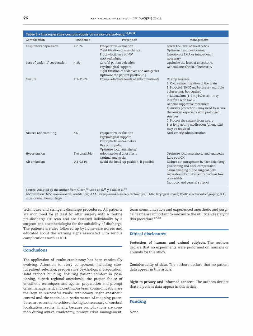
Table 3 – Intraoperative complications of awake craniotomy.16,28,29
Complication Incidence Prevention Management
Respiratory depression 2–18% Preoperative evaluationTight titration of anestheticsProphylactic use of NIVAAA technique
Lower the level of anestheticsOptimize head positioningInsertion of LMA or intubation, ifnecessary
Loss of patients’ cooperation 4.2% Careful patient selectionPsychological supportTight titration of sedatives and analgesicsOptimize the patient positioning
Optimize the level of anestheticsGeneral anesthesia, if necessary
Seizure 2.1–11.6% Ensure adequate levels of anticonvulsants To stop seizures:2. Cold saline irrigation of the brain3. Propofol (10–30 mg boluses) – multipleboluses may be required4. Midazolam (1–2 mg boluses) – mayinterfere with ECoGGeneral supportive measures:1. Airway protection - may need to securethe airway, especially with prolongedseizures2. Protect the patient from injury3. A long-acting medication (phenytoin)may be required
Nausea and vomiting 4% Preoperative evaluationPsychological supportProphylactic anti-emeticsUse of propofolOptimize local anesthesia
Anti-emetic administration
Hypertension Not available Adequate local anesthesiaOptimal analgesia
Optimize local anesthesia and analgesiaRule out ICH
Air embolism 0.3–0.64% Avoid the head-up position, if possible Reduce air entrapment by Trendelenbergpositioning and neck compressionSaline flushing of the surgical fieldAspiration of air, if a central venous lineis availableInotropic and general support
Source: Adapted by the author from Olsen,16 Lobo et al.28 y Balki et al.29
Abbreviation: NIV: non-invasive ventilation; AAA: asleep–awake–asleep techniques; LMA: laryngeal mask; EcoG: electrocorticography; ICH:intra-cranial hemorrhage.
techniques and stringent discharge procedures. All patientsare monitored for at least 6 h after surgery with a routinepre-discharge CT scan and are assessed individually by asurgeon and anesthesiologist for the suitability of discharge.The patients are also followed up by home-care nurses andeducated about the warning signs associated with seriouscomplications such as ICH.
Conclusions
The application of awake craniotomy has been continuallyevolving. Attention to every component, including care-ful patient selection, preoperative psychological preparation,solid rapport building, ensuring patient comfort in posi-tioning, superb regional anesthesia, the proper choice ofanesthetic techniques and agents, preparation and promptcrisis management, and continuous team communication, arethe keys to successful awake craniotomy. Tight anestheticcontrol and the meticulous performance of mapping proce-dures are essential to achieve the highest accuracy of cerebrallocalization results. Finally, because complications are com-mon during awake craniotomy, prompt crisis management,
team communication and experienced anesthetic and surgi-cal teams are important to maximize the utility and safety ofthis procedure.27–40
Ethical disclosures
Protection of human and animal subjects. The authorsdeclare that no experiments were performed on humans oranimals for this study.
Confidentiality of data. The authors declare that no patientdata appear in this article.
Right to privacy and informed consent. The authors declarethat no patient data appear in this article.
Funding
None.
r e v c o l o m b a n e s t e s i o l . 2 0 1 5;43(S1):22–28 27
Conflicts of interest
The author has no conflicts of interest to declare.
r e f e r e n c e s
1. Bulsara KR, Johnson J, Villavicencio AT. Improvements inbrain tumor surgery: the modern history of awakecraniotomies. Neurosurg Focus. 2005;18:e5.
2. Szelényi A, Bello L, Duffau H, Fava E, Feigl GC, Galanda M,et al. Intraoperative electrical stimulation in awakecraniotomy: methodological aspects of current practice.Neurosurg Focus. 2010;28:E7.
3. Nguyen HS, Sundaram SV, Mosier KM, Cohen-Gadol AA. Amethod to map the visual cortex during an awakecraniotomy. J Neurosurg. 2011;114:922–6.
4. Chui J, Manninen P, Valiante T, Venkatraghavan L. Theanesthetic considerations of intraoperativeelectrocorticography during epilepsy surgery. Anesth Analg.2013;117:479–86.
5. Larkin M. Neurosurgeons wake up to awake-brain surgery.Lancet. 1999;353:1772.
6. Taylor MD, Bernstein M. Awake craniotomy with brainmapping as the routine surgical approach to treating patientswith supratentorial intraaxial tumors: a prospective trial of200 cases. J Neurosurg. 1999;90:35–41.
7. Serletis D, Bernstein M. Prospective study of awakecraniotomy used routinely and nonselectively forsupratentorial tumors. J Neurosurg. 2007;107:1–6.
8. Brown T, Shah AH, Bregy A, Shah NH, Thambuswamy M,Barbarite E, et al. Awake craniotomy for brain tumorresection: the rule rather than the exception? J NeurosurgAnesthesiol. 2013;25:240–7.
9. Sacko O, Lauwers-Cances V, Brauge D, Sesay M, Brenner A,Roux FE. Awake craniotomy vs surgery under generalanesthesia for resection of supratentorial lesions.Neurosurgery. 2011;68:1192–8, discussion 1198–9.
10. Gupta DK, Chandra PS, Ojha BK, Sharma BS, Mahapatra AK,Mehta VS. Awake craniotomy versus surgery under generalanesthesia for resection of intrinsic lesions of eloquent cortex– a prospective randomised study. Clin Neurol Neurosurg.2007;109:335–43.
11. Erickson KM, Cole DJ. Anesthetic considerations for awakecraniotomy for epilepsy and functional neurosurgery.Anesthesiol Clin. 2012;30:241–68.
12. Nossek E, Matot I, Shahar T, Barzilai O, Rapoport Y, Gonen T,et al. Failed awake craniotomy: a retrospective analysis in 424patients undergoing craniotomy for brain tumor. J Neurosurg.2013;118:243–9.
13. Santini B, Talacchi A, Casagrande F, Casartelli M, Savazzi S,Procaccio F, et al. Eligibility criteria and psychological profilesin patient candidates for awake craniotomy: a pilot study. JNeurosurg Anesthesiol. 2012;24:209–16.
14. Klimek M, Verbrugge SJ, Roubos S, van der Most E, Vincent AJ,Klein J. Awake craniotomy for glioblastoma in a 9-year-oldchild. Anaesthesia. 2004;59:607–9.
15. Sarang A, Dinsmore J. Anaesthesia for awake craniotomy –evolution of a technique that facilitates awake neurologicaltesting. Br J Anaesth. 2003;90:161–5.
16. Olsen KS. The asleep–awake technique usingpropofol-remifentanil anaesthesia for awake craniotomy forcerebral tumours. Eur J Anaesthesiol. 2008;25:662–9.
17. Skucas AP, Artru AA. Anesthetic complications of awakecraniotomies for epilepsy surgery. Anesth Analg.2006;102:882–7.
18. Gignac E, Manninen PH, Gelb AW. Comparison of fentanyl,sufentanil and alfentanil during awake craniotomy forepilepsy. Can J Anaesth. 1993;40 5 Pt 1:421–4.
19. Manninen PH, Balki M, Lukitto K, Bernstein M. Patientsatisfaction with awake craniotomy for tumor surgery: acomparison of remifentanil and fentanyl in conjunction withpropofol. Anesth Analg. 2006;102:237–42.
20. Keifer JC, Dentchev D, Little K, Warner DS, Friedman AH, BorelCO. A retrospective analysis of a remifentanil/propofolgeneral anesthetic for craniotomy before awake functionalbrain mapping. Anesth Analg. 2005;101:502–8.
21. Bekker AY, Kaufman B, Samir H, Doyle W. The use ofdexmedetomidine infusion for awake craniotomy. AnesthAnalg. 2001;92:1251–3.
22. Souter MJ, Rozet I, Ojemann JG, Souter KJ, Holmes MD, Lee L,et al. Dexmedetomidine sedation during awake craniotomyfor seizure resection: effects on electrocorticography. JNeurosurg Anesthesiol. 2007;19:38–44.
23. Bustillo MA, Lazar RM, Finck AD, Fitzsimmons B, Berman MF,Pile-Spellman J, et al. Dexmedetomidine may impaircognitive testing during endovascular embolization ofcerebral arteriovenous malformations: a retrospective casereport series. J Neurosurg Anesthesiol. 2002;14:209–12.
24. Bernstein M. Outpatient craniotomy for brain tumor: a pilotfeasibility study in 46 patients. Can J Neurol Sci.2001;28:120–4.
25. Blanshard HJ, Chung F, Manninen PH, Taylor MD, Bernstein M.Awake craniotomy for removal of intracranial tumor:considerations for early discharge. Anesth Analg.2001;92:89–94.
26. Boulton M, Bernstein M. Outpatient brain tumor surgery:innovation in surgical neurooncology. J Neurosurg.2008;108:649–54.
27. Carrabba G, Venkatraghavan L, Bernstein M. Day surgeryawake craniotomy for removing brain tumours: technicalnote describing a simple protocol. Minim Invasive Neurosurg.2008;51:208–10.
28. Lobo F, Beiras A. Propofol and remifentanil effect-siteconcentrations estimated by pharmacokinetic simulation andbispectral index monitoring during craniotomy withintraoperative awakening for brain tumor resection. JNeurosurg Anesthesiol. 2007;19:183–9.
29. Balki M, Manninen PH, McGuire GP, El-Beheiry H, Bernstein M.Venous air embolism during awake craniotomy in a supinepatient. Can J Anaesth. 2003;50:835–8.
30. Bonhomme V, Franssen C, Hans P. Awake craniotomy. Eur JAnaesthesiol. 2009;26:906–12.
31. Costello TG. Awake craniotomy and multilingualism:language testing during anaesthesia for awake craniotomy ina bilingual patient. J Clin Neurosci. 2014;12:123–4.
32. Milian M, Tatagiba M, Feigl GC. Patient response to awakecraniotomy – a summary overview. Acta Neurochir (Wien).2014;156:1063–70.
33. Shinoura N, Midorikawa A, Yamada R, Hana T, Saito A,Hiromitsu K, et al. Awake craniotomy for brain lesions withinand near the primary motor area: a retrospective analysis offactors associated with worsened paresis in 102 consecutivepatients. Surg Neurol Int. 2013;4:149.
34. Bilotta F, Stazi E, Titi L, Lalli D, Delfini R, Santoro A, et al.Diagnostic work up for language testing in patientsundergoing awake craniotomy for brain lesions in languageareas. Br J Neurosurg. 2013.
35. Garavaglia MM, Das S, Cusimano MD, Crescini C, Mazer CD,Hare GM, et al. Anesthetic approach to high-risk patients andprolonged awake craniotomy using dexmedetomidine andscalp block. J Neurosurg Anesthesiol. 2013.
36. Bilotta F, Titi L, Lanni F, Stazi E, Rosa G. Traininganesthesiology residents in providing anesthesia for awake

28 r e v c o l o m b a n e s t e s i o l . 2 0 1 5;43(S1):22–28
craniotomy: learning curves and estimate of needed caseload. J Clin Anesth. 2013;25:359–66.
37. Hansen E, Seemann M, Zech N, Doenitz C, Luerding R,Brawanski A. Awake craniotomies without any sedation: theawake–awake–awake technique. Acta Neurochir (Wien).2013;155:1417–24.
38. Ouyang MW, McDonagh DL, Phillips-Bute B, James ML,Friedman AH, Gan TJ. Comparison of postoperative nauseabetween benign and malignant brain tumor patientsundergoing awake craniotomy: a retrospective analysis. CurrMed Res Opin. 2013;29:1039–44.
39. Ouyang MW, McDonagh DL, Phillips-Bute B, James ML,Friedman AH, Gan TJ. Does midline shift predictpostoperative nausea in brain tumor patients undergoingawake craniotomy? A retrospective analysis. Curr Med ResOpin. 2013;29:1033–8.
40. Nossek E, Matot I, Shahar T, Barzilai O, Rapoport Y, Gonen T,et al. Intraoperative seizures during awake craniotomy:incidence and consequences: analysis of 477 patients.Neurosurgery. 2013;73:135–40, discussion 140.