Revision of the smiling worms, genera Prosadenoporus Bu¨rger, … · 2010-03-06 · Bu¨rger...
39
Revision of the smiling worms, genera Prosadenoporus Bu ¨rger, 1890 and Pantinonemertes Moore and Gibson, 1981 and description of a new species Prosadenoporus floridensis sp. nov. (Prosorhochmidae; Hoplonemertea; Nemertea) from Florida and Belize Svetlana A. Maslakova a * and Jon L. Norenburg b a Oregon Institute of Marine Biology, University of Oregon, Charleston, OR, USA; b National Museum of Natural History, Smithsonian Institution, Washington, DC, USA (Received 31 December 2007; final version received 3 April 2008) The hoplonemertean genera Prosadenoporus Bu ¨ rger, 1890 and Pantinonemertes Moore and Gibson, 1981 are revised and synonymized based on a morphological re-evaluation. We redefine Prosadenoporus Bu ¨ rger, 1890 on the basis of characters held in common by the eight species: Prosadenoporus agricola (Willemoes-Suhm, 1874) comb. nov., Prosadenoporus arenarius Bu ¨ rger, 1890, Prosadenoporus spectaculum (Yamaoka, 1940) comb. nov., Prosadenoporus winsori (Moore and Gibson, 1981) comb. nov., Prosadenoporus enalios (Moore and Gibson, 1981) comb. nov., Prosadenoporus mooreae (Gibson, 1982b) comb. nov., Prosadenoporus mortoni (Gibson, 1990) comb. nov. and Prosadenoporus fujianensis (Sun, 2001) comb. nov. We describe a new semi-terrestrial species Prosadenoporus floridensis sp. nov. from Belize and Florida and compare its morphology to other species of Prosadenoporus. The average sequence divergence of P. floridensis sp. nov. from other congeners is 9.15% (16S) and 10.65% (COI) and 7.8% and 10.3% respectively from the nearest sequenced congener P. mortoni. Keywords: nemertea; Prosadenoporus; Pantinonemertes; Prosorhochmidae; semi- terrestrial Introduction Members of the hoplonemertean family Prosorhochmidae are distinguished by a crescent-shaped epithelial head groove – the ‘‘prosorhochmid smile’’. Like the majority of nemerteans, many prosorhochmids are marine, but some occupy semi- terrestrial, terrestrial or even arboreal habitats. The transition to terrestriality and the morphological adaptations that might have facilitated this are the subject of much discussion (Moore 1985; Moore and Gibson 1985; Gibson and Moore 1989). Established by Bu ¨ rger (1895), the family Prosorhochmidae initially included three genera: Prosorhochmus Keferstein, 1862, Geonemertes Semper, 1863 and Prosadenoporus Bu ¨ rger, 1890. Two features stand out in the original diagnosis – the enormously developed cephalic glands and hermaphroditism (Bu ¨ rger 1895). At this time, hermaphroditism can no longer serve as a defining feature of the family because many prosorhochmids with separate sexes have been discovered. However, the massive cephalic glands and large frontal organ through which some of these glands discharge are characteristic of all prosorhochmids. Although the ‘‘most terrestrial’’ of the species actually do not have the prosorhochmid smile, it appears *Corresponding author. Email: [email protected] Journal of Natural History Vol. 42, Nos. 25–28, July 2008, 1689–1727 ISSN 0022-2933 print/ISSN 1464-5262 online # 2008 Taylor & Francis DOI: 10.1080/00222930802130286 http://www.informaworld.com
Transcript of Revision of the smiling worms, genera Prosadenoporus Bu¨rger, … · 2010-03-06 · Bu¨rger...

Revision of the smiling worms, genera Prosadenoporus Burger, 1890 andPantinonemertes Moore and Gibson, 1981 and description of a newspecies Prosadenoporus floridensis sp. nov. (Prosorhochmidae;Hoplonemertea; Nemertea) from Florida and Belize
Svetlana A. Maslakovaa* and Jon L. Norenburgb
aOregon Institute of Marine Biology, University of Oregon, Charleston, OR, USA; bNationalMuseum of Natural History, Smithsonian Institution, Washington, DC, USA
(Received 31 December 2007; final version received 3 April 2008)
The hoplonemertean genera Prosadenoporus Burger, 1890 and PantinonemertesMoore and Gibson, 1981 are revised and synonymized based on a morphologicalre-evaluation. We redefine Prosadenoporus Burger, 1890 on the basis of charactersheld in common by the eight species: Prosadenoporus agricola (Willemoes-Suhm,1874) comb. nov., Prosadenoporus arenarius Burger, 1890, Prosadenoporusspectaculum (Yamaoka, 1940) comb. nov., Prosadenoporus winsori (Moore andGibson, 1981) comb. nov., Prosadenoporus enalios (Moore and Gibson, 1981)comb. nov., Prosadenoporus mooreae (Gibson, 1982b) comb. nov.,Prosadenoporus mortoni (Gibson, 1990) comb. nov. and Prosadenoporusfujianensis (Sun, 2001) comb. nov. We describe a new semi-terrestrial speciesProsadenoporus floridensis sp. nov. from Belize and Florida and compare itsmorphology to other species of Prosadenoporus. The average sequence divergenceof P. floridensis sp. nov. from other congeners is 9.15% (16S) and 10.65% (COI)and 7.8% and 10.3% respectively from the nearest sequenced congener P. mortoni.
Keywords: nemertea; Prosadenoporus; Pantinonemertes; Prosorhochmidae; semi-terrestrial
Introduction
Members of the hoplonemertean family Prosorhochmidae are distinguished by a
crescent-shaped epithelial head groove – the ‘‘prosorhochmid smile’’. Like themajority of nemerteans, many prosorhochmids are marine, but some occupy semi-
terrestrial, terrestrial or even arboreal habitats. The transition to terrestriality and
the morphological adaptations that might have facilitated this are the subject of
much discussion (Moore 1985; Moore and Gibson 1985; Gibson and Moore 1989).
Established by Burger (1895), the family Prosorhochmidae initially included
three genera: Prosorhochmus Keferstein, 1862, Geonemertes Semper, 1863 and
Prosadenoporus Burger, 1890. Two features stand out in the original diagnosis – the
enormously developed cephalic glands and hermaphroditism (Burger 1895). At thistime, hermaphroditism can no longer serve as a defining feature of the family
because many prosorhochmids with separate sexes have been discovered. However,
the massive cephalic glands and large frontal organ through which some of these
glands discharge are characteristic of all prosorhochmids. Although the ‘‘most
terrestrial’’ of the species actually do not have the prosorhochmid smile, it appears
*Corresponding author. Email: [email protected]
Journal of Natural History
Vol. 42, Nos. 25–28, July 2008, 1689–1727
ISSN 0022-2933 print/ISSN 1464-5262 online
# 2008 Taylor & Francis
DOI: 10.1080/00222930802130286
http://www.informaworld.com

that the well-developed frontal organ and cephalic glands, associated with the smile
in other prosorhochmids, might have played an important role in the conquest of
land.
Over the years, the content of the family varied, and at one time
Prosorhochmidae encompassed up to 19 different genera, including all known
terrestrial and some of the freshwater forms (Gibson 1972, 1982a). Following the
revision of the terrestrial nemerteans (Moore and Gibson 1981), and the genera
Prosorhochmus Keferstein, 1862 (Gibson and Moore 1985) and Prosadenoporus
Burger, 1890 (Moore and Gibson 1988), the family was redefined based on the
characters interpreted as being held in common by the four genera Geonemertes
Semper, 1863, Prosadenoporus Burger, 1890, Prosorhochmus Keferstein, 1862 and
Pantinonemertes Moore and Gibson, 1981 (Moore and Gibson 1988, p. 85). As part
of an ongoing revision of the family Prosorhochmidae (Maslakova et al. 2005;
Maslakova and Norenburg 2008) here we revise the genera Prosadenoporus and
Pantinonemertes.
The genus Prosadenoporus currently includes one species: Prosadenoporus
arenarius Burger, 1890 (Moore and Gibson 1988). The genus was established by
Burger (1890) for four briefly described species from Indonesia mainly distinguished
by size and colour: P. arenarius Burger, 1890, P. badiovagatus Burger, 1890, P.
janthinus Burger, 1890 and P. oleaginus Burger, 1890. Each description was based on
a single preserved specimen. Punnet (1901) briefly described a fifth member of the
genus, P. buergeri, based on a single specimen from a geographically distant area –
the Laccadive Islands. None of the five species is sufficiently described to allow
unambiguous identification and Burger’s material is almost certainly lost. Moore
and Gibson (1988) re-evaluated Punnet’s material deposited at the Cambridge
Museum of Zoology: one tube labelled Prosadenoporus buergeri, which contained
fragments of proboscis and posterior body insufficient for identification, and one
tube labelled Prosadenoporus sp. from Kedah Province on Malay Peninsula with
several preserved specimens which they sectioned and examined. Moore and Gibson
(1988) invalidated four species as nomina dubia, retaining Prosadenoporus arenarius
Burger, 1890 as the type species of the genus. They provided a detailed description of
the internal anatomy of the species, arguing that Punnet’s Malaysian specimens
cannot be distinguished from Burger’s description of P. arenarius from Indonesia
and are, therefore, conspecific.
The genus Pantinonemertes currently includes nine valid species: P. agricola
(Willemoes-Suhm, 1874), P. spectaculum (Yamaoka, 1940), P. winsori Moore and
Gibson, 1981 (Figures 1B, 1E and 1F), P. enalios Moore and Gibson, 1981, P.
mooreae Gibson, 1982b (Figures 1C and D), P. californiensis Gibson et al., 1982, P.
mortoni Gibson, 1990, P. daguilarensis Gibson and Sundberg, 1992 and P. fujianensis
Sun, 2001. It was first established for the taxon encompassing Geonemertes agricola
(Willemoes-Suhm, 1874), P. enalios Moore and Gibson, 1981 and P. winsori Moore
and Gibson, 1981, with the latter designated as the type species (Moore and Gibson,
1981). However, as pointed out by Chernyshev (1992), Pantinonemertes is a junior
synonym of Neonemertes – the genus established by Girard (1893) for Tetrastemma
agricola Willemoes-Suhm, 1874. Subsequently, Tetrastemma spectaculum Yamaoka,
1940 was re-described and transferred to Pantinonemertes (Gibson, 1990) and five
new species were described, including two species whose affiliation with the genus is
questionable: P. californiensis and P. daguilarensis.
1690 S.A. Maslakova and J.L. Norenburg

We describe a new semi-terrestrial prosorhochmid species from Florida and
Belize whose morphology closely resembles that of Pantinonemertes and
Prosadenoporus (Figures 1A, 2A, 2B, 3–6 and 10I). To make a thorough
comparison with the previously described species we re-evaluated all the available
type and voucher material and recollected fresh specimens of as many species of
both genera as possible for morphological and molecular study. In the process we
came to recognize that the morphological differences between Pantinonemertes
and Prosadenoporus are elusive and the two genera should be synonymized; in
which case, according to the principle of priority (International Code of
Zoological Nomenclature, Article 23), Prosadenoporus Burger, 1890 takes
precedence over both Pantinonemertes Moore and Gibson, 1981 and
Neonemertes Girard, 1893. The following revision incorporates this change of
generic names.
Material and methods
Specimen preparation
Characters of external appearance and stylet apparatus were documented in living
specimens as described in Maslakova and Norenburg (2008). Measurements such
as body length and width were taken on live specimens relaxed by gradual addition
of 7.5% MgCl2. For histology, specimens were relaxed in MgCl2, fixed for 24 h in
4% formaldehyde made in local seawater, briefly rinsed in tap water and post-fixed
in Hollande’s cupri-picri-formal-acetic fluid for 48–72 h. After post-fixation
specimens were transferred to 70% ethanol for long-term storage. Specimens
were dehydrated in a standard alcohol series, embedded in Tissueprep Paraffin
(56u C m.p.), serially sectioned at 8 mm and stained using Crandall’s polychrome
protocol – a combined variant of the Mallory, Gomori, Koneff and
Gurr-McConail techniques (Frank Crandall, National Museum of Natural
History, Smithsonian Institution,Washington, DC, personal communication to
authors).
DNA extraction, amplification and sequencing
Tissue samples for molecular work were obtained for the following species:
Prosadenoporus mortoni comb. nov., Prosadenoporus mooreae comb. nov.,
Prosadenoporus winsori comb. nov., Prosadenoporus floridensis sp. nov. and
Pantinonemertes californiensis (Table 1). Specimens were preserved in 95% ethanol
and stored at room temperature or 220uC short-term and at 280uC long-term.
DNA was extracted using Qiagen DNeasy Kits. Partial sequences of genes
encoding for cytochrome oxidase subunit I (COI), 658 bp long, and mitochondrial
large subunit rRNA (16S), 458–467 bp long, were PCR-amplified using universal
primers: 16sar-L [cgcctgtttatcaaaaacat] and 16sbr-H [ccggtctgaactcagatcacgt] from
Palumbi et al. (1991) for 16S, and LCO1490 [ggtcaacaaatcataaagatattgg] and
HCO2198 [taaacttcagggtgaccaaaaaatca] from Folmer et al. (1994) for COI. In
addition, a pair of nemertean specific internal primers were designed for each gene
to amplify fragments where DNA degradation appeared to be a problem: P2619-
Nem16S-intF [acaagaagacccttttgagct] and P2620-Nem16S-intR [taaagct-
caaaagggtcttctt] for 16S, and P2638-COIF3-nemert [gtctagraatrttgctcatgctg] and
Journal of Natural History 1691

P2683-COIR-nemert [ctyccagcatgwgcaayatt] for COI. PCR amplification was
performed as described by us (Maslakova and Norenburg, 2008). The purified
PCR products were sequenced on a 3100 ABI Capillary DNA Sequencer; both
strands were sequenced at least once and proofread using Sequencher 4.1.4 (Gene
Codes Corporation). Sequences are deposited with the GeneBank (see Table 1 for
accession numbers).
Stylet measurements
To determine whether interspecific differences in the stylet and basis measurements
were statistically significant we performed a one-way ANOVA correcting for
multiple comparisons with Tukey–Kramer HSD method using the statistical
software package JMP 6.0.3 (SAS Institute Inc.). Stylet measurements used in the
analysis are presented in Table 2. Cases where individual measurements were not
reported or where only a single data point was available could not be used in the
analysis. However, these measurements were used to adjust the range of variation in
species diagnoses. Data for statistical comparisons were only available for
Prosadenoporus winsori comb. nov., Prosadenoporus mooreae comb. nov. and
Prosadenoporus floridensis sp. nov.
Results
Class: HOPLONEMERTEA
Order: MONOSTILIFERA
Family: PROSORHOCHMIDAE Burger, 1895
Diagnosis
Monostiliferous, marine, brackish-water, semi-terrestrial or terrestrial hoplonemer-
teans with rhynchocoel extending most or all the body length, body wall containing
distinct outer circular and inner longitudinal muscle layers and a delicate layer of
diagonal muscles between; longitudinal musculature may appear to be anteriorly
divided (i.e. some of the proboscis insertion muscles may be oriented obliquely and
longitudinally); four simple eyes; proboscis may be small or large, and sometimes
used in locomotion; stylet basis in most species characteristically truncated; head in
most species with characteristic dorsal horizontal epithelial fold (the prosorhochmid
smile); cerebral sensory organs anterior or anterolateral to brain, with simple
unbranched cerebral canals, in most species opening ventro-laterally via reduced
cerebral organ furrows; cephalic glands well developed, with distinct granular
proteinaceous components (orange-G and acidophilic glands) in addition to
basophilic mucus lobules; frontal organ represented by an exceptionally well-
developed tubular canal, typically with laterally specialized epithelium; middorsal
blood vessel with a single vascular plug; typically gonochoric and oviparous,
occasionally hermaphroditic and viviparous.
Composition
As a result of our proposed synonymization of Pantinonemertes Moore and Gibson,
1981 with Prosadenoporus Burger, 1890 the family now contains three genera:
1692 S.A. Maslakova and J.L. Norenburg

Prosorhochmus Keferstein, 1862 (type genus), Prosadenoporus Burger, 1890(5Pantinonemertes Moore and Gibson, 1981 part, new synonymy) and
Geonemertes Semper, 1863.
Geographic distribution
Atlantic coast of the British Isles, France, Spain, USA (FL and SC) and Bermuda;
Caribbean (Belize); Adriatic Sea (coast of Italy and Croatia); Mediterranean Sea
(coast of Italy, France, Chafarinas Islands); Black Sea (Russian coast); Pacific coast
of USA (Puget Sound, WA to CA, Hawaii) and Chile; Hong Kong, China (Fujian
Province), north-eastern coast of Australia (Queensland), Indopacific Islands
(Noordwachter Is. off Sulawesi, Palau Bidan off Malay Peninsula, Papua New
Guinea, Japan, Seychelles, Sri Lanka, Sulawesi, Pelew Is., Caroline Is., Samoan Is.,Kei Is., Mauritius, Samarai, the Philippines), the West Indies (Dominica, Jamaica).
Prosadenoporus Burger, 1890
Prosadenoporus Burger, 1890.
Neonemertes Girard, 1893. New synonymy. Type species Tetrastemma agricola
Willemoes-Suhm, 1874 by monotypy.
Pantinonemertes Moore and Gibson, 1981 (part). New synonymy. Type species
Pantinonemertes winsori Moore and Gibson, 1981 by original designation.
Type species
Prosadenoporus arenarius Burger, 1890, by subsequent designation (Moore and
Gibson 1988).
Etymology
‘‘Pro’’5before, forward, in front of, L.; ‘‘adeno, adenos’’5gland, G.; ‘‘poros’’5hole,
G. Likely refers to the presence of well-developed frontal organ [anterior pore]
through which mucus cephalic glands discharge.
Table 1. GenBank accession numbers and specimen data for molecular analysis.
Species Accession
numbers
Collecting information (specimen numbers)
Pantinonemertes
californiensis
EF157597,
EF157585
Coll. Crandall, Tomales Bay, CA, USA (#4-15-92)
Prosadenoporus
floridensis sp. nov.
EF157596,
EF157583,
EF157584
Coll. JLN, Link Port, FL, USA (#2-22-98-2); Coll.
SAM, Twin Keys, Belize (#178-14-2-00-1)
Prosadenoporus
mooreae comb.
nov.
EF157595,
EF157582
Coll. SAM, 21 March 2003, type locality in Picnic Bay
and in Cockle Bay, Magnetic Island, Australia (#778-
03-23-03-1,2,3)
Prosadenoporus
mortoni comb. nov.
EF157593,
EF157580
Coll. Sun, Nan Ao Island and Xiamen; Fujian Province;
China (#910913, #990909-2B, #9909-1A)
Prosadenoporus
winsori comb. nov.
EF157594,
EF157581
Coll. SAM, 18 March 2003, type locality on the banks of
Ross River, Townsville, Australia (#777-03-18-03-5)
Journal of Natural History 1693

Table 2. Measurements of the stylet apparatus in Prosadenoporus species.
Species Specimen Body
length
(mm)
Stylet
length
(S; mm)
Basis
length
(B; mm)
S:B ratio Source
P. floridensis sp.
nov.
1 N/A 165 250 0.66 Collected JLN, Belize
P. floridensis sp.
nov.
2 N/A 98 196 0.5 Collected JLN, Belize
P. floridensis sp.
nov.
3 N/A 158 269 0.59 Collected SAM, Belize
P. floridensis sp.
nov.
4 N/A 159 371 0.43 Collected SAM, Belize
P. floridensis sp.
nov.
5 N/A 146 372 0.39 Collected SAM, Belize
P. floridensis sp.
nov.
6 12 92 140 0.66 Collected JLN, Florida
P. floridensis sp.
nov.
7 25 130 250 0.52 Collected JLN, Florida
P. floridensis sp.
nov.
8 40 130 300 0.43 Collected JLN, Florida
P. floridensis sp.
nov.
9 22 115 230 0.5 Collected JLN, Florida
P. floridensis sp.
nov.
10 17 140 240 0.58 Collected JLN, Florida
P. floridensis sp.
nov.
11 18 115 165 0.7 Collected JLN, Florida
P. floridensis sp.
nov.
12 16 120 192 0.63 Collected JLN, Florida
P. mooreae comb.
nov.
1 N/A 50 150 0.33 Gibson (1982b)
P. mooreae comb.
nov.
1 N/A 130 140 0.92 Collected SAM,
Magnetic Island,
QLD, Australia
P. mooreae comb.
nov.
2 N/A 120 130 0.92 Collected SAM,
Magnetic Island,
QLD, Australia
P. mooreae comb.
nov.
3 N/A 120 160 0.75 Collected SAM,
Magnetic Island,
QLD, Australia
P. winsori comb.
nov.*
N/A N/A 200–250 950 NA Moore and Gibson
(1981)
P. winsori comb.
nov.
1 180 220 850 0.26 Collected SAM,
Magnetic Island,
QLD, Australia
P. winsori comb.
nov.
2 65 120 490 0.25 Collected SAM,
Magnetic Island,
QLD, Australia
P. winsori comb.
nov.
3 N/A 360 720 0.5 Collected SAM,
Magnetic Island,
QLD, Australia
1694 S.A. Maslakova and J.L. Norenburg

Diagnosis
Monostiliferous marine, brackish-water, semi-terrestrial or terrestrial hoplonemer-
teans with a horizontal cephalic epithelial fold (prosorhochmid smile) (Figures 1D,
1E and 2B). Four eyes (Figures 1A, 1D, 1E and 2B). Body-wall musculature well
developed with a delicate layer of diagonal muscles between outer circular and inner
longitudinal muscle layers (Figures 3A and 3B). Rhynchocoel full body-length, with
wall composed of distinct outer circular and inner longitudinal muscle layers
(Figure 3K). Proboscis small or massive, may be used for rapid locomotion. If
massive, proboscis often with increased number of proboscis nerves and accessory
stylet pouches, longer stylet (S) and basis (B) and decreased S:B ratio (Table 3).
Tubular frontal organ is long and exceptionally well developed, with laterally
differentiated epithelium (Figures 3C, 4I–L, 5G–I, 7A–I, 7L, 8A–D and 9C–F).
Cephalic glands extensive, reaching into stomach-pylorus region and containing
basophilic (mucus) components (Figure 3C) discharging through frontal organ and
via improvised epidermal ducts and acidophilic (proteinaceous) components,
staining red (red glands) (Figure 5H) and orange with Mallory trichrome and its
modifications (orange-G glands) (Figures 5J, 6B and 6D). Proteinaceous cephalic
glands discharge via improvised ducts scattered throughout epidermis. Cerebral
sensory organs small, anterior or antero-lateral to brain, with simple un-forked
canal, opening ventro-laterally into reduced cerebral organ furrows (Figures 2A, 5A,
5B and 5J). Neurochord cells and neurochords present in some species (Figures 5E,
5F, 7J, 8E, 8F and 9G), but not necessarily together. Lateral nerve cords without
accessory nerves (e.g. Figures 4G and 7J). Oesophagus divisible into anterior region
supplied with acidophilic glands (Figures 3F, 3I, 3J, 5J, 6A and 6B), and posterior
non-glandular region (Figures 3G and 6C). Caecum long, anteriorly bifid, with
numerous lateral diverticula on each side (Figure 6E). Blood system with three main
longitudinal vessels, without transverse connectives; mid-dorsal blood vessel with
single vascular plug (Figure 4F); cephalic vascular loop recurved. Extracellular
matrix (parenchyma) may be very extensive, especially in large species. Excretory
system well developed, with binucleate terminal nephridial cells reinforced by
transverse support bars (Figures 5C, 5D and 7K), thin-walled canals and
large number of inconspicuous nephridiopores. Gonochoric or hermaphroditic.
Oviparous or viviparous.
Species Specimen Body
length
(mm)
Stylet
length
(S; mm)
Basis
length
(B; mm)
S:B ratio Source
P. fujianensis comb.
nov.
1 N/A 120 380 0.32 Sun (2001)
P. agricola comb.
nov.*
N/A N/A 80–95 75–90 NA Coe (1904)
P. enalios comb.
nov.
1 N/A 110 170 0.65 Moore and Gibson
(1981)
P. mortoni comb.
nov.
1 N/A 125 123 1 Collected Sun, Fujian
province, China
Note: *Cases where individual measurements are not reported.
Table 2. (Continued).
Journal of Natural History 1695

Composition
Prosadenoporus includes nine species: P. agricola (Willemoes-Suhm, 1874) comb.
nov., P. arenarius Burger, 1890, P. spectaculum (Yamaoka, 1940) comb. nov., P.
winsori (Moore and Gibson, 1981) comb. nov., P. enalios (Moore and Gibson, 1981)
comb. nov., P. mooreae (Gibson, 1982b) comb. nov., P. mortoni (Gibson, 1990)
comb. nov., P. fujianensis (Sun, 2001) comb. nov., and P. floridensis sp. nov.
Geographic distribution
Atlantic coast of the USA (FL) and Bermuda; Caribbean (Belize); Pacific coast
of USA (Puget Sound, WA to CA); Hong Kong, China (Fujian Province),
north-eastern coast of Australia (Queensland), Indonesian Islands (Noordwachter
Figure 1. External appearance of members of Prosadenoporus. (A) Female and male of
Prosadenoporus floridensis sp. nov.; (B) Prosadenoporus winsori comb. nov. on a piece of rotten
mangrove penetrated by teredinid burrows (its natural habitat); (C–D) Prosadenoporus
mooreae comb. nov.; (E–F) anterior end of P. winsori (E5lateral view; F5dorsal view). Notes:
cg, cerebral ganglia; pf, posterior cephalic furrow; sm, prosorhochmid smile. Scales: (A and
C), 3 mm; (B), 20 mm; (D), 0.5 mm; (E and F), 1 mm.
1696 S.A. Maslakova and J.L. Norenburg

Island off Sulawesi and Ambon Island [Amboina]), Palau Bidan (Kedah Province)
off Malay Peninsula, Laccadive Islands.
Prosadenoporus agricola (Willemoes-Suhm, 1874), new combination
(Figure 10F; Tables 2 and 3)
Tetrastemma agricola (Willemoes-Suhm 1874; Moseley 1879; Hubrecht 1887; Verrill
1902).
Neonemertes agricola (Girard 1893; Joubin 1894; Friedrich 1955).
Geonemertes agricola (Burger 1895, 1897–1907, 1904; Coe 1904, 1905, 1929, 1939,
1940; Crozier 1917; Pantin 1947, 1961, 1969; Gibson 1972; Moore and Moore 1972;
Moore 1973, 1975a, 1975b).
Pantinonemertes agricola (Moore and Gibson 1981; Gibson et al. 1982; Gibson
1982b; Gibson, 1990; Sundberg 1989; Gibson and Sundberg 1992; 2001; Moore et al.
2001; Sun 2001).
Etymology
The name reflects terrestrial habitat of this species (agricola5farmer, L).
Type material
Types were never designated. Original material almost certainly lost.
Material examined
Prosadenoporus agricola (Willemoes-Suhm, 1874) comb. nov. Series of sections of
one mature and one juvenile specimen are held at the University Museum of
Zoology, Cambridge, UK (coll. Boden, B. 1951, Bermuda).
Diagnosis
Prosadenoporus agricola comb. nov. differs from most other Prosadenoporus species in
being hermaphroditic and viviparous (Table 3). It differs from Prosadenoporus arenarius,
the only other hermaphroditic congener, whose life history is unknown, by having fewer
proboscis nerves 12–15 (compared with 15–17) and possessing neurochord cells. Central
stylet (S) 80–95 mm long, basis (B) rounded, pear-shaped or truncated 75–90 mm long,
two accessory stylet pouches. It is not possible to determine mean S:B ratio because only
ranges of stylet and basis lengths are available. Data at hand are insufficient to determine
whether these metrics are significantly different from those of other species.
Habitat and distribution
Along the shores of mangrove swamps, under stones and logs on moist, silty soil or
inside burrows of earthworms above high-water mark and, during wet season, on
adjacent hillsides. Associated with a heteronemertean Lineus sp., earthworms,
nematodes and, sometimes, insects. Smaller individuals also occur below the high-
water mark. Found in Hungary Bay, Bayley’s Bay, Walsingham Bay, on shores of
Castle Harbor, near the ‘‘Causeway’’, at Hamilton Harbor and other localities on
Bermuda Island. Once common on Bermuda (Coe 1904, p. 532), this species had last
been collected in 1966 during an extensive search of the islands by Dr B. Boden, which
yielded very few specimens, all obtained from a remote place. More recent commercial
Journal of Natural History 1697

Ta
ble
3.
Mo
rph
olo
gic
al
cha
ract
eris
tics
of
Pro
sad
eno
po
rus
spec
ies.
Sp
ecie
sH
ab
ita
tB
od
yco
lou
rin
life
Sex
/lif
eh
isto
ryN
euro
-
cho
rds
Neu
ro-
cho
rdce
lls
P.
flo
rid
ensi
ssp
.n
ov
.S
emi-
terr
estr
ial
Lig
ht
yel
low
ish
,o
ran
ge-
bro
wn
,o
liv
e-ta
no
r
gre
enis
h-b
row
nd
ors
all
y,
da
rkes
tin
the
do
rsa
l
mid
lin
e,o
ff-w
hit
eto
yel
low
ish
crea
mv
entr
all
y
Go
no
cho
ric,
ov
ipa
rou
sA
bse
nt
Pre
sen
t
P.
are
na
riu
s?
Un
kn
ow
n,
pre
serv
edsp
ecim
ens
un
ifo
rmly
du
ll
bro
wn
Her
ma
ph
rod
itic
,
pre
sum
ab
lyo
vip
aro
us
Ab
sen
tA
bse
nt
P.
ag
rico
laS
emi-
terr
estr
ial,
terr
estr
ial
Va
ries
fro
mu
nif
orm
mil
k-w
hit
eo
rg
ray
ish
-wh
ite
to
ora
ng
e,ro
sy,
gre
enis
h-b
row
no
rg
ray
ish
-bro
wn
do
rsa
lly
,a
nd
pa
leg
ray
or
wh
itis
hv
entr
all
y
Her
ma
ph
rod
itic
,
viv
ipa
rou
s
Ab
sen
tP
rese
nt
P.
win
sori
Sem
i-te
rres
tria
lP
urp
lish
-bro
wn
do
rsa
lly
wit
ha
na
rro
w,
pa
le
mid
-do
rsa
llo
ng
itu
din
al
stri
pe
an
teri
orl
y,
pa
le
crea
mto
gra
yis
h-w
hit
ev
entr
all
y,
inte
nsi
fyin
gto
pa
lere
dd
ish
-bro
wn
po
ster
iorl
y
Go
no
cho
ric,
pre
sum
ab
ly
ov
ipa
rou
s
Pre
sen
tP
rese
nt
P.
fuji
an
ensi
sS
emi-
terr
estr
ial
Gra
yis
h-b
lack
do
rsa
lly
,p
ale
wh
itis
hv
entr
all
y,
da
rk
pig
men
tco
nce
ntr
ate
da
lon
gth
ed
ors
al
mid
lin
e
form
ing
an
arr
ow
stri
pe
Go
no
cho
ric,
pre
sum
ab
ly
ov
ipa
rou
s
Ab
sen
tP
rese
nt
P.
mo
ore
ae
Ma
rin
ein
tert
ida
lD
ors
al
colo
ur
gra
yis
h-g
reen
wit
ha
da
rk-g
reen
to
bla
ckn
arr
ow
lon
git
ud
ina
lst
rip
e,v
entr
al
sid
e
crea
mto
pa
leg
reen
ish
Go
no
cho
ric,
pre
sum
ab
ly
ov
ipa
rou
s
Ab
sen
tA
bse
nt
P.
mo
rto
ni
Ma
rin
ein
tert
ida
lD
ors
al
colo
ur
oli
ve-
gre
enw
ith
ad
ark
-gre
enn
arr
ow
lon
git
ud
ina
lst
rip
e,v
entr
al
sid
ey
ello
wis
h-w
hit
e
Go
no
cho
ric,
pre
sum
ab
ly
ov
ipa
rou
s
Ab
sen
tA
bse
nt
P.
ena
lio
sM
ari
ne
inte
rtid
al
Pa
lefa
wn
-bro
wn
,o
ran
ge-
bro
wn
or
ora
ng
ed
ors
all
y,
dee
pes
tp
igm
enta
tio
na
pp
eari
ng
ov
era
nte
rio
rp
air
of
eyes
,v
entr
all
ycr
eam
,o
ff-w
hit
eo
rp
ale
ora
ng
e
Go
no
cho
ric,
pre
sum
ab
ly
ov
ipa
rou
s
Ab
sen
tA
bse
nt
P.
spec
tacu
lum
Sem
i-te
rres
tria
lO
ff-w
hit
eto
crea
my
-bro
wn
,sp
eck
led
wit
hd
ark
-bro
wn
do
rsa
lly
an
dla
tera
lly
,m
ore
den
sely
tow
ard
do
rsa
l
mid
lin
e;ch
ara
cter
isti
csp
ecta
cle-
sha
ped
pa
tter
no
f
da
rk-b
row
na
rou
nd
eyes
?A
bse
nt
Pre
sen
t
1698 S.A. Maslakova and J.L. Norenburg

Ta
ble
3.
(Co
nti
nu
ed).
Sp
ecie
sS
ha
pe
of
sty
let
ba
sis
Sty
let
len
gth
(S;
mm)
Ba
sis
len
gth
(B;
mm)
S:B
rati
o
Nu
mb
ero
f
acc
esso
ryst
yle
t
po
uch
es
Nu
mb
ero
f
pro
bo
scis
ner
ves
P.
flo
rid
ensi
ssp
.n
ov
.T
run
cate
d9
2–
16
5
(mea
n1
30
.7)
14
0–
37
2
(mea
n2
47
.9)
0.3
9–
0.7
(mea
n0
.55
)
21
1–
14
P.
are
na
riu
s?
??
??
15
–1
7
P.
ag
rico
laR
ou
nd
ed,
pea
r-sh
ap
edo
rtr
un
cate
d
80
–9
57
5–
90
?2
12
–1
5
P.
win
sori
Tru
nca
ted
20
0–
36
0
(mea
n2
33
.3)
49
0–
85
0
(mea
n6
86
.7)
0.2
5–
0.5
(mea
n0
.34
)
6–
92
2–
24
P.
fuji
an
ensi
sT
run
cate
d1
20
38
00
.32
51
9–
20
P.
mo
ore
ae
Elo
ng
ate
d,
na
rro
wa
nd
rou
nd
ed
50
–1
30
(mea
n1
05
)
13
0–
16
0
(mea
n1
45
)
0.3
3–
0.9
2
(mea
n0
.73
)
21
5–
16
P.
mo
rto
ni
Elo
ng
ate
d,
na
rro
wa
nd
rou
nd
ed
12
51
23
1.0
?1
4
P.
ena
lio
sT
run
cate
d1
10
17
00
.65
?1
4–
16
P.
spec
tacu
lum
??
??
21
8–
22
Journal of Natural History 1699

development has left very little suitable habitat and the species has apparently not been
found since (Moore et al. 2001; W. Sterrer, personal communication to SAM).
Remarks
Coe’s (1904) anatomical account of the species mentions 13–15 proboscis nerves.
SAM observed 12 proboscis nerves in the available voucher specimen in the
collection of University Museum of Zoology, Cambridge, UK.
Prosadenoporus arenarius Burger, 1890
Prosadenoporus arenarius (Burger 1890, 1895, 1904; Moore and Gibson 1988).
Etymology
The species name means ‘‘living in sand’’ (L).
Type material
Type material almost certainly does not exist; not known from the most likely
repositories – Museum fur Naturkunde, Berlin; Zoologisches Institut, Universitat
Gottingen; Stazione Zoologica, Naples.
Material examined
Prosadenoporus arenarius Burger, 1890. Series of transverse sections (slides 1–3) of
hermaphroditic specimen from Punnett’s collection (Palau Bidan off Malay
Peninsula) held at the University Museum of Zoology, Cambrige, UK.
Diagnosis
Prosadenoporus arenarius differs from all other Prosadenoporus species except
Prosadenoporus agricola comb. nov. by being hermaphroditic (Moore and Gibson
1988, p. 81) and from the Bermudan P. agricola by having 15–17 proboscis nerves
(compared with 12–15) and by lacking neurochord cells (Table 3). Characters of the
proboscis armature are unknown.
Habitat and distribution
Habitat is unknown, except in as much as the name of the species implies. Found in
Java Sea (Noordwachter Island off northwestern Sulawesi) and Palau Bidan (Kedah
Province, off Malay Peninsula).
Remarks
As mentioned above, Burger’s material is almost certainly lost. A tube with several
specimens labelled ‘‘Prosadenoporus sp.’’ from Punnett’s collection from Palau Bidan
(Kedah Province, Malay Peninsula) was deposited at the University Museum of
Zoology in Cambridge, UK. Moore and Gibson (1988) sectioned some of these
specimens and re-described Prosadenoporus arenarius Burger, 1890, arguing that
Punnett’s specimens fit the original (very brief) description of P. arenarius. However,
1700 S.A. Maslakova and J.L. Norenburg

some evidence suggests that Burger’s P. arenarius from Indonesia and Punnet’s
Prosadenoporus sp. from Malaysia might represent different species.
First, the specimen described by Burger (1890) from the Noordwachter Island off
Sulawesi had a broad dark-brown, mid-dorsal stripe on a grayish-green background,
while Punnett’s specimens were uniformly dull brown (Moore and Gibson 1988).
Second, and more important, Burger’s diagnosis of the genus mentions neurochord
cells and neurochords. Moore and Gibson (1988) report about Punnett’s specimens
that ‘‘few neurochord cells are present, located near the inner margins of the dorsal
cerebral lobes and neurochords can be traced in the main nerve cords’’ (p. 80).
However, their figure 10 (Moore and Gibson 1988, p. 78, fig. 10) suggests that
they mistake the type III neurons for neurochord cells, both defined by Burger
(1895). Our reinvestigation of Punnett’s material showed no traces of neurochord
cells or neurochords. Finally, Burger’s specimen had 12 proboscis nerves, while
there are 17 proboscis nerves in Punnett’s material. Although we have little
confidence that Burger’s description and Punnett’s material belong to the same
species, lack of the type or other informative material thwarts resolution of the
question.
Moore and Gibson (1988, p. 82) provided an amended diagnosis of the genus
Prosadenoporus. The main difference between Pantinonemertes Moore and Gibson,
1981 and Prosadenoporus Burger, 1890 is the presence of a second cephalic vascular
loop described for Prosadenoporus by Moore and Gibson (1988, p. 81, fig. 18).
However, the two authors of the present paper, as well as Frank Crandall (National
Museum of Natural History, Smithsonian Institution, personal communication),
independently confirmed the presence of a single cephalic vascular loop upon re-
examining Punnett’s material. This eliminates the main distinction between the two
genera and provides grounds for synonymizing Prosadenoporus Burger, 1890 and
Pantinonemertes Moore and Gibson, 1981.
Prosadenoporus enalios (Moore and Gibson, 1981), new combination
(Figures 7A–C and 10E; Tables 2 and 3).
Pantinonemertes enalios (Moore and Gibson 1981; Gibson 1982b, 1990; Gibson et al.
1982; Sundberg 1989; Gibson and Sundberg 1992; Sun 2001).
Etymology
The species name reflects the fully marine habitat of the species; (enalios5in the sea, G).
Type material
Prosadenoporus enalios (Moore and Gibson, 1981) comb. nov. Sections of holotype
W5900 and paratype W5899 are held at the Australian Museum, Sydney, Australia.
Material examined
Prosadenoporus enalios (Moore and Gibson, 1981) comb. nov. Holotype W5900,
paratype W5899. Additional sectioned specimen 1978-12-3, coll. by R. Gibson from
Magnetic Island, Queensland, Australia held at the Natural History Museum,
London, UK.
Journal of Natural History 1701

Diagnosis
Prosadenoporus enalios comb. nov. differs from P. fujianensis, P. winsori and P.
spectaculum by paler colouration and the absence of neurochord cells. Additionally, it
differs from P. winsori in lacking neurochords. It differs from P. agricola and P.
arenarius by being gonochoric, and from P. mortoni and P. mooreae by lacking
characteristic greenish striped colouration and by having a truncated basis of central
stylet (Figure 10E). It differs from P. floridensis sp. nov. by the absence of neurochord
cells and having 14–16 proboscis nerves (compared with 11–14). Number of accessory
stylet pouches unknown. Central stylet (S) 110 mm long, basis (B) truncated 170 mm
long, S:B ratio 0.65 (Moore and Gibson 1981). Data at hand are insufficient to
determine whether these metrics are significantly different from those of other species.
Habitat and distribution
In silty mud beneath rocks and coral boulders, mid- to lower-shore, intertidal. Nelly
Bay and Picnic Bay on Magnetic Island, Queensland and on Pelorus Island, Palm
Island Group, Great Barrier Reef, Australia.
Remarks
Original description mentions 14–15 proboscis nerves (Moore and Gibson 1981).
However, we observed 16 nerves in the holotype. Although it appears to have been
abundant at the time of original description, SAM failed to re-collect any from the
type locality and other localities nearby in March 2003 despite the successful
recollection of P. mooreae from the same area.
Prosadenoporus floridensis, new species
(Figures 1A, 2A, 2B, 3–6 and 10I; Tables 1–4)
Etymology
The species is named after Florida, its type locality.
Type material
Type specimens are deposited at the US National Museum of Natural History
(NMNH), Smithsonian Institution, Washington, DC, USA. The holotype – mature
female USNM 1098692 consists of two series of transverse sections of anterior and
posterior body regions in addition to unsectioned proboscis and a fragment of
midbody in 70% ethanol (coll. JLN, May 1992, type locality in FL, USA). Paratypes
– mature male USNM 1098693 (two series of transverse sections of anterior and
posterior, coll. JLN, May 1992, Link Port, FL, USA) and mature female USNM
1098696 (a series of longitudinal frontal sections of the anterior and a series of
transverse sections of the posterior, coll. C. Glasby, 1986, Twin Cays, Belize).
Additional voucher specimens (sectioned and unsectioned) are stored at the NMNH
and assigned the following catalog numbers USNM 1098701, 1098702, 1098713,
1098714, 1014302–1014306, 1098694, 1098695, 1098697–10987000, 1098703–
1098712.
1702 S.A. Maslakova and J.L. Norenburg

Material examined
Holotype and paratypes USNM 1098692, 1098693 and 1098696. Sections of voucher
specimens from Belize and Florida USNM 1098701, 1098702, 1098713, 1098714,
1098697–109869700, 1098704, 1098706, 1098708–1098710 and 1098712.
Diagnosis
Prosadenoporus floridensis sp. nov. is not known to possess any morphological
apomorphies. It differs from P. arenarius in being gonochoric and from P. agricola in
being gonochoric and oviparous. It differs from P. winsori by paler colouration
(compare Figures 1A and 1B) and by lacking neurochords (Figures 4G and 7J). It
differs from both P. winsori and P. fujianensis by having but a single pair of
accessory stylet pouches (Table 3), and from P. mooreae and P. mortoni by lacking
characteristic greenish striped colouration (compare Figures 1A and 1C) and by
Figure 2. (A–B) Diagrams of anterior end of Prosadenoporus floridensis sp. nov. (A5ventral
view; B5dorsal view); (C–D) Pantinonemertes californiensis (C5ventral view; D5dorsal view).
Notes: cof, cerebral organ furrow; pf, posterior cephalic furrow; rhst, rhynchostome; sm,
prosorhochmid smile.
Journal of Natural History 1703

having a truncated basis of central stylet (Figures 10A, 10B and 10I). It differs from
P. enalios by having neurochord cells (Figures 5E and 5F) and fewer proboscis
nerves (11–14 compared with 14–16), and from P. spectaculum by lacking a distinct
spectacle head colour pattern and by having 11–14 proboscis nerves (compared with
22–24). Two accessory stylet pouches. Central stylet (S) 92–165 mm long,
average5130.7 mm, basis (B) truncated 140–372 mm long, 247.9 mm average, S:B
ratio 0.39–0.7, average50.55. All three stylet metrics are significantly different from
those of P. winsori (p50.05) and the S:B ratio is significantly different from that of P.
mooreae (p50.05). Data for other species are insufficient to make the statistical
comparisons. The average sequence divergence between P. floridensis and other
Prosadenoporus species is 9.15% (16S) and 10.65% (COI) (Table 4).
Type locality, habitat and distribution
Prosadenoporus floridensis is common underneath and on the under surface of large
rocks partially embedded in coarse moist sand in the supra-littoral zone in the type
locality, locally known as Link Port, near Harbor Branch Oceanographic Institution
in Fort Pierce, Florida, USA on the shore of the Indian River Lagoon
(27u3296.180N, 80u20947.140W). Nemerteans were found from the top of oligochaete
zone up into wrack zone (a vertical distance of about 20 cm). Additional specimens
were collected from Twin Cays, Belize (16u49927.930N, 88u6912.630W) from a similar
habitat, underneath large logs embedded in moist sand in the supra-littoral zone at
the edge of mangroves.
Description
External appearance. Prosadenoporus floridensis is a relatively large species with
maximum recorded length of reproductive specimens 70–75 mm and width up to
1.2 mm. Females tend to be larger than males. The colour in life ranges from light
yellowish, orange-brown to olive tan or greenish-brown dorsally and from off-white
to deeper yellowish-cream ventrally. The colour is darkest in the dorsal midline,
sometimes creating an appearance of a very faint median stripe (Figure 1A). There is
a rather sharp lateral transition from darker dorsal to paler ventral colouration. The
body is broadest in the middle part, slightly narrower at the anterior and gradually
tapering toward the posterior to end in a bluntly rounded tip. The dorsal surface is
vaulted, so that the body is oval to round in cross-section, similar to other semi-
terrestrial members of the genus – for example, P. winsori.
The head is blunt with a characteristic vertical anterior notch giving it a
somewhat bilobed appearance, otherwise not demarcated from the body (Figures 2A
and 2B). A dorsal horizontal epidermal fold anterior to the eyes separates two
ventral apical lobes from a median dorsal lobe, forming the characteristic
prosorhochmid ‘‘smile’’ (Figure 2B). The four large dark-brown to black eyes are
situated at the anterior margin of the brain in a rectangle; the anterior pair is slightly
larger than the posterior. The distance between the eyes of the anterior pair and those of
the posterior pair is about the same as the distance between the two pairs (Figure 2B).
The rudimentary cerebral organ furrows appear as a pair of inconspicuous latero-
ventral, whitish, semi-circular grooves between the anterior and posterior pairs of eyes;
these are not visible from the dorsal side (Figures 2A and B). The faint posterior cephalic
1704 S.A. Maslakova and J.L. Norenburg

furrow forms a shallow, dorsal, posteriorly directed ‘‘V’’ immediately behind the brain
(Figure 2B). The rhynchostome opening is subterminal.
Body wall, musculature and parenchyma. Epidermis is of typical hoplonemertean
structure (Figure 3A). Dermis is represented by a thin layer of extracellular matrix.
Body-wall musculature consists of an outer circular layer and an inner longitudinal
layer. Diagonal (oblique) muscle fibres situated between the circular and long-
itudinal musculature of the body wall form a very delicate but distinct layer best
visualized in longitudinal tangential sections (Figure 3B).
The proboscis insertion comprises mostly longitudinal and oblique muscle fibres,
which form a more or less separate inner layer of longitudinal muscles that joins the
outer longitudinal muscle layer behind the brain. A radial component of the
proboscis insertion is weakly developed or absent. In other words, precerebral
septum is not well defined and the longitudinal musculature is anteriorly split into an
inner layer composed of proboscis insertion muscles and outer layer – the body-wall
longitudinal muscles proper (Figures 3C and 4I). Individual fibres from the inner
portion of the longitudinal musculature continue into the head as cephalic retractors.
Dorso-ventral muscles are well developed (Figure 3D). Isolated muscle fibres
oriented dorso-ventrally, obliquely and horizontally are found in the precerebral
region (Figure 3E). The longitudinal muscle fibres surrounding rhynchodeum
continue posteriorly as an almost continuous thin sheet of muscles surrounding
foregut (Figure 3F); these are often referred to as the ‘‘splanchnic musculature’’. At
the brain level the foregut muscles are reinforced by longitudinal muscle fibres
originating at the proboscis insertion. Foregut or splanchnic muscles continue as a
delicate, sometimes indistinct layer of fibres surrounding the stomach (Figure 3H).
The amorphous extracellular matrix, the so-called ‘‘parenchyma’’, is very sparse but
is otherwise unremarkable.
Proboscis apparatus. The proboscis pore opens at the tip of the head (Figures 2A and
2B) and leads into a short, thin-walled rhynchodeum lined with squamous
epithelium. It was not possible with light microscopy to determine whether
rhynchodeal epithelial cells are ciliated. The rhynchodeal musculature comprises a
delicate layer of longitudinal and circular muscle fibres. There is no localized
concentration of circular muscle fibers representing a distinct rhynchodeal sphincter.
The rhynchocoel reaches 75–100% of body length. Its wall is of typical
distromatonemertean (Thollesson and Norenburg 2003) structure and contains
separate outer circular and inner longitudinal muscle layers (Figure 3K). The
thickness of the layers changes dramatically with the state of contraction of the
animal.
The proboscis is thin, longer than the body, cream-coloured, and used in escape
reaction. Anteriorly the proboscis wall consists of a non-glandular epithelium, a thin
layer of circular muscles and a thick layer of longitudinal muscles supplied with
proboscis nerves (Figures 3L and 4A). Further posteriad, the wall of the anterior
chamber is composed of a tall glandular epithelium, arranged into conical papillae, a
thin layer of extracellular matrix, an outer circular muscle layer, a longitudinal
muscle layer divided into two concentric layers by the neural sheath, a delicate layer
of inner circular muscles, and a thin endothelium (Figures 4B and 4C). The neural
sheath bears 11–14 proboscis nerves (Figure 4B).
Journal of Natural History 1705

Figure 3. Histological sections of Prosadenoporus floridensis sp. nov. (dorsal5up; ventral5
down unless otherwise noted). (A) Transverse section through the body wall; (B) longitudinal
(tangential) section through the body wall showing diagonal muscle fibres; (C) longitudinal
saggital section through the head (anterior to the left) showing frontal organ, cerebral ganglia
and voluminous basophilic cephalic glands; (D) transverse section showing well-developed
dorso-ventral musculature in the midgut region (arrowhead); (E) transverse section through
precerebral region showing rhynchodeum, oesophagus and an ocellus; (F) transverse section
through the anterior oesophagus showing acidophilic oesophageal glands and ‘‘splanchnic’’
musculature (arrowheads); (G) transverse section through the posterior oesophagus showing
ciliated oesophageal epithelium lacking acidophilic glands; (H) transverse section through the
stomach showing ‘‘splanchnic’’ musculature (arrowheads); (I) transverse section through
the anterior rhynchodeum; (J) transverse section through the rhynchodeum at the level of the
proboscis insertion (ventral to the right); (K) transverse section through the rhynchocoel wall
(L) transverse section through anterior-most portion of proboscis (near proboscis insertion)
showing distinct proboscis nerves (arrowheads). Notes: bgl, basophilic cephalic glands; cg,
cerebral ganglia; cm, circular muscles; ep, epidermis; oes, oesophagus; fo, frontal organ; int,
intestine; lm, longitudinal muscles; lnc, lateral nerve cord; ov, ovary; pb, proboscis; pc, ocellus;
ps, proboscis insertion muscles; rhc, rhynchocoel; rhd, rhynchodeum; stm, stomach. Scales:
(A, B), (F–L), 50 mm; (D–E), 100 mm; (C), 200 mm.
1706 S.A. Maslakova and J.L. Norenburg

Figure 4. Histological sections of Prosadenoporus floridensis sp. nov. Transverse sections
through (A) the proboscis insertion, and (B) anterior proboscis showing proboscis nerves
(arrowheads); (C) transverse section through the proboscis showing inner circular muscles;
(D–E) transverse sections through the cephalic blood vessels, demonstrating ‘‘valves’’ and
‘‘pouches’’ (arrowheads); (F) transverse section through the vascular plug (arrowhead); (G)
transverse section through the lateral nerve cord showing nerve cord muscles (arrowhead); (H)
transverse section through the midgut region of a mature female showing large oocytes in the
ovary; (I) longitudinal (sagittal) section through the frontal organ (arrowhead) and
precerebral region (anterior to the left); (J–L) a series of transverse sections through the
anterior (J), middle (K) and posterior (L) portions of the frontal organ showing lateral
acidophilic regions (arrowheads). Notes: bgl, basophilic cephalic glands; cg, cerebral ganglia;
cm, circular muscles; fo, frontal organ; icm, inner circular musculature; int, intestine; lm,
longitudinal musculature; lnc, lateral nerve cord; lum, blood vessel lumen; oct, oocytes; pb,
proboscis; pep, proboscis epithelium; rhc, rhynchocoel; rhd, rhynchodeum. Scales: (D–F),
25 mm; (A, B, C, G and J–L), 50 mm; (H–I), 200 mm.
Journal of Natural History 1707
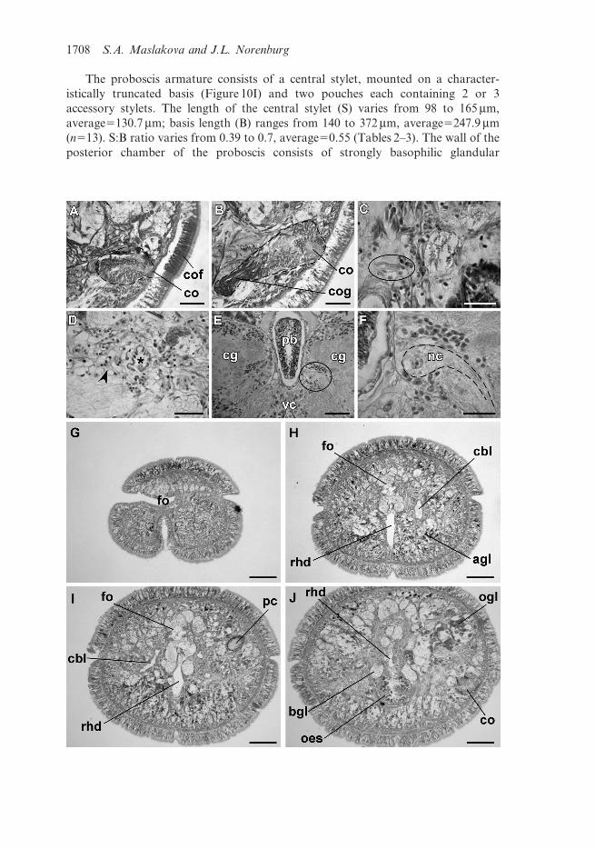
The proboscis armature consists of a central stylet, mounted on a character-
istically truncated basis (Figure 10I) and two pouches each containing 2 or 3
accessory stylets. The length of the central stylet (S) varies from 98 to 165 mm,
average5130.7 mm; basis length (B) ranges from 140 to 372 mm, average5247.9 mm
(n513). S:B ratio varies from 0.39 to 0.7, average50.55 (Tables 2–3). The wall of the
posterior chamber of the proboscis consists of strongly basophilic glandular
1708 S.A. Maslakova and J.L. Norenburg

epithelium organized into papillae, outer longitudinal muscle layer, thin inner
circular muscle layer, and a delicate endothelium.
Alimentary canal. The oesophagus opens into the rhynchodeum in front of the
proboscis insertion (Figure 3E). It is enclosed by longitudinal muscle fibres, which
are confluent with the rhynchodeal musculature and continue posteriorly as the
musculature of the stomach (Figures 3F and 3H). As in all other species of
Prosadenoporus, the anterior portion of oesophageal epithelium is richly supplied
with finely granular acidophilic gland cells, staining deep red with Crandall’s
trichrome technique (Figures 3F, 3J, 5J, 6A and 6B). Posteriorly the oesophageal
epithelium is ciliated and conspicuously lacks acidophilic or basophilic glands
(Figures 3G and 6C). This part sometimes is referred to in the literature as the
anterior stomach (see, for example, Moore and Gibson 1981, p. 184); however, it
more resembles oesophagus in morphology.
The stomach is of typical hoplonemertean structure with densely ciliated, deeply
folded epithelium, containing numerous basophilic and acidophilic glands
(Figures 3H and 6D). The intestinal caecum is well developed, anteriorly bifid and
bears numerous lateral diverticula throughout its length (Figure 6E). The anterior
caecal diverticula reach the posterior portion of the dorsal cerebral ganglia.
Intestinal diverticula are lobed.
Blood system. The blood system comprises paired lateral and an unpaired mid-dorsal
blood vessel, joined precerebrally via a suprarhynchodeal loop and posteriorly via a
supra-anal loop. As is characteristic of Prosadenoporus, the suprarhynchodeal
(cephalic) loop in P. floridensis sp. nov. is recurved: i.e., the lateral blood vessels run
forward into the tip of the head before curving up and backward to anastomose in
the midline just behind the posterior chamber of the frontal organ. The mid-dorsal
blood vessel originates near the ventral cerebral commissure from the right cephalic
vessel (traced in two specimens) and immediately penetrates the rhynchocoel floor to
form a single vascular plug. The wall of the vascular plug consists of thickened
endothelium of the blood vessel, a thin layer of extracellular matrix and a modified
Figure 5. Prosadenoporus floridensis sp. nov. (A) Slightly oblique transverse section through
the cerebral organ furrow and anterior portion of the cerebral organ (outlined) showing
cerebral organ gland; (B) transverse section through the posterior portion of the cerebral
organ (outlined) showing posterior lobe of the cerebral organ gland; (C) transverse section
through the terminal region of nephridia showing a particularly distinct binucleate flame cell
(circled); (D) transverse section through the cephalic blood vessel (asterisk) showing the
nephridial tubules and flame cells (arrowhead) crowded around the blood vessel; (E and F)
transverse sections through the brain, showing a neurochord cell (E circled; F outlined); (G–J)
a series of slightly oblique transverse sections from anterior tip to oesophagus: (G) the
prosorhochmid smile; (H) the frontal organ, (I) the anterior eye and cephalic blood vessel, (J)
one of the cerebral organs and anterior oesophagus. Notes: agl, acidophilic cephalic glands;
bgl, basophilic cephalic glands; cbl, cephalic vascular loop; cg, cerebral ganglia; co, cerebral
organ; cof, cerebral organ furrow; cog, cerebral organ gland; oes, oesophagus; fo, frontal
organ; nc, neurochord cell; ogl, orange-G cephalic glands; pc, ocellus; pb, proboscis; rhd,
rhynchodeum; vc, ventral brain commissure. Scales: (C and F), 20 mm; (D), 30 mm; (A, B and
E), 50 mm; (G–J), 100 mm.
Journal of Natural History 1709

rhynchocoel endothelium (Figure 4F). No transverse connectives linking mid-dorsal
and lateral blood vessels in the intestinal region were found by us. The blood vessels
are thin-walled with few ‘‘valves’’ and ‘‘pouches’’ (Figures 4D and 4E).
Nervous system. As in other nemerteans, the brain consists of two ventral and two
dorsal ganglia, joined by ventral (subrhynchocoelic) and dorsal (suprarhynchocoelic)
commissures, respectively (Figures 3C, 4I, 5E, 6B and 6C). The dorsal ganglia aremore widely separated than the ventral. A thin, but distinct outer neurilemma
encloses the brain as a whole, but there is no inner neurilemma dividing the fibrous
and ganglionic tissues. Notably, there is a pair of large and conspicuous neurochord
cells close to the inner side of the ventral cerebral ganglia in vicinity of the ventral
cerebral commissure (Figures 5E and 5F). However, no neurochords are found in the
lateral nerve cords (Figure 4G).
The lateral nerve cords contain a single fibrous core throughout their length, i.e.
there are no accessory nerve cords. As observed in most monostiliferans studied inthe last three decades, each lateral nerve cord contains a single delicate muscle
bundle, consisting of 3–7 fibres and running within or adjacent to the fibrous core,
near its dorsal border. In addition, there are several less conspicuous muscle fibres
running along the inner lateral side of the fibrous core (Figure 4G). Muscle fibres
associated with the lateral nerve cords can be traced to their origin near the proboscis
insertion. Numerous cephalic nerves lead anteriorly from the brain lobes to supply
various structures of the head. A pair of stout nerves originating from the ventral
brain lobes supplies the cerebral sensory organs. Paired proboscis nerve trunksoriginate from the ventral brain lobes near the ventral cerebral commissure and
branch before entering the proboscis (Figure 4A).
Frontal organ. The well-developed frontal organ opens at the tip of the head. Its
elongated ciliated canal, about 200–400 mm long, extends about half way to brain
(Figures 3C and 4I) and is lined by regionally differentiated epithelium. The ventral
and dorsal walls as well as the posterior extremity of the canal, through which the
basophilic mucus cephalic glands discharge, have a vacuolated appearance and bear
long, sparsely distributed cilia. The lateral walls of the canal, sometimes bearing amore or less distinct groove, comprise strongly acidophilic epithelium clad in short
densely arranged cilia (Figures 4L and 5G–I). The acidophilic appearance results
from the densely arranged elongated nuclei of the ciliated cells. Unlike some other
species of the genus, such as P. winsori, P. mooreae and P. spectaculum, the frontal
organ canal does not exhibit any noticeable ‘‘twisting’’ (compare Figures 4J–L, 5G–I
to 7A–C, 7F–I, 8A–D and 9C–F).
Cephalic glands. Cephalic glands are extremely well developed. As in other
Prosadenoporus, they include three types of cells: basophilic lobules with vacuolatedappearance (mucus glands), coarsely granular proteinaceous gland cells staining
golden-yellow to brown with Mallory trichrome or orange with Crandall’s method
(orange-G glands), and finely granular proteinaceous acidophilic cells, staining red
with Mallory or Crandall’s technique (acidophilic or red glands).
Mucus glands open into the frontal organ (Figures 3C and 4I), as well as through
the epidermis via multiple improvised ducts. Mucus glands are found precerebrally
around the frontal organ: organized into more compact lobes dorsally and ventrally
1710 S.A. Maslakova and J.L. Norenburg

and interspersed with the orange-G glands laterally (Figures 3C, 3F, 3H–J, 4I–L and
5H–J). Two major ventro-lateral lobes of the mucus gland descend closely on both sides
of the rhynchodeum and run posteriad ventro-lateral to the foregut (Figures 3F, 3I, 5J
and 6A–D). At the level of proboscis insertion dorsal lobes of mucus glands abound
above the rhynchodeum (Figures 5J and 6A). Numerous smaller lobes are found
between the body wall and internal organs laterally. In the cerebral region, mucus
glands occupy the dorso-medial region between the dorsal body wall and rhynchocoel,
as well as the region ventro-lateral to the brain ganglia. Ventro-lateral mucus tracts are
very prominent postcerebrally (Figure 6D). Further posteriad, ventro-lateral lobes
gradually become displaced by the caecal diverticula, while dorso-lateral lobes remain
prominent (Figure 6E). Mucus glands gradually decrease in number toward the
posterior and disappear at the end of pylorus.
Acidophilic glands are well developed and, for the most part, restricted to the
ventro-lateral precerebral region (Figures 5H and 5I). Individual acidophilic gland
cells are found dorsally in the precerebral region directly underneath the body wall
interspersed with the mucus cephalic glands (Figures 5H–J). Ventral acidophilic
glands largely disappear posterior to the proboscis insertion, while the scattered
dorsal gland cells can be found in the cerebral region.
Orange-G glands are extremely well developed. Overall, they present a
distribution typical for the genus. Two dorso-lateral tracts of orange-G glands first
appear at the level of the frontal organ and reach as far back as the end of the
pylorus. In the precerebral region, orange-G glands are found laterally between the
ventral acidophilic glands and dorsal mucus glands, and dorso-laterally, where they
intersperse with mucus glands (Figure 5J). The orange-G glands are very abundant
postcerebrally above the lateral nerve cords where they are interspersed with the
mucus glands (Figure 6D).
Cerebral organs. The compact paired cerebral organs are situated in front of the brain
between the anterior and posterior pairs of eyes. Each organ opens ventro-laterally into
a reduced anterior cephalic furrow (Figure 2A). The latter are shallow semi-circular
grooves lined by strongly acidophilic ciliated epithelium (Figure 5A). Cerebral organ
canals are not branched. Two types of gland cells can be distinguished in the cerebral
organs. The coarsely granular cells, staining dark reddish-brown with Crandall’s
method are found at anterior face of the cerebral organ (Figure 5A). The second type of
cell is finely granular, stains less intensively red to brownish purple and forms a single
lobe on the posterior face of each organ (Figure 5B). This posterior glandular lobe of
cerebral organs may extend under the brain.
Excretory system. The collecting tubules of the excretory system are thin-walled and
inconspicuous. Terminal flame cells are small, binucleate and reinforced by five to
seven very thin and indistinct transverse support bars. Flame cells are typically
observed embedded in the extracellular matrix in the vicinity of blood vessels, and
are particularly numerous around the cephalic blood vessels (Figures 5C and 5D).
Collecting tubules open to the outside via numerous inconspicuous thin-walled
nephridioducts. With effort, few flame cells and nephridioducts can be detected past
foregut region, but it is impossible to determine the full extent of the nephridial
system in this species with light microscopy.
Journal of Natural History 1711

Figure 6. (A–E) A series of slightly oblique transverse sections of Prosadenoporus floridensis
sp. nov. from precerebral region to pyloric region (proboscis is missing in this individual): (A)
Section through the posterior eyes immediately in front of cerebral ganglia; (B) cerebral
ganglia, ventral cerebral commissure and anterior oesophagus; (C) cerebral ganglia and
posterior oesophagus; (D) stomach; (E) pylorus and caecum. (F) transverse section through
the midgut region of another specimen showing ripe testes. Notes: bgl, basophilic cephalic
glands; cae, caecum; cbl, cephalic vascular loop; cdiv, caecal diverticulum; cg, cerebral ganglia;
cog, cerebral organ gland; dc, dorsal cerebral commissure; oes, oesophagus; int, intestine; lbv,
lateral blood vessel; lnc, lateral nerve cord; ogl, orange-G cephalic glands; pb, proboscis; pc,
ocellus; pyl, pylorus; rhc, rhynchocoel; rhd, rhynchodeum; stm, stomach; ts, testis; vc, ventral
brain commissure. Scales: (A–F), 100 mm.
1712 S.A. Maslakova and J.L. Norenburg
Reproductive system and life history. Sexes are separate. All sectioned sexually
mature individuals possessed either ovaries (Figure 4H) or testes (Figure 6F) andnever mixed gonads. Reproductive males and females were observed from January
to May in Florida. Males and females can be distinguished by the colour of mature
gonads: testes are whitish, while the ovaries are pinkish-orange, due to the colour
of mature oocytes. Males also tend to be smaller than females. Mature ovaries
contain three to seven oocytes each. Mature egg is about 380 mm in diameter, very
yolky, pinkish-orange to brownish-orange and enclosed within an egg envelop
approximately 430 mm in diameter. In captivity, several females laid clutches of
100–150 eggs on walls of glass or plastic containers above the water line.
Development is encapsulated. Gastrulation occurs on the second day of development
(approximately 36 h) and ciliation is obvious on the third day. Large and yolky cells ofepidermis, possibly corresponding to the transitory larval epidermis, described in some
other hoplonemertean larvae (Maslakova and Malakhov 1999; Maslakova and von
Dohren 2007), are apparent on the sixth day of development. Yolky, teardrop-shaped to
vermiform juveniles about 2 mm long hatch from the egg envelopes in about 6–10 days,
after which they may remain crawling within the mucus clutch for a few more days. Two-
week old juveniles have brain, ocelli and proboscis with a central stylet on a basis.
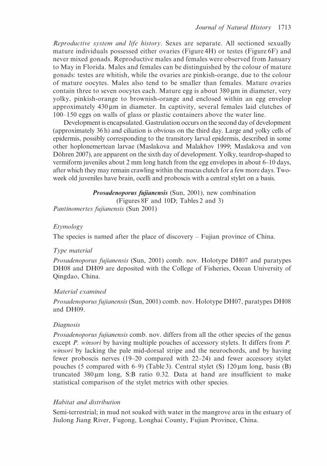
Prosadenoporus fujianensis (Sun, 2001), new combination
(Figures 8F and 10D; Tables 2 and 3)
Pantinomertes fujianensis (Sun 2001)
Etymology
The species is named after the place of discovery – Fujian province of China.
Type material
Prosadenoporus fujianensis (Sun, 2001) comb. nov. Holotype DH07 and paratypes
DH08 and DH09 are deposited with the College of Fisheries, Ocean University of
Qingdao, China.
Material examined
Prosadenoporus fujianensis (Sun, 2001) comb. nov. Holotype DH07, paratypes DH08
and DH09.
Diagnosis
Prosadenoporus fujianensis comb. nov. differs from all the other species of the genus
except P. winsori by having multiple pouches of accessory stylets. It differs from P.
winsori by lacking the pale mid-dorsal stripe and the neurochords, and by having
fewer proboscis nerves (19–20 compared with 22–24) and fewer accessory stylet
pouches (5 compared with 6–9) (Table 3). Central stylet (S) 120 mm long, basis (B)
truncated 380 mm long, S:B ratio 0.32. Data at hand are insufficient to make
statistical comparison of the stylet metrics with other species.
Habitat and distribution
Semi-terrestrial; in mud not soaked with water in the mangrove area in the estuary of
Jiulong Jiang River, Fugong, Longhai County, Fujian Province, China.
Journal of Natural History 1713

Remarks
The original description mentions 19 proboscis nerves in all three specimens,
however, our re-investigation showed that the paratypes have 19 and the holotype
has 20 proboscis nerves. Morphologically, this species most resembles another
mangrove-dwelling species – P. winsori from Queensland, Australia. Unfortunately,
Figure 7. Microscopic anatomy of Prosadenoporus species. (A–C) A series of transverse sections
through the frontal organ of Prosadenoporus enalios comb. nov.; relative position of the lateral
acidophilic regions (arrowheads) indicate a noticeable twist of the frontal organ canal; (D) a
longitudinal sagittal section through frontal organ of Prosadenoporus mooreae comb. nov.
(anterior to the right); (E–I) a series of slightly oblique transverse sections through the frontal
organ of P. mooreae comb. nov.; lateral acidophilic regions of the frontal organ (arrowheads),
note the twisting of the frontal organ canal; (J) transverse section through lateral nerve cord of
Prosadenoporus winsori comb. nov. showing the tentative neurochord (arrowhead); (K) binucleate
terminal cells of nephridia (flame cells) of P. winsori comb. nov. (arrowheads); (L) a longitudinal
sagittal section through the frontal organ of P. winsori comb. nov. (anterior to the left). Notes: bgl,
basophilic cephalic glands; cg, cerebral ganglia; fo, frontal organ; lnc, lateral nerve cord; rhd,
rhynchodeum. Scales: (A–C), and (E–I), 50 mm; (D), 100 mm; (J), 30 mm; (K), 20 mm; (L), 200 mm.
1714 S.A. Maslakova and J.L. Norenburg

tissue for molecular analysis was not available to us to compare the sequence
divergence between the two species.
Prosadenoporus mooreae (Gibson, 1982b), new combination
(Figures 1C, 1D, 7D–I and 10B; Tables 1–4)
Pantinonemertes mooreae (Gibson 1982b, 1990; Sundberg 1989; Gibson and
Sundberg 1992; Sun 2001)
Etymology
The species is named after Dr Janet Moore of the Department of Zoology,
University of Cambridge, as a tribute to her work on the terrestrial nemerteans of
the world.
Type material
Prosadenoporus mooreae (Gibson, 1982b) comb. nov. Sections of holotype W5903
and paratype W5904 are held at the Australian Museum, Sydney, Australia.
Material examined
Prosadenoporus mooreae (Gibson, 1982b) comb. nov. Holotype W5903 and paratype
W5904. Several additional specimens (most sectioned) collected by SAM in March
2003 from Picnic Bay and Cockle Bay, Magnetic Island, Queensland, Australia held
at the National Museum of Natural History in Washington, DC, USA (USNM
1087356, 1087357, 1087359–1087361, 1087358). One unsectioned specimen from the
same collecting trip G20028 is held in 70% ethanol at the Museum of Tropical
Queensland, Townsville, Queensland, Australia.
Diagnosis
Prosadenoporus mooreae comb. nov. does not have any known morphological
apomorphies. It differs from all other species of the genus except P. mortoni by
having a distinct greenish colour pattern with a dark-green to black mid-dorsal
longitudinal stripe (Figures 1C and 1D). Similar to P. mortoni, P. mooreae has a very
slender rounded stylet basis (Figure 10B). With the exception of the number of
Table 4. Percentage sequence divergence between species of Prosadenoporus species and
Pantinonemertes californiensis (%, 16S/COI).
P. winsori P. mooreae P. mortoni P. floridensis sp.
nov.
P. winsori – – – –
P. mooreae 7.8/10.2 – – –
P. mortoni 7.9/10.6 2.4/4.1 – –
P. floridensis sp. nov. 10.3/10.8 7.8/10.3 8.6/10.4 –
P. californiensis 7.3/13.1 7.6/12.6 7.6/12.8 9.9/11.1
Journal of Natural History 1715

proboscis nerves (15–16 vs. 14) these two species are morphologically indistinguish-
able (Table 2, 3) and represent the closest molecular pair in the genus, exhibiting
sequence divergence of 2.4% (16S) and 4.1% (COI) (Tables 1 and 4). Two accessory
stylet pouches. Central stylet 50–130 mm long, average5105 mm, basis (B) rounded
130–160 mm long, average5145 mm, S:B ratio 0.33–0.92, average50.73. All three
stylet metrics are significantly different from those of P. winsori. The S:B ratio is
significantly different from that of Prosadenoporus floridensis (p50.05). Data at hand
are insufficient to make the statistical comparisons with other species.
Habitat and distribution
In silty mud beneath rocks and coral boulders, mid- to low-shore, intertidal. Picnic
Bay and Cockle Bay, Magnetic Island, Queensland, Australia.
Remarks
Two specimens were collected originally in Picnic Bay by R. Gibson in 1975. SAM
collected nine additional specimens from the nearby Cockle Bay in March 2003, and
two from Picnic Bay. Gibson (1982b) reported only one measurement for the stylet
apparatus (S:B ratio50.33), which is very different from that observed by SAM
(average S:B for three specimens50.86). With the data at hand it is not possible to
determine whether the difference is statistically significant. We pooled our
measurements with Gibson’s and adjusted the means for the species accordingly.
Contrary to Gibson’s observations, SAM found that P. mooreae uses its
proboscis for rapid locomotion. Upon the slightest contact the worm everts its
Figure 8. (A–D) A series of transverse sections through the frontal organ of Prosadenoporus
winsori comb. nov.; relative position of the lateral acidophilic regions (arrowheads) indicate a
noticeable twist of the frontal organ canal; (E) Neurochord cell of P. winsori comb. nov.
(circled, insert); (F) Neurochord cell of Prosadenoporus fujianensis comb. nov. (circled, insert).
Notes: bgl, basophilic cephalic glands; cg, cerebral ganglia; fo, frontal organ; pb, proboscis.
Scales: (A–F), 100 mm; inserts, 50 mm.
1716 S.A. Maslakova and J.L. Norenburg

proboscis and quickly draws it back in, pulling itself into a crevice and out of reach
of the collector.
Prosadenoporus mortoni (Gibson, 1990), new combination
(Figure 10A; Tables 1–4)
Pantinonemertes mortoni (Gibson 1990; Gibson and Sundberg 1992; Sun 2001)
Etymology
The species is named in honour of Professor Brian Morton, as a tribute to his
considerable involvement in furthering the knowledge of the marine flora and fauna
of the Hong Kong region.
Type material
Prosadenoporus mortoni (Gibson, 1990) comb. nov. Sections of the holotype 1987-4-
54 are deposited at the Natural History Museum in London, UK (coll. P. G. Oliver
and A. S. Y. Mackie. Starfish Bay, Hong Kong).
Material examined
Prosadenoporus mortoni (Gibson, 1990) comb. nov. Holotype 1987-4-54. Additional
material coll. by Dr Shichun Sun, Nan Ao Island and Xiamen, Fujian Province,
China; sectioned and deposited at the National Museum of Natural History in
Washington, DC, USA (USNM 1020576–1020577).
Diagnosis
Prosadenoporus mortoni comb. nov. does not have any known morphological
apomorphies. It differs from all other species of the genus except P. mooreae by
having a distinct colour pattern with a greenish-black mid-dorsal longitudinal stripe.
P. mortoni shares with P. mooreae the characteristic slender rounded basis of central
stylet (Figure 10A) and is nearly indistinguishable from it morphologically. The two
species represent the closest molecular pair in the genus exhibiting sequence
divergence of 2.4% (16S) and 4.1% (COI) (Tables 1 and 4). Number of accessory
stylet pouches unknown. Central stylet (S) 125 mm long, basis (B) rounded 123 mm
long, S:B ratio 1.0. Data at hand are insufficient to make the statistical comparisons
of stylet metrics with other species.
Habitat and distribution
Under rocks on coarse sand, lower shore; Starfish Bay, Hong Kong; Xiamen and
Nan Ao Island, Fujian Province, China.
Remarks
The original description is based on a single specimen from Hong Kong and has no
account of the stylet apparatus. Dr Shichun Sun kindly provided three additional
specimens collected and identified by him from Fujian Province in China, used here
Journal of Natural History 1717
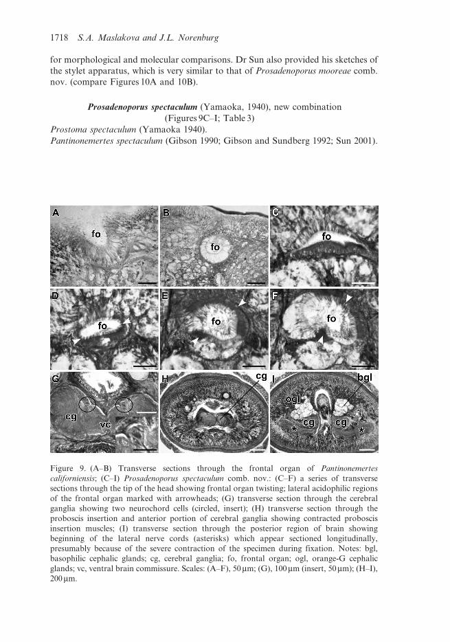
for morphological and molecular comparisons. Dr Sun also provided his sketches of
the stylet apparatus, which is very similar to that of Prosadenoporus mooreae comb.
nov. (compare Figures 10A and 10B).
Prosadenoporus spectaculum (Yamaoka, 1940), new combination
(Figures 9C–I; Table 3)
Prostoma spectaculum (Yamaoka 1940).
Pantinonemertes spectaculum (Gibson 1990; Gibson and Sundberg 1992; Sun 2001).
Figure 9. (A–B) Transverse sections through the frontal organ of Pantinonemertes
californiensis; (C–I) Prosadenoporus spectaculum comb. nov.: (C–F) a series of transverse
sections through the tip of the head showing frontal organ twisting; lateral acidophilic regions
of the frontal organ marked with arrowheads; (G) transverse section through the cerebral
ganglia showing two neurochord cells (circled, insert); (H) transverse section through the
proboscis insertion and anterior portion of cerebral ganglia showing contracted proboscis
insertion muscles; (I) transverse section through the posterior region of brain showing
beginning of the lateral nerve cords (asterisks) which appear sectioned longitudinally,
presumably because of the severe contraction of the specimen during fixation. Notes: bgl,
basophilic cephalic glands; cg, cerebral ganglia; fo, frontal organ; ogl, orange-G cephalic
glands; vc, ventral brain commissure. Scales: (A–F), 50 mm; (G), 100 mm (insert, 50 mm); (H–I),
200 mm.
1718 S.A. Maslakova and J.L. Norenburg

Etymology
The name reflects the unique spectacle-shaped pattern of pigmentation on the head
of this species.
Type material
Type material almost certainly lost.
Material examined
Prosadenoporus spectaculum (Yamaoka, 1940) comb. nov. Two sectioned specimens
1987-2-43 and 1987-2-42 held at the Museum of Natural History, London, UK.
Diagnosis
Prosadenoporus spectaculum comb. nov. is distinguished from other species in the
genus by the characteristic spectacle or helmet-shaped pattern of pigmentation on
the head. Additionally, P. spectaculum differs from P. winsori in lacking neurochords
and from both P. winsori and P. fujianensis by having a single pair of accessory stylet
pouches. It differs from P. mooreae, P. mortoni, P. enalios and P. arenarius by having
neurochord cells (Figure 9G) and from P. agricola and P. floridensis by having more
proboscis nerves (18–22 compared with 11–14 in P. floridensis and 12–15 in P.
agricola). The shape and size of stylet and basis are not known.
Habitat and distribution
Originally described from Naha and Tinen, Riukiu Islands, Japan; habitat unknown.
Three specimens on which the re-description by Gibson (1990) is based were
collected from rock crevices and clefts just below high tide level in Starfish Bay and
Tai Mong Tsai, Hong Kong.
Remarks
Gibson (1990) stated that one of the diagnostic features of this species is presence of
the split precerebral septum (Kirsteuer 1974). Our re-investigation of one of the
Hong Kong specimens deposited at the British Museum of Natural History revealed
that the structure of the precerebral septum (i.e. proboscis insertion muscles) in this
species is the same as in other members of the genus; that is there is no distinct
precerebral septum. However, we observed that the voucher specimen is severely
contracted – as evident from the position of lateral nerve cords – which causes
longitudinal muscle fibres in the proboscis insertion to appear as radial, giving the
impression of the ‘‘split precerebral septum’’ (Figures 9H and 9I).
Prosadenoporus winsori (Moore and Gibson, 1981), new combination
(Figures 1B, 1E, 1F, 7J–L, 8A–E, 10G and 10H; Tables 1–4)
Pantinonemertes winsori (Moore and Gibson 1981).
Etymology
The species is named after Dr Leigh Winsor, who first drew attention to the species.
Journal of Natural History 1719
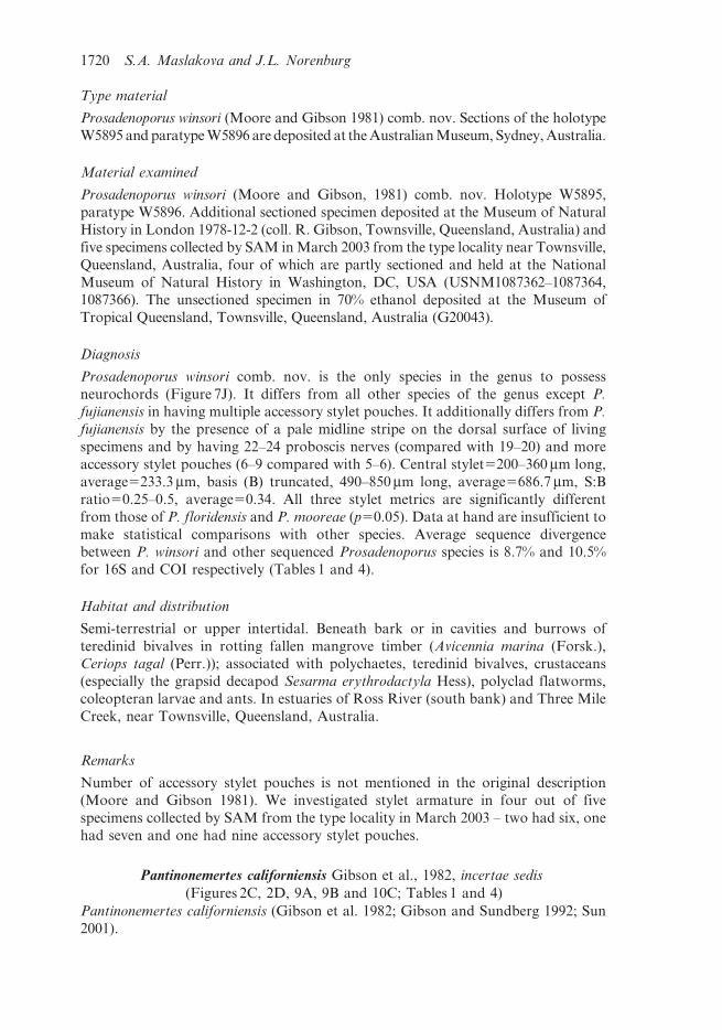
Type material
Prosadenoporus winsori (Moore and Gibson 1981) comb. nov. Sections of the holotype
W5895 and paratype W5896 are deposited at the Australian Museum, Sydney, Australia.
Material examined
Prosadenoporus winsori (Moore and Gibson, 1981) comb. nov. Holotype W5895,
paratype W5896. Additional sectioned specimen deposited at the Museum of Natural
History in London 1978-12-2 (coll. R. Gibson, Townsville, Queensland, Australia) and
five specimens collected by SAM in March 2003 from the type locality near Townsville,
Queensland, Australia, four of which are partly sectioned and held at the National
Museum of Natural History in Washington, DC, USA (USNM1087362–1087364,1087366). The unsectioned specimen in 70% ethanol deposited at the Museum of
Tropical Queensland, Townsville, Queensland, Australia (G20043).
Diagnosis
Prosadenoporus winsori comb. nov. is the only species in the genus to possess
neurochords (Figure 7J). It differs from all other species of the genus except P.
fujianensis in having multiple accessory stylet pouches. It additionally differs from P.
fujianensis by the presence of a pale midline stripe on the dorsal surface of living
specimens and by having 22–24 proboscis nerves (compared with 19–20) and more
accessory stylet pouches (6–9 compared with 5–6). Central stylet5200–360 mm long,
average5233.3 mm, basis (B) truncated, 490–850 mm long, average5686.7 mm, S:B
ratio50.25–0.5, average50.34. All three stylet metrics are significantly different
from those of P. floridensis and P. mooreae (p50.05). Data at hand are insufficient to
make statistical comparisons with other species. Average sequence divergencebetween P. winsori and other sequenced Prosadenoporus species is 8.7% and 10.5%
for 16S and COI respectively (Tables 1 and 4).
Habitat and distribution
Semi-terrestrial or upper intertidal. Beneath bark or in cavities and burrows of
teredinid bivalves in rotting fallen mangrove timber (Avicennia marina (Forsk.),Ceriops tagal (Perr.)); associated with polychaetes, teredinid bivalves, crustaceans
(especially the grapsid decapod Sesarma erythrodactyla Hess), polyclad flatworms,
coleopteran larvae and ants. In estuaries of Ross River (south bank) and Three Mile
Creek, near Townsville, Queensland, Australia.
Remarks
Number of accessory stylet pouches is not mentioned in the original description
(Moore and Gibson 1981). We investigated stylet armature in four out of five
specimens collected by SAM from the type locality in March 2003 – two had six, one
had seven and one had nine accessory stylet pouches.
Pantinonemertes californiensis Gibson et al., 1982, incertae sedis
(Figures 2C, 2D, 9A, 9B and 10C; Tables 1 and 4)
Pantinonemertes californiensis (Gibson et al. 1982; Gibson and Sundberg 1992; Sun
2001).
1720 S.A. Maslakova and J.L. Norenburg

Type material
Type material deposited at the Museum of Natural History in London, UK consists
of sections of the holotype (female) 1981.1.2 and paratype (male) 1981.1.3.
Material examined
We examined sectioned specimens from the personal collection of Frank Crandall
(National Museum of Natural History, Smithsonian Institution, Washington DC,
USA) collected from Millerton Creek marsh between Hwy 1 and the bay just
opposite the Hwy 1 gate to the Borello ranch, Tomales Bay, CA, USA (FC031,
FC034–37, FC085, FC112) and sections of one specimen collected by F. Crandall in
July 1981 from the Bolsa Chica Ecological Reserve, CA, USA (a California State
Figure 10. Central stylets: (A) Prosadenoporus mortoni comb. nov. (a sketch of stylet apparatus
provided by Dr Shichun Sun); (B) Prosadenoporus mooreae comb. nov. (specimen from the type
locality coll. by SAM); (C) Pantinonemertes californiensis (after Gibson et al. 1982, p. 471, fig. 3b);
(D) Prosadenoporus fujianensis comb. nov. (redrawn from Sun 2001, p. 205, fig. 23); (E)
Prosadenoporus enalios comb. nov. (redrawn from Moore and Gibson 1981, p. 183, plate III,
fig. c); (F) Prosadenoporus agricola comb. nov. (redrawn from Coe 1904. p. 571. plate 23, fig. 3);
(G–H) Prosadenoporus winsori comb. nov. (specimen from the type locality coll. by SAM); (I)
Prosadenoporus floridensis sp. nov. Note: Scales: (A, B, E, F), 100 mm; (C, D, G, H, I), 500 mm.
Journal of Natural History 1721

designated area near Huntington Beach) (FC102–104, FC 109–111). We anticipate
these to be deposited with the NMNH. We also examined additional specimens
collected by F. Crandall from Tomales Bay, CA, USA deposited at the NMNH:
USNM 64138 (70% ethanol) and USNM 1076584–1076587 (slides).
Pantinonemertes daguilarensis Gibson and Sundberg, 1992, incertae sedis
Pantinonemertes daguilarensis (Gibson and Sundberg 1992; Sun 2001).
Type material
Pantinonemertes daguilarensis Gibson and Sundberg, 1992. Sections of the holotype
1990-7-5, and paratypes 1990-7-6 and 1990-7-7 are deposited at the Museum of
Natural History in London, UK.
Material examined
Pantinonemertes daguilarensis Gibson and Sundberg, 1992. Holotype 1990-7-5,
paratypes 1990-7-6 and 1990-7-7.
Discussion and conclusions
Synonymization of Pantinonemertes and Prosadenoporus
According to the amended diagnosis of the currently monotypic genus
Prosadenoporus by Moore and Gibson (1988, p. 82), the main difference between
the Pantinonemertes Moore and Gibson, 1981 and Prosadenoporus Burger, 1890 is
the presence of a second cephalic vascular loop, described for Prosadenoporus by
Moore and Gibson (1988, p. 81, fig. 18). We re-examined sections of the voucher
specimen of the only valid species of Prosadenoporus – P. arenarius Burger, 1890 and
were not able to confirm the presence of a second cephalic vascular loop. We also
could not find any other morphological characters that could distinguish
Prosadenoporus from Pantinonemertes. Therefore, we propose to synonymize
Prosadenoporus Burger, 1890 and Pantinonemertes Moore and Gibson, 1981.
According to the principle of priority (ICZN Art. 23), Prosadenoporus Burger,
1890 takes precedence over Pantinonemertes Moore and Gibson, 1981.
Prosadenoporus floridensis sp. nov. – a new semi-terrestrial prosorhochmid
We describe a new semi-terrestrial prosorhochmid species from Florida and Belize.
The presence of the prosorhochmid smile, reduced cerebral organ furrows, extremely
well-developed frontal organ with laterally differentiated epithelium, well-developed
cephalic glands including basophilic, acidophilic and orange-G glands and truncated
stylet basis support the prosorhochmid affinity of this species. However, these
characters are shared by Prosadenoporus and Prosorhochmus species. Characters that
distinguish this species from Prosorhochmus and support its position within
Prosadenoporus include: thin-walled nephridial canals; extreme multiplication of
the nephridiopores; binucleate flame cells reinforced by transverse support bars;
presence of acidophilic glands in the anterior esophagus; elongated shape of the
frontal organ; and recurved cephalic vascular loop.
1722 S.A. Maslakova and J.L. Norenburg
Prosadenoporus floridensis differs from the closest geographical congener, a
semi-terrestrial species from Bermuda P. agricola, by being gonochoric and
oviparous. The average sequence divergence between P. floridensis and other
Prosadenoporus species is 9.15% (16S) and 10.65% (COI) and 7.8% (16S) and
10.3% (COI) from the nearest sequenced congener P. mortoni, which is comparable
to the sequence divergence between other sequenced species of Prosadenoporus
(Tables 1 and 4).
Two species of uncertain taxonomic affinity
Pantinonemertes daguilarensis, a fully marine species from Hong Kong, reaches a
maximum length of 200 mm and width of 0.75 mm. Colour in life varies from light
grey with a brown tinge to pale pink; the posterior half of the intestinal region being
darker gray or purplish-gray. Unlike all other Pantinonemertes (now
Prosadenoporus) species, P. daguilarensis has a distinct cephalic lobe, numerous
eyes situated in groups or rows, lacks orange-G cephalic glands and its frontal organ
is a simple epithelial pit. Furthermore, it lacks the prosorhochmid smile and lateral
differentiation of the epithelium in the frontal organ canal, has a planar (as opposed
to recurved) cephalic vascular loop, forked cerebral organ canals, lacks acidophilic
glands in the anterior esophagus, possesses an accessory nerve in the lateral nerve
cords, has thick-walled nephridial canals, and only a few nephridiopores.
Additionally, upon reinvestigation of the type and paratype specimens, we have
not been able to find any terminal nephridial flame cells characteristic of
Prosadenoporus species (confirmed by Frank Crandall, personal communication to
authors), although they are described by Gibson and Sundberg (1992) even as having
distinct support bars.
Pantinonemertes daguilarensis originally was placed in the genus Pantinonemertes
by Gibson and Sundberg (1992) based on the presence of anteriorly split body-wall
longitudinal muscle, simple cephalic vascular loop, presence of diagonal musculature
in the body wall, lack of precerebral septum, cephalic retractor muscles derived only
from the outer portion of the divided longitudinal musculature, and a rhynchocoel
near full body length. We argue that these characters are not sufficient to justify
placement of this species within Prosadenoporus. First, a diagonal muscle layer in the
body wall is present in almost all monostiliferan species studied by us and it is easily
missed when only cross-sections are investigated. It cannot serve as a diagnostic
character. Simple vascular loop and rhynchocoel near full body length are so widely
distributed among the monostiliferous hoplonemerteans that they most likely
represent plesiomorphic similarity. It is the alternatives that are unusual and
characteristic. Finally, anteriorly split longitudinal musculature, or something
interpreted as such, is reported to occur in over 25 monostiliferan genera
(Chernyshev 2002), and derivation of the cephalic retractor muscles is not a
sufficiently well-studied character to be the sole basis for generic placement. Because
the differences from other species of the genus are substantial and the only
similarities are likely plesiomorphies, we argue that Pantinonemertes daguilarensis
does not belong in Prosadenoporus. Until more data are available to clarify position
of this species, it may be referred to as species incertae sedis.
Pantinonemertes californiensis, an oviparous, dioecious, semi-terrestrial species
from the coast of California, reaches a maximum length of 265 mm and width of
Journal of Natural History 1723

3 mm. Dorsal colour in life is greenish-brown to grayish-green, shading to a lighter
greenish-tan laterally; pigmentation is darker in the dorsal midline, forming a stripe
of very dark green without clearly delimited margins; ventral surface is light cream to
tan and head is almost the same colour. Mature oocytes are characteristically coral
red, while testes show through the body wall as salmon pink or cream-coloured
opaque masses.
Unlike other prosorhochmids, Pantinonemertes californiensis has numerous
eyes situated in four groups or rows (compared with four simple eyes) (Figure 2D),
oblique cerebral organ furrows (compared with reduced ventro-lateral arches)
(Figures 2C and 2D) and a weakly developed frontal organ (Figures 9A and 9B).
However, similar to other prosorhochmids, P. californiensis possesses well-
developed cephalic glands, including basophilic and proteinaceous (red and
orange-G) cells, reaching well into foregut region and some into intestinal region.
Unlike species of Prosadenoporus, P. californiensis lacks the prosorhochmid
smile (Figure 2D) and epithelial differentiation of the frontal organ canal
(Figures 9A and 9B). In this species, the frontal organ is but a small
undifferentiated ciliated pit at the tip of the head. Pantinonemertes californiensis
resembles Prosadenoporus species in having binucleate flame cells reinforced by
distinct support bars and thin-walled nephridial canals opening via a large number
of nephridiopores.
Similar to the Prosadenoporus species occupying semi-terrestrial and terrestrial
habitats, such as P. winsori and P. fujianensis, Pantinonemertes californiensis has a
large proboscis with multiple accessory stylet pouches. Morphological cladistic
analysis of the family Prosorhochmidae with several hoplonemertean outgroups
places P. californiensis outside Prosadenoporus, while molecular analysis based on
partial sequences of 16S and COI and the combined morphological and molecular
analysis place it either as a sister clade to Prosadenoporus or outside of the
Prosorhochmidae (Maslakova 2005). Average sequence divergence between P.
californiensis and Prosadenoporus species is 8.1% for 16S and 12.4% for COI;
comparable to, if slightly larger than, the sequence divergence between such species
as P. winsori and P. mooreae. Until more data are available to clarify its position,
this species also may best be treated as incertae sedis.
It had been argued previously that Pantinonemertes was erected invalidly because
the monotypic Neonemertes agricola (Willemoes-Suhm, 1874) was among the
originally included nominal species (Chernyshev 1992), rendering Pantinonemertes
an objective junior synonym and unavailable. If Pantinonemertes is treated as valid,
as it functionally has been since 1981, our action here would be to treat it as a junior
subjective synonym and potentially available, except that we transfer the type species
to Prosadenoporus. Thus, either way, Pantinonemertes is unavailable for P.
californiensis and P. daguilarensis. With respect to these two species, we anticipate
useful phylogenetic realignment and consequent taxonomic actions as DNA
sequence data for other distromatonemerteans become available in the near future,
but we see no practical or useful immediate options. This is especially so with respect
to P. californiensis, which now is well known from California to Puget Sound and is
the subject of a significant number of publications. For this reason, we can envision
supporting, within the context of the ICZN, a proposal to make the name available.
This latter route is preferable to creating new monotypic genera as an almost certain
short-lived solution.
1724 S.A. Maslakova and J.L. Norenburg

Acknowledgements
The authors thank Barbara Littman (National Museum of Natural History, Smithsonian
Institution, Washington, DC, USA) for assistance in preparation of histological slides, Lee
Weigt, Ken Shallop, Ken Wurdack, Jeff Bates and other researchers and staff at the
Laboratory of Analytical Biology (Smithsonian Institution) for help with molecular work.
Unpublished data, photomicrographs, sketches and specimens were kindly provided by Frank
Crandall (NMNH, Smithsonian Institution, Washington, DC, USA) and Shichun Sun
(College of Fisheries, University of Quingdao, China). Many curators, collection managers
and other biologists worldwide assisted with loans and field-work. The authors are especially
grateful to Michael Akam, Ray Symonds and Janet Moore (University Museum of Zoology,
Cambridge, UK), Sheila Halsey (Museum of Natural History, London, UK), Shichun Sun
(College of Fisheries, University of Quingdao, China), staff of the Western Australian
Museum (Perth, Australia), staff of the Australian Museum (Sydney, Australia) and staff of
the Museum of Tropical Queensland (Townsville, Australia). SAM especially thanks Michael
Akam, Janet Moore and Cassandra Extavour for their hospitality and assistance during her
visit to the University Museum of Zoology in Cambridge, UK. The field-work in Australia
was made possible by the generous help of Leigh Winsor, late Peter Arnold, Carden Wallace,
Gilianne Brodie, Estelle Davies, Mark O’Callaghan and the staff of the Orpheus Island
Research Station. SAM is grateful to the owners of ‘‘The Rocks’’ in Townsville: Jeanne, Joe
and Sebastian, for their hospitality. The field-work in Belize was assisted by Cheryl Thacker.
The authors thank Mary Rice and the staff of the Smithsonian Marine Station at Fort Pierce
for their help and assistance during our work in Florida. This project was supported by the US
National Science Foundation (DEB-9712463 to J. Norenburg and D. Lipscomb). The authors
further acknowledge material and financial support of the George Washington University and
the Smithsonian Institution. This is contribution number 733 of the Smithsonian Marine
Station at Fort Pierce and number 821 of the Smithsonian Caribbean Coral Reef Ecosystem
Station (Carrie Bow Cay, Belize).
References
Burger O. 1890. Untersuchungen uber die anatomie und histologie der nemertinen nebst
beitragen zur systematik [Studies of anatomy and histology of nemerteans with contribution
to systematics]. Z Wiss Zool. 50:1–277. German.
Burger O. 1895. Die nemertinen des golfes von Neapel und der angrenzenden Meeres-
Abschnitte [Nemerteans of the Gulf of Naples and surrounding waters]. Fauna and Flora
des Golfes von Neapel und der angrenzenden Meeres-Abschnitte [Fauna and Flora of the
Gulf of Naples and surrounding waters], Vol. 22. Berlin (Germany): Verlag von R.
Friedlander & Sohn. German. p. 1–743.
Burger O. 1897–1907. Nemertini (Schnurwurmer). In: Bronn HG, editor. Klassen und
Ordnungen des Tier-Reichs. Vol. 4, Supplement. Leipzig (Germany): C. F. Winter. p.
1–542.
Burger O. 1904. Nemertini. Tierreich 20:1–151.
Chernyshev AV. 1992. O nazvaniyakh nekotoruikh nemertin [About the names of some
nemerteans]. Zool Zh. 71:134–136. Russian.
Chernyshev AV. 2002. Opisanie novogo vida roda Poseidonemertes (Nemertea, Monostilifera)
s obosnovaniem semeistva Poseideonmemertidae [Description of a new species of the
genus Poseidonemertes (Nemertea, Monostilifera) with establishment of the family
Poseidonemertidae]. Zool Zh. 81:909–916. Russian with English summary.
Coe WR. 1904. The anatomy and development of the terrestrial nemertean Geonemertes
agricola of Bermuda. Proc Bost Soc Nat Hist. 31:531–570.
Journal of Natural History 1725

Coe WR. 1905. Nemerteans of the west and northwest coasts of America. Bulletin of the
Museum of Comparative Zoology at Harvard College 47:1–318.
Coe WR. 1929. The excretory organs of terrestrial nemerteans. Biol Bull. 56:306–311.
Coe WR. 1939. Sexual phases in terrestrial nemerteans. Biol Bull. 76:416–427.
Coe WR. 1940. Revision of the nemertean fauna of the Pacific coasts of North, Central, and
northern South America. Allan Hancock Pacific Expeditions 2:247–322.
Crozier WJ. 1917. Note on the habitat of Geonemertes agricola. Am Nat. 51:758–760.
Folmer O, Black M, Hoeh W, Lutz R, Vrijenhoek R. 1994. DNA primers for amplification of
mitochondrial cytochrome c oxidase subunit I from diverse metazoan invertebrates. Mol
Mar Biol Biotech 3:294–299.
Friedrich H. 1955. Beitrage zu einer Synopsis der Gattungen der Nemertini monostilifera
nebst Bestimmungsschlussel [Contribution to synopsis of monostiliferan nemertean
genera with identification key]. Z Wiss Zool Abt A. 158:133–192. German.
Gibson R. 1972. Nemerteans. London (UK): Hutchinson and Co Ltd. 224 p.
Gibson R. 1982a. Nemertea. In: Parker SP, editor. Synopsis and classification of living
organisms. Vol. 1. New York: McGraw-Hill. p. 823–846.
Gibson R. 1982b. Nemerteans of the Great Barrier Reef. 5. Enopla Hoplonemertea
(Monostilifera). Zool J Linn Soc. 75:269–296.
Gibson R. 1990. The macrobenthic nemertean fauna of Hong Kong. In: Morton B, editor.
The Marine Flora and Fauna of Hong Kong and Southern China. Hong Kong: Hong
Kong University Press. p. 33–212.
Gibson R, Moore J. 1985. The genus Prosorhochmus Keferstein, 1862 (Hoplonemertea). J
Zool. 206:145–162.
Gibson R, Moore J. 1989. Functional requirements for the invasion of land and freshwater
habitats by nemertean worms. J Zool. 219:517–526.
Gibson R, Moore J, Crandall FB. 1982. A new semi-terrestrial nemertean from California. J
Zool. 196:463–474.
Gibson R, Sundberg P. 1992. Three new nemerteans from Hong Kong. In: Morton B, editor,
Hong Kong: Hong Kong University Press. p. 97–129.
Gibson R, Sundberg P. 2001. Some nemerteans (Nemertea) from Queensland and the Great
Barrier Reef, Australia. Zool Sci. 18:1259–1273.
Girard C. 1893. Recherches sur les planaries et les nemertiens de l’Amerique du Nord [Studies
on the flatworms and the nemerteans of the North America]. Ann Sci Nat. Ser. 7.
15:145–310. French.
Hubrecht AAW. 1887. Report on the nemertea collected by HMS Challenger during the years
1873–76. Report of the scientific results of the voyage of the HMS Challenger during the
years 1873–76. Zoology 19:1–150.
International Commission on Zoological Nomenclature. 1999. International Code of
Zoological Nomenclature. Ride WDL, Cogger HG, Dupuis C, Kraus O, Minelli A,
Thompson FC, Tubbs PK, editors, London (UK): The International Trust for
Zoological Nomenclature.
Joubin L. 1894. Les Nemertiens [The Nemerteans]. In: Blanchard R, de Guerne J, editors.
Fauna Francaise. Paris (France): Societe d’editions Scientifiques. p. 1–235. French.
Kirsteuer E. 1974. Description of Poseidonemertes caribensis sp. n., and discussion of other
taxa of Hoplonemertini Monostilifera with divided longitudinal musculature in the body
wall. Zool Scr. 3:153–166.
Maslakova SA. 2005. Systematics and evolution of smiling worms (Fam. Prosorhochmidae,
Hoplonemertea, Nemertea) [PhD Thesis]. [Washington (DC)]: George Washington
University.
Maslakova SA, Malakhov VV. 1999. Hidden larva in the Hoplonemertini order of
nemerteans. Doklady Akademii Nauk. 366(6):849–852.
1726 S.A. Maslakova and J.L. Norenburg

Maslakova SA, Norenburg JL. 2008. Revision of the smiling worms, genus Prosorhochmus
Keferstein 1862, and description of a new species, Prosorhochmus belizeanus sp. nov.
(Prosorhochmidae; Hoplonemertea; Nemertea) from Florida and Belize. J Nat Hist
42(17-20):1219–1260.
Maslakova SA, Thiel M, Vasquez N, Norenburg JL. 2005. The smile of Amphiporus nelsoni
Sanchez, 1973 (Nemertea: Hoplonemertea: Monostilifera: Amphiporidae) leads to a
redescription and a change in family. Proc Biol Soc Wash. 118:483–498.
Maslakova SA, von Dohren J. 2007. Enigmatic transitory epidermis in planktonic larval
development of a hoplonemertean Paranemertes peregrina and its significance in
evolution of development in nemerteans (Phylum Nemertea; Lophotrochozoa). Integr
Comp Biol. 47:e1–e152.
Moore J. 1973. Land nemertines of New Zealand. Zool J Linn Soc. 52:293–313.
Moore J. 1975a. A land nemertine from Mauritius. Mauritius Inst Bull. 8:27–32.
Moore J. 1975b. Land nemertines of Australia. Zool J Linn Soc. 56:23–43.
Moore J. 1985. The distribution and evolution of terrestrial nemertines. Am Zool. 25:15–21.
Moore J, Gibson R. 1981. The Geonemertes problem (Nemertea). J Zool. 194:175–201.
Moore J, Gibson R. 1985. The evolution and comparative physiology of terrestrial and
freshwater nemerteans. Biol Rev. 60:257–312.
Moore J, Gibson R. 1988. Marine relatives of terrestrial nemerteans – the genus
Prosadenoporus Burger, 1890 (Hoplonemertea). Hydrobiologia 156:75–86.
Moore J, Gibson R, Jones HD. 2001. Terrestrial nemerteans thirty years on. Hydrobiologia
456:1–6.
Moore J, Moore NW. 1972. Land nemertines of Madeira and the Azores. Boletim do Museu
Municipal do Funchal. 26:31–44.
Moseley HN. 1879. Corals. Report of the scientific results of the voyage of the HMS
Challenger during years 1873–1876. 2:26–27.
Palumbi S, Martin A, Romano S, McMillan WO, Stice L, Grabowski G. 1991. The simple
fools guide to PCR Version 2.0. Honolulu (HI): Department of Zoology and Kewalo
Marine Laboratory, University of Hawaii.
Pantin CFA. 1947. The nephridia of Geonemertes dendyi. Q J Microsc Sci. 88:15–25.
Pantin CFA. 1961. Geonemertes: a study in island life. Proc Linn Soc. Lon. 172:137–152.
Pantin CFA. 1969. The genus Geonemertes. B Brit Mus – Nat Hist: Zool. 18:261–310.
Punnet RC. 1901. Nemerteans. In: Gardiner JS, editor. The fauna and geography of the
Maldive and Laccadive archipelagoes. Cambridge (UK): Cambridge University Press. p.
101–118.
Semper C. 1863. Reisebericht. Briefliche mittheilung am A. Kolliker [Account of the journey
by Dr Carl Semper. Brief communication to A. Kolliker]. Z Wiss Zool Abt A.
13:558–570. German.
Sun S. 2001. A new mangrove-dwelling nemertean from China. Hydrobiologia 456:199–209.
Sundberg P. 1989. Phylogeny and cladistic classification of terrestrial nemerteans – the genera
Pantinonemertes Moore and Gibson and Geonemertes Semper. Zool J Linn Soc.
95:363–372.
Thollesson M, Norenburg JL. 2003. Ribbon worm relationships: a phylogeny of the phylum
Nemertea. Proc Roy Soc-B. 270:407–415.
Verrill AE. 1902. The Bermuda Islands, their scenery, climate, productions, physiography,
natural history, and geology; with sketches of their early history and the changes due to
man. T Connecticut Acad Arts Sci. 11:413–956.
Willemoes-Suhm R. 1874. On a land-nemertean found in the Bermudas. Ann Mag Nat Hist.
Ser 4. 13:409–411.
Yamaoka T. 1940. Two nemerteans from the Riukiu Islands. Annot Zool Japon. 19:13–18.
Journal of Natural History 1727