Review of Histology/Histopathology and Airway Diseases … · • Disease of the conducting airways...
33
1 Pulmonary Diseases: Structure-Function Correlation I Review of Histology/Histopathology and Airway Diseases (Obstructive) Alain C. Borczuk, M.D. Department of Pathology Pulmonary Diseases: Structure-Function Correlation I • Overview – Two lectures will follow the structure/function section of the syllabus: • Lecture 1 - Histology/histopathology review and Airways disease. • Lecture 2 - Interstitial and parenchymal disease, and vascular disease.
Transcript of Review of Histology/Histopathology and Airway Diseases … · • Disease of the conducting airways...

1
Pulmonary Diseases: Structure-Function Correlation I
Review of Histology/Histopathology and Airway Diseases (Obstructive)
Alain C. Borczuk, M.D.Department of Pathology
Pulmonary Diseases: Structure-Function Correlation I
• Overview– Two lectures will follow the structure/function
section of the syllabus:• Lecture 1 - Histology/histopathology review
and Airways disease.• Lecture 2 - Interstitial and parenchymal
disease, and vascular disease.

2
Pulmonary Diseases: Structure-Function Correlation I
Goals:• To review microanatomy/histology of normal lung and compare to pathologic alterations within those elements• To observe the relationship between structural/morphologic manifestation of diseases to measurable functional parameters using prototypical diseases of the airways • To describe the pathology, Gross and microscopic, of these pulmonary diseases.

3
Pulmonary Diseases: Structure-Function Correlation I
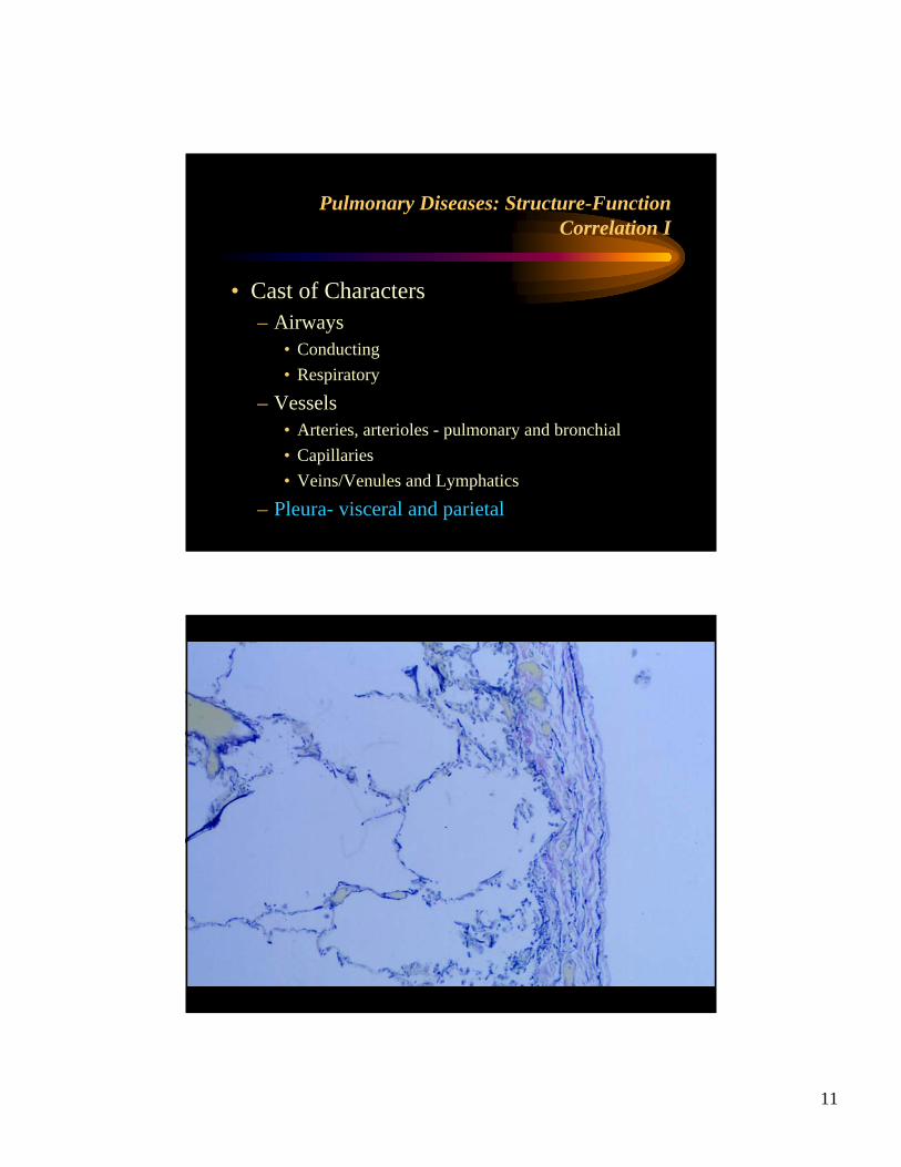
• Cast of Characters– Airways
• Conducting• Respiratory
– Vessels• Arteries, arterioles - pulmonary and bronchial• Capillaries• Veins/Venules and Lymphatics
– Pleura- visceral and parietal
Pulmonary Diseases: Structure-Function Correlation I
• Airways Conducting Zone•Trachea •Bronchi - ciliated and goblet cells, elastic tissue, smooth muscle, glands, cartilage
•Bronchioles - (1 mm) -No cartilage or bronchial glands, ciliated lining,no goblet cells, smooth muscle
• Cell types– CILIATED CELL -
beating of cilia contribute to mucociliary elevator
– GOBLET CELL -Mucus secretion
– BASAL CELL - reserve cell
– KULCHITSKY CELL -neuroendocrine cells.
4
Normal airway

5
Squamous metaplasia
6
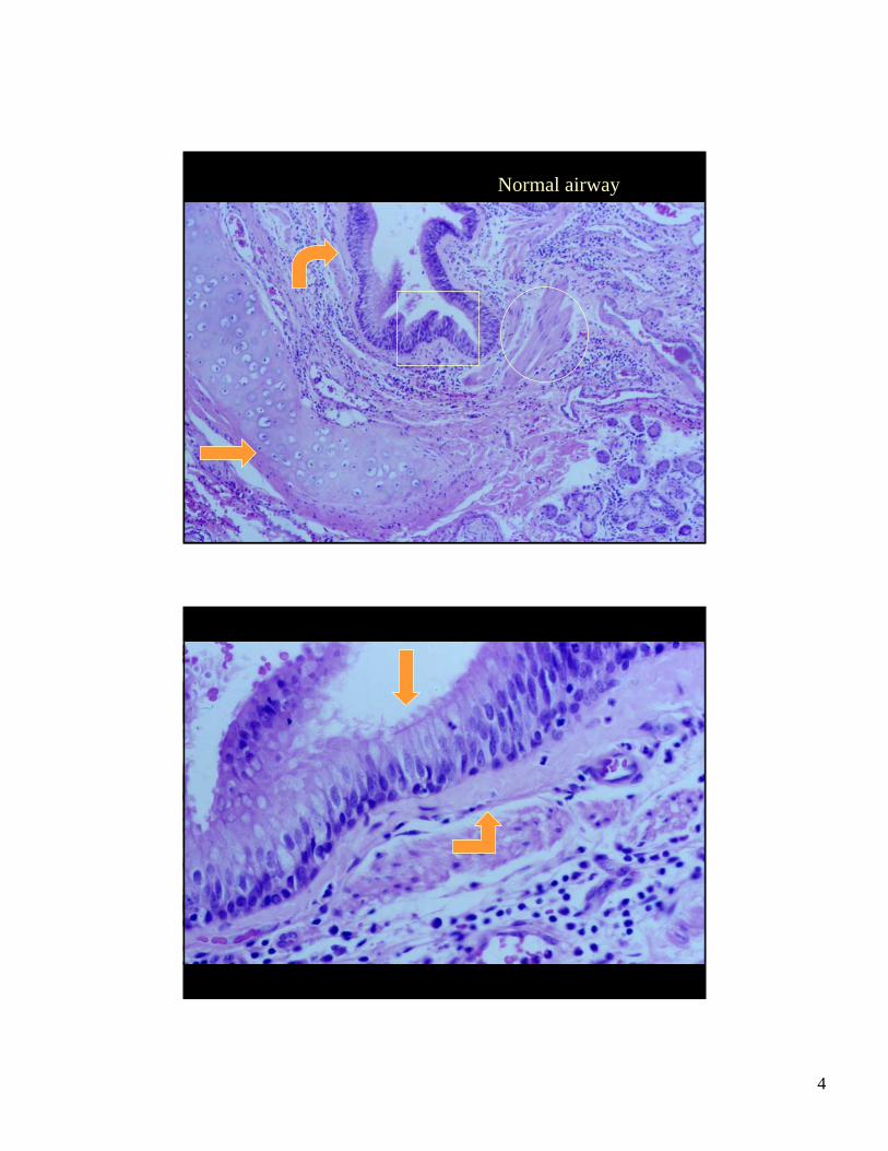
Pulmonary Diseases: Structure-Function Correlation I
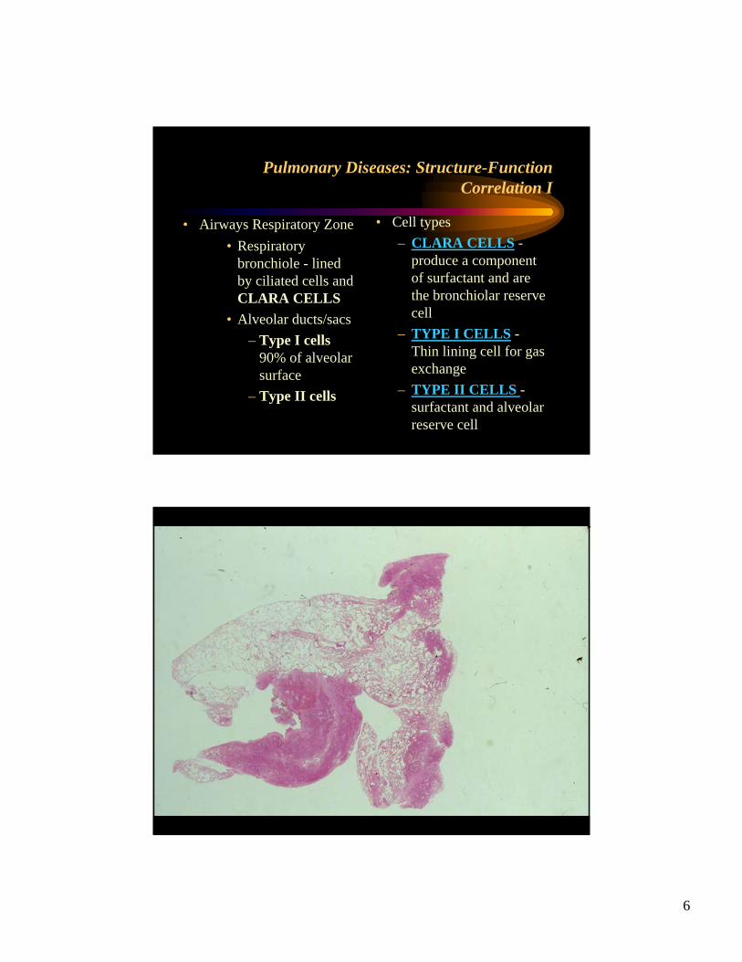
• Airways Respiratory Zone• Respiratory
bronchiole - lined by ciliated cells and CLARA CELLS
• Alveolar ducts/sacs– Type I cells
90% of alveolar surface
– Type II cells
• Cell types– CLARA CELLS -
produce a component of surfactant and are the bronchiolar reserve cell
– TYPE I CELLS -Thin lining cell for gas exchange
– TYPE II CELLS -surfactant and alveolar reserve cell
7

8

9
Pulmonary Diseases: Structure-Function Correlation I
• Vessels - Pulmonary–Arteries/arterioles - travel and divide with bronchi and bronchioles
– Produce capillary bed in alveoli for gas exchange
–Venules collect capillary blood into lobular septa, forming veins and joining at the hilum.
• Vessels - Bronchial– Artery from aorta– Supplies bronchial tree
up to respiratory bronchiole
– Venous drainage to azygous/hemiazygous

10
11
Pulmonary Diseases: Structure-Function Correlation I
• Cast of Characters– Airways
• Conducting• Respiratory
– Vessels• Arteries, arterioles - pulmonary and bronchial• Capillaries• Veins/Venules and Lymphatics
– Pleura- visceral and parietal

12
Pulmonary Diseases: Structure-Function Correlation I
• Disease of the acini and interstitium1) Replacement of air with fluid, inflammatory
cells or cellular debris2) Thickening of alveolar walls and interstitium3) Destruction of acinar walls
• Disease of the conducting airways• Disease of the pulmonary vasculature

13
Pulmonary Diseases: Structure-Function Correlation I
• Disease of the acini and interstitium1) Replacement of air with fluid, inflammatory
cells or cellular debris2) Thickening of alveolar walls and interstitium3) Destruction of acinar walls
• Disease of the conducting airways• Disease of the pulmonary vasculature
Pulmonary Diseases: Structure-Function Correlation I
• Disease of the conducting airways– Asthma– Chronic bronchitis– Bronchiectasis

14
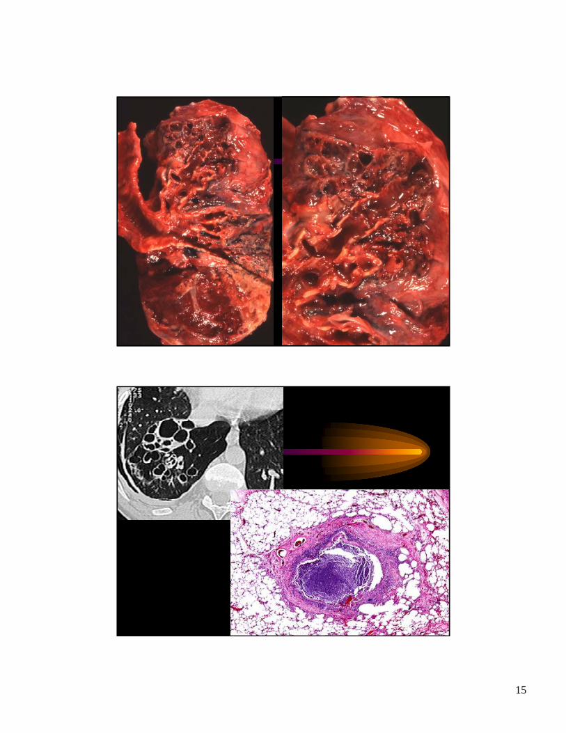
Disease of the conducting airways -Bronchiectasis
• Dilatation of bronchi and bronchioles, usually due to necrosis of wall and obstruction– Foreign body– Mucoid impaction– Cystic fibrosis– Immotile cilia– Chronic bronchitis
and infection
• Gross Pathol. - Dilated bronchi, filled with mucus or pus, lower lobes.
• Microscopic -– Can have acute and
chronic inflammation– Varying degrees of
fibrosis
15

16
17
Pulmonary Diseases: Structure-Function Correlation I
• Disease of the conducting airways– Asthma– Chronic bronchitis– Bronchiectasis
Disease of the conducting airways -ASTHMA
• Bronchospasm, usually reversible, due to allergic or non-allergic stimuli.
• Anatomic targets -bronchial epithelium and smooth muscle.
• Inflammation• Obstructive disease
• Gross pathology– hyperinflation, severe if
status asthmaticus– Mucus plugging
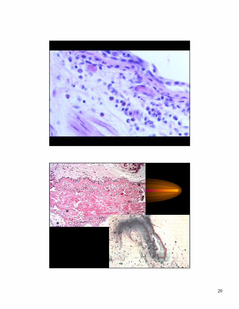
• Microscopic– Smooth muscle hypertrophy– Inflammation, eosinophils– Basement membrane
thickening– edema

18

19
20

21
Disease of the conducting airways -ASTHMA
• Gross pathology–hyperinflation–Mucus plugging
• Microscopic–Smooth muscle hypertrophy–Inflammation, eosinophils–Basement membrane thickening
–edema
Functional significance• Total lung capacity -
increased during attack
• Work of breathing increased due to airway resistance
• Airway resistance increased, on expiration more than inspiration
Pulmonary Diseases: Structure-Function Correlation I
• Disease of the conducting airways– Asthma– Chronic bronchitis– Bronchiectasis
22
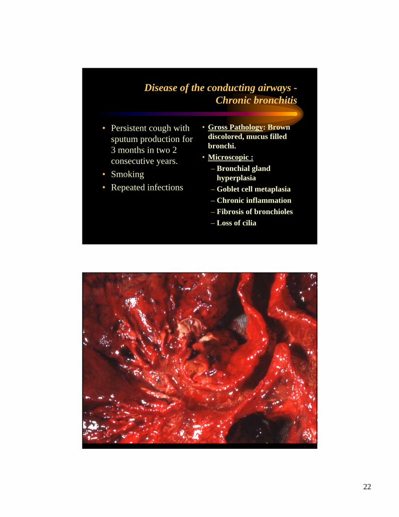
Disease of the conducting airways -Chronic bronchitis
• Persistent cough with sputum production for 3 months in two 2 consecutive years.
• Smoking• Repeated infections
• Gross Pathology: Brown discolored, mucus filled bronchi.
• Microscopic :– Bronchial gland
hyperplasia– Goblet cell metaplasia– Chronic inflammation– Fibrosis of bronchioles– Loss of cilia

23

24

25
Disease of the conducting airways -Chronic bronchitis
• Gross Pathology: Brown discolored, mucus filled bronchi.
• Microscopic :– Bronchial gland
hyperplasia– Goblet cell metaplasia– Chronic inflammation– Fibrosis of bronchiolar
walls– Loss of cilia
• Functional Significance– Airway resistance, due to
mucus, edema and narrowing. Obstructive disease
– Degree of obstruction determines extent of V/Q mismatch
– Lung capacity normal– Right heart failure and
pulmonary hypertension can occur

26
Pulmonary Diseases: Structure-Function Correlation I
• Disease of the acini and interstitium1) Replacement of air with fluid, inflammatory
cells or cellular debris2) Thickening of alveolar walls and interstitium3) Destruction of acinar walls
• Disease of the conducting airways• Disease of the pulmonary vasculature
From NEJM 2000;343:270

27
Destruction of acinar walls -Emphysema
• Obstructive disease• Involves the airway distal to the terminal
conducting bronchiole• Airway wall is damaged, and fibrosis can be
present.• Is classified by pattern/ location of damage
within the respiratory acinus
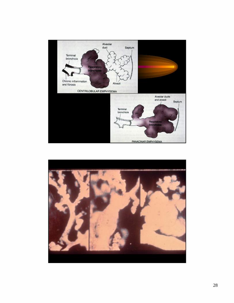
Destruction of acinar walls -Emphysema
• Centriacinar (Centrilobular)– Smoking– Damage is to the respiratory bronchiole. When
severe disease develops, whole acinus involved.– Upper lobes, especially apical portions most affected
• Panacinar (Panlobular)– Damage is to the entire acinar unit from respiratory
bronchiole to alveolar sac– More severe at bases, but is more diffuse than CLE– Alpha -1 antitrypsin deficiency
28

29
Destruction of acinar walls -Emphysema
• Pathogenesis– Protease/Antiprotease hypothesis
• Imbalance between neutrophil derived elastaseand deficiency in anti-elastase activity from alpha-1-antitrypsin
• Neutrophil elastase is unchecked, causing tissue destruction
• Smoking causes more rapid evolution of panacinar emphysema.
Destruction of acinar walls -Emphysema
• Pathogenesis– Protease/Antiprotease hypothesis
• In panacinar emphysema, deficiency in alpha 1 anti-trypsinis a genetic defect
• In centrilobular emphysema, the interplay of cigarette smoke, acquired deactivation of A1AT activity and activation of a perhaps broader spectrum of proteases may be significant. These may include proteinase 3, cathepsinsand metal metalloproteinases (1,2,9,12)
• Other inhibitors of protease activity may also play a role – e.g. TIMPs

30
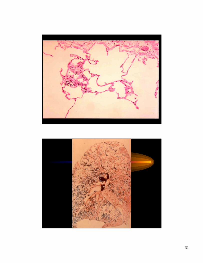
Destruction of acinar walls -Emphysema
• Gross pathology–Upper lobe, irregularly
dilated airspaces–Thin walled and grossly
apparent• Microscopic
–Dilated spaces, alongside normal alveoli
–Anthracotic pigment
• Gross Pathology– Lower lobe, more
uniformly dilated spaces
– Voluminous lungs• Microscopic
– Dilated spaces, uniformly dilated.
CENTRILOBULAR VS. PANACINAR
31

32
Destruction of acinar walls -Emphysema
• Gross pathology–Upper lobe, irregularly
dilated airspaces–Thin walled and grossly
apparent• Microscopic
–Dilated spaces, alongside normal alveoli
–Anthracotic pigment
• Gross Pathology– Lower lobe, more
uniformly dilated spaces
– Voluminous lungs• Microscopic
– Dilated spaces, uniformly dilated.
CENTRILOBULAR VS. PANACINAR

33
Destruction of acinar walls -Emphysema
• Gross pathology–Upper lobe, irregularly
dilated airspaces–Thin walled and grossly
apparent• Microscopic
–Dilated spaces, alongside normal alveoli
–Anthracotic pigment
• Total lung capacity increase• Lung compliance increased
(elastin destruction)• V/Q mismatch mild - airway
and capillary destruction• Recoil decreased; lose radial
traction on airways Obstructive; worsens on forced expiration
STRUCTURAL VS. FUNCTIONAL